Article

Investigation of Conditions for Capture of Live Legionella pneumophila with Polyclonal and Recombinant Antibodies

1
Laboratory for Nano-Technologies and Nano-Characterization (LN2)-CNRS UMI-3463, Interdisciplinary Institute for Technological Innovation (3IT), Université de Sherbrooke, 3000 Boul. de l’Université, Sherbrooke, QC J1K 0A5, Canada
2
Department of Microbiology and Infectiology, Faculty of Medicine and Health Science, Université de Sherbrooke, 3001 12th Avenue North, Sherbrooke, QC J1H 5N4, Canada
3
Department of Electrical and Computer Engineering, Faculty of Engineering, Université de Sherbrooke, 2500 Boul. de l’Université, Sherbrooke, QC J1K 2R1, Canada
4
Department of Biotechnology, Technische Universität Braunschweig, Spielmannstrasse 7, 38106 Braunschweig, Germany
5
CIUSSS de l’Estrie-Centre Hospitalier Universitaire de Sherbrooke, 3001, 12th Avenue North, Sherbrooke, QC J1H 5N4, Canada
*
Author to whom correspondence should be addressed.
Biosensors 2022, 12(6), 380; https://doi.org/10.3390/bios12060380
Submission received: 19 April 2022 / Revised: 16 May 2022 / Accepted: 27 May 2022 / Published: 31 May 2022
(This article belongs to the Special Issue Microbial Toxins and Pathogen Biodetection)

Abstract

:
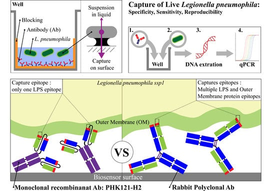
Since Legionella pneumophila has caused punctual epidemics through various water systems, the need for a biosensor for fast and accurate detection of pathogenic bacteria in industrial and environmental water has increased. In this report, we evaluated conditions for the capture of live L. pneumophila on a surface by polyclonal antibodies (pAb) and recombinant antibodies (recAb) targeting the bacterial lipopolysaccharide. Using immunoassay and PCR quantification, we demonstrated that, when exposed to live L. pneumophila in PBS or in a mixture containing other non-target bacteria, recAb captured one third fewer L. pneumophila than pAb, but with a 40% lower standard deviation, even when using the same batch of pAb. The presence of other bacteria did not interfere with capture nor increase background by either Ab. Increased reproducibility, as manifested by low standard deviation, is a characteristic that is coveted for biosensing. Hence, the recAb provided a better choice for immune adhesion in biosensors even though it was slightly less sensitive than pAb. Polyclonal or recombinant antibodies can specifically capture large targets such as whole bacteria, and this opens the door to multiple biosensor approaches where any of the components of the bacteria can then be measured for detection or characterisation.

Graphical Abstract

1. Introduction

Legionella pneumophila is a gram-negative bacterium that is ubiquitous in water sources, environmental or man-made [1]. Sixteen serogroups of L. pneumophila are defined by the various components of the surface, but principally due to the variability of the lipopolysaccharide (LPS) [2]. In the environment, L. pneumophila is able to survive, despite its nutritional dependence on cysteine and iron, by having a high resilience in water [2,3] and by infecting protozoa, especially amoeba [4]. When engulfed by amoeba or human macrophages, they prevent lysosome fusion after phagocytosis and use nutriments of the host to divide and eventually cause host cell death [5]. This ability to survive intracellularly makes them pathogenic for humans, with different levels of pathogenicity depending on the individual’s immunologic strength and the pathogenic potential of the microorganism. The severity of the infection varies from asymptomatic, Flu-like symptoms, Pontiac fever, to Legionellosis and even death [6]. Even though 16 serogroups exist for L. pneumophila, 75% of infections, including outbreaks, are caused by L. pneumophila ssp 1 [7,8]. Due to its ubiquitous presence and proliferation in man-made water systems, Legionellosis outbreaks usually originate from water sources, especially those in industry. Since the first reported L. pneumophila epidemic that occurred in 1976 [9], outbreaks have occurred in man-made water [10] structures, with the following two vibrant examples occurring recently: the outbreaks in New York City in 2015 (138 cases and 16 deaths) [11] and in Quebec City in 2012 (182 infected and 13 deaths) [12]. In both occurrences, cooling tower water (CTW) was the source as follows: L. pneumophila infected microdroplets were dispersed from large structures such as skyscrapers or industrial buildings. A CTW environment provides suitable conditions for the growth of L. pneumophila in bacterial biofilms and protozoa. Driven by the 2012 Quebec City epidemic, new intervention guidelines for Legionella were implemented in 2015 in Quebec province [13], requiring that CTW must be monitored for the presence of Legionella at least once a month in Quebec. In recent legislation, similar guidelines have been established for New York City and State [14]. Even if PCR can provide results in 2–3 h after sample collection and transport to a laboratory, culture on agar plates remains necessary to validate the PCR results, and this usually requires a 5-day incubation but up to 14 days of obligatory plate observation before the culture can be considered negative [13]. Indeed, overestimation of positivity by PCR is possible due to the detection of DNA from non-viable and even dead bacteria, and confirmation by culture remains necessary [13]. Both culture and PCR require highly qualified personnel and time, which, during an epidemic, can cost lives. Therefore, the development of biosensors could provide fast, quasi-real-time, and even prospective control of L. pneumophila and thus avoid or curtail future outbreaks [15].
Several biosensing platforms have been investigated for the detection of L. pneumophila, such as quartz crystal microbalance (QCM) [16], surface plasmon resonance (SPR) [16,17] and electrical impedance spectroscopy (EIS) [16]. Based on the photonic characteristics of digital photocorrosion (DIP) of GaAs/AlGaAs heterostructures functionalized with antibodies, our group [18] has developed DIP based biosensors allowing the detection of Escherichia coli K12 at 103 CFU/mL [19,20] and heat-inactivated L. pneumophila ssp 1 at 103 CFU/mL [21,22].
The sensitivity of such biosensors is highly dependent on their ability to specifically capture the targeted bacteria, which is very dependent on the molecules used for capture. Antibodies have been shown to enhance the specificity of the reaction, as polyclonal antibodies provide stronger antibody-target bonds or affinity, which might improve sensitivity but could give rise to more false positives [23,24,25]. However, monoclonal and especially recombinant antibodies, have also shown increased reproducibility [16]. Most previous studies have used antibodies to capture individual proteins and lipopolysaccharides or alternatively, to agglutinate larger entities such as whole bacteria or viruses. Capture and retention of large entities on a surface has not been extensively examined. This may prove useful for SPR, DIP, and other detection strategies, which could benefit from the presence of large targets. Furthermore, living bacteria could be further characterised to provide a second level of authentication or additional information such as antimicrobial sensitivity. As an alternative to antibodies, we have also employed capture with short-ligand and sandwich antimicrobial peptide architectures that allowed detection of L. pneumophila at 1–2 × 10² CFU/mL [26].
In the present study, we report the performance of a recombinant antibody (recAb) specific to L. pneumophila ssp 1 LPS [21] as compared to polyclonal Ab (pAb) to capture whole, live bacteria on a surface. The sensitivity, specificity, and reproducibility of pAb and recAb to capture L. pneumophila were evaluated to select an optimal strategy for biosensing.

2. Materials and Methods

2.1. Materials

L. pneumophila ssp 1 expressing Green Fluorescent Protein (GFP L. pneumophila), a strain transformed with an Isopropyl B-D-1-thiogalactopyranoside (IPTG)-inductive plasmid producing GFP maintained by chloramphenicol was kindly provided by Prof. Sébastien Faucher (McGill University, Montréal, QC, Canada). L. pneumophila ssp 5 [27], E. coli, Pseudomonas aeruginosa, Bacillus subtilis, and Staphylococcus aureus were provided by the department of microbiology of the Centre Hospitalier Universitaire de Sherbrooke (Sherbrooke, QC, Canada). Buffered charcoal yeast extract (BCYE) agar was obtained from Becton, Dickinson, and Company (Bergen County, MD, USA). Luria-Bertani (LB) plates and LB broth were obtained from EMD chemicals (Darmstadt, Germany). L-cysteine, Iron (III)-pyrophosphate, ethylenediaminetetraacetic acid (EDTA), ethanolamine, Legionella enrichment broth and Sodium dodecyl sulfate (SDS) were purchased from Sigma-Aldrich (Oakville, ON, Canada). Rabbit polyclonal anti-Legionella pneumophila Ab (pAb) was purchased from Virostat, Inc. (Portland, ME, USA). The human recombinant scFv antibody fragment against Legionella LPS was obtained by antibody phage display and fused to human Fc to form PHK121-H2h [28]. Costar 3590 96-well microtitre plates were obtained from Corning Inc. (Corning, NY, USA). Phosphate-buffered saline (PBS) and Tris-HCl were purchased from Amresco (Solon, OH, USA) and Invitrogen (Carlsbad, CA, USA), respectively. Triton X-100 and Tween-20 were obtained from Fisher scientific (Branchburg, NJ, USA). All solutions were made in PBS, except Ethanolamine which was dissolved in sterile deionized (DI) water. Molecular grade water was obtained from Wisent Inc. (Saint-Jean-Baptiste, QC, Canada). The Plate shaker (PMX-01E) and the Ultrasonicator Branson 200 (40 kHz) were purchased from Fujirebio inc (Shanghai, China) and Branson Ultrasonics corp. (Brookfield, CT, USA), respectively. For qPCR, the complete master mix was obtained from the protein purification service of the Department of microbiology and infectiology (Université de Sherbrooke, QC, Canada) and PCR system LightCycler® 480 II (Roche diagnostics, Indianapolis, Indiana, USA) was used for all samples. Primers and probes were obtained from Integrated DNA technologies (Coralville, IA, USA).

2.2. Bacterial Culture

In preliminary experiments, we occasionally observed overgrowth with strains resembling L. pneumophila, so we employed a strain-producing GFP to provide a double guarantee of the identity of the bacteria tested as follows: it does not grow on BCYE plates without L-Cys, and colonies appear green on BCYE plates overlaid with chloramphenicol, which induces GFP production. L. pneumophila ssp 5 was also confirmed by its lack of growth on BCYE plates without L-Cys. L. pneumophila was plated and grown on BCYE agar plates coated with chloramphenicol for 4–5 days at 37 °C. An isolated colony was inoculated in Legionella enrichment broth (L.ER) supplemented with iron-pyrophosphate and L-cysteine and incubated for 24–48 h at 37 °C with shaking (~100 rpm). E. coli, B. subtilis and S. aureus were cultivated on LB plates overnight at 37 °C (O/N, 37 °C) then an isolated colony was inoculated into LB broth and grown (O/N, 37 °C) with shaking (~100 rpm). Exponential phase cultures of L. pneumophila were used as their outer membranes varied only slightly from viable but non-culturable L. pneumophila found in nutriment lacking environments [2], such as CTW. They were confirmed by having neutral pH [29] and no brown colour [30] (see Appendix A.1). The same protocol was applied to P. aeruginosa, but at a temperature of 25 °C. The bacterial concentrations were quantified by optical density measurements at 600 nm (OD600 nm). OD600 nm of 0.1 was 6 × 10⁷ CFU/mL for L. pneumophila, 2.5 × 10⁸ CFU/mL for both E. coli and S. aureus [31], and 2.5 × 10⁷ CFU/mL for both B. subtilis [32] and P. aeruginosa [33], respectively. All bacterial suspensions were prepared in PBS.

2.3. Immuno-Capture of Live Bacteria on 96 Well Plates

As a proxy for capture of bacteria with Ab on GaAs plates or other biosensor surfaces, L. pneumophila was captured with Ab on 96 well Costar plates. Ab solutions at 100 µg/mL in PBS were incubated in the plate wells for 1 h at room temperature (RT). Following adsorption, the following two blocking steps were carried out: first with BSA 2% in PBS and then with ethanolamine (pH 8, 1 M) in DI water. Both blocking solutions (200 µL/well) were incubated in the wells for 1 h at RT. After blocking, the wells were washed 3 times with PBS. For bacterial capture, 100 µL of L. pneumophila suspensions were added to each well (1 h, RT). Finally, 3 successive washes were made with PBS + Tween 0.05% (PBST). The effect of 0.05% Tween in PBS on the viability of L. pneumophila was evaluated (see Appendix A.2). To test the impact of Tween on reducing non-specific and antibody-specific capture, experiments were performed with or without Tween 0.05% in the bacterial washing step. All described incubations were performed with shaking (~500 rpm). For background level wells, the step of adding Ab solution was replaced by adding PBS instead, while all other steps remain unchanged.

2.4. Effect of Antibody Pre-Sonication on Bacterial Capture

In order to test the effect of sonication on possible rAb aggregation, 100 µL aliquots of Ab were pre-sonicated in a sonication bath at 4 °C in glass tubes for various times to reduce possible Ab aggregation before adding the Ab solution to the wells. This step was performed in a walk-in cold room at 4 °C. The ensuing blocking and washing steps remained the same as described in the previous section.

2.5. Direct DNA Extraction

An adaptation of the EtNa DNA extraction [34] protocol was chosen for direct DNA extraction of different microorganisms from Costar plates because of the limited material requirements and minimal loss of DNA. The DNA suspension solution from the EtNa protocol (Triton X-100 1%, Tween 20 0.05%, Tris-HCL 50 mM, EDTA 0.1 mM, pH 8) was used to detach bacteria captured on the plate wells. After adding 200 µL of the DNA extraction solution to the wells, the plate was heated to 85 °C for 30 min in a water bath, then cooled on ice for 5 min, quickly centrifuged and placed at −15 °C O/N. After thawing, the samples were aliquoted into microtubes and used for PCR.

2.6. Polymerase Chain Reaction (qPCR)

A 2 µL sample of the direct DNA extract was added to 8 µL of amplification solution in a LightCycler PCR plate as follows: 0.25 µM MIP forward and reverse primers, 0.15 µM MIP probe, (and/or 0.1 µM 16S rDNA forward and reverse primers, 0.075 µM 16S rDNA probe), 5 µL of 2X Taqman master mix (produced by the protein and purification service of the University of Sherbrooke, Sherbrooke, QC, Canada), completed to 8 µL with Mol Grade H2O. MIP gene primers and probes were employed for specific detection of L. pneumophila [35], as shown in Table 1. For universal amplification, 16S ribosomal DNA (16S rDNA) primers and probes, adapted from the CDC protocol [36], were added, as shown in Table 1. PCR program (activation: 95 °C-15 s; 50 amplification cycle: 95 °C-15 s, 60 °C-1 min) was optimized for the primers and probes. After amplification, the LightCycler programme determined the cycle threshold (Ct) of detection.

2.7. Bacteria Quantification Curve

Triplicate 100 µL volumes containing concentrations from 103 to 108 CFU/mL of bacteria were added to a Costar plate in 1 log10 increments to obtain 6 concentrations of L. pneumophila. The addition of 100 µL of 2X concentrated DNA extraction solution to the wells resulted in concentrations from 5 × 10² to 5 × 10⁷ CFU/mL of L. pneumophila. DNA extraction of the bacteria added to the Costar plate was carried out as described above and amplified by qPCR. Quantification curve wells were included in each experiment to assess the variability of extraction and amplification of positive controls and to obtain sufficient replicates for the quantification curve (Figure 1). Ct values were plotted against bacterial concentrations to give a linear calibration curve. Experimental Ct values obtained from experiments were employed to quantify the number of immobilised bacteria by interpolation with the calibration curve.

3. Results and Discussion

3.1. Calibration Curve

Following DNA extraction of L. pneumophila from suspensions with concentrations ranging from 5 × 10² to 5 × 10⁷ CFU/mL, qPCR amplification was carried out, and Ct values were determined with two sets of primers and probes [37]. Standard curves generated by linear regression revealed an inverse linear correlation between Ct and the concentration of L. pneumophila (Figure 1).
Lower concentrations were not tested as the Ct for 5 × 10² CFU or less showed a plateau, indicating that the detection limit of the system had been reached. This is not surprising as, theoretically, there would be only one bacterium in a 2 µL aliquot from a bacterial solution with 5 × 10² CFU/mL, so lower concentrations of bacteria might be expected to generate negative qPCR results. The difference in Ct values between the two curves for the same bacterial concentration was due to the presence of three copies of the 16S rDNA sequence [38] but only a single copy of the MIP gene per Legionella bacteria [39,40]. The calibration curve was used to estimate the number of immobilised bacteria in the plate wells, with the MIP gene curve used for L. pneumophila and the 16S gene curve for L. pneumophila and other bacteria. Each capture test included calibration curve wells that also served as positive controls for the experiment.

3.2. Optimal Bacterial Concentration for Incubation in Ab-Coated Wells

We had previously observed that the optimal concentration of the solution of Ab to be deposited on GaAs or microplate surfaces was 100 µg/mL [41]. Hence, we used this concentration to evaluate the saturation level of the two Ab used to capture L. pneumophila.
To estimate the saturation level of an Ab-coated surface, Ab-coated wells were exposed to various concentrations of bacteria. It was presumed that in order to compare the ability of pAb and recAb to capture bacteria, it would be necessary to use bacterial concentrations that did not saturate the surface, as results would be expected to plateau with saturating concentrations and variation in conditions might not affect the values. The highest concentration tested was 109 CFU/mL, and it provided the highest bacterial capture, as shown in Figure 2.
When suspensions at 109 CFU/mL were added to wells coated with Ab, 2.2 × 10⁶ CFU/mL (0.22% of the input bacteria) were captured with pAb and 1.5 × 10⁶ CFU/mL (0.15%) with recAb, whereas without Ab, a non-specific capture of 3.6 × 10⁴ CFU/mL (0.0035%) was observed. Exposure to suspensions at lower concentrations resulted in specific capture of 4.1 × 10⁵ ± 2.2 × 10⁵ (0.41%) CFU/mL by pAb and 2.8 × 10⁵ ± 1.1 × 10⁵ (0.28%) CFU/mL by recAb at 108 CFU/mL. The attachment of 3.0 × 10⁴ ± 1.7 × 10³ (0.30%) CFU/mL by pAb and 1.5 × 10⁴ ± 9.0 × 10³ (0.15%) CFU/mL by recAb were obtained by exposure to 107 CFU/mL of L. pneumophila. Non-specific attachment of 5.8 × 10³ ± 4.8 × 10³ (0.0057%) and 1.0 × 10³ ± 1.6 × 10² (0.01%) CFU/mL were obtained with exposure of 108 CFU/mL and 107 CFU/mL, respectively. The exposure to suspensions at lower concentrations (108 CFU/mL and 107 CFU/mL) resulted in lower bacterial retention than the 109 CFU/mL, but similar capture percentages, indicating that capture was not less efficient. Hence, the 108 and 107 CFU/mL concentrations did not saturate the surface, and we do not have evidence that even 109 CFU/mL saturated the surface. The percentage of capture on wells without Ab increased slightly with a decrease in the exposure concentration. This could be attributed to the saturation of non-specific capture sites on the wells with increasing concentrations of bacteria.
These results confirmed the use of 108 or 107 CFU/mL as exposure concentrations for experimentation as they did not saturate the surface. This would allow us to evaluate the variation in capture by Ab under different conditions. They also maintained values for non-specific detection of >103 CFU/mL, which is within the range of the calibration curve.

3.3. Effect of Pre-Sonication of Antibodies on Bacterial Capture

Since only 0.1–1% of the total bacteria in the suspension were captured by Ab attached to the wells, we hypothesized that we lost some of the Ab capacity due to the presence of Ab aggregates. To reduce potential Ab aggregation, the Ab solutions were sonicated before adsorption to the wells to enhance bacterial capture. Figure 3 shows that different durations of sonication for both recAb and pAb did not significantly improve bacterial capture.
This result was expected for pAb due to its good solubility as it is produced by an organism for activity in solution [42]. Since the added scFv fragment may interfere with the solubility of recAb and cause self-aggregation [42], we checked this solution for aggregation. However, we did not observe that sonication improved capture efficiency, so we concluded that it was ineffective for breaking up aggregates or that aggregation did not pose a problem under our conditions.

3.4. Specificity of Polyclonal and Recombinant Antibodies

To evaluate Ab specificity, wells coated with pAb or recAb were exposed to different non-target bacteria (E. coli, S. aureus, B. subtilis, and P. aeruginosa). L. pneumophila ssp 5 was also used to test the selectivity towards other Legionella serogroups. Results based on the amplification of a universal 16S RNA showed that the average value of the capture of non-target bacteria to wells with pAb or recAb did not vary significantly from the background adhesion to the No-Ab wells, as shown in Figure 4, but the higher non-specific attachment of B. subtilis and P. aeruginosa to wells without Ab.
This indicated that Ab wells did not capture non-target bacteria significantly. However, L. pneumophila ssp 5 did show significant capture with recAb (2.0 × 10³ CFU/mL), albeit nearly 10 times less than the capture of L. pneumophila ssp 1 (1.5 × 10⁴ CFU/mL).
It was surprising that pAb did not capture more L. pneumophila ssp 5. Indeed, pAb is directed towards multiple components of L. pneumophila ssp 1, as indicated by the manufacturer, Virostat, who reported some cross-reactivity of this Ab with other serogroups of L. pneumophila [43,44]. However, this reactivity may not be towards components that allow the capture of L. pneumophila. LPS, or serogroup-specific LPS epitopes, may be the preferred target that allows the capture of these bacteria in the current experiments. When it was formulated, recAb was chosen to detect the LPS of ssp 1, and indeed, it was found highly specific for human isolates of serogroup 1 of L. pneumophila and did not detect an environmental serogroup 1, nor a serogroup 6 strain [22].
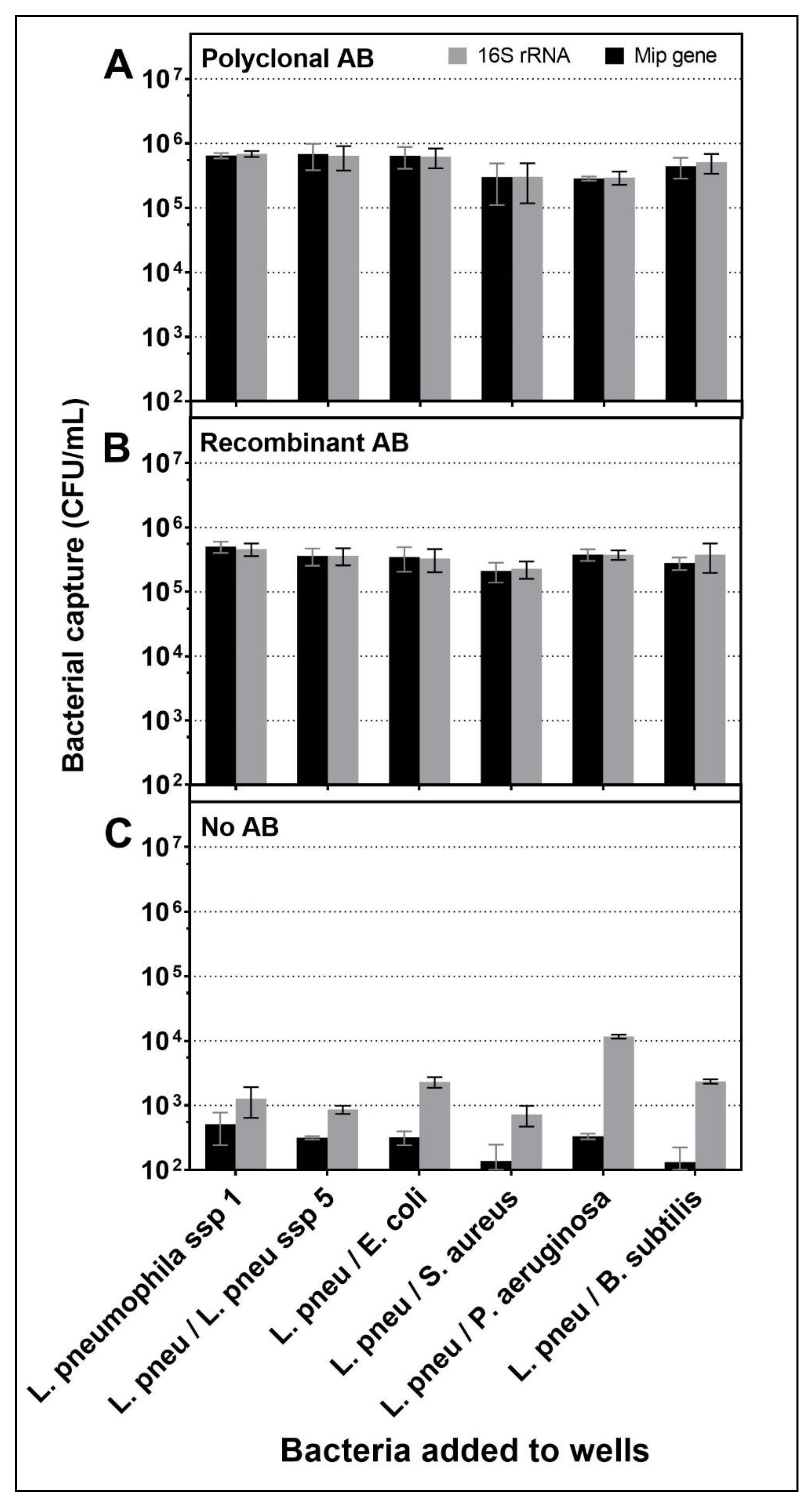
For further evaluation of specificity, mixtures of L. pneumophila at a concentration of 107 CFU/mL were added together with competitor bacteria to evaluate the interference of other bacteria present in the solution with the adhesion of L. pneumophila to the surface of the wells as follows: E. coli; S. aureus; L. pneumophila sg5 at a concentration of 107 CFU/mL (ratio 1:1) and B. subtilis; P. aeruginosa at a concentration of 106 CFU/mL (ratio 10:1). Both sets of primers were used on all sample wells to evaluate the total number of bacteria captured on the surface vs. the number of Legionella. The results indicated that the presence of other bacteria did not interfere significantly with the attachment of L. pneumophila to the surface (Figure 5A,B, black bars).
It was observed that other bacteria did not adhere to the surface in the presence of attached L. pneumophila, as there was no increase in the 16S rDNA (grey columns) compared to the MIP (black columns) when other bacteria were added. The highest rate of non-specific attachment in wells not coated with Ab was observed with P. aeruginosa as follows: 1.2 × 10⁴ CFU/mL (Figure 5C), but this high non-specific attachment did not affect capture levels of L. pneumophila with Ab even when 16S primers and probe were used for evaluation (Figure 5A,B). This observation was also noted with all other bacteria mixtures. This result suggested that the Ab-L. pneumophila complex absorbed to the surface had acted as a blocking agent, preventing further binding of the non-target bacteria.
Considering specificity, both recAb and pAb showed excellent results when exposed to a variety of bacteria, making them both adequate choices for biosensor use.

3.5. Evaluation of Sensitivity and Reproducibility of Polyclonal and Recombinant Antibodies

From the various experiments (Figure 2, Figure 3, Figure 4 and Figure 5), both pAb and recAb showed comparable sensitivity under the same stringent conditions. pAb showed slightly higher numbers of captured bacteria compared to recAb. A compilation of capture levels and the standard deviation obtained for L. pneumophila captured with each Ab and quantified with 16S rDNA primers (from Figure 5) is presented in Figure 6.
These data indicate that pAb captured 4.32 × 10⁵ ± 2.72 × 10⁵ CFU/mL of live L. pneumophila, whereas recAb captured 2.99 × 10⁵ ± 1.64 × 10⁵ CFU/mL.
A slightly lower (70%) capture efficiency was observed for recAb compared to pAb; however, a slightly lower (60%) standard deviation was also observed for recAb compared to pAb, indicating that recAb captured L. pneumophila more consistently and more reproducibly than pAb. Several factors could contribute to these differences. RecAb is directed towards a single epitope of the L. pneumophila ssp 1 LPS, whereas pAb is probably composed of antibodies directed towards multiple epitopes of the LPS and perhaps other proteins as well. These other epitopes are not necessary for efficient capture but might contribute slightly to the strength of attachment to the target. On the other hand, reaction with other epitopes did not result in the capture of serogroup 5 L. pneumophila by pAb, perhaps because they are not as solidly anchored in the cell wall or membrane, or not as exposed as the LPS epitope targeted by recAb. It is interesting to note that recAb provided efficient capture, demonstrating that single epitopes of LPS can be a useful target for capturing bacteria and can be employed in biosensors.
It is to be expected that there will be variation between batches of pAb since they are produced in rabbits or other mammals where individual variation is observed. Since the production of a recAb does not require synthesis by a living vertebrate organism, it should be highly reproducible and provide identical specificity. The increased variation of results that we observed with pAb could not be attributed to batch-to-batch dissimilarity as we employed only a single batch for all of our tests. We hypothesize that it might be due to variation associated with the multiple epitopes targeted. We could further hypothesise that the reproducibility of pAb would have been even lower with multiple batches, considering reports in the literature that there are batch-to-batch differences from one animal to another for pAb [45].
It is generally considered that polyclonal antibodies will have higher apparent binding capacities than monoclonal or recombinant antibodies due to the variety of epitopes targeted compared with the single epitope target of a monoclonal or recombinant antibody [23,24,25]. The recAb that we employed was almost as efficient as pAb, so it is no doubt directed towards a dominant epitope that allows efficient capture. The increased reproducibility that we observed with recAb is no doubt a considerable advantage over pAb for use in biosensors, especially considering that its capture efficiency was almost as high as that of pAb. In the future, the use of multiclonal antibodies, a mixture of monoclonal recombinant antibodies with a completely defined composition, further promises to provide detection reagents that combine the advantages of both recombinant and polyclonal antibodies [45].

4. Conclusions

With the ubiquity of L. pneumophila in water sources, the development of an effective biosensor is imperative to improve prospective and preventive action toward outbreaks. We report two important steps towards attaining this objective. A new recombinant antibody targeting the LPS of L. pneumophila spp1 has been developed [28], which we have evaluated in the development of biosensors for the capture of whole L. pneumophila. We have also developed a stringent washing protocol that maintains the viability of L. pneumophila. Polyclonal and recombinant antibodies have been shown to specifically capture large targets, such as whole bacteria, and this opens the door to multiple biosensor approaches where any of the components of the bacteria can be measured for detection or characterisation, such as we have used with PCR detection of bacterial DNA sequences.
Through the advancement of biotechnology, reproducible production processes have been developed to manufacture recAb in order to forgo the use of animal and pAb production with their inherent variation and animal welfare concerns. Although they target only single epitopes, we have demonstrated that recAb are capable of capturing intact target bacteria. The capture of L. pneumophila at different concentrations by recAb and pAb in different environments showed comparable sensitivity and specificity. However, the lower standard deviations, indicating a more reproducible capture, favour recAb, even compared with a unique batch of pAb. For biosensors, precision is crucial in order to obtain constant, reproducible, and stable results for repeated tests.
As these results are in favour of the use of recombinant antibodies under pristine laboratory conditions, it is still necessary to confirm these results with cooling tower water or other environmental water samples using surfaces of different biosensors, such as those based on gold surfaces or on digital photocorrosion of GaAs/AlGaAs nanoheterostructures.

Author Contributions

Writing (original draft, reviewing, and editing), conceptualization, methodology, investigation, L.P.; writing (reviewing and editing), investigation (bacteria culture), W.M.H.; investigation J.C.; conceptualization, writing (reviewing and editing), S.D. and S.L.; supervising, conceptualization, validation, writing (reviewing and editing) J.J.D. and E.H.F. All authors have read and agreed to the published version of the manuscript.

Funding

This work was supported by the Fonds de recherche Nature et Technologie du Québec (FRQNT Grant 2015-PR-184056 to EHF and JJD) and the Natural Sciences and Engineering Research Council of Canada (NSERC SPG Grant 2016-494057 to EHF and JJD).

Institutional Review Board Statement

Not applicable for studies not involving humans or animals.

Informed Consent Statement

Not applicable for studies not involving humans.

Acknowledgments

The authors are indebted to Sébastien Faucher (McGill University, Montréal, QC, Canada) for providing L. pneumophila ssp 1 expressing Green Fluorescent Protein.

Conflicts of Interest

S.D. is cofounder of antibody engineering companies including Yumab and Abcalis. The other authors declare no competing financial interest.

Appendix A

Appendix A.1. Colour of Media and pH to Monitor Bacterial Growth

In preliminary experiments, we occasionally observed overgrowth with strains resembling L. pneumophila, so we employed a strain producing GFP to provide a double guarantee of the identity of the bacteria tested: it does not grow on BCYE plates without L-Cys, and colonies appear green on BCYE plates due to GFP production. L. pneumophila ssp 5 was also confirmed by its lack of growth on BCYE plates without L-Cys. Cultures on agar plates were used only to ensure that the bacterial strains were indeed L. pneumophila and to start liquid cultures.
Liquid cultures were chosen to reduce the aggregation of Legionella caused by biofilm production on agar plates and thus generate a more uniform suspension of bacteria for antibody-dependent capture on surfaces. Using Legionella enrichment broth (L.ER), an OD600 of 0.8 was attained at 48 h, as shown in Figure A1, while conserving the same tint of yellow from the beginning of growth, indicating that the bacteria were still in exponential phase (see below).
Figure A1. Optical density at 600 nm (OD 600) (top) and pH (bottom) of cultures of Legionella pneumophila ssp 1 or L. pneumophila ssp 5 grown in Legionella enrichment broth (L.ER) supplemented or not (indicated by minus-sign) with Fe-pyrophosphate or L-cysteine (L-cys).
Biosensors 12 00380 g0a1
After 72 h of culture, Legionella entered the stationary phase, and the medium started browning due to the production of homogentisic acid (HGA) [30]. HGA is produced by Legionella on quorum sensing at high bacterial concentrations and is used to inhibit the growth of other bacterial species. However, in aerobic environments, HGA is oxidised and polymerised into HGA-melanine, causing a browning of the surrounding environment. The browning of the medium is thus a visual indicator that the culture has entered the stationary phase and should not be used for experiments where exponential phase growth is required. Using yeast extract as a nutrient source and at an optimal pH of 6.8 for Legionella, the cultures became more alkaline with growth, unlike most other bacteria, which usually acidify the medium [29]. After 5 days in the stationary phase, the pH did not decrease further, as Legionella was still intact and viable despite the lack of nutrients. If Legionella had died and lysed, the cytosol content would have been liberated and this would probably have changed the pH. After the stationary phase, it would be expected that Legionella enters a latency phase where the bacteria stop dividing and move towards a more infectious phenotype [3] before entering a viable but non-culturable (VBNC) phenotype [2].

Appendix A.2. Effect of Detergents on the Viability of L. pneumophila

To investigate the effect of different detergents used in washing solutions on the viability of L. pneumophila and to ensure that there was no lysis of the target bacteria during the washing process, different detergent solutions containing Tween-20, Triton X-100 or SDS were tested at different concentrations. For SDS and Triton, 0.02; 0.04; 0.06; 0.08; 0.1; 0.2; 2 and 4% solutions in PBS were prepared, whereas for Tween, the tested concentrations were: 0.1; 0.2; 0.5; 0.1; 1.5; 2 and 4%. A volume of 200 µL of each detergent solution was mixed with 200 µL of L. pneumophila suspension at 103 CFU/mL was prepared in PBS from an exponential L.ER broth. After 5 min of exposure, 100 µL of the bacteria-detergent solution was spread on each of three BCYE agar plates. Two control tests were also carried out: (1) a solution of PBS without detergent was used as a positive control, and (2) a bacterial suspension in 1% bleach was used as a negative control. After 72-h of incubation, colonies were counted, and the results were normalised with respect to the positive control.
Detergent was added to washing solutions to ensure an efficient washing of the plates and to avoid the non-specific attachment of bacteria. To ensure that bacteria had not been degraded by detergents and that only bacterial outer membrane shells were still bound to the surface by Ab, we evaluated the toxicity of detergents, assuming that if the bacteria were still viable, they would be intact, whereas if dead, they might have ruptured. Exposing Legionella to different detergents showed that 0.05% and even 0.1% Tween-20 did not affect its viability, as shown in Figure A2.
Figure A2. Colony counts of L. pneumophila exposed to various concentrations of Tween-20 for 5 min and plated on BCYE (n = 3). Count values were normalized against the control PBS exposure without detergent. Errors bars indicate standard deviation.
Biosensors 12 00380 g0a2
This result demonstrates that 0.05% Tween-20 is not toxic at these concentrations and could be used to attempt to reduce the non-specific adhesion of bacteria. However, the use of SDS and triton X-100 at concentrations ranging from 0.01% to 2% resulted in the complete inactivation of L. pneumophila (not shown). The observation of more than 100% viability for between 0.05 and 0.1% concentrations of Tween-20 could be due to the reduction of bacterial aggregation due to mixing and the appearance of more individual bacterial colonies on the plates.

References

  1. Rhoads, W.J.; Pruden, A.; Edwards, M.A. Interactive effects of corrosion, copper, and chloramines on legionella and mycobacteria in hot water plumbing. Environ. Sci. Technol. 2017, 51, 7065–7075. [Google Scholar] [CrossRef] [PubMed]
  2. Schrammel, B.; Petzold, M.; Cervero-Arago, S.; Sommer, R.; Luck, C.; Kirschner, A. Persistent presence of outer membrane epitopes during short- and long-term starvation of five Legionella pneumophila strains. BMC Microbiol. 2018, 18, 75. [Google Scholar] [CrossRef] [PubMed]
  3. Li, L.; Mendis, N.; Trigui, H.; Faucher, S.P. Transcriptomic changes of Legionella pneumophila in water. BMC Genom. 2015, 16, 637. [Google Scholar] [CrossRef] [PubMed]
  4. Cunha, B.A.; Burillo, A.; Bouza, E. Legionnaires’ disease. Lancet 2016, 387, 376–385. [Google Scholar] [CrossRef]
  5. Brown, S.A.; Palmer, K.L.; Whiteley, M. Revisiting the host as a growth medium. Nat. Rev. Microbiol. 2008, 6, 657–666. [Google Scholar] [CrossRef] [Green Version]
  6. McDade, J.E. Legionella and the Prevention of Legionellosis. Emerg. Infect. Dis. 2008, 14, 1006. [Google Scholar] [CrossRef]
  7. Diederen, B.M. Legionella spp. and Legionnaires’ disease. J. Infect. 2008, 56, 1–12. [Google Scholar] [CrossRef]
  8. Shevchuk, O.; Jager, J.; Steinert, M. Virulence properties of the Legionella pneumophila cell envelope. Front. Microbiol. 2011, 2, 74. [Google Scholar] [CrossRef] [Green Version]
  9. Fraser, D.W.; Tsai, T.R.; Orenstein, W.; Parkin, W.E.; Beecham, H.J.; Sharrar, R.G.; Harris, J.; Mallison, G.F.; Martin, S.M.; McDade, J.E.; et al. Legionnaires’ disease: Description of an epidemic of pneumonia. N. Engl. J. Med. 1977, 297, 1189–1197. [Google Scholar] [CrossRef]
  10. Centre For Disease Control. Communicable Disease Control Chapter I–Management of Specific Diseases Legionella Outbreak Investigation and Control; National Library of Medicine: Bethesda, MD, USA, 2018; p. 42. [Google Scholar]
  11. Lapierre, P.; Nazarian, E.; Zhu, Y.; Wroblewski, D.; Saylors, A.; Passaretti, T.; Hughes, S.; Tran, A.; Lin, Y.; Kornblum, J.; et al. Legionnaires’ disease outbreak caused by endemic strain of Legionella pneumophila, New York, NY, USA, 2015. Emerg. Infect. Dis. 2017, 23, 1784–1791. [Google Scholar] [CrossRef]
  12. Levesque, S.; Plante, P.L.; Mendis, N.; Cantin, P.; Marchand, G.; Charest, H.; Raymond, F.; Huot, C.; Goupil-Sormany, I.; Desbiens, F.; et al. Genomic characterization of a large outbreak of Legionella pneumophila serogroup 1 strains in Quebec City, 2012. PLoS ONE 2014, 9, e103852. [Google Scholar] [CrossRef] [PubMed]
  13. Ministère de la Santé et des Services Sociaux (MSSS). Guide d’Intervention-La Legionellose; Ministère de la Santé et des Services Sociaux: Quebec, QC, Canada, 2015.
  14. NYC Health. Cooling Tower Requirements: What Building Owners Should Know. 2016. Available online: https://www1.nyc.gov/assets/doh/downloads/pdf/cd/cooling-tower-FAQs.pdf (accessed on 1 November 2021).
  15. Berkelman, R.L.; Pruden, A. Prevention of Legionnaires’ disease in the 21st century by advancing science and public health practice. Emerg. Infect. Dis. 2017, 23, 1905–1907. [Google Scholar] [CrossRef] [Green Version]
  16. Zeng, X.; Shen, Z.; Mernaugh, R. Recombinant antibodies and their use in biosensors. Anal. Bioanal. Chem. 2012, 402, 3027–3038. [Google Scholar] [CrossRef] [PubMed] [Green Version]
  17. Trilling, A.K.; Hesselink, T.; van Houwelingen, A.; Cordewener, J.H.; Jongsma, M.A.; Schoffelen, S.; van Hest, J.C.; Zuilhof, H.; Beekwilder, J. Orientation of llama antibodies strongly increases sensitivity of biosensors. Biosens. Bioelectron. 2014, 60, 130–136. [Google Scholar] [CrossRef] [PubMed]
  18. Nazemi, E.; Hassen, W.M.; Frost, E.H.; Dubowski, J.J. Growth of Escherichia coli on the GaAs (001) surface. Talanta 2018, 178, 69–77. [Google Scholar] [CrossRef] [PubMed]
  19. Nazemi, E.; Aithal, S.; Hassen, W.M.; Frost, E.H.; Dubowski, J.J. GaAs/AlGaAs heterostructure based photonic biosensor for rapid detection of Escherichia coli in phosphate buffered saline solution. Sens. Actuators B Chem. 2015, 207, 556–562. [Google Scholar] [CrossRef]
  20. Nazemi, E.; Hassen, W.M.; Frost, E.H.; Dubowski, J.J. Monitoring growth and antibiotic susceptibility of Escherichia coli with photoluminescence of GaAs/AlGaAs quantum well microstructures. Biosens. Bioelectron. 2017, 93, 234–240. [Google Scholar] [CrossRef]
  21. Aziziyan, M.R.; Hassen, W.M.; Morris, D.; Frost, E.H.; Dubowski, J.J. Photonic biosensor based on photocorrosion of GaAs/AlGaAs quantum heterostructures for detection of Legionella pneumophila. Biointerphases 2016, 11, 019301. [Google Scholar] [CrossRef] [Green Version]
  22. Aziziyan, M.R.; Hassen, W.M.; Sharma, H.; Shirzaei Sani, E.; Annabi, N.; Frost, E.H.; Dubowski, J.J. Sodium dodecyl sulfate decorated Legionella pneumophila for enhanced detection with a GaAs/AlGaAs nanoheterostructure biosensor. Sens. Actuators B Chem. 2020, 304, 127007. [Google Scholar] [CrossRef]
  23. Lipman, N.S.; Jackson, L.R.; Trudel, L.J.; Weis-Garcia, F. Monoclonal versus polyclonal antibodies: Distinguishing characteristics, applications, and information resources. ILAR J. 2005, 46, 258–268. [Google Scholar] [CrossRef] [Green Version]
  24. Le Berre, M.; Kane, M. Biosensor-based assay for domoic acid: Comparison of performance using polyclonal, monoclonal, and recombinant antibodies. Anal. Lett. 2006, 39, 1587–1598. [Google Scholar] [CrossRef]
  25. Alcocer, M.J.; Doyen, C.; Lee, H.A.; Morgan, M.R. Properties of polyclonal, monoclonal, and recombinant antibodies recognizing the organophosphorus pesticide chlorpyrifos-ethyl. J. Agric. Food Chem. 2000, 48, 4053–4059. [Google Scholar] [CrossRef] [PubMed]
  26. Islam, M.A.; Hassen, W.M.; Tayabali, A.F.; Dubowski, J.J. Short Ligand, Cysteine-modified warnericin RK antimicrobial peptides favor highly sensitive detection of Legionella pneumophila. ACS Omega 2021, 6, 1299–1308. [Google Scholar] [CrossRef] [PubMed]
  27. Bedard, E.; Levesque, S.; Martin, P.; Pinsonneault, L.; Paranjape, K.; Lalancette, C.; Dolce, C.E.; Villion, M.; Valiquette, L.; Faucher, S.P.; et al. Energy conservation and the promotion of Legionella pneumophila growth: The probable role of heat exchangers in a nosocomial outbreak. Infect. Control Hosp. Epidemiol. 2016, 37, 1475–1480. [Google Scholar] [CrossRef] [Green Version]
  28. Kuhn, P.; Thiem, S.; Steinert, M.; Purvis, D.; Lugmayr, V.; Treutlein, U.; Plobner, L.; Leiser, R.M.; Hust, M.; Dubel, S. Human Anti-Lipopolysaccharid (LPS) antibodies against Legionella with high species specificity. Hum. Antibodies 2017, 26, 29–38. [Google Scholar] [CrossRef] [Green Version]
  29. Sánchez-Clemente, R.; Igeño, M.I.; Población, A.G.; Guijo, M.I.; Merchán, F.; Blasco, R. Study of pH changes in media during bacterial growth of several environmental strains. Proceedings 2018, 2, 1297. [Google Scholar] [CrossRef] [Green Version]
  30. Levin, T.C.; Goldspiel, B.P.; Malik, H.S. Density-dependent resistance protects Legionella pneumophila from its own antimicrobial metabolite, HGA. eLife 2019, 8, e46086. [Google Scholar] [CrossRef]
  31. Chang, Y.C.; Yang, C.Y.; Sun, R.L.; Cheng, Y.F.; Kao, W.C.; Yang, P.C. Rapid single cell detection of Staphylococcus aureus by aptamer-conjugated gold nanoparticles. Sci. Rep. 2013, 3, 1863. [Google Scholar] [CrossRef]
  32. Arrieta, M.C.; Leskiw, B.K.; Kaufman, W.R. Antimicrobial activity in the egg wax of the African cattle tick Amblyomma hebraeum (Acari: Ixodidae). Exp. Appl. Acarol. 2006, 39, 297–313. [Google Scholar] [CrossRef]
  33. Dong-Ju, K. Relation of microbial biomass to counting units for Pseudomonas aeruginosa. Afr. J. Microbiol. Res. 2012, 6, 4620–4646. [Google Scholar] [CrossRef]
  34. Vingataramin, L.; Frost, E.H. A single protocol for extraction of gDNA from bacteria and yeast. Biotechniques 2015, 58, 120–125. [Google Scholar] [CrossRef] [PubMed]
  35. Moosavian, M.; Moradzadeh, M.; Ghadiri, A.; Saki, M. Isolation and identification of Legionella spp. in environmental water sources based on macrophage infectivity potentiator (mip) gene sequencing in southwest Iran. AIMS Microbiol. 2019, 5, 223–231. [Google Scholar] [CrossRef] [PubMed]
  36. Chen, L.; Mediavilla, J.R.; Endimiani, A.; Rosenthal, M.E.; Zhao, Y.; Bonomo, R.A.; Kreiswirth, B.N. Multiplex Real-Time PCR Detection of Klebsiella pneumoniae Carbapenemase (KPC) and New Delhi metallo-β-lactamase (NDM-1). J. Clin. Microbiol. 2011, 49, 579–585. [Google Scholar] [CrossRef] [Green Version]
  37. Yang, S.; Lin, S.; Kelen, G.D.; Quinn, T.C.; Dick, J.D.; Gaydos, C.A.; Rothman, R.E. Quantitative multiprobe PCR assay for simultaneous detection and identification to species level of bacterial pathogens. J. Clin. Microbiol. 2002, 40, 3449–3454. [Google Scholar] [CrossRef] [PubMed] [Green Version]
  38. Amaro, F.; Gilbert, J.A.; Owens, S.; Trimble, W.; Shuman, H.A. Whole-genome sequence of the human pathogen Legionella pneumophila serogroup 12 strain 570-CO-H. J. Bacteriol. 2012, 194, 1613–1614. [Google Scholar] [CrossRef] [PubMed] [Green Version]
  39. Wellinghausen, N.; Frost, C.; Marre, R. Detection of legionellae in hospital water samples by quantitative real-time LightCycler PCR. Appl. Environ. Microbiol. 2001, 67, 3985–3993. [Google Scholar] [CrossRef] [PubMed] [Green Version]
  40. Rantakokko-Jalava, K.; Jalava, J. Development of conventional and real-time PCR assays for detection of Legionella DNA in respiratory specimens. J. Clin. Microbiol. 2001, 39, 2904–2910. [Google Scholar] [CrossRef] [Green Version]
  41. Choiniere, S.; Frost, E.H.; Dubowski, J.J. Binding strategies for capturing and growing Escherichia coli on surfaces of biosensing devices. Talanta 2019, 192, 270–277. [Google Scholar] [CrossRef] [Green Version]
  42. Tiller, K.E.; Tessier, P.M. Advances in Antibody Design. Annu. Rev. Biomed. Eng. 2015, 17, 191–216. [Google Scholar] [CrossRef] [Green Version]
  43. Virostat Inc. Legionella pneumophila. 2018. Available online: https://www.virostat-inc.com/products1/legionella-pneumonia (accessed on 5 December 2021).
  44. Abcam. Anti-Legionella pneumophila Antibody (ab20943). 2021. Available online: https://www.abcam.com/legionella-pneumophila-antibody-ab20943.html (accessed on 10 March 2022).
  45. Groff, K.; Brown, J.; Clippinger, A.J. Modern affinity reagents: Recombinant antibodies and aptamers. Biotechnol. Adv. 2015, 33, 1787–1798. [Google Scholar] [CrossRef] [Green Version]
Figure 1. Correlation of Ct values derived from second derivative of PCR amplification using 16S ribosomal DNA universal bacterial primers and MIP gene primers specific for L. pneumophila with bacterial suspensions at different concentrations in microplate wells (n = 15). The results were fitted for MIP and 16S DNA as Ct = 40.532 ± 3.117 × log(10) and 38.487 ± 2.953 × log(10), respectively. Error bars are shown to indicate standard deviation.
Biosensors 12 00380 g001
Figure 2. Number of bacteria captured per well with pAb (Black), recAb (grey) or without Ab (white) for different concentrations of L. pneumophila added to wells (n = 3, except where indicated differently on the bottom of the bar). The results were quantified by qPCR (MIP primers). Error bars are shown to indicate standard deviation.
Biosensors 12 00380 g002
Figure 3. Normalized number of bacteria captured in wells coated with different pre-sonicated Ab and then exposed to L. pneumophila at 108 CFU/mL (n = 3). Capture was quantified by qPCR (MIP primers) and normalized relative to control wells not coated with Ab (background value) (n = 2). Error bars are shown to indicate standard deviation.
Biosensors 12 00380 g003
Figure 4. Capture of different bacteria in wells coated with pAb or recAb. The concentration of bacteria added to the wells was 107 CFU/mL for L. pneumophila; E. coli; S. aureus; L. pneumophila ssp 5 and 106 CFU/mL for B. subtilis; P aeruginosa (n = 3). The bacterial capture levels were quantified by qPCR using 16S rDNA primers and probe. Error bars are shown to indicate standard deviation.
Biosensors 12 00380 g004
Figure 5. Bacteria captured in wells coated with (A) pAb, (B) recAb, and (C) control no Ab, exposed to various suspensions of live bacteria. L. pneumophila ssp 1 at 107 CFU/mL was used alone or mixed with other bacteria at 107 CFU/mL for E. coli, S. aureus, L. pneumophila ssp 5 (ratio 1:1) and 106 CFU/mL for B. subtilis and P. aeruginosa (Ratio 10:1) mix (n = 3). Capture was quantified by qPCR with 16S rDNA universal primers (grey) or MIP primers (black). Error bars are shown to indicate standard deviation.
Biosensors 12 00380 g005
Figure 6. Compilation of number of L. pneumophila captured in wells coated with pAb (left) and recAb (right) exposed to 107 CFU/mL L. pneumophila from Figure 5. Data were quantified with qPCR by employing 16S rDNA universal primers. Error bars are shown to indicate standard deviation.
Biosensors 12 00380 g006
Table 1. Taqman PCR primers and probes.
BacteriaNameSequenceAbsorption/Emmision
LegionellaMip
pneumophila Forward5′-TTGTCTTATAGCATTGGTGCCG-3′
Reverse5′-CCAATTGAGCGCCACTCATAG-3′
Probe5′-(6-FAM)-CGGAAGCAA(ZEN)TGGCTAAAGGCATGCA–(IABkFQ)-3′495/520 nm
Universal1 rDNA
Forward5′-TGGAGCATGTGGTTTAATTCGA-3′
Reverse5′-TGCGGGACTTAACCCAAC A-3′
Probe5′-(5CY5)-CACGAGCTGACGACARCCATGCA–(3BHQ_2)-3′646/520 nm
Publisher’s Note: MDPI stays neutral with regard to jurisdictional claims in published maps and institutional affiliations.