A Brief Review of In Situ and Operando Electrochemical Analysis of Bacteria by Scanning Probes


Abstract
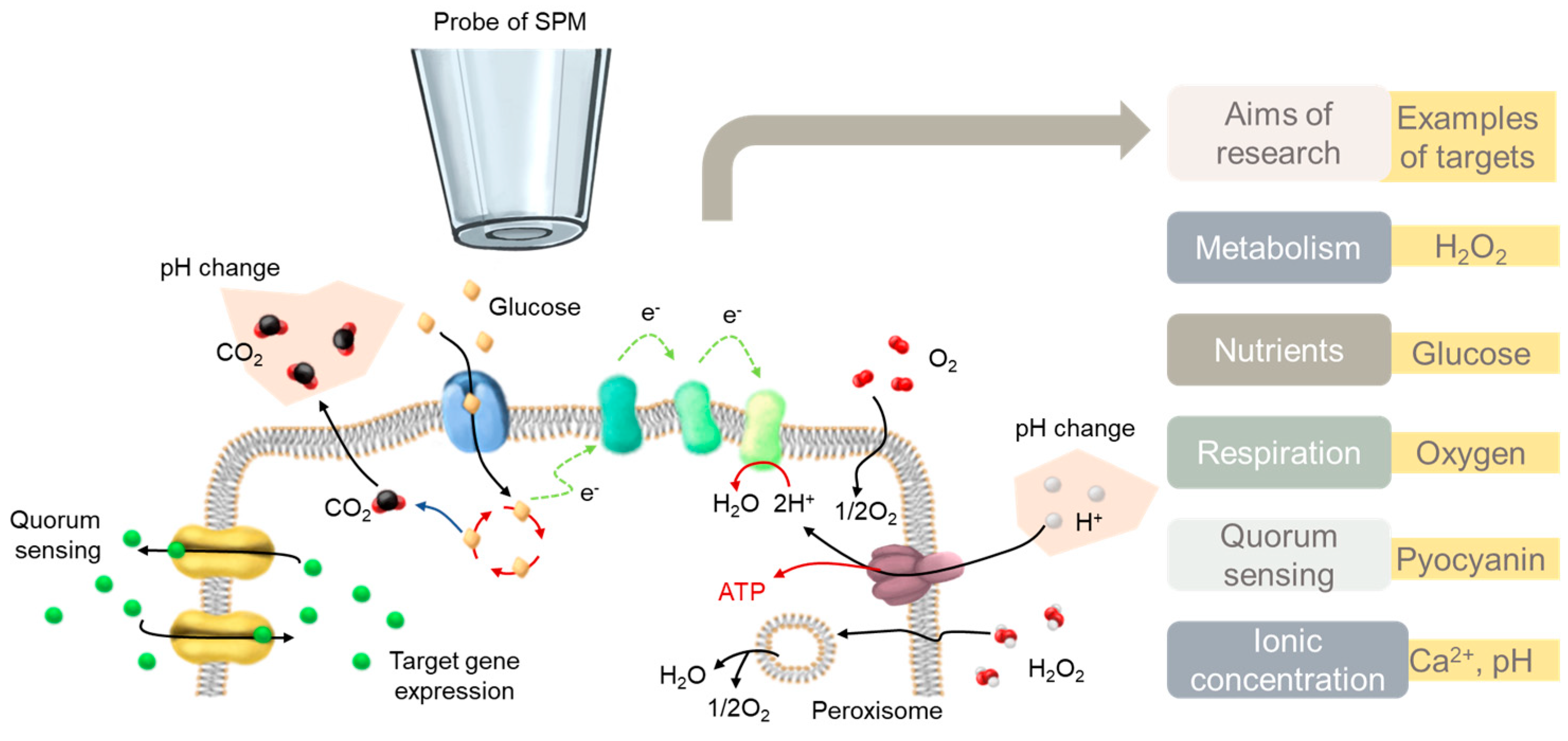
:1. Introduction
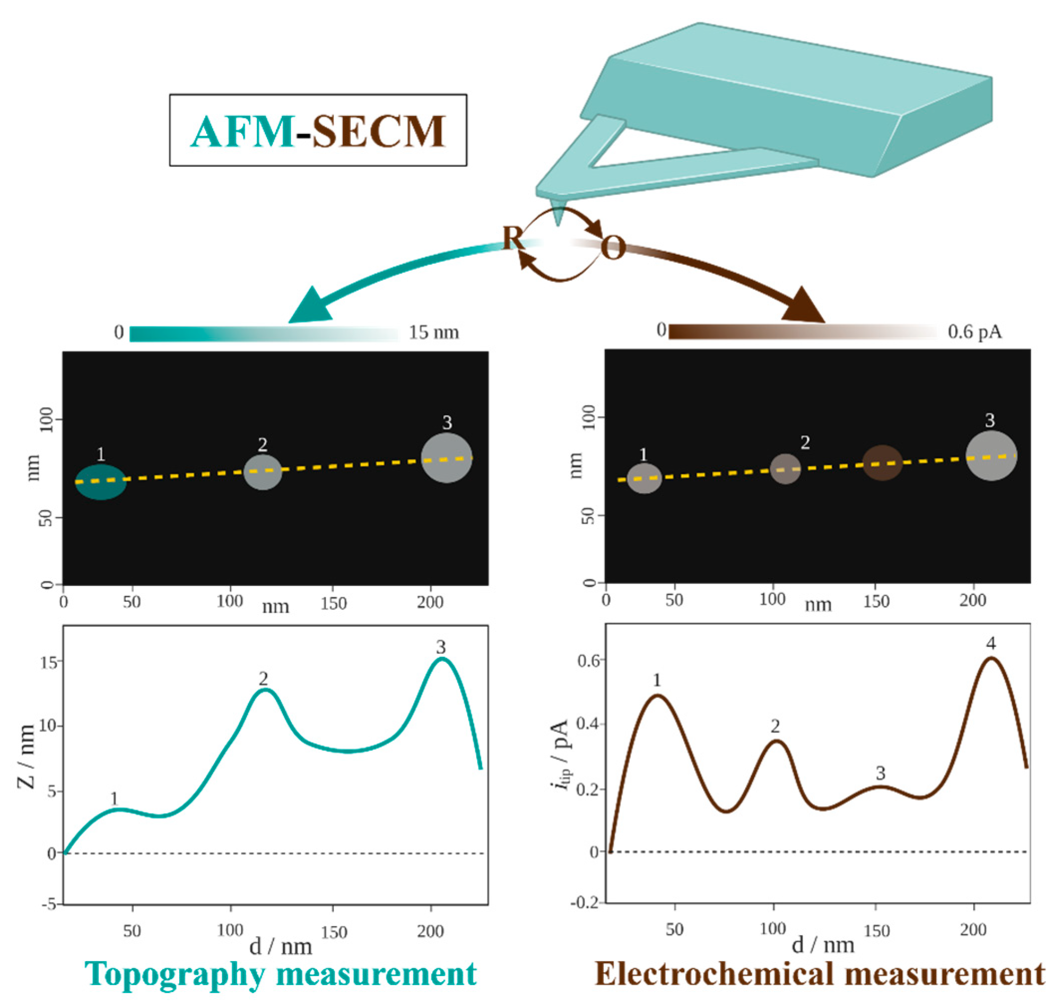
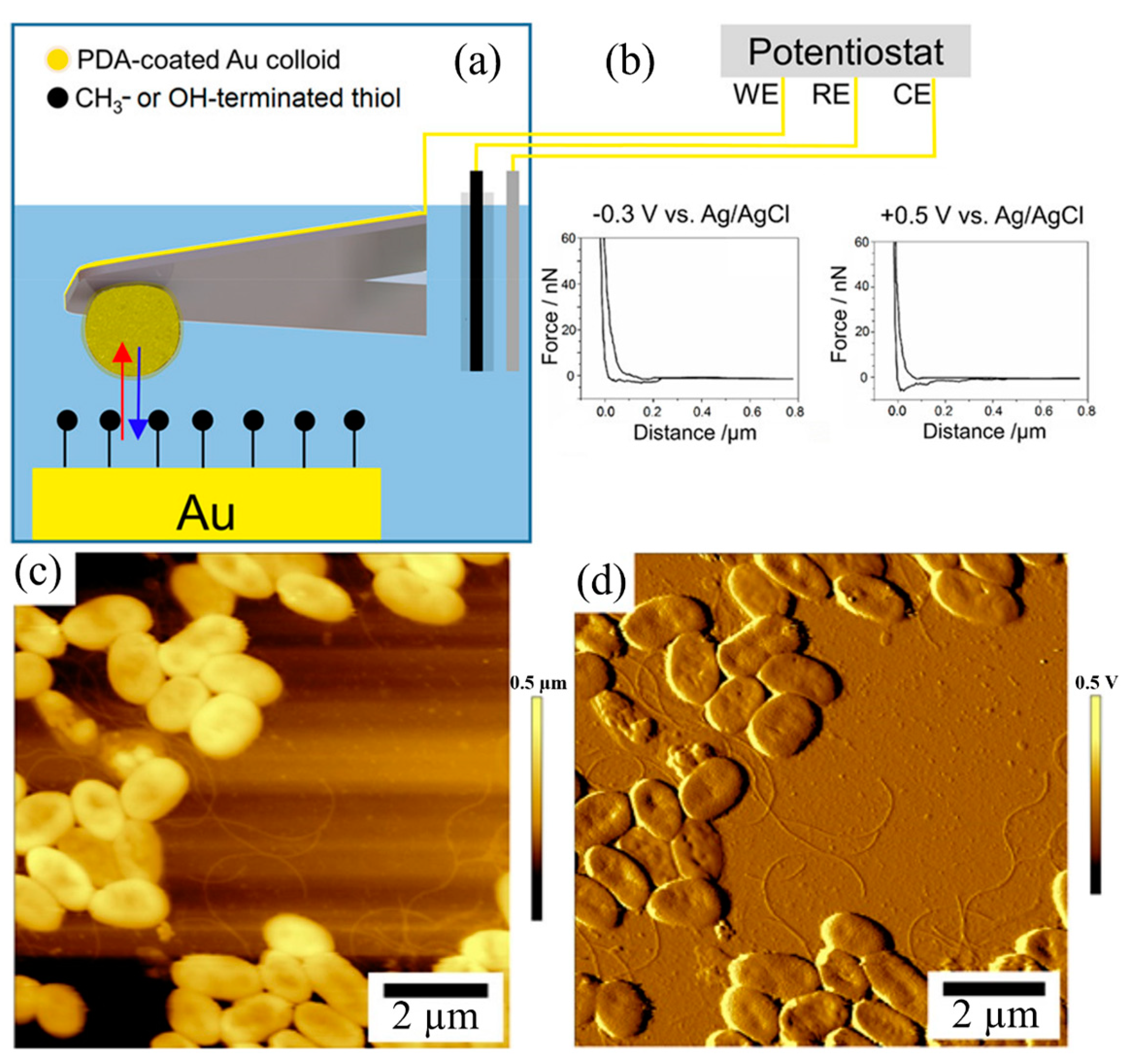
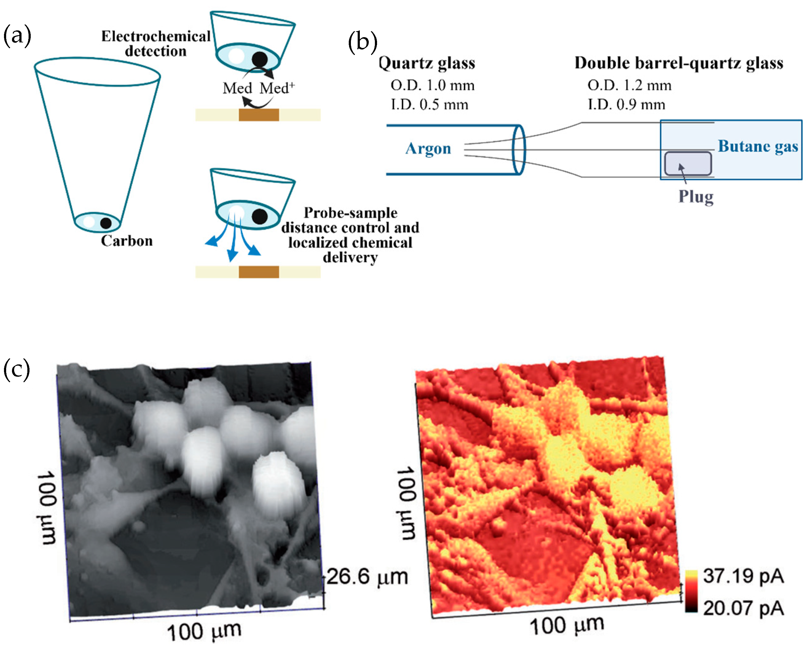
2. The Basic Instrumentation of SECM
3. The Recent Development of Using Scanning Probes for Bacteria Analysis
3.1. SECM Probes and Complementary Techniques
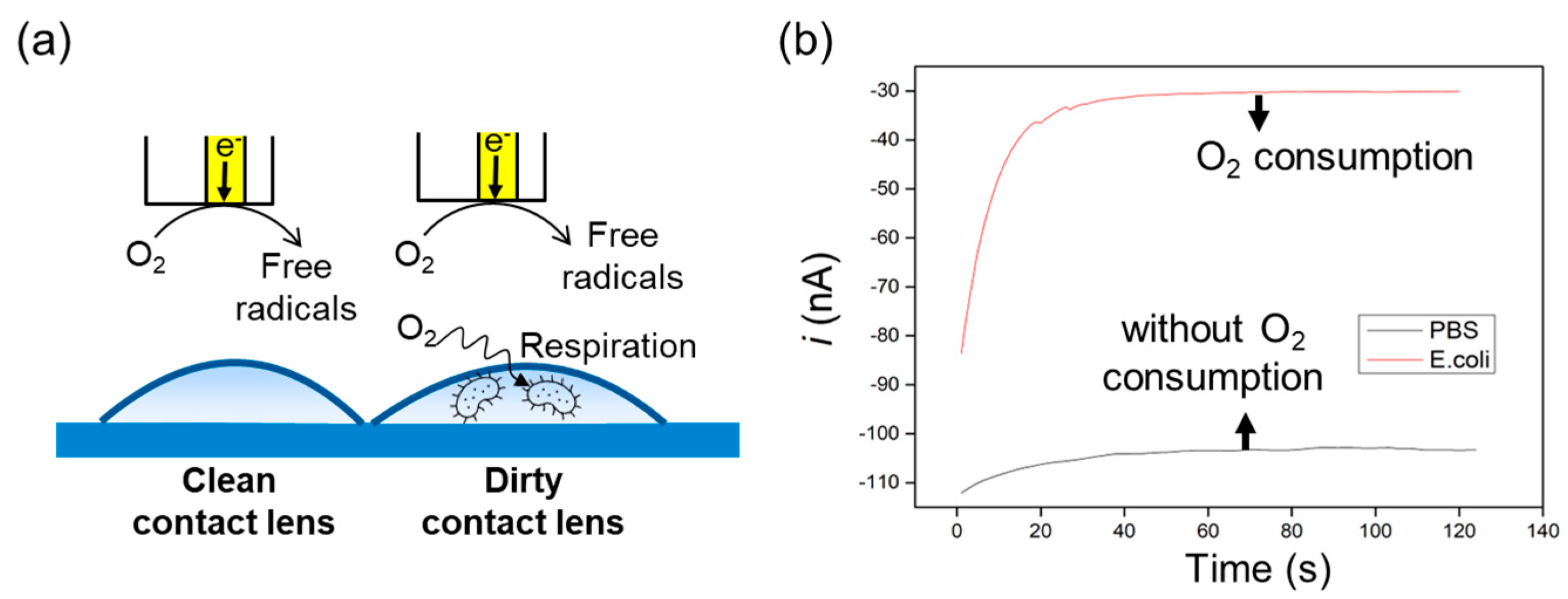
3.2. Measurements of O2 Consumption in Bacteria
3.3. Measurements of the Interactions of Metal Ions with Bacteria
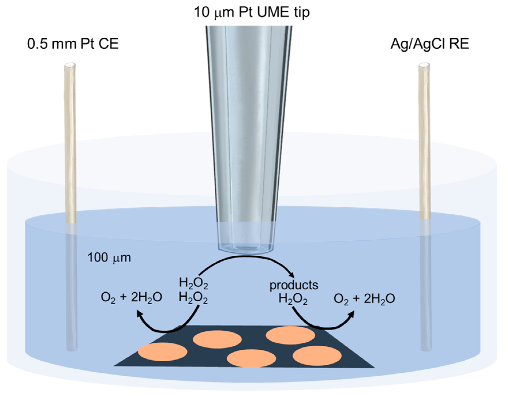
3.4. Detection of Hydrogen Peroxide
3.5. pH and ROS Measurement
3.6. The Research on Quorum Sensing
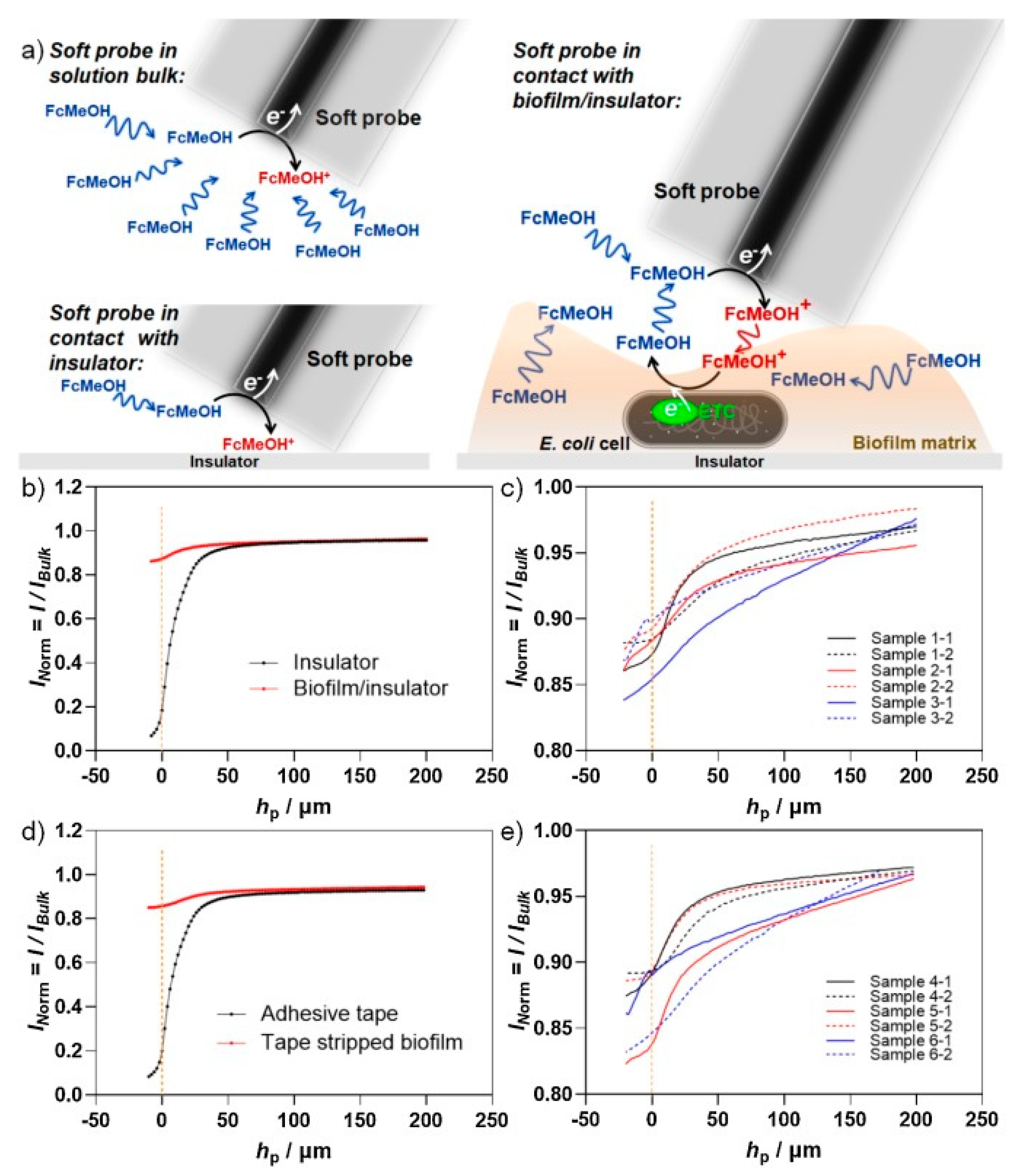
3.7. Direct Biofilm Imaging Using a Soft-Probe SECM
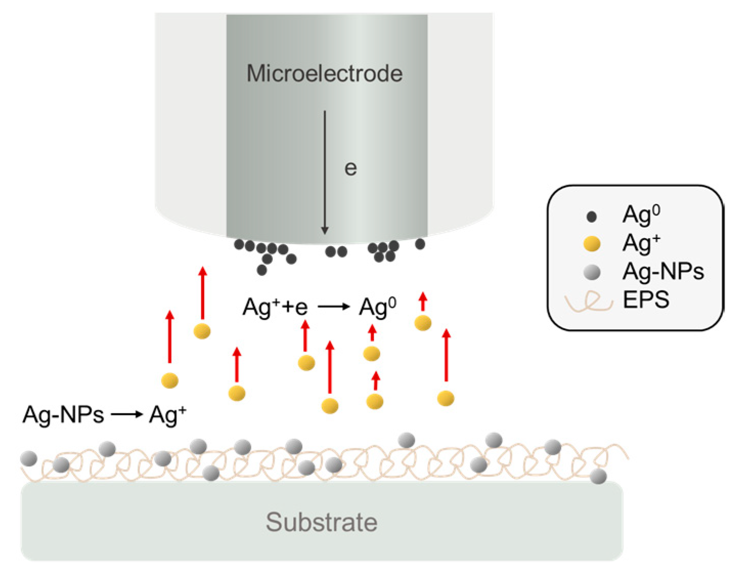
3.8. Characterization of the Growth of Bacteriogenic Metal Nanoparticles
4. Conclusions and Future Perspectives
Author Contributions
Funding
Institutional Review Board Statement
Informed Consent Statement
Data Availability Statement
Conflicts of Interest
References
- Mah, T.-F.C.; O’Toole, G.A. Mechanisms of biofilm resistance to antimicrobial agents. Trends Microbiol. 2001, 9, 34–39. [Google Scholar] [CrossRef]
- Sauer, K.; Stoodley, P.; Goeres, D.M.; Hall-Stoodley, L.; Burmølle, M.; Stewart, P.S.; Bjarnsholt, T. The biofilm life cycle: Expanding the conceptual model of biofilm formation. Nat. Rev. Microbiol. 2022, 20, 608–620. [Google Scholar] [CrossRef] [PubMed]
- Bian, K.; Gerber, C.; Heinrich, A.J.; Müller, D.J.; Scheuring, S.; Jiang, Y. Scanning probe microscopy. Nat. Rev. Methods Prim. 2021, 1, 36. [Google Scholar] [CrossRef]
- Gewirth, A.A.; Niece, B.K. Electrochemical Applications of in Situ Scanning Probe Microscopy. Chem. Rev. 1997, 97, 1129–1162. [Google Scholar] [CrossRef]
- Yang, C.; Su, T.; Hua, Y.; Zhang, L. Electrochemical scanning probe microscopies for artificial photosynthesis. Nano Res. 2023, 16, 4013–4028. [Google Scholar] [CrossRef]
- Zhou, Y.; Zhang, J.; Zhao, Y.; Liu, Y.; Li, F. Bacterial Biofilm Probed by Scanning Electrochemical Microscopy: A Review. ChemElectroChem 2022, 9, e202200470. [Google Scholar] [CrossRef]
- Casero, E.; Vázquez, L.; Parra-Alfambra, A.M.; Lorenzo, E. AFM, SECM and QCM as useful analytical tools in the characterization of enzyme-based bioanalytical platforms. Analyst 2010, 135, 1878–1903. [Google Scholar] [CrossRef]
- Konno, A.; Ino, K.; Utagawa, Y.; Shiku, H. Electrochemical imaging for cell analysis in microphysiological systems. Curr. Opin. Electrochem. 2023, 39, 101270. [Google Scholar] [CrossRef]
- Kranz, C. Recent advancements in nanoelectrodes and nanopipettes used in combined scanning electrochemical microscopy techniques. Analyst 2013, 139, 336–352. [Google Scholar] [CrossRef]
- Krieg, M.; Fläschner, G.; Alsteens, D.; Gaub, B.M.; Roos, W.H.; Wuite, G.J.L.; Gaub, H.E.; Gerber, C.; Dufrêne, Y.F.; Müller, D.J. Atomic force microscopy-based mechanobiology. Nat. Rev. Phys. 2019, 1, 41–57. [Google Scholar] [CrossRef] [Green Version]
- Mirkin, M.V.; Nogala, W.; Velmurugan, J.; Wang, Y. Scanning electrochemical microscopy in the 21st century. Update 1: Five years after. Phys. Chem. Chem. Phys. 2011, 13, 21196–21212. [Google Scholar] [CrossRef]
- Schulte, A.; Nebel, M.; Schuhmann, W. Scanning Electrochemical Microscopy in Neuroscience. Annu. Rev. Anal. Chem. 2010, 3, 299–318. [Google Scholar] [CrossRef] [PubMed]
- Wittstock, G.; Burchardt, M.; Pust, S.E.; Shen, Y.; Zhao, C. Scanning Electrochemical Microscopy for Direct Imaging of Reaction Rates. Angew. Chem. Int. Ed. 2007, 46, 1584–1617. [Google Scholar] [CrossRef] [PubMed]
- Zhu, C.; Huang, K.; Siepser, N.P.; Baker, L.A. Scanning Ion Conductance Microscopy. Chem. Rev. 2021, 121, 11726–11768. [Google Scholar] [CrossRef] [PubMed]
- Lin, T.E.; Rapino, S.; Girault, H.H.; Lesch, A. Electrochemical imaging of cells and tissues. Chem. Sci. 2018, 9, 4546–4554. [Google Scholar] [CrossRef] [Green Version]
- Preet, A.; Lin, T.-E. A Review: Scanning Electrochemical Microscopy (SECM) for Visualizing the Real-Time Local Catalytic Activity. Catalysts 2021, 11, 594. [Google Scholar] [CrossRef]
- Lin, Y.-H.; Tsai, C.-N.; Chen, P.-F.; Lin, Y.-T.; Darvishi, S.; Girault, H.H.; Lin, T.-Y.; Liao, M.-Y.; Lin, T.-E. AI-Assisted Fusion of Scanning Electrochemical Microscopy Images Using Novel Soft Probe. ACS Meas. Sci. Au 2022, 2, 576–583. [Google Scholar] [CrossRef]
- Lin, T.-E.; Zhu, Y.; Hsu, Y.-T.; Liu, F.-Y.; Lin, Y.-P.; Cheng, C.-M. In situ detection of multitarget impurities on contact lens by electrochemical scanning probe. Sens. Actuators B Chem. 2023, 374, 132855. [Google Scholar] [CrossRef]
- Lin, Y.-T.; Preet, A.; Chiu, Y.-P.; Yip, B.-S.; Girault, H.H.; Darvishi, S.; Wang, L.; Lin, T.-E. Communication—Scanning Electrochemical Microscopy Analysis of Interleukin-6 in Oral Cancer. ECS J. Solid. State Sci. Technol. 2020, 9, 115028. [Google Scholar] [CrossRef]
- Lin, T.-E.; Lu, Y.-J.; Sun, C.-L.; Pick, H.; Chen, J.-P.; Lesch, A.; Girault, H.H. Soft Electrochemical Probes for Mapping the Distribution of Biomarkers and Injected Nanomaterials in Animal and Human Tissues. Angew. Chem. Int. Ed. 2017, 56, 16498–16502. [Google Scholar] [CrossRef]
- Lin, T.-E.; Lesch, A.; Li, C.-L.; Girault, H.H. Mapping the antioxidant activity of apple peels with soft probe scanning electrochemical microscopy. J. Electroanal. Chem. 2017, 786, 120–128. [Google Scholar] [CrossRef] [Green Version]
- Bondarenko, A.; Lin, T.-E.; Stupar, P.; Lesch, A.; Cortés-Salazar, F.; Girault, H.H.; Pick, H. Fixation and Permeabilization Approaches for Scanning Electrochemical Microscopy of Living Cells. Anal. Chem. 2016, 88, 11436–11443. [Google Scholar] [CrossRef] [PubMed] [Green Version]
- Polcari, D.; Dauphin-Ducharme, P.; Mauzeroll, J. Scanning Electrochemical Microscopy: A Comprehensive Review of Experimental Parameters from 1989 to 2015. Chem. Rev. 2016, 116, 13234–13278. [Google Scholar] [CrossRef] [PubMed]
- Bonazza, G.; Girault, H.H.; Lesch, A.; Daniele, S. Simultaneous local sensing of two chemical properties with Dual Soft Probe Scanning Electrochemical Microscopy. Electrochim. Acta 2023, 462, 142752. [Google Scholar] [CrossRef]
- Darvishi, S.; Pick, H.; Oveisi, E.; Girault, H.H.; Lesch, A. Soft-probe-scanning electrochemical microscopy reveals electrochemical surface reactivity of E. coli biofilms. Sens. Actuators B Chem. 2021, 334, 129669. [Google Scholar] [CrossRef]
- Pribil, M.M.; Cortés-Salazar, F.; Andreyev, E.A.; Lesch, A.; Karyakina, E.E.; Voronin, O.G.; Girault, H.H.; Karyakin, A.A. Rapid optimization of a lactate biosensor design using soft probes scanning electrochemical microscopy. J. Electroanal. Chem. 2014, 731, 112–118. [Google Scholar] [CrossRef] [Green Version]
- Cortés-Salazar, F.; Träuble, M.; Li, F.; Busnel, J.-M.; Gassner, A.-L.; Hojeij, M.; Wittstock, G.; Girault, H.H. Soft Stylus Probes for Scanning Electrochemical Microscopy. Anal. Chem. 2009, 81, 6889–6896. [Google Scholar] [CrossRef] [Green Version]
- Lesch, A.; Momotenko, D.; Cortés-Salazar, F.; Wirth, I.; Tefashe, U.M.; Meiners, F.; Vaske, B.; Girault, H.H.; Wittstock, G. Fabrication of soft gold microelectrode arrays as probes for scanning electrochemical microscopy. J. Electroanal. Chem. 2012, 666, 52–61. [Google Scholar] [CrossRef] [Green Version]
- Cortés-Salazar, F.; Momotenko, D.; Lesch, A.; Wittstock, G.; Girault, H.H. Soft Microelectrode Linear Array for Scanning Electrochemical Microscopy. Anal. Chem. 2010, 82, 10037–10044. [Google Scholar] [CrossRef] [Green Version]
- Cortés-Salazar, F.; Momotenko, D.; Girault, H.H.; Lesch, A.; Wittstock, G. Seeing Big with Scanning Electrochemical Microscopy. Anal. Chem. 2011, 83, 1493–1499. [Google Scholar] [CrossRef] [Green Version]
- Clausmeyer, J.; Schuhmann, W. Nanoelectrodes: Applications in electrocatalysis, single-cell analysis and high-resolution electrochemical imaging. TrAC Trends Anal. Chem. 2016, 79, 46–59. [Google Scholar] [CrossRef]
- Sarkar, S.; Herath, A.C.; Mukherjee, D.; Mandler, D. Ionic strength induced local electrodeposition of ZnO nanoparticles. Electrochim. Acta 2022, 429, 140986. [Google Scholar] [CrossRef]
- Saikrithika, S.; Shaju, A.; Dinesh, B.; Kumar, A.S. In-situ scanning electrochemical microscopy interrogation on open-circuit release of toxic Ni2+ ion from Ni-containing carbon nanomaterials and nickel-hexacyanoferrate formation in physiological pH and its thiol-electrocatalysis relevance. Electrochim. Acta 2022, 405, 139806. [Google Scholar] [CrossRef]
- Klementiev, A.D.; Jin, Z.; Whiteley, M. Micron Scale Spatial Measurement of the O2 Gradient Surrounding a Bacterial Biofilm in Real Time. MBio 2020, 11, e02536-20. [Google Scholar] [CrossRef]
- Rudolph, D.; Bates, D.; DiChristina, T.J.; Mizaikoff, B.; Kranz, C. Detection of Metal-reducing Enzyme Complexes by Scanning Electrochemical Microscopy. Electroanalysis 2016, 28, 2459–2465. [Google Scholar] [CrossRef]
- Holt, K.B.; Bard, A.J. Interaction of Silver(I) Ions with the Respiratory Chain of Escherichia coli: An Electrochemical and Scanning Electrochemical Microscopy Study of the Antimicrobial Mechanism of Micromolar Ag+. Biochemistry 2005, 44, 13214–13223. [Google Scholar] [CrossRef]
- Moreira, R.; Schütz, M.K.; Libert, M.; Tribollet, B.; Vivier, V. Influence of hydrogen-oxidizing bacteria on the corrosion of low carbon steel: Local electrochemical investigations. Bioelectrochemistry 2014, 97, 69–75. [Google Scholar] [CrossRef] [Green Version]
- Aponso, S.; Ummadi, J.G.; Davis, H.; Ferracane, J.; Koley, D. A Chemical Approach to Optimizing Bioactive Glass Dental Composites. J. Dent. Res. 2019, 98, 194–199. [Google Scholar] [CrossRef] [PubMed]
- Joshi, V.S.; Sheet, P.S.; Cullin, N.; Kreth, J.; Koley, D. Real-Time Metabolic Interactions between Two Bacterial Species Using a Carbon-Based pH Microsensor as a Scanning Electrochemical Microscopy Probe. Anal. Chem. 2017, 89, 11044–11052. [Google Scholar] [CrossRef] [PubMed]
- Harris, D.; Ummadi, J.G.; Thurber, A.R.; Allau, Y.; Verba, C.; Colwell, F.; Torres, M.E.; Koley, D. Real-time monitoring of calcification process by Sporosarcina pasteurii biofilm. Analyst 2016, 141, 2887–2895. [Google Scholar] [CrossRef] [PubMed]
- Hu, Z.; Jin, J.; Abruña, H.D.; Houston, P.L.; Hay, A.G.; Ghiorse, W.C.; Shuler, M.L.; Hidalgo, G.; Lion, L.W. Spatial Distributions of Copper in Microbial Biofilms by Scanning Electrochemical Microscopy. Environ. Sci. Technol. 2007, 41, 936–941. [Google Scholar] [CrossRef] [PubMed]
- Qian, H.C.; Chang, W.W.; Cui, T.Y.; Li, Z.; Guo, D.W.; Kwok, C.T.; Tam, L.M.; Zhang, D.W. Multi-mode scanning electrochemical microscopic study of microbiologically influenced corrosion mechanism of 304 stainless steel by thermoacidophilic archaea. Corros. Sci. 2021, 191, 109751. [Google Scholar] [CrossRef]
- Daboss, S.; Lin, J.; Godejohann, M.; Kranz, C. Redox Switchable Polydopamine-Modified AFM-SECM Probes: A Probe for Electrochemical Force Spectroscopy. Anal. Chem. 2020, 92, 8404–8413. [Google Scholar] [CrossRef] [PubMed]
- Caniglia, G.; Sportelli, M.C.; Heinzmann, A.; Picca, R.A.; Valentini, A.; Barth, H.; Mizaikoff, B.; Cioffi, N.; Kranz, C. Silver-fluoropolymer (Ag-CFX) films: Kinetic study of silver release, and spectroscopic-microscopic insight into the inhibition of P. fluorescens biofilm formation. Anal. Chim. Acta 2022, 1212, 339892. [Google Scholar] [CrossRef]
- Liu, X.; Ramsey, M.M.; Chen, X.; Koley, D.; Whiteley, M.; Bard, A.J. Real-time mapping of a hydrogen peroxide concentration profile across a polymicrobial bacterial biofilm using scanning electrochemical microscopy. Proc. Natl. Acad. Sci. USA 2011, 108, 2668–2673. [Google Scholar] [CrossRef] [Green Version]
- Joshi, V.S.; Kreth, J.; Koley, D. Pt-Decorated MWCNTs–Ionic Liquid Composite-Based Hydrogen Peroxide Sensor To Study Microbial Metabolism Using Scanning Electrochemical Microscopy. Anal. Chem. 2017, 89, 7709–7718. [Google Scholar] [CrossRef]
- Jayathilake, N.M.; Koley, D. Glucose Microsensor with Covalently Immobilized Glucose Oxidase for Probing Bacterial Glucose Uptake by Scanning Electrochemical Microscopy. Anal. Chem. 2020, 92, 3589–3597. [Google Scholar] [CrossRef]
- Lin, M.-H.; Mehraeen, S.; Cheng, G.; Rusinek, C.; Chaplin, B.P. Role of Near-Electrode Solution Chemistry on Bacteria Attachment and Poration at Low Applied Potentials. Environ. Sci. Technol. 2020, 54, 446–455. [Google Scholar] [CrossRef] [PubMed]
- Nguyen, A.T.; Goswami, S.; Ferracane, J.; Koley, D. Real-time monitoring of the pH microenvironment at the interface of multispecies biofilm and dental composites. Anal. Chim. Acta 2022, 1201, 339589. [Google Scholar] [CrossRef] [PubMed]
- Sheet, P.S.; Koley, D. Dendritic Hydrogel Bioink for 3D Printing of Bacterial Microhabitat. ACS Appl. Bio Mater. 2019, 2, 5941–5948. [Google Scholar] [CrossRef] [PubMed]
- She, Z.; Topping, K.; Dong, B.; Shamsi, M.H.; Kraatz, H.-B. An unexpected use of ferrocene. A scanning electrochemical microscopy study of a toll-like receptor array and its interaction with E. coli. Chem. Commun. 2017, 53, 2946–2949. [Google Scholar] [CrossRef] [Green Version]
- She, Z.; Topping, K.; Shamsi, M.H.; Wang, N.; Chan, N.W.C.; Kraatz, H.-B. Investigation of the Utility of Complementary Electrochemical Detection Techniques to Examine the in Vitro Affinity of Bacterial Flagellins for a Toll-Like Receptor 5 Biosensor. Anal. Chem. 2015, 87, 4218–4224. [Google Scholar] [CrossRef]
- Abucayon, E.; Ke, N.; Cornut, R.; Patelunas, A.; Miller, D.; Nishiguchi, M.K.; Zoski, C.G. Investigating Catalase Activity Through Hydrogen Peroxide Decomposition by Bacteria Biofilms in Real Time Using Scanning Electrochemical Microscopy. Anal. Chem. 2014, 86, 498–505. [Google Scholar] [CrossRef]
- Connell, J.L.; Kim, J.; Shear, J.B.; Bard, A.J.; Whiteley, M. Real-time monitoring of quorum sensing in 3D-printed bacterial aggregates using scanning electrochemical microscopy. Proc. Natl. Acad. Sci. USA 2014, 111, 18255–18260. [Google Scholar] [CrossRef] [Green Version]
- Borghese, R.; Malferrari, M.; Brucale, M.; Ortolani, L.; Franchini, M.; Rapino, S.; Borsetti, F.; Zannoni, D. Structural and electrochemical characterization of lawsone-dependent production of tellurium-metal nanoprecipitates by photosynthetic cells of Rhodobacter capsulatus. Bioelectrochemistry 2020, 133, 107456. [Google Scholar] [CrossRef]
- Battistel, D.; Baldi, F.; Gallo, M.; Faleri, C.; Daniele, S. Characterisation of biosynthesised silver nanoparticles by scanning electrochemical microscopy (SECM) and voltammetry. Talanta 2015, 132, 294–300. [Google Scholar] [CrossRef]
- Tian, X.; Wu, R.; Li, X.; Wu, X.; Jiang, Y.; Zhao, F. Feedback current production by a ferrous mediator revealing the redox properties of Shewanella oneidensis MR-1. J. Electroanal. Chem. 2022, 916, 116387. [Google Scholar] [CrossRef]
- Almeida, E.; Puri, S.; Elangovan, S.; Kim, J.; Ramsey, M. Corynebacterium matruchotii fitness enhancement of adjacent streptococci by multiple mechanisms. bioRxiv 2022. [Google Scholar] [CrossRef]
- Verba, C.; Thurber, A.R.; Alleau, Y.; Koley, D.; Colwell, F.; Torres, M.E. Mineral changes in cement-sandstone matrices induced by biocementation. Int. J. Greenh. Gas. Control. 2016, 49, 312–322. [Google Scholar] [CrossRef] [Green Version]
- Zhang, W.; Wu, H.; Hsing, I.-M. Real-Time Label-Free Monitoring of Shewanella oneidensis MR-1 Biofilm Formation on Electrode during Bacterial Electrogenesis Using Scanning Electrochemical Microscopy. Electroanalysis 2015, 27, 648–655. [Google Scholar] [CrossRef]
- Hansma, H.G. Varieties of imaging with scanning probe microscopes. Proc. Natl. Acad. Sci. USA 1999, 96, 14678–14680. [Google Scholar] [CrossRef] [Green Version]
- Caniglia, G.; Kranz, C. Scanning electrochemical microscopy and its potential for studying biofilms and antimicrobial coatings. Anal. Bioanal. Chem. 2020, 412, 6133–6148. [Google Scholar] [CrossRef]
- Cremin, K.; Jones, B.A.; Teahan, J.; Meloni, G.N.; Perry, D.; Zerfass, C.; Asally, M.; Soyer, O.S.; Unwin, P.R. Scanning Ion Conductance Microscopy Reveals Differences in the Ionic Environments of Gram-Positive and Negative Bacteria. Anal. Chem. 2020, 92, 16024–16032. [Google Scholar] [CrossRef]
- Guerret-Legras, L.; Audibert, J.F.; Dubacheva, G.V.; Miomandre, F. Combined scanning electrochemical and fluorescence microscopies using a tetrazine as a single redox and luminescent (electrofluorochromic) probe. Chem. Sci. 2018, 9, 5897–5905. [Google Scholar] [CrossRef] [Green Version]
- Guerret-Legras, L.; Audibert, J.F.; Ojeda, I.M.G.; Dubacheva, G.V.; Miomandre, F. Combined SECM-fluorescence microscopy using a water-soluble electrofluorochromic dye as the redox mediator. Electrochim. Acta 2019, 305, 370–377. [Google Scholar] [CrossRef]
- Goldberg, B.S.; Ackerman, M.E. Antibody-mediated complement activation in pathology and protection. Immunol. Cell Biol. 2020, 98, 305–317. [Google Scholar] [CrossRef] [PubMed]
- Kasas, S.; Fellay, B.; Cargnello, R. Observation of the action of penicillin on bacillus subtilis using atomic force microscopy: Technique for the preparation of bacteria. Surf. Interface Anal. 1994, 21, 400–401. [Google Scholar] [CrossRef]
- Ong, Y.-L.; Razatos, A.; Georgiou, G.; Sharma, M.M. Adhesion Forces between E. coli Bacteria and Biomaterial Surfaces. Langmuir 1999, 15, 2719–2725. [Google Scholar] [CrossRef]
- Kasas, S.; Thomson, N.H.; Smith, B.L.; Hansma, H.G.; Zhu, X.; Guthold, M.; Bustamante, C.; Kool, E.T.; Kashlev, M.; Hansma, P.K. Escherichia coli RNA Polymerase Activity Observed Using Atomic Force Microscopy. Biochemistry 1997, 36, 461–468. [Google Scholar] [CrossRef]
- Lyubchenko, Y.L.; Shlyakhtenko, L.S. Visualization of supercoiled DNA with atomic force microscopy in situ. Proc. Natl. Acad. Sci. USA 1997, 94, 496–501. [Google Scholar] [CrossRef] [Green Version]
- Shlyakhtenko, L.S.; Potaman, V.N.; Sinden, R.R.; Lyubchenko, Y.L. Structure and dynamics of supercoil-stabilized DNA cruciforms11Edited by I. Tinoco. J. Mol. Biol. 1998, 280, 61–72. [Google Scholar] [CrossRef] [PubMed] [Green Version]
- Efremov, Y.M.; Okajima, T.; Raman, A. Measuring viscoelasticity of soft biological samples using atomic force microscopy. Soft Matter 2020, 16, 64–81. [Google Scholar] [CrossRef] [PubMed]
- Wang, J.; Qian, Y.; Li, L.; Qiu, X. Atomic Force Microscopy and Molecular Dynamics Simulations for Study of Lignin Solution Self-Assembly Mechanisms in Organic–Aqueous Solvent Mixtures. ChemSusChem 2020, 13, 4420–4427. [Google Scholar] [CrossRef] [PubMed]
- Walsh, D.A.; Fernández, J.L.; Mauzeroll, J.; Bard, A.J. Scanning Electrochemical Microscopy. 55. Fabrication and Characterization of Micropipet Probes. Anal. Chem. 2005, 77, 5182–5188. [Google Scholar] [CrossRef] [PubMed]
- Comstock, D.J.; Elam, J.W.; Pellin, M.J.; Hersam, M.C. Integrated Ultramicroelectrode-Nanopipet Probe for Concurrent Scanning Electrochemical Microscopy and Scanning Ion Conductance Microscopy. Anal. Chem. 2010, 82, 1270–1276. [Google Scholar] [CrossRef] [PubMed]
- Takahashi, Y.; Shevchuk, A.I.; Novak, P.; Zhang, Y.; Ebejer, N.; Macpherson, J.V.; Unwin, P.R.; Pollard, A.J.; Roy, D.; Clifford, C.A.; et al. Multifunctional Nanoprobes for Nanoscale Chemical Imaging and Localized Chemical Delivery at Surfaces and Interfaces. Angew. Chem. Int. Ed. 2011, 50, 9638–9642. [Google Scholar] [CrossRef]
- Takahashi, Y.; Shevchuk, A.I.; Novak, P.; Babakinejad, B.; Macpherson, J.; Unwin, P.R.; Shiku, H.; Gorelik, J.; Klenerman, D.; Korchev, Y.E.; et al. Topographical and electrochemical nanoscale imaging of living cells using voltage-switching mode scanning electrochemical microscopy. Proc. Natl. Acad. Sci. USA 2012, 109, 11540–11545. [Google Scholar] [CrossRef] [Green Version]
- Yip, N.-C.; Rawson, F.J.; Tsang, C.W.; Mendes, P.M. Real-time electrocatalytic sensing of cellular respiration. Biosens. Bioelectron. 2014, 57, 303–309. [Google Scholar] [CrossRef] [Green Version]
- Fu, H.; Yuan, J.; Gao, H. Microbial oxidative stress response: Novel insights from environmental facultative anaerobic bacteria. Arch. Biochem. Biophys. 2015, 584, 28–35. [Google Scholar] [CrossRef]
- Pecchielan, G.; Battistel, D.; Daniele, S. Scanning Electrochemical Microscopy and Voltammetric Investigation of Silver Nanoparticles Embedded within a Nafion Membrane. ChemElectroChem 2016, 3, 2297–2304. [Google Scholar] [CrossRef]
- Lovley, D.R.; Coates, J.D. Novel forms of anaerobic respiration of environmental relevance. Curr. Opin. Microbiol. 2000, 3, 252–256. [Google Scholar] [CrossRef]
- Nealson, K.H.; Belz, A.; McKee, B. Breathing metals as a way of life: Geobiology in action. Antonie Van Leeuwenhoek 2002, 81, 215–222. [Google Scholar] [CrossRef] [PubMed]
- Kreve, S.; Reis, A.C.D. Bacterial adhesion to biomaterials: What regulates this attachment? A review. Jpn. Dent. Sci. Rev. 2021, 57, 85–96. [Google Scholar] [CrossRef] [PubMed]
- Yao, S.; Hao, L.; Zhou, R.; Jin, Y.; Huang, J.; Wu, C. Multispecies biofilms in fermentation: Biofilm formation, microbial interactions, and communication. Compr. Rev. Food Sci. Food Saf. 2022, 21, 3346–3375. [Google Scholar] [CrossRef]
- Sionov, R.V.; Steinberg, D. Targeting the Holy Triangle of Quorum Sensing, Biofilm Formation, and Antibiotic Resistance in Pathogenic Bacteria. Microorganisms 2022, 10, 1239. [Google Scholar] [CrossRef]
- Meirelles, L.A.; Newman, D.K. Both toxic and beneficial effects of pyocyanin contribute to the lifecycle of Pseudomonas aeruginosa. Mol. Microbiol. 2018, 110, 995–1010. [Google Scholar] [CrossRef] [PubMed] [Green Version]
- Saravanan, A.; Kumar, P.S.; Karishma, S.; Vo, D.-V.N.; Jeevanantham, S.; Yaashikaa, P.R.; George, C.S. A review on biosynthesis of metal nanoparticles and its environmental applications. Chemosphere 2021, 264, 128580. [Google Scholar] [CrossRef]


| Research Goals | Redox Mediators or Solutions Added | Ref. |
|---|---|---|
| O2 consumption of Pseudomonas aeruginosa | (ferrocenylmethyl)trimethylammonium ion (FcMTMA+) | [34] |
| Fe, Mn, and O2 consumption of Shewanella oneidensis | Tris-acetate buffer, Ru(NH3)6Cl3 | [35] |
| O2 consumption, antimicrobial mechanism of Ag+ in Escherichia coli | Electrolytes with NaNO3 and glucose | [36] |
| H2 consumption of Shewanella oneidensis | M1 solution prepared by the authors | [37] |
| pH and release of Ca2+ against Streptococcus mutans | Artificial saliva | [38] |
| Metabolic interactions of Streptococcus gordonii and pathogenic Streptococcus mutans | Artificial saliva and sugar | [39] |
| Calcification process of Sporosarcina pasteurii | Brine solution containing urea | [40] |
| Copper concentration near Escherichia coli biofilm | Hydroxymethyl ferrocene | [41] |
| Metallosphaera cuprina associated with Fe2+ consumption | FcMeOH or FeCl2 | [42] |
| Adhesion of Pseudomonas fluorescens | KCl solution | [43] |
| Antimicrobial efficiency of silver-fluoropolymer (Ag-CFX) films against Pseudomonas fluorescens | Phosphate-buffered saline (PBS) | [44] |
| Hydrogen peroxide production of Streptococcus gordonii and interaction with Aggregatibacter actinomycetemcomitans | Chemically defined medium (CDM) culture solution with glucose | [45] |
| Hydrogen peroxide produced by Streptococcus gordonii | Glucose and artificial saliva solution | [46] |
| Glucose consumption of Streptococcus mutans | Artificial saliva solution and ferrocyanide | [47] |
| pH, ROS measurement, and bacteria attachment and poration of Pseudomonas aeruginosa | PBS, Fe(CN)63–/4– and Ru(NH3)63+/2+ | [48] |
| pH changes in dental-plaque-derived multi-species biofilm | Artificial saliva and FcMeOH | [49] |
| Characterization of a 3D-printed hydrogel with Streptococcus mutans and Escherichia coli | FcMeOH, phosphate-buffered saline (PBS) | [50] |
| Electron transfer train of ampicillin-resistant Escherichia coli on the tape | FcMeOH | [25] |
| Toll-like receptor array and its interaction with Escherichia coli | Ferrocene derivatives and electrolytes | [51] |
| Interaction of toll-like receptor 5 and Salmonella typhimurium and Bacillus subtilis | K4Fe(CN)6 | [52] |
| Catalase activity of γ-Protebacteria-Vibrionaceae biofilms | Artificial seawater | [53] |
| Pseudomonas aeruginosa quantifies pyocyanin of QS | FcMeOH | [54] |
| Production of tellurium metal nanoprecipitates by Rhodobacter capsulatus | Phosphate-buffered saline (PBS) and lawsone | [55] |
| AgNPs biosynthesized by a Klebsiella oxytoca | K3IrCl6 | [56] |
| Redox properties of Shewanella oneidensis | FcMeOH | [57] |
| Corynebacterium matruchotii fitness enhancement in adjacent streptococci mitis | FcMeOH | [58] |
| The pH of biocementation induced by Sporosarcina pasteurii | Brine solution and urea | [59] |
| Biofilm formation of Shewanella oneidensis | Ru(NH3)6Cl3 | [60] |
| Bacteria contamination in contact lenses, including Escherichia coli, Diphtheroid, Pseudomonas aeruginosa, Staphylococcus aureus, Fusobacterium nucleatum, Unidentified Gram(+) Bacilli | Phosphate-buffered saline (PBS), FcMeOH | [18] |
Disclaimer/Publisher’s Note: The statements, opinions and data contained in all publications are solely those of the individual author(s) and contributor(s) and not of MDPI and/or the editor(s). MDPI and/or the editor(s) disclaim responsibility for any injury to people or property resulting from any ideas, methods, instructions or products referred to in the content. |
© 2023 by the authors. Licensee MDPI, Basel, Switzerland. This article is an open access article distributed under the terms and conditions of the Creative Commons Attribution (CC BY) license (https://creativecommons.org/licenses/by/4.0/).
Share and Cite
Lin, T.-E.; Darvishi, S. A Brief Review of In Situ and Operando Electrochemical Analysis of Bacteria by Scanning Probes. Biosensors 2023, 13, 695. https://doi.org/10.3390/bios13070695
Lin T-E, Darvishi S. A Brief Review of In Situ and Operando Electrochemical Analysis of Bacteria by Scanning Probes. Biosensors. 2023; 13(7):695. https://doi.org/10.3390/bios13070695
Chicago/Turabian StyleLin, Tzu-En, and Sorour Darvishi. 2023. "A Brief Review of In Situ and Operando Electrochemical Analysis of Bacteria by Scanning Probes" Biosensors 13, no. 7: 695. https://doi.org/10.3390/bios13070695