Non-Invasive Biosensing for Healthcare Using Artificial Intelligence: A Semi-Systematic Review


Abstract
:1. Introduction
2. Common Deep Learning Architectures in Biosensing
2.1. Convolutional Neural Networks (CNNs)
2.2. Long Short-Term Memory Networks (LSTMs)
2.3. Autoencoders
2.4. Transformers
2.5. Model Selection Considerations
2.6. Commercial Use of Deep Learning Models for Biosensing
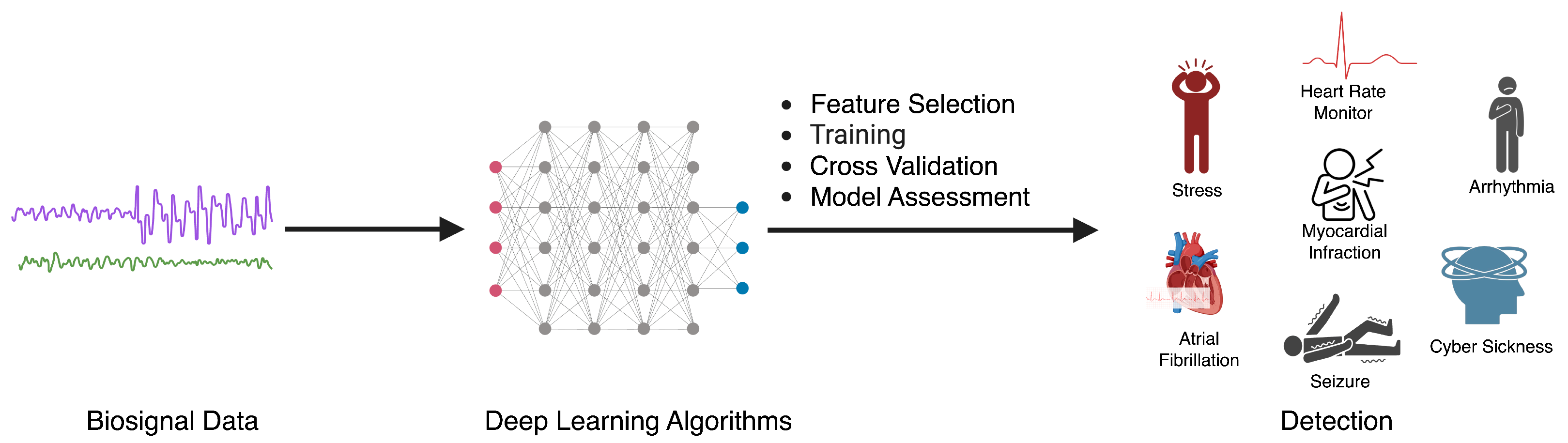
3. Sensor Modalities and Corresponding Health Applications
3.1. Deep Learning with EEG
3.2. Deep Learning with EDA
3.3. Deep Learning with ECG
3.4. Consideration for Selecting Biosensors for a Digital Health Application
4. Digital Health Applications using Biosensors
4.1. Remote Patient Monitoring
4.2. Digital Diagnostics
4.3. Adaptive Digital Interventions
4.4. Considerations for Integrating Wearable Sensors in Digital Health Systems
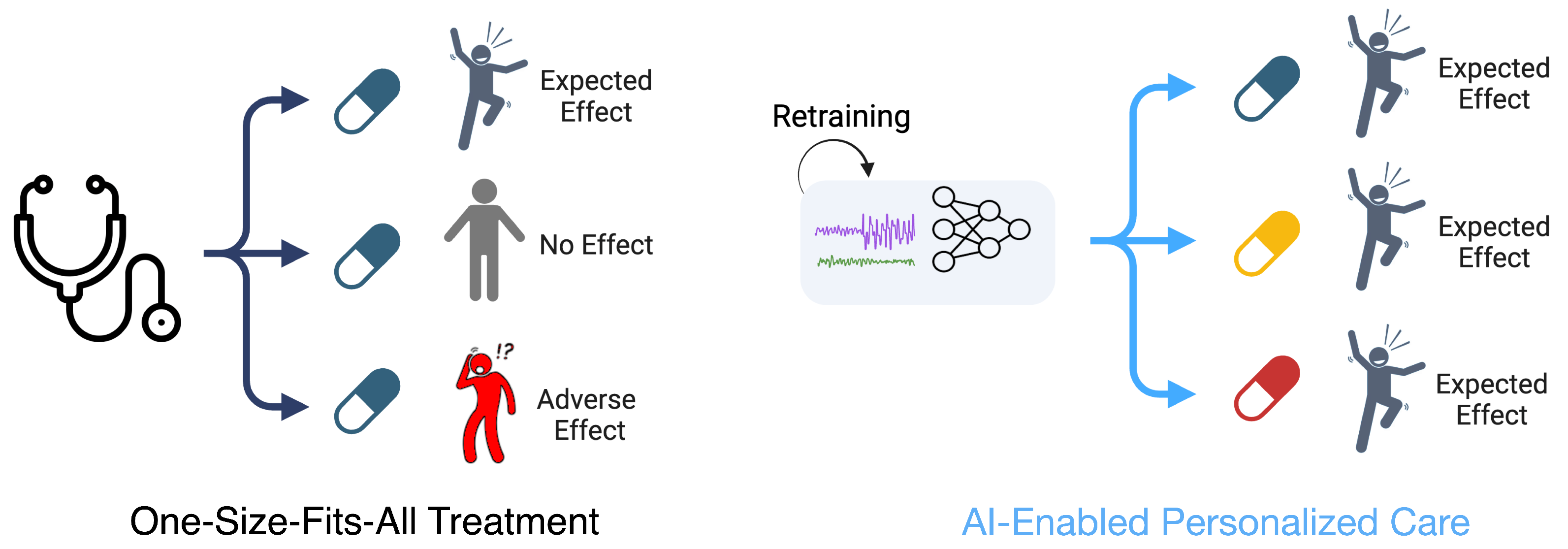
5. Challenges and Opportunities
5.1. Challenges
5.2. Opportunities
6. Conclusions
Funding
Acknowledgments
Conflicts of Interest
References
- Dong, S.; Wang, P.; Abbas, K. A survey on deep learning and its applications. Comput. Sci. Rev. 2021, 40, 100379. [Google Scholar] [CrossRef]
- Zhou, M.; Tian, C.; Cao, R.; Wang, B.; Niu, Y.; Hu, T.; Guo, H.; Xiang, J. Epileptic seizure detection based on EEG signals and CNN. Front. Neuroinform. 2018, 12, 95. [Google Scholar] [CrossRef]
- Mao, W.; Fathurrahman, H.; Lee, Y.; Chang, T. EEG dataset classification using CNN method. J. Phys. Conf. Ser. 2020, 1456, 012017. [Google Scholar] [CrossRef]
- Liu, Q.; Cai, J.; Fan, S.Z.; Abbod, M.F.; Shieh, J.S.; Kung, Y.; Lin, L. Spectrum analysis of EEG signals using CNN to model patient’s consciousness level based on anesthesiologists’ experience. IEEE Access 2019, 7, 53731–53742. [Google Scholar] [CrossRef]
- Khanday, N.Y.; Sofi, S.A. Taxonomy, state-of-the-art, challenges and applications of visual understanding: A review. Comput. Sci. Rev. 2021, 40, 100374. [Google Scholar] [CrossRef]
- Venugopalan, S.; Xu, H.; Donahue, J.; Rohrbach, M.; Mooney, R.; Saenko, K. Translating videos to natural language using deep recurrent neural networks. arXiv 2014, arXiv:1412.4729. [Google Scholar]
- Saltepe, B.; Bozkurt, E.U.; Güngen, M.A.; Çiçek, A.E.; Şeker, U.Ö.Ş. Genetic circuits combined with machine learning provides fast responding living sensors. Biosens. Bioelectron. 2021, 178, 113028. [Google Scholar] [CrossRef] [PubMed]
- Esmaeili, F.; Cassie, E.; Nguyen, H.P.T.; Plank, N.O.; Unsworth, C.P.; Wang, A. Predicting analyte concentrations from electrochemical aptasensor signals using LSTM recurrent networks. Bioengineering 2022, 9, 529. [Google Scholar] [CrossRef] [PubMed]
- Hinton, G.E.; Salakhutdinov, R.R. Reducing the dimensionality of data with neural networks. Science 2006, 313, 504–507. [Google Scholar] [CrossRef]
- Vincent, P.; Larochelle, H.; Lajoie, I.; Bengio, Y.; Manzagol, P.A.; Bottou, L. Stacked denoising autoencoders: Learning useful representations in a deep network with a local denoising criterion. J. Mach. Learn. Res. 2010, 11, 3371–3408. [Google Scholar]
- Ng, A. Sparse autoencoder. CS294A Lect. Notes 2011, 72, 1–19. [Google Scholar]
- Saadatnejad, S.; Oveisi, M.; Hashemi, M. LSTM-based ECG classification for continuous monitoring on personal wearable devices. IEEE J. Biomed. Health Inform. 2019, 24, 515–523. [Google Scholar] [CrossRef]
- Sun, Y.; Jin, W.; Si, X.; Zhang, X.; Cao, J.; Wang, L.; Yin, S.; Ming, D. Continuous Seizure Detection Based on Transformer and Long-Term iEEG. IEEE J. Biomed. Health Inform. 2022, 26, 5418–5427. [Google Scholar] [CrossRef]
- Yildirim, O.; San Tan, R.; Acharya, U.R. An efficient compression of ECG signals using deep convolutional autoencoders. Cogn. Syst. Res. 2018, 52, 198–211. [Google Scholar] [CrossRef]
- Chien, H.Y.S.; Goh, H.; Sandino, C.M.; Cheng, J.Y. MAEEG: Masked Auto-encoder for EEG Representation Learning. In Proceedings of the NeurIPS Workshop, New Orleans, LA, USA, 28 November 2022. [Google Scholar]
- Nazaret, A.; Tonekaboni, S.; Darnell, G.; Ren, S.; Sapiro, G.; Miller, A.C. Modeling Heart Rate Response to Exercise with Wearable Data. In Proceedings of the NeurIPS, New Orleans, LA, USA, 28 November 2022. [Google Scholar]
- Slyusarenko, K.; Fedorin, I. Smart alarm based on sleep stages prediction. In Proceedings of the 2020 42nd Annual International Conference of the IEEE Engineering in Medicine & Biology Society (EMBC), Montreal, QC, Canada, 20–24 July 2020; pp. 4286–4289. [Google Scholar] [CrossRef]
- Zhang, H.; Zhu, L.; Nathan, V.; Kuang, J.; Kim, J.; Gao, J.A.; Olgin, J. Towards early detection and burden estimation of atrial fibrillation in an ambulatory free-living environment. In Proceedings of the ACM on Interactive, Mobile, Wearable and Ubiquitous Technologies; ACM: New York, NY, USA, 2021; Volume 5, pp. 1–19. [Google Scholar]
- Kinnunen, H.; Rantanen, A.; Kenttä, T.; Koskimäki, H. Feasible assessment of recovery and cardiovascular health: Accuracy of nocturnal HR and HRV assessed via ring PPG in comparison to medical grade ECG. Physiol. Meas. 2020, 41, 04NT01. [Google Scholar] [CrossRef]
- Song, T.; Zheng, W.; Song, P.; Cui, Z. EEG Emotion Recognition Using Dynamical Graph Convolutional Neural Networks. IEEE Trans. Affect. Comput. 2020, 11, 532–541. [Google Scholar] [CrossRef]
- Park, C.; Choi, G.; Kim, J.; Kim, S.; Kim, T.J.; Min, K.; Jung, K.Y.; Chong, J. Epileptic seizure detection for multi-channel EEG with deep convolutional neural network. In Proceedings of the 2018 International Conference on Electronics, Information, and Communication (ICEIC), Honolulu, HI, USA, 24–27 January 2018; pp. 1–5. [Google Scholar]
- Tsinalis, O.; Matthews, P.M.; Guo, Y.; Zafeiriou, S. Automatic sleep stage scoring with single-channel EEG using convolutional neural networks. arXiv 2016, arXiv:1610.01683. [Google Scholar]
- Roy, A.M. An efficient multi-scale CNN model with intrinsic feature integration for motor imagery EEG subject classification in brain-machine interfaces. Biomed. Signal Process. Control 2022, 74, 103496. [Google Scholar] [CrossRef]
- Leeb, R.; Brunner, C.; Müller-Putz, G. Available online: http://www.bbci.de/competition/iv/ (accessed on 6 March 2021).
- Raghu, S.; Sriraam, N.; Temel, Y.; Rao, S.V.; Kubben, P.L. EEG based multi-class seizure type classification using convolutional neural network and transfer learning. Neural Netw. 2020, 124, 202–212. [Google Scholar] [CrossRef]
- Obeid, I.; Picone, J. The temple university hospital EEG data corpus. Front. Neurosci. 2016, 10, 196. [Google Scholar] [CrossRef]
- Fouladi, S.; Safaei, A.A.; Mammone, N.; Ghaderi, F.; Ebadi, M. Efficient deep neural networks for classification of Alzheimer’s disease and mild cognitive impairment from scalp EEG recordings. Cogn. Comput. 2022, 14, 1247–1268. [Google Scholar] [CrossRef]
- Wang, Q.; Wang, Z.; Xiong, R.; Liao, X.; Tan, X. A Method for Classification and Evaluation of Pilot’s Mental States Based on CNN. Comput. Syst. Sci. Eng. 2023, 46, 1999–2020. [Google Scholar] [CrossRef]
- Zheng, W.L.; Lu, B.L. Investigating Critical Frequency Bands and Channels for EEG-Based Emotion Recognition with Deep Neural Networks. IEEE Trans. Auton. Ment. Dev. 2015, 7, 162–175. [Google Scholar] [CrossRef]
- Parajuli, M.; Amara, A.W.; Shaban, M. Deep-learning detection of mild cognitive impairment from sleep electroencephalography for patients with Parkinson’s disease. PLoS ONE 2023, 18, e0286506. [Google Scholar] [CrossRef] [PubMed]
- Göker, H. Detection of alzheimer’s disease from electroencephalography (EEG) signals using multitaper and ensemble learning methods. Uludağ Üniversitesi Mühendislik Fakültesi Dergisi 2023, 28, 141–152. [Google Scholar] [CrossRef]
- Li, Y.; Shen, Y.; Fan, X.; Huang, X.; Yu, H.; Zhao, G.; Ma, W. A novel EEG-based major depressive disorder detection framework with two-stage feature selection. BMC Med. Inform. Decis. Mak. 2022, 22, 209. [Google Scholar] [CrossRef]
- Masad, I.S.; Alqudah, A.; Qazan, S. Automatic classification of sleep stages using EEG signals and convolutional neural networks. PLoS ONE 2024, 19, e0297582. [Google Scholar] [CrossRef]
- Khalighi, S.; Sousa, T.; Santos, J.M.; Nunes, U. ISRUC-Sleep: A comprehensive public dataset for sleep researchers. Comput. Methods Programs Biomed. 2016, 124, 180–192. [Google Scholar] [CrossRef]
- Abbasi, S.F.; Abbasi, Q.H.; Saeed, F.; Alghamdi, N.S. A convolutional neural network-based decision support system for neonatal quiet sleep detection. Math. Biosci. Eng. 2023, 20, 17018–17036. [Google Scholar] [CrossRef]
- Tao, T.; Gao, Y.; Jia, Y.; Chen, R.; Li, P.; Xu, G. A Multi-Channel Ensemble Method for Error-Related Potential Classification Using 2D EEG Images. Sensors 2023, 23, 2863. [Google Scholar] [CrossRef]
- Wei, L.; Ventura, S.; Ryan, M.A.; Mathieson, S.; Boylan, G.B.; Lowery, M.; Mooney, C. Deep-spindle: An automated sleep spindle detection system for analysis of infant sleep spindles. Comput. Biol. Med. 2022, 150, 106096. [Google Scholar] [CrossRef] [PubMed]
- Barnes, L.D.; Lee, K.; Kempa-Liehr, A.W.; Hallum, L.E. Detection of sleep apnea from single-channel electroencephalogram (EEG) using an explainable convolutional neural network (CNN). PLoS ONE 2022, 17, e0272167. [Google Scholar] [CrossRef] [PubMed]
- Zhang, G.Q.; Cui, L.; Mueller, R.; Tao, S.; Kim, M.; Rueschman, M.; Mariani, S.; Mobley, D.; Redline, S. The National Sleep Research Resource: Towards a sleep data commons. J. Am. Med. Inform. Assoc. 2018, 25, 1351–1358. [Google Scholar] [CrossRef] [PubMed]
- Shaban, M.; Amara, A.W. Resting-state electroencephalography based deep-learning for the detection of Parkinson’s disease. PLoS ONE 2022, 17, e0263159. [Google Scholar] [CrossRef] [PubMed]
- Rockhill, A.P.; Jackson, N.; George, J.; Aron, A.; Swann, N.C. UC San Diego Resting State EEG Data from Patients with Parkinson’s Disease. 2020. Available online: https://openneuro.org/datasets/ds002778/versions/1.0.5 (accessed on 1 January 2020).
- Ghosh, R.; Phadikar, S.; Deb, N.; Sinha, N.; Das, P.; Ghaderpour, E. Automatic Eyeblink and Muscular Artifact Detection and Removal From EEG Signals Using k-Nearest Neighbor Classifier and Long Short-Term Memory Networks. IEEE Sens. J. 2023, 23, 5422–5436. [Google Scholar] [CrossRef]
- Ghosh, R.; Deb, N.; Sengupta, K.; Phukan, A.; Choudhury, N.; Kashyap, S.; Phadikar, S.; Saha, R.; Das, P.; Sinha, N.; et al. SAM 40: Dataset of 40 subject EEG recordings to monitor the induced-stress while performing Stroop color-word test, arithmetic task, and mirror image recognition task. Data Brief 2022, 40, 107772. [Google Scholar] [CrossRef] [PubMed]
- Michielli, N.; Acharya, U.R.; Molinari, F. Cascaded LSTM recurrent neural network for automated sleep stage classification using single-channel EEG signals. Comput. Biol. Med. 2019, 106, 71–81. [Google Scholar] [CrossRef] [PubMed]
- Goldberger, A.L.; Amaral, L.A.; Glass, L.; Hausdorff, J.M.; Ivanov, P.C.; Mark, R.G.; Mietus, J.E.; Moody, G.B.; Peng, C.K.; Stanley, H.E. PhysioBank, PhysioToolkit, and PhysioNet: Components of a new research resource for complex physiologic signals. Circulation 2000, 101, e215–e220. [Google Scholar] [CrossRef]
- Soleymani, M.; Asghari-Esfeden, S.; Fu, Y.; Pantic, M. Analysis of EEG Signals and Facial Expressions for Continuous Emotion Detection. IEEE Trans. Affect. Comput. 2016, 7, 17–28. [Google Scholar] [CrossRef]
- Soleymani, M.; Lichtenauer, J.; Pun, T.; Pantic, M. A Multimodal Database for Affect Recognition and Implicit Tagging. IEEE Trans. Affect. Comput. 2012, 3, 42–55. [Google Scholar] [CrossRef]
- Alessandrini, M.; Biagetti, G.; Crippa, P.; Falaschetti, L.; Luzzi, S.; Turchetti, C. EEG-Based Neurodegenerative Disease Classification using LSTM Neural Networks. In Proceedings of the 2023 IEEE Statistical Signal Processing Workshop (SSP), Hanoi, Vietnam, 2–5 July 2023; pp. 428–432. [Google Scholar]
- Zhang, S.; Wang, J.; Pei, L.; Liu, K.; Gao, Y.; Fang, H.; Zhang, R.; Zhao, L.; Sun, S.; Wu, J.; et al. Interpretable CNN for ischemic stroke subtype classification with active model adaptation. BMC Med. Inform. Decis. Mak. 2022, 22, 1–12. [Google Scholar] [CrossRef] [PubMed]
- Alvi, A.M.; Siuly, S.; Wang, H. A long short-term memory based framework for early detection of mild cognitive impairment from EEG signals. IEEE Trans. Emerg. Top. Comput. Intell. 2022, 7, 375–388. [Google Scholar] [CrossRef]
- Kashefpoor, M.; Rabbani, H.; Barekatain, M. Automatic diagnosis of mild cognitive impairment using electroencephalogram spectral features. J. Med. Signals Sens. 2016, 6, 25–32. [Google Scholar] [PubMed]
- Khan, S.U.; Jan, S.U.; Koo, I. Robust Epileptic Seizure Detection Using Long Short-Term Memory and Feature Fusion of Compressed Time–Frequency EEG Images. Sensors 2023, 23, 9572. [Google Scholar] [CrossRef] [PubMed]
- Mohammad, A.; Siddiqui, F.; Alam, M.A.; Idrees, S.M. Tri-model classifiers for EEG based mental task classification: Hybrid optimization assisted framework. BMC Bioinform. 2023, 24, 406. [Google Scholar] [CrossRef] [PubMed]
- Fang, W.; Tang, L.; Pan, J. AGL-Net: An efficient neural network for EEG-based driver fatigue detection. J. Integr. Neurosci. 2023, 22, 146. [Google Scholar] [CrossRef] [PubMed]
- Najafi, T.; Jaafar, R.; Remli, R.; Wan Zaidi, W.A. A classification model of EEG signals based on rnn-lstm for diagnosing focal and generalized epilepsy. Sensors 2022, 22, 7269. [Google Scholar] [CrossRef] [PubMed]
- Mendonça, F.; Mostafa, S.S.; Freitas, D.; Morgado-Dias, F.; Ravelo-García, A.G. Multiple Time Series Fusion based on LSTM: An application to CAP A phase classification using EEG. Int. J. Environ. Res. Public Health 2022, 19, 10892. [Google Scholar] [CrossRef] [PubMed]
- Phutela, N.; Relan, D.; Gabrani, G.; Kumaraguru, P.; Samuel, M. Stress classification using brain signals based on LSTM network. Comput. Intell. Neurosci. 2022, 2022, 7607592. [Google Scholar] [CrossRef]
- Muhammad, G.; Hossain, M.S.; Kumar, N. EEG-Based Pathology Detection for Home Health Monitoring. IEEE J. Sel. Areas Commun. 2021, 39, 603–610. [Google Scholar] [CrossRef]
- Amin, S.U.; Alsulaiman, M.; Muhammad, G.; Mekhtiche, M.A.; Hossain, M.S. Deep Learning for EEG motor imagery classification based on multi-layer CNNs feature fusion. Future Gener. Comput. Syst. 2019, 101, 542–554. [Google Scholar] [CrossRef]
- Brunner, C.; Leeb, R.; Müller-Putz, G.; Schlögl, A.; Pfurtscheller, G. BCI Competition 2008–Graz Data Set A; Institute for Knowledge Discovery (Laboratory of Brain-Computer Interfaces), Graz University of Technology: Graz, Austria, 2008; Volume 16, pp. 1–6. [Google Scholar]
- Schirrmeister, R.T.; Springenberg, J.T.; Fiederer, L.D.J.; Glasstetter, M.; Eggensperger, K.; Tangermann, M.; Hutter, F.; Burgard, W.; Ball, T. Deep learning with convolutional neural networks for EEG decoding and visualization. Hum. Brain Mapp. 2017, 38, 5391–5420. [Google Scholar] [CrossRef] [PubMed]
- Ieracitano, C.; Mammone, N.; Hussain, A.; Morabito, F.C. A novel multi-modal machine learning based approach for automatic classification of EEG recordings in dementia. Neural Netw. 2020, 123, 176–190. [Google Scholar] [CrossRef] [PubMed]
- Andrzejak, R.G.; Lehnertz, K.; Mormann, F.; Rieke, C.; David, P.; Elger, C.E. Indications of nonlinear deterministic and finite-dimensional structures in time series of brain electrical activity: Dependence on recording region and brain state. Phys. Rev. E 2001, 64, 061907. [Google Scholar] [CrossRef] [PubMed]
- Waqar, H.; Xiang, J.; Zhou, M.; Hu, T.; Ahmed, B.; Shapor, S.H.; Iqbal, M.S.; Raheel, M. Towards classifying epileptic seizures using entropy variants. In Proceedings of the 2019 IEEE Fifth International Conference on Big Data Computing Service and Applications (BigDataService), Newark, CA, USA, 4–9 April 2019; pp. 296–300. [Google Scholar]
- Bark, B.; Nam, B.; Kim, I.Y. SelANet: Decision-assisting selective sleep apnea detection based on confidence score. BMC Med. Inform. Decis. Mak. 2023, 23, 190. [Google Scholar] [CrossRef] [PubMed]
- Ghassemi, M.M.; Moody, B.E.; Lehman, L.W.H.; Song, C.; Li, Q.; Sun, H.; Mark, R.G.; Westover, M.B.; Clifford, G.D. You snooze, you win: The physionet/computing in cardiology challenge 2018. In Proceedings of the 2018 Computing in Cardiology Conference (CinC), Maastricht, The Netherlands, 23–26 September 2018; Volume 45, pp. 1–4. [Google Scholar]
- Srinivasan, S.; Dayalane, S.; Mathivanan, S.k.; Rajadurai, H.; Jayagopal, P.; Dalu, G.T. Detection and classification of adult epilepsy using hybrid deep learning approach. Sci. Rep. 2023, 13, 17574. [Google Scholar] [CrossRef] [PubMed]
- Miltiadous, A.; Gionanidis, E.; Tzimourta, K.D.; Giannakeas, N.; Tzallas, A.T. DICE-Net: A Novel Convolution-Transformer Architecture for Alzheimer Detection in EEG Signals. IEEE Access 2023, 11, 71840–71858. [Google Scholar] [CrossRef]
- Miltiadous, A.; Tzimourta, K.D.; Afrantou, T.; Ioannidis, P.; Grigoriadis, N.; Tsalikakis, D.G.; Angelidis, P.; Tsipouras, M.G.; Glavas, E.; Giannakeas, N.; et al. A Dataset of Scalp EEG Recordings of Alzheimer’s Disease, Frontotemporal Dementia and Healthy Subjects from Routine EEG. Data 2023, 8, 95. [Google Scholar] [CrossRef]
- Rukhsar, S.; Tiwari, A.K. Lightweight convolution transformer for cross-patient seizure detection in multi-channel EEG signals. Comput. Methods Programs Biomed. 2023, 242, 107856. [Google Scholar] [CrossRef]
- Shoeb, A.; Edwards, H.; Connolly, J.; Bourgeois, B.; Treves, S.T.; Guttag, J. Patient-specific seizure onset detection. Epilepsy Behav. 2004, 5, 483–498. [Google Scholar] [CrossRef]
- Chen, J.; Zhang, Y.; Pan, Y.; Xu, P.; Guan, C. A transformer-based deep neural network model for SSVEP classification. Neural Netw. 2023, 164, 521–534. [Google Scholar] [CrossRef]
- Nakanishi, M.; Wang, Y.; Wang, Y.T.; Jung, T.P. A comparison study of canonical correlation analysis based methods for detecting steady-state visual evoked potentials. PLoS ONE 2015, 10, e0140703. [Google Scholar] [CrossRef] [PubMed]
- Wang, Y.; Chen, X.; Gao, X.; Gao, S. A benchmark dataset for SSVEP-based brain–computer interfaces. IEEE Trans. Neural Syst. Rehabil. Eng. 2016, 25, 1746–1752. [Google Scholar] [CrossRef]
- Lih, O.S.; Jahmunah, V.; Palmer, E.E.; Barua, P.D.; Dogan, S.; Tuncer, T.; Garcia, S.; Molinari, F.; Acharya, U.R. EpilepsyNet: Novel automated detection of epilepsy using transformer model with EEG signals from 121 patient population. Comput. Biol. Med. 2023, 164, 107312. [Google Scholar] [CrossRef] [PubMed]
- Tasci, I.; Tasci, B.; Barua, P.D.; Dogan, S.; Tuncer, T.; Palmer, E.E.; Fujita, H.; Acharya, U.R. Epilepsy detection in 121 patient populations using hypercube pattern from EEG signals. Inf. Fusion 2023, 96, 252–268. [Google Scholar] [CrossRef]
- Huang, X.; Shirahama, K.; Irshad, M.T.; Nisar, M.A.; Piet, A.; Grzegorzek, M. Sleep stage classification in children using self-attention and Gaussian noise data augmentation. Sensors 2023, 23, 3446. [Google Scholar] [CrossRef]
- Huang, X.; Shirahama, K.; Li, F.; Grzegorzek, M. Sleep stage classification for child patients using DeConvolutional Neural Network. Artif. Intell. Med. 2020, 110, 101981. [Google Scholar] [CrossRef] [PubMed]
- Zhong, X.; Liu, G.; Dong, X.; Li, C.; Li, H.; Cui, H.; Zhou, W. Automatic Seizure Detection Based on Stockwell Transform and Transformer. Sensors 2023, 24, 77. [Google Scholar] [CrossRef] [PubMed]
- Giannakakis, G.; Grigoriadis, D.; Giannakaki, K.; Simantiraki, O.; Roniotis, A.; Tsiknakis, M. Review on psychological stress detection using biosignals. IEEE Trans. Affect. Comput. 2019, 13, 440–460. [Google Scholar] [CrossRef]
- Healey, J.A.; Picard, R.W. Detecting stress during real-world driving tasks using physiological sensors. IEEE Trans. Intell. Transp. Syst. 2005, 6, 156–166. [Google Scholar] [CrossRef]
- Ganapathy, N.; Veeranki, Y.R.; Swaminathan, R. Convolutional neural network based emotion classification using electrodermal activity signals and time-frequency features. Expert Syst. Appl. 2020, 159, 113571. [Google Scholar] [CrossRef]
- Koelstra, S.; Muhl, C.; Soleymani, M.; Lee, J.S.; Yazdani, A.; Ebrahimi, T.; Pun, T.; Nijholt, A.; Patras, I. Deap: A database for emotion analysis; using physiological signals. IEEE Trans. Affect. Comput. 2011, 3, 18–31. [Google Scholar] [CrossRef]
- Pinzon-Arenas, J.O.; Kong, Y.; Chon, K.H.; Posada-Quintero, H.F. Design and Evaluation of Deep Learning Models for Continuous Acute Pain Detection Based on Phasic Electrodermal Activity. IEEE J. Biomed. Health Inform. 2023, 27, 4250–4260. [Google Scholar] [CrossRef]
- Posada-Quintero, H.F.; Kong, Y.; Nguyen, K.; Tran, C.; Beardslee, L.; Chen, L.; Guo, T.; Cong, X.; Feng, B.; Chon, K.H. Using electrodermal activity to validate multilevel pain stimulation in healthy volunteers evoked by thermal grills. Am. J. Physiol.-Regul. Integr. Comp. Physiol. 2020, 319, R366–R375. [Google Scholar] [CrossRef] [PubMed]
- Islam, T.; Washington, P. Individualized Stress Mobile Sensing Using Self-Supervised Pre-Training. Appl. Sci. 2023, 13, 12035. [Google Scholar] [CrossRef]
- Schmidt, P.; Reiss, A.; Duerichen, R.; Marberger, C.; Van Laerhoven, K. Introducing wesad, a multimodal dataset for wearable stress and affect detection. In Proceedings of the 20th ACM International Conference on Multimodal Interaction, Boulder, CO, USA, 16–20 October 2018; pp. 400–408. [Google Scholar]
- Eom, S.; Eom, S.; Washington, P. SIM-CNN: Self-Supervised Individualized Multimodal Learning for Stress Prediction on Nurses Using Biosignals. In Workshop on Machine Learning for Multimodal Healthcare Data, Proceedings of the First International Workshop, ML4MHD 2023, Honolulu, HI, USA, 29 July 2023; Springer: Cham, Switzerland, 2023; pp. 155–171. [Google Scholar]
- Hosseini, S.; Gottumukkala, R.; Katragadda, S.; Bhupatiraju, R.T.; Ashkar, Z.; Borst, C.W.; Cochran, K. A multimodal sensor dataset for continuous stress detection of nurses in a hospital. Sci. Data 2022, 9, 255. [Google Scholar] [CrossRef]
- Gouverneur, P.J.; Li, F.; M. Szikszay, T.; M. Adamczyk, W.; Luedtke, K.; Grzegorzek, M. Classification of Heat-Induced Pain Using Physiological Signals. In Information Technology in Biomedicine; Springer: Berlin/Heidelberg, Germany, 2021; pp. 239–251. [Google Scholar]
- Qu, C.; Che, X.; Ma, S.; Zhu, S. Bio-physiological-signals-based vr cybersickness detection. CCF Trans. Pervasive Comput. Interact. 2022, 4, 268–284. [Google Scholar] [CrossRef]
- Kim, J.; Kim, W.; Oh, H.; Lee, S.; Lee, S. A deep cybersickness predictor based on brain signal analysis for virtual reality contents. In Proceedings of the IEEE/CVF International Conference on Computer Vision, Seoul, Republic of Korea, 27 October–2 November 2019; pp. 10580–10589. [Google Scholar]
- Pouromran, F.; Lin, Y.; Kamarthi, S. Personalized Deep Bi-LSTM RNN based model for pain intensity classification using EDA signal. Sensors 2022, 22, 8087. [Google Scholar] [CrossRef] [PubMed]
- Lin, Y.; Xiao, Y.; Wang, L.; Zhu, W.; Schreiber, K.L. Experimental exploration of objective human pain assessment using multimodal sensing signals. Front. Neurosci. 2022, 16, 831627. [Google Scholar] [CrossRef]
- Liaqat, S.; Dashtipour, K.; Rizwan, A.; Usman, M.; Shah, S.A.; Arshad, K.; Assaleh, K.; Ramzan, N. Personalized wearable electrodermal sensing-based human skin hydration level detection for sports, health and wellbeing. Sci. Rep. 2022, 12, 3715. [Google Scholar] [CrossRef]
- Da Silva, H.P.; Guerreiro, J.; Lourenço, A.; Fred, A.; Martins, R. BITalino: A novel hardware framework for physiological computing. In Proceedings of the International Conference on Physiological Computing Systems, Lisbon, Portugal, 7–9 January 2014; SciTePress: Setúbal, Portugal, 2014; Volume 2, pp. 246–253. [Google Scholar]
- Kuttala, R.; Subramanian, R.; Oruganti, V.R.M. Hierarchical Autoencoder Frequency Features for Stress Detection. IEEE Access 2023, 11, 103232–103241. [Google Scholar] [CrossRef]
- Abadi, M.K.; Subramanian, R.; Kia, S.M.; Avesani, P.; Patras, I.; Sebe, N. DECAF: MEG-based multimodal database for decoding affective physiological responses. IEEE Trans. Affect. Comput. 2015, 6, 209–222. [Google Scholar] [CrossRef]
- Markova, V.; Ganchev, T.; Kalinkov, K. Clas: A database for cognitive load, affect and stress recognition. In Proceedings of the 2019 International Conference on Biomedical Innovations and Applications (BIA), Varna, Bulgaria, 8–9 November 2019; pp. 1–4. [Google Scholar]
- Beh, W.K.; Wu, Y.H. MAUS: A dataset for mental workload assessmenton N-back task using wearable sensor. arXiv 2021, arXiv:2111.02561. [Google Scholar]
- Albuquerque, I.; Tiwari, A.; Parent, M.; Cassani, R.; Gagnon, J.F.; Lafond, D.; Tremblay, S.; Falk, T.H. Wauc: A multi-modal database for mental workload assessment under physical activity. Front. Neurosci. 2020, 14, 549524. [Google Scholar] [CrossRef] [PubMed]
- Yu, S.; El Atrache, R.; Tang, J.; Jackson, M.; Makarucha, A.; Cantley, S.; Sheehan, T.; Vieluf, S.; Zhang, B.; Rogers, J.L.; et al. Artificial intelligence-enhanced epileptic seizure detection by wearables. Epilepsia 2023, 64, 3213–3226. [Google Scholar] [CrossRef]
- Li, Y.; Li, K.; Chen, J.; Wang, S.; Lu, H.; Wen, D. Pilot Stress Detection Through Physiological Signals Using a Transformer-Based Deep Learning Model. IEEE Sens. J. 2023, 23, 11774–11784. [Google Scholar] [CrossRef]
- Wang, H.; Naghavi, M.; Allen, C.; Barber, R.; Bhutta, Z.; Carter, A. A systematic analysis for the Global Burden of Disease Study 2015. Lancet 2016, 388, 1459–1544. [Google Scholar] [CrossRef]
- Lilly, L.S. Pathophysiology of Heart Disease: A Collaborative Project of Medical Students and Faculty; Lippincott Williams & Wilkins: Philadelphia, PA, USA, 2012. [Google Scholar]
- George, S.; Rodriguez, I.; Ipe, D.; Sager, P.T.; Gussak, I.; Vajdic, B. Computerized extraction of electrocardiograms from continuous 12-lead holter recordings reduces measurement variability in a thorough QT study. J. Clin. Pharmacol. 2012, 52, 1891–1900. [Google Scholar] [CrossRef]
- Benali, R.; Bereksi Reguig, F.; Hadj Slimane, Z. Automatic classification of heartbeats using wavelet neural network. J. Med. Syst. 2012, 36, 883–892. [Google Scholar] [CrossRef]
- Huang, J.; Chen, B.; Yao, B.; He, W. ECG Arrhythmia Classification Using STFT-Based Spectrogram and Convolutional Neural Network. IEEE Access 2019, 7, 92871–92880. [Google Scholar] [CrossRef]
- Moody, G.B.; Mark, R.G. The impact of the MIT-BIH arrhythmia database. IEEE Eng. Med. Biol. Mag. 2001, 20, 45–50. [Google Scholar] [CrossRef]
- Yao, Q.; Wang, R.; Fan, X.; Liu, J.; Li, Y. Multi-class Arrhythmia detection from 12-lead varied-length ECG using Attention-based Time-Incremental Convolutional Neural Network. Inf. Fusion 2020, 53, 174–182. [Google Scholar] [CrossRef]
- Jahmunah, V.; Ng, E.; Tan, R.S.; Oh, S.L.; Acharya, U.R. Explainable detection of myocardial infarction using deep learning models with Grad-CAM technique on ECG signals. Comput. Biol. Med. 2022, 146, 105550. [Google Scholar] [CrossRef] [PubMed]
- Acharya, U.R.; Fujita, H.; Oh, S.L.; Hagiwara, Y.; Tan, J.H.; Adam, M. Application of deep convolutional neural network for automated detection of myocardial infarction using ECG signals. Inf. Sci. 2017, 415, 190–198. [Google Scholar] [CrossRef]
- Andersen, R.S.; Peimankar, A.; Puthusserypady, S. A deep learning approach for real-time detection of atrial fibrillation. Expert Syst. Appl. 2019, 115, 465–473. [Google Scholar] [CrossRef]
- Colloca, R.; Johnson, A.E.; Mainardi, L.; Clifford, G.D. A Support Vector Machine approach for reliable detection of atrial fibrillation events. In Proceedings of the Computing in Cardiology 2013, Zaragoza, Spain, 22–25 September 2013; pp. 1047–1050. [Google Scholar]
- Darmawahyuni, A.; Nurmaini, S.; Rachmatullah, M.N.; Tutuko, B.; Sapitri, A.I.; Firdaus, F.; Fansyuri, A.; Predyansyah, A. Deep learning-based electrocardiogram rhythm and beat features for heart abnormality classification. PeerJ Comput. Sci. 2022, 8, e825. [Google Scholar] [CrossRef] [PubMed]
- Jahan, M.S.; Mansourvar, M.; Puthusserypady, S.; Wiil, U.K.; Peimankar, A. Short-term atrial fibrillation detection using electrocardiograms: A comparison of machine learning approaches. Int. J. Med. Inform. 2022, 163, 104790. [Google Scholar] [CrossRef] [PubMed]
- Moody, G. A new method for detecting atrial fibrillation using RR intervals. Proc. Comput. Cardiol. 1983, 10, 227–230. [Google Scholar]
- Ehrlich, F.; Bender, J.; Malberg, H.; Goldammer, M. Automatic Sleep Arousal Detection Using Heart Rate From a Single-Lead Electrocardiogram. In Proceedings of the 2022 Computing in Cardiology (CinC), Tampere, Finland, 4–7 September 2022; Volume 498, pp. 1–4. [Google Scholar]
- Quan, S.F.; Howard, B.V.; Iber, C.; Kiley, J.P.; Nieto, F.J.; O’Connor, G.T.; Rapoport, D.M.; Redline, S.; Robbins, J.; Samet, J.M.; et al. The sleep heart health study: Design, rationale, and methods. Sleep 1997, 20, 1077–1085. [Google Scholar]
- Liu, Y.; Liu, J.; Qin, C.; Jin, Y.; Li, Z.; Zhao, L.; Liu, C. A deep learning-based acute coronary syndrome-related disease classification method: A cohort study for network interpretability and transfer learning. Appl. Intell. 2023, 53, 25562–25580. [Google Scholar] [CrossRef]
- Topalidis, P.I.; Baron, S.; Heib, D.P.; Eigl, E.S.; Hinterberger, A.; Schabus, M. From Pulses to Sleep Stages: Towards Optimized Sleep Classification Using Heart-Rate Variability. Sensors 2023, 23, 9077. [Google Scholar] [CrossRef] [PubMed]
- Loh, H.W.; Ooi, C.P.; Oh, S.L.; Barua, P.D.; Tan, Y.R.; Molinari, F.; March, S.; Acharya, U.R.; Fung, D.S.S. Deep neural network technique for automated detection of ADHD and CD using ECG signal. Comput. Methods Programs Biomed. 2023, 241, 107775. [Google Scholar] [CrossRef] [PubMed]
- Donati, M.; Olivelli, M.; Giovannini, R.; Fanucci, L. ECG-Based Stress Detection and Productivity Factors Monitoring: The Real-Time Production Factory System. Sensors 2023, 23, 5502. [Google Scholar] [CrossRef] [PubMed]
- Shen, C.P.; Freed, B.C.; Walter, D.P.; Perry, J.C.; Barakat, A.F.; Elashery, A.R.A.; Shah, K.S.; Kutty, S.; McGillion, M.; Ng, F.S.; et al. Convolution neural network algorithm for shockable arrhythmia classification within a digitally connected automated external defibrillator. J. Am. Heart Assoc. 2023, 12, e026974. [Google Scholar] [CrossRef] [PubMed]
- Kim, D.H.; Lee, G.; Kim, S.H. An ECG stitching scheme for driver arrhythmia classification based on deep learning. Sensors 2023, 23, 3257. [Google Scholar] [CrossRef] [PubMed]
- Yoon, T.; Kang, D. Bimodal CNN for cardiovascular disease classification by co-training ECG grayscale images and scalograms. Sci. Rep. 2023, 13, 2937. [Google Scholar] [CrossRef] [PubMed]
- Kumar, S.; Mallik, A.; Kumar, A.; Del Ser, J.; Yang, G. Fuzz-ClustNet: Coupled fuzzy clustering and deep neural networks for Arrhythmia detection from ECG signals. Comput. Biol. Med. 2023, 153, 106511. [Google Scholar] [CrossRef] [PubMed]
- Bousseljot, R.; Kreiseler, D.; Schnabel, A. Nutzung der EKG-Signaldatenbank CARDIODAT der PTB über das Internet; De Gruyter: Berlin, Germany, 1995. [Google Scholar]
- Farag, M.M. A tiny matched filter-based cnn for inter-patient ecg classification and arrhythmia detection at the edge. Sensors 2023, 23, 1365. [Google Scholar] [CrossRef]
- Pokaprakarn, T.; Kitzmiller, R.R.; Moorman, R.; Lake, D.E.; Krishnamurthy, A.K.; Kosorok, M.R. Sequence to Sequence ECG Cardiac Rhythm Classification Using Convolutional Recurrent Neural Networks. IEEE J. Biomed. Health Inform. 2022, 26, 572–580. [Google Scholar] [CrossRef]
- Moss, T.J.; Lake, D.E.; Moorman, J.R. Local dynamics of heart rate: Detection and prognostic implications. Physiol. Meas. 2014, 35, 1929. [Google Scholar] [CrossRef]
- Yeh, C.Y.; Chang, H.Y.; Hu, J.Y.; Lin, C.C. Contribution of different subbands of ECG in sleep apnea detection evaluated using filter bank decomposition and a convolutional neural network. Sensors 2022, 22, 510. [Google Scholar] [CrossRef] [PubMed]
- Penzel, T.; Moody, G.B.; Mark, R.G.; Goldberger, A.L.; Peter, J.H. The apnea-ECG database. In Proceedings of the Computers in Cardiology 2000. Vol. 27 (Cat. 00CH37163), Cambridge, MA, USA, 24–27 September 2000; pp. 255–258. [Google Scholar]
- Yildirim, O.; Baloglu, U.B.; Tan, R.S.; Ciaccio, E.J.; Acharya, U.R. A new approach for arrhythmia classification using deep coded features and LSTM networks. Comput. Methods Programs Biomed. 2019, 176, 121–133. [Google Scholar] [CrossRef] [PubMed]
- Yildirim, O. A novel wavelet sequence based on deep bidirectional LSTM network model for ECG signal classification. Comput. Biol. Med. 2018, 96, 189–202. [Google Scholar] [CrossRef] [PubMed]
- Zhang, C.J.; Tang, F.Q.; Cai, H.P.; Qian, Y.F. Heart failure classification using deep learning to extract spatiotemporal features from ECG. BMC Med. Inform. Decis. Mak. 2024, 24, 17. [Google Scholar] [CrossRef] [PubMed]
- Johnson, A.E.; Pollard, T.J.; Shen, L.; Lehman, L.w.H.; Feng, M.; Ghassemi, M.; Moody, B.; Szolovits, P.; Anthony Celi, L.; Mark, R.G. MIMIC-III, a freely accessible critical care database. Sci. Data 2016, 3, 160035. [Google Scholar] [CrossRef] [PubMed]
- Nawaz, M.; Ahmed, J. Cloud-based healthcare framework for real-time anomaly detection and classification of 1-D ECG signals. PLoS ONE 2022, 17, e0279305. [Google Scholar] [CrossRef]
- De Marco, F.; Ferrucci, F.; Risi, M.; Tortora, G. Classification of QRS complexes to detect Premature Ventricular Contraction using machine learning techniques. PLoS ONE 2022, 17, e0268555. [Google Scholar] [CrossRef]
- Kumar, D.; Peimankar, A.; Sharma, K.; Domínguez, H.; Puthusserypady, S.; Bardram, J.E. Deepaware: A hybrid deep learning and context-aware heuristics-based model for atrial fibrillation detection. Comput. Methods Programs Biomed. 2022, 221, 106899. [Google Scholar] [CrossRef]
- Xia, Y.; Zhang, H.; Xu, L.; Gao, Z.; Zhang, H.; Liu, H.; Li, S. An Automatic Cardiac Arrhythmia Classification System with Wearable Electrocardiogram. IEEE Access 2018, 6, 16529–16538. [Google Scholar] [CrossRef]
- Belkadi, M.A.; Daamouche, A.; Melgani, F. A deep neural network approach to QRS detection using autoencoders. Expert Syst. Appl. 2021, 184, 115528. [Google Scholar] [CrossRef]
- Sugimoto, K.; Kon, Y.; Lee, S.; Okada, Y. Detection and localization of myocardial infarction based on a convolutional autoencoder. Knowl.-Based Syst. 2019, 178, 123–131. [Google Scholar] [CrossRef]
- National Institute of General Medical Sciences and National Institute of Biomedical Imaging and Bioengineering. PhysioBank. 2018. Available online: https://physionet.org/physiobank/ (accessed on 1 June 2018).
- Siouda, R.; Nemissi, M.; Seridi, H. ECG beat classification using neural classifier based on deep autoencoder and decomposition techniques. Prog. Artif. Intell. 2021, 10, 333–347. [Google Scholar] [CrossRef]
- Thill, M.; Konen, W.; Wang, H.; Back, T. Temporal convolutional autoencoder for unsupervised anomaly detection in time series. Appl. Soft Comput. 2021, 112, 107751. [Google Scholar] [CrossRef]
- Arslan, N.N.; Ozdemir, D.; Temurtas, H. ECG heartbeats classification with dilated convolutional autoencoder. Signal Image Video Process. 2024, 18, 417–426. [Google Scholar] [CrossRef]
- Moody, G.B.; Mark, R.G.; Goldberger, A.L. PhysioNet: A web-based resource for the study of physiologic signals. IEEE Eng. Med. Biol. Mag. 2001, 20, 70–75. [Google Scholar] [CrossRef] [PubMed]
- Singh, P.; Sharma, A. Attention-based convolutional denoising autoencoder for two-lead ECG denoising and arrhythmia classification. IEEE Trans. Instrum. Meas. 2022, 71, 4007710. [Google Scholar] [CrossRef]
- Silva, R.; Fred, A.; Plácido da Silva, H. Morphological autoencoders for beat-by-beat atrial fibrillation detection using single-lead ecg. Sensors 2023, 23, 2854. [Google Scholar] [CrossRef] [PubMed]
- Clifford, G.D.; Liu, C.; Moody, B.; Li-wei, H.L.; Silva, I.; Li, Q.; Johnson, A.; Mark, R.G. AF classification from a short single lead ECG recording: The PhysioNet/computing in cardiology challenge 2017. In Proceedings of the 2017 Computing in Cardiology (CinC), Rennes, France, 24–27 September 2017; pp. 1–4. [Google Scholar]
- Tutuko, B.; Darmawahyuni, A.; Nurmaini, S.; Tondas, A.E.; Naufal Rachmatullah, M.; Teguh, S.B.P.; Firdaus, F.; Sapitri, A.I.; Passarella, R. DAE-ConvBiLSTM: End-to-end learning single-lead electrocardiogram signal for heart abnormalities detection. PLoS ONE 2022, 17, e0277932. [Google Scholar] [CrossRef] [PubMed]
- Laguna, P.; Mark, R.G.; Goldberg, A.; Moody, G.B. A database for evaluation of algorithms for measurement of QT and other waveform intervals in the ECG. In Proceedings of the Computers in cardiology 1997, Lund, Sweden, 7–10 September 1997; pp. 673–676. [Google Scholar]
- Che, C.; Zhang, P.; Zhu, M.; Qu, Y.; Jin, B. Constrained transformer network for ECG signal processing and arrhythmia classification. BMC Med. Inform. Decis. Mak. 2021, 21, 184. [Google Scholar] [CrossRef]
- Liu, F.; Liu, C.; Zhao, L.; Zhang, X.; Wu, X.; Xu, X.; Liu, Y.; Ma, C.; Wei, S.; He, Z.; et al. An open access database for evaluating the algorithms of electrocardiogram rhythm and morphology abnormality detection. J. Med. Imaging Health Inform. 2018, 8, 1368–1373. [Google Scholar] [CrossRef]
- Meng, L.; Tan, W.; Ma, J.; Wang, R.; Yin, X.; Zhang, Y. Enhancing dynamic ECG heartbeat classification with lightweight transformer model. Artif. Intell. Med. 2022, 124, 102236. [Google Scholar] [CrossRef] [PubMed]
- Cai, Z.; Liu, C.; Gao, H.; Wang, X.; Zhao, L.; Shen, Q.; Ng, E.; Li, J. An open-access long-term wearable ECG database for premature ventricular contractions and supraventricular premature beat detection. J. Med. Imaging Health Inform. 2020, 10, 2663–2667. [Google Scholar] [CrossRef]
- Hu, R.; Chen, J.; Zhou, L. A transformer-based deep neural network for arrhythmia detection using continuous ECG signals. Comput. Biol. Med. 2022, 144, 105325. [Google Scholar] [CrossRef] [PubMed]
- Akan, T.; Alp, S.; Bhuiyan, M.A.N. ECGformer: Leveraging transformer for ECG heartbeat arrhythmia classification. arXiv 2024, arXiv:2401.05434. [Google Scholar]
- Zou, C.; Djajapermana, M.; Martens, E.; Müller, A.; Rückert, D.; Müller, P.; Steger, A.; Becker, M.; Wolfgang, U. DWT-CNNTRN: A Convolutional Transformer for ECG Classification with Discrete Wavelet Transform. In Proceedings of the 2023 45th Annual International Conference of the IEEE Engineering in Medicine & Biology Society (EMBC), Sydney, Australia, 24–27 July 2023; pp. 1–6. [Google Scholar]
- Alshareef, M.S.; Alturki, B.; Jaber, M. A transformer-based model for effective and exportable IoMT-based stress detection. In Proceedings of the GLOBECOM 2022-2022 IEEE Global Communications Conference, Rio de Janeiro, Brazil, 4–8 December 2022; pp. 1158–1163. [Google Scholar] [CrossRef]
- Lu, P.; Creagh, A.P.; Lu, H.Y.; Hai, H.B.; Consortium, V.; Thwaites, L.; Clifton, D.A. 2D-WinSpatt-Net: A Dual Spatial Self-Attention Vision Transformer Boosts Classification of Tetanus Severity for Patients Wearing ECG Sensors in Low-and Middle-Income Countries. Sensors 2023, 23, 7705. [Google Scholar] [CrossRef] [PubMed]
- Van, H.M.T.; Van Hao, N.; Quoc, K.P.N.; Hai, H.B.; Yen, L.M.; Nhat, P.T.H.; Duong, H.T.H.; Thuy, D.B.; Zhu, T.; Greeff, H.; et al. Vital sign monitoring using wearable devices in a Vietnamese intensive care unit. BMJ Innov. 2021, 7. [Google Scholar] [CrossRef]
- Liu, T.; Si, Y.; Yang, W.; Huang, J.; Yu, Y.; Zhang, G.; Zhou, R. Inter-patient congestive heart failure detection using ECG-convolution-vision transformer network. Sensors 2022, 22, 3283. [Google Scholar] [CrossRef]
- Baim, D.S.; Colucci, W.S.; Monrad, E.S.; Smith, H.S.; Wright, R.F.; Lanoue, A.; Gauthier, D.F.; Ransil, B.J.; Grossman, W.; Braunwald, E. Survival of patients with severe congestive heart failure treated with oral milrinone. J. Am. Coll. Cardiol. 1986, 7, 661–670. [Google Scholar] [CrossRef]
- Chi, W.N.; Reamer, C.; Gordon, R.; Sarswat, N.; Gupta, C.; White VanGompel, E.; Dayiantis, J.; Morton-Jost, M.; Ravichandran, U.; Larimer, K.; et al. Continuous Remote Patient Monitoring: Evaluation of the Heart Failure Cascade Soft Launch. Appl. Clin. Inform. 2021, 12, 1161–1173. [Google Scholar] [CrossRef]
- Idrees, A.K.; Idrees, S.K.; Ali-Yahiya, T.; Couturier, R. Multibiosensor Data Sampling and Transmission Reduction With Decision-Making for Remote Patient Monitoring in IoMT Networks. IEEE Sens. J. 2023, 23, 15140–15152. [Google Scholar] [CrossRef]
- Idrees, A.K.; Abd Alhussein, D.; Harb, H. Energy-efficient multisensor adaptive sampling and aggregation for patient monitoring in edge computing based IoHT networks. J. Ambient. Intell. Smart Environ. 2023, 15, 235–253. [Google Scholar] [CrossRef]
- Alam, M.G.R.; Haw, R.; Kim, S.S.; Azad, M.A.K.; Abedin, S.F.; Hong, C.S. EM-Psychiatry: An Ambient Intelligent System for Psychiatric Emergency. IEEE Trans. Ind. Inform. 2016, 12, 2321–2330. [Google Scholar] [CrossRef]
- Ganti, V.G.; Gazi, A.H.; An, S.; Srivatsa, V.A.; Nevius, B.N.; Nichols, C.J.; Carek, A.M.; Fares, M.; Abdulkarim, M.; Hussain, T.; et al. Wearable Seismocardiography-Based Assessment of Stroke Volume in Congenital Heart Disease. J. Am. Heart Assoc. 2022, 11, e026067. [Google Scholar] [CrossRef] [PubMed]
- Shaik, T.; Tao, X.; Higgins, N.; Li, L.; Gururajan, R.; Zhou, X.; Acharya, U.R. Remote patient monitoring using artificial intelligence: Current state, applications, and challenges. Wiley Interdiscip. Rev. Data Min. Knowl. Discov. 2023, 13, e1485. [Google Scholar] [CrossRef]
- Larimer, K.; Wegerich, S.; Splan, J.; Chestek, D.; Prendergast, H.; Vanden Hoek, T. Personalized Analytics and a Wearable Biosensor Platform for Early Detection of COVID-19 Decompensation (DeCODe): Protocol for the Development of the COVID-19 Decompensation Index. JMIR Res. Protoc. 2021, 10, e27271. [Google Scholar] [CrossRef] [PubMed]
- Annis, T.; Pleasants, S.; Hultman, G.; Lindemann, E.; Thompson, J.A.; Billecke, S.; Badlani, S.; Melton, G.B. Rapid implementation of a COVID-19 remote patient monitoring program. J. Am. Med. Inform. Assoc. 2020, 27, 1326–1330. [Google Scholar] [CrossRef] [PubMed]
- Catarinucci, L.; de Donno, D.; Mainetti, L.; Palano, L.; Patrono, L.; Stefanizzi, M.L.; Tarricone, L. An IoT-Aware Architecture for Smart Healthcare Systems. IEEE Internet Things J. 2015, 2, 515–526. [Google Scholar] [CrossRef]
- Alizadeh, M.; Shaker, G.; Martins De Almeida, J.C.; Morita, P.P.; Safavi-Naeini, S. Remote Monitoring of Human Vital Signs Using mm-Wave FMCW Radar. IEEE Access 2019, 7, 54958–54968. [Google Scholar] [CrossRef]
- Kenaan, A.; Li, K.; Barth, I.; Johnson, S.; Song, J.; Krauss, T.F. Guided mode resonance sensor for the parallel detection of multiple protein biomarkers in human urine with high sensitivity. Biosens. Bioelectron. 2020, 153, 112047. [Google Scholar] [CrossRef]
- Ben Halima, H.; Bellagambi, F.G.; Hangouet, M.; Alcacer, A.; Pfeiffer, N.; Heuberger, A.; Zine, N.; Bausells, J.; Elaissari, A.; Errachid, A. A novel electrochemical strategy for NT-proBNP detection using IMFET for monitoring heart failure by saliva analysis. Talanta 2023, 251, 123759. [Google Scholar] [CrossRef]
- Amin, M.; Abdullah, B.M.M.; Wylie, S.R.R.; Rowley-Neale, S.J.J.; Banks, C.E.E.; Whitehead, K.A.A. The Voltammetric Detection of Cadaverine Using a Diamine Oxidase and Multi-Walled Carbon Nanotube Functionalised Electrochemical Biosensor. Nanomaterials 2023, 13, 36. [Google Scholar] [CrossRef] [PubMed]
- Goswami, P.P.; Deshpande, T.; Rotake, D.R.; Singh, S.G. Near perfect classification of cardiac biomarker Troponin-I in human serum assisted by SnS2-CNT composite, explainable ML, and operating-voltage-selection-algorithm. Biosens. Bioelectron. 2023, 220, 114915. [Google Scholar] [CrossRef] [PubMed]
- So, S.; Khalaf, A.; Yi, X.; Herring, C.; Zhang, Y.; Simon, M.A.; Akcakaya, M.; Lee, S.; Yun, M. Induced bioresistance via BNP detection for machine learning-based risk assessment. Biosens. Bioelectron. 2021, 175, 112903. [Google Scholar] [CrossRef] [PubMed]
- Samman, N.; El-Boubbou, K.; Al-Muhalhil, K.; Ali, R.; Alaskar, A.; Alharbi, N.K.; Nehdi, A. MICaFVi: A Novel Magnetic Immuno-Capture Flow Virometry Nano-Based Diagnostic Tool for Detection of Coronaviruses. Biosensors 2023, 13, 553. [Google Scholar] [CrossRef] [PubMed]
- Kumar, N.; Towers, D.; Myers, S.; Galvin, C.; Kireev, D.; Ellington, A.D.; Akinwande, D. Graphene Field Effect Biosensor for Concurrent and Specific Detection of SARS-CoV-2 and Influenza. ACS Nano 2023, 17, 18629–18640. [Google Scholar] [CrossRef] [PubMed]
- Le Brun, G.; Hauwaert, M.; Leprince, A.; Glinel, K.; Mahillon, J.; Raskin, J.P. Electrical Characterization of Cellulose-Based Membranes towards Pathogen Detection in Water. Biosensors 2021, 11, 57. [Google Scholar] [CrossRef] [PubMed]
- Li, Y.; Liu, X.; Guo, J.; Zhang, Y.; Guo, J.; Wu, X.; Wang, B.; Ma, X. Simultaneous Detection of Inflammatory Biomarkers by SERS Nanotag-Based Lateral Flow Assay with Portable Cloud Raman Spectrometer. Nanomaterials 2021, 11, 1496. [Google Scholar] [CrossRef] [PubMed]
- Zhang, Y.; Hu, Y.; Jiang, N.; Yetisen, A.K. Wearable artificial intelligence biosensor networks. Biosens. Bioelectron. 2023, 219, 114825. [Google Scholar] [CrossRef]
- Kim, S.Y.; Lee, J.C.; Seo, G.; Woo, J.H.; Lee, M.; Nam, J.; Sim, J.Y.; Kim, H.R.; Park, E.C.; Park, S. Computational Method-Based Optimization of Carbon Nanotube Thin-Film Immunosensor for Rapid Detection of SARS-CoV-2 Virus. Small Sci. 2022, 2, 2100111. [Google Scholar] [CrossRef]
- Krokidis, M.G.; Dimitrakopoulos, G.N.; Vrahatis, A.G.; Tzouvelekis, C.; Drakoulis, D.; Papavassileiou, F.; Exarchos, T.P.; Vlamos, P. A Sensor-Based Perspective in Early-Stage Parkinson’s Disease: Current State and the Need for Machine Learning Processes. Sensors 2022, 22, 409. [Google Scholar] [CrossRef]
- Soundararajan, R.; Prabu, A.V.; Routray, S.; Malla, P.P.; Ray, A.K.; Palai, G.; Faragallah, O.S.; Baz, M.; Abualnaja, M.M.; Eid, M.M.A.; et al. Deeply Trained Real-Time Body Sensor Networks for Analyzing the Symptoms of Parkinson’s Disease. IEEE Access 2022, 10, 63403–63421. [Google Scholar] [CrossRef]
- Wang, J.B.; Cadmus-Bertram, L.A.; Natarajan, L.; White, M.M.; Madanat, H.; Nichols, J.F.; Ayala, G.X.; Pierce, J.P. Wearable Sensor/Device (Fitbit One) and SMS Text-Messaging Prompts to Increase Physical Activity in Overweight and Obese Adults: A Randomized Controlled Trial. Telemed. E-Health 2015, 21, 782–792. [Google Scholar] [CrossRef] [PubMed]
- Baca-Motes, K.; Edwards, A.M.; Waalen, J.; Edmonds, S.; Mehta, R.R.; Ariniello, L.; Ebner, G.S.; Talantov, D.; Fastenau, J.M.; Carter, C.T.; et al. Digital recruitment and enrollment in a remote nationwide trial of screening for undiagnosed atrial fibrillation: Lessons from the randomized, controlled mSToPS trial. Contemp. Clin. Trials Commun. 2019, 14, 100318. [Google Scholar] [CrossRef] [PubMed]
- Elzinga, W.O.; Prins, S.; Borghans, L.G.J.M.; Gal, P.; Vargas, G.A.; Groeneveld, G.J.; Doll, R.J. Detection of Clenbuterol-Induced Changes in Heart Rate Using At-Home Recorded Smartwatch Data: Randomized Controlled Trial. JMIR Form. Res. 2021, 5, e31890. [Google Scholar] [CrossRef] [PubMed]
- Fascio, E.; Vitale, J.A.; Sirtori, P.; Peretti, G.; Banfi, G.; Mangiavini, L. Early Virtual-Reality-Based Home Rehabilitation after Total Hip Arthroplasty: A Randomized Controlled Trial. J. Clin. Med. 2022, 11, 1766. [Google Scholar] [CrossRef] [PubMed]
- Goldstein, N.; Eisenkraft, A.; Arguello, C.J.; Yang, G.J.; Sand, E.; Ishay, A.B.; Merin, R.; Fons, M.; Littman, R.; Nachman, D.; et al. Exploring Early Pre-Symptomatic Detection of Influenza Using Continuous Monitoring of Advanced Physiological Parameters during a Randomized Controlled Trial. J. Clin. Med. 2021, 10, 5202. [Google Scholar] [CrossRef] [PubMed]
- Gatsios, D.; Antonini, A.; Gentile, G.; Marcante, A.; Pellicano, C.; Macchiusi, L.; Assogna, F.; Spalletta, G.; Gage, H.; Touray, M.; et al. Feasibility and Utility of mHealth for the Remote Monitoring of Parkinson Disease: Ancillary Study of the PD_manager Randomized Controlled Trial. JMIR Mhealth Uhealth 2020, 8, e16414. [Google Scholar] [CrossRef] [PubMed]
- Browne, S.H.; Umlauf, A.; Tucker, A.J.; Low, J.; Moser, K.; Garcia, J.G.; Peloquin, C.A.; Blaschke, T.; Vaida, F.; Benson, C.A. Wirelessly observed therapy compared to directly observed therapy to confirm and support tuberculosis treatment adherence: A randomized controlled trial. PLoS Med. 2019, 16, e1002891. [Google Scholar] [CrossRef]
- Zhang, J.; Kailkhura, B.; Han, T.Y.J. Leveraging uncertainty from deep learning for trustworthy material discovery workflows. ACS Omega 2021, 6, 12711–12721. [Google Scholar] [CrossRef]
- Radin, J.M.; Quer, G.; Jalili, M.; Hamideh, D.; Steinhubl, S.R. The hopes and hazards of using personal health technologies in the diagnosis and prognosis of infections. Lancet Digit. Health 2021, 3, e455–e461. [Google Scholar] [CrossRef]
- Zhang, K.; Wang, J.; Liu, T.; Luo, Y.; Loh, X.J.; Chen, X. Machine learning-reinforced noninvasive biosensors for healthcare. Adv. Healthc. Mater. 2021, 10, 2100734. [Google Scholar] [CrossRef] [PubMed]
- Krittanawong, C.; Rogers, A.J.; Johnson, K.W.; Wang, Z.; Turakhia, M.P.; Halperin, J.L.; Narayan, S.M. Integration of novel monitoring devices with machine learning technology for scalable cardiovascular management. Nat. Rev. Cardiol. 2021, 18, 75–91. [Google Scholar] [CrossRef] [PubMed]
- Hezarjaribi, N.; Fallahzadeh, R.; Ghasemzadeh, H. A machine learning approach for medication adherence monitoring using body-worn sensors. In Proceedings of the 2016 Design, Automation & Test in Europe Conference & Exhibition (DATE), Dresden, Germany, 14–18 March 2016; pp. 842–845. [Google Scholar]
- Jourdan, T.; Debs, N.; Frindel, C. The contribution of machine learning in the validation of commercial wearable sensors for gait monitoring in patients: A systematic review. Sensors 2021, 21, 4808. [Google Scholar] [CrossRef] [PubMed]
- Tucker, C.S.; Behoora, I.; Nembhard, H.B.; Lewis, M.; Sterling, N.W.; Huang, X. Machine learning classification of medication adherence in patients with movement disorders using non-wearable sensors. Comput. Biol. Med. 2015, 66, 120–134. [Google Scholar] [CrossRef]
- Fuest, J.; Tacke, M.; Ullman, L.; Washington, P. Individualized, self-supervised deep learning for blood glucose prediction. medRxiv 2023, 2023-08. [Google Scholar]
- Kargarandehkordi, A.; Washington, P. Personalized Prediction of Stress-Induced Blood Pressure Spikes in Real Time from FitBit Data using Artificial Intelligence: A Research Protocol. medRxiv 2023, 2023-12. [Google Scholar]
- Kargarandehkordi, A.; Washington, P. Computer Vision Estimation of Stress and Anxiety Using a Gamified Mobile-based Ecological Momentary Assessment and Deep Learning: Research Protocol. medRxiv 2023, 2023-04. [Google Scholar]
- Li, J.; Washington, P. A Comparison of Personalized and Generalized Approaches to Emotion Recognition Using Consumer Wearable Devices: Machine Learning Study. arXiv 2023, arXiv:2308.14245. [Google Scholar] [CrossRef]
- Washington, P. Personalized Machine Learning using Passive Sensing and Ecological Momentary Assessments for Meth Users in Hawaii: A Research Protocol. medRxiv 2023, 2023-08. [Google Scholar]


| Neural Networks | Application | Ref. No. | Dataset Sample Size | Performance |
|---|---|---|---|---|
| CNN | motor imagery classification | [23] | 9 [24] | accuracy: |
| seizure classification | [25] | 352 [26] | accuracy: | |
| Alzheimer’s and mild cognitive impairment classification | [27] | 90 | accuracy: | |
| mental state classification | [28] | 12/15 [29] | accuracy from [28]: and accuracy from [29]: | |
| mild cognitive impairment classification | [30] | 36 | accuracy, sensitivity, specificity, and AUC all above | |
| Alzheimer’s classification | [31] | 48 | accuracy: and F1-score: | |
| depression classification | [32] | 92 | accuracy: and F1-score: | |
| sleep stage classification | [33] | 100 [34] | accuracy: | |
| quiet neonatal sleep classification | [35] | 19 [34] | accuracy: | |
| error-related potential (ErrP) classification | [36] | 6 | accuracy: 86.46% | |
| sleep spindle classification | [37] | 141 | sensitivity: 91.9% to 96.5% and specificity: 95.3% to 96.7% | |
| sleep apnea classification | [38] | 2650 [39] | accuracy: 69.9% | |
| Parkinson’s classification | [40] | 15 [41] | AUC: 0.99 | |
| LSTM | eye-blink and muscular artifact classification | [42] | 40 [43] | accuracy: |
| sleep stage classification | [44] | 40 [45] | PCC for 4-class classification: and 2-class classification: | |
| emotion quantification from facial expressions | [46] | 28 [47] | RMSE: | |
| neurodegenerative disease classification | [48] | 68 | accuracy: | |
| ischemic stroke subtype classification | [49] | 2310 | AUC: | |
| early mild cognitive impairment classification | [50] | 27 [51] | accuracy: , sensitivity: , and specificity: | |
| epileptic seizure detection | [52] | 10 | accuracy for three-class and four-class: 95%, accuracy for five-class: 93.5% | |
| mental task classification | [53] | 32 | F1-score: 85% | |
| driver fatigue detection | [54] | 23 | accuracy: 87.3% | |
| focal and feneralized epilepsy detection | [55] | 50 | accuracy: 96.1% | |
| cyclic alternating pattern (CAP) classification | [56] | 16 | ROC: 0.82, accuracy: ranging from 77% to 79% | |
| stress classification | [57] | 40 | accuracy: 93.17% | |
| Autoencoder | epileptic seizure detection | [58] | 5 [26] | 2-Class classification accuracy: and multi-class classification accuracy: |
| motor imagery classification | [59] | 9 [60]/14 [61] | accuracy from first dataset: and accuracy from second dataset: | |
| epileptic seizure detection | [62] | 5 [63]/21 [64] | accuracy from [63]: and accuracy from [64]: | |
| Alzheimer’s classification | [27] | 90 | accuracy: | |
| sleep apnea classification | [65] | 994 [66] | accuracy: | |
| epilepsy classification | [67] | 23 | accuracy: | |
| Transformer | Alzheimer’s classification | [68] | 88 [69] | accuracy: |
| seizure detection | [70] | 22 [71] | accuracy: and F1-score: | |
| steady-state visual evoked potential (SSVEP) classification | [72] | 10 [73]/35 [74] | accuracy from [73]: and accuracy from [74]: | |
| epilepsy detection | [75] | 121 [76] | accuracy: | |
| sleep stage classification | [77] | 21 [78] | accuracy: and F1-score: | |
| seizure detection | [79] | 23 | accuracy: 96.15% |
| Neural Networks | Application | Ref. No. | Dataset Sample Size | Performance |
|---|---|---|---|---|
| CNN | emotion classification | [82] | 32 [83] | F1-score for valence and arousal: % and %, respectively |
| acute pain classification | [84] | 38 [85] | accuracy: % | |
| stress detection | [86] | 15 [87] | RMSE: less than | |
| stress detection | [88] | 15 [89] | accuracy: % and F1-Score: % | |
| heat-induced pain classification | [90] | 10 | accuracy: | |
| LSTM | acute pain classification | [84] | 38 [85] | accuracy: % and F1-score: % |
| cybersickness classification | [91] | 9 [92] | accuracy: % | |
| pain intensity classification | [93] | 29 [94] | F1-score: 81% and AUC: 0.93 | |
| skin hydration level classification | [95] | 16 [96] | accuracy: 97.83% | |
| Autoencoder | stress detection | [97] | 58 [98]/ | |
| 62 [99]/ | accuracies for [98,99,100,101]: %, %, 88% and %, respectively; | |||
| 22 [100]/ | F1-scores for [98,99,100,101]: , , and , respectively | |||
| 48 [101] | ||||
| epileptic seizure detection | [102] | 166 | sensitivity: 83.9% and false positive rate: 35.3% | |
| Transformer | stress detection | [103] | 14 [103] | accuracy for 2-class task: %, 3-class task: %, and 4-class task: % |
| Neural Networks | Application | Ref. No. | Dataset Sample Size | Performance |
|---|---|---|---|---|
| CNN | arrhythmia detection | [108] | 47 [45,109] | 1D CNN accuracy: and 2D CNN accuracy: |
| multi-class arrhythmia detection | [110] | 6877 [110] | F1-score: | |
| myocardial infarction detection | [111] | 290 [45,109] | accuracy: | |
| myocardial infarction detection | [112] | 11 [45,109] | accuracy with noise: % and without noise: % | |
| atrial fibrillation detection | [113] | 89 [114] | specificity: and sensitivity: | |
| heart abnormality classification | [115] | 480 | accuracy: | |
| short-term atrial fibrillation detection | [116] | 25 [117] | F1-score: | |
| automatic arousal detection | [118] | 6600 [119] | AUC: | |
| acute coronary syndrome-related disease classification | [120] | - | accuracy: | |
| sleep classification | [121] | 136 | accuracy: 86.3% | |
| ADHD and CD classification | [122] | 123 | accuracy: 96.04% | |
| stress detection | [123] | - | accuracy: 88.4% | |
| shockable arrhythmia classification | [124] | 18 | AUC: 0.995 | |
| driver arrhythmia classification | [125] | - | accuracy: 88.99% | |
| cardiovascular disease classification | [126] | 10,646 | accuracy: 95.08% | |
| arrhythmia detection | [127] | 47 [117]/290 [128] | accuracy for [117]: 98.66% and accuracy for [128]: 95.79% | |
| inter-patient ECG classification and arrhythmia detection | [129] | 47 [117] | accuracy: 98.18% | |
| cardiac rhythm classification | [130] | 1928 [131] | F1-score: 89% | |
| sleep apnea detection | [132] | 70 [133] | per-recording accuracy: 100% and per-minute accuracy: 85.8% | |
| LSTM | arrhythmia classification | [134] | 47 [45,109] | accuracy: |
| ECG signal classification | [135] | 47 [45,109] | accuracy: % | |
| heart failure classification | [136] | 40,000 [137] | accuracy: 99.09% | |
| real-time anomaly detection and classification of 1D ECG signals | [138] | 162 | accuracy: 100% | |
| premature ventricular contraction | [139] | 47 [109] | accuracy: 98.5% | |
| atrial fibrillation detection | [140] | 47 [109] | accuracy: 93.05% | |
| Autoencoder | cardiac arrhythmia classification | [141] | 47 [45,109] | accuracy for VEB: % and accuracy for SVEB: % |
| QRS detection | [142] | 47 [45,109] | accuracy: % | |
| detection and localization of myocardial infarction | [143] | 52 [45]/148 [144] | MI detection accuracy: % and MI localization accuracy: >99% | |
| ECG beat classification | [145] | 47 [45,109] | OAA-MLP accuracy: % and OAO-MLP accuracy: % | |
| anomaly detection | [146] | 47 [45,109] | F1-score: 93% | |
| ECG heartbeat classification | [147] | 47 [148] | accuracy: | |
| atrial fibrillation classification | [149] | 25 [45] | accuracy: | |
| beat-by-beat atrial fibrillation detection | [150] | 12,186 [151]/25 [109] | F1-score for [151]: 88% and F1-score for [109]: 87% and | |
| heart abnormalities detection | [152] | 105 [153] | accuracy: 98.59% | |
| Transformer | arrhythmia classification | [154] | 6877 [155] | F1-Score: |
| ECG heartbeat classification | [156] | 10 [157] | accuracy: % | |
| arrhythmia detection | [158] | 47/25 [45,109] | 4-categories accuracy: %, 8-categories accuracy: %, and binary classification accuracy: % | |
| heartbeat arrhythmia classification | [159] | 337 | accuracy: | |
| ECG classification | [160] | 110 [155] | accuracy: and F1-score: | |
| stress detection | [161] | 15 [87] | F1-score: | |
| classification of tetanus severity | [162] | 110 [163] | F1-score: 88% | |
| inter-patient congestive heart failure detection | [164] | 18 [45]/15 [165] | accuracy: 98.88% |
| Reference No. | Application |
|---|---|
| [166] | continuous remote patient monitoring in heart failure management |
| [167] | remote patient monitoring optimization in IoMT networks |
| [168] | energy-efficient patient monitoring in IoHT networks |
| [169] | ambient intelligent system for psychiatric emergencies |
| [170] | stroke volume monitoring in congenital heart disease via wearable technology |
| [172] | COVID-19 decompensation detection via wearable biosensors |
| [173] | evaluating remote patient monitoring and education technology for COVID-19 symptoms |
| [174] | IoT-Aware smart hospital system for patient and asset monitoring |
| [175] | remote human vital signs monitoring with a 77 GHz FMCW radar |
| Reference No. | Application |
|---|---|
| [179] | early detection of acute myocardial infarction |
| [178] | real-time early detection of cadaverine for periodontal disease diagnostics and personalized treatment plans |
| [176] | simultaneous detection of protein biomarkers in urine in point-of-care settings |
| [177] | heart failure diagnosis with electrochemical sensor from biomarkers in saliva |
| [180] | detection of BNP biomarkers in serum |
| [181] | alternative to PCR tests to detect coronaviruses, including MERS-CoV and SARS-CoV-2 |
| [182] | detection of SARS-CoV-2 and influenza using liquid-gated graphene field-effect transistors |
| [183] | public health surveillance through pathogen detection in water |
| [184] | detection of serum amyloid A (SAA) and C-reactive protein (CRP) biomarkers |
| Ref. No. | Clinical Endpoint | Application Effectiveness | Summary |
|---|---|---|---|
| [189] | Moderate to Vigorous PA (MVPA) | 4.3 min/week increase vs. controls | The intervention using a wearable device (Fitbit One) and SMS prompts showed a short-term increase in physical activity among overweight and obese adults, but the effect was not sustained beyond the first week of the 6-week study period. |
| [190] | Atrial Fibrillation Detection Rate | 9.4% enrollment increase with optimized campaign vs. baseline | The mSToPS trial focused on screening for undiagnosed atrial fibrillation (AF) using a wearable ECG sensor patch, targeting individuals at increased risk. The study emphasized the importance of early detection of AF, a significant contributor to stroke and mortality, to potentially initiate preventative treatment and reduce health risks. |
| [191] | Heart Rate (Awake/Asleep) | Detected clenbuterol effect from Day 3 (Awake, +8.79 bpm, ) and Night 1 (Asleep, +3.79 bpm, ) | This study successfully demonstrated the potential of using smartwatch-based heart rate monitoring to detect clenbuterol-induced changes in heart rate during clinical trials, proving particularly effective and sensitive while participants were asleep. |
| [192] | Global Perceived Effect (GPE) | Higher in VRRS () vs. Control (); p < | The study demonstrated that early virtual-reality-based home rehabilitation (VRRS) after Total Hip Arthroplasty was as effective as traditional rehabilitation in improving functional outcomes, with participants in the VRRS group expressing higher satisfaction with their rehabilitation program. This indicates that VRRS can be a viable and patient-preferred alternative to conventional methods, warranting further exploration. |
| [193] | Influenza Prediction Accuracy | 81% accuracy 2 days before major symptoms | The study demonstrated employing wearable technology for continuous monitoring of physiological parameters for early flu detection and surveillance, offering insights into the natural progression of the disease and facilitating timely healthcare interventions during outbreaks. |
| [194] | System Compliance | Median 11.57 days of use out of 14-day period (87% completion rate) | This study validated the mHealth system’s ability to passively and unobtrusively monitor and evaluate Parkinson’s disease symptoms, including an evaluation algorithm, indicating its potential to enhance disease management and patient care in real-life settings. Future research is needed to confirm these benefits and to further explore the system’s impact on disease management. |
| [195] | Medication Adherence | 30% increase in confirmed daily doses with WOT (93% with WOT vs. 63% with DOT) | The study demonstrated that Wirelessly Observed Therapy (WOT), a digital patient self-management system involving an edible ingestion sensor, wearable patch, and mobile device, accurately detected medication ingestions and confirmed daily adherence to tuberculosis (TB) treatment more effectively than Directly Observed Therapy (DOT). |
Disclaimer/Publisher’s Note: The statements, opinions and data contained in all publications are solely those of the individual author(s) and contributor(s) and not of MDPI and/or the editor(s). MDPI and/or the editor(s) disclaim responsibility for any injury to people or property resulting from any ideas, methods, instructions or products referred to in the content. |
© 2024 by the authors. Licensee MDPI, Basel, Switzerland. This article is an open access article distributed under the terms and conditions of the Creative Commons Attribution (CC BY) license (https://creativecommons.org/licenses/by/4.0/).
Share and Cite
Islam, T.; Washington, P. Non-Invasive Biosensing for Healthcare Using Artificial Intelligence: A Semi-Systematic Review. Biosensors 2024, 14, 183. https://doi.org/10.3390/bios14040183
Islam T, Washington P. Non-Invasive Biosensing for Healthcare Using Artificial Intelligence: A Semi-Systematic Review. Biosensors. 2024; 14(4):183. https://doi.org/10.3390/bios14040183
Chicago/Turabian StyleIslam, Tanvir, and Peter Washington. 2024. "Non-Invasive Biosensing for Healthcare Using Artificial Intelligence: A Semi-Systematic Review" Biosensors 14, no. 4: 183. https://doi.org/10.3390/bios14040183
APA StyleIslam, T., & Washington, P. (2024). Non-Invasive Biosensing for Healthcare Using Artificial Intelligence: A Semi-Systematic Review. Biosensors, 14(4), 183. https://doi.org/10.3390/bios14040183