Recent Advances in Lateral Flow Assays for Viral Protein Detection with Nanomaterial-Based Optical Sensors


Abstract
:1. Introduction
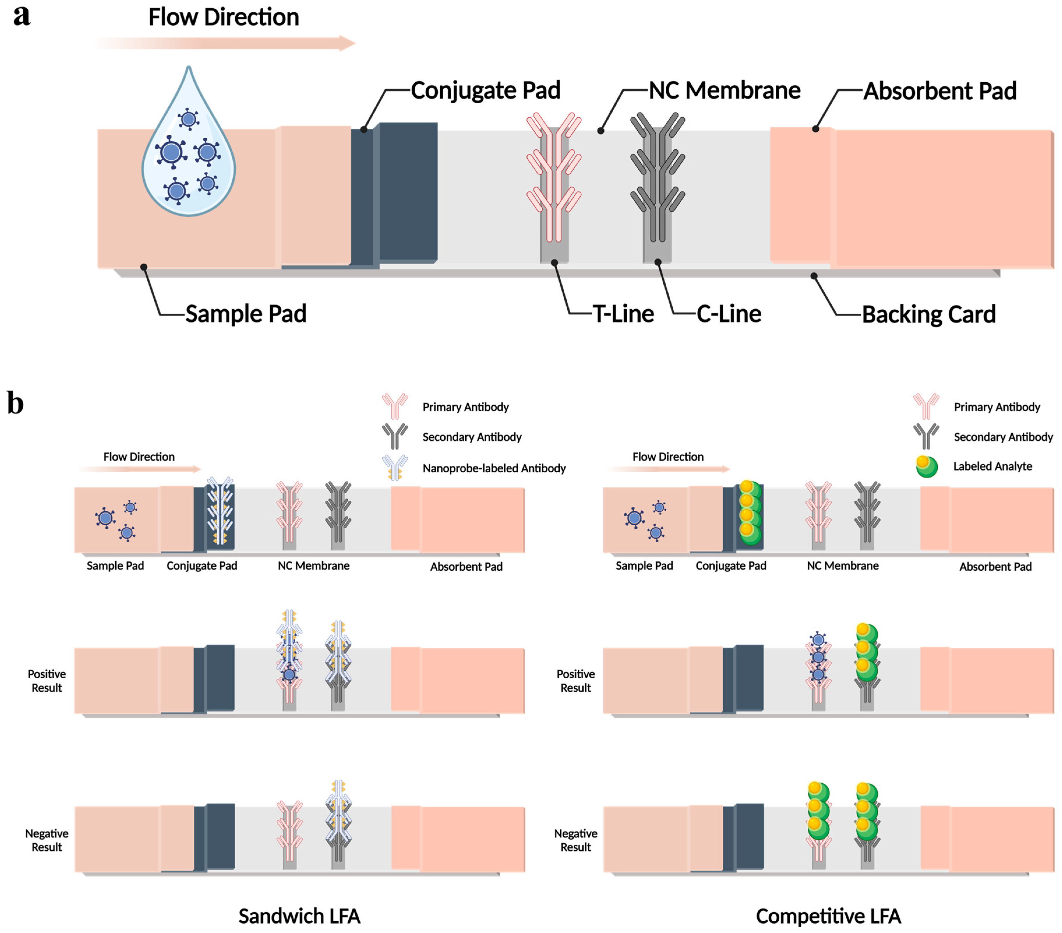
2. LFA Components, Structures, and Principles

3. Colorimetric-Based LFAs for Viral Protein Detection
4. Fluorescence-Based LFAs for Viral Protein Detection
5. SERS-Based LFAs for Viral Protein Detection
6. Conclusions
Author Contributions
Funding
Institutional Review Board Statement
Informed Consent Statement
Data Availability Statement
Conflicts of Interest
References
- Dance, A. Beyond Coronavirus: The Virus Discoveries Transforming Biology. Nature 2021, 595, 22–25. [Google Scholar] [CrossRef]
- Dronina, J.; Samukaite-Bubniene, U.; Ramanavicius, A. Towards Application of CRISPR-Cas12a in the Design of Modern Viral DNA Detection Tools (Review). J. Nanobiotechnol. 2022, 20, 41. [Google Scholar] [CrossRef]
- Baker, R.E.; Mahmud, A.S.; Miller, I.F.; Rajeev, M.; Rasambainarivo, F.; Rice, B.L.; Takahashi, S.; Tatem, A.J.; Wagner, C.E.; Wang, L.-F.; et al. Infectious Disease in an Era of Global Change. Nat. Rev. Microbiol. 2022, 20, 193–205. [Google Scholar] [CrossRef] [PubMed]
- Ka-Wai Hui, E. Reasons for the Increase in Emerging and Re-Emerging Viral Infectious Diseases. Microbes Infect. 2006, 8, 905–916. [Google Scholar] [CrossRef]
- Edelson, P.J.; Harold, R.; Ackelsberg, J.; Duchin, J.S.; Lawrence, S.J.; Manabe, Y.C.; Zahn, M.; LaRocque, R.C. Climate Change and the Epidemiology of Infectious Diseases in the United States. Clin. Infect. Dis. 2023, 76, 950–956. [Google Scholar] [CrossRef]
- Russell, R.C. Survival of Insects in the Wheel Bays of a Boeing 747B Aircraft on Flights between Tropical and Temperate Airports. Bull. World Health Organ. 1987, 65, 659–662. [Google Scholar] [PubMed]
- Roberts, J.D.; Tehrani, S.O. Environments, Behaviors, and Inequalities: Reflecting on the Impacts of the Influenza and Coronavirus Pandemics in the United States. Int. J. Environ. Res. Public Health 2020, 17, 4484. [Google Scholar] [CrossRef] [PubMed]
- Flerlage, T.; Boyd, D.F.; Meliopoulos, V.; Thomas, P.G.; Schultz-Cherry, S. Influenza Virus and SARS-CoV-2: Pathogenesis and Host Responses in the Respiratory Tract. Nat. Rev. Microbiol. 2021, 19, 425–441. [Google Scholar] [CrossRef]
- Drosten, C.; Günther, S.; Preiser, W.; van der Werf, S.; Brodt, H.-R.; Becker, S.; Rabenau, H.; Panning, M.; Kolesnikova, L.; Fouchier, R.A.M.; et al. Identification of a Novel Coronavirus in Patients with Severe Acute Respiratory Syndrome. N. Engl. J. Med. 2003, 348, 1967–1976. [Google Scholar] [CrossRef]
- Cauchemez, S.; Fraser, C.; Van Kerkhove, M.D.; Donnelly, C.A.; Riley, S.; Rambaut, A.; Enouf, V.; van der Werf, S.; Ferguson, N.M. Middle East Respiratory Syndrome Coronavirus: Quantification of the Extent of the Epidemic, Surveillance Biases, and Transmissibility. Lancet Infect. Dis. 2014, 14, 50–56. [Google Scholar] [CrossRef]
- de Wit, E.; van Doremalen, N.; Falzarano, D.; Munster, V.J. SARS and MERS: Recent Insights into Emerging Coronaviruses. Nat. Rev. Microbiol. 2016, 14, 523–534. [Google Scholar] [CrossRef] [PubMed]
- Mofijur, M.; Fattah, I.M.R.; Alam, M.A.; Islam, A.B.M.S.; Ong, H.C.; Rahman, S.M.A.; Najafi, G.; Ahmed, S.F.; Uddin, M.d.A.; Mahlia, T.M.I. Impact of COVID-19 on the Social, Economic, Environmental and Energy Domains: Lessons Learnt from a Global Pandemic. Sustain. Prod. Consum. 2021, 26, 343–359. [Google Scholar] [CrossRef] [PubMed]
- Hou, Y.J.; Okuda, K.; Edwards, C.E.; Martinez, D.R.; Asakura, T.; Dinnon, K.H.; Kato, T.; Lee, R.E.; Yount, B.L.; Mascenik, T.M.; et al. SARS-CoV-2 Reverse Genetics Reveals a Variable Infection Gradient in the Respiratory Tract. Cell 2020, 182, 429–446.e14. [Google Scholar] [CrossRef] [PubMed]
- Chakraborty, I.; Maity, P. COVID-19 Outbreak: Migration, Effects on Society, Global Environment and Prevention. Sci. Total Environ. 2020, 728, 138882. [Google Scholar] [CrossRef]
- Piot, P.; Bartos, M.; Ghys, P.; Walker, N.; Schwartländer, B. The Global Impact of HIV/AIDS. Nature 2001, 410, 968–973. [Google Scholar] [CrossRef] [PubMed]
- Omoleke, S.A.; Mohammed, I.; Saidu, Y. Ebola Viral Disease in West Africa: A Threat to Global Health, Economy and Political Stability. J. Public Health Afr. 2016, 7, 534. [Google Scholar] [CrossRef] [PubMed]
- de Graaf, M.; van Beek, J.; Koopmans, M.P.G. Human Norovirus Transmission and Evolution in a Changing World. Nat. Rev. Microbiol. 2016, 14, 421–433. [Google Scholar] [CrossRef] [PubMed]
- Nikookar, S.H.; Fazeli-Dinan, M.; Enayati, A.; Zaim, M. Zika; a Continuous Global Threat to Public Health. Environ. Res. 2020, 188, 109868. [Google Scholar] [CrossRef] [PubMed]
- Sharma, A.; Ghosh, D.; Divekar, N.; Gore, M.; Gochhait, S.; Shireshi, S.S. Comparing the Socio-economic Implications of the 1918 Spanish Flu and the COVID-19 Pandemic in India: A Systematic Review of Literature. Int. Soc. Sci. J. 2021, 71, 23–36. [Google Scholar] [CrossRef]
- Carter, L.J.; Garner, L.V.; Smoot, J.W.; Li, Y.; Zhou, Q.; Saveson, C.J.; Sasso, J.M.; Gregg, A.C.; Soares, D.J.; Beskid, T.R.; et al. Assay Techniques and Test Development for COVID-19 Diagnosis. ACS Cent. Sci. 2020, 6, 591–605. [Google Scholar] [CrossRef]
- Shen, M.; Zhou, Y.; Ye, J.; Abdullah AL-Maskri, A.A.; Kang, Y.; Zeng, S.; Cai, S. Recent Advances and Perspectives of Nucleic Acid Detection for Coronavirus. J. Pharm. Anal. 2020, 10, 97–101. [Google Scholar] [CrossRef] [PubMed]
- Shafie, M.H.; Antony Dass, M.; Ahmad Shaberi, H.S.; Zafarina, Z. Screening and Confirmation Tests for SARS-CoV-2: Benefits and Drawbacks. Beni-Suef Univ. J. Basic Appl. Sci. 2023, 12, 6. [Google Scholar] [CrossRef] [PubMed]
- Xu, M.; Wang, D.; Wang, H.; Zhang, X.; Liang, T.; Dai, J.; Li, M.; Zhang, J.; Zhang, K.; Xu, D.; et al. COVID-19 Diagnostic Testing: Technology Perspective. Clin. Transl. Med. 2020, 10, e158. [Google Scholar] [CrossRef] [PubMed]
- Shahrajabian, M.H.; Sun, W. Various Techniques for Molecular and Rapid Detection of Infectious and Epidemic Diseases. Lett. Org. Chem. 2023, 20, 779–801. [Google Scholar] [CrossRef]
- Tahamtan, A.; Ardebili, A. Real-Time RT-PCR in COVID-19 Detection: Issues Affecting the Results. Expert Rev. Mol. Diagn. 2020, 20, 453–454. [Google Scholar] [CrossRef] [PubMed]
- Liu, R.; Fu, A.; Deng, Z.; Li, Y.; Liu, T. Promising Methods for Detection of Novel Coronavirus SARS-CoV-2. View 2020, 1, e4. [Google Scholar] [CrossRef] [PubMed]
- Corman, V.M.; Landt, O.; Kaiser, M.; Molenkamp, R.; Meijer, A.; Chu, D.K.; Bleicker, T.; Brünink, S.; Schneider, J.; Schmidt, M.L.; et al. Detection of 2019 Novel Coronavirus (2019-NCoV) by Real-Time RT-PCR. Eurosurveillance 2020, 25, 2000045. [Google Scholar] [CrossRef] [PubMed]
- Li, C.; Debruyne, D.N.; Spencer, J.; Kapoor, V.; Liu, L.Y.; Zhou, B.; Pandey, U.; Bootwalla, M.; Ostrow, D.; Maglinte, D.T.; et al. Highly Sensitive and Full-Genome Interrogation of SARS-CoV-2 Using Multiplexed PCR Enrichment Followed by next-Generation Sequencing. biorXiv 2020. [CrossRef]
- Kurosaki, Y.; Martins, D.B.G.; Kimura, M.; Catena, A.d.S.; Borba, M.A.C.S.M.; Mattos, S.d.S.; Abe, H.; Yoshikawa, R.; de Lima Filho, J.L.; Yasuda, J. Development and Evaluation of a Rapid Molecular Diagnostic Test for Zika Virus Infection by Reverse Transcription Loop-Mediated Isothermal Amplification. Sci. Rep. 2017, 7, 13503. [Google Scholar] [CrossRef]
- Shirato, K.; Yano, T.; Senba, S.; Akachi, S.; Kobayashi, T.; Nishinaka, T.; Notomi, T.; Matsuyama, S. Detection of Middle East Respiratory Syndrome Coronavirus Using Reverse Transcription Loop-Mediated Isothermal Amplification (RT-LAMP). Virol. J. 2014, 11, 139. [Google Scholar] [CrossRef]
- Shirato, K.; Semba, S.; El-Kafrawy, S.A.; Hassan, A.M.; Tolah, A.M.; Takayama, I.; Kageyama, T.; Notomi, T.; Kamitani, W.; Matsuyama, S.; et al. Development of Fluorescent Reverse Transcription Loop-Mediated Isothermal Amplification (RT-LAMP) Using Quenching Probes for the Detection of the Middle East Respiratory Syndrome Coronavirus. J. Virol. Methods 2018, 258, 41–48. [Google Scholar] [CrossRef] [PubMed]
- Choi, S.; Goryll, M.; Sin, L.Y.M.; Wong, P.K.; Chae, J. Microfluidic-Based Biosensors toward Point-of-Care Detection of Nucleic Acids and Proteins. Microfluid. Nanofluidics 2011, 10, 231–247. [Google Scholar] [CrossRef]
- Aydin, S. A Short History, Principles, and Types of ELISA, and Our Laboratory Experience with Peptide/Protein Analyses Using ELISA. Peptides 2015, 72, 4–15. [Google Scholar] [CrossRef] [PubMed]
- Wu, L.; Li, G.; Xu, X.; Zhu, L.; Huang, R.; Chen, X. Application of Nano-ELISA in Food Analysis: Recent Advances and Challenges. TrAC Trends Anal. Chem. 2019, 113, 140–156. [Google Scholar] [CrossRef]
- Mohit, E.; Rostami, Z.; Vahidi, H. A Comparative Review of Immunoassays for COVID-19 Detection. Expert Rev. Clin. Immunol. 2021, 17, 573–599. [Google Scholar] [CrossRef] [PubMed]
- Stadlbauer, D.; Amanat, F.; Chromikova, V.; Jiang, K.; Strohmeier, S.; Arunkumar, G.A.; Tan, J.; Bhavsar, D.; Capuano, C.; Kirkpatrick, E.; et al. SARS-CoV-2 Seroconversion in Humans: A Detailed Protocol for a Serological Assay, Antigen Production, and Test Setup. Curr. Protoc. Microbiol. 2020, 57, e100. [Google Scholar] [CrossRef] [PubMed]
- Krähling, V.; Halwe, S.; Rohde, C.; Becker, D.; Berghöfer, S.; Dahlke, C.; Eickmann, M.; Ercanoglu, M.S.; Gieselmann, L.; Herwig, A.; et al. Development and Characterization of an Indirect ELISA to Detect SARS-CoV-2 Spike Protein-Specific Antibodies. J. Immunol. Methods 2021, 490, 112958. [Google Scholar] [CrossRef]
- Urusov, A.E.; Zherdev, A.V.; Dzantiev, B.B. Towards Lateral Flow Quantitative Assays: Detection Approaches. Biosensors 2019, 9, 89. [Google Scholar] [CrossRef]
- Andryukov, B.G. Six Decades of Lateral Flow Immunoassay: From Determining Metabolic Markers to Diagnosing COVID-19. AIMS Microbiol. 2020, 6, 280–304. [Google Scholar] [CrossRef]
- Pilavaki, E.; Demosthenous, A. Optimized Lateral Flow Immunoassay Reader for the Detection of Infectious Diseases in Developing Countries. Sensors 2017, 17, 2673. [Google Scholar] [CrossRef]
- Banerjee, R.; Jaiswal, A. Recent Advances in Nanoparticle-Based Lateral Flow Immunoassay as a Point-of-Care Diagnostic Tool for Infectious Agents and Diseases. Analyst 2018, 143, 1970–1996. [Google Scholar] [CrossRef] [PubMed]
- Grant, B.D.; Anderson, C.E.; Alonzo, L.F.; Garing, S.H.; Williford, J.R.; Baughman, T.A.; Rivera, R.; Glukhova, V.A.; Boyle, D.S.; Dewan, P.K.; et al. A SARS-CoV-2 Coronavirus Nucleocapsid Protein Antigen-Detecting Lateral Flow Assay. PLoS ONE 2021, 16, e0258819. [Google Scholar] [CrossRef] [PubMed]
- Kim, K.; Kim, M.-J.; Kim, D.W.; Kim, S.Y.; Park, S.; Park, C.B. Clinically Accurate Diagnosis of Alzheimer’s Disease via Multiplexed Sensing of Core Biomarkers in Human Plasma. Nat. Commun. 2020, 11, 119. [Google Scholar] [CrossRef] [PubMed]
- Borrebaeck, C.A.K. Precision Diagnostics: Moving towards Protein Biomarker Signatures of Clinical Utility in Cancer. Nat. Rev. Cancer 2017, 17, 199–204. [Google Scholar] [CrossRef] [PubMed]
- Jin, Z.; Mantri, Y.; Retout, M.; Cheng, Y.; Zhou, J.; Jorns, A.; Fajtova, P.; Yim, W.; Moore, C.; Xu, M.; et al. A Charge-Switchable Zwitterionic Peptide for Rapid Detection of SARS-CoV-2 Main Protease. Angew. Chem. 2022, 134, e202112995. [Google Scholar] [CrossRef]
- Harshita; Wu, H.F.; Kailasa, S.K. Recent Advances in Nanomaterials-based Optical Sensors for Detection of Various Biomarkers (Inorganic Species, Organic and Biomolecules). Luminescence 2022, 38, 954–998. [Google Scholar] [CrossRef] [PubMed]
- Singh, H.; Bamrah, A.; Bhardwaj, S.K.; Deep, A.; Khatri, M.; Kim, K.-H.; Bhardwaj, N. Nanomaterial-Based Fluorescent Sensors for the Detection of Lead Ions. J. Hazard. Mater. 2021, 407, 124379. [Google Scholar] [CrossRef] [PubMed]
- Bhardwaj, N.; Bhardwaj, S.K.; Bhatt, D.; Lim, D.K.; Kim, K.-H.; Deep, A. Optical Detection of Waterborne Pathogens Using Nanomaterials. TrAC Trends Anal. Chem. 2019, 113, 280–300. [Google Scholar] [CrossRef]
- Qiu, S.; Zhang, H.; Shi, Z.; Li, H.; Zhou, Z.-K. Ultrasensitive Refractive Index Sensing Based on Hybrid High-Q Metasurfaces. J. Phys. Chem. C 2023, 127, 8263–8270. [Google Scholar] [CrossRef]
- Zhang, L.; Fu, Q.; Tan, Y.; Li, X.; Deng, Y.; Zhou, Z.-K.; Zhou, B.; Xia, H.; Chen, H.; Qiu, C.-W.; et al. Metaoptronic Multiplexed Interface for Probing Bioentity Behaviors. Nano Lett. 2021, 21, 2681–2689. [Google Scholar] [CrossRef]
- O’Farrell, B. Evolution in Lateral Flow–Based Immunoassay Systems. In Lateral Flow Immunoassay; Humana Press: Totowa, NJ, USA, 2009; pp. 1–33. [Google Scholar]
- Asghari, S.; Ekrami, E.; Barati, F.; Avatefi, M.; Mahmoudifard, M. The Role of the Nanofibers in Lateral Flow Assays Enhancement: A Critical Review. Int. J. Polym. Mater. Polym. Biomater. 2023, 72, 1191–1204. [Google Scholar] [CrossRef]
- Tsai, T.-T.; Huang, T.-H.; Chen, C.-A.; Ho, N.Y.-J.; Chou, Y.-J.; Chen, C.-F. Development a Stacking Pad Design for Enhancing the Sensitivity of Lateral Flow Immunoassay. Sci. Rep. 2018, 8, 17319. [Google Scholar] [CrossRef] [PubMed]
- Dowlatshahi, S.; Shabani, E.; Abdekhodaie, M.J. Serological Assays and Host Antibody Detection in Coronavirus-Related Disease Diagnosis. Arch. Virol. 2021, 166, 715–731. [Google Scholar] [CrossRef] [PubMed]
- Mak, W.C.; Beni, V.; Turner, A.P.F. Lateral-Flow Technology: From Visual to Instrumental. TrAC Trends Anal. Chem. 2016, 79, 297–305. [Google Scholar] [CrossRef]
- Younes, N.; Yassine, H.M.; Kourentzi, K.; Tang, P.; Litvinov, D.; Willson, R.C.; Abu-Raddad, L.J.; Nasrallah, G.K. A Review of Rapid Food Safety Testing: Using Lateral Flow Assay Platform to Detect Foodborne Pathogens. Crit. Rev. Food Sci. Nutr. 2023, 12, 1–23. [Google Scholar] [CrossRef] [PubMed]
- Bahadır, E.B.; Sezgintürk, M.K. Lateral Flow Assays: Principles, Designs and Labels. TrAC Trends Anal. Chem. 2016, 82, 286–306. [Google Scholar] [CrossRef]
- Parolo, C.; Sena-Torralba, A.; Bergua, J.F.; Calucho, E.; Fuentes-Chust, C.; Hu, L.; Rivas, L.; Álvarez-Diduk, R.; Nguyen, E.P.; Cinti, S.; et al. Tutorial: Design and Fabrication of Nanoparticle-Based Lateral-Flow Immunoassays. Nat. Protoc. 2020, 15, 3788–3816. [Google Scholar] [CrossRef] [PubMed]
- Santos, B.S.A.S.; Cunha, J.L.R.; Carvalho, I.C.; Costa, J.M.C.; Longo, B.C.; Galinari, G.C.F.; Diniz, P.H.S.M.; Mendes, G.M.M.; Fonseca, F.G.; Abrahão, J.S.; et al. Nanotechnology Meets Immunology towards a Rapid Diagnosis Solution: The COVID-19 Outbreak Challenge. RSC Adv. 2022, 12, 31711–31728. [Google Scholar] [CrossRef]
- Pramanik, A.; Gao, Y.; Patibandla, S.; Gates, K.; Ray, P.C. Bioconjugated Nanomaterial for Targeted Diagnosis of SARS-CoV-2. Acc. Mater. Res. 2022, 3, 134–148. [Google Scholar] [CrossRef]
- Derakhshan, M.A.; Amani, A.; Faridi-Majidi, R. State-of-the-Art of Nanodiagnostics and Nanotherapeutics against SARS-CoV-2. ACS Appl. Mater. Interfaces 2021, 13, 14816–14843. [Google Scholar] [CrossRef]
- Shen, Y.; Shen, G. Signal-Enhanced Lateral Flow Immunoassay with Dual Gold Nanoparticle Conjugates for the Detection of Hepatitis B Surface Antigen. ACS Omega 2019, 4, 5083–5087. [Google Scholar] [CrossRef]
- Toubanaki, D.K.; Margaroni, M.; Prapas, A.; Karagouni, E. Development of a Nanoparticle-Based Lateral Flow Strip Biosensor for Visual Detection of Whole Nervous Necrosis Virus Particles. Sci. Rep. 2020, 10, 6529. [Google Scholar] [CrossRef] [PubMed]
- Huang, C.; Wen, T.; Shi, F.-J.; Zeng, X.-Y.; Jiao, Y.-J. Rapid Detection of IgM Antibodies against the SARS-CoV-2 Virus via Colloidal Gold Nanoparticle-Based Lateral-Flow Assay. ACS Omega 2020, 5, 12550–12556. [Google Scholar] [CrossRef] [PubMed]
- Lee, A.S.; Kim, S.M.; Kim, K.R.; Park, C.; Lee, D.-G.; Heo, H.R.; Cha, H.J.; Kim, C.S. A Colorimetric Lateral Flow Immunoassay Based on Oriented Antibody Immobilization for Sensitive Detection of SARS-CoV-2. Sens. Actuators B Chem. 2023, 379, 133245. [Google Scholar] [CrossRef] [PubMed]
- Cavalera, S.; Colitti, B.; Rosati, S.; Ferrara, G.; Bertolotti, L.; Nogarol, C.; Guiotto, C.; Cagnazzo, C.; Denina, M.; Fagioli, F.; et al. A Multi-Target Lateral Flow Immunoassay Enabling the Specific and Sensitive Detection of Total Antibodies to SARS COV-2. Talanta 2021, 223, 121737. [Google Scholar] [CrossRef] [PubMed]
- Oh, H.-K.; Kim, K.; Park, J.; Im, H.; Maher, S.; Kim, M.-G. Plasmon Color-Preserved Gold Nanoparticle Clusters for High Sensitivity Detection of SARS-CoV-2 Based on Lateral Flow Immunoassay. Biosens. Bioelectron. 2022, 205, 114094. [Google Scholar] [CrossRef] [PubMed]
- Rong, Z.; Wang, Q.; Sun, N.; Jia, X.; Wang, K.; Xiao, R.; Wang, S. Smartphone-Based Fluorescent Lateral Flow Immunoassay Platform for Highly Sensitive Point-of-Care Detection of Zika Virus Nonstructural Protein 1. Anal. Chim. Acta 2019, 1055, 140–147. [Google Scholar] [CrossRef] [PubMed]
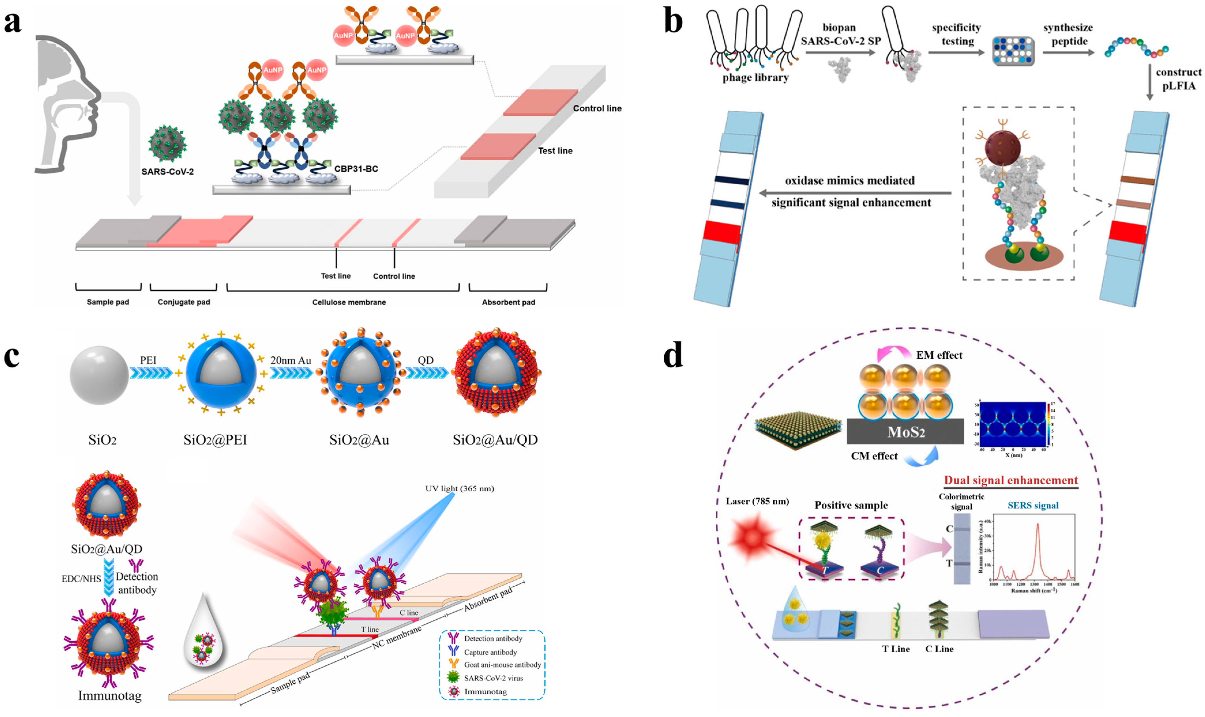
- Dong, T.; Zhang, X.; Yuan, J.; Lin, Z.; Yin, P.; Yu, H.; Wang, M.; Liu, A. Sensitive Lateral Flow Immunoassay Based on Specific Peptide and Superior Oxidase Mimics with a Universal Dual-Mode Significant Signal Amplification. Anal. Chem. 2023, 95, 12532–12540. [Google Scholar] [CrossRef] [PubMed]
- Han, H.; Wang, C.; Yang, X.; Zheng, S.; Cheng, X.; Liu, Z.; Zhao, B.; Xiao, R. Rapid Field Determination of SARS-CoV-2 by a Colorimetric and Fluorescent Dual-Functional Lateral Flow Immunoassay Biosensor. Sens. Actuators B Chem. 2022, 351, 130897. [Google Scholar] [CrossRef]
- Yu, Q.; Li, J.; Zheng, S.; Xia, X.; Xu, C.; Wang, C.; Wang, C.; Gu, B. Molybdenum Disulfide-Loaded Multilayer AuNPs with Colorimetric-SERS Dual-Signal Enhancement Activities for Flexible Immunochromatographic Diagnosis of Monkeypox Virus. J. Hazard. Mater. 2023, 459, 132136. [Google Scholar] [CrossRef]
- Sena-Torralba, A.; Álvarez-Diduk, R.; Parolo, C.; Piper, A.; Merkoçi, A. Toward Next Generation Lateral Flow Assays: Integration of Nanomaterials. Chem. Rev. 2022, 122, 14881–14910. [Google Scholar] [CrossRef] [PubMed]
- Zhang, J.; Gui, X.; Zheng, Q.; Chen, Y.; Ge, S.; Zhang, J.; Xia, N. An HRP-labeled Lateral Flow Immunoassay for Rapid Simultaneous Detection and Differentiation of Influenza A and B Viruses. J. Med. Virol. 2019, 91, 503–507. [Google Scholar] [CrossRef] [PubMed]
- Axelrod, T.; Eltzov, E.; Marks, R.S. Capture-Layer Lateral Flow Immunoassay: A New Platform Validated in the Detection and Quantification of Dengue NS1. ACS Omega 2020, 5, 10433–10440. [Google Scholar] [CrossRef] [PubMed]
- Ren, W.; Irudayaraj, J. Paper-Based Test for Rapid On-Site Screening of SARS-CoV-2 in Clinical Samples. Biosensors 2021, 11, 488. [Google Scholar] [CrossRef] [PubMed]
- Huang, Y.; Ren, J.; Qu, X. Nanozymes: Classification, Catalytic Mechanisms, Activity Regulation, and Applications. Chem. Rev. 2019, 119, 4357–4412. [Google Scholar] [CrossRef] [PubMed]
- Duan, D.; Fan, K.; Zhang, D.; Tan, S.; Liang, M.; Liu, Y.; Zhang, J.; Zhang, P.; Liu, W.; Qiu, X.; et al. Nanozyme-Strip for Rapid Local Diagnosis of Ebola. Biosens. Bioelectron. 2015, 74, 134–141. [Google Scholar] [CrossRef] [PubMed]
- Liu, D.; Ju, C.; Han, C.; Shi, R.; Chen, X.; Duan, D.; Yan, J.; Yan, X. Nanozyme Chemiluminescence Paper Test for Rapid and Sensitive Detection of SARS-CoV-2 Antigen. Biosens. Bioelectron. 2021, 173, 112817. [Google Scholar] [CrossRef]
- Panferov, V.G.; Safenkova, I.V.; Zherdev, A.V.; Dzantiev, B.B. The Steadfast Au@Pt Soldier: Peroxide-Tolerant Nanozyme for Signal Enhancement in Lateral Flow Immunoassay of Peroxidase-Containing Samples. Talanta 2021, 225, 121961. [Google Scholar] [CrossRef] [PubMed]
- Sun, Y.; Xie, Z.; Pei, F.; Hu, W.; Feng, S.; Hao, Q.; Liu, B.; Mu, X.; Lei, W.; Tong, Z. Trimetallic Au@Pd@Pt Nanozyme-Enhanced Lateral Flow Immunoassay for the Detection of SARS-CoV-2 Nucleocapsid Protein. Anal. Methods 2022, 14, 5091–5099. [Google Scholar] [CrossRef]
- Cheng, X.; Yang, X.; Tu, Z.; Rong, Z.; Wang, C.; Wang, S. Graphene Oxide-Based Colorimetric/Fluorescence Dual-Mode Immunochromatography Assay for Simultaneous Ultrasensitive Detection of Respiratory Virus and Bacteria in Complex Samples. J. Hazard. Mater. 2023, 459, 132192. [Google Scholar] [CrossRef]
- Li, J.; Liang, P.; Zhao, T.; Guo, G.; Zhu, J.; Wen, C.; Zeng, J. Colorimetric and Raman Dual-Mode Lateral Flow Immunoassay Detection of SARS-CoV-2 N Protein Antibody Based on Ag Nanoparticles with Ultrathin Au Shell Assembled onto Fe3O4 Nanoparticles. Anal. Bioanal. Chem. 2023, 415, 545–554. [Google Scholar] [CrossRef]
- Wang, C.; Yang, X.; Gu, B.; Liu, H.; Zhou, Z.; Shi, L.; Cheng, X.; Wang, S. Sensitive and Simultaneous Detection of SARS-CoV-2-Specific IgM/IgG Using Lateral Flow Immunoassay Based on Dual-Mode Quantum Dot Nanobeads. Anal. Chem. 2020, 92, 15542–15549. [Google Scholar] [CrossRef]
- Liu, Y.; Lv, Y.; Chen, W.; Yang, X.; Cheng, X.; Rong, Z.; Wang, S. Development of a Fluorescent Immunochromatographic Assay Based on Quantum Dot-Functionalized Two-Dimensional Monolayer Ti3C2 MXene Nanoprobes for the Simultaneous Detection of Influenza A Virus and SARS-CoV-2. ACS Appl. Mater. Interfaces 2023, 15, 35872–35883. [Google Scholar] [CrossRef] [PubMed]
- Liang, P.; Guo, Q.; Zhao, T.; Wen, C.-Y.; Tian, Z.; Shang, Y.; Xing, J.; Jiang, Y.; Zeng, J. Ag Nanoparticles with Ultrathin Au Shell-Based Lateral Flow Immunoassay for Colorimetric and SERS Dual-Mode Detection of SARS-CoV-2 IgG. Anal. Chem. 2022, 94, 8466–8473. [Google Scholar] [CrossRef]
- Roberts, A.; Prakashan, D.; Dhanze, H.; Gandham, R.K.; Gandhi, S.; Sharma, G.T. Immuno-Chromatic Probe Based Lateral Flow Assay for Point-of-Care Detection of Japanese Encephalitis Virus NS1 Protein Biomarker in Clinical Samples Using a Smartphone-Based Approach. Nanoscale Adv. 2022, 4, 3966–3977. [Google Scholar] [CrossRef]
- Panferov, V.G.; Safenkova, I.V.; Varitsev, Y.A.; Zherdev, A.V.; Dzantiev, B.B. Enhancement of Lateral Flow Immunoassay by Alkaline Phosphatase: A Simple and Highly Sensitive Test for Potato Virus X. Microchim. Acta 2018, 185, 25. [Google Scholar] [CrossRef] [PubMed]
- Vafabakhsh, M.; Dadmehr, M.; Kazemi Noureini, S.; Es’haghi, Z.; Malekkiani, M.; Hosseini, M. Paper-Based Colorimetric Detection of COVID-19 Using Aptasenor Based on Biomimetic Peroxidase like Activity of ChF/ZnO/CNT Nano-Hybrid. Spectrochim. Acta A Mol. Biomol. Spectrosc. 2023, 301, 122980. [Google Scholar] [CrossRef]
- Wang, K.; Liu, X.; Liang, X.; Jiang, Y.; Wen, C.-Y.; Zeng, J. Near-Infrared Responsive Ag@Au Nanoplates with Exceptional Stability for Highly Sensitive Colorimetric and Photothermal Dual-Mode Lateral Flow Immunoassay. Anal. Chem. 2024, 96, 3216. [Google Scholar] [CrossRef] [PubMed]
- Atta, S.; Zhao, Y.; Li, J.Q.; Vo-Dinh, T. Dual-Modal Colorimetric and Surface-Enhanced Raman Scattering (SERS)-Based Lateral Flow Immunoassay for Ultrasensitive Detection of SARS-CoV-2 Using a Plasmonic Gold Nanocrown. Anal. Chem. 2024, 96, 4783–4790. [Google Scholar] [CrossRef]
- Xu, L.-D.; Zhang, Q.; Ding, S.-N.; Xu, J.-J.; Chen, H.-Y. Ultrasensitive Detection of Severe Fever with Thrombocytopenia Syndrome Virus Based on Immunofluorescent Carbon Dots/SiO2 Nanosphere-Based Lateral Flow Assay. ACS Omega 2019, 4, 21431–21438. [Google Scholar] [CrossRef]
- Zhou, Y.; Chen, Y.; Liu, W.; Fang, H.; Li, X.; Hou, L.; Liu, Y.; Lai, W.; Huang, X.; Xiong, Y. Development of a Rapid and Sensitive Quantum Dot Nanobead-Based Double-Antigen Sandwich Lateral Flow Immunoassay and Its Clinical Performance for the Detection of SARS-CoV-2 Total Antibodies. Sens. Actuators B Chem. 2021, 343, 130139. [Google Scholar] [CrossRef] [PubMed]
- Nguyen, A.V.T.; Dao, T.D.; Trinh, T.T.T.; Choi, D.-Y.; Yu, S.-T.; Park, H.; Yeo, S.-J. Sensitive Detection of Influenza a Virus Based on a CdSe/CdS/ZnS Quantum Dot-Linked Rapid Fluorescent Immunochromatographic Test. Biosens. Bioelectron. 2020, 155, 112090. [Google Scholar] [CrossRef] [PubMed]
- Xu, L.-D.; Du, F.-L.; Zhu, J.; Ding, S.-N. Luminous Silica Colloids with Carbon Dot Incorporation for Sensitive Immunochromatographic Assay of Zika Virus. Analyst 2021, 146, 706–713. [Google Scholar] [CrossRef] [PubMed]
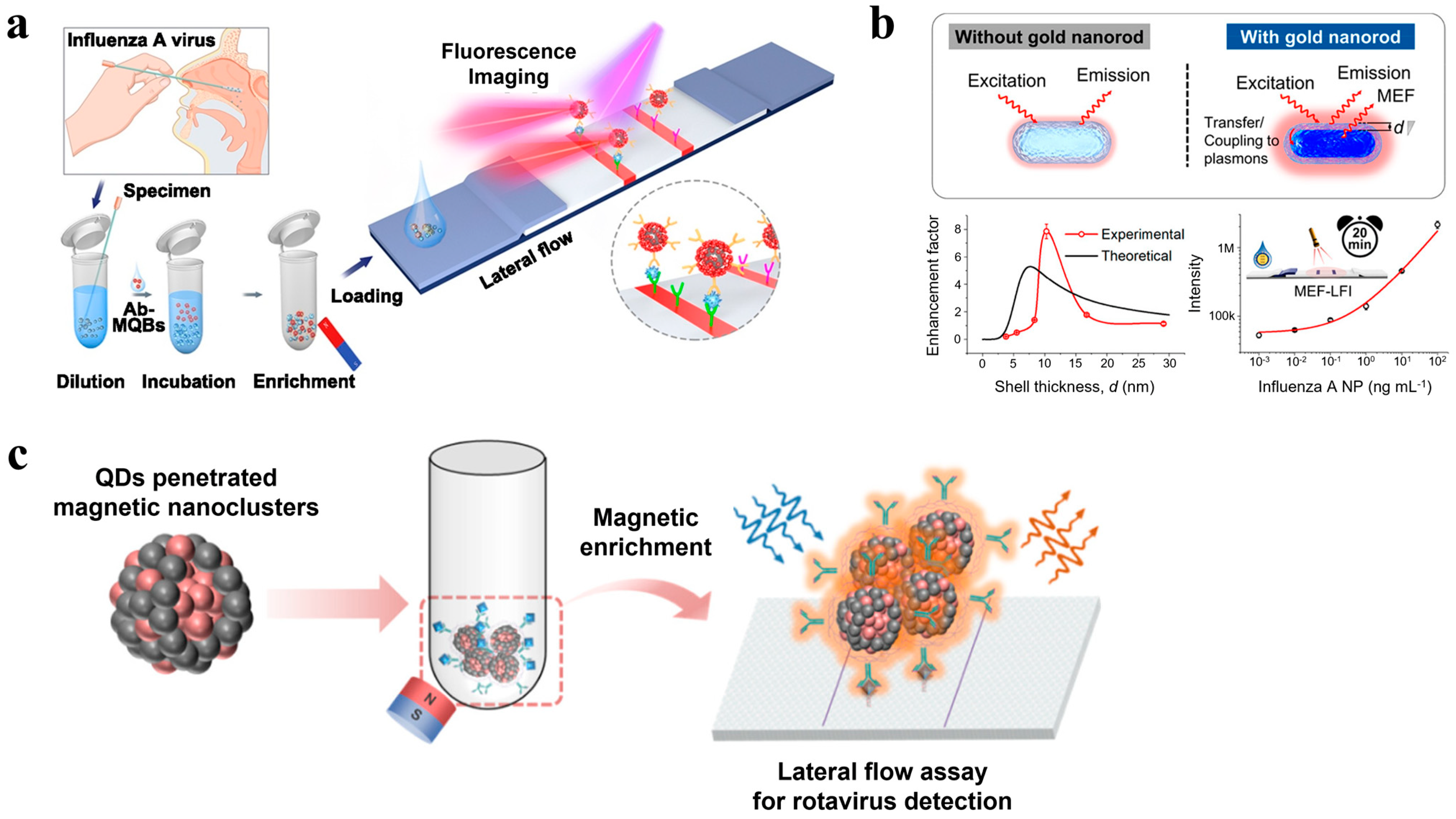
- Bai, Z.; Wei, H.; Yang, X.; Zhu, Y.; Peng, Y.; Yang, J.; Wang, C.; Rong, Z.; Wang, S. Rapid Enrichment and Ultrasensitive Detection of Influenza A Virus in Human Specimen Using Magnetic Quantum Dot Nanobeads Based Test Strips. Sens. Actuators B Chem. 2020, 325, 128780. [Google Scholar] [CrossRef] [PubMed]
- Kang, K.; Huang, L.; Ouyang, C.; Du, J.; Yang, B.; Chi, Y.; He, S.; Ying, L.; Chen, G.; Wang, J. Development, Performance Evaluation, and Clinical Application of a Rapid SARS-CoV-2 IgM and IgG Test Kit Based on Automated Fluorescence Immunoassay. J. Med. Virol. 2021, 93, 2838–2847. [Google Scholar] [CrossRef] [PubMed]
- Chen, R.; Ren, C.; Liu, M.; Ge, X.; Qu, M.; Zhou, X.; Liang, M.; Liu, Y.; Li, F. Early Detection of SARS-CoV-2 Seroconversion in Humans with Aggregation-Induced Near-Infrared Emission Nanoparticle-Labeled Lateral Flow Immunoassay. ACS Nano 2021, 15, 8996–9004. [Google Scholar] [CrossRef] [PubMed]
- Zhang, C.; Zhou, L.; Du, K.; Zhang, Y.; Wang, J.; Chen, L.; Lyu, Y.; Li, J.; Liu, H.; Huo, J.; et al. Foundation and Clinical Evaluation of a New Method for Detecting SARS-CoV-2 Antigen by Fluorescent Microsphere Immunochromatography. Front. Cell. Infect. Microbiol. 2020, 10, 553837. [Google Scholar] [CrossRef] [PubMed]
- Mao, M.; Wu, F.; Shi, X.; Huang, Y.; Ma, L. Ultrasensitive Detection of COVID-19 Virus N Protein Based on p-Toluenesulfonyl Modified Fluorescent Microspheres Immunoassay. Biosensors 2022, 12, 437. [Google Scholar] [CrossRef]
- Wei-Wen Hsiao, W.; Sharma, N.; Le, T.-N.; Cheng, Y.-Y.; Lee, C.-C.; Vo, D.-T.; Hui, Y.Y.; Chang, H.-C.; Chiang, W.-H. Fluorescent Nanodiamond-Based Spin-Enhanced Lateral Flow Immunoassay for Detection of SARS-CoV-2 Nucleocapsid Protein and Spike Protein from Different Variants. Anal. Chim. Acta 2022, 1230, 340389. [Google Scholar] [CrossRef]
- Xu, G.; Zeng, S.; Zhang, B.; Swihart, M.T.; Yong, K.-T.; Prasad, P.N. New Generation Cadmium-Free Quantum Dots for Biophotonics and Nanomedicine. Chem. Rev. 2016, 116, 12234–12327. [Google Scholar] [CrossRef]
- Chen, Z.; Zhang, Z.; Zhai, X.; Li, Y.; Lin, L.; Zhao, H.; Bian, L.; Li, P.; Yu, L.; Wu, Y.; et al. Rapid and Sensitive Detection of Anti-SARS-CoV-2 IgG, Using Lanthanide-Doped Nanoparticles-Based Lateral Flow Immunoassay. Anal. Chem. 2020, 92, 7226–7231. [Google Scholar] [CrossRef] [PubMed]
- Le, T.-N.; Hsiao, W.W.-W.; Cheng, Y.-Y.; Lee, C.-C.; Huynh, T.-T.; Pham, D.M.; Chen, M.; Jen, M.-W.; Chang, H.-C.; Chiang, W.-H. Spin-Enhanced Lateral Flow Immunoassay for High-Sensitivity Detection of Nonstructural Protein NS1 Serotypes of the Dengue Virus. Anal. Chem. 2022, 94, 17819–17826. [Google Scholar] [CrossRef]
- Hong, D.; Jo, E.-J.; Bang, D.; Jung, C.; Lee, Y.E.; Noh, Y.-S.; Shin, M.G.; Kim, M.-G. Plasmonic Approach to Fluorescence Enhancement of Mesoporous Silica-Coated Gold Nanorods for Highly Sensitive Influenza A Virus Detection Using Lateral Flow Immunosensor. ACS Nano 2023, 17, 16607–16619. [Google Scholar] [CrossRef] [PubMed]
- Fu, H.E.; Koo, T.M.; Kim, M.S.; Ko, M.J.; Park, B.C.; Oh, K.; Cho, Y.; Jung, J.-W.; Kim, S.; Jang, W.S.; et al. Magnetic-Fluorescent Nanocluster Lateral Flow Assay for Rotavirus Detection. ACS Appl. Nano Mater. 2023, 6, 5789–5798. [Google Scholar] [CrossRef]
- Zhou, J.; Yang, Y.; Zhang, C. Toward Biocompatible Semiconductor Quantum Dots: From Biosynthesis and Bioconjugation to Biomedical Application. Chem. Rev. 2015, 115, 11669–11717. [Google Scholar] [CrossRef] [PubMed]
- Wang, C.; Cheng, X.; Liu, L.; Zhang, X.; Yang, X.; Zheng, S.; Rong, Z.; Wang, S. Ultrasensitive and Simultaneous Detection of Two Specific SARS-CoV-2 Antigens in Human Specimens Using Direct/Enrichment Dual-Mode Fluorescence Lateral Flow Immunoassay. ACS Appl. Mater. Interfaces 2021, 13, 40342–40353. [Google Scholar] [CrossRef] [PubMed]
- Li, Z.; Wang, A.; Zhou, J.; Chen, Y.; Liu, H.; Liu, Y.; Zhang, Y.; Ding, P.; Zhu, X.; Liang, C.; et al. A Universal Fluorescent Immunochromatography Assay Based on Quantum Dot Nanoparticles for the Rapid Detection of Specific Antibodies against SARS-CoV-2 Nucleocapsid Protein. Int. J. Mol. Sci. 2022, 23, 6225. [Google Scholar] [CrossRef] [PubMed]
- Chen, W.; Chen, H.; Liu, Y.; Wei, H.; Wang, Y.; Rong, Z.; Liu, X. An Integrated Fluorescent Lateral Flow Assay for Multiplex Point-of-Care Detection of Four Respiratory Viruses. Anal. Biochem. 2022, 659, 114948. [Google Scholar] [CrossRef] [PubMed]
- Srivastav, S.; Dankov, A.; Adanalic, M.; Grzeschik, R.; Tran, V.; Pagel-Wieder, S.; Gessler, F.; Spreitzer, I.; Scholz, T.; Schnierle, B.; et al. Rapid and Sensitive SERS-Based Lateral Flow Test for SARS-CoV2-Specific IgM/IgG Antibodies. Anal. Chem. 2021, 93, 12391–12399. [Google Scholar] [CrossRef] [PubMed]
- Lu, M.; Joung, Y.; Jeon, C.S.; Kim, S.; Yong, D.; Jang, H.; Pyun, S.H.; Kang, T.; Choo, J. Dual-Mode SERS-Based Lateral Flow Assay Strips for Simultaneous Diagnosis of SARS-CoV-2 and Influenza a Virus. Nano Converg. 2022, 9, 39. [Google Scholar] [CrossRef]
- Borzenkov, M.; Chirico, G.; D’Alfonso, L.; Sironi, L.; Collini, M.; Cabrini, E.; Dacarro, G.; Milanese, C.; Pallavicini, P.; Taglietti, A.; et al. Thermal and Chemical Stability of Thiol Bonding on Gold Nanostars. Langmuir 2015, 31, 8081–8091. [Google Scholar] [CrossRef] [PubMed]
- Lu, X.; Mei, T.; Guo, Q.; Zhou, W.; Li, X.; Chen, J.; Zhou, X.; Sun, N.; Fang, Z. Improved Performance of Lateral Flow Immunoassays for Alpha-Fetoprotein and Vanillin by Using Silica Shell-Stabilized Gold Nanoparticles. Microchim. Acta 2019, 186, 2. [Google Scholar] [CrossRef] [PubMed]
- Wang, F.; Widejko, R.G.; Yang, Z.; Nguyen, K.T.; Chen, H.; Fernando, L.P.; Christensen, K.A.; Anker, J.N. Surface-Enhanced Raman Scattering Detection of PH with Silica-Encapsulated 4-Mercaptobenzoic Acid-Functionalized Silver Nanoparticles. Anal. Chem. 2012, 84, 8013–8019. [Google Scholar] [CrossRef] [PubMed]
- Jeon, J.; Lee, S.H.; Joung, Y.; Kim, K.; Choi, N.; Choo, J. Improvement of Reproducibility and Thermal Stability of Surface-Enhanced Raman Scattering-Based Lateral Flow Assay Strips Using Silica-Encapsulated Gold Nanoparticles. Sens. Actuators B Chem. 2020, 321, 128521. [Google Scholar] [CrossRef]
- Liu, H.; Dai, E.; Xiao, R.; Zhou, Z.; Zhang, M.; Bai, Z.; Shao, Y.; Qi, K.; Tu, J.; Wang, C.; et al. Development of a SERS-Based Lateral Flow Immunoassay for Rapid and Ultra-Sensitive Detection of Anti-SARS-CoV-2 IgM/IgG in Clinical Samples. Sens. Actuators B Chem. 2021, 329, 129196. [Google Scholar] [CrossRef] [PubMed]
- Zhang, Y.; Wu, G.; Wei, J.; Ding, Y.; Wei, Y.; Liu, Q.; Chen, H. Rapid and Sensitive Detection of Rotavirus by Surface-Enhanced Raman Scattering Immunochromatography. Microchim. Acta 2021, 188, 3. [Google Scholar] [CrossRef] [PubMed]
- Jia, X.; Liu, Z.; Peng, Y.; Hou, G.; Chen, W.; Xiao, R. Automatic and Sensitive Detection of West Nile Virus Non-Structural Protein 1 with a Portable SERS–LFIA Detector. Microchim. Acta 2021, 188, 206. [Google Scholar] [CrossRef] [PubMed]
- Liang, J.; Wu, L.; Wang, Y.; Liang, W.; Hao, Y.; Tan, M.; He, G.; Lv, D.; Wang, Z.; Zeng, T.; et al. SERS/Photothermal-Based Dual-Modal Lateral Flow Immunoassays for Sensitive and Simultaneous Antigen Detection of Respiratory Viral Infections. Sens. Actuators B Chem. 2023, 389, 133875. [Google Scholar] [CrossRef]
- Shen, H.; Xie, K.; Huang, L.; Wang, L.; Ye, J.; Xiao, M.; Ma, L.; Jia, A.; Tang, Y. A Novel SERS-Based Lateral Flow Assay for Differential Diagnosis of Wild-Type Pseudorabies Virus and GE-Deleted Vaccine. Sens. Actuators B Chem. 2019, 282, 152–157. [Google Scholar] [CrossRef]
- Xiao, M.; Xie, K.; Dong, X.; Wang, L.; Huang, C.; Xu, F.; Xiao, W.; Jin, M.; Huang, B.; Tang, Y. Ultrasensitive Detection of Avian Influenza A (H7N9) Virus Using Surface-Enhanced Raman Scattering-Based Lateral Flow Immunoassay Strips. Anal. Chim. Acta 2019, 1053, 139–147. [Google Scholar] [CrossRef]
- Shen, H.; Song, E.; Wang, Y.; Meng, L.; Dong, J.; Lin, B.; Huang, D.; Guan, Z.; Yang, C.; Zhu, Z. In Situ Raman Enhancement Strategy for Highly Sensitive and Quantitative Lateral Flow Assay. Anal. Bioanal. Chem. 2022, 414, 507–513. [Google Scholar] [CrossRef] [PubMed]
- Gandra, N.; Hendargo, H.C.; Norton, S.J.; Fales, A.M.; Palmer, G.M.; Vo-Dinh, T. Tunable and Amplified Raman Gold Nanoprobes for Effective Tracking (TARGET): In Vivo Sensing and Imaging. Nanoscale 2016, 8, 8486–8494. [Google Scholar] [CrossRef]
- Ngo, H.T.; Gandra, N.; Fales, A.M.; Taylor, S.M.; Vo-Dinh, T. Sensitive DNA Detection and SNP Discrimination Using Ultrabright SERS Nanorattles and Magnetic Beads for Malaria Diagnostics. Biosens. Bioelectron. 2016, 81, 8–14. [Google Scholar] [CrossRef] [PubMed]
- Yap, L.W.; Chen, H.; Gao, Y.; Petkovic, K.; Liang, Y.; Si, K.J.; Wang, H.; Tang, Z.; Zhu, Y.; Cheng, W. Bifunctional Plasmonic-Magnetic Particles for an Enhanced Microfluidic SERS Immunoassay. Nanoscale 2017, 9, 7822–7829. [Google Scholar] [CrossRef] [PubMed]
- Wang, C.; Wang, J.; Li, P.; Rong, Z.; Jia, X.; Ma, Q.; Xiao, R.; Wang, S. Sonochemical Synthesis of Highly Branched Flower-like Fe3O4@SiO2@Ag Microcomposites and Their Application as Versatile SERS Substrates. Nanoscale 2016, 8, 19816–19828. [Google Scholar] [CrossRef] [PubMed]
- Wang, Y.; Gao, T.; Wang, K.; Zou, B.; Zhang, X.; Du, Z.; Zhou, S. Magnetic-Based Silver Composite Microspheres with Nanosheet-Assembled Shell for Effective SERS Substrate. J. Mater. Chem. C Mater. 2013, 1, 2441–2447. [Google Scholar] [CrossRef]
- Wang, C.; Wang, C.; Wang, X.; Wang, K.; Zhu, Y.; Rong, Z.; Wang, W.; Xiao, R.; Wang, S. Magnetic SERS Strip for Sensitive and Simultaneous Detection of Respiratory Viruses. ACS Appl. Mater. Interfaces 2019, 11, 19495–19505. [Google Scholar] [CrossRef] [PubMed]
- Xie, Z.; Feng, S.; Pei, F.; Xia, M.; Hao, Q.; Liu, B.; Tong, Z.; Wang, J.; Lei, W.; Mu, X. Magnetic/Fluorescent Dual-Modal Lateral Flow Immunoassay Based on Multifunctional Nanobeads for Rapid and Accurate SARS-CoV-2 Nucleocapsid Protein Detection. Anal. Chim. Acta 2022, 1233, 340486. [Google Scholar] [CrossRef] [PubMed]
- Liu, Z.; Wang, C.; Zheng, S.; Yang, X.; Han, H.; Dai, Y.; Xiao, R. Simultaneously Ultrasensitive and Quantitative Detection of Influenza A Virus, SARS-CoV-2, and Respiratory Syncytial Virus via Multichannel Magnetic SERS-Based Lateral Flow Immunoassay. Nanomedicine 2023, 47, 102624. [Google Scholar] [CrossRef]
- Liu, X.; Yang, X.; Wang, C.; Liu, Q.; Ding, Y.; Xu, S.; Wang, G.; Xiao, R. A Nanogap-Enhanced SERS Nanotag–Based Lateral Flow Assay for Ultrasensitive and Simultaneous Monitoring of SARS-CoV-2 S and NP Antigens. Microchim. Acta 2024, 191, 104. [Google Scholar] [CrossRef]
- Huang, L.; Tian, S.; Zhao, W.; Liu, K.; Ma, X.; Guo, J. Multiplexed Detection of Biomarkers in Lateral-Flow Immunoassays. Analyst 2020, 145, 2828–2840. [Google Scholar] [CrossRef] [PubMed]
- Ghasemi, F.; Fahimi-Kashani, N.; Bigdeli, A.; Alshatteri, A.H.; Abbasi-Moayed, S.; Al-Jaf, S.H.; Merry, M.Y.; Omer, K.M.; Hormozi-Nezhad, M.R. Paper-Based Optical Nanosensors—A Review. Anal. Chim. Acta 2023, 1238, 340640. [Google Scholar] [CrossRef] [PubMed]
- Liu, J.; Li, M.; Man, Q.; Huang, L.; Wang, J.; Gao, M.; Zhang, X. Naked-Eye Readout Distance Quantitative Lateral Flow Assay Based on the Permeability Changes of Enzyme-Catalyzed Hydrogelation. Anal. Chem. 2023, 95, 8011–8019. [Google Scholar] [CrossRef] [PubMed]
- Kim, S.K.; Lee, J.U.; Jeon, M.J.; Kim, S.-K.; Hwang, S.-H.; Hong, M.E.; Sim, S.J. Bio-Conjugated Nanoarchitectonics with Dual-Labeled Nanoparticles for a Colorimetric and Fluorescent Dual-Mode Serological Lateral Flow Immunoassay Sensor. RSC Adv. 2023, 13, 27225–27232. [Google Scholar] [CrossRef] [PubMed]
- Misawa, K.; Yamamoto, T.; Hiruta, Y.; Yamazaki, H.; Citterio, D. Text-Displaying Semiquantitative Competitive Lateral Flow Immunoassay Relying on Inkjet-Printed Patterns. ACS Sens. 2020, 5, 2076–2085. [Google Scholar] [CrossRef]
- Yang, D.; Hu, C.; Zhang, H.; Geng, S. Recent Developments in Paper-Based Sensors with Instrument-Free Signal Readout Technologies (2020–2023). Biosensors 2024, 14, 36. [Google Scholar] [CrossRef]


| Target | Nanoprobe | LoD | Reference | |
|---|---|---|---|---|
| Au NP | HBsAg | Au NP | 1.8 ng/mL | [62] |
| Nodavirus | Au NP | 6 × 10⁻³ TCID50 | [63] | |
| SARS-CoV-2 | PLASCOP Au NP cluster | 0.038 ng/mL | [67] | |
| Japanese Encephalitis Virus | Au NP | 10 pg/mL | [86] | |
| SARS-CoV-2 | Au NP | 0.2 mg/mL | [59] | |
| SARS-CoV-2 | Au NP | 5 × 10⁴ copies/mL | [65] | |
| Enzyme reaction | IAV, IBV | HRP | 0.001~0.00025 HA units (IAV), 0.016~0.004 HA units (IBV) | [73] |
| Dengue virus | HRP | 5 ng/mL | [74] | |
| SARS-CoV-2 | HRP-labeled Fe3O4/Au core–shell magnetic NP | 400 PFU/mL | [75] | |
| PVX | ALP-labeled Au NP | 0.3 ng/mL | [87] | |
| SARS-CoV-2 | Au@Pd@Pt nanozyme | 0.037 ng/mL | [80] | |
| SARS-CoV-2 | ChF/ZnO/CNT nanohybrid | 0.05 pg/mL | [88] | |
| SARS-CoV-2 | PDA@MnO2 nanocomposite | 8.01 pg/mL | [69] | |
| Dual detection | SARS-CoV-2 | SiO2@Au NP/QD | 1 ng/mL, 33 pg/mL (*C, F) | [70] |
| IAV | GO-Au/QD-QD | 5 × 10⁴ copies/mL, 891 copies/mL (*C, F) | [81] | |
| MPXV | MoS2@Au–Au | 0.2 ng/mL, 0.002 ng/mL (*C, S) | [71] | |
| SARS-CoV-2 | Fe3O4-AgMBA@Au NP | 10−8 mg/mL, 0.08 pg/mL (*C, S) | [82] | |
| IAV, SARS-CoV-2 | Ti3C2-QD | 1 ng/mL, 2.4 pg/mL (IAV) 1 ng/mL, 6.2 pg/mL (SARS-CoV-2) (*C, F) | [84] | |
| SARS-CoV-2 | AgMBA@Au | 10−6 mg/mL, 0.22 pg/mL (*C, S) | [85] | |
| SARS-CoV-2 | Ag@Au triangular nanoplate | 1 ng/mL, 40 pg/mL (*C, P) | [89] | |
| SARS-CoV-2 | Plasmonic-active Au nanocrown | 91.24 pg/mL, 57.21 fg/mL (*C, S) | [90] |
| Target | Nanoprobe | LoD | Reference | |
|---|---|---|---|---|
| Fluorophore | SARS-CoV-2 | Fluorescent microsphere | 100 ng/mL | [98] |
| SARS-CoV-2 | Aggregation-induced emission810 NP | 0.236 μg/mL (IgM), 0.125 μg/mL (IgG) | [97] | |
| Dengue NS1 serotypes (DV1, DV2, DV3, DV4) | FND | 0.33, 0.24, 0.10, 1.33 ng/mL | [103] | |
| IAV | Cy5-mSiO2@GNR | 0.52 pg /mL | [104] | |
| SARS-CoV-2 | Fluorescent microsphere | 0.01 ng/mL | [105] | |
| QD | Zika virus | QD microsphere | 0.045 ng/mL | [68] |
| Thrombocytopenia syndrome virus | Fluorescent carbon dots (CDs)/SiO2 nanosphere | 10 pg/mL | [91] | |
| IAV | CdSe/CdS/ZnS QD | 2.5 HAU/mL (H1N1), 0.63 HAU/mL (H3N2) | [93] | |
| Zika virus | Fluorescent CD-based silica colloid | 10 pg/mL | [94] | |
| IAV | Magnetic-QD NB | 22 pfu/mL | [95] | |
| SARS-CoV-2 | MagTQD | 0.5 pg/mL | [107] | |
| SARS-CoV-2, IAV, IBV, ADV | QD NB | 0.01, 0.05, 0.31, 0.40 ng/mL | [109] | |
| Rotavirus | CFNC | 1.0 × 101 TCID50/mL | [105] |
| Target | Nanoprobe | LoD | Reference | |
|---|---|---|---|---|
| Noble metal | Pseudorabies virus | AuAg4−ATP@Ag NP | 5 ng/mL | [120] |
| Avian influenza virus | AuAg4−ATP@Ag NP | 0.0018 HAU | [121] | |
| Dengue virus, Zika virus | MGITC-labeled Si-Au NP | 1.906 μg/mL | [115] | |
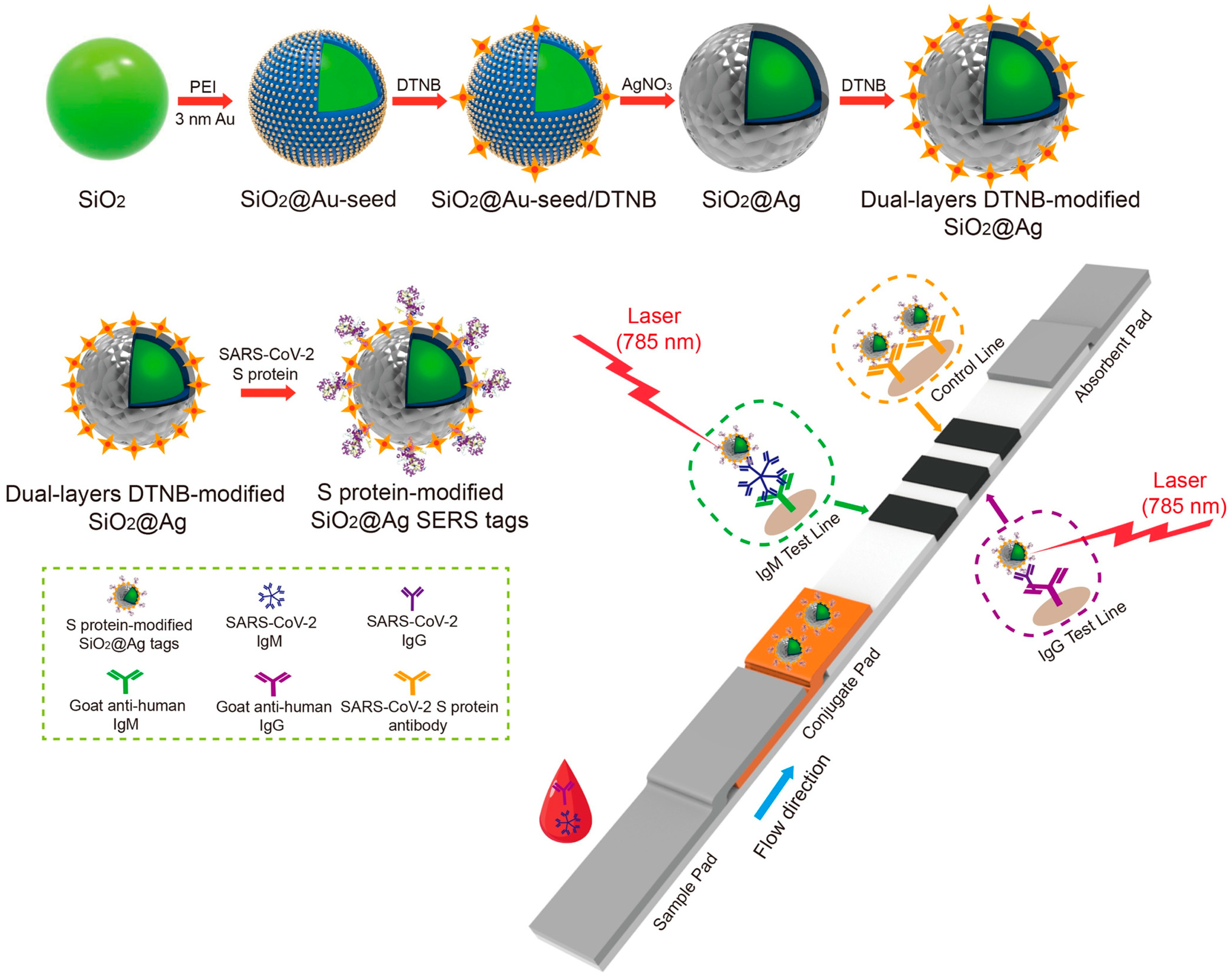
| SARS-CoV-2 | dual-layer DTNB-modified SiO2@Ag NP | 1 pg/mL | [116] | |
| Rotavirus | Au/DTNB/Ag/DTNB | 8 pg/mL | [117] | |
| West Nile virus | Au@Ag NP | 0.2 × 10² copy/μL | [118] | |
| SARS-CoV-2 | NIR-797-ITC-labeled Au nanostar | 100 fg/mL | [110] | |
| SARS-CoV-2, IAV | MGITC-labeled Au NP | 5.2 PFU/mL (SARS-CoV-2), 23 HAU/mL (IAV) | [111] | |
| Magnetic NP | IAV, human ADV | Fe3O4@Ag magnetic tag | 50 pfu/mL, 10 pfu/mL | [128] |
| SARS-CoV-2 | MagDQD | 0.235 ng/mL, 0.012 ng/mL (*M, F) | [129] | |
| IAV, SARS-CoV-2, respiratory syncytial virus | Fe3O4/DTNB@Au/DTNB | 85 copies/mL, 8 pg/mL 8 pg/mL | [130] | |
| SARS-CoV-2 | DTNB–encoded satellite Fe3O4@Au SERS tag | 23 pg/mL, 2 pg/mL | [131] |
Disclaimer/Publisher’s Note: The statements, opinions and data contained in all publications are solely those of the individual author(s) and contributor(s) and not of MDPI and/or the editor(s). MDPI and/or the editor(s) disclaim responsibility for any injury to people or property resulting from any ideas, methods, instructions or products referred to in the content. |
© 2024 by the authors. Licensee MDPI, Basel, Switzerland. This article is an open access article distributed under the terms and conditions of the Creative Commons Attribution (CC BY) license (https://creativecommons.org/licenses/by/4.0/).
Share and Cite
Kim, M.J.; Haizan, I.; Ahn, M.J.; Park, D.-H.; Choi, J.-H. Recent Advances in Lateral Flow Assays for Viral Protein Detection with Nanomaterial-Based Optical Sensors. Biosensors 2024, 14, 197. https://doi.org/10.3390/bios14040197
Kim MJ, Haizan I, Ahn MJ, Park D-H, Choi J-H. Recent Advances in Lateral Flow Assays for Viral Protein Detection with Nanomaterial-Based Optical Sensors. Biosensors. 2024; 14(4):197. https://doi.org/10.3390/bios14040197
Chicago/Turabian StyleKim, Min Jung, Izzati Haizan, Min Ju Ahn, Dong-Hyeok Park, and Jin-Ha Choi. 2024. "Recent Advances in Lateral Flow Assays for Viral Protein Detection with Nanomaterial-Based Optical Sensors" Biosensors 14, no. 4: 197. https://doi.org/10.3390/bios14040197
APA StyleKim, M. J., Haizan, I., Ahn, M. J., Park, D.-H., & Choi, J.-H. (2024). Recent Advances in Lateral Flow Assays for Viral Protein Detection with Nanomaterial-Based Optical Sensors. Biosensors, 14(4), 197. https://doi.org/10.3390/bios14040197