Recent Advances in Bacterial Detection Using Surface-Enhanced Raman Scattering


Abstract
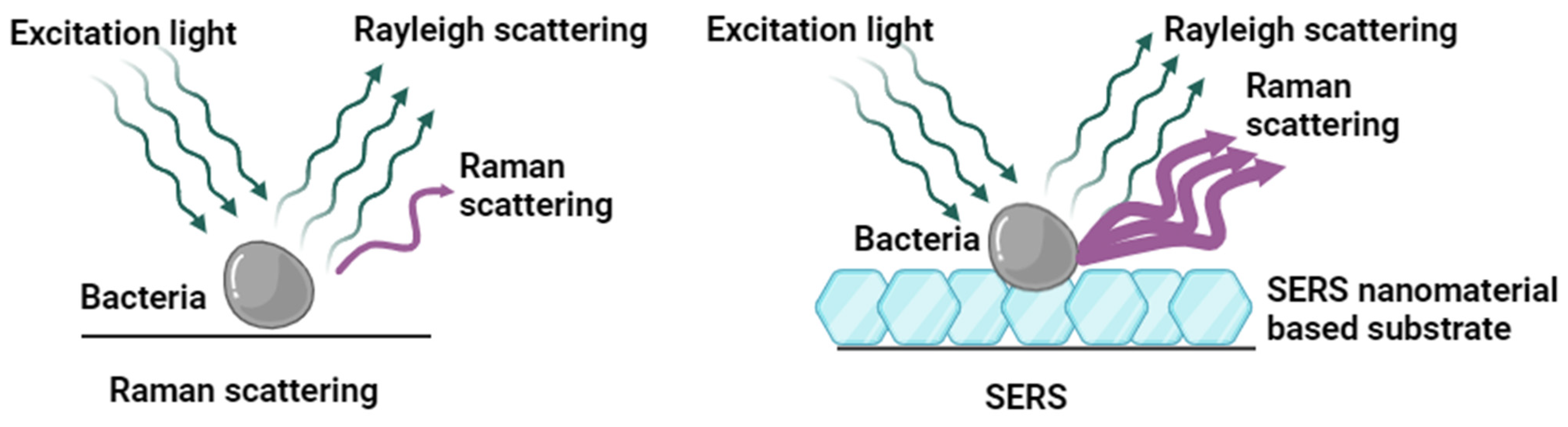
:1. Introduction
2. Overview of Bacterial SERS Detection and Analytes
2.1. Different SERS Detection Methods: Label-Free and Label-Based
2.2. Target Analytes for SERS Bacterial Detection
3. Label-Free Bacterial SERS Detection
3.1. SERS-Based Bacterial Gene Probe
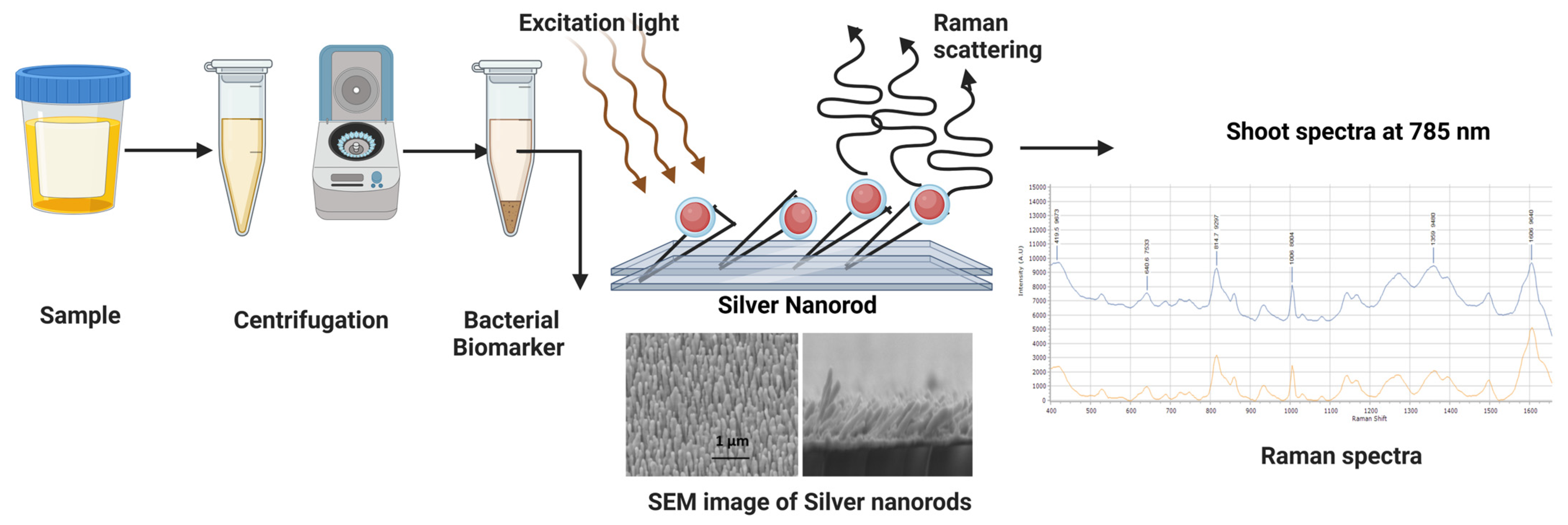
3.2. Biomarker-Based Detection
3.3. Bacterial Whole Cell Detection
4. Enhancing SERS Detection Performance
4.1. Different Types of SERS Substrates with Enhanced Sensitivity
4.2. Bacterial Concentration Methods
4.3. Microfluidic SERS-Based Detection
4.4. Differentiation of Spectra Using Chemometric Analysis
4.5. AI/ML-Enabled SERS Detection
Reproducibility of SERS
4.6. Detection of Microbes in Complex Samples
5. Challenges and Opportunities
6. Conclusions
Author Contributions
Funding
Institutional Review Board Statement
Informed Consent Statement
Data Availability Statement
Conflicts of Interest
References
- Gopinath, S.C.; Tang, T.H.; Chen, Y.; Citartan, M.; Lakshmipriya, T. Bacterial detection: From microscope to smartphone. Biosens. Bioelectron. 2014, 60, 332–342. [Google Scholar] [CrossRef] [PubMed]
- Wells-Bennik, M.H.; Eijlander, R.T.; den Besten, H.M.; Berendsen, E.M.; Warda, A.K.; Krawczyk, A.O.; Nierop Groot, M.N.; Xiao, Y.; Zwietering, M.H.; Kuipers, O.P.; et al. Bacterial Spores in Food: Survival, Emergence, and Outgrowth. Annu. Rev. Food Sci. Technol. 2016, 7, 457–482. [Google Scholar] [CrossRef] [PubMed]
- Kim, S.O.; Kim, S.S. Bacterial pathogen detection by conventional culture-based and recent alternative (polymerase chain reaction, isothermal amplification, enzyme linked immunosorbent assay, bacteriophage amplification, and gold nanoparticle aggregation) methods in food samples: A review. J. Food Saf. 2021, 41, e12870. [Google Scholar]
- Wu, X.; Chen, J.; Park, B.; Huang, Y.W.; Zhao, Y. Advances in Applied Nanotechnology for Agriculture; American Chemical Society: Washington, DC, USA, 2013; Volume 1143, pp. 85–108. [Google Scholar]
- Sur, U.K. Surface-enhanced Raman spectroscopy: Recent advancement of Raman spectroscopy. Resonance 2010, 15, 154–164. [Google Scholar] [CrossRef]
- Kneipp, K.; Kneipp, H.; Itzkan, I.; Dasari, R.R.; Feld, M.S. Ultrasensitive chemical analysis by Raman spectroscopy. Chem. Rev. 1999, 99, 2957–2976. [Google Scholar] [CrossRef] [PubMed]
- Tian, Z.-Q.; Ren, B.; Li, J.-F.; Yang, Z.-L. Expanding generality of surface-enhanced Raman spectroscopy with borrowing SERS activity strategy. Chem. Commun. 2007, 3514–3534. [Google Scholar] [CrossRef] [PubMed]
- Campion, A.; Kambhampati, P. Surface-enhanced Raman scattering. Chem. Soc. Rev. 1998, 27, 241–250. [Google Scholar] [CrossRef]
- Kneipp, K.; Wang, Y.; Kneipp, H.; Perelman, L.T.; Itzkan, I.; Dasari, R.; Feld, M.S. Single molecule detection using surface-enhanced Raman scattering (SERS). Phys. Rev. Lett. 1997, 78, 1667–1670. [Google Scholar] [CrossRef]
- Nie, S.M.; Emery, S.R. Probing single molecules and single nanoparticles by surface-enhanced Raman scattering. Science 1997, 275, 1102–1106. [Google Scholar] [CrossRef]
- Xu, H.X.; Aizpurua, J.; Kall, M.; Apell, P. Electromagnetic contributions to single-molecule sensitivity in surface-enhanced Raman scattering. Phys. Rev. E 2000, 62, 4318–4324. [Google Scholar] [CrossRef]
- Xu, H.X.; Bjerneld, E.J.; Kall, M.; Borjesson, L. Spectroscopy of single hemoglobin molecules by surface enhanced Raman scattering. Phys. Rev. Lett. 1999, 83, 4357–4360. [Google Scholar] [CrossRef]
- Liu, H.; Zhang, L.; Lang, X.; Yamaguchi, Y.; Iwasaki, H.; Inouye, Y.; Xue, Q.; Chen, M. Single molecule detection from a large-scale SERS-active Au79Ag21 substrate. Sci. Rep. 2011, 1, 112. [Google Scholar] [CrossRef] [PubMed]
- Smith, T.J.; O’Connor, L.; Glennon, M.; Maher, M. Molecular diagnostics in food safety: Rapid detection of food-borne pathogens. Ir. J. Agric. Food Res. 2000, 39, 309–319. [Google Scholar]
- Jarvis, R.M.; Goodacre, R. Characterisation and identification of bacteria using SERS. Chem. Soc. Rev. 2008, 37, 931–936. [Google Scholar] [CrossRef] [PubMed]
- Murphy, S.; Huang, L.; Kamat, P.V. Reduced graphene oxide–silver nanoparticle composite as an active SERS material. J. Phys. Chem. C 2013, 117, 4740–4747. [Google Scholar] [CrossRef]
- Tang, X.; Hao, Q.; Hou, X.; Lan, L.; Li, M.; Yao, L.; Zhao, X.; Ni, Z.; Fan, X.; Qiu, T. Exploring and Engineering 2D Transition Metal Dichalcogenides toward Ultimate SERS Performance. Adv. Mater. 2024, 36, 2312348. [Google Scholar] [CrossRef] [PubMed]
- Maznichenko, D.; Venkatakrishnan, K.; Tan, B. Stimulating multiple SERS mechanisms by a nanofibrous three-dimensional network structure of titanium dioxide (TiO2). J. Phys. Chem. C 2013, 117, 578–583. [Google Scholar] [CrossRef]
- Zong, C.; Ge, M.; Pan, H.; Wang, J.; Nie, X.; Zhang, Q.; Zhao, W.; Liu, X.; Yu, Y. In situ synthesis of low-cost and large-scale flexible metal nanoparticle–polymer composite films as highly sensitive SERS substrates for surface trace analysis. RSC Adv. 2019, 9, 2857–2864. [Google Scholar] [CrossRef] [PubMed]
- Hu, B.; Pu, H.; Sun, D.W. Multifunctional cellulose based substrates for SERS smart sensing: Principles, applications and emerging trends for food safety detection. Trends Food Sci. Technol. 2021, 110, 304–320. [Google Scholar] [CrossRef]
- Alexander, T.A.; Pellegrino, P.M.; Gillespie, J.B. Near-infrared surface-enhanced-Raman-scattering-mediated detection of single optically trapped bacterial spores. Appl. Spectrosc. 2003, 57, 1340–1345. [Google Scholar] [CrossRef] [PubMed]
- Efrima, S.; Bronk, B.V. Silver colloids impregnating or coating bacteria. J. Phys. Chem. B 1998, 102, 5947–5950. [Google Scholar] [CrossRef]
- Zeiri, L.; Bronk, B.V.; Shabtai, Y.; Eichler, J.; Efrima, S. Surface-enhanced Raman spectroscopy as a tool for probing specific biochemical components in bacteria. Appl. Spectrosc. 2004, 58, 33–40. [Google Scholar] [CrossRef] [PubMed]
- Premasiri, W.R.; Moir, D.T.; Klempner, M.S.; Krieger, N.; Jones, G.; Ziegler, L.D. Characterization of the Surface Enhanced Raman Scattering (SERS) of bacteria. J. Phys. Chem. B 2005, 109, 312–320. [Google Scholar] [CrossRef]
- Fan, M.; Andrade, G.F.; Brolo, A.G. A review on the fabrication of substrates for surface enhanced Raman spectroscopy and their applications in analytical chemistry. Anal. Chim. Acta 2011, 693, 7–25. [Google Scholar] [CrossRef] [PubMed]
- Sil, S.; Kuhar, N.; Acharya, S.; Umapathy, S. Is chemically synthesized graphene ‘really’ a unique substrate for SERS and fluorescence quenching? Sci. Rep. 2013, 3, 3336. [Google Scholar] [CrossRef] [PubMed]
- Ma, L.; Huang, Y.; Hou, M.; Xie, Z.; Zhang, Z. Silver Nanorods Wrapped with Ultrathin Al2O3 Layers Exhibiting Excellent SERS Sensitivity and Outstanding SERS Stability. Sci. Rep. 2015, 5, 12890. [Google Scholar] [CrossRef] [PubMed]
- Dinish, U.S.; Balasundaram, G.; Chang, Y.T.; Olivo, M. Actively targeted in vivo multiplex detection of intrinsic cancer biomarkers using biocompatible SERS nanotags. Sci. Rep. 2014, 4, 4075. [Google Scholar] [CrossRef] [PubMed]
- Lin, X.M.; Cui, Y.; Xu, Y.H.; Ren, B.; Tian, Z.Q. Surface-enhanced Raman spectroscopy: Substrate-related issues. Anal. Bioanal. Chem. 2009, 394, 1729–1745. [Google Scholar] [CrossRef] [PubMed]
- Liu, Y.; Zhou, H.; Hu, Z.; Yu, G.; Yang, D.; Zhao, J. Label and label-free based surface-enhanced Raman scattering for pathogen bacteria detection: A review. Biosens. Bioelectron. 2017, 94, 131–140. [Google Scholar] [CrossRef] [PubMed]
- Guven, B.; Basaran-Akgul, N.; Temur, E.; Tamer, U.; Boyacı, İ.H. SERS-based sandwich immunoassay using antibody coated magnetic nanoparticles for Escherichia coli enumeration. Analyst 2011, 136, 740–748. [Google Scholar] [CrossRef] [PubMed]
- Xu, X.; Ma, X.; Wang, H.; Wang, Z. Aptamer based SERS detection of Salmonella typhimurium using DNA-assembled gold nanodimers. Microchim. Acta 2018, 185, 325. [Google Scholar] [CrossRef] [PubMed]
- Yuan, K.; Mei, Q.; Guo, X.; Xu, Y.; Yang, D.; Sánchez, B.J.; Sheng, B.; Liu, C.; Hu, Z.; Yu, G.; et al. Antimicrobial peptide based magnetic recognition elements and Au@ Ag-GO SERS tags with stable internal standards: A three in one biosensor for isolation, discrimination and killing of multiple bacteria in whole blood. Chem. Sci. 2018, 9, 8781–8795. [Google Scholar] [CrossRef] [PubMed]
- Zhou, X.; Hu, Z.; Yang, D.; Xie, S.; Jiang, Z.; Niessner, R.; Haisch, C.; Zhou, H.; Sun, P. Bacteria detection: From powerful SERS to its advanced compatible techniques. Adv. Sci. 2020, 7, 2001739. [Google Scholar] [CrossRef] [PubMed]
- Fan, C.; Hu, Z.Q.; Mustapha, A.; Lin, M.S. Rapid detection of food- and waterborne bacteria using surface-enhanced Raman spectroscopy coupled with silver nanosubstrates. Appl. Microbiol. Biotechnol. 2011, 92, 1053–1061. [Google Scholar] [CrossRef] [PubMed]
- Kahraman, M.; Zamaleeva, A.I.; Fakhrullin, R.F.; Culha, M. Layer-by-layer coating of bacteria with noble metal nanoparticles for surface-enhanced Raman scattering. Anal. Bioanal. Chem. 2009, 395, 2559–2567. [Google Scholar] [CrossRef] [PubMed]
- Zhou, H.B.; Yang, D.T.; Mircescu, N.; Ivleva, N.P.; Schwarzmeier, K.; Wieser, A.; Schubert, S.; Niessner, R.; Haisch, C. Surface-enhanced Raman scattering detection of bacteria on microarrays at single cell levels using silver nanoparticles. Microchim. Acta 2015, 182, 2259–2266. [Google Scholar] [CrossRef]
- Knauer, M.; Ivleva, N.P.; Niessner, R.; Haisch, C. A flow-through microarray cell for the online SERS detection of antibody-captured E. coli bacteria. Anal. Bioanal. Chem. 2012, 402, 2663–2667. [Google Scholar] [CrossRef] [PubMed]
- Zhou, H.B.; Yang, D.T.; Ivleva, N.P.; Mircescu, N.E.; Niessner, R.; Haisch, C. SERS Detection of Bacteria in Water by in Situ Coating with Ag Nanoparticles. Anal. Chem. 2014, 86, 1525–1533. [Google Scholar] [CrossRef]
- Walter, A.; Marz, A.; Schumacher, W.; Rosch, P.; Popp, J. Towards a fast, high specific and reliable discrimination of bacteria on strain level by means of SERS in a microfluidic device. Lab Chip 2011, 11, 1013–1021. [Google Scholar] [CrossRef] [PubMed]
- Cam, D.; Keseroglu, K.; Kahraman, M.; Sahin, F.; Culha, M. Multiplex identification of bacteria in bacterial mixtures with surface-enhanced Raman scattering. J. Raman Spectrosc. 2010, 41, 484–489. [Google Scholar] [CrossRef]
- Laucks, M.L.; Sengupta, A.; Junge, K.; Davis, E.J.; Swanson, B.D. Comparison of psychro-active arctic marine bacteria and common mesophillic bacteria using surface-enhanced Raman spectroscopy. Appl. Spectrosc. 2005, 59, 1222–1228. [Google Scholar] [CrossRef] [PubMed]
- Sengupta, A.; Laucks, M.L.; Davis, E.J. Surface-enhanced Raman spectroscopy of bacteria and pollen. Appl. Spectrosc. 2005, 59, 1016–1023. [Google Scholar] [CrossRef] [PubMed]
- Kahraman, M.; Yazici, M.M.; Sahin, F.; Culha, M. Convective assembly of bacteria for surface-enhanced Raman scattering. Langmuir 2008, 24, 894–901. [Google Scholar] [CrossRef] [PubMed]
- Culha, M.; Kahraman, M.; Cam, D.; Sayin, I.; Keseroglu, K. Rapid identification of bacteria and yeast using surface-enhanced Raman scattering. Surf. Interface Anal. 2010, 42, 462–465. [Google Scholar] [CrossRef]
- Cao, Y.; Lv, M.Y.; Xu, H.J.; Svec, F.; Tan, T.W.; Lv, Y.Q. Planar monolithic porous polymer layers functionalized with gold nanoparticles as large-area substrates for sensitive surface-enhanced Raman scattering sensing of bacteria. Anal. Chim. Acta 2015, 896, 111–119. [Google Scholar] [CrossRef] [PubMed]
- Zhang, L.Y.; Tian, B.; Huang, Y.H.; Gu, B.; Ju, P.; Luo, Y.; Tang, J.; Wang, L. Classification and prediction of Klebsiella pneumoniae strains with different MLST allelic profiles via SERS spectral analysis. PeerJ 2023, 11, e16161. [Google Scholar] [CrossRef] [PubMed]
- Najafi, R.; Mukherjee, S.; Hudson, J.; Sharma, A.; Banerjee, P. Development of a rapid capture-cum-detection method for Escherichia coli O157 from apple juice comprising nano-immunomagnetic separation in tandem with surface enhanced Raman scattering. Int. J. Food Microbiol. 2014, 189, 89–97. [Google Scholar] [CrossRef] [PubMed]
- Jia, H.W.; Wang, W.Q.; Qiu, L.; Zhang, N.N.; Ge, H.H.; Wang, J. Fabricating a Long-Range Ordered 3D Bimetallic Nanoassembly with Edge-On Substrate for Highly Sensitive SERS Sensing of Escherichia coli Bacteria. Plasmonics 2015, 10, 1889–1894. [Google Scholar] [CrossRef]
- Szymborski, T.; Witkowska, E.; Adamkiewicz, W.; Waluk, J.; Kaminska, A. Electrospun polymer mat as a SERS platform for the immobilization and detection of bacteria from fluids. Analyst 2014, 139, 5061–5064. [Google Scholar] [CrossRef] [PubMed]
- Madiyar, F.R.; Bhana, S.; Swisher, L.Z.; Culbertson, C.T.; Huang, X.H.; Li, J. Integration of a nanostructured dielectrophoretic device and a surface-enhanced Raman probe for highly sensitive rapid bacteria detection. Nanoscale 2015, 7, 3726–3736. [Google Scholar] [CrossRef] [PubMed]
- Temur, E.; Boyaci, I.H.; Tamer, U.; Unsal, H.; Aydogan, N. A highly sensitive detection platform based on surface-enhanced Raman scattering for Escherichia coli enumeration. Anal. Bioanal. Chem. 2010, 397, 1595–1604. [Google Scholar] [CrossRef] [PubMed]
- Sengupta, A.; Mujacic, M.; Davis, E.J. Detection of bacteria by surface-enhanced Raman spectroscopy. Anal. Bioanal. Chem. 2006, 386, 1379–1386. [Google Scholar] [CrossRef] [PubMed]
- Cheng, I.F.; Lin, C.C.; Lin, D.Y.; Chang, H. A dielectrophoretic chip with a roughened metal surface for on-chip surface-enhanced Raman scattering analysis of bacteria. Biomicrofluidics 2010, 4, 034104. [Google Scholar] [CrossRef] [PubMed]
- Bodelon, G.; Montes-Garcia, V.; Lopez-Puente, V.; Hill, E.H.; Hamon, C.; Sanz-Ortiz, M.N.; Rodal-Cedeira, S.; Costas, C.; Celiksoy, S.; Perez-Juste, I.; et al. Detection and imaging of quorum sensing in Pseudomonas aeruginosa biofilm communities by surface-enhanced resonance Raman scattering. Nat. Mater. 2016, 15, 1203–1211. [Google Scholar] [CrossRef] [PubMed]
- Wu, X.M.; Chen, J.; Li, X.B.; Zhao, Y.P.; Zughaier, S.M. Culture-free diagnostics of Pseudomonas aeruginosa infection by silver nanorod array based SERS from clinical sputum samples. Nanomed. Nanotechnol. Biol. Med. 2014, 10, 1863–1870. [Google Scholar] [CrossRef] [PubMed]
- Cheng, I.F.; Chen, T.Y.; Lu, R.J.; Wu, H.W. Rapid identification of bacteria utilizing amplified dielectrophoretic force-assisted nanoparticle-induced surface-enhanced Raman spectroscopy. Nanoscale Res. Lett. 2014, 9, 324. [Google Scholar] [CrossRef] [PubMed]
- Wang, H.Y.; Zhou, Y.F.; Jiang, X.X.; Sun, B.; Zhu, Y.; Wang, H.; Su, Y.Y.; He, Y. Simultaneous Capture, Detection, and Inactivation of Bacteria as Enabled by a Surface-Enhanced Raman Scattering Multifunctional Chip. Angew. Chem. Int. Ed. 2015, 54, 5132–5136. [Google Scholar] [CrossRef] [PubMed]
- Wang, J.; Wu, X.; Wang, C.; Rong, Z.; Ding, H.; Li, H.; Li, S.; Shao, N.; Dong, P.; Xiao, R.; et al. Facile Synthesis of Au-Coated Magnetic Nanoparticles and Their Application in Bacteria Detection via a SERS Method. ACS Appl. Mater. Interfaces 2016, 8, 19958–19967. [Google Scholar] [CrossRef] [PubMed]
- Lin, D.H.; Qin, T.Q.; Wang, Y.Q.; Sun, X.Y.; Chen, L.X. Graphene Oxide Wrapped SERS Tags: Multifunctional Platforms toward Optical Labeling, Photothermal Ablation of Bacteria, and the Monitoring of Killing Effect. ACS Appl. Mater. Interfaces 2014, 6, 1320–1329. [Google Scholar] [CrossRef] [PubMed]
- Kang, T.; Yoo, S.M.; Yoon, I.; Lee, S.Y.; Kim, B. Patterned multiplex pathogen DNA detection by Au particle-on-wire SERS sensor. Nano Lett. 2010, 10, 1189–1193. [Google Scholar] [CrossRef] [PubMed]
- Wu, X.M.; Han, C.Q.; Chen, J.; Huang, Y.W.; Zhao, Y.P. Rapid Detection of Pathogenic Bacteria from Fresh Produce by Filtration and Surface-Enhanced Raman Spectroscopy. JOM 2016, 68, 1156–1162. [Google Scholar] [CrossRef]
- Chu, H.Y.; Huang, Y.W.; Zhao, Y.P. Silver nanorod arrays as a surface-enhanced Raman scattering substrate for foodborne pathogenic bacteria detection. Appl. Spectrosc. 2008, 62, 922–931. [Google Scholar] [CrossRef] [PubMed]
- Duan, N.; Chang, B.Y.; Zhang, H.; Wang, Z.P.; Wu, S.J. Salmonella typhimurium detection using a surface-enhanced Raman scattering-based aptasensor. Int. J. Food Microbiol. 2016, 218, 38–43. [Google Scholar] [CrossRef] [PubMed]
- Wang, Y.L.; Lee, K.; Irudayaraj, J. Silver Nanosphere SERS Probes for Sensitive Identification of Pathogens. J. Phys. Chem. C 2010, 114, 16122–16128. [Google Scholar] [CrossRef]
- Osorio-Roman, I.O.; Aroca, R.F.; Astudillo, J.; Matsuhiro, B.; Vasquez, C.; Perez, J.M. Characterization of bacteria using its O-antigen with surface-enhanced Raman scattering. Analyst 2010, 135, 1997–2001. [Google Scholar] [CrossRef] [PubMed]
- Ravindranath, S.P.; Kadam, U.S.; Thompson, D.K.; Irudayaraj, J. Intracellularly grown gold nanoislands as SERS substrates for monitoring chromate, sulfate and nitrate localization sites in remediating bacteria biofilms by Raman chemical imaging. Anal. Chim. Acta 2012, 745, 1–9. [Google Scholar] [CrossRef] [PubMed]
- Yang, X.; Gu, C.; Qian, F.; Li, Y.; Zhang, J.Z. Highly Sensitive Detection of Proteins and Bacteria in Aqueous Solution Using Surface-Enhanced Raman Scattering and Optical Fibers. Anal. Chem. 2011, 83, 5888–5894. [Google Scholar] [CrossRef] [PubMed]
- Mosier-Boss, P.A.; Sorensen, K.C.; George, R.D.; Obraztsova, A. SERS substrates fabricated using ceramic filters for the detection of bacteria. Spectrochim. Acta A 2016, 153, 591–598. [Google Scholar] [CrossRef] [PubMed]
- Ravindranath, S.P.; Henne, K.L.; Thompson, D.K.; Irudayaraj, J. Raman Chemical Imaging of Chromate Reduction Sites in a Single Bacterium Using Intracellularly Grown Gold Nanoislands. ACS Nano 2011, 5, 4729–4736. [Google Scholar] [CrossRef] [PubMed]
- Sivanesan, A.; Witkowska, E.; Adamkiewicz, W.; Dziewit, L.; Kaminska, A.; Waluk, J. Nanostructured silver-gold bimetallic SERS substrates for selective identification of bacteria in human blood. Analyst 2014, 139, 1037–1043. [Google Scholar] [CrossRef] [PubMed]
- Cheng, H.W.; Huan, S.Y.; Wu, H.L.; Shen, G.L.; Yu, R.Q. Surface-Enhanced Raman Spectroscopic Detection of a Bacteria Biomarker Using Gold Nanoparticle Immobilized Substrates. Anal. Chem. 2009, 81, 9902–9912. [Google Scholar] [CrossRef] [PubMed]
- Zhang, L.; Xu, J.; Mi, L.; Gong, H.; Jiang, S.; Yu, Q. Multifunctional magnetic-plasmonic nanoparticles for fast concentration and sensitive detection of bacteria using SERS. Biosens. Bioelectron. 2012, 31, 130–136. [Google Scholar] [CrossRef] [PubMed]
- Kudo, H.; Itoh, T.; Kashiwagi, T.; Ishikawa, M.; Takeuchi, H.; Ukeda, H. Surface enhanced Raman scattering spectroscopy of Ag nanoparticle aggregates directly photo-reduced on pathogenic bacterium (Helicobacter pylori). J. Photochem. Photobiol. A 2011, 221, 181–186. [Google Scholar] [CrossRef]
- Wu, X.M.; Huang, Y.W.; Park, B.; Tripp, R.A.; Zhao, Y.P. Differentiation and classification of bacteria using vancomycin functionalized silver nanorods array based surface-enhanced Raman spectroscopy and chemometric analysis. Talanta 2015, 139, 96–103. [Google Scholar] [CrossRef] [PubMed]
- Xie, Y.F.; Xu, L.; Wang, Y.Q.; Shao, J.D.; Wang, L.; Wang, H.Y.; Qian, H.; Yao, W.R. Label-free detection of the foodborne pathogens of Enterobacteriaceae by surface-enhanced Raman spectroscopy. Anal. Methods 2013, 5, 946–952. [Google Scholar] [CrossRef]
- Mircescu, N.E.; Zhou, H.; Leopold, N.; Chiş, V.; Ivleva, N.P.; Niessner, R.; Wieser, A.; Haisch, C. Towards a receptor-free immobilization and SERS detection of urinary tract infections causative pathogens. Anal. Bioanal. Chem. 2014, 406, 3051–3058. [Google Scholar] [CrossRef] [PubMed]
- Stevens, K.A.; Jaykus, L.A. Bacterial separation and concentration from complex sample matrices: A review. Crit. Rev. Microbiol. 2004, 30, 7–24. [Google Scholar] [CrossRef] [PubMed]
- Yang, Y.; Xu, B.; Haverstick, J.; Ibtehaz, N.; Muszyński, A.; Chen, X.; Chowdhury, M.E.; Zughaier, S.M.; Zhao, Y. Differentiation and classification of bacterial endotoxins based on surface enhanced Raman scattering and advanced machine learning. Nanoscale 2022, 14, 8806–8817. [Google Scholar] [CrossRef] [PubMed]
- De Marchi, S.; Bodelón, G.; Vázquez-Iglesias, L.; Liz-Marzán, L.M.; Pérez-Juste, J.; Pastoriza-Santos, I. Surface-enhanced Raman scattering (SERS) imaging of bioactive metabolites in mixed bacterial populations. Appl. Mater. Today 2019, 14, 207–215. [Google Scholar] [CrossRef]
- Lee, T.; Lim, J.; Park, K.; Lim, E.K.; Lee, J.J. Peptidoglycan-binding protein metamaterials mediated enhanced and selective capturing of Gram-positive bacteria and their specific, ultra-sensitive, and reproducible detection via surface-enhanced Raman scattering. ACS Sens. 2020, 5, 3099–3108. [Google Scholar] [CrossRef]
- Zhou, Z.; Xiao, R.; Cheng, S.; Wang, S.; Shi, L.; Wang, C.; Qi, K.; Wang, S. A universal SERS-label immunoassay for pathogen bacteria detection based on Fe3O4@ Au-aptamer separation and antibody-protein A orientation recognition. Anal. Chim. Acta 2021, 1160, 338421. [Google Scholar] [CrossRef] [PubMed]
- Shvalya, V.; Vasudevan, A.; Modic, M.; Abutoama, M.; Skubic, C.; Nadižar, N.; Zavašnik, J.; Vengust, D.; Zidanšek, A.; Abdulhalim, I.; et al. Bacterial DNA recognition by SERS active plasma-coupled nanogold. Nano Lett. 2022, 22, 9757–9765. [Google Scholar] [CrossRef] [PubMed]
- Wu, X.; Chen, J.; Zhao, Y.; Zughaier, S.M. Rapid detection of Pseudomonas aeruginosa biomarkers in biological fluids using surface-enhanced Raman spectra. In Smart Biomedical and Physiological Sensor Technology XI; SPIE: Bellingham, WA, USA, 2014; Volume 9107, pp. 25–34. [Google Scholar]
- Elsayeh, M.; Kandil, A.H. Detection and identification system of bacteria and bacterial endotoxin based on Raman spectroscopy. Int. J. Adv. Comput. Sci. Appl. 2016, 7, 28. [Google Scholar] [CrossRef]
- Vo-Dinh, T.; Houck, K.; Stokes, D.L. Surface-Enhanced Raman Gene Probes. Anal. Chem. 1994, 66, 3379–3383. [Google Scholar] [CrossRef] [PubMed]
- Strelau, K.K.; Brinker, A.; Schnee, C.; Weber, K.; Moller, R.; Popp, J. Detection of PCR products amplified from DNA of epizootic pathogens using magnetic nanoparticles and SERS. J. Raman Spectrosc. 2011, 42, 243–250. [Google Scholar] [CrossRef]
- van Lierop, D.; Faulds, K.; Graham, D. Separation Free DNA Detection Using Surface Enhanced Raman Scattering. Anal. Chem. 2011, 83, 5817–5821. [Google Scholar] [CrossRef] [PubMed]
- Efrima, S.; Zeiri, L. Understanding SERS of bacteria. J. Raman Spectrosc. 2009, 40, 277–288. [Google Scholar] [CrossRef]
- Rusciano, G.; Capriglione, P.; Pesce, G.; Abete, P.; Carnovale, V.; Sasso, A. Raman spectroscopy as a new tool for early detection of bacteria in patients with cystic fibrosis. Laser Phys. Lett. 2013, 10, 075603. [Google Scholar] [CrossRef]
- Willemse-Erix, D.F.M.; Scholtes-Timmerman, M.J.; Jachtenberg, J.W.; van Leeuwen, W.B.; Horst-Kreft, D.; Schut, T.C.B.; Deurenberg, R.H.; Puppels, G.J.; van Belkum, A.; Vos, M.C.; et al. Optical Fingerprinting in Bacterial Epidemiology: Raman Spectroscopy as a Real-Time Typing Method. J. Clin. Microbiol. 2009, 47, 652–659. [Google Scholar] [CrossRef] [PubMed]
- Willemse-Erix, D.F.M.; Jachtenberg, J.W.; Schut, T.B.; van Leeuwen, W.; van Belkum, A.; Puppels, G.; Maquelin, K. Towards Raman-based epidemiological typing of Pseudomonas aeruginosa. J. Biophotonics 2010, 3, 506–511. [Google Scholar] [CrossRef] [PubMed]
- Liu, C.Y.; Han, Y.Y.; Shih, P.H.; Lian, W.N.; Wang, H.H.; Lin, C.H.; Hsueh, P.R.; Wang, J.K.; Wang, Y.L. Rapid bacterial antibiotic susceptibility test based on simple surface-enhanced Raman spectroscopic biomarkers. Sci. Rep. 2016, 6, 23375. [Google Scholar] [CrossRef] [PubMed]
- Zhang, X.Y.; Young, M.A.; Lyandres, O.; Van Duyne, R.P. Rapid detection of an anthrax biomarker by surface-enhanced Raman spectroscopy. J. Am. Chem. Soc. 2005, 127, 4484–4489. [Google Scholar] [CrossRef] [PubMed]
- Cowcher, D.P.; Xu, Y.; Goodacre, R. Portable, Quantitative Detection of Bacillus Bacterial Spores Using Surface-Enhanced Raman Scattering. Anal. Chem. 2013, 85, 3297–3302. [Google Scholar] [CrossRef] [PubMed]
- Xiang, S.; Ge, C.; Li, S.; Chen, L.; Wang, L.; Xu, Y. In situ detection of endotoxin in bacteriostatic process by SERS chip integrated array microchambers within bioscaffold nanostructures and SERS tags. ACS Appl. Mater. Interfaces 2020, 12, 28985–28992. [Google Scholar] [CrossRef] [PubMed]
- Wu, X.; Zhao, Y.; Zughaier, S.M. Highly sensitive detection and differentiation of endotoxins derived from bacterial pathogens by surface-enhanced raman scattering. Biosensors 2021, 11, 234. [Google Scholar] [CrossRef] [PubMed]
- Paul, A.M.; Fan, Z.; Sinha, S.S.; Shi, Y.L.; Le, L.D.; Bai, F.W.; Ray, P.C. Bioconjugated Gold Nanoparticle Based SERS Probe for Ultrasensitive Identification of Mosquito-Borne Viruses Using Raman Fingerprinting. J. Phys. Chem. C 2015, 119, 23669–23675. [Google Scholar] [CrossRef] [PubMed]
- Radzol, A.R.; Lee, K.Y.; Mansor, W.; Omar, I.S. PCA criterion for SVM (MLP) classifier for flavivirus biomarker from salivary SERS spectra at febrile stage. In Proceedings of the Annual International Conference of the IEEE Engineering in Medicine and Biology Society, Orlando, FL, USA, 16–20 August 2016; pp. 6206–6209. [Google Scholar]
- Rebroşová, K.; Śiler, M.; Samek, O.; Riżicka, F.; Bernatová, S.; Holá, V.; Ježek, J.; Zemánek, P.; Sokolová, J.; Petráš, P. Rapid identification of staphylococci by Raman spectroscopy. Sci. Rep. 2017, 7, 14846. [Google Scholar] [CrossRef] [PubMed]
- Yan, S.; Wang, S.; Qiu, J.; Li, M.; Li, D.; Xu, D.; Li, D.; Liu, Q. Raman spectroscopy combined with machine learning for rapidly detecting food-borne pathogens at the single-cell level. Talanta 2021, 226, 122195. [Google Scholar] [CrossRef] [PubMed]
- Zhang, W.; He, S.; Hong, W.; Wang, P. A Review of Raman-Based Technologies for Bacterial Identification and Antimicrobial Susceptibility Testing. Photonics 2022, 9, 133. [Google Scholar] [CrossRef]
- Wang, L.; Liu, W.; Tang, J.W.; Wang, J.J.; Liu, Q.H.; Wen, P.B.; Wang, M.M.; Pan, Y.C.; Gu, B.; Zhang, X. Applications of Raman spectroscopy in bacterial infections: Principles, advantages, and shortcomings. Front. Microbiol. 2021, 12, 683580. [Google Scholar] [CrossRef] [PubMed]
- Wu, W.; Liu, L.; Dai, Z.; Liu, J.; Yang, S.; Zhou, L.; Xiao, X.; Jiang, C.; Roy, V.A. Low-Cost, Disposable, Flexible and Highly Reproducible Screen Printed SERS Substrates for the Detection of Various Chemicals. Sci. Rep. 2015, 5, 10208. [Google Scholar]
- Biju, V.; Pan, D.; Gorby, Y.A.; Fredrickson, J.; McLean, J.; Saffarini, D.; Lu, H.P. Combined spectroscopic and topographic characterization of nanoscale domains and their distributions of a redox protein on bacterial cell surfaces. Langmuir 2007, 23, 1333–1338. [Google Scholar] [CrossRef] [PubMed]
- Leyton, P.; Lizama-Vergara, P.A.; Campos-Vallette, M.M.; Becker, M.I.; Clavijo, E.; Reyes, I.C.; Vera, M.; Jerez, C.A. Surface enhanced Raman spectrum of nanometric molecular systems. J. Chil. Chem. Soc. 2005, 50, 725–730. [Google Scholar]
- Kahraman, M.; Keseroglu, K.; Culha, M. On Sample Preparation for Surface-Enhanced Raman Scattering (SERS) of Bacteria and the Source of Spectral Features of the Spectra. Appl. Spectrosc. 2011, 65, 500–506. [Google Scholar] [CrossRef] [PubMed]
- Domenici, F.; Bizzarri, A.R.; Cannistraro, S. Surface-enhanced Raman scattering detection of wild-type and mutant p53 proteins at very low concentration in human serum. Anal. Biochem. 2012, 421, 9–15. [Google Scholar] [CrossRef]
- Hu, H.; Wang, J.; Yi, X.; Lin, K.; Meng, S.; Zhang, X.; Jiang, C.; Tang, Y.; Wang, M.; He, J.; et al. Stain-free Gram staining classification of pathogens via single-cell Raman spectroscopy combined with machine learning. Anal. Methods 2022, 14, 4014–4020. [Google Scholar] [CrossRef] [PubMed]
- Sengupta, A.; Brar, N.; Davis, E.J. Bioaerosol detection and characterization by surface-enhanced Raman spectroscopy. J. Colloid Interface Sci. 2007, 309, 36–43. [Google Scholar] [CrossRef] [PubMed]
- Latteyer, F.; Peisert, H.; Gohring, N.; Peschel, A.; Chasse, T. Vibrational and electronic characterisation of Staphylococcus aureus wall teichoic acids and relevant components in thin films. Anal. Bioanal. Chem. 2010, 397, 2429–2437. [Google Scholar] [CrossRef] [PubMed]
- Vogel, H.; Jähnig, F. Models for the structure of outer-membrane proteins of Escherichia coli derived from raman spectroscopy and prediction methods. J. Mol. Biol. 1986, 190, 191–199. [Google Scholar] [CrossRef] [PubMed]
- Uchiyama, T.; Sonoyama, M.; Hamada, Y.; Komatsu, M.; Dukor, R.K.; Nafie, L.A.; Oosawa, K. Raman spectroscopic study on the L-type straight flagellar filament of Salmonella. Vib. Spectrosc. 2006, 42, 192–194. [Google Scholar] [CrossRef]
- Hall, S.; McDermott, C.; Anoopkumar-Dukie, S.; McFarland, A.J.; Forbes, A.; Perkins, A.V.; Davey, A.K.; Chess-Williams, R.; Kiefel, M.J.; Arora, D.; et al. Cellular effects of pyocyanin, a secreted virulence factor of Pseudomonas aeruginosa. Toxins 2016, 8, 236. [Google Scholar] [CrossRef] [PubMed]
- Xue, L.; Chen, Y.Y.; Yan, Z.; Lu, W.; Wan, D.; Zhu, H. Staphyloxanthin: A potential target for antivirulence therapy. Infect. Drug Resist. 2019, 12, 2151–2160. [Google Scholar] [CrossRef] [PubMed]
- Stiles, P.L.; Dieringer, J.A.; Shah, N.C.; Van Duyne, R.P. Surface-enhanced Raman spectroscopy. Annu. Rev. Anal. Chem. 2008, 1, 601–626. [Google Scholar] [CrossRef] [PubMed]
- Hu, F.; Lin, H.; Zhang, Z.; Liao, F.; Shao, M.; Lifshitz, Y.; Lee, S.T. Smart liquid SERS substrates based on Fe3O4/Au nanoparticles with reversibly tunable enhancement factor for practical quantitative detection. Sci. Rep. 2014, 4, 7204. [Google Scholar] [CrossRef] [PubMed]
- Guzelian, A.A.; Sylvia, J.M.; Janni, J.A.; Clauson, S.L.; Spencer, K.M. SERS of whole-cell bacteria and trace levels of biological molecules. In Vibrational Spectroscopy-Based Sensor Systems; SPIE: Bellingham, WA, USA, 2002; Volume 4577, pp. 182–192. [Google Scholar]
- Zhang, B.; Wang, H.; Lu, L.; Ai, K.; Zhang, G.; Cheng, X. Large-Area Silver-Coated Silicon Nanowire Arrays for Molecular Sensing Using Surface-Enhanced Raman Spectroscopy. Adv. Funct. Mater. 2008, 18, 2348–2355. [Google Scholar] [CrossRef]
- Wu, X.M.; Xu, C.; Tripp, R.A.; Huang, Y.W.; Zhao, Y.P. Detection and differentiation of foodborne pathogenic bacteria in mung bean sprouts using field deployable label-free SERS devices. Analyst 2013, 138, 3005–3012. [Google Scholar] [CrossRef] [PubMed]
- Wang, Y.; Ravindranath, S.; Irudayaraj, J. Separation and detection of multiple pathogens in a food matrix by magnetic SERS nanoprobes. Anal. Bioanal. Chem. 2011, 399, 1271–1278. [Google Scholar] [CrossRef] [PubMed]
- Liu, T.T.; Lin, Y.H.; Hung, C.S.; Liu, T.J.; Chen, Y.; Huang, Y.C.; Tsai, T.H.; Wang, H.H.; Wang, D.W.; Wang, J.K.; et al. A High Speed Detection Platform Based on Surface-Enhanced Raman Scattering for Monitoring Antibiotic-Induced Chemical Changes in Bacteria Cell Wall. PLoS ONE 2009, 4, e5470. [Google Scholar] [CrossRef] [PubMed]
- Nima, Z.A.; Mahmood, M.; Xu, Y.; Mustafa, T.; Watanabe, F.; Nedosekin, D.A.; Juratli, M.A.; Fahmi, T.; Galanzha, E.I.; Nolan, J.P.; et al. Circulating tumor cell identification by functionalized silver-gold nanorods with multicolor, super-enhanced SERS and photothermal resonances. Sci. Rep. 2014, 4, 4752. [Google Scholar] [CrossRef] [PubMed]
- Zhang, Y.; Li, X.; Xue, B.; Kong, X.; Liu, X.; Tu, L.; Chang, Y. A facile and general route to synthesize silica-coated SERS tags with the enhanced signal intensity. Sci. Rep. 2015, 5, 14934. [Google Scholar] [CrossRef] [PubMed]
- Zhao, Y. On the Measurements of the Surface-Enhanced Raman Scattering Spectrum: Effective Enhancement Factor, Optical Configuration, Spectral Distortion, and Baseline Variation. Nanomaterials 2023, 13, 2998. [Google Scholar] [CrossRef] [PubMed]
- Kneipp, K.; Moskovits, M.; Kneipp, H. Surface-Enhanced Raman Scattering: Physics and Applications; Springer: Berlin/Heidelberg, Germany, 2006. [Google Scholar]
- Liu, T.Y.; Tsai, K.T.; Wang, H.H.; Chen, Y.; Chen, Y.H.; Chao, Y.C.; Chang, H.H.; Lin, C.H.; Wang, J.K.; Wang, Y.L. Functionalized arrays of Raman-enhancing nanoparticles for capture and culture-free analysis of bacteria in human blood. Nat. Commun. 2011, 2, 538. [Google Scholar] [CrossRef] [PubMed]
- Kaur, V.; Tanwar, S.; Kaur, G.; Sen, T. DNA-Origami-Based Assembly of Au@Ag Nanostar Dimer Nanoantennas for Label-Free Sensing of Pyocyanin. ChemPhysChem Eur. J. Chem. Phys. Phys. Chem. 2021, 22, 160–167. [Google Scholar] [CrossRef] [PubMed]
- Liu, T.Y.; Chen, Y.; Wang, H.H.; Huang, Y.L.; Chao, Y.C.; Tsai, K.T.; Cheng, W.C.; Chuang, C.Y.; Tsai, Y.H.; Huang, C.Y.; et al. Differentiation of Bacteria Cell Wall Using Raman Scattering Enhanced by Nanoparticle Array. J. Nanosci. Nanotechnol. 2012, 12, 5004–5008. [Google Scholar] [CrossRef] [PubMed]
- He, L.L.; Deen, B.D.; Pagel, A.H.; Diez-Gonzalez, F.; Labuza, T.P. Concentration, detection and discrimination of Bacillus anthracis spores in orange juice using aptamer based surface enhanced Raman spectroscopy. Analyst 2013, 138, 1657–1659. [Google Scholar] [CrossRef] [PubMed]
- Ravindranath, S.P.; Wang, Y.L.; Irudayaraj, J. SERS driven cross-platform based multiplex pathogen detection. Sens. Actuators B Chem. 2011, 152, 183–190. [Google Scholar] [CrossRef]
- Lu, X.N.; Samuelson, D.R.; Xu, Y.H.; Zhang, H.W.; Wang, S.; Rasco, B.A.; Xu, J.; Konkel, M.E. Detecting and Tracking Nosocomial Methicillin-Resistant Staphylococcus aureus Using a Microfluidic SERS Biosensor. Anal. Chem. 2013, 85, 2320–2327. [Google Scholar] [CrossRef] [PubMed]
- Srivastava, S.K.; Hamo, H.B.; Kushmaro, A.; Marks, R.S.; Gruner, C.; Rauschenbach, B.; Abdulhalim, I. Highly sensitive and specific detection of E. coli by a SERS nanobiosensor chip utilizing metallic nanosculptured thin films. Analyst 2015, 140, 3201–3209. [Google Scholar] [CrossRef] [PubMed]
- Zhang, K.; Wang, Z.; Liu, H.; Perea-López, N.; Ranasinghe, J.C.; Bepete, G.; Minns, A.M.; Rossi, R.M.; Lindner, S.E.; Huang, S.X.; et al. Understanding the excitation wavelength dependence and thermal stability of the SARS-CoV-2 receptor-binding domain using surface-enhanced raman scattering and machine learning. ACS Photonics 2022, 9, 2963–2972. [Google Scholar] [CrossRef] [PubMed]
- Jadhav, S.A.; Biji, P.; Panthalingal, M.K.; Krishna, C.M.; Rajkumar, S.; Joshi, D.S.; Sundaram, N. Development of integrated microfluidic platform coupled with Surface-enhanced Raman Spectroscopy for diagnosis of COVID-19. Med. Hypotheses 2021, 146, 110356. [Google Scholar] [CrossRef] [PubMed]
- Tadesse, L.F.; Safir, F.; Ho, C.S.; Hasbach, X.; Khuri-Yakub, B.; Jeffrey, S.S.; Saleh, A.A.E.; Dionne, J. Toward rapid infectious disease diagnosis with advances in surface-enhanced Raman spectroscopy. J. Chem. Phys. 2020, 152, 240902. [Google Scholar] [CrossRef] [PubMed]
- Liu, C.Y.; Wang, S.Y.; Xu, S.P.; Xu, W.Q. SERS and EEM Fluorescence Spectral Distinction of Sudan I and Paprika Red in Food. Chem. J. Chin. Univ. 2013, 34, 2505–2510. [Google Scholar]
- Verma, T.; Podder, S.; Mehta, M.; Singh, S.; Singh, A.; Umapathy, S.; Nandi, D. Raman spectroscopy reveals distinct differences between two closely related bacterial strains, Mycobacterium indicus pranii and Mycobacterium intracellulare. Anal. Bioanal. Chem. 2019, 411, 7997–8009. [Google Scholar] [CrossRef]
- Ueno, H.; Kato, Y.; Tabata, K.V.; Noji, H. Revealing the metabolic activity of persisters in mycobacteria by single-cell D2O Raman imaging spectroscopy. Anal. Chem. 2019, 91, 15171–15178. [Google Scholar] [CrossRef] [PubMed]
- Perumal, J.; Dinish, U.S.; Bendt, A.K.; Kazakeviciute, A.; Fu, C.Y.; Ong, I.L.H.; Olivo, M. Identification of mycolic acid forms using surface-enhanced Raman scattering as a fast detection method for tuberculosis. Int. J. Nanomed. 2018, 13, 6029–6038. [Google Scholar] [CrossRef] [PubMed]
- Naja, G.; Bouvrette, P.; Hrapovic, S.; Luong, J.H.T. Raman-based detection of bacteria using silver nanoparticles conjugated with antibodies. Analyst 2007, 132, 679–686. [Google Scholar] [CrossRef] [PubMed]
- Huang, P.J.; Tay, L.L.; Tanha, J.; Ryan, S.; Chau, L.K. Single-domain antibody-conjugated nanoaggregate-embedded beads for targeted detection of pathogenic bacteria. Chem. Eur. J. 2009, 15, 9330–9334. [Google Scholar] [CrossRef] [PubMed]
- Tay, L.; Huang, P.J.; Tanha, J.; Ryan, S.; Wu, X.; Hulse, J.; Chau, L.K. Detection of Staphylococci aureus cells with single domain antibody functionalized Raman nanoparobes. Photonics North 2007, 34, 831–855. [Google Scholar]
- Cullum, B.M.; Hankus, M.E.; Kiser, J.B.; Sun, J. COLL 140-Multiplexed SERS nanosensors and SERS nano-imaging probes for pathogen detection. In Abstracts of Papers of the American Chemical Society; American Chemical Society: Washington, DC, USA, 2009; Volume 238. [Google Scholar]
- Hennigan, S.L.; Driskell, J.D.; Dluhy, R.A.; Zhao, Y.P.; Tripp, R.A.; Waites, K.B.; Krause, D.C. Detection of Mycoplasma pneumoniae in Simulated and True Clinical Throat Swab Specimens by Nanorod Array-Surface-Enhanced Raman Spectroscopy. PLoS ONE 2010, 5, e13633. [Google Scholar] [CrossRef] [PubMed]
- Patel, I.S.; Premasiri, W.R.; Moir, D.T.; Ziegler, L.D. Barcoding bacterial cells: A SERS-based methodology for pathogen identification. J. Raman Spectrosc. 2008, 39, 1660–1672. [Google Scholar] [CrossRef] [PubMed]
- Premasiri, W.R.; Gebregziabher, Y.; Ziegler, L.D. On the Difference between Surface-Enhanced Raman Scattering (SERS) Spectra of Cell Growth Media and Whole Bacterial Cells. Appl. Spectrosc. 2011, 65, 493–499. [Google Scholar] [CrossRef] [PubMed]
- Liu, Y.L.; Chen, Y.R.; Nou, X.W.; Chao, K.L. Potential of surface-enhanced Raman spectroscopy for the rapid identification of Escherichia coli and Listeria monocytogenes cultures on silver colloidal nanoparticles. Appl. Spectrosc. 2007, 61, 824–831. [Google Scholar] [CrossRef] [PubMed]
- Stephen, K.E.; Homrighausen, D.; DePalma, G.; Nakatsu, C.H.; Irudayaraj, J. Surface enhanced Raman spectroscopy (SERS) for the discrimination of Arthrobacter strains based on variations in cell surface composition. Analyst 2012, 137, 4280–4286. [Google Scholar] [CrossRef] [PubMed]
- Nguyen, C.Q. Machine Learning for SERS Quantitative Detection of Pyocyanin. Ph.D. Thesis, University of California, Irvine, CA, USA, 2018. [Google Scholar]
- de Siqueira Oliveira, F.S.; Giana, H.E.; Silveira, L., Jr. Discrimination of selected species of pathogenic bacteria using near-infrared Raman spectroscopy and principal components analysis. J. Biomed. Opt. 2012, 17, 107004. [Google Scholar] [CrossRef] [PubMed]
- Hu, S.; Gu, F.; Chen, M.; Wang, C.; Li, J.; Yang, J.; Wang, G.; Zhou, Z.; Yang, Y. A novel method for identifying and distinguishing Cryptococcus neoformans and Cryptococcus gattii by surface-enhanced Raman scattering using positively charged silver nanoparticles. Sci. Rep. 2020, 10, 12480. [Google Scholar] [CrossRef] [PubMed]
- Prucek, R.; Ranc, V.; Kvitek, L.; Panacek, A.; Zboril, R.; Kolar, M. Reproducible discrimination between Gram-positive and Gram-negative bacteria using surface enhanced Raman spectroscopy with infrared excitation. Analyst 2012, 137, 2866–2870. [Google Scholar] [CrossRef] [PubMed]
- Rho, E.; Kim, M.; Cho, S.H.; Choi, B.; Park, H.; Jang, H.; Jung, Y.S.; Jo, S. Separation-free bacterial identification in arbitrary media via deep neural network-based SERS analysis. Biosens. Bioelectron. 2022, 202, 113991. [Google Scholar] [CrossRef] [PubMed]
- Ciloglu, F.U.; Caliskan, A.; Saridag, A.M.; Kilic, I.H.; Tokmakci, M.; Kahraman, M.; Aydin, O. Drug-resistant Staphylococcus aureus bacteria detection by combining surface-enhanced Raman spectroscopy (SERS) and deep learning techniques. Sci. Rep. 2021, 11, 18444. [Google Scholar] [CrossRef] [PubMed]
- Kumar, A.; Islam, M.R.; Zughaier, S.M.; Chen, X.; Zhao, Y. Precision classification and quantitative analysis of bacteria biomarkers via surface-enhanced Raman spectroscopy and machine learning. Spectrochim. Acta Part A Mol. Biomol. Spectrosc. 2024, 320, 124627. [Google Scholar] [CrossRef] [PubMed]
- Ho, C.S.; Jean, N.; Hogan, C.A.; Blackmon, L.; Jeffrey, S.S.; Holodniy, M.; Banaei, N.; Saleh, A.A.E.; Ermon, S.; Dionne, J. Rapid identification of pathogenic bacteria using Raman spectroscopy and deep learning. Nat. Commun. 2019, 10, 4927. [Google Scholar] [CrossRef] [PubMed]
- Contreras, J.; Mostafapour, S.; Popp, J.; Bocklitz, T. Siamese Networks for Clinically Relevant Bacteria Classification based on Raman Spectroscopy. Molecules 2024, 29, 1061. [Google Scholar] [CrossRef] [PubMed]
- Liu, W.; Tang, J.W.; Lyu, J.W.; Wang, J.J.; Pan, Y.C.; Shi, X.Y.; Liu, Q.H.; Zhang, X.; Gu, B.; Wang, L. Discrimination between carbapenem-resistant and carbapenem-sensitive Klebsiella pneumoniae strains through computational analysis of surface-enhanced Raman spectra: A pilot study. Microbiol. Spectr. 2022, 10, e02409-21. [Google Scholar] [CrossRef] [PubMed]
- Ju, Y.; Neumann, O.; Bajomo, M.; Zhao, Y.; Nordlander, P.; Halas, N.J.; Patel, A. Identifying surface-enhanced raman spectra with a raman library using machine learning. ACS Nano 2023, 17, 21251–21261. [Google Scholar] [CrossRef] [PubMed]
- Shi, L.; Xu, L.; Xiao, R.; Zhou, Z.; Wang, C.; Wang, S.; Gu, B. Rapid, quantitative, high-sensitive detection of Escherichia coli O157: H7 by gold-shell silica-core nanospheres-based surface-enhanced Raman scattering lateral flow immunoassay. Front. Microbiol. 2020, 11, 596005. [Google Scholar] [CrossRef] [PubMed]
- Wei, C.; Li, M.; Zhao, X. Surface-Enhanced Raman Scattering (SERS) with Silver Nano Substrates Synthesized by Microwave for Rapid Detection of Foodborne Pathogens. Front. Microbiol. 2018, 9, 2857. [Google Scholar] [CrossRef] [PubMed]
- Gao, S.; He, L. Development of a filtration-based SERS mapping platform for specific screening of Salmonella enterica serovar Enteritidis. Anal. Bioanal. Chem. 2019, 411, 7899–7906. [Google Scholar] [CrossRef] [PubMed]
- Appaturi, J.N.; Pulingam, T.; Thong, K.L.; Muniandy, S.; Ahmad, N.; Leo, B.F. Rapid and sensitive detection of Salmonella with reduced graphene oxide-carbon nanotube based electrochemical aptasensor. Anal. Biochem. 2020, 589, 113489. [Google Scholar] [CrossRef] [PubMed]
- Josset, S.; Keller, N.; Lett, M.C.; Ledoux, M.J.; Keller, V. Numeration methods for targeting photoactive materials in the UV-A photocatalytic removal of microorganisms. Chem. Soc. Rev. 2008, 37, 744–755. [Google Scholar] [CrossRef] [PubMed]
- Neugebauer, U.; Rösch, P.; Popp, J. Raman spectroscopy towards clinical application: Drug monitoring and pathogen identification. Int. J. Antimicrob. Agents 2015, 46, S35–S39. [Google Scholar] [CrossRef] [PubMed]
- Yang, D.; Zhou, H.; Dina, N.E.; Haisch, C. Portable bacteria-capturing chip for direct surface-enhanced Raman scattering identification of urinary tract infection pathogens. R. Soc. Open Sci. 2018, 5, 180955. [Google Scholar] [CrossRef]
- Kastanos, E.; Kyriakides, A.; Hadjigeorgiou, K.; Pitris, C. A novel method for bacterial UTI diagnosis using Raman spectroscopy. Int. J. Spectrosc. 2012, 2012, 195317. [Google Scholar] [CrossRef]
- Li, Y.; Shi, Z.; Radauer-Preiml, I.; Andosch, A.; Casals, E.; Luetz-Meindl, U.; Cobaleda, M.; Lin, Z.; Jaberi-Douraki, M.; Italiani, P.; et al. Bacterial endotoxin (lipopolysaccharide) binds to the surface of gold nanoparticles, interferes with biocorona formation and induces human monocyte inflammatory activation. Nanotoxicology 2017, 11, 1157–1175. [Google Scholar] [CrossRef] [PubMed]
- Sin, M.L.; Mach, K.E.; Wong, P.K.; Liao, J.C. Advances and challenges in biosensor-based diagnosis of infectious diseases. Expert Rev. Mol. Diagn. 2014, 14, 225–244. [Google Scholar] [CrossRef] [PubMed]
- Yan, S.; Qiu, J.; Guo, L.; Li, D.; Xu, D.; Liu, Q. Development overview of Raman-activated cell sorting devoted to bacterial detection at single-cell level. Appl. Microbiol. Biotechnol. 2021, 105, 1315–1331. [Google Scholar] [CrossRef] [PubMed]
- Carlomagno, C.; Bertazioli, D.; Gualerzi, A.; Picciolini, S.; Banfi, P.I.; Lax, A.; Messina, E.; Navarro, J.; Bianchi, L.; Caronni, A.; et al. COVID-19 salivary Raman fingerprint: Innovative approach for the detection of current and past SARS-CoV-2 infections. Sci. Rep. 2021, 11, 4943. [Google Scholar] [CrossRef] [PubMed]


| Bacteria Strains | Detection Method | Substrates | Limit of Detection | Condition of Detection |
|---|---|---|---|---|
| Escherichia coli | Label-free detection | Silver nanoparticles | Down to single cell | Liquid (Lab test) [35,36,37] |
| 4.3 × 10³ cells/mL | Liquid (Lab test) [38] | |||
| 2.5 × 10² cell/mL | Liquid (Lab test) [39] | |||
| - | Liquid (Lab test) [23,40,41,42,43,44,45,46] | |||
| Planar monolithic porous polymer layers functionalized with gold nanoparticles | - | Solid (Lab test) [47] | ||
| magnetite–gold magnetic nanoparticles | 102 CFU/mL | Liquid (in apple juice) [48,49] | ||
| Vancomycin-coated long-range ordered 3D nanoassembly of gold/silver core–shell nanorods with edge-on substrate | - | Solid (Lab test) [50] | ||
| A polymer mat covered a layer of gold | - | Solid (in blood, urine, water or milk) [51] | ||
| Label-based detection | Iron oxide-gold core–shell nanoovals; QSY21 as target | 210 CFU/mL | Liquid (Lab test) [52] | |
| Citrate-stabilized gold nanosphere and hexadecyltrimethylammonium bromide (CTAB)-stabilized gold nanorod particles | 2.0 × 10² CFU/mL | Liquid (in water sample) [53] | ||
| Pseudomonas | Label-free detection | Silver nanoparticles | 103 CFU/mL | Liquid (Lab test) [54] |
| Roughened metal shelter | 106 CFU/mL | Liquid (in diluted blood) [55] | ||
| Au@pNIPAM hydrogel with embedded Au nanorods and mesostructured Au@TiO2 substrate with a mesoporous TiO2 thin film over a submonolayer of Au nanospheres | 3.4 × 10⁷ CFU /mL | Liquid (in vivo) [56] | ||
| Label-based detection | Silver nanorod array; pyocyanin as the biomarker | 5 ppb; 2.38 × 10⁻⁸ mol/L | Solid (in clinical sputum samples: wounds and urine specimens) [57] | |
| Staphylococcus | Label-free detection | Silver nanoparticles | Down to single-cell | Liquid (Lab test) [35,36] |
| Silver nanoparticles | - | Liquid (in diluted blood) [58] | ||
| Silicon wafer decorated with silver nanoparticles | 102 cells/mL | Solid (in human blood) [59] | ||
| Label-based detection | Au-coated magnetic nanoparticles core/shell nanocomposites; DTNB as target | 10 cells/mL | Liquid (Lab test) [60] | |
| GA-modified Au@Rubpy/L-GO SERS tags | - | Liquid (Lab test) [61] | ||
| Gold nanoparticle-on-wire; DNAs as target | 10 pmol/L | Liquid (Lab test) [62] | ||
| Salmonella | Label-free detection | Vancomycin-coated silver nanorod | 100 CFU/mL | Solid (in fresh produce) [63] |
| Silver nanoparticles | - | Liquid (Lab test) [43] | ||
| Silver nanorod array substrates | Down to single cell | Solid (Lab test) [64] | ||
| Au@Ag core/shell nanoparticles | 15 CFU/mL | Liquid (Lab test) [65] | ||
| Ag nanocrystals into Ag nanospheres | 10 CFU/mL | Liquid (Lab test) [66] | ||
| Salmonella | Label-based detection | Silver nanoparticles; O-antigen as target | - | Liquid (Lab test) [67] |
| Shewanella | Label-free detection | Biofilms cultivated on gold-coated glass slides, gold nanoislands | - | Liquid (Lab test) [68] |
| Tip-coated multimode fiber, liquid core photonic crystal fiber | 106 cells/mL | Liquid (Lab test) [69] | ||
| Ag or Au colloidal particles onto a rigid, ceramic filter | - | Liquid (Lab test) [70] | ||
| Label-based detection | Gold nanoislands; the intracellular bioreduction of two stable valence forms of chromate | Down to single cell | Liquid (Lab test) [71] | |
| Bacillus | Label-free detection | Rough silver (colloidal) film | - | Liquid (Lab test) [72] |
| Thin gold layer on an electrochemically roughened nanoscopic silver substrate | - | Solid (in human blood) [73] | ||
| Label-based detection | AuNPs/PVP/Au; dipicolinic acid as a biomarker | ~106 (SERS EF) | Liquid (Lab test) [74] | |
| Fe3O4–Au core–shell nanoparticles | - | Liquid (Lab test) [74] | ||
| Other bacteria | ||||
| Helicobacter pylori | Label-free detection | Silver nanoparticles | ~1011 (SERS EF) | Solid (Lab test) [75] |
| Listeria monocytogenes | Label-free detection | silver nanoparticles | Down to single cell | Liquid (Lab test) [35] |
| Klebsiella | Label-free detection | Vancomycin-coated silver nanorod | Bacterial strain level | Solid (Lab test) [75,76] |
| Citrobacter | Label-free detection | Vancomycin-coated silver nanorod | Bacterial strain level | Solid (Lab test) [75,76] |
| Proteus | Label-free detection | silver nanoparticles | Bacterial strain level | Liquid (Lab test) [9,77] |
| Arthrobacter | Label-free detection | silver nanoparticles | - | Liquid (in soil and groundwater) [78] |
| Sphingomonas | Label-free detection | silver nanoparticles | - | Liquid (in soil and groundwater) [78] |
| Shigella sonnei | Label-free detection | silver nanoparticles | - | Liquid (Lab test) [45] |
| Mycobacterium smegmatis | Label-free detection | Silver on anodic aluminum oxide nanoparticle arrays | - | Solid (Lab test) [29] |
| Erwinia amylovara | Label-free detection | silver nanoparticles | - | Liquid (Lab test) [45] |
| Stenotrophomonas maltophilia | Label-based detection | Gold nanoparticle-on-wire; DNAs as target | 10 pmol/L | Liquid (Lab test) [61] |
| Vibrio vulnificus | Label-based detection | Gold nanoparticle-on-wire; DNAs as target | 10 pmol/L | Liquid (Lab test) [61] |
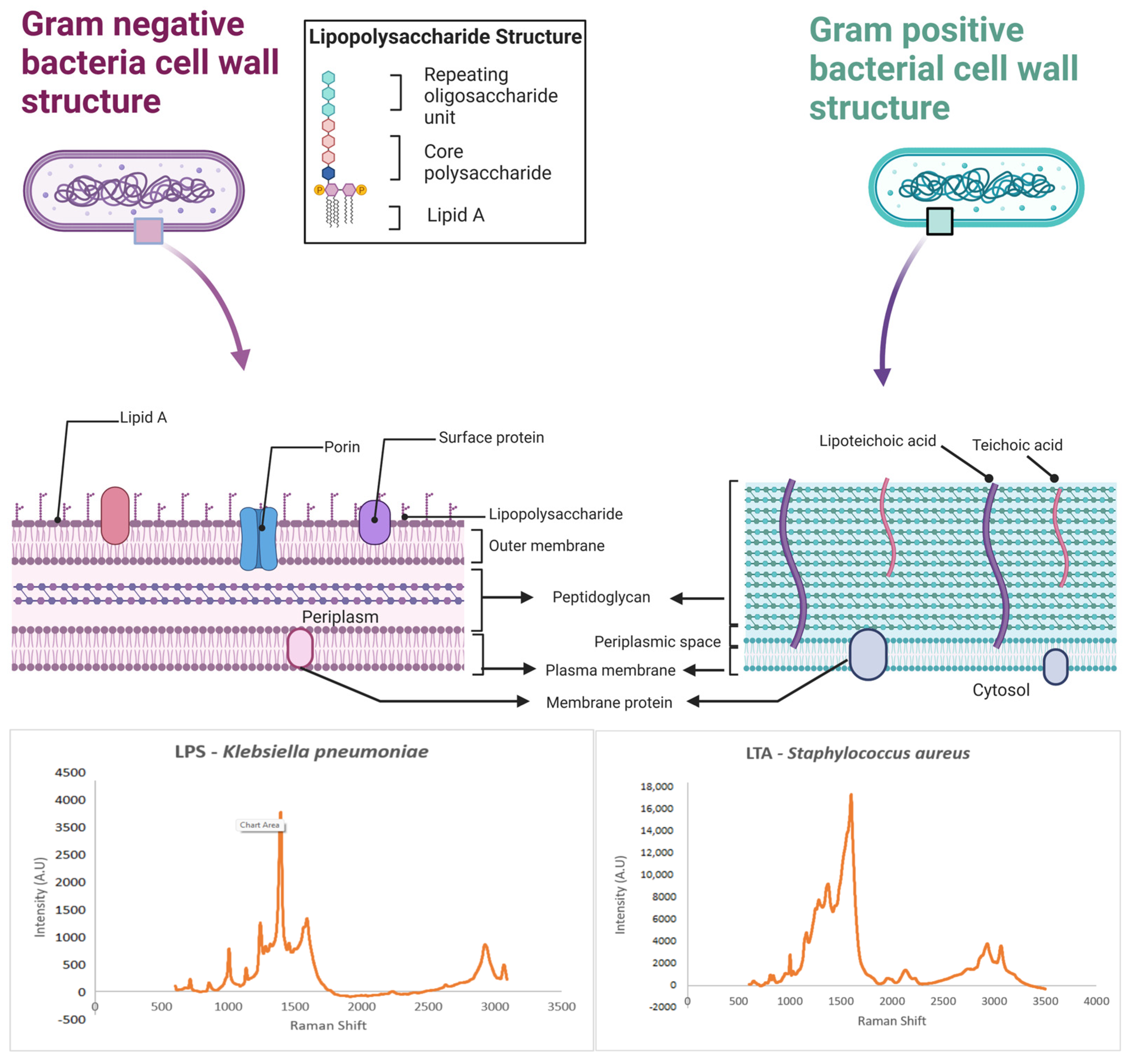
| Structure | Chemical Constituents | Gram |
|---|---|---|
| Cell wall | ||
| Peptidoglycan [110] | Alternating polymers of NAM (N-Acetylglucosamine) and NAG (N-acetylmuramic acid) | +/− |
| Teichoic Acid [111] | Polyribitol phosphate or glycerol phosphate is cross-linked to peptidoglycan. | + |
| Lipoteichoic Acid [112] | Lipid-linked teichoic acid. | + |
| Periplasmic Space [112] | proteases, phosphatases, lipases, nucleases, and carbohydrate-degrading enzymes | − |
| Outer Membrane [112] | Phospholipids with saturated fatty acids. | − |
| Proteins [112] | Porins and lipoproteins transport proteins. | − |
| Lipopolysaccharide [112] | Lipid A and core polysaccharide | − |
| Other external structures | ||
| Capsule [113] | Polysaccharides (disaccharides and trisaccharides) and polypeptides. | +/− |
| Pili [113] | Pilin and adhesins. | +/− |
| Flagellum [113] | Motor proteins, flagellin. | +/− |
| Biomarker Proteins [114,115] | For example, M proteins of streptococci and O antigen. Staphyloxanthin for Staphylococcus sp. Pyocyanin for Pseudomonas sp. | +/− + − |
| Other internal structures | ||
| Metabolic products [108] | ATP, NAD, and NADP+ | +/− |
| Proteins [9] | Metabolic proteins | +/− |
| DNA or RNA [9] | Nucleotides | +/− |
| Chemicals | Peak Position (cm−1) | Tentative Peak Assignments | Chemicals | Peak Position (cm−1) | Tentative Peak Assignments |
|---|---|---|---|---|---|
| Cell wall | Other external structures | ||||
| Peptidoglycan (NAG) [110] SERS (514.5 nm) | 699 | N/A | Capsule | N/A | |
| 815 | N/A | Pili | N/A | ||
| 964 | N/A | Flagellum [113] Raman (532 nm) | 903 | N/A | |
| 1059 | N/A | 945 | Skeletal CCN deformation | ||
| 1236 | N/A | ||||
| 1279 | N/A | 1003 | Phe | ||
| 1374 | N/A | 1246 | Helix | ||
| 1394 | N/A | 1320 | N/A | ||
| 1536 | N/A | 1453 | CH2 rocking | ||
| 1638 | N/A | 1662 | Amide I | ||
| Teichoic acid [111] Raman (532 nm) | 964 | POH bending | Other internal structures | ||
| 1250 | PO- bending | Cell plasma SERS [108] (514.5 nm) | 735 | N/A | |
| 1212 | CN bending | 1330 | N/A | ||
| 1322 | CHOH bending | 780 | N/A | ||
| 1452 | CH | 1050 | N/A | ||
| 1646 | Amid II | 1125 | N/A | ||
| Lipoteichoic acid [111,112] | Similar to teichoic acid | 1230 | N/A | ||
| 1435 | N/A | ||||
| Periplasmic space [112] | N/A | Metabolic products (4-ATP) [108] SERS (632.8 nm) | 1089 | NH2 rocking | |
| 1176 | CH bending | ||||
| Outer membrane proteins (Porins and OmpA) [112] Raman (514.5 nm) | 1553 | Trp | 1211 | CN bending | |
| 1579 | Trp | 1286 | CH stretching | ||
| 1602 | Phe | 1492 | CC stretching and CH bending | ||
| 1613 | Tyr | ||||
| 1669 | Amide | 1593 | CC stretching and NH2 bend. | ||
| 1734 | N/A | ||||
| Lipopolysaccharide [112] Raman (514.5 nm) | 1612 | N/A | Internal proteins [9] SERS-gold (830 nm) | 1250 | Amide III |
| 1652 | N/A | 1322 | Adenine, guanine, and Tyr | ||
| 1726 | N/A | 1003 | C(CC) aromatic ring (Phe) | ||
| N/A Not available | 1081 | V(PO) in oligonucleotides | |||
| DNA/RNA [9] SERS-gold (830 nm) | 546 | CO and POC bending | |||
| 795 | V(PO2) and v(CC) ring breathing | ||||
| 816 | CO and POC | ||||
| 853 | 1,4 glysosidic link | ||||
| Chemometric Methods | SERS Substrates | Bacterial Samples | Number of Bacteria | Results and Conclusions |
|---|---|---|---|---|
| DFA-HCA; PCA [9] | Silver colloid | Clinical bacterial isolates from patients with UTI (Escherichia coli; Klebsiella oxytoca; Klebsiella pneumoniae; Citrobacter freundii; and Enterococcus spp. and Proteus mirabilis) | 6 species, 5 strains | Discriminate between distinct species and discriminate Escherichia coli on strain level. |
| PCA, HCA, and DFA based on the “barcoding method” [146] | Au-nanoparticle-covered SiO2 substrate | Bacillus thuringiensis; Bacillus cereus; Bacillus anthracis; Bacillus licheniformis; Mycobacterium smegmatis; Mycobacterium fortuitum; Escherichia coli; Salmonella typhimurium | 8 species | Species and strain separation |
| PCA, HCA, and PLS-DA [145] | AgNR | Mycoplasma pneumonia and clinical throat swab | 1 specie, 3 strains | The throat swab samples spiked with M. pneumonia, and actual clinical throat swab samples were correctly classified. |
| PCA [35] | Internal deposition of silver nanoparticles | Staphylococcus epidermidis and Escherichia coli O157:H7 | 2 species | Differentiate Staphylococcus. epidermidis, Escherichia coli O157:H7, and their 1:1 ratio mixer |
| PCA [147] | Au, ion-doped SiO2 sol–gel | Kembolar pneumonia, Escherichia coli, Pseudomonas aeruginosa, Enterococcus faecalis, and Staphylococcus aureus | 4 species, 2 strains | Discriminate SERS spectra of different bacteria and the culture media in which they are grown. |
| PCA and SVM [40] | Silver colloid incorporates a microfluidic device | Escherichia coli | 9 strains | Classification between strains with a high correct rate |
| PCA [153] | Silver nanoparticles | Enterococcus faecalis; Streptococcus pyogenes; Acinetobacter baumannii; Klebsiella pneumoniae | 4 species | Discrimination between G+ and G-bacterial genera |
| PCA, LDA, and HCA [149] | Roughened gold-coated glass slides | Arthrobacter strains | 14 strains | Distinct molecular differences on the surface of fourteen closely related Arthrobacter strains; liquid and solid cultures are distinguished |
| PCA [73] | Magnetic–plasmonic Fe3O4–Au core–shell nanoparticles (Au-MNPs) | Acinetobacter calcoaceticus, Escherichia coli K12, and Pseudomonas aeruginosa | 3 species | Discriminate between species |
| PCA and HCA [76] | Gold nanoparticles (GNPs) | Salmonella typhimurium ATCC 50013, Salmonella O7HZ10, Shigella boydii CMCC51514, Shigella sonnei CMCC51529, Shigella dysenteriae CMCC51252, Citrobacter freundii ATCC43864, and Enterobacter sakazakii 154 | 6 species, 2 strains | Discriminate between species and serotypes |
| PCA [63] | AgNR | Generic Escherichia coli; Escherichia coli O157:H7; Staphylococcus aureus; Salmonella typhimurium 1925-1 poultry isolate, and Escherichia coli DH 5a | 3 species, 3 serotypes | Distinguish between distinct species, differentiate pure cell samples from mixed cell samples, and classify different bacterial strains. |
| PCA and PLS-DA [120] | VAN AgNR | Salmonella enterica serotype Anatum, Salmonella enterica serotype Cubana, Salmonella enterica serotype Stanley, Salmonella Enteritidis, Escherichia coli O157:H7, and Staphylococcus epidermidis | 3 species, 4 serotypes | Differentiate between species and serotypes in mung bean sprout samples |
| PCA and machine learning algorithm—RamanNet [79] | AgNR | E. coli, S. typhmirium, S. minnesota, S. mileloti, P. aeruginosa, M. catarrhalis, H. pylori GU2, F. tularensis LVS, E. coli 0128B12, E. coli 011B4, E. coli J5, and E. coli H100 | 6 species, 7 strains | Discriminate between distinct species and discriminate on strain level |
| PCA [96] | AgNR | E. coli, S. typhimurium, S. minnesota, V. cholerae, Rhizobium species R. CE3, and R. NGR, as well as Neisseria meningitidis | 6 species | SERS spectra can be used to differentiate between the different enteric LPS |
Disclaimer/Publisher’s Note: The statements, opinions and data contained in all publications are solely those of the individual author(s) and contributor(s) and not of MDPI and/or the editor(s). MDPI and/or the editor(s) disclaim responsibility for any injury to people or property resulting from any ideas, methods, instructions or products referred to in the content. |
© 2024 by the authors. Licensee MDPI, Basel, Switzerland. This article is an open access article distributed under the terms and conditions of the Creative Commons Attribution (CC BY) license (https://creativecommons.org/licenses/by/4.0/).
Share and Cite
Hassan, M.; Zhao, Y.; Zughaier, S.M. Recent Advances in Bacterial Detection Using Surface-Enhanced Raman Scattering. Biosensors 2024, 14, 375. https://doi.org/10.3390/bios14080375
Hassan M, Zhao Y, Zughaier SM. Recent Advances in Bacterial Detection Using Surface-Enhanced Raman Scattering. Biosensors. 2024; 14(8):375. https://doi.org/10.3390/bios14080375
Chicago/Turabian StyleHassan, Manal, Yiping Zhao, and Susu M. Zughaier. 2024. "Recent Advances in Bacterial Detection Using Surface-Enhanced Raman Scattering" Biosensors 14, no. 8: 375. https://doi.org/10.3390/bios14080375