Abstract
Copper-thiolate nanostructures, formed through the self-assembly of cysteine (Cys) and glutathione (GSH) with copper ions, offer a versatile platform for redox-active applications due to their structural stability and chemical functionality. In this study, Cu-Cys-GSH nanoparticles were synthesized and employed to develop a capacitive biosensor for the ultralow concentration detection of hydrogen peroxide (H2O2). The detection mechanism leverages a Fenton-like reaction, where H2O2 interacts with Cu-Cys-GSH nanoparticles to generate hydroxyl radicals (·OH) through redox cycling between Cu2+ and Cu+ ions. These redox processes induce changes in the sensor’s surface charge and dielectric properties, enabling highly sensitive capacitive sensing at gold interdigitated electrodes (IDEs). The influence of chirality on sensing performance was investigated by synthesizing nanoparticles with both L- and D-cysteine enantiomers. Comparative analysis revealed that the stereochemistry of cysteine impacts the catalytic activity and sensor response, with Cu-L-Cys-GSH nanoparticles exhibiting superior performance. Specifically, the biosensor achieved a linear detection range from 1.0 fM to 1.0 pM and demonstrated an ultra-sensitive detection limit of 21.8 aM, outperforming many existing methods for H2O2 detection. The sensor’s practical performance was further validated using milk and saliva samples, yielding high recovery rates and confirming its robustness and accuracy for real-world applications. This study offers a disposable, low-cost sensing platform compatible with sustainable healthcare practices and facilitates easy integration into point-of-care diagnostic systems.
1. Introduction
Hydrogen peroxide (H2O2) is a crucial biomolecule involved in various physiological and pathological processes, particularly in oxidative stress, immune response, and inflammation [1,2]. As a key reactive oxygen species (ROS), abnormal levels of H2O2 are closely associated with the progression of chronic diseases, including cancer, neurodegenerative disorders, and cardiovascular conditions [3,4,5,6,7,8]. Therefore, the development of highly sensitive and selective detection methods for H2O2 in biological samples is essential for early disease diagnosis, clinical monitoring, and biomedical research.
Nanomaterials have gained significant attention as biosensing platforms due to their unique physicochemical properties, including high surface area, tunable electronic states, and excellent catalytic activity [9,10,11,12,13,14,15,16,17,18]. The integration of nanomaterials into biosensors has significantly improved their analytical performance and utilization in the biomedical field. In the context of H2O2 detection, nanoparticles such as gold, silver, platinum, iron oxide, rhodium, nickel oxide, copper, and bimetallic nanoparticles have been widely used to enhance sensor performance [19,20,21]. Silver nanoparticles deposited on reduced graphene oxide and cerium(IV) oxide nanocomposites have been employed for nonenzymatic electrochemical detection of H2O2, demonstrating high sensitivity and stability [22]. Similarly, platinum-doped graphene sheets combined with cerium(IV) oxide have been integrated into screen-printed electrodes for selective H2O2 sensing, showcasing improved electron transfer and catalytic activity [23]. Additionally, Fe3O4-Au magnetic nanoparticles have been utilized to immobilize horseradish peroxidase on graphene-based electrodes, enabling sensitive electrochemical H2O2 biosensing [24]. Rhodium nanoparticles have been employed to modify graphite electrodes, facilitating the electrochemical detection of H2O2 even in the presence of oxygen and from tea extracts, highlighting their robust catalytic properties [25]. Furthermore, platinum-modified multi-walled carbon nanotube electrodes have been fabricated for rapid and quantitative H2O2 detection, particularly in food safety applications with high sensitivity and reproducibility [26]. Copper-based nanoparticles, particularly those incorporating cysteine and glutathione ligands, have attracted interest due to their redox activity and potential role in catalytic processes. Studies have shown that Cu-Cys nanoparticles exhibit self-assembly behavior and can engage in redox cycling between Cu2+ and Cu+, making them suitable for reactions involving H2O2 [27]. The addition of glutathione to form Cu-Cys-GSH nanoparticles further enhances their stability and reactivity, which has been explored in contexts such as chemodynamic therapy and oxidative stress modulation. The interaction of Cu-Cys-GSH nanoparticles with H2O2 leads to a redox cycling process where Cu2+ is reduced to Cu+ in the presence of glutathione, followed by the Cu+-catalyzed decomposition of H2O2 to produce highly reactive hydroxyl radicals (·OH) [28]. This mechanism mimics the classical Fenton reaction observed with iron but operates more efficiently at neutral or slightly acidic pH, making it highly suitable for biosensing applications [29]. Recent studies have demonstrated that Cu-Cys-GSH nanoparticles exhibit peroxidase-mimicking activity, catalyzing the oxidation of substrates such as 3,3′,5,5′-tetramethylbenzidine (TMB) in the presence of H2O2, confirming their role in ROS generation and redox cycling. Specifically, the oxidation of TMB to its oxidized form (oxTMB) was observed through a significant increase in UV–vis absorption, whereas the presence of GSH reversed this process by reducing oxTMB back to TMB, leading to a measurable decrease in absorption [30].
Although chromatography, colorimetry, chemiluminescence, and fluorometry are widely used for H2O2 detection, they are often limited by background interference, limited sensitivity thresholds, high operational costs, and the requirement for complex, specialized instrumentation. Additionally, many approaches rely heavily on labeling strategies, which can further complicate procedures and hinder real-time analysis. Capacitive biosensors have emerged as a promising alternative, offering label-free, real-time monitoring of molecular interactions based on changes in dielectric properties and surface charge distribution at the electrode interface. These sensors rely on variations in capacitance caused by biomolecular interactions, making them highly sensitive for detecting ultralow concentrations of analytes. Additionally, recent advances in AC electrokinetics (ACEK) effects have demonstrated the potential to further enhance capacitive sensing by actively directing biomolecules toward the electrode surface, overcoming the limitations of passive diffusion [31,32,33,34]. The ACEK effect, including AC electrothermal (ACET) flow, enables long-range transport of analytes, ensuring effective interaction with the sensor surface, thereby accelerating detection speed and improving sensitivity [35].
The integration of Cu-Cys-GSH nanoparticles with ACEK-assisted capacitive biosensing provides a novel and highly efficient strategy for H2O2 detection. The catalytic activity of these nanoparticles, driven by the Fenton-like reaction, induces dynamic changes in surface charge and dielectric properties upon H2O2 interaction. This process enhances the capacitive signal, enabling real-time and highly selective detection at femtomolar concentrations. Additionally, the stereochemistry of cysteine in Cu-Cys-GSH nanoparticles influences catalytic efficiency, further affecting sensor performance [29].
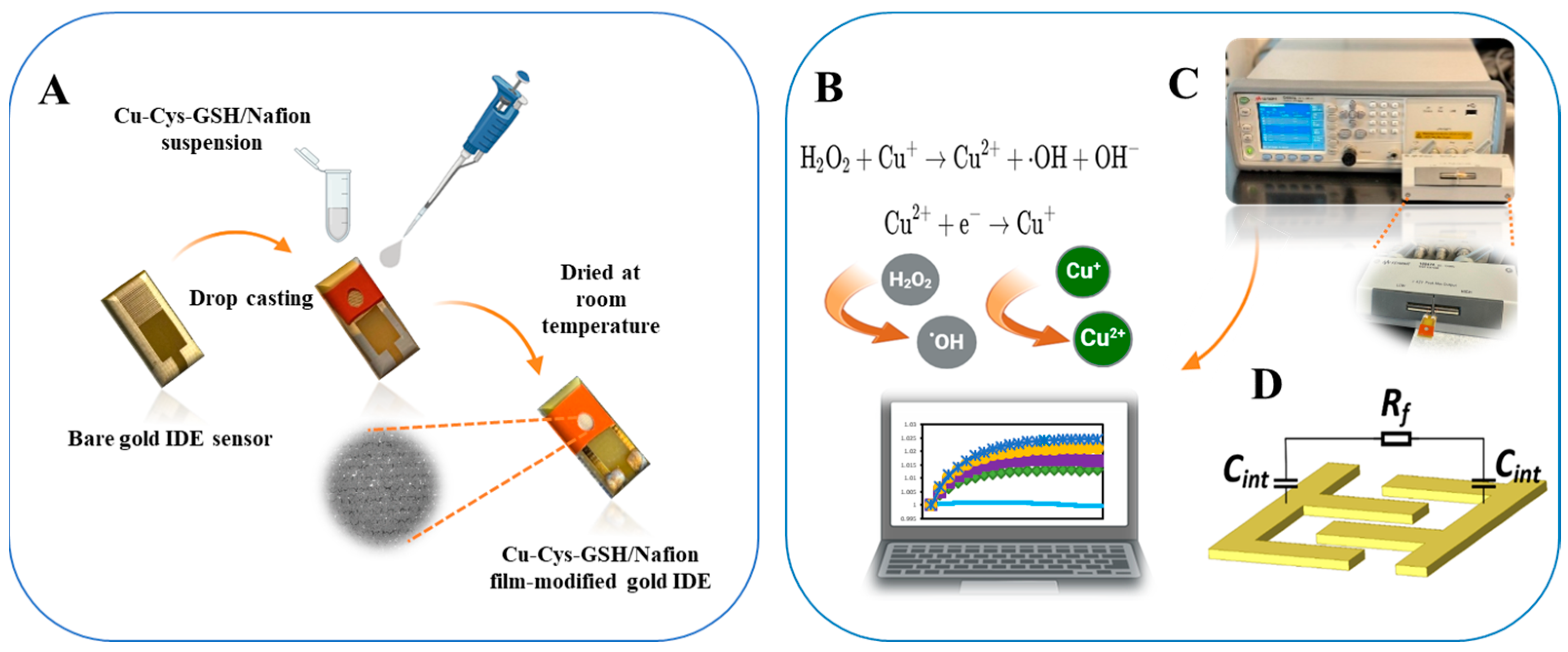
This study aims to develop a Cu-Cys-GSH nanoparticle–based capacitive biosensor for ultra-trace H2O2 detection while investigating the chiral effects on sensor performance. The detection mechanism leverages a Fenton-like reaction, wherein Cu2+ and Cu+ ions undergo redox cycling in the presence of H2O2, generating hydroxyl radicals (·OH) (Figure 1B). This process alters the interfacial capacitance at the gold interdigitated electrode surface, enabling highly sensitive detection. Furthermore, investigating the influence of nanoparticle chirality on sensor performance provides insights into the role of stereochemistry in catalytic efficiency and signal transduction. The applicability of the biosensor under real-world conditions is validated using milk samples. This work demonstrates the potential of Cu-Cys-GSH nanoparticles in capacitive biosensing, offering a low-cost, disposable platform for real-world applications in point-of-care testing.
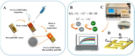
Figure 1.
Schematic diagram of (A) functionalization of the gold-IDE sensor with Cu-Cys-GSH/Nafion film, (B) Fenton-like reaction mechanism involved in detection, (C) measurement setup for ACEK-capacitive sensing, where interfacial capacitance is acquired at a fixed voltage through an LCR meter and transferred to a computer for processing, and (D) equivalent circuit model representing interfacial capacitance.
2. Materials and Methods
2.1. Reagents and Apparatus
Copper(II) chloride dihydrate (CuCl2·2H2O) (˃99%), sodium hydroxide (NaOH), L-cysteine (˃98%), D-cysteine (98%), L-alanine (99%), L-aspartic acid (99%), dopamine hydrochloride (99%), phosphate-buffered saline (PBS, 10X) at pH 7.4 were purchased from Thermo Scientific (Waltham, MA, USA). Acetone, isopropyl alcohol (IPA), and Nafion were supplied by Sigma-Aldrich (Darmstadt, Germany). L-glutathione (GSH) reduced was bought from Cayman Chemical (Ann Arbor, MI, USA). Testing buffer 0.1X PBS pH 7.4 was prepared by diluting 10X PBS with Milli-Q water.
The morphologies of the sensor and chiral nanoparticles were characterized using scanning electron microscopy (SEM TM-4000Plus, Hitachi, Tokyo, Japan). X-ray diffraction (XRD) patterns of nanoparticles were recorded on a Panalytical Empyrean q-2q diffractometer (Malvern Panalytical, Malvern, UK). Sufficient counting statistics were achieved using a 0.02626° 2q step scan from 3 to 70°, with an exposure time of 114.75 s per step and a revolution spin rate of 2 s. Fourier transform infrared spectra (FTIR) were obtained on a Thermo Scientific FTIR spectrometer (Waltham, MA, USA). A precision LCR meter (Keysight® E4980A, Santa Rosa, CA, USA) was utilized for electrical measurements.
2.2. Preparation of Cu-Cys-GSH Nanoparticles
NaOH (2 mmol) and either L- or D-cysteine (2 mmol) were dissolved in deionized water and slowly added dropwise into an aqueous CuCl2 solution under stirring for 5 min to facilitate the formation of Cu-Cys nanoparticles. The resulting Cu(II)-Cys nanoparticles were then separated via centrifugation and washed sequentially with deionized water and alcohol. This synthesis method was first introduced in [28].
The Cu(II)-Cys nanoparticles were mixed with 50 mM L-GSH under a nitrogen atmosphere and stirred for 2 h. The resulting Cu-Cys-GSH nanoparticles were collected via centrifugation and washed with deionized water [30].
2.3. Preparation of Cu-Cys-GSH Nanoparticle–Functionalized Gold IDE Sensor
The preparation of the gold IDEs with a metallic trilayer structure (Cu/Ni/Au) involved sequential surface treatment, electroplating, and cleaning. The width of the IDE finger and the gap between adjacent fingers are 100 μm. Initially, the electrodes underwent mechanical polishing using polishing pads to achieve a smooth and uniform surface. Subsequently, a thorough cleaning procedure was performed by sequentially rinsing the electrodes with acetone, isopropanol (IPA), and Milli-Q water, followed by air drying to eliminate any residual contaminants. A thin gold layer was electrodeposited onto the surface using a two-electrode configuration. Following electroplating, the electrodes underwent an additional cleaning cycle using acetone, IPA, and Milli-Q water, followed by air drying. To enhance surface hydrophilicity, the gold IDEs were subjected to UV–ozone treatment for 25 min. Finally, a 2 mm-thick silicone chamber was affixed to the electrode surface to secure the samples for subsequent experimental procedures.
A total of 15 mg of Cu-Cys-GSH nanoparticles was dispersed in 1 mL of 0.5% Nafion solution to form a homogenous dispersion under vigorous ultrasonication for about 1 h. Then, 8 µL of the resulting Cu-Cys-GSH/Nafion dispersion was dropped onto the surface of gold IDEs and was kept at room temperature till dry (Figure 1A).
2.4. Sample Preparation
To evaluate the applicability of the developed biosensor, H2O2 detection was performed in milk. Milk samples, purchased from a local market, were centrifuged at 10,000 rpm for 10 min to sediment fats, proteins, and other macromolecular components [36]. The clear supernatant was collected and diluted 10-fold with 0.05X PBS. Spiked recovery experiments were conducted by introducing H2O2 at concentrations of 10 fM, 100 fM, and 1 pM into the samples before the detection.
2.5. Capacitance Measurement
H2O2 stock solutions were diluted with 0.05X PBS buffer to obtain the analytical samples with concentrations ranging from 1 fM to 1 pM. During the measurement process, 15 μL of H2O2 solution was introduced into the chamber of the IDE sensor, which was then linked to an LCR meter for data acquisition and waited 5 min (Figure 1C). An AC signal of 1 kHz at 300 mV was applied to the IDE, and capacitance readings were recorded continuously for 10 s. The measured capacitance values were then normalized relative to the initial capacitance, and the rate of capacitance change was computed in terms of percentage variation per minute (dC/dt expressed in %/min). Sensor repeatability and consistency were evaluated by reporting the results as the mean ± standard deviation (SD) of measurements from three independently prepared sensors. The baseline capacitance change rate was determined using blank 0.05X PBS. The 1 kHz frequency was selected based on our previous study [37]. The voltage was optimized through experimental evaluation to achieve the best sensing performance.
3. Sensing Mechanism
3.1. Interfacial Capacitance Sensing
When an electrode is immersed in an electrolyte solution, charge accumulation occurs at the electrode surface, leading to the formation of an electric double layer (EDL). This layer consists of oppositely charged counter ions near the surface, forming an interfacial capacitance [38]. When an AC signal is applied, the solid–liquid interface can be represented by an equivalent circuit, as illustrated in Figure 1D, where Rf denotes the charge transfer resistance, and Cint corresponds to the interfacial capacitance, characterizing the capacitive behavior at the electrode–electrolyte interface. The application of an AC signal (300 mV at 1 kHz) enables the measurement of capacitance changes at the interface, which are sensitive to surface modifications induced by molecular interactions. As described in our previous studies [35,39,40,41], changes in surface morphology, whether through nanoparticle deposition or biomolecular immobilization, can significantly impact both the dielectric environment and surface morphology. These surface modifications enhance the capacitive response by increasing the interfacial area available for electric double-layer formation.
The initial capacitance is described using the following equation:
where Aint is the surface area of the capacitor. ε and d are the relative permittivity and thickness of the EDL, respectively. Following nanoparticle deposition and the Fenton-like reaction, interfacial capacitance (Cint,final) can be expressed as follows:
where Afinal is the effective surface area of the final capacitor. The relative change in capacitance is given as follows:
In this study, the IDE sensor with a functionalized Cu-Cys-GSH nanoassembly layer operated as a dynamic sensing platform for H2O2 detection through capacitance variation. Functionalization with Cu-Cys-GSH nanoparticles increased the surface roughness and effective area of the electrode, leading to an initial rise in Cint. The subsequent introduction of H2O2 triggered a Fenton-like reaction at the modified surface, as shown in Figure 1B, further altering the interface and enhancing Cint through catalytic surface restructuring.
3.2. ACEK Effects
AC electrokinetic (ACEK) effects encompass a range of electrokinetic phenomena that occur when an inhomogeneous AC electric field is applied to an aqueous solution. These effects include dielectrophoresis (DEP), AC electroosmosis (ACEO), and AC electrothermal (ACET) flow, each of which contributes to the movement of particles or fluid within the system [40,41]. The interplay of these mechanisms significantly influences the capacitive sensing performance of the proposed biosensor for H2O2 detection.
Dielectrophoresis (DEP) is a phenomenon in which neutral particles suspended in a solution experience motion due to polarization effects at the particle–liquid interface in response to the applied electric field gradient [42]. While DEP is a dominant force in systems involving micro- and nanoscale particles, its influence is negligible in this study because H2O2 is a small molecule that does not undergo significant polarization under the applied electric field.
AC electro-osmosis (ACEO) refers to the induced fluid flow at electrode surfaces under an inhomogeneous alternating current (AC) electric field. It plays a crucial role in microfluidic applications by enhancing mass transport and analyte mixing without external pumps. When an electrode is energized, counter ions in the electrolyte solution are attracted to the surface, forming the EDL. The inhomogeneous AC field creates a tangential electric field component, causing ion movement within the EDL. This motion generates shear stress, which drags the surrounding fluid, resulting in AC electro-osmotic flow. The magnitude of this flow depends on factors such as AC voltage, frequency, electrolyte conductivity, electrode geometry, and EDL thickness. At low to moderate AC frequencies (100 Hz to a few kHz), ACEO-driven flows dominate and can significantly enhance analyte transport in microfluidic biosensors. By directing biomolecules and nanoparticles toward the sensing region, ACEO improves detection sensitivity and reaction kinetics [43,44]. The velocity of ACEO flow (uACEO) is expressed as follows:
where εm is the permittivity of the solution, η is the fluid viscosity, ξ is the zeta potential at the electrode interface and Et is the parallel component of the applied electric field. However, ACEO is strongest in low-conductivity solutions (<0.085 S/m) and becomes less effective as the conductivity increases [45]. Since the current study employs 0.05X PBS (≈0.08 S/m), which is close to this threshold, ACEO effects may still contribute slightly to fluid motion but are expected to be weak compared to ACET. AC electrothermal (ACET) flow, in contrast, arises due to temperature gradients generated by the non-uniform electric field at the electrode surface. These temperature variations lead to changes in the solution’s conductivity and permittivity, creating a convective flow that enhances the movement of H2O2 molecules toward the electrode surface [46]. The velocity of ACET-driven fluid motion (uACET) is given as follows:
where V is the applied voltage amplitude, r is the distance between adjacent electrode fingers, which was equal to 50 μm in this study, T is the absolute temperature, and k is the thermal conductivity of the solution. Since ACET flow is enhanced in solutions with higher conductivity, it plays a role in this study. Both ACEO and ACET effects facilitate the efficient transport of H2O2 molecules toward the Cu-Cys-GSH/Nafion-modified IDE sensor, enhancing detection sensitivity and reducing response time.
4. Results and Discussion
4.1. Sensor Characterization
The surface morphology of the Cu-Cys-GSH nanoparticle–functionalized sensor and the synthesized Cu-L-Cys-GSH and Cu-D-Cys-GSH nanoparticles was analyzed using scanning electron microscopy (SEM), as shown in Figure 2. The low-magnification SEM image (Figure 2a, ×35) shows that the entire gold IDE sensor surface is uniformly coated with the Cu-Cys-GSH/Nafion film, confirming the successful surface modification via drop-casting. At medium magnification (Figure 2b, ×100), a network-like microstructure is observed, indicating the formation of interconnected nanoparticle aggregates within the Nafion matrix. The highest magnification (Figure 2c, ×1000) reveals a porous and granular texture, characteristic of nanostructured surfaces, where densely packed nanoparticles contribute to a high surface-to-volume ratio. This hierarchical nanostructure plays a crucial role in facilitating Fenton-like catalytic activity, enhancing charge redistribution, and improving the capacitive sensing response. Figure 2d,e reveals that both nanoparticle variants exhibit a granular and aggregated structure, suggesting successful self-assembly. The overall morphology appears similar between Cu-L-Cys-GSH and Cu-D-Cys-GSH, with no visually distinct differences in particle distribution or surface texture.
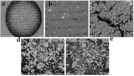
Figure 2.
SEM images of (a–c) the electrode surface covered Cu-Cys-GSH/Nafion film at different scales, (d) Cu-L-Cys-GSH nanoparticles, and (e) Cu-D-Cys-GSH nanoparticles.
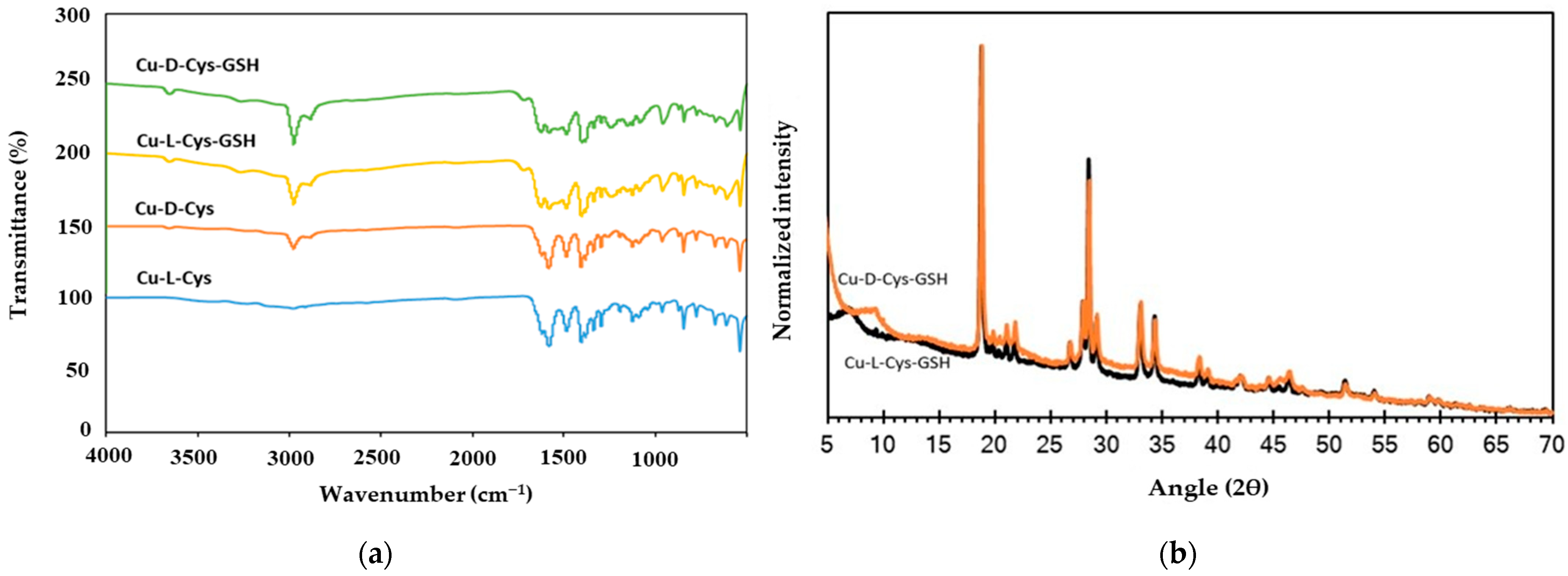
The FTIR spectra of Cu-L-Cys, Cu-D-Cys, Cu-L-Cys-GSH, and Cu-D-Cys-GSH nanoparticles, as shown in Figure 3a, confirm the successful interaction of cysteine and glutathione with copper through characteristic functional group vibrations. The broad absorption bands observed around 3200–3500 cm⁻1 correspond to O-H and N-H stretching vibrations, indicating the presence of hydroxyl and amine groups, which contribute to metal coordination. The peaks in the 1600–1700 cm⁻1 range are attributed to the C=O stretching of carboxyl groups, confirming metal-carboxylate interactions, which are common in amino acid–functionalized copper nanoparticles [47]. The disappearance of the thiol (-SH) peak around 2550–2600 cm⁻1 indicates that sulfur binds to copper through the formation of a Cu-S bond [29]. The peak around 611 cm⁻1 in Cu-Cys-GSH samples corresponds to Cu-S stretching vibrations, further confirming Cu-thiol interactions [29]. The additional peaks in the 1000–1200 cm⁻1 region correspond to C-N stretching from amine groups and asymmetric vibrations of C-O-C bonds, which indicate strong metal-ligand coordination with glutathione. The spectral differences between Cu-L-Cys-GSH and Cu-D-Cys-GSH suggest potential variations in molecular interactions due to chirality, affecting the self-assembly behavior of these nanoparticles. These findings confirm the formation of well-defined Cu-Cys and Cu-Cys-GSH nanoparticles with strong Cu-ligand interactions, validating their structural integrity.
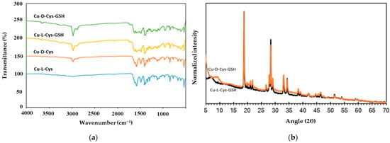
Figure 3.
(a) FTIR spectra of synthesized chiral nanoparticles and (b) XRD patterns of Cu-L-Cys-GSH and Cu-D-Cys-GSH nanoparticles.
X-ray diffraction (XRD) patterns shown in Figure 3b provide structural insights into the crystalline and amorphous phases of Cu-Cys-GSH nanoparticles. The presence of sharp diffraction peaks at 18.84°, 28.4°, and 34.4° indicates the characteristic diffraction planes associated with the incorporation of cysteine in the nanoparticle structure [29]. Additionally, the peaks observed around 29.2° suggest the formation of Cu(II)-thiolate complexes, confirming the coordination of copper with sulfur-containing ligands. The broad background signal at lower angles suggests the presence of amorphous regions, which may be attributed to the self-assembled nanostructure of Cu-Cys-GSH.
4.2. Optimization of Measurement Conditions
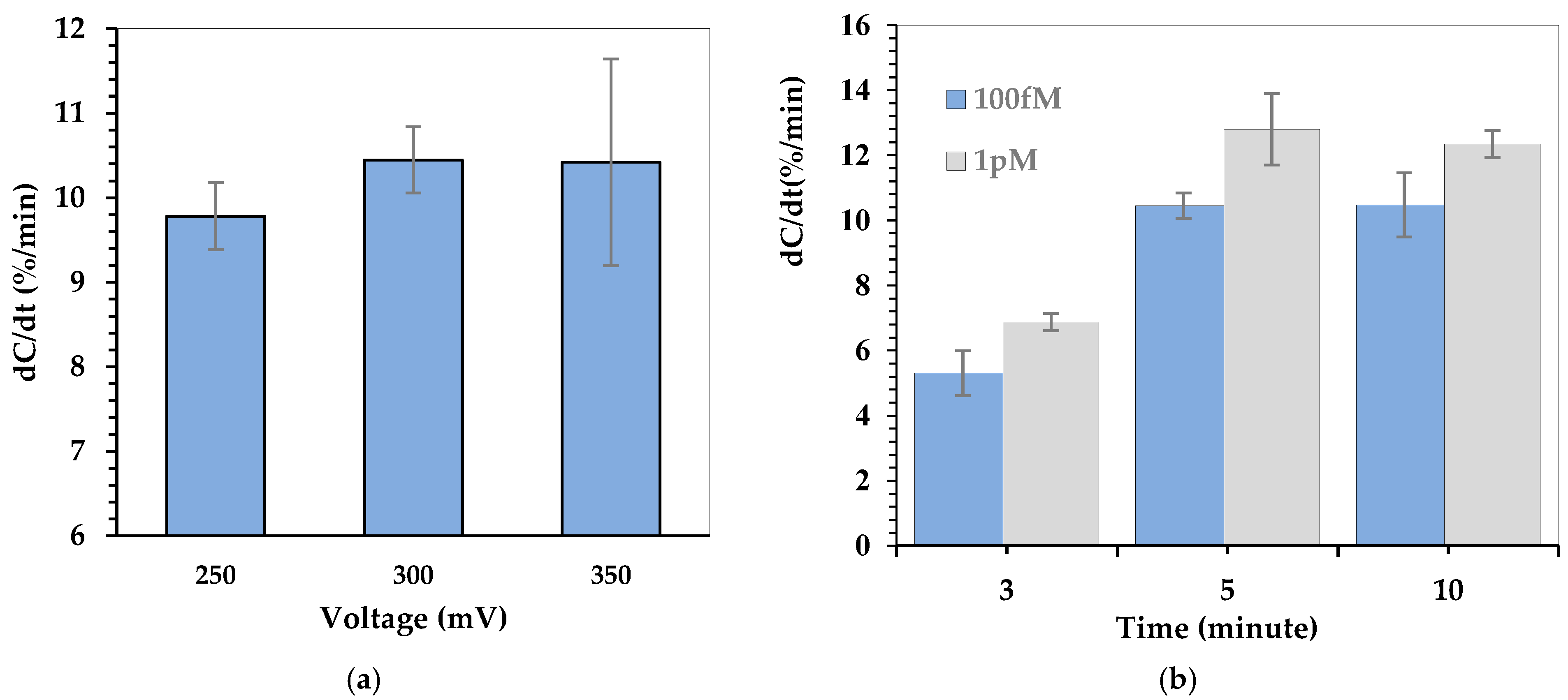
To ensure optimal performance of the Cu-L-Cys-GSH nanoparticle–functionalized capacitive biosensor, the effects of applied AC voltage and incubation time were investigated systematically. Figure 4 presents the capacitance change rate (dC/dt, %/min) under different testing conditions to determine the optimum voltage amplitude and incubation duration for reliable H2O2 detection. Figure 4a illustrates the effect of applied voltage (250, 300, and 350 mV) on the capacitance change rate (dC/dt, %/min). The influence of AC voltage on the sensor’s capacitance response was evaluated at a fixed frequency of 1 kHz with an H2O2 concentration of 100 fM. The results show that increasing the applied voltage from 250 mV to 300 mV enhances the capacitance response, suggesting improved charge redistribution and analyte interaction due to ACEK effects. However, at 350 mV, the variation in the signal increases, indicating possible instability at higher voltages. Based on these results, 300 mV was selected as the optimal voltage for further measurements. Figure 4b illustrates the effect of incubation time on the capacitance change rate (dC/dt, %/min) at two different H2O2 concentrations (100 fM and 1 pM). The results show that as the incubation time increases, the capacitance response also increases, indicating that a longer reaction time allows for more effective interaction between H2O2 and the Cu-L-Cys-GSH/Nafion film–modified sensor surface. The sensor response reaches a peak at 5 min, suggesting that sufficient reaction has occurred to induce a stable capacitive signal. Extending the incubation time does not significantly enhance the response. This behavior suggests that after 5 min, the interaction between H2O2 and the sensor surface stabilizes, and additional time may only lead to signal saturation rather than increased sensitivity.
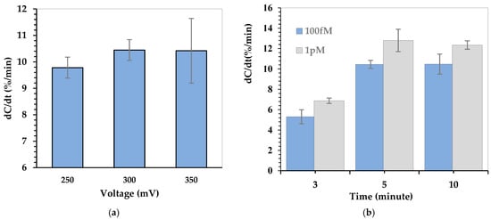
Figure 4.
Normalized capacitance response for (a) applied voltage optimization and (b) incubation time optimization.
4.3. Dose Response of Cu-Cys-GSH Nanoparticle–Functionalized Sensor
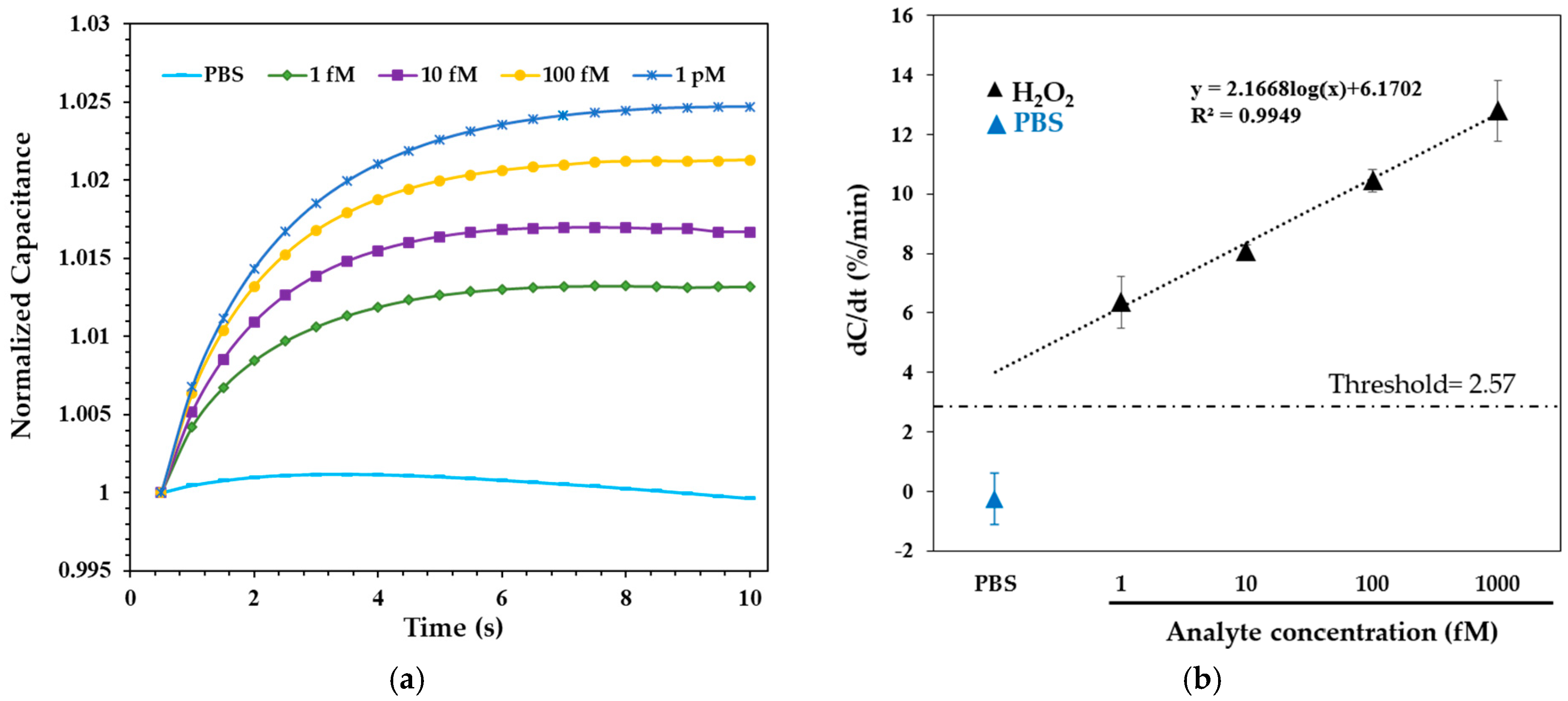
H2O2 solutions ranging from 1 fM to 1 pM in 0.05X PBS were applied to the Cu-L-Cys-GSH/Nafion film–modified electrode surface and incubated for 5 min before measurement. Following the incubation, the normalized capacitance was recorded for 10 s under the optimized AC conditions. Figure 5a presents the normalized capacitance-time profiles following incubation with H2O2. A concentration-dependent increase in capacitance was observed, where higher H2O2 levels led to a more rapid and pronounced rise within the 10-s measurement window. This trend is attributed to enhanced charge accumulation at the sensor interface, driven by the Fenton-like reaction between H2O2 and Cu-Cys-GSH nanoparticles.
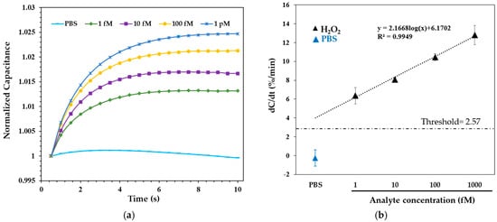
Figure 5.
Capacitance response of H2O2 in 0.05X PBS. (a) Transient capacitance curves over 10 s. (b) Normalized dose-dependent response. Data are presented as mean ± SD from three independent replicates. Measurements were performed at 300 mV and 1 kHz.
Figure 5b shows the corresponding dose-response curve, where the capacitance change rate (dC/dt, %/min) is plotted against H2O2 concentrations ranging from 1 fM to 1 pM, with logarithmic scaling applied in the data fitting. The sensor exhibited excellent linearity across this range, following the equation dC/dt = 2.1668 log10(x) + 6.1702, with a correlation coefficient (R2) of 0.9949, where x is the H2O2 concentration in femtomolar (fM). The detection threshold, calculated as three times the standard deviation of the blank [32], was determined to be 2.57%/min. Substituting this value into the dose-response equation yielded a limit of detection (LOD) of 21.8 aM. These results establish the Cu-Cys-GSH nanoparticle–functionalized capacitive biosensor as a highly sensitive and stable system for H2O2 detection.
4.4. Influence of Chirality on Cu-Cys-GSH Nanoparticle–Functionalized Sensor Response
To investigate the influence of chirality on sensor performance, we evaluated the capacitive responses of sensors functionalized with Cu-L-Cys-GSH and Cu-D-Cys-GSH nanoparticles across a range of H2O2 concentrations (Figure 6). The experimental results indicate that the sensor modified with Cu-L-Cys-GSH nanoparticles consistently outperformed the sensor modified with Cu-D-Cys-GSH nanoparticles at all tested concentrations, ranging from 1 fM to 1 pM. Specifically, Cu-L-Cys-GSH nanoparticles exhibited higher sensitivity (greater dC/dt values) compared to their D-enantiomer counterparts.
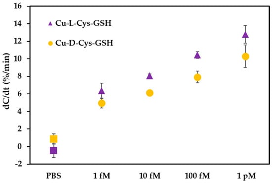
Figure 6.
Influence of chirality on the capacitive response (dC/dt, %/min) of sensors functionalized with Cu-L-Cys-GSH and Cu-D-Cys-GSH nanoparticles across varying concentrations of H2O2 (1 fM to 1 pM). The data indicate enhanced sensor sensitivity for the L-enantiomer nanoparticles compared to the D-enantiomer counterparts.
This difference in performance can be attributed to distinct chiral interactions between the nanoparticles and endogenous biomolecules such as glutathione. As supported by previous research [29], Cu-L-Cys nanoparticles demonstrated a higher affinity toward L-GSH due to a more energetically favorable binding process, confirmed by isothermal titration calorimetry and molecular dynamics simulations. The stronger affinity between L-enantiomer nanoparticles and L-GSH accelerates the redox reaction between them, significantly enhancing the catalytic efficiency of the subsequent Fenton-like reaction [29]. The redox conversion of Cu(II) to Cu(I) is facilitated by more efficient electron transfer. Specifically, tighter molecular interactions reduce the electron transfer distance and lower the activation energy barrier. Additionally, stronger binding creates a localized high concentration of L-GSH at the nanoparticle surface, further enhancing the redox cycling rate. This improved electron transfer efficiency ultimately strengthens the catalytic performance of the Fenton-like reaction. The increased catalytic activity of Cu-L-Cys-GSH nanoparticles results in enhanced generation of hydroxyl radicals (·OH), thereby amplifying the sensor’s response through changes in capacitance, driven by intensified local redox cycling and shifts in conductivity.
4.5. Selectivity
Selectivity is a key parameter in the development of biosensors, particularly when applied to complex biological environments containing diverse interfering species. To evaluate the selectivity of the Cu-L-Cys-GSH/Nafion-functionalized capacitive sensor, potential interferents such as amino acids (L-alanine and L-aspartic acid) and the electroactive neurotransmitter dopamine were selected.
Figure 7 presents the selectivity performance of the sensor when exposed to these interferents in comparison to H2O2. The non-target species were tested at concentrations of 100 fM and 1 pM using an AC signal of 1 kHz and 300 mV. All non-target species exhibited lower responses than that of 1 fM H2O2 under identical conditions. Specifically, at a concentration of 1 fM, H2O2 yielded a capacitance change rate (dC/dt) of 6.36 ± 0.865%/min, while L-alanine, L-aspartic acid, and dopamine at 1 pM exhibited dC/dt values of 5.86 ± 0.43%/min, 6.35 ± 0.57%/min, and 6.14 ± 1.065%/min, respectively. Therefore, the sensor demonstrates good specificity for H2O2, with a selectivity of approximately 1000:1. The low standard deviation values further support the reproducibility across different electrodes.
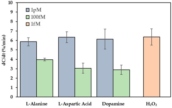
Figure 7.
Selectivity of the Cu-L-Cys-GSH/Nafion-functionalized sensor. Sensor responses toward H2O2 (1 fM) and potential interfering species (L-alanine, L-aspartic acid, and dopamine) at concentrations of 100 fM and 1 pM in 0.05X PBS.
The significant discrimination between H2O2 and the interfering species can be attributed to the catalytic specificity of the Cu-L-Cys-GSH nanoparticles, which facilitated a Fenton-like reaction exclusively with H2O2, resulting in amplified charge redistribution and enhanced capacitive transduction. In contrast, the non-catalytic species, including amino acids and dopamine, likely induced only minor surface perturbations or weak, non-specific adsorption, and a substantially attenuated capacitance response.
4.6. Sample Analysis
The reliability of the Cu-L-Cys-GSH nanoparticle–functionalized capacitive biosensor was validated through the analysis of spiked milk samples. As presented in Table 1, the sensor exhibited excellent recovery rates, ranging from 109.2% to 126.2% in milk across H2O2 concentrations of 10 fM, 100 fM, and 1 pM. The relative standard deviation (RSD) values remained consistently low (<0.6%), underscoring the high precision of the sensor in complex matrices. Saliva testing data are included in the Supplementary Materials.

Table 1.
Detection of H2O2 in milk samples using Cu-L-Cys-GSH/Nafion film–modified sensor.
These results confirm the sensor’s capability for accurate and consistent detection of trace levels of H2O2 in real samples, demonstrating its robustness and applicability for practical diagnostic applications.
5. Conclusions
In this study, we developed a capacitive biosensor functionalized with Cu-Cys-GSH nanoparticles for highly sensitive detection of H2O2. The detection mechanism is based on a Fenton-like reaction, where redox cycling between Cu2+ and Cu+ generates hydroxyl radicals (·OH), causing localized alterations in electrolyte composition, ionic mobility, and charge distribution at the electrode interface. The influence of nanoparticle chirality was also explored, demonstrating that sensors modified with Cu-L-Cys-GSH nanoparticles exhibited superior capacitive responses compared to the D-enantiomer. This difference can be attributed to stereochemistry-dependent interactions with glutathione, influencing catalytic efficiency. Analysis using spiked milk demonstrated the sensor’s high accuracy and precision. The findings confirm the effectiveness of Cu-Cys-GSH nanoparticles in enhancing capacitive biosensor performance through chirality-dependent catalytic mechanisms. Based on these results, the sensing platform holds potential for extension toward detecting diverse biological and chemical targets, particularly redox-active species, for future applications in clinical diagnostics, food safety analysis, and environmental monitoring.
Supplementary Materials
The following supporting information can be downloaded at: https://www.mdpi.com/article/10.3390/bios15050315/s1, Table S1: Detection of H2O2 in saliva samples using Cu-L-Cys-GSH/Nafion film-modified sensor.
Author Contributions
Conceptualization, D.Y.A. and J.J.W.; methodology, D.Y.A. and J.J.W.; investigation, D.Y.A.; resources, J.J.W. and J.C.; data curation, D.Y.A.; writing—original draft preparation, D.Y.A., J.J.W. and J.C.; writing—review and editing, D.Y.A., J.J.W. and J.C.; visualization, D.Y.A.; funding acquisition, J.J.W. All authors have read and agreed to the published version of the manuscript.
Funding
This research was funded by the USDA NIFA, grant number 2023-67021-40613.
Informed Consent Statement
Informed consent was obtained from all subjects involved in the study.
Data Availability Statement
The original contributions presented in this study are included in the article. Further inquiries can be directed to the corresponding author.
Acknowledgments
The author, Duygu Yilmaz Aydin, would like to thank The Scientific and Technological Research Council of Türkiye (TUBITAK) for supporting this research within the scope of the 2219 International Postdoctoral Research Fellowship Program.
Conflicts of Interest
The authors declare no conflicts of interest.
References
- Andrés, C.M.C.; Pérez De La Lastra, J.M.; Juan, C.A.; Plou, F.J.; Pérez-Lebeña, E. Chemistry of Hydrogen Peroxide Formation and Elimination in Mammalian Cells, and Its Role in Various Pathologies. Stresses 2022, 2, 256–274. [Google Scholar] [CrossRef]
- Pravda, J. Hydrogen Peroxide and Disease: Towards a Unified System of Pathogenesis and Therapeutics. Mol. Med. 2020, 26, 41. [Google Scholar] [CrossRef] [PubMed]
- Upadhyay, S.; Vaish, S.; Dhiman, M. Hydrogen Peroxide-Induced Oxidative Stress and Its Impact on Innate Immune Responses in Lung Carcinoma A549 Cells. Mol. Cell. Biochem. 2019, 450, 135–147. [Google Scholar] [CrossRef]
- Jomova, K.; Raptova, R.; Alomar, S.Y.; Alwasel, S.H.; Nepovimova, E.; Kuca, K.; Valko, M. Reactive Oxygen Species, Toxicity, Oxidative Stress, and Antioxidants: Chronic Diseases and Aging. Arch. Toxicol. 2023, 97, 2499–2574. [Google Scholar] [CrossRef]
- Houldsworth, A. Role of Oxidative Stress in Neurodegenerative Disorders: A Review of Reactive Oxygen Species and Prevention by Antioxidants. Brain Commun. 2023, 6, fcad356. [Google Scholar] [CrossRef] [PubMed]
- Teleanu, D.M.; Niculescu, A.-G.; Lungu, I.I.; Radu, C.I.; Vladâcenco, O.; Roza, E.; Costăchescu, B.; Grumezescu, A.M.; Teleanu, R.I. An Overview of Oxidative Stress, Neuroinflammation, and Neurodegenerative Diseases. Int. J. Mol. Sci. 2022, 23, 5938. [Google Scholar] [CrossRef]
- Anik, M.I.; Mahmud, N.; Masud, A.A.; Khan, M.I.; Islam, M.N.; Uddin, S.; Hossain, M.K. Role of Reactive Oxygen Species in Aging and Age-Related Diseases: A Review. ACS Appl. Bio Mater. 2022, 5, 4028–4054. [Google Scholar] [CrossRef]
- Rauf, A.; Khalil, A.A.; Awadallah, S.; Khan, S.A.; Abu-Izneid, T.; Kamran, M.; Hemeg, H.A.; Mubarak, M.S.; Khalid, A.; Wilairatana, P. Reactive Oxygen Species in Biological Systems: Pathways, Associated Diseases, and Potential Inhibitors—A Review. Food Sci. Nutr. 2024, 12, 675–693. [Google Scholar] [CrossRef]
- Robby, A.I.; Park, S.Y. Recyclable Metal Nanoparticle-Immobilized Polymer Dot on Montmorillonite for Alkaline Phosphatase-Based Colorimetric Sensor with Photothermal Ablation of Bacteria. Anal. Chim. Acta 2019, 1082, 152–164. [Google Scholar] [CrossRef]
- Zhao, Y.; Peng, N.; Gao, W.; Hu, F.; Zhang, C.; Wei, X. ZnS and Reduced Graphene Oxide Nanocomposite-Based Non-Enzymatic Biosensor for the Photoelectrochemical Detection of Uric Acid. Biosensors 2024, 14, 488. [Google Scholar] [CrossRef]
- Fang, Y.; Li, C.; Bo, J.; Henzie, J.; Yamauchi, Y.; Asahi, T. Chiral Sensing with Mesoporous Pd@Pt Nanoparticles. ChemElectroChem 2017, 4, 1832–1835. [Google Scholar] [CrossRef]
- Zhou, J.; Liu, C.; Zhong, Y.; Luo, Z.; Wu, L. A Review of Current Developments in Functionalized Mesoporous Silica Nanoparticles: From Synthesis to Biosensing Applications. Biosensors 2024, 14, 575. [Google Scholar] [CrossRef]
- Harish, V.; Tewari, D.; Gaur, M.; Yadav, A.B.; Swaroop, S.; Bechelany, M.; Barhoum, A. Review on Nanoparticles and Nanostructured Materials: Bioimaging, Biosensing, Drug Delivery, Tissue Engineering, Antimicrobial, and Agro-Food Applications. Nanomaterials 2022, 12, 457. [Google Scholar] [CrossRef]
- Li, C.; Wang, Y.; Jiang, H.; Wang, X. Biosensors Based on Advanced Sulfur-Containing Nanomaterials. Sensors 2020, 20, 3488. [Google Scholar] [CrossRef] [PubMed]
- Purohit, B.; Vernekar, P.R.; Shetti, N.P.; Chandra, P. Biosensor Nanoengineering: Design, Operation, and Implementation for Biomolecular Analysis. Sens. Int. 2020, 1, 100040. [Google Scholar] [CrossRef]
- Karakuş, E.; Erdemir, E.; Demirbilek, N.; Liv, L. Colorimetric and Electrochemical Detection of SARS-CoV-2 Spike Antigen with a Gold Nanoparticle-Based Biosensor. Anal. Chim. Acta 2021, 1182, 338939. [Google Scholar] [CrossRef] [PubMed]
- Lee, C.-Y.; Wu, L.-P.; Chou, T.-T.; Hsieh, Y.-Z. Functional Magnetic Nanoparticles–Assisted Electrochemical Biosensor for Eosinophil Cationic Protein in Cell Culture. Sens. Actuators B Chem. 2018, 257, 672–677. [Google Scholar] [CrossRef]
- Hwang, K.Y.; Brown, D.; Attanayake, S.B.; Luu, D.; Nguyen, M.D.; Lee, T.R.; Phan, M.-H. Signal Differentiation of Moving Magnetic Nanoparticles for Enhanced Biodetection and Diagnostics. Biosensors 2025, 15, 116. [Google Scholar] [CrossRef]
- Chen, S.; Yuan, R.; Chai, Y.; Hu, F. Electrochemical Sensing of Hydrogen Peroxide Using Metal Nanoparticles: A Review. Microchim. Acta 2013, 180, 15–32. [Google Scholar] [CrossRef]
- Dhara, K.; Mahapatra, D.R. Recent Advances in Electrochemical Nonenzymatic Hydrogen Peroxide Sensors Based on Nanomaterials: A Review. J. Mater. Sci. 2019, 54, 12319–12357. [Google Scholar] [CrossRef]
- Giaretta, J.E.; Duan, H.; Oveissi, F.; Farajikhah, S.; Dehghani, F.; Naficy, S. Flexible Sensors for Hydrogen Peroxide Detection: A Critical Review. ACS Appl. Mater. Interfaces 2022, 14, 20491–20505. [Google Scholar] [CrossRef]
- Yao, Z.; Yang, X.; Wu, F.; Wu, W.; Wu, F. Synthesis of Differently Sized Silver Nanoparticles on a Screen-Printed Electrode Sensitized with a Nanocomposites Consisting of Reduced Graphene Oxide and Cerium(IV) Oxide for Nonenzymatic Sensing of Hydrogen Peroxide. Microchim. Acta 2016, 183, 2799–2806. [Google Scholar] [CrossRef]
- Yang, X.; Ouyang, Y.; Wu, F.; Hu, Y.; Zhang, H.; Wu, Z. In Situ & Controlled Preparation of Platinum Nanoparticles Dopping into Graphene Sheets@cerium Oxide Nanocomposites Sensitized Screen Printed Electrode for Nonenzymatic Electrochemical Sensing of Hydrogen Peroxide. J. Electroanal. Chem. 2016, 777, 85–91. [Google Scholar] [CrossRef]
- Yang, X.; Xiao, F.-B.; Lin, H.-W.; Wu, F.; Chen, D.-Z.; Wu, Z.-Y. A Novel H2O2 Biosensor Based on Fe3O4–Au Magnetic Nanoparticles Coated Horseradish Peroxidase and Graphene Sheets–Nafion Film Modified Screen-Printed Carbon Electrode. Electrochim. Acta 2013, 109, 750–755. [Google Scholar] [CrossRef]
- Gatselou, V.A.; Giokas, D.L.; Vlessidis, A.G.; Prodromidis, M.I. Rhodium Nanoparticle-Modified Screen-Printed Graphite Electrodes for the Determination of Hydrogen Peroxide in Tea Extracts in the Presence of Oxygen. Talanta 2015, 134, 482–487. [Google Scholar] [CrossRef]
- Chou, T.-C.; Wu, K.-Y.; Hsu, F.-X.; Lee, C.-K. Pt-MWCNT Modified Carbon Electrode Strip for Rapid and Quantitative Detection of H2O2 in Food. J. Food Drug Anal. 2018, 26, 662–669. [Google Scholar] [CrossRef] [PubMed]
- Bain, D.; Russier-Antoine, I.; Yuan, H.; Kolay, S.; Maclot, S.; Moulin, C.; Salmon, E.; Brevet, P.-F.; Pniakowska, A.; Olesiak-Bańska, J.; et al. Solvent-Induced Aggregation of Self-Assembled Copper–Cysteine Nanoparticles Reacted with Glutathione: Enhancing Linear and Nonlinear Optical Properties. Langmuir 2023, 39, 16554–16561. [Google Scholar] [CrossRef]
- Ma, B.; Wang, S.; Liu, F.; Zhang, S.; Duan, J.; Li, Z.; Kong, Y.; Sang, Y.; Liu, H.; Bu, W.; et al. Self-Assembled Copper–Amino Acid Nanoparticles for in Situ Glutathione “AND” H2O2 Sequentially Triggered Chemodynamic Therapy. J. Am. Chem. Soc. 2019, 141, 849–857. [Google Scholar] [CrossRef]
- Wang, S.; Zhao, Y.; Yao, S.; Wang, Z.; Zhang, Z.; Wen, K.; Ma, B.; Li, L. Chirality of Copper–Amino Acid Nanoparticles Determines Chemodynamic Cancer Therapeutic Outcome. Small 2024, 20, 2309328. [Google Scholar] [CrossRef]
- Liu, M.; Yan, C.; Ye, Q.; Sun, X.; Han, J. Discrimination and Quantification of Glutathione by Cu+-Based Nanozymes. Biosensors 2023, 13, 827. [Google Scholar] [CrossRef]
- Cui, H.; Cheng, C.; Lin, X.; Wu, J.; Chen, J.; Eda, S.; Yuan, Q. Rapid and Sensitive Detection of Small Biomolecule by Capacitive Sensing and Low Field AC Electrothermal Effect. Sens. Actuators B Chem. 2016, 226, 245–253. [Google Scholar] [CrossRef]
- Mirzajani, H.; Cheng, C.; Vafaie, R.H.; Wu, J.; Chen, J.; Eda, S.; Aghdam, E.N.; Ghavifekr, H.B. Optimization of ACEK-Enhanced, PCB-Based Biosensor for Highly Sensitive and Rapid Detection of Bisphenol a in Low Resource Settings. Biosens. Bioelectron. 2022, 196, 113745. [Google Scholar] [CrossRef] [PubMed]
- Li, S.; Cui, H.; Yuan, Q.; Wu, J.; Wadhwa, A.; Eda, S.; Jiang, H. AC Electrokinetics-Enhanced Capacitive Immunosensor for Point-of-Care Serodiagnosis of Infectious Diseases. Biosens. Bioelectron. 2014, 51, 437–443. [Google Scholar] [CrossRef]
- Qi, H.; Xiao, L.; Wu, J.; Lv, L.; Hu, X.; Zhuang, Y.; Liu, X.; Zhao, W.; You, F.; Zhang, J.; et al. One-Step and Real-Time Detection of Hg2+ in Brown Rice Flour Using a Biosensor Integrated with AC Electrothermal Enrichment. Food Chem. 2023, 416, 135823. [Google Scholar] [CrossRef] [PubMed]
- Jiang, Y.; Huang, J.; Wu, J.; Eda, S. A Rapid, Sensitive, and Simple-to-Use Biosensor for on-Site Detection of Attomolar Level microRNA Biomarkers from Serum Extracellular Vesicles. Sens. Actuators B Chem. 2022, 369, 132314. [Google Scholar] [CrossRef]
- Kumar, D.; Kumar, V.; Sachdev, A.; Matai, I. Electrochemical Microfluidic Sensor Based on hBN-CeO2@Cyt c Hydrogel-Modified SPCE for the Detection of Hydrogen Peroxide. Ionics 2024, 30, 8559–8575. [Google Scholar] [CrossRef]
- Qi, H.; Zhao, M.; Liang, H.; Wu, J.; Huang, Z.; Hu, A.; Wang, J.; Lu, Y.; Zhang, J. Rapid Detection of Trace Cu2+ Using an L-cysteine Based Interdigitated Electrode Sensor Integrated with AC Electrokinetic Enrichment. Electrophoresis 2019, 40, 2699–2705. [Google Scholar] [CrossRef]
- Wu, J. Interactions of Electrical Fields with Fluids:Laboratory-on-a-Chip Applications. IET Nanobiotechnol. 2008, 2, 14–27. [Google Scholar] [CrossRef] [PubMed]
- Wan, N.; Jiang, Y.; Huang, J.; Oueslati, R.; Eda, S.; Wu, J.; Lin, X. Rapid and Sensitive Detection of miRNA Based on AC Electrokinetic Capacitive Sensing for Point-of-Care Applications. Sensors 2021, 21, 3985. [Google Scholar] [CrossRef]
- Zhang, J.; Wang, J.; Wu, J.; Qi, H.; Wang, C.; Fang, X.; Cheng, C.; Yang, W. Rapid Detection of Ultra-Trace Nanoparticles Based on ACEK Enrichment for Semiconductor Manufacturing Quality Control. Microfluid. Nanofluid 2019, 23, 2. [Google Scholar] [CrossRef]
- Qi, H.; Wang, B.; Liang, H.; Wu, J.J.; Ni, T.; Huang, Z.; Lu, Y.; Zhang, J. A Nanofluidic Sensor for Real-Time Detection of Ultratrace Contaminant Particles in IC Fabrication. IEEE Sens. J. 2021, 21, 755–764. [Google Scholar] [CrossRef]
- Yao, J.; Zhao, K.; Lou, J.; Zhang, K. Recent Advances in Dielectrophoretic Manipulation and Separation of Microparticles and Biological Cells. Biosensors 2024, 14, 417. [Google Scholar] [CrossRef]
- Yuan, Q.; Wu, J.; Greenbaum, E.; Evans, B.R. A Resettable In-Line Particle Concentrator Using AC Electrokinetics for Distributed Monitoring of Microalgae in Source Waters. Sens. Actuators B Chem. 2017, 244, 265–274. [Google Scholar] [CrossRef]
- Wu, J. Ac Electro-Osmotic Micropump by Asymmetric Electrode Polarization. J. Appl. Phys. 2008, 103, 024907. [Google Scholar] [CrossRef]
- Lin, X.; Cheng, C.; Terry, P.; Chen, J.; Cui, H.; Wu, J. Rapid and Sensitive Detection of Bisphenol a from Serum Matrix. Biosens. Bioelectron. 2017, 91, 104–109. [Google Scholar] [CrossRef] [PubMed]
- Lian, M.; Islam, N.; Wu, J. AC Electrothermal Manipulation of Conductive Fluids and Particles for Lab-Chip Applications. IET Nanobiotechnol. 2007, 1, 36–43. [Google Scholar] [CrossRef]
- Soomro, R.A.; Nafady, A.; Sirajuddin; Memon, N.; Sherazi, T.H.; Kalwar, N.H. L-Cysteine Protected Copper Nanoparticles as Colorimetric Sensor for Mercuric Ions. Talanta 2014, 130, 415–422. [Google Scholar] [CrossRef]


Disclaimer/Publisher’s Note: The statements, opinions and data contained in all publications are solely those of the individual author(s) and contributor(s) and not of MDPI and/or the editor(s). MDPI and/or the editor(s) disclaim responsibility for any injury to people or property resulting from any ideas, methods, instructions or products referred to in the content. |
© 2025 by the authors. Licensee MDPI, Basel, Switzerland. This article is an open access article distributed under the terms and conditions of the Creative Commons Attribution (CC BY) license (https://creativecommons.org/licenses/by/4.0/).