Review

Recent Advances in Fluorescent Arylboronic Acids for Glucose Sensing

by
Jon Stefan Hansen
*,† and
Jørn Bolstad Christensen
Department of Chemistry, University of Copenhagen, Thorvaldsensvej 40, DK-1871 Frederiksberg C, Copenhagen, Denmark
*
Author to whom correspondence should be addressed.
These authors contributed equally to this work.
Biosensors 2013, 3(4), 400-418; https://doi.org/10.3390/bios3040400
Submission received: 24 October 2013 / Revised: 13 November 2013 / Accepted: 2 December 2013 / Published: 10 December 2013
(This article belongs to the Special Issue Recent Advances in Glucose Sensors)

Abstract

:
Continuous glucose monitoring (CGM) is crucial in order to avoid complications caused by change in blood glucose for patients suffering from diabetes mellitus. The long-term consequences of high blood glucose levels include damage to the heart, eyes, kidneys, nerves and other organs, among others, caused by malign glycation of vital protein structures. Fluorescent monitors based on arylboronic acids are promising candidates for optical CGM, since arylboronic acids are capable of forming arylboronate esters with 1,2-cis-diols or 1,3-diols fast and reversibly, even in aqueous solution. These properties enable arylboronic acid dyes to provide immediate information of glucose concentrations. Thus, the replacement of the commonly applied semi-invasive and non-invasive techniques relying on glucose binding proteins, such as concanavalin A, or enzymes, such as glucose oxidase, glucose dehydrogenase and hexokinases/glucokinases, might be possible. The recent progress in the development of fluorescent arylboronic acid dyes will be emphasized in this review.

Graphical Abstract

1. Introduction

Complications caused by change in blood glucose for patients suffering from diabetes mellitus can be avoided by stringent personal control offered by continuous glucose monitoring (CGM) and corresponding insulin administration. Damage to the heart, eyes, kidneys, nerves and other organs is, among other reasons, caused by high glucose concentrations, i.e., hyperglycemia. Proper glycemic control is, however, a difficult task, because a plethora of factors influence the glucose concentration, such as the timing of meals, the type and dosing of insulin, exercise, infections, etc. A key property of a continuous monitor is the constant surveillance and predictability of future blood glucose concentrations compared to intermittent monitoring, despite the fact that the latter displays a higher accuracy [1,2,3]. CGM can minimize unwanted fluctuations in blood glucose with a minimum operating effort in comparison to an intermittent monitor, which is invasive and inconvenient [4].
Semi-invasive glucose monitors provide significantly more comfort and less pain compared to their invasive counterparts [4,5]. These monitors are based on very sensitive fluorometric techniques, i.e., recording the change in emission intensity and fluorescence lifetime. The lectin concanavalin A (ConA) [6] enzymes, such as glucose oxidase (GOx) [7], glucose dehydrogenase [8], hexokinase/glucokinase [9], bacterial glucose binding proteins [10] and boronic acid derivatives [11] are usually employed.
The challenges to overcome for semi-invasive monitors are the demands for miniaturization, the long-term stability of the enzyme and transducer, oxygen deprivation, in vivo calibration, powering, short stability, baseline shift, safety and convenience [12,13,14,15,16]. In order to minimize discomfort associated with implementation, a very tiny size and proper sensor shape is required. Usually, inflammatory response to the implementable sensor in the subcutaneous tissue leads to the formation of fibrous capsules around the chemical sensors.
The elusive goal is the development of a reliable non-invasive glucose monitoring device. Non-invasive monitoring has been directed toward measurements in tears, saliva or sweat. However, the reliability of these approaches has been widely discussed [17,18,19].

2. Fluorescent Arylboronic Acid Sensors

Arylboronic acids are small and flexible molecules, which are capable of forming cyclic esters with 1,2-cis-diols and 1,3-diols, fast and reversibly in aqueous solution. Thus, fluorescent arylboronic acid sensors have gained increased interest over the last decades [11,20,21,22,23], due to the desirable properties, such as high sensitivity, detection through skin and less need for reference measurements. The main concepts in the design of fluorescent sensors are Photoinduced Electron Transfer (PET), Fluorescence Resonance Energy Transfer (FRET) and Internal Charge Transfer (ICT) [24,25,26,27,28].
Fluorescent systems can conveniently be divided into systems containing free fluorescent sensors and immobilized systems.
James and co-workers have been working on arylboronic acid-based sensors for more than two decades, exploiting the PET mechanism for sensing [29,30,31,32].
The employed fluorescent dyes were based on anthracene [29,30], pyrene [31,32,33] and chiral binaphthols [34,35,36] coupled with diboronic acids to obtain superior d-glucose selectivity. Recent studies showed that the binol sensor was exhibiting a significant enantioselectivity for the binding of d-glucose at pH 6 (KR/KS = 1.4:1) [36]. KR is 2.01 × 10² M−1, while KS = 1.46 × 10² M−1. Chiral discrimination was, however, not exhibited at pH 8, since d-glucose was bound with a similar strength by sensor S1 and R1. KR is 2.42 × 10² M−1, while KS = 2.32 × 10² M−1 at pH 8. The λex is 305 nm, and the λem is 374 nm. The binol sensor, 1, is shown in Figure 1. The sensor, however, showed significantly higher binding affinity and chiral discrimination to important sugar alcohols, such as d-sorbitol, d-mannitol and xylitol. For d-sorbitol, KR/KS = 1.09/5.88 at pH 6, and KR/KS = 0.1 at pH 8. KR is 1.09 × 10³ M−1, and KS is 5.88 × 10³ M−1 at pH 6.0, while KR is 1.13 × 10³ M−1 and 1.13 × 10⁴ M−1 at pH 8.0. All measurements were carried out in 52.1 w/w% methanol in water NaCl ionic buffer, due to the poor aqueous solubility of the binol sensor.
Figure 1. The chiral binol sensor, 1, developed by James and co-workers [36].
Biosensors 03 00400 g001
James and co-workers also recently prepared two chiral diboronic acid sensors, 2 and 3, both based on pyrene units [37]. Sensor 2 contains a naphthyl unit, while sensor 3 contains a p-methoxyphenyl unit. The most significant chiral discrimination is exhibited by sensor 2, where KR = 1,177 M−1, and KS = 314 M−1 for binding of d-glucose. This gives KR/KS = 3.7. KR = 828 M−1 and KS = 586 M−1 for the binding of d-glucose by sensor 3, which gives KR/KS = 1.4. A Job-plot of sensor R2 indicates 1:1 binding stoichiometry.
The d-fructose binding strength is very similar to the binding strength of d-glucose by sensor 2. Furthermore, this sensor does not exhibit chiral discrimination. James and co-workers also developed the achiral sensors, 4, 5 and 6, which all exhibit a strong binding toward d-glucose. Binding constants are very similar, i.e., K around 1,400 M−1 to 1,500 M−1. The d-fructose binding was found to be slightly weaker, with binding constants varying between 800 M−1 and 1,000 M−1. The sensors 26 are shown in Figure 2.
This study indicates the importance of built-in chirality in arylboronic acid sensors in order to obtain chiral discrimination in the binding of chiral molecules, such as saccharides. The sensors could be improved by adjusting the wavelength to the near-infrared area, enabling monitoring through skin. Improvement of the water solubility needs to be done in order to monitor physiological d-glucose in diabetes patients.
Research by Larkin et al. [38] and Collins et al. [39] has shown that the intramolecular dative B–N interaction responsible for the PET-mechanism almost is limited to systems without polar solvents. 11B-NMR and X-ray crystallography have revealed that a solvent molecule is inserted in all cases in a hydroxylic solvent, forming the zwitterionic complex shown in Scheme 1. Therefore, the PET-mechanism is predominantly facilitated by the extended protonation of the amino group, rather than B–N interaction, according to these studies. However, these observations are limited to sugars forming diester complexes with arylboronic acids, and not to, e.g., d-fructose binding, where triester formation is expected.
Figure 2. The aryl diboronic acid sensors, 26, synthesized and tested by James and co-workers [37].
Biosensors 03 00400 g002
Scheme 1. Formation of the corresponding boronate ester of an o-aminoalkyl phenylboronic acid by inclusion of a hydroxylic solvent molecule [38,39].
Biosensors 03 00400 g012
Christensen and co-workers evaluated a fluorescent d-glucose sensor, 7, based on the boron-dipyrromethene (BODIPY)-fluorophore, earlier reported by DiCesare and Lakowicz [40,41]. This sensor exhibited a significant increase in emission intensity at 510 nm upon d-glucose addition. However, this increase was found to depend on the buffered environment at physiological pH (7.4). The overall increase in intensity was highest in a 52.3 w/w% methanolic phosphate buffer, i.e., over 700%, whereas the increase was only around 30% in a phosphate buffer and a saline buffered solution. The increase in emission intensity was attributed to a reduced oxidative quenching upon formation of the aryl monoboronate. The calculated d-glucose binding constants were found to be buffer-dependent, with K = 19 M−1 (highest) and K = 10 M−1 (lowest) in the methanolic phosphate buffer and the saline buffer, respectively. The exhibited d-fructose binding strength was also buffer-dependent, displaying the weakest binding strength in the methanolic phosphate buffer with K =172 M−1 and the strongest complexation in the saline buffer with K = 455 M−1. This afforded a ratio K(d-glc)/K(d-frc) = 1:9 and K(d-glc)/K(d-frc) = 1:48. This row of experiments shows the buffer importance of binding affinity and underlines that binding constants are only comparable in similarly buffered environments. Thus, it is important to note that fluctuations in the blood environment may modulate d-glucose selectivity. The BODIPY sensor, 7, is shown in Scheme 2, along with the proposed oxidative quenching.
Scheme 2. Proposed oxidative quenching of the BODIPY sensor, 7, in the neutral form. The willingness to accept electrons decreases upon boronate formation, which increases the emission intensity [41].
Biosensors 03 00400 g013
Scheme 3. (Top) ortho-substituted aryl monoboronic acids, 8 and 9, incorporated into a BODIPY fluorophore. (Bottom) Proposed reductive quenching for the diminished tridentate D-fructose binding of sensor 9, caused by the presence of an ortho-positioned methyl substituent [42].
Biosensors 03 00400 g014
Christensen and co-workers also investigated the same type of BODIPY arylboronic acid, 8 and 9, containing ortho-substituents [42]. This research showed that incorporation of fluorine in the ortho-position to the boronic acid created a sensor that was capable of binding d-glucose with a binding strength matching the mean value of the fluctuating blood glucose concentrations found in diabetics ([d-glc] varies between 2 mM and 30 mM). The association constant for d-glucose binding is very close to 60 M−1 in the three tested buffered solutions, i.e., a saline buffer, a phosphate buffer and a 52.3 w/w% methanolic phosphate buffer. This assay robustness is very desirable for a future biological application of this sensor. The ortho-methylated BODIPY sensor, on the other hand, displayed a very weak d-glucose binding, i.e., K < 2 M−1, which underlines the importance of a sufficiently low pKa for proper d-glucose binding. Both sensors, 8 and 9, are shown in Scheme 3.
d-fructose selectivity was, in both cases, not reduced significantly by the presence of the ortho-substituent. However, the d-fructose binding affinity by the ortho-methylated sensor was reduced significantly by one order of magnitude [43]. The binding response was opposite of that observed for the ortho-fluorinated sensor, i.e., exhibition of reduced emission intensity. Earlier investigations have shown reduced d-fructose selectivity for small derivatives of phenylboronic acid containing ortho-substituents. The studies were performed by UV-Vis titrations with the colorimetric competitive assay Alizarin Red Sodium (ARS) and further rationalized by calculations at the B3LYP/6-31G(d)-level with the Gaussian [44]. The reported ortho-fluorinated d-glucose sensor could be modulated to bind d-fructose with a reduced affinity, and the wavelength of emission could be modulated towards the near-infrared area in order to monitor through tissue.
Singaram and co-workers have developed a lot of FRET-based viologen arylboronic acid sugar sensors. These sensors are capable of operating in aqueous solution at physiological pH and are highly sensitive to d-glucose at the physiological range [45,46,47,48,49,50,51]. Viologens are electron-deficient molecules, which enable them to act as quenchers for anionic dyes. Singaram and co-workers mixed their viologens with the water soluble anionic dye, pyranine. Pyranine can be excited by visible light, displaying a high fluorescence quantum yield, with λex and λem of 461.8 nm and 511 nm, respectively.
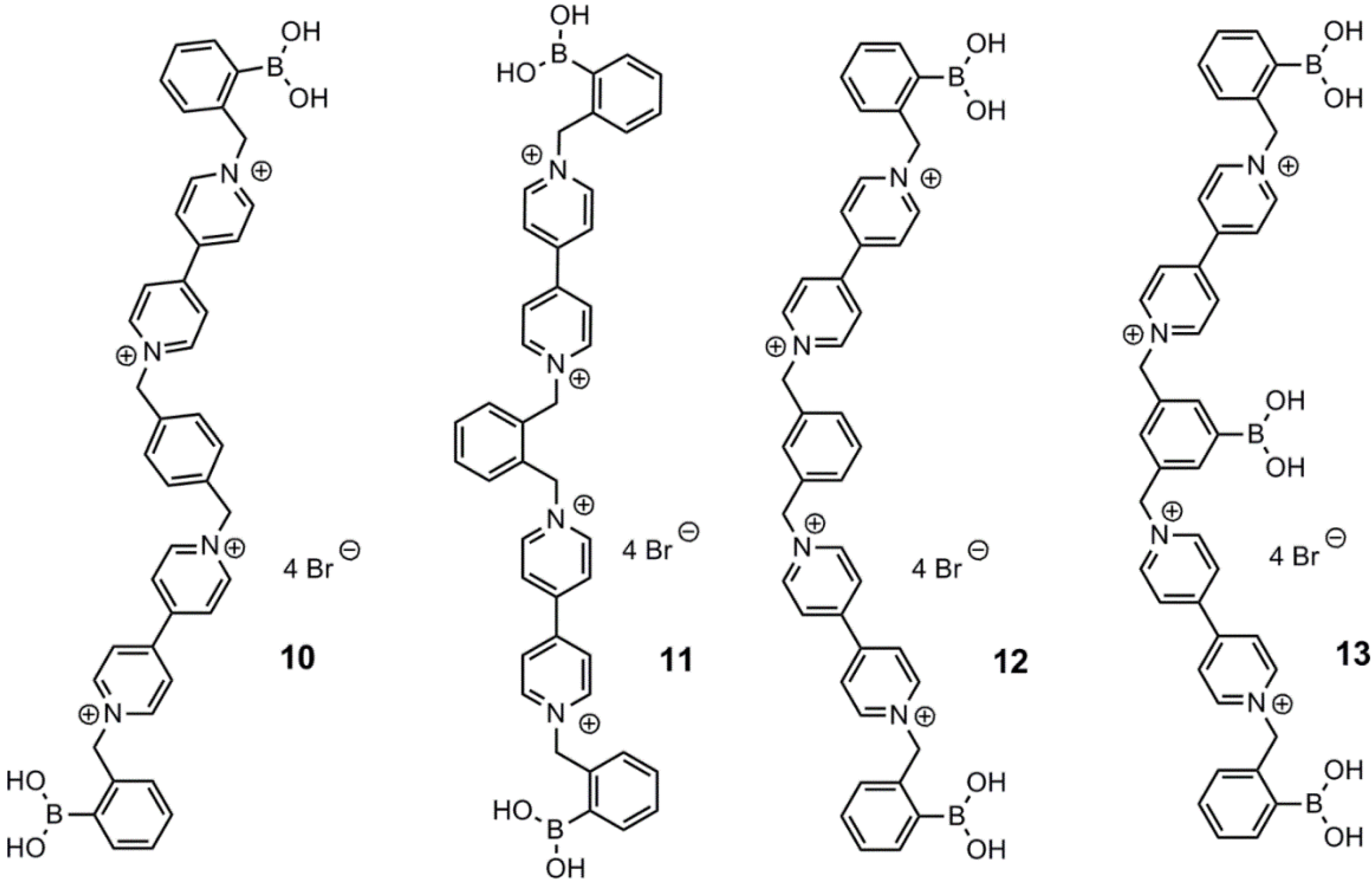
Figure 3. Viologens, 1013, developed and tested by Singaram and co-workers for the structural dependency on their quenching effect [48].
Biosensors 03 00400 g003
Recently, four quadrupole charged viologens, 1013, were developed in order to elucidate how the structural features influenced their respective quenching abilities [48]. The structures of 1013 are depicted in Figure 3. The quenching ability was found to increase in the order 11 < 12 < 13 < 10, which was ascribed to the decreasing tendency of pimer formation, caused by intramolecular stacking of the π-system in the viologens. In comparison to former published work [46], 1013 are superior quenchers. This study indicates that the presence of the bisviologen core plays a greater part in the quenching rather than the charges. The highest d-glucose modulation was achieved with 13. This was ascribed to a higher degree of negative charge in comparison to 1012. 10 and 12 were found to be far better quenchers than 11. This phenomenon may be attributed to their poorer ability to form pimers. The highest quenching constant was obtained with 10, followed by 13, with K = 6.8 × 10⁵ M−1 and K = 4.4 × 10⁵ M−1, respectively.
The viologens were further employed in glucose sensing hydrogels [49,50].
Feng and co-workers used the same two-component strategy with compounds 1517 [52], earlier reported by Singaram and co-workers [45]. The employed fluorophore, azo compound, 14, was found to emit fluorescence with λem = 585 nm, and maximum absorbance was recorded at λ = 530 nm. A significant d-glucose response from 0.4 mM to 150 mM was found. However, no binding constant was reported. Compounds 1517 were also used together with CdTe quantum dots, where the increase in emission intensity at 635 nm for the quantum dots was monitored upon the addition of d-glucose to a mixture of quencher and CdTe quantum dots [53]. The quenching abilities were found to decrease in the order 15 > 16 > 17. Thus, the highest increase by addition of d-glucose was found with 17. The structures of the three viologens, 1517, and the azo compound, 14, are shown in Figure 4.
Figure 4. Singaram’s viologen arylboronic acids, 1517, employed by Feng and co-workers, using azo compound 14 as the fluorescent dye [52].
Biosensors 03 00400 g004
Feng and co-workers also reported a dye for selective d-fructose recognition in the presence of other monosaccharides, such as d-mannose, d-galactose and d-arabinose, using pyranine as the fluorescent moiety [54,55].
Singaram’s and Feng’s systems are a great step in the development of glucose sensing devices. However, d-glucose selectivity is crucial and may be achieved by modulating the boronic acid distance, in order to bind in a cooperative manner. The wavelength should also be altered toward the near-infrared area in order to monitor through skin. The systems are, however, limited to in vitro monitoring, since dilution in vivo may be a problem for proper glucose readout.
Wu et al. developed a stilbene-based aryl diboronic acid, which is capable of recognizing d-glucose selectively in comparison to d-fructose [56]. The sensoric system relies on the formation of a dimeric supramolecular complex of two stilbene derivatives within the cavity of a γ-cyclodextrin, 18, which enables studies in aqueous media. The dimeric complex is stabilized due to bidentate binding of d-glucose by adjacent boronate units. The π-π-stacking upon dimer formation with the binding of d-glucose was observed to redshift the emission. However, the redshift was only modest, i.e., λem = 438 nm → λem = 448 nm (λex = 376 nm). d-fructose, which only stabilized the inclusion of a monomeric stilbene aryl diboronic acid within the γ-cyclodextrin cavity, did not give rise to a wavelength shift. The presence of the dimeric stilbene complex was proven by an induced exciton-coupled circular dichroism (CD)-signal. The d-glucose association constant was determined to be K = 1,048 M−1 at pH 10.5. The interaction of the γ-cyclodextrin complex with d-glucose and d-fructose is shown in Scheme 4.
Scheme 4. Interaction of d-glucose and d-fructose with the γ-cyclodextrin complex, 18, forming two different inclusion complexes [56].
Biosensors 03 00400 g015
d-glucose binding was found to be superior in a urine sample containing other saccharides, such as d-fructose, d-galactose and d-mannose, with a readout error less than 30%. This makes the sensor suitable for in vitro monitoring of d-glucose.
Liu and co-workers reported a tetraphenylene-based fluorescent sensor, 19, capable of forming oligomeric complexes with d-glucose [57]. Complex formation with d-glucose afforded exciplex formation, redshifting the emitted fluorescence significantly (λem = 440 nm → λem = 485 nm, λex = 365 nm). The oligomeric complexes were formed as a consequence of the capability of d-glucose to form esters with two arylboronic acid units. This property is not present in other monosaccharides, such as d-fructose, d-galactose and d-mannose. Due to low aqueous solubility of the tetraphenylene sensor, the measurements were performed at high pH (10.5) in an aqueous carbonate buffer. The formed complexes are illustrated in Scheme 5.
Scheme 5. The weakly emissive 2:1 d-glucose-tetraphenylene diboronic acid complex of 19 (top). A significant redshift at 45 nm occurred, due to the formation of the emissive cyclic oligomer with d-glucose and the tetraphenylene diboronic acid sensor (bottom right). The linear oligomer is also redshifting in a similar manner [57].
Biosensors 03 00400 g016
Studies with samples all containing d-glucose and one of the saccharides, d-fructose, d-galactose and d-mannose, were performed. The affinity was the following order: d-fructose > d-galactose ≈ d-mannose > d-glucose. However, only d-glucose gave rise to an emission increase at 485 nm. This means that the reported sensor is d-glucose selective both in the presence and in the absence of the other mentioned saccharides. The association constant was, however, not reported for binding of d-glucose.
Saito et al. reported a water soluble long-wavelength squarylium cyanine boronic acid dye, 20, for the detection of monosaccharides [58]. This dye was found to exhibit superior d-fructose binding selectivity in comparison to other tested monosaccharides. The studies were conducted in an aqueous carbonate buffer at pH 10, with λex = 630 nm and λem = 660 nm. An enhancement factor of 18 for the emission intensity was obtained in the presence of 20 mM d-fructose. The association constants for the binding of d-fructose and d-glucose was K = 628 M−1 and K = 8 M−1, respectively. This dye could be suitable for d-glucose monitoring through skin, due to the long wavelength of excitation and emission. The d-glucose selectivity must, however, be enhanced significantly and the d-glucose binding strength modulated towards physiological d-glucose levels. The reported dye, 20, is shown in Figure 5.
Wang and co-workers recently prepared water soluble, fluorescent and enantiomeric α-amidoboronic acids, 21 and 22, attached to a naphthalene unit [59]. The two sensors did not show any chiral discrimination in the binding strength towards the tested monosaccharides. The apparent association constants at pH 7.4 of 21 with d-fructose, d-glucose and d-sorbitol were 55 M−1, 1.6 M−1 and 100 M−1, whereas the association constants of 22 with the same sugars were 46 M−1, 1.5 M−1 and 102 M−1, respectively. λex = 280 nm, and λem = 334 nm. Sensor 21 and 22 are depicted in Figure 5. The change in fluorescence was attributed to the through-space interaction by the change in the hybridization of the boron core upon sugar binding. The performed studies also showed a significant decrease in quantum yield upon sugar binding for all saccharides in comparison to the obtained quantum yield without sugar. For sensor 21, the yields were for free arylboronic acid, d-sorbitol-complex, d-glucose-complex, d-fructose-complex: 15.3%, 8.5%, 6.0% and 0.5% respectively. These values were about the same as recorded for 22.
Figure 5. The near-infrared squarylium cyanine boronic acid dye, 20, reported by Saito and co-workers [58], and the two enantiomeric α-amidoboronic acids, 21 and 22, reported by Wang and co-workers [59].
Biosensors 03 00400 g005
Several other articles describing the newest research in the field of fluorescent arylboronic acids for glucose detection have been published covering topics, such as supramolecular boronic acid complexes with cyclodextrins [60], benzoxazole alanine boronic acid derivatives [61], boronic acid anthraquinone derivatives via the glucose oxidase (GOx) enzymatic reaction [62], and the general timescale question of boronic acid binding with sugars in aqueous solution at physiological pH [63].

3. Immobilized Systems

Immobilization of the sensing system offers many advantages for integration into a device, and this has been the topic for a number of research groups. Strano et al. [64,65] described a system consisting of aromatic boronic acids, which form stable complexes with single-walled carbon nanotubes (SWCNTs). The SWCNTs were solubilized in micelles in water by the surfactant, sodium cholate. SWCNTs are fluorescent in the near-infrared area, but upon binding of aromatic boronic acids, the fluorescence is quenched, and reappears upon the binding of carbohydrates, as shown in Figure 6. The altered emission spectra are depicted in Figure 7. The effect of fluorescence change was, however, dependent upon the structure of the boronic acid, and ensembles of 30 boronic acids in total were screened.
Qiu and co-workers [66] recently reported that combining an N,N′-bis(boronic acid) alkylated 4,4′-bipyridine with grapheme quantum dots gave a system that only became fluorescent after binding of glucose to the bis(boronic acid), which detached it from the quantum dot. They did not, however, investigate any selectivity for specific carbohydrates.
Manju and Sreenivasan [67] described a system-based gold nanoparticle coated with dextrane and functionalized with Rhodamine B and 3-aminobenzeneboronic acid, where binding of glucose to the boronic acid leads to emission from Rhodamine B. The system showed a good linear relationship between glucose concentration and emission, but the effect of interference from fructose was not investigated.
Figure 6. Binding of arylboronic acids to the surface of single-walled carbon nanotubes (SWCNTs) cause quenching of the near-infrared fluorescence, which reappears upon binding of glucose (Reprinted with permission from [64]. Copyright 2012 American Chemical Society).
Biosensors 03 00400 g006
Figure 7. Modulation of the fluorescence upon binding of glucose to SWCNT-coated with 4-chlorophenylboronic acid (Reprinted with permission from [64]. Copyright 2012 American Chemical Society).
Biosensors 03 00400 g007
An amphiphilic d-glucose selective aryl monoboronic acid sensor, 23, containing a hydrophobic pyrene unit has been reported by James and co-workers [68]. They found that high d-glucose selectivity is facilitated by the formation of a 1:2 glucose-sensor complex, which is capable of forming aggregates in an aqueous buffer. d-glucose is capable of forming a boronate diester twice, which is exploited in this system. The positively charged sensor containing a pyridinium moiety becomes zwitterionic at boronate formation, which is facilitated by d-glucose binding. The sensor displays ratiometric properties, since the wavelength of fluorescence is redshifted significantly upon aggregate formation. The redshift is explained by the exciplex formation by the stacking of pyrene units, λem = 377 nm → λem = 510 nm. Phenylboronic acid (PBA) was used as a mask for d-fructose, since d-fructose is preferentially bound by PBA. This PBA addition enhanced the d-glucose selectivity further. Sensor 23 is shown in Figure 8, along with an illustration of the different binding mode of d-glucose and d-fructose. Thus, this system outlines the stoichiometry dependence, since there is a clear discrimination between glucose and fructose. The fluorescence spectra are shown in Figure 9.
The optimal conditions were found to be 0.10 mM of the sensor to make a compromise between sensitivity and the ease of aggregation control. The association constant for binding of d-glucose was found to be K = 1,378 M−1, whereas K = 353 M−1 was found for d-fructose binding. Despite the good d-glucose selectivity, the discovered sensor system is, however, not suitable for in vivo monitoring of blood glucose. The limitations are due to the dilution removing the aggregate. The wavelength of fluorescence is also not in the near-infrared area, which is a required property in order to minimize light absorption by the skin.
Figure 8. Cartoon representation of the different types of aggregates formed upon binding of glucose and fructose [68]. PBA, phenylboronic acid.
Biosensors 03 00400 g008
Figure 9. Fluorescence spectra recorded in a carbonate buffer (pH = 10) containing 2% methanol. (a) Glucose and (b) fructose, both in a concentration range from 0–10 mM. Insert I (a) shows the ratio between the excimer and monomer as a function of concentration and carbohydrate (Reprinted with permission from [68]. Copyright 2013 American Chemical Society).
Biosensors 03 00400 g009
The potential of pyridinium containing arylboronic acids in polyol sensing was further explored through surface bound arylboronates and, e.g., modulation of exciplex formation in solution [69,70].
Incorporation of the complex between Alizarin Red S (ARS) and 2-formylbenzene boronic acid into aqueous micelles gives fluorescent micelles, where the fluorescence decreases upon adding glucose, due to the preferred binding of glucose to the boronic acid. This has been shown by Ngeontae and co-workers [71], who used simple surfactants, such as cetyltrimethylammonium bromide (CTAB), for the formation of the micelles. They observed an increased sensitivity upon using the micellar formulation, but the specificity of the system was fructose > xylose > glucose > maltose > lactose. Functional vesicles having boronic acids as part of the head groups have been investigated by Karpichev and co-workers [72], where vesicles were formed from an N-alkyl pyridium 3-boronic acid in water and binding of diols were measured using the Alizarin Red Sodium (ARS)-assay. The system, however, showed higher affinity for fructose than for glucose.
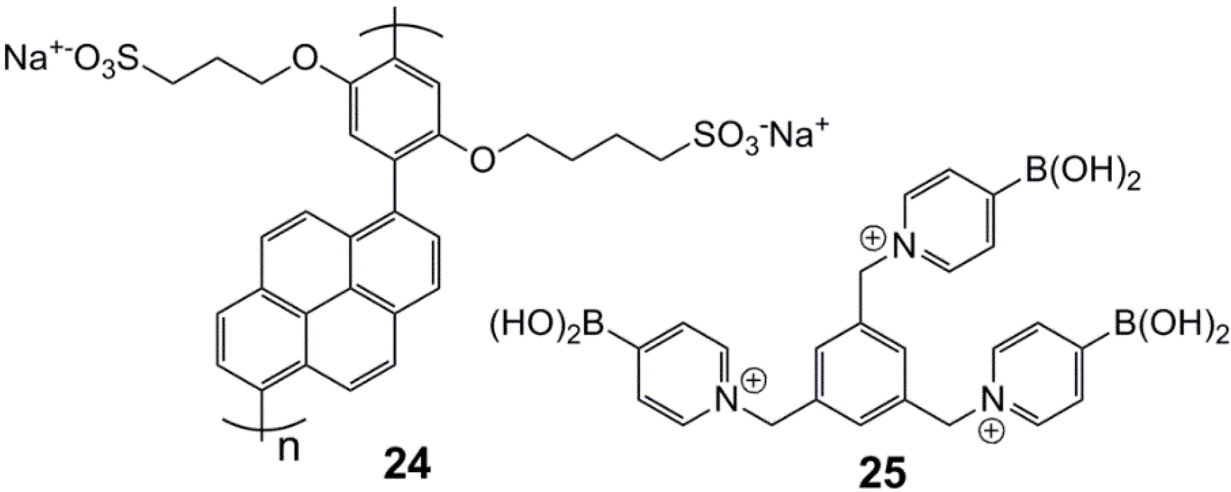
Polymer-based systems are interesting candidates for in vivo (diagnostics) and in vitro measurements of glucose, and a recent example of a system for diagnostics comes from Feng et al. [73], where a tridentate pyridinium boronic acid forms a non-fluorescent complex with a pyrene-containing polymeric polyelectrolyte (Figure 10 and Figure 11). The system shows some discrimination, but still, the main problem is achieving selectivity for glucose in the presence of fructose.
Figure 10. Poly(p-phenylene-pyrene)-sulfonate, 24, and tris(pyridiniumboronic acid), 25 [73].
Biosensors 03 00400 g010
Figure 11. Fluorescence intensity at 467 nm for the system after the addition of 50.0 mM of monosaccharides at pH 7.4 buffer solution. Glu = glucose, Fru = fructose, Man = mannose, Rib = ribose, Ara = arabinose, Xyl = xylose (reproduced from [73]).
Biosensors 03 00400 g011
Another polymer-monomer-based system is the combination of an acrylamide copolymer containing benzene boronic acid units in combination with Bordeaux R, which is a red azo dye. This system, reported by Zhou and Li [74], formed a polymeric gel, where the addition of glucose leads to quenching of the fluorescence of the dye through the swelling of the gel.
Takeuchi and co-workers developed polyacrylamide-based boronic acid hydrogel fibers that were tested in vitro, demonstrating that it is possible to monitor the glucose concentration by transdermal measurements in a mouse model [75]. The fibers were implanted under the skin on the ears of the mice, showed only little inflammation and had a device lifetime of 140 days.

4. Conclusion

In conclusion, fluorescent sensors based on arylboronic acids are very promising candidates for the elusive goal of performing noninvasive monitoring of d-glucose or other biologically important carbohydrates. This is due to the high sensitivity of fluorescence spectroscopy, which enables detection of very small changes in the analytical environment.
The aryl monoboronic acid sensors generally favor binding of d-fructose over d-glucose, which is attributed to the different binding mode, i.e., tridentate d-fructose binding versus bidentate d-glucose binding. BODIPY sensor 8 showed binding of d-glucose at the physiological level, but d-fructose was bound significantly more strongly [42]. Superior d-glucose selectivity is achieved by complexation with aryl diboronic acid sensors. Aryl diboronic acid dyes are usually saturated at physiological d-glucose levels, i.e., [d-glc] ≈ 5 mM, and these sensors are usually not water soluble, due to the large hydrophobic scaffold, which is required in order to place the boronic acid units in an appropriate cooperative distance. Aryl diboronic acid equivalents have been formed by aggregate formation with d-glucose [68] or by inclusion of lipophilic aryl monoboronic acids into cyclodextrin cavities [56]. Fluorescent properties, such as wavelengths of excitation and emission, and potential fluorescence lifetime experiments are to be optimized in future systems. The emphasis may also be on systems with fewer tendencies to photobleach or oxidize. Proper glucose-readout may be monitored through skin. Thus, a suitable glucose sensor may be tattooed into the subcutaneous layer of human skin in order to perform optimal regulation of blood glucose in diabetics, providing safety and comfort. The dye must be near-infrared to avoid skin absorption of light; otherwise, an optical fiber is needed, which is painful, due to the surgical procedure of implantation.
Ultimately, blood glucose can be determined noninvasively in the case of a proper tear glucose/blood glucose correlation.

Acknowledgments

Financial support from Forsknings og Innovationsstyrrelsen, Teknologi og Produktion (FTP) is gratefully acknowledged, along with a fruitful collaboration with Novo Nordisk A/S and the University of Copenhagen, Department of Chemistry.

Conflicts of Interest

The authors declare no conflict of interest.

References

  1. Mastrototaro, J.J. The minimed continuous glucose monitoring system. Diabetes Technol. Ther. 2000, 2, 13–18. [Google Scholar] [CrossRef]
  2. Buckingham, B.A.; Beck, R.W.; Tamborlane, W.V.; Boland, E.A.; Chase, H.P.; Tansey, M.J.; Mauras, N.; Weinzimmer, S.A.; Ruedy, K.J.; Booth, A.D.; Kollman, C.; Diabetes Research in Children Network (DirecNet) Study Group. The accuracy of the CGMS in children with type 1 diabetes: Results of the diabetes research in children network (DirecNet) accuracy study. Diabetes Technol. Ther. 2003, 5, 781–789. [Google Scholar] [CrossRef]
  3. Facchinetti, A.; Sparacino, G.; Guerra, S.; Lujif, Y.M.; DeVries, J.H.; Mader, J.K.; Ellmerer, M.; Benesch, C.; Heinemann, L.; Bruttomesso, D.; Avogaro, A.; Cobelli, C. Real-time improvement of continuous glucose monitoring accuracy. Diabetes Care 2013, 36, 793–800. [Google Scholar] [CrossRef]
  4. Smith, J.L. The Pursuit of Noninvasive Glucose: Hunting the Deceitful Turkey. Available online: http://www.mendosa.com/noninvasive.glucose.pdf (accessed on 24 October 2013).
  5. Yoo, E.-H.; Lee, S.-Y. Glucose biosensors: An overview of use in clinical practice. Sensors 2010, 10, 4558–4576. [Google Scholar] [CrossRef]
  6. Mansouri, S.; Schultz, J.S. A miniature optical glucose sensor based on affinity binding. Biotechnology 1984, 2, 885–890. [Google Scholar] [CrossRef]
  7. De Marcos, S.; Galindo, J.; Sierra, J.F.; Galban, J.; Castillo, J.R. An optical glucose biosensor based on derived glucose oxidase immobilised onto a sol-gel matrix. Sens. Actuator. B 1999, 57, 227–232. [Google Scholar]
  8. D’Auria, S.; Di Cesare, N.; Gryczynski, Z.; Rossi, M.; Lakowicz, J.R. A thermophilic apoglucose dehydrogenase as a nonconsuming glucose sensor. Biochem. Biophys. Res. Commun. 2000, 274, 727–731. [Google Scholar] [CrossRef]
  9. Maity, H.; Maity, N.C.; Jarori, G.K. Time-resolved fluorescence of tryptophans in yeast hexokinase-PI: Effect of subunit dimerization and ligand binding. J. Photochem. Photobiol. B 2000, 55, 20–26. [Google Scholar] [CrossRef]
  10. Gilardi, G.; Mei, G.; Rosato, N.; Agro, A.F.; Cass, A.E.G. Spectroscopic properties of an engineered maltose binding protein. Protein Eng. 1997, 10, 479–486. [Google Scholar] [CrossRef]
  11. Hall, D.G. Boronic Acids: Preparation, Application in Organic Synthesis and Medicine; Wiley-VCH: Berlin, Germany, 2005. [Google Scholar]
  12. Wang, J. Electrochemical glucose biosensors. Chem. Rev. 2008, 108, 814–825. [Google Scholar] [CrossRef]
  13. Reach, G.; Wilson, G.S. Can continuous glucose monitoring be used for the treatment of diabetes? Anal. Chem. 1992, 64, 381A–386A. [Google Scholar]
  14. Wickramasinghe, Y.; Yang, Y.; Spencer, S.A. Current problems and potential techniques in in vivo glucose monitoring. J. Fluoresc. 2004, 14, 513–520. [Google Scholar] [CrossRef]
  15. Wilson, G.S.; Gifford, R. Biosensors for real-time measurements. Biosens. Bioelectron. 2005, 20, 2388–2403. [Google Scholar] [CrossRef]
  16. Vadgama, P.; Desai, M.; Crump, P. Electrochemical transducers for in vivo monitoring. Electroanalysis 1991, 3, 597–606. [Google Scholar] [CrossRef]
  17. LeBlanc, J.M.; Haas, C.E.; Vicente, G.; Colon, L.A. Evaluation of lacrimal fluid as an alternative for monitoring glucose in critically ill patients. Intensive Care Med. 2005, 31, 1442–1445. [Google Scholar] [CrossRef]
  18. Baca, J.T.; Taormina, C.R.; Feingold, E.; Finegold, D.N.; Grabowski, J.J.; Asher, S.A. Mass spectral determination of fasting tear glucose concentrations in nondiabetic volunteers. Clin. Chem. 2007, 53, 1370–1372. [Google Scholar] [CrossRef]
  19. Baca, J.T.; Finegold, D.N.; Asher, S.A. Tear glucose analysis for the noninvasive detection and monitoring of diabetes mellitus. Ocul. Surf. 2007, 5, 280–293. [Google Scholar] [CrossRef]
  20. Yang, W.; Gao, X.; Wang, B. Boronic acids as potential pharmaceutical agents. Med. Res. Rev. 2003, 23, 346–368. [Google Scholar]
  21. Springsteen, G.; Wang, B. A detailed examination of boronic acid-diol complexation. Tetrahedron 2002, 58, 5291–5300. [Google Scholar] [CrossRef]
  22. Cao, H.; Heagy, M.D. Fluorescent chemosensors for carbohydrates: A decade’s worth of bright spies for saccharides in review. J. Fluoresc. 2004, 14, 569–584. [Google Scholar] [CrossRef]
  23. James, T.D.; Phillips, M.D.; Shinkai, S. Boronic Acids in Saccharide Recognition; Royal Society of Chemistry: London, UK, 2006. [Google Scholar]
  24. Pickup, J.C.; Hussain, F.; Evans, N.D.; Rolinsky, O.J.; Birch, D.S.J. Fluorescence based glucose sensors. Biosens. Bioelectron. 2005, 20, 2555–2565. [Google Scholar] [CrossRef]
  25. Geddes, C.D. Reviews in Fluorescence; Springer-Verlag: New York, NY, USA, 2008. [Google Scholar]
  26. De Silva, A.P.; Gunaratne, H.Q.N.; Gunnlaugsson, T.; McCoy, C.P.; Maxwell, P.R.S.; Rademacher, J.T.; Rice, T.E. Photoionic devices with receptor-functionalized fluorophores. Pure Appl. Chem. 1996, 68, 1443–1448. [Google Scholar] [CrossRef]
  27. De Silva, A.P.; Gunaratne, H.Q.N.; McCoy, C.P. Molecular photoionic and logic gates with bright fluorescence and off-on digital action. J. Am. Chem. Soc. 1997, 119, 7891–7892. [Google Scholar]
  28. De Silva, A.P.; Gunaratne, H.Q.N.; Gunnlaugsson, T.; Huxley, A.J.M.; McCoy, C.P.; Rademacher, J.T.; Rice, T.E. Signaling recognition events with fluorescent sensors and switches. Chem. Rev. 1997, 97, 1515–1566. [Google Scholar]
  29. James, T.D.; Sandanayake, K.; Shinkai, S. Novel photoinduced electron-transfer sensor for saccharides based on the interaction of boronic acid and amine. J. Chem. Soc. Chem. Commun. 1994, 47, 477–478. [Google Scholar]
  30. James, T.D.; Sandanayake, K.R.A.S.; Iguchi, R.; Shinkai, S. Novel saccharide photoinduced electron transfer sensors based on the interaction between boronic acid and amine. J. Am. Chem. Soc. 1995, 117, 8982–8987. [Google Scholar] [CrossRef]
  31. Arimori, S.; Bell, M.L.; Oh, C.S.; Frimat, K.A.; James, T.D. Modular fluorescence sensors for saccharides. Chem. Commun. 2001, 183, 1836–1837. [Google Scholar]
  32. Arimori, S.; Bell, M.L.; Oh, C.S.; Frimat, K.A.; James, T.D. Modular fluorescence sensors for saccharides. J. Chem. Soc. Perkin Trans. 1 2002, 803–808. [Google Scholar] [CrossRef]
  33. Arimori, S.; Bell, M.L.; Oh, C.S.; James, T.D. A modular fluorescence intramolecular energy transfer saccharide sensor. Org. Lett. 2002, 4, 4249–4251. [Google Scholar] [CrossRef]
  34. Zhao, J.; Davidson, M.G.; Mahon, M.F.; Kociak-Köhn, G.; James, T.D. An enantioselective fluorescent sensor for sugar acids. J. Am. Chem. Soc. 2004, 126, 16179–16186. [Google Scholar] [CrossRef]
  35. Zhao, J.; Fyles, T.M.; James, T.D. Chiral binol-bisboronic acid as fluorescence sensor for sugar acids. Angew. Chem. Int. Ed. Engl. 2004, 43, 3461–3464. [Google Scholar] [CrossRef]
  36. Liang, X.; James, T.D.; Zhao, J. 6,6′-Bis-substituted binol boronic acids as enantioselective and chemoselective fluorescent chemosensors for d-sorbitol. Tetrahedron 2008, 64, 1309–1315. [Google Scholar] [CrossRef]
  37. Xing, Z.; Wang, H.-C.; Cheng, Y.; Zhu, C.; James, T.D.; Zhao, J. Selective saccharide recognition using modular diboronic acid fluorescent sensors. Eur. J. Org. Chem. 2012, 1223–1229. [Google Scholar] [CrossRef]
  38. Larkin, J.D.; Fossey, J.S.; James, T.D.; Brooks, B.R.; Bock, C.W. A computational investigation of the nitrogen-boron interaction in o-(N,N-dialkylaminomethyl)-arylboronate systems. J. Phys. Chem. A 2010, 114, 12531–12539. [Google Scholar] [CrossRef]
  39. Collins, B.E.; Sorey, S.; Hargrove, A.E.; Shabbir, S.H.; Lynch, V.M.; Anslyn, E.V. Probing intramolecular B–N interactions in ortho-aminomethyl arylboronic acids. J. Org. Chem. 2009, 74, 4055–4060. [Google Scholar] [CrossRef]
  40. DiCesare, N.; Lakowicz, J.R. Fluorescent probe for monosaccharides based on a functionalized boron-dipyrromethene with a boronic acid group. Tetrahedron Lett. 2001, 42, 9105–9108. [Google Scholar] [CrossRef]
  41. Hansen, J.S.; Petersen, J.F.; Hoeg-Jensen, T.; Christensen, J.B. Buffer and sugar concentration dependent fluorescence response of a BODIPY-based aryl monoboronic acid sensor. Tetrahedron Lett. 2012, 53, 5852–5855. [Google Scholar] [CrossRef]
  42. Hansen, J.S.; Ficker, M.; Petersen, J.F.; Christensen, J.B.; Hoeg-Jensen, T. Ortho-substituted fluorescent aryl monoboronic acid displays physiological binding of d-glucose. Tetrahedron Lett. 2013, 54, 1849–1852. [Google Scholar] [CrossRef]
  43. Van Duin, M.; Peters, J.A.; Kieboom, A.P.G.; van Bekkum, H. Studies on borate esters 1: The pH dependence of the stability of esters of boric acid and in aqueous media as studied by 11B-NMR. Tetrahedron 1984, 40, 2901–2911. [Google Scholar] [CrossRef]
  44. Hansen, J.S.; Christensen, J.B.; Solling, T.I.; Jakobsen, P.; Hoeg-Jensen, T. Ortho-substituted aryl monoboronic acids have improved selectivity for d-glucose relative to d-fructose and l-lactate. Tetrahedron 2011, 67, 1334–1340. [Google Scholar]
  45. Camara, J.N.; Suri, J.T.; Cappucio, F.E.; Wessling, R.A.; Singaram, B. Boronic acid substituted viologen based optical sugar sensors: Modulated quenching with viologen as a method for monosaccharide detection. Tetrahedron Lett. 2002, 43, 1139–1141. [Google Scholar] [CrossRef]
  46. Cordes, D.B.; Gamsey, S.; Sharrett, Z.; Miller, A.; Thoniyot, P.; Wessling, R.A.; Singaram, B. The interaction of boronic acid-substituted viologens with pyranine: The effects of quencher charge on fluorescence quenching and glucose response. Langmuir 2005, 21, 6540–6547. [Google Scholar] [CrossRef]
  47. Cordes, D.B.; Miller, A.; Gamsey, S.; Singaram, B. Simultaneous use of multiple fluorescent reporter dyes for glucose sensing in aqueous solution. Anal. Bioanal. Chem. 2007, 387, 2767–2773. [Google Scholar] [CrossRef]
  48. Sharrett, Z.; Gamsey, S.; Levine, P.; Cunningham-Bryant, D.; Vilozny, B.; Schiller, A.; Wessling, R.A.; Singaram, B. Boronic acid-appended bis-viologens as a new family of viologens for glucose sensing. Tetrahedron Lett. 2008, 49, 300–304. [Google Scholar] [CrossRef]
  49. Thoniyot, P.; Cappucio, F.E.; Gamsey, S.; Cordes, D.B.; Wessling, R.A.; Singaram, B. Continuous glucose detection with fluorescent thin-film hydrogels. 2. Fiber optic sensor fabrication and in vitro testing. Diabetes Technol. Ther. 2006, 8, 279–287. [Google Scholar] [CrossRef]
  50. Gamsey, S.; Suri, J.T.; Wessling, R.A.; Singaram, B. Continuous glucose sensing using boronic acid-substituted viologens in fluorescent hydrogels: Linker effects and extension to fiber optics. Langmuir 2006, 22, 9067–9074. [Google Scholar] [CrossRef]
  51. Sharrett, Z.; Gamsey, S.; Fat, J.; Cunningham-Bryant, D.; Wessling, R.A.; Singaram, B. The effect of boronic acid acidity on performance of viologen-based boronic acids in a two-component optical glucose-sensing system. Tetrahedron Lett. 2007, 48, 5125–5129. [Google Scholar] [CrossRef]
  52. Wang, Z.; Lei, H.; Zhou, C.; Liang, F.; Feng, L. Optical probe for d-glucose based on cationic polymer quencher/receptor and anionic dye in aqueous solution. Sens. Actuator. B 2012, 163, 202–206. [Google Scholar] [CrossRef]
  53. Feng, L.; Wang, Y.; Liang, F.; Wang, X.; Zhang, L. Detection of glucose based on reversible “on-off” fluorescence systems in aqueous solution. Sens. Actuator. B 2011, 156, 499–503. [Google Scholar] [CrossRef]
  54. Feng, L.; Wang, Y.; Liang, F.; Ming, X.; Wang, X. Highly selective recognition of monosaccharides based on two-component system in aqueous solution. Tetrahedron 2011, 67, 3175–3180. [Google Scholar]
  55. Feng, L.; Liang, F.; Wang, Y.; Wang, X. A facile probe for dfructose with fluorescence “on-off-on” switch ensemble. Sens. Actuator. B 2012, 173, 575–579. [Google Scholar]
  56. Wu, X.; Lin, L.-R.; Huang, Y.-J.; Li, Z.; Jiang, Y.-B. A 2:2 stilbeneboronic acid-γ-cyclodextrin fluorescent ensemble highly selective for glucose in aqueous solutions. Chem. Commun. 2012, 48, 4362–4364. [Google Scholar] [CrossRef]
  57. Liu, Y.; Deng, C.; Tang, L.; Qin, A.; Hu, R.; Sun, J.Z.; Tang, B.Z. Specific detection of d-glucose by a tetraphenylene-based fluorescent sensor. J. Am. Chem. Soc. 2011, 133, 660–663. [Google Scholar] [CrossRef]
  58. Saito, S.; Massie, T.L.; Maeda, T.; Nakazumi, H.; Colyer, C.L. A long-wavelength fluorescent squarylium cyanine dye possessing boronic acid for sensing monosaccharides and glycoproteins with high enhancement in aqueous solution. Sensors 2012, 12, 5420–5431. [Google Scholar] [CrossRef]
  59. Jin, S.; Zhu, C.; Li, M.; Wang, B. Identification of the first fluorescent a-amidoboronic acid that change fluorescent properties upon sugar binding. Bioorg. Med. Chem. Lett. 2009, 19, 1596–1599. [Google Scholar] [CrossRef]
  60. Kumai, M.; Kozuka, S.; Samizo, M.; Hashimoto, T.; Suzuki, I.; Hayashita, T. Glucose recognition by a supramolecular complex of boronic acid fluorophore with boronic acid-modified cyclodextrin in water. Anal. Sci. 2012, 28, 121–126. [Google Scholar] [CrossRef]
  61. Guzow, K.; Jazdzewska, D.; Wiczk, W. 3-[2-(Boronophenyl)benzoxazol-5-yl]alanine derivatives as fluorescent monosaccharide sensors. Tetrahedron 2012, 68, 9240–9248. [Google Scholar] [CrossRef]
  62. Wannajuk, K.; Jamkatoke, M.; Tuntulani, T.; Tomapatanaget, B. Highly specific-glucose fluorescence sensing based on boronic anthraquinone derivatives via the GOx enzymatic reaction. Tetrahedron 2012, 68, 8899–8904. [Google Scholar] [CrossRef]
  63. Ni, N.; Laughlin, S.; Wang, Y.; Feng, Y.; Zheng, Y.; Wang, B. Probing the general time scale question of boronic acid binding with sugars in aqueous solution at physiological pH. Bioorg. Med. Chem. 2012, 20, 2957–2961. [Google Scholar] [CrossRef]
  64. Yum, K.; Ahn, J.-H.; McNicholas, T.P.; Barone, P.W.; Mu, B.; Kim, J.-H.; Jain, R.M.; Strano, M.S. Boronic acid library for selective, reversible near-infrared fluorescence quenching of surfactant suspended single-walled carbon nanotubes in response to glucose. ACS Nano 2012, 6, 819–830. [Google Scholar]
  65. Mu, B.; McNicholas, T.P.; Zhang, J.; Hilmer, A.J.; Jin, Z.; Reuel, N.F.; Kim, J.-H.; Yum, K.; Strano, M.S. A structure-function relationship for the optical modulation of phenyl boronic acid-grafted, polyethylene glycol-wrapped single-walled carbon nanotubes. J. Am. Chem. Soc. 2012, 134, 17620–17627. [Google Scholar] [CrossRef]
  66. Li, Y.-H.; Zhang, L.; Huang, J.; Liang, R.-P.; Qiu, J.-D. Fluorescent graphene quantum dots with a boronic acid appened bipyridinium salt to sense monosaccharides in aqueous solution. Chem. Commun. 2013, 5180–5182. [Google Scholar] [CrossRef]
  67. Manju, S.; Sreenivasan, K. Detection of glucose in synthetic tear fluid using dually functionalized gold nanoparticles. Talanta 2011, 85, 2643–2649. [Google Scholar] [CrossRef]
  68. Huang, Y.-J.; Ouyang, W.-J.; Wu, X.; Li, Z.; Fossey, J.S.; James, T.D.; Jiang, Y.-B. Glucose sensing via aggregation and the use of “knock-out” binding to improve selectivity. J. Am. Chem. Soc. 2013, 135, 1700–1703. [Google Scholar] [CrossRef]
  69. Huang, Y.-J.; Jiang, Y.-B.; Fossey, J.S.; James, T.D.; Marken, F. Assembly of N-hexadecyl-pyridinium-4-boronic acid hexafluorophosphate monolayer films with catechol sensing selectivity. J. Mater. Chem. 2010, 20, 8305–8310. [Google Scholar] [CrossRef]
  70. Huang, Y.-J.; Jiang, Y.-B.; Bull, S.D.; Fossey, J.S.; James, T.D. Diols and anions can control the formation of an exciplex between a pyridinium boronic acid with an aryl group connected via a propylene linker. Chem. Commun. 2010, 46, 8180–8182. [Google Scholar] [CrossRef]
  71. Ngamdee, K.; Noipa, T.; Martwiset, S.; Tuntulani, T.; Ngeontae, W. Enhancement of sensitivity of glucose sensors from alizarin-boronic acid adducts in aqueous micelles. Sens. Actuator. B 2011, 160, 129–138. [Google Scholar] [CrossRef]
  72. Savsunenko, O.; Matondo, H.; Franceschi-Messant, S.; Perez, E.; Popov, A.F.; Rico-Lattes, I.; Lattes, A.; Karpichev, Y. Functionalized vesicles based on amphiphilic boronic acids: A system for recognizing biologically important polyols. Langmuir 2013, 29, 3207–3213. [Google Scholar] [CrossRef]
  73. Feng, L.; Yin, N.; Wang, X.; Wang, Z. Fluorescence probe for monosaccharide based on anionic polyelectrolyte and cationic pyridine quaternary ammonium salt. Sens. Actuator. B 2013, 181, 730–734. [Google Scholar] [CrossRef]
  74. Li, Y.; Zhou, S. A simple method to fabricate fluorescent glucose sensor based on dye-complexed microgels. Sens. Actuator. B 2013, 177, 792–799. [Google Scholar] [CrossRef]
  75. Heo, Y.J.; Shibata, H.; Okitsu, T.; Kawanishi, T.; Takeuchi, S. Long-term in vivo glucose monitoring using fluorescent hydrogel fibers. PNAS 2011, 108, 13399–13403. [Google Scholar] [CrossRef]