Abstract
The eradication of endodontic pathogens continues to be the focus of the search for new root canal system (RCS) disinfection strategies. This scoping review provides a comprehensive synthesis of antimicrobial photodynamic therapy (aPDT) using nanoparticles (NPs) as an alternative to optimize RCS disinfection. A systematic search up to March 2021 was carried out using the MEDLINE, EMBASE, Scopus, Lilacs, Central Cochrane Library, and BBO databases. We included studies focused on evaluating the activation of NPs by aPDT in inoculated root canals of human or animal teeth or bacterial cultures in the laboratory. The selection process and data extraction were carried out by two researchers independently. A qualitative synthesis of the results was performed. A total of seventeen studies were included, of which twelve showed a substantial antibacterial efficacy, two assessed the substantivity of the disinfection effect, and three showed low cytotoxicity. No adverse effects were reported. The use of functionalized NPs with photosensitizer molecules in aPDT has been shown to be effective in reducing the bacteria count, making it a promising alternative in endodontic disinfection. Further studies are needed to assess the development of this therapy in in vivo conditions, with detailed information about the laser parameters used to allow the development of safe and standardized protocols.
1. Introduction
The success of endodontic disinfection depends on the eradication of microbes and their by-products from the root canal system (RCS) [1]. Bacterial biofilm is considered to be the main etiological cause of primary and secondary/persistent infection [2]. Existing endodontic treatment strategies have proved insufficient to reduce endodontic pathogenic microorganisms below detection limits [3,4]. The unpredictable nature of the anatomy of the RCS, which consists of accessory canals, isthmuses, lateral canals, apical deltas, and recesses from C-shaped or oval/flattened canals, hinders the adequate action of antiseptic solutions [5,6]. New advanced disinfection approaches are therefore required for the effective eradication of microbial biofilms in endodontic infections. Antimicrobial photodynamic therapy (aPDT) is a promising alternative therapy proposed to combat a broad spectrum of endodontic biofilm-mediated infectious diseases [7,8,9]. It is of particular interest because of its simple approach, a visible light source of a suitable wavelength, generally a low-intensity diode or light-emitting diode (LED), is absorbed by a non-toxic photosensitizer (PS) [10]. The light sources cited for aPDT in the literature are laser, light-emitting diodes (LED) and halogen lamps [11,12]. Of these the most commonly used is the diode laser, due to its portability, ease of handling and low cost compared to other lasers [13]. Furthermore, the range of wavelengths emitted by diode laser coincides with the electronic absorption spectrum of most of the PS available [14]. Phenothiazines (synthetic non-porphyrin compounds), such as methylene blue (MB) and toluidine blue O (TBO), are the most widely studied PS in endodontics [12]. The main disadvantage of conventional PS is their poor solubility in water [15]; an uncontrollable drug-release profile, poor target selectivity, and low extinction coefficient also hamper their clinical action [16]. Therefore, a promising approach to improve the performance of PS within RCS is to encapsulate them in nanostructured materials [17]. In recent works, indocyanine green (ICG) [18,19], rose bengal (RB) [20,21] and erythrosine (ER) [22] have been cited as PS with optimal properties for nanoencapsulation and incorporation into aPDT in endodontic treatment. Nanoparticles (NPs) are defined as submicroscopic particles between 1 nm and 100 nm in size [13,23]. It has been seen that NPs can potentially improve the therapeutic efficacy of pharmaceuticals by promoting better bioavailability, serum stability, and pharmacokinetics of the drug [24]. According to the literature, nano-formulations provide better penetration and allow slow, controlled release of active ingredients at target sites [25]. Their most important feature is their large external surface area, as well as their high surface/volume ratio, factors that determine their physicochemical properties [26]. Different combinations irrigant/sealer can cause subsequent changes in the interfacial shear strength (ISS) of dentin, which may subsequently affect the post-restoration process in the root canal [27]. However, it has been shown that irrigation with NPs does not negatively interfere with the hardness and elastic modulus of dentin [28] or the bond strength and permeability of the interface of bonded fiberglass posts with resin [29]. The use of PS-loaded NPs is a modern strategy in endodontics; it is considered to increase the antimicrobial efficacy of aPDT [30,31]. In recent years, several PSs associated with polymeric [20,21,32,33,34,35,36,37], metallic [18,19,38], and carbon-based NPs [17,39] have been tested in vitro, ex vivo, or in vivo in combination with aPDT. Published studies on the antibacterial action of PS-based NPs complexes and aPDT are remarkably heterogeneous and show great variability in their respective methodologies. This is particularly true due to differences in the PS agents and types of NP used, light activation parameters, bacterial organization (planktonic cells or biofilm) and different bacterial species; this heterogeneity makes it difficult to determine the potential benefit of this new therapeutic combination. Thus, the aim of this scoping review was to address the current status of new PS-loaded NPs complexes used for antimicrobial purposes in endodontics when activated by aPDT.
2. Results
2.1. Selection of Sources of Evidence
A total of 1384 articles were identified from searches of electronic databases, from which 563 duplicates were eliminated. Of the 821 remaining, 805 were excluded based on the title and abstract, and 16 full text articles were retrieved and assessed for eligibility. Of these, two were excluded because they evaluate aPDT without NPs, or evaluate NPs without PS. Three additional articles were included after a manual search of the reference, giving a final total of 17 articles considered eligible for this scoping review. A flow diagram of the selection process is shown in Figure 1.
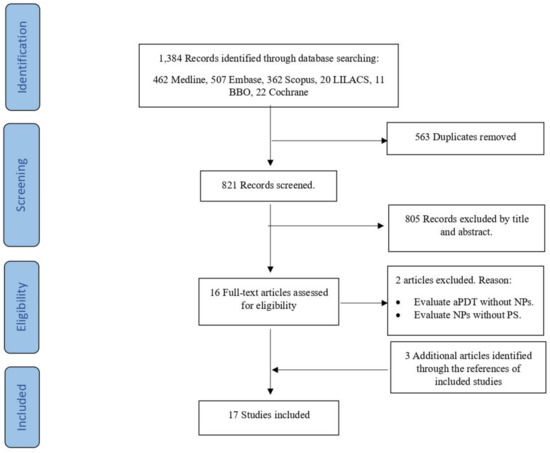
Figure 1.
Flow diagram of selection of sources of evidence. aPDT, antimicrobial photodynamic therapy; NPs, nanoparticles; PS, photosensitizer.
2.1.1. Characteristics of Sources of Evidence
The main characteristics of the articles included are summarized in Table 1. They were published between 2010 and 2020. Eight studies were from Canada, four from Iran, two from the USA, and one each from Taiwan, India, and Turkey. According to the study design, ten articles were in vitro, five ex vivo, one in vivo, and one incorporated both ex vivo and in vivo designs. The distribution of NPs and PS used were as follows: three articles studied silver NPs (AgNPs), two of which used toluidine blue (TB) as PS, and one used ICG. Chitosan (CS) NPs were analyzed by eight articles, five with rose bengal (RB), three with methylene blue (MB) and RB, and one with erythrosine (ER). Nano-graphene oxide (NGO) NPs were evaluated in two articles: one used ICG, and the other curcumin. The following compounds were each treated in one study: nano-metal organic frameworks (MOFs)-ICG, silica NPs (SiO2–NH2)-RB, and poly (lactic co-glycolic acid) (PLGA) NPs-MB. Turning to the lasers and their wavelengths, seven articles utilized a diode laser at wavelengths of 50, 620, 665, and 810 nm, three articles a non-coherent light at 540 and 660 nm, two a broad-spectrum lamp at 540 and 660 nm; one a white light source at 540 nm and 660 nm; one a green light source at 540 nm; and one article used a high-power LED array at 540 nm. Two articles did not mention the laser type, although they did mention the wavelengths used, 540 and 630 nm. The duration of light application in a single activation was from 30 s to 60 min, with an average of 9.0 min.

Table 1.
Summary of studies included.
2.1.2. Critical Appraisal within Sources of Evidence
In the overall assessment, 12 articles (70.6%) were considered reliable without restrictions, and five (29.4%) were classified as reliable with restrictions. Table S1 shows the methodological quality for each article. In the “test substance identification” criterion group, two articles (11.8%) did not report the purity of the substance or information on its physicochemical properties; not reporting this information is prejudicial to the transparency of the experiments performed and may affect the quality of their results. In the second criterion group “test system characterization”, all articles (100%) were rated with the highest score. The third criterion group evaluates the “study design description”; five articles (29.4%) did not report the minimum information required in the “description of study design”, and were thus classified as reliable with restrictions. Most of the studies did not describe laser application. Two articles (11.8%) did not mention the type of laser used, although the wavelength used was mentioned. In five articles (29.4%) the time of light application was not mentioned. Thirteen articles (76.5%) did not mention the number of light activations. Twelve articles (70.6%) did not describe how the light was applied. The critical evaluation tool considered only the frequency and duration of exposure as minimum information required. However, all the above-mentioned could lead to a misinterpretation of the study results. It is important to emphasize that the basis of aPDT is light exposure; therefore, a detailed description of this step is imperative. In the fourth criterion group “study results documentation”, all the articles were rated with the highest score. The fifth and final criterion was “plausibility of study design and data”; only one article (5.9%) did not score the maximum for this criterion due to selective outcome reporting.
2.2. Synthesis of Results
2.2.1. Antibacterial Efficacy
Twelve studies evaluated the efficacy of aPDT through the colony-forming units (CFU/mL), percentage of reduction in colony count (% RCC), bacterial survival (log CFU mL−1), or percentage of biofilm formation. The main results are shown in Table 2. All groups of PS-based NPs achieved an efficacy greater than 90%, except for the MOFs-ICG groups [19], which showed an effectiveness up to 62.67%. Total bacterial eradication was achieved by seven studies, four evaluated CSRB-NPs [20,35,37,41], one evaluated ER-CS [22], one evaluated AgNPs-TBO [42], and the other evaluated SiO2–NH2–RB [40]. The studies also showed an efficacy greater than 90% compared to laser alone [18], PS plus aPDT (MB and RB) [20,40,41], and NPs alone (AgNPs) [18].
Table 2.
Treatment efficacy data.
2.2.2. Penetration Capability
One study evaluated the penetration of functionalized NPs into the bacterial biofilm, although this photoactivation was not evaluated [34]. Shrestha et al. [34] evaluated the uptake of CSRB-NPs and RB in the three-dimensional biofilm structure under confocal laser scanning microscopy (CLSM) in an in vitro study design. It was found that 1 mL of CSRB-NPs for 15 min penetrated more deeply (52 μm) into the 7-day-old E. faecalis biofilm with a higher uptake than RB under the same conditions.
2.2.3. Substantivity of the Disinfecting Effect
Two studies assessed CSRB-NPs, RB, or MB efficacy in the presence of bovine serum albumin (BSA) or pulp tissue. The efficacy was evaluated immediately after aPDT, and 24 h after aPDT, based on bacterial survival percentage. It was observed that total bacterial eradication was achieved 24 h after CSRB-NPs activation. In contrast, RB and MB showed high bacterial survival 24 h after treatment (Table 3).
Table 3.
Antibacterial substantivity and impact of inhibitors.
2.2.4. Adverse Effects and Possible Toxicity in Adjacent Tissues
None of the included studies reported adverse effects in adjacent tissues. Three authors studied possible toxicity in adjacent tissues. Cell viability was assessed in macrophage cells, human embryonic kidney cell line, and in mouse fibroblast cells treated with NPs functionalized with PS. Low cytotoxicity of aPDT was noted when CSRB-NPs [35,36] and TBO–AgNP were evaluated. CSRB-NPs also exhibited no cytotoxicity prior to light irradiation; however, they presented a certain level of cytotoxicity in one study after aPDT. No such increase in the level of cytotoxicity was observed with RB [35] (Table 4).
Table 4.
Toxicity in adjacent tissues.
2.2.5. Antibacterial Efficacy of Functionalized NPs in the Absence of Light
Light activation is the principle of aPDT; however, in some studies a disinfecting effect was noted even before light application. This ability to become activated in the absence of light is known as the dark toxicity of substrates [22]. Two studies using CSRB-NPs [35] and ER/CS-NPs [22] showed decreased bacterial survival in the dark. In the case of CSRB-NPs, the effect increased with increasing concentration of the agent. One study evaluated Al-101, Fe-88, and Fe-101 functionalized with ICG. A reduction in CFUs was achieved by all three groups [19] (Table 5).
Table 5.
Antibacterial efficacy in absence of light.
2.2.6. Impact of Tissue Inhibitors on Antibacterial Efficacy
Dentin, dentin matrix, and lipopolysaccharides (LPS) have a smaller inhibitory effect than pulp tissues and BSA [20,35]. There was an important inhibitory effect of pulp, which supports the assertion that its elimination should be a priority. However, even in the presence of pulp tissue, complete efficacy was achieved by CSRB-NPs after 24 h. By contrast, RB or MB were unable to eradicate bacteria completely in the presence of pulp tissue. When BSA was evaluated, complete bacterial eradication was observed using CSRB-NPs. By contrast, only 3% bacterial eradication was achieved when MB was used [20] (Table 3).
2.2.7. Ability of PS-Based NPs to Neutralize Pro-Inflammatory Agents
The ability of antimicrobial agents to neutralize LPS was explored in two studies by Shrestha et al. [21,36] in an in vitro and in vivo study design, respectively. The in vitro study evaluated the neutralization of LPS by CSRB-NPs and MB. The production of nitric oxide (NO), interleukin-6 (IL-6), and tumor necrosis factor-alfa (TNF-α) by macrophages was used to determine the inflammatory potential of LPS. The results showed that aPDT using CSRB-NPs and MB was effective in neutralizing LPS, subsequently reducing their inflammatory potential [36]. In the in vivo study, neotissue formation was assessed on LPS-contaminated teeth implanted into guinea pigs’ mandibles. The best result was achieved by the combination therapy based on NaOCl and photoactivated CSRB-NPs. Treatment with NaOCl alone did not achieve total elimination of LPS.
3. Discussion
The aim of our scoping review was to address the current status of new PS-NP complexes used for antimicrobial purposes in endodontics when activated by aPDT. After the selection process, 17 studies met our inclusion criteria. These studies showed substantial effectiveness in antibacterial efficacy, penetration ability and adequate substantivity. No adverse effects were reported.
3.1. Antibacterial Efficacy
The high antibacterial efficiency shown by PS-NPs may be due to the physico-chemical characteristics of the NPs, including their ultra-small size, higher chemical reactivity, and large surface area/mass ratio [35,43]. The different nanocomplexes studied have optimal adhesion capacities to the biofilm and permeability of the bacterial cell membrane, which translates into improved effectiveness of aPDT [22,44]. They also exhibited antibacterial properties, such as a high affinity for the bacterial cell membrane and ability to penetrate deeply into biofilms, thus provoking effective disruption of their structure. All of these characteristics are considered relevant in a clinical setting [20,34]. Furthermore, a transmission electron microscopy (TEM) study determined that the localization of PS-loaded NPs was mainly visualized in the bacterial cell wall, corroborating previous reports [22,34]. It should be noted that the concentration of PS when incorporated into NP is one-fifth of its concentration in conventional aPDT, reducing the probability of cytotoxicity and tooth discoloration [39]. The studies included in this scoping review confirm that the nanostructure makes PS more stable, and improves its antimicrobial properties.
3.2. Penetration Capability
The penetration capability into bacterial biofilms is a significant problem when total microbial eradication is sought. The conjugation of PS with nanoscale cationic particles such as CS also influenced physicochemical interaction with the cell wall, and contributed to the anti-biofilm effect [37]. CS has shown the ability to intercalate with extracellular DNA and irreversibly modify the biofilm structure [45]. In this regard, it is recommended that biofilms of three weeks’ maturation be used in the study methodology to test new antimicrobial therapies, since they have already reached abundant EPS thickness [46]. Although the antimicrobial results were favorable, further studies are needed to determine the penetration of other NP-PS combinations.
3.3. Substantivity of the Disinfecting Effect
A prolonged antimicrobial effect is a desirable characteristic when evaluating a disinfectant agent in endodontics. The articles included showed a considerable reduction in the bacterial survival percentage 24 h after PS-NP light activation, even in the presence of tissue inhibitors [20,41]; however, this prolonged efficacy was not present in the PS groups alone [20,35]. The results suggested that treating the dentin surface with NPs could prevent bacterial adherence, thus avoiding bacterial recolonization and the formation of biofilms [34]. However, it must be considered that the light delivery system into the RCS through a thin optical fiber must be effective, allowing the light to be transmitted homogeneously even in the most apical third of the root [47].
3.4. Adverse Effects and Possible Toxicity in Adjacent Tissues
From a clinical point of view, the antimicrobial action of photoactivation of a PS should exhibit extensive destruction of pathogens with minimal damage to host tissues [22]. None of the articles included mentioned any adverse effects; however, dental staining and discoloration have been reported as an adverse effect of conventional aPDT when MB is used as PS [48,49], being more marked in an application interval of 10 min than one of 5 min [50]. The use of a nanocarrier to transport the PS clearly reduces tooth discoloration, favoring the clinical application of aPDT in endodontics. The toxicity in adjacent tissues was evaluated in three different cell lines, reaching low [35,38] or even zero toxicity [36]. Moreover, the concentration of TBO which reduces 99.9% of bacterial cells does not show any significant cytotoxic effect on human fibroblasts in culture [51].
3.5. Antibacterial Efficacy of Functionalized NPs in the Absence of Light
Chen et al. [22] indicated that an ideal aPDT should induce the antimicrobial effect only upon light illumination. This is proposed as a key factor for allowing penetration into dentinal tubules and correct distribution inside the bacterial biofilm. It has been observed that PS does not exhibit toxicity in the dark [52] and its action is dependent on the application of light irradiation [31,38]. This could potentially cause beneficial effects if we consider the limitations on access of light to all the anatomical complexities of root canals. Controlled activation in the absence of light may enhance disinfection in those hard-to-reach areas. Although activation in the absence of light is the subject of discussion among the authors, the studies included in this review showed that the functionalized NPs used present certain bacterial killing rates in the absence of light (Table 5) [19,22,35]. More studies are needed to develop this topic.
3.6. Impact of Tissue Inhibitors on Antibacterial Efficacy
Different organic tissues present in root canals, such as necrotic pulp tissue, dentin debris, bacterial endotoxins, bacterial LPS, and serum, appear to play an important role in the effect of aPDT [53]. These are different tissue inhibitors that impact negatively on the interaction of PS and the bacterial cell and reduce the half-life of the singlet oxygen produced in aPDT [54]. Thus, they significantly decrease the antibacterial effectiveness of aPDT [37]. Two studies have shown that the action of photoactivated cationic MB and anionic RB dyes is limited in the presence of BSA and pulp tissue, showing little or no effectiveness at 24 h [20,35]. However, it has been seen that incorporating a photosensitizer into nanostructures increases their antimicrobial effectiveness when activated by light with a certain wavelength in the presence of tissue inhibitors. By contrast, photoactivated MB and RB were not able to remove bacterial cells successfully [20]. The study models employed to evaluate endodontic disinfection are commonly far removed from natural conditions. This explains the use of monospecific biofilms, which grow on a sterilized tooth or well plates without the interaction of any organic material [17,18,39]. The incorporation of tissue inhibitors or bacterial toxins into the study model could improve the representation of clinical conditions, as a realistic way of analyzing antimicrobial substances and demonstrating results close to reality in the early stages of an investigation [20,41].
3.7. Ability of PS-Based NPs to Neutralize Pro-Inflammatory Agents
Effective disinfection of the RCS is achieved with the removal of both bacterial biofilms and their by-products, which provides the basis for regeneration and healing of the affected apical tissue [3]. However, conventional chemo-mechanical therapy is ineffective in many cases in eliminating/inactivating bacterial modulins such as endotoxins/LPS from infected root dentin [55]. The activation of CS-NPs has been shown to be effective in neutralizing LPS [56]. Furthermore, the aPDT activation of CSRB-NPs has been shown to be effective in the inactivation of bacterial endotoxins, particularly of LPS obtained from P. aeruginosa [36]. Therefore, due to their antibacterial effectiveness, plus their ability to inactivate endotoxins, CSRB-NPs activated by aPDT could be an effective alternative for reducing the expression of inflammatory cytokines from macrophage cells [36]. Furthermore, in an in vivo experimental design, it was observed that photoactivated functionalized NPs (CSRB-NPs) favored the formation of healthy tissue, suggesting the effective inactivation of LPS bound to dentin, corroborating results reported previously [21].
3.8. Limitations
The present scoping review has some limitations. First, although we were systematic in our review, it is possible that we may have failed to identify all studies. However, we believe that this has been minimized by the sensitive search strategy used, the additional search of references by hand, and the double independent review process followed. In addition, the grey literature was systematically reviewed. Second, we only selected studies published in English, Spanish, or Portuguese; therefore, our results could have missed important studies written in other languages. Third, quality assessment was hampered by poor reporting in the studies included. We tried to contact the authors for more information but did not get a satisfactory response. One of the main areas that is not clear due to the lack of information is that the protocols used in light activation; how the energy activates the PS, and for how long could affect the effectiveness of the therapy. It is not feasible for a clinical therapy to use long activation times. Consequently, results of studies with excessively high light application times could lead to lack of clinical applicability, and misrepresentation of the real effectiveness a therapy could achieve.
3.9. Implications for Practice and Research
The use of PS-loaded nanostructures will clearly help to reduce the worldwide antibiotic resistance generated in recent decades. Their easy application in the endodontic field would imply an improvement in the success rate of treatments, improving patients’ quality of life. In order to achieve a real therapeutic impact, the market price of nanocomplexes needs to be affordable for most clinicians. However, further studies are needed to assess the development of this therapy in in vivo conditions. Future research must include detailed information about the laser parameters employed to provide evidence that allows the development of safe protocols.
4. Materials and Methods
4.1. Protocol and Registration
This scoping review was reported according to the Preferred Reporting Items for Systematic Reviews and Meta-Analyses Extension for Scoping Reviews [57]. The protocol is available and can be accessed at https://osf.io/ztv9u/?view_only=759adc2a837346f3b31c031cb1704280 (accessed on 29 July 2021).
4.2. Eligibility Criteria
We included primary studies focused on evaluating the activation of NPs by aPDT in inoculated root canals of human or animal teeth (ex vivo or in vivo) or bacterial cultures in laboratory (in vitro) against endodontopathogenic microorganisms, published in English, Spanish, or Portuguese. There were no restrictions on publication dates. Secondary studies, non-experimental studies, editorials, letters, case reports, case series, dissertations, expert opinions, and book chapters were excluded.
4.3. Sources of Information and Search Strategy
A systematic search of the literature up to 22 March 2021 was conducted using the Medline, Embase, Scopus, Lilacs, Central Cochrane Library, and BBO databases. The search strategy used in Medline was: ((nanoparticle* OR NPs OR “Nanoparticles”[Mesh])) AND ((root canal OR endodontic OR “Root Canal Therapy”[Mesh] OR pulpectomy OR “Pulpectomy”[Mesh])). The adapted search strategies performed in the other databases are shown in the Supplementary Materials (Table S2). The gray literature was explored on 22 March 2021 consulting the OpenGrey information system (http://www.opengrey.eu/ accessed on 29 July 2021) and EThOS British Library (https://ethos.bl.uk/ accessed on 29 July 2021) using the following terms: root canal, endodontic, nanoparticles, photodynamic therapy. Additionally, (https://clinicaltrials.gov/ accessed on 29 July 2021) was consulted up to March 2021 to identify registered clinical trials. The reference lists of selected articles were screened to detect other potentially eligible studies.
4.4. Selection of Sources of Evidence
All references identified were exported into the Research Information Systems (RIS) file and uploaded into the EndNote software, where duplicates were automatically eliminated. Articles were then exported into the Rayyan online software (https://rayyan.qcri.org/ accessed on 29 July 2021) for selection on 22 March 2021. The blind mode was activated so that each reviewer’s activity would be hidden from the others. Two reviewers (N.B. and P.B.) independently performed the selection of the studies by title and abstract, and then by full text according to the eligibility criteria. If there was a discrepancy, a consensus was reached. The reviewers were not blinded to the authors or journals. The reasons for exclusions were recorded.
4.5. Data Charting Process
Data extraction was performed by two reviewers (N.B. and P.B.). One reviewer collected the required information from the selected articles and the second crosschecked all the data. The following items were extracted from each article using a predefined excel form: study identification information, type of study, study objective, number of samples, study model, description of the study model, microorganism employed, organizational form of the bacteria studied, type of light used (laser name and wavelength), laser parameters, NP and PS used, effectiveness of treatment, control and experimental group (number of samples and components used), duration of light application and number of activations, diameter of the tip, tip work position, and principal conclusions of the study. Disagreements were resolved through discussion and mutual agreement between the two reviewers.
4.6. Critical Appraisal
Selected articles were analyzed by two reviewers (N.B. and P.B.) independently according to the Toxicological data Reliability Assessment Tool (ToxRTool) [58] using a predetermined Microsoft Excel® file. This tool consists of two different sections, one for in vivo and one for in vitro data. The ex vivo analysis was included in the in vitro section. The tool includes 21 criteria for in vivo studies and 18 criteria for in vitro studies. In addition, criteria are subdivided into five groups: I. test substance identification; II. test system characterization; III. study design description; IV. study results documentation; and V. plausibility of study design and data. Each criterion can be assigned either a “1” (one point), i.e., “criterion met”, or a “0” (no point), i.e., “criterion not met”. The total points assigned to a given study led to the proposal of a reliability category (1 to 3). For in vivo studies, a sum of 18–21 points put the study in the first reliability category (“reliable without restrictions”), 13–17 points in the second category (“reliable with restrictions”), and less than 13 points in the third category (“not reliable”). In vitro studies awarded 15–18 points were placed in the first category, 11–14 points in the second category, and less than 11 in the third category. For both types of studies, there was a fourth category (“not assignable”) if the documentation was insufficient (reviews, handbooks, other secondary sources). In addition, there were minimum information requirements. If these criteria were not met, the tool assigned a lower data reliability category, regardless of the total score obtained [58].
4.7. Synthesis of Results
Finally, the results were synthesized following the recommendations of Green et al. [59]. A narrative overview model, which is a broad narrative synthesis of formerly published studies, was constructed. Tables were used to present information on the antibacterial efficacy of the therapy, substantivity of the disinfecting effect and impact of inhibitors, toxicity in adjacent tissues, and antibacterial efficacy in the absence of light. When articles showed more than one efficacy result, the mean and standard deviation of the numerical data were calculated. If the article only showed its results information in graphs, images, or figures, the numerical data were extracted using the Web Plot Digitizer tool 4.2 for Mac software.
5. Conclusions
The use of functionalized NPs with PS molecules in aPDT has been shown to be effective in reducing the bacteria count, making it a promising alternative in endodontic disinfection. Further studies are needed to assess the development of this therapy in in vivo conditions, with detailed information about the laser parameters used to allow the development of safe and standardized protocols.
Supplementary Materials
The following are available online at https://www.mdpi.com/article/10.3390/antibiotics10091064/s1, Table S1: Critical appraisal details; Table S2: Search strategies performed in electronic databases.
Author Contributions
Conceptualization, P.B. and N.B.; methodology, C.Z. and N.B.; validation, P.B., E.S.-S. and C.Z.; formal analysis, P.B., E.S.-S. and C.Z.; investigation, P.B. and N.B.; resources, N.B. and P.B.; data curation, N.B.; writing—original draft preparation, P.B. and N.B.; writing—review and editing, P.B. and E.S.-S.; supervision, C.Z. and E.S.-S.; project administration, P.B.; funding acquisition, P.B. and E.S.-S. All authors have read and agreed to the published version of the manuscript.
Funding
The authors are grateful for DIUFRO project DI21-0060. This project was partially funded by the Research Directorate of Universidad de La Frontera.
Institutional Review Board Statement
Not applicable.
Informed Consent Statement
Not applicable.
Conflicts of Interest
The authors declare no conflict of interest.
References
- Siqueira Junior, J.F.; Rôças, I.D.N.; Marceliano-Alves, M.F.; Pérez, A.R.; Ricucci, D. Unprepared root canal surface areas: Causes, clinical implications, and therapeutic strategies. Braz. Oral. Res. 2018, 32, e65. [Google Scholar] [CrossRef] [Green Version]
- Bouillaguet, S.; Manoil, D.; Girard, M.; Louis, J.; Gaïa, N.; Leo, S.; Schrenzel, J.; Lazarevic, V. Root Microbiota in Primary and Secondary Apical Periodontitis. Front. Microbiol. 2018, 9, 2374. [Google Scholar] [CrossRef] [PubMed] [Green Version]
- Pérez, A.R.; Ricucci, D.; Vieira, G.C.; Provenzano, J.C.; Alves, F.R.; Marceliano-Alves, M.F.; Rôças, I.D.N.; Siqueira Junior, J.F. Cleaning, Shaping, and Disinfecting Abilities of 2 Instrument Systems as Evaluated by a Correlative Micro-computed Tomographic and Histobacteriologic Approach. J. Endod. 2020, 46, 846–857. [Google Scholar] [CrossRef] [PubMed]
- Laukkanen, E.; Vehkalahti, M.M.; Kotiranta, A.K. Impact of type of tooth on outcome of non-surgical root canal treatment. Clin. Oral. Investig. 2019, 23, 4011–4018. [Google Scholar] [CrossRef] [PubMed] [Green Version]
- Martins, J.N.R.; Marques, D.; Silva, E.J.N.L.; Caramês, J.; Versiani, M.A. Prevalence Studies on Root Canal Anatomy Using Cone-beam Computed Tomographic Imaging: A Systematic Review. J. Endod. 2019, 45, 372–386. [Google Scholar] [CrossRef]
- Betancourt, P.; Sierra, J.M.; Camps-Font, O.; Arnabat-Domínguez, J.; Viñas, M. Er,Cr:YSGG Laser-Activation Enhances Antimicrobial and Antibiofilm Action of Low Concentrations of Sodium Hypochlorite in Root Canals. Antibiotics 2019, 8, 232. [Google Scholar] [CrossRef] [Green Version]
- Hu, X.; Huang, Y.Y.; Wang, Y.; Wang, X.; Hamblin, M.R. Antimicrobial Photodynamic Therapy to Control Clinically Relevant Biofilm Infections. Front. Microbiol. 2018, 9, 1299. [Google Scholar] [CrossRef] [Green Version]
- Chiniforush, N.; Pourhajibagher, M.; Shahabi, S.; Bahador, A. Clinical Approach of High Technology Techniques for Control and Elimination of Endodontic Microbiota. J. Lasers Med. Sci. 2015, 6, 139–150. [Google Scholar] [CrossRef] [PubMed]
- Oda, D.F.; Duarte, M.A.H.; Andrade, F.B.; Moriyama, L.T.; Bagnato, V.S.; de Moraes, I.G. Antimicrobial action of photodynamic therapy in root canals using LED curing light, curcumin and carbopol gel. Int. Endod. J. 2019, 52, 1010–1019. [Google Scholar] [CrossRef] [PubMed]
- Plotino, G.; Grande, N.M.; Mercade, M. Photodynamic therapy in endodontics. Int. Endod. J. 2019, 52, 760–774. [Google Scholar] [CrossRef] [Green Version]
- Kübler, A.C. Photodynamic therapy. Med. Laser Appl. 2005, 20, 37–45. [Google Scholar] [CrossRef]
- Nagata, J.Y.; Hioka, N.; Kimura, E.; Batistela, V.R.; Terada, R.S.S.; Graciano, A.X.; Baesso, M.L.; Hayacibara, M.F. Antibacterial photodynamic therapy for dental caries: Evaluation of the photosensitizers used and light source properties. Photodiagn. Photodyn. Ther. 2012, 9, 122–131. [Google Scholar] [CrossRef] [PubMed]
- Jerjes, W.; Upile, T.; Betz, C.S.; El Maaytah, M.; Abbas, S.; Wright, A.; Hopper, C. The application of photodynamic therapy in the head and neck. Dent. Update 2007, 34, 478–480. [Google Scholar] [CrossRef]
- Wilson, B.C.; Patterson, M.S. The physics, biophysics and technology of photodynamic therapy. Phys. Med. Biol. 2008, 53, R61–R109. [Google Scholar] [CrossRef] [PubMed]
- Qi, M.; Chi, M.; Sun, X.; Xie, X.; Weir, M.D.; Oates, T.W.; Zhou, Y.; Wang, L.; Bai, Y.; Xu, H.H. Novel nanomaterial-based antibacterial photodynamic therapies to combat oral bacterial biofilms and infectious diseases. Int. J. Nanomed. 2019, 14, 6937–6956. [Google Scholar] [CrossRef] [Green Version]
- Yin, R.; Agrawal, T.; Khan, U.; Gupta, G.K.; Rai, V.; Huang, Y.Y.; Hamblin, M.R. Antimicrobial photodynamic inactivation in nanomedicine: Small light strides against bad bugs. Nanomedicine 2015, 10, 2379–2404. [Google Scholar] [CrossRef] [Green Version]
- Ghorbanzadeh, R.; Assadian, H.; Chiniforush, N.; Parker, S.; Pourakbari, B.; Ehsani, B.; Alikhani, M.I.; Bahador, A. Modulation of virulence in Enterococcus faecalis cells surviving antimicrobial photodynamic inactivation with reduced graphene oxide-curcumin: An ex vivo biofilm model. Photodiagn. Photodyn. Ther. 2020, 29, 101643. [Google Scholar] [CrossRef] [PubMed]
- Afkhami, F.; Akbari, S.; Chiniforush, N. Enterococcus faecalis elimination in root canals using silver nanoparticles, photodynamic therapy, diode laser, or laser-activated nanoparticles: An in vitro study. J. Endod. 2016, 43, 279–282. [Google Scholar] [CrossRef]
- Golmohamadpour, A.; Bahramian, B.; Khoobi, M.; Pourhajibagher, M.; Barikani, H.R.; Bahador, A. Antimicrobial photodynamic therapy assessment of three indocyanine green-loaded metal-organic frameworks against Enterococcus faecalis. Photodiagn. Photodyn. Ther. 2018, 23, 331–338. [Google Scholar] [CrossRef]
- Shrestha, A.; Kishen, A. Antibacterial efficacy of photosensitizer functionalized biopolymeric nanoparticles in the presence of tissue inhibitors in root canal. J. Endod. 2014, 40, 566–570. [Google Scholar] [CrossRef]
- Shrestha, A.; Friedman, S.; Torneck, C.D.; Kishen, A. Bioactivity of Photoactivated Functionalized Nanoparticles Assessed in Lipopolysaccharide-contaminated Root Canals In vivo. J. Endod. 2018, 44, 104–110. [Google Scholar] [CrossRef] [Green Version]
- Chen, C.P.; Chen, C.T.; Tsai, T. Chitosan nanoparticles for antimicrobial photodynamic inactivation: Characterization and in vitro investigation. Photochem. Photobiol. 2012, 88, 570–576. [Google Scholar] [CrossRef] [PubMed]
- Allaker, R.P.; Memarzadeh, K. Nanoparticles and the control of oral infections. Int. J. Antimicrob. Agents 2014, 43, 95–104. [Google Scholar] [CrossRef] [PubMed]
- Chenthamara, D.; Subramaniam, S.; Ramakrishnan, S.G.; Krishnaswamy, S.; Essa, M.M.; Lin, F.H.; Qoronfleh, M.W. Therapeutic efficacy of nanoparticles and routes of administration. Biomater. Res. 2019, 23, 1–29. [Google Scholar] [CrossRef]
- Sans-Serramitjana, E.; Fusté, E.; Martínez-Garriga, B.; Merlos, A.; Pastor, M.; Pedraz, J.L.; Esquisabel, A.; Bachiller, D.; Vinuesa, T.; Viñas, M. Killing effect of nanoencapsulated colistin sulfate on Pseudomonas aeruginosa from cystic fibrosis patients. J. Cyst-Fibros. 2016, 15, 611–618. [Google Scholar] [CrossRef] [Green Version]
- Jaramillo, A.F.; Riquelme, S.A.; Sánchez-Sanhueza, G.; Medina, C.; Solís-Pomar, F.; Rojas, D.; Montalba, C.; Melendrez, M.F.; Pérez-Tijerina, E. Comparative Study of the Antimicrobial Effect of Nanocomposites and Composite Based on Poly (butylene adipate-co-terephthalate) Using Cu and Cu/Cu2O Nanoparticles and CuSO4. Nanoscale Res. Lett. 2019, 14, 158. [Google Scholar] [CrossRef] [PubMed] [Green Version]
- Grassi, F.R.; Pappalettere, C.; Di-Comite, M.; Corsalini, M.; Mori, G.; Ballini, A.; Crincoli, V.; Pettini, F.; Rapone, B.; Boccaccio, A. Effect of different irrigating solutions and endodontic sealers on bond strength of the dentin-post interface with and without defects. Int. J. Med. Sci. 2012, 9, 642–654. [Google Scholar] [CrossRef] [PubMed] [Green Version]
- Suzuki, T.Y.U.; Gallego, J.; Assunção, W.G.; Briso, A.L.F.; Dos Santos, P.H. Influence of silver nanoparticle solution on the mechanical properties of resin cements and intrarradicular dentin. PLoS ONE 2019, 14, e0217750. [Google Scholar]
- Suzuki, T.Y.U.; Pereira, M.A.; Gomes-Filho, J.E.; Wang, L.; Assunção, W.G.; Santos, P.H.D. Do irrigation solutions influence the bond interface between glass fiber posts and dentin? Braz. Dent. J. 2019, 30, 106–116. [Google Scholar] [CrossRef] [Green Version]
- Perni, S.; Prokopovich, P.; Pratten, J.; Parkin, I.P.; Wilson, M. Nanoparticles: Their potential use in antibacterial photodynamic therapy. Photochem. Photobiol. Sci. 2011, 10, 712–720. [Google Scholar] [CrossRef] [PubMed]
- Fuückel, B.; Roberts, D.A.; Cheng, Y.Y.; Clady, R.G.; Piper, R.B.; Ekins-Daukes, N.J.; Crossley, M.J.; Schmidt, T.W. Singlet oxygen mediated photochemical upconversion of NIR light. J. Phys. Chem. Lett. 2011, 2, 966–971. [Google Scholar] [CrossRef]
- Pagonis, T.C.; Chen, J.; Fontana, C.R.; Devalapally, H.; Ruggiero, K.; Song, X.; Foschi, F.; Dunham, J.; Skobe, Z.; Yamazaki, H.; et al. Nanoparticle-based endodontic antimicrobial photodynamic therapy. J. Endod. 2010, 36, 322–328. [Google Scholar] [CrossRef] [Green Version]
- DaSilva, L.; Finer, Y.; Friedman, S.; Basrani, B.; Kishen, A. Biofilm formation within the interface of bovine root dentin treated with conjugated chitosan and sealer containing chitosan nanoparticles. J. Endod. 2013, 39, 249–253. [Google Scholar] [CrossRef] [Green Version]
- Shrestha, A.; Kishen, A. Antibiofilm efficacy of photosensitizer-functionalized bioactive nanoparticles on multispecies biofilm. J. Endod. 2014, 40, 1604–1610. [Google Scholar] [CrossRef]
- Shrestha, A.; Hamblin, M.R.; Kishen, A. Photoactivated rose bengal functionalized chitosan nanoparticles produce antibacterial/biofilm activity and stabilize dentin-collagen. Nanomedicine 2014, 10, 491–501. [Google Scholar] [CrossRef] [PubMed] [Green Version]
- Shrestha, A.; Cordova, M.N.; Kishen, A. Photoactivated polycationic bioactive chitosan nanoparticles inactivate bacterial endotoxins. J. Endod. 2015, 41, 686–691. [Google Scholar] [CrossRef] [PubMed]
- Shrestha, A.; Kishen, A. Polycationic chitosan-conjugated photosensitizer for antibacterial photodynamic therapy. Photochem. Photobiol. 2012, 88, 577–583. [Google Scholar] [CrossRef] [PubMed]
- Misba, L.; Kulshrestha, S.; Khan, A.U. Antibiofilm action of a toluidine blue O-silver nanoparticle conjugate on Streptococcus mutans: A mechanism of type I photodynamic therapy. Biofouling 2016, 32, 313–328. [Google Scholar] [CrossRef]
- Akbari, T.; Pourhajibagher, M.; Hosseini, F.; Chiniforush, N.; Gholibegloo, E.; Khoobi, M.; Shahabi, S.; Bahador, A. The effect of indocyanine green loaded on a novel nano-graphene oxide for high performance of photodynamic therapy against Enterococcus faecalis. Photodiagn. Photodyn. Ther. 2017, 20, 148–153. [Google Scholar] [CrossRef]
- Guo, Y.; Rogelj, S.; Zhang, P. Rose Bengal-decorated silica nanoparticles as photosensitizers for inactivation of gram-positive bacteria. Nanotechnology 2010, 21, 065102. [Google Scholar] [CrossRef]
- Shrestha, A.; Hamblin, M.R.; Kishen, A. Characterization of a conjugate between Rose Bengal and chitosan for targeted antibiofilm and tissue stabilization effects as a potential treatment of infected dentin. Antimicrob. Agents Chemother. 2012, 56, 4876–4884. [Google Scholar] [CrossRef] [PubMed] [Green Version]
- Aydın, H.; Er, K.; Kuştarcı, A.; Akarsu, M.; Gençer, G.M.; Er, H.; Felek, R. Antibacterial activity of silver nanoparticles activated by photodynamic therapy in infected root canals. Dent. Med. Probl. 2020, 57, 393–400. [Google Scholar] [CrossRef]
- Shrestha, A.; Kishen, A. Antibacterial nanoparticles in endodontics: A review. J. Endod. 2016, 42, 1417–1426. [Google Scholar] [CrossRef]
- Tsai, T.; Chien, H.F.; Wang, T.H.; Huang, C.T.; Ker, Y.B.; Chen, C.T. Chitosan augments photodynamic inactivation of gram-positive and gram-negative bacteria. Antimicrob. Agents Chemother. 2011, 55, 1883–1890. [Google Scholar] [CrossRef] [Green Version]
- Kishen, A.; Upadya, M.; Tegos, G.P.; Hamblin, M.R. Efflux pump inhibitor potentiates antimicrobial photodynamic inactivation of Enterococcus faecalis biofilm. Photochem. Photobiol. 2010, 86, 1343–1349. [Google Scholar] [CrossRef] [Green Version]
- Kishen, A.; Haapasalo, M. Biofilm models and methods of biofilm assessment. Endod. Topics. 2010, 22, 58–78. [Google Scholar] [CrossRef]
- George, R.; Walsh, L.J. Performance assessment of novel side firing safe tips for endodontic applications. J. Biomed. Opt. 2011, 16, 048004. [Google Scholar] [CrossRef] [PubMed] [Green Version]
- Carvalho, E.D.S.; Mello, I.; Albergaria, S.J.; Habitante, S.M.; Lage-Marques, J.L.; Raldi, D.P. Effect of chemical substances in removing methylene blue after photodynamic therapy in root canal treatment. Photomed. Laser Surg. 2011, 29, 559–563. [Google Scholar] [CrossRef]
- Ramalho, K.M.; Cunha, S.R.; Mayer-Santos, E.; de Paula Eduardo, C.; de Freitas, P.M.; Aranha, A.C.C.; Moura-Netto, C. In vitro evaluation of methylene blue removal from root canal after Photodynamic Therapy. Photodiagn. Photodyn. Ther. 2017, 20, 248–252. [Google Scholar] [CrossRef]
- Figueiredo, R.A.; Anami, L.C.; Mello, I.; Carvalho, E.D.S.; Habitante, S.M.; Raldi, D.P. Tooth discoloration induced by endodontic phenothiazine dyes in photodynamic therapy. Photomed. Laser Surg. 2014, 32, 458–462. [Google Scholar] [CrossRef] [PubMed]
- Kashef, N.; Abadi, G.R.S.; Djavid, G.E. Photodynamic inactivation of primary human fibroblasts by methylene blue and toluidine blue O. Photodiag. Photodyn. Ther. 2012, 9, 355–358. [Google Scholar] [CrossRef]
- Alfirdous, R.A.; Garcia, I.M.; Balhaddad, A.A.; Collares, F.M.; Martinho, F.C.; Melo, M.A.S. Advancing Photodynamic Therapy for Endodontic Disinfection with Nanoparticles: Present Evidence and Upcoming Approaches. Appl. Sci. 2021, 11, 4759. [Google Scholar] [CrossRef]
- Shrestha, A.; Kishen, A. The effect of tissue inhibitors on the Antibacterial Activity of Chitosan Nanoparticles and Photodynamic Therapy. J. Endod. 2012, 38, 1275–1278. [Google Scholar] [CrossRef] [PubMed]
- Bhatti, M.; MacRobert, A.; Meghji, S.; Henderson, B.; Wilson, M. Effect of dosimetric and physiological factors on the lethal photosensitization of Porphyromonas gingivalis in vitro. Photochem. Photobiol. 1997, 65, 1026–1031. [Google Scholar] [CrossRef] [PubMed]
- Gomes, B.P.; Martinho, F.C.; Vianna, M.E. Comparison of 2.5% sodium hypochlorite and 2% chlorhexidine gel on bacterial lipopolysaccharide reduction from primarily infected root canals. J. Endod. 2009, 35, 1350–1353. [Google Scholar] [CrossRef]
- Yoon, H.J.; Moon, M.E.; Park, H.S.; Im, S.Y.; Kim, Y.H. Chitosan oligosaccharide (COS) inhibits LPS-induced inflammatory effects in RAW 264.7 macrophage cells. Biochem. Biophys. Res. Commun. 2007, 358, 954–959. [Google Scholar] [CrossRef]
- Tricco, A.C.; Lillie, E.; Zarin, W.; O’Brien, K.K.; Colquhoun, H.; Levac, D.; Moher, D.; Peters, M.; Horsley, T.; Weeks, L.; et al. PRISMA extension for scoping reviews (PRISMA-ScR): Checklist and explanation. Ann. Intern. Med. 2018, 169, 467–473. [Google Scholar] [CrossRef] [PubMed] [Green Version]
- Schneider, K.; Schwarz, M.; Burkholder, I.; Kopp-Schneider, A.; Edler, L.; Kinsner-Ovaskainen, A.; Hartung, T.; Hoffmann, S. “ToxRTool”, a new tool to assess the reliability of toxicological data. Toxicol. Lett. 2009, 189, 138–144. [Google Scholar] [CrossRef]
- Green, B.N.; Johnson, C.D.; Adams, A. Writing narrative literature reviews for peer-reviewed journals: Secrets of the trade. J. Chiropr. Med. 2006, 5, 101–117. [Google Scholar] [CrossRef] [Green Version]
Publisher’s Note: MDPI stays neutral with regard to jurisdictional claims in published maps and institutional affiliations. |
© 2021 by the authors. Licensee MDPI, Basel, Switzerland. This article is an open access article distributed under the terms and conditions of the Creative Commons Attribution (CC BY) license (https://creativecommons.org/licenses/by/4.0/).