Article

Influence of Sub-Inhibitory Dosage of Cefotaxime on Multidrug Resistant Staphylococcus haemolyticus Isolated from Sick Neonatal Care Unit

by
Madhurima Chakraborty
1,
Taniya Bardhan
1,
Manjari Basu
2 and
Bornali Bhattacharjee
1,3,*
1
National Institute of Biomedical Genomics, Kalyani 741251, West Bengal, India
2
College of Medicine & Jawaharlal Nehru Memorial Hospital, Kalyani 741235, West Bengal, India
3
Amity Institute of Biotechnology, Amity University, Kolkata 700135, West Bengal, India
*
Author to whom correspondence should be addressed.
Antibiotics 2022, 11(3), 360; https://doi.org/10.3390/antibiotics11030360
Submission received: 25 January 2022 / Revised: 24 February 2022 / Accepted: 4 March 2022 / Published: 8 March 2022

Abstract

:
Staphylococcus haemolyticus has emerged to be a frequently encountered late-onset sepsis pathogen among newborn infants. Critical care of neonates involves substantial usage of antibiotics and these pathogens are often exposed to sub-optimal doses of antibiotics which can augment maintenance of selection determinants and a range of physiological effects, prime among them being biofilm formation. Therefore, in this study, the outcome of a sub-inhibitory dosage of a commonly prescribed third-generation antibiotic, cefotaxime (CTX), on multidrug resistant (MDR) S. haemolyticus, was investigated. A total of 19 CTX-resistant, MDR and 5 CTX-susceptible strains isolated from neonates were included. Biofilm-forming abilities of S. haemolyticus isolates in the presence of sub-optimal CTX (30 μg/mL) were determined by crystal violet assays and extracellular DNA (eDNA) quantitation. CTX was found to significantly enhance biofilm production among the non-susceptible isolates (p-valueWilcoxintest—0.000008) with an increase in eDNA levels (p-valueWilcoxintest—0.000004). Further, in the absence of antibiotic selection in vitro, populations of MDR isolates, JNM56C1 and JNM60C2 remained antibiotic non-susceptible after >500 generations of growth. These findings demonstrate that sub-optimal concentration of CTX induces biofilm formation and short-term non-exposure to antibiotics does not alter non-susceptibility among S. haemolyticus isolates under the tested conditions.

Graphical Abstract

1. Introduction

Coagulase-negative staphylococci (CoNS) are currently one of the major causes of device-related infections by forming biofilms, particularly in immunocompromised patients [1,2] and the methicillin-resistant species are known to be dominant colonizers among neonates admitted to intensive care units [3]. Among the CoNS species, S. haemolyticus is the leading cause of late-onset sepsis (LOS) in neonates and also plays a very important role in hospital-acquired infections worldwide [4,5]. Our work on nosocomial nasal colonization among preterm neonates also shows that S. haemolyticus is a dominant colonizer (Supplementary Table S1). S. haemolyticus is known for acquiring antibiotic resistance (MDR) which contributes to the establishment of more virulent clones [6] and many isolates have the ability to form biofilms [7,8,9].
Across the world, approximately 2.4 million children lose their lives in the first month of birth each year and India contributes majorly to this. India has a neonate mortality rate of 21.7% [10] and 20–41% of preterm neonates admitted to tertiary care hospitals succumb to sepsis [11], often ascribed to multidrug resistant (MDR) bacteria [12]. Among the causal pathogens, members of the Enterobacteriaceae and Moraxellaceae families dominate, but coagulase-negative staphylococci (CoNS) have been found to contribute significantly [13,14]. Along similar lines, it has been observed by the German Neonatal Network (GNN) that weekly screening for MDR colonizers has brought down the rate of LOS essentially due to the decrease in CoNS colonization [15]. It is also noteworthy to observe that with the rise in ß-lactam resistance and mecA gene carriage among clinical S. haemolyticus isolates, numerous combination therapies are increasingly being devised to treat LOS [16,17].
When bacterial populations are exposed to antibiotics, resistance clones often emerge due to chromosomal mutations or horizontal gene transfer [18]. Many groups have also shown that biofilms, in general, have considerably higher tolerance to antimicrobial agents as compared to planktonic cells [19,20]. Exposure to sub-optimal concentrations of antibiotics does not inhibit bacterial growth [21]. However, given that each class of antibiotic targets a molecule or enzyme with an essential function, it has been observed that sub-MIC doses of antibiotics influence bacterial pathogenicity, stress response, motility, and biofilm formation [22]. Along similar lines, it has been shown that sub-lethal concentrations of an alcohol-based disinfectant also enhances multi-species biofilms when pathogens are exposed to different stress factors i.e., antimicrobial exposure and nutrient-poor environments [23]. Twenty-seven percent of erythromycin-resistant Staphylococcus epidermidis have been found to exhibit biofilm induction by 0.25 MIC [24]. Kaplan et al. showed biofilm induction in the presence of low dose of β-lactam antibiotics in Staphylococcus aureus strains [25,26]. In yet another study, biofilm formation was found to be enhanced by sub-inhibitory concentrations of cell wall synthesis inhibitors in Enterococcus faecalis [27]. Several studies have also found that sub-optimal doses of antibiotics can induce biofilm development in Gram-negative species [28,29].
The Sick Newborn Care Units (SNCUs) are known for considerable use of antibiotics and empiric use is also rampant [30]. Further, it is also unclear as to how many generations it would take after discontinuation of an antibiotic for a resistant bacterial population to become susceptible again [31]. To address each of these questions, we focused on a known LOS pathogen, S. haemolyticus. The experiments were carried out to understand if a sub-optimal dosage of one of the most prescribed antibiotics, cefotaxime (CTX), a third-generation cephalosporin, results in enhanced biofilm formation in the laboratory. Finally, short-term 15-day evolution experiments were carried out to evaluate if discontinuation of the antibiotic could result in loss of non-susceptibility.

2. Results

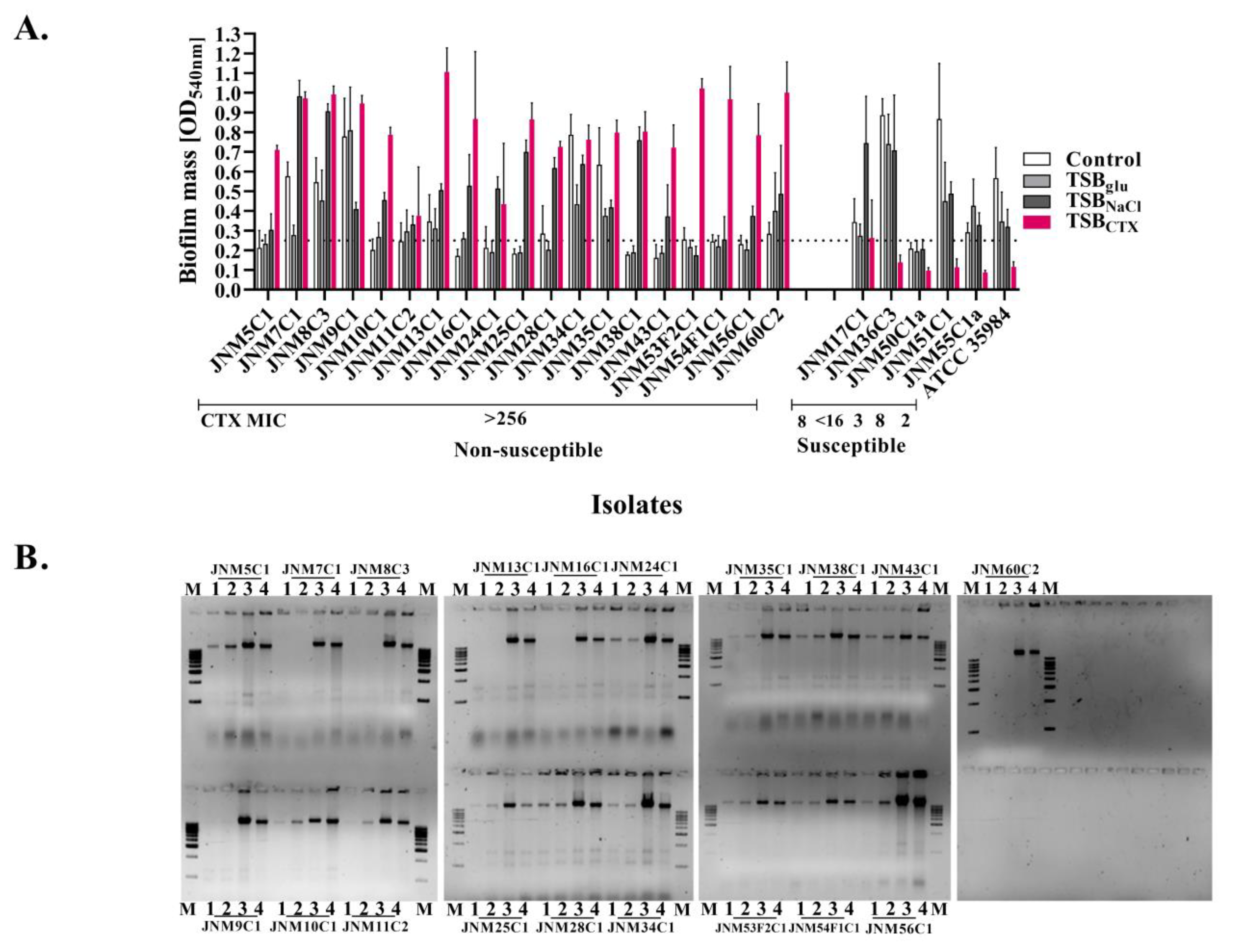
2.1. Susceptibility Profiling

The antibiotic susceptibilities of a total of 24 S. haemolyticus isolates were characterized to identify the MDR strains and appropriate controls. A total of five antibiotics including CTX were tested. Nineteen isolates (79.16%) were non-susceptible to CTX with MIC values of >256 µg/mL followed by MRP (n = 13, 54.17%; MIC: 6–32 µg/mL). A total of 18 out of 19 isolates harbored the mecA gene (94.74%) and 11 were amplification positive for the blaZ gene (57.89%). Out of the five CTX sensitive isolates, all were susceptible to AK, MRP and three were susceptible to GEN (Figure 1A, Supplementary Table S2). Two CTX susceptible isolates with MIC of 8 µg/mL (JNM17C1 and JNM51C1) were also found to harbor the resistance genes (Figure 1B, Supplementary Table S2).

2.2. Biofilm Enhancement and eDNA Release among the Isolates

In the presence of sub-inhibitory concentrations of CTX (30μg/mL), biofilm production was enhanced significantly (p-valueWilcoxintest—0.000008) among all the non-susceptible isolates. By contrast, there was a reduction in median optical density values among the susceptible isolates, presumably due to cell death (Figure 2A, Supplementary Table S3). All except for one isolate (JNM50C1a) grown in TSBglu and TSBNaCl were identified to be biofilm producers after 24 h. The growth media strongly influenced biofilm formation and a total of 14 out of 24 (58.33%) isolates produced biofilms in TSBglu whereas 22 (91.67%) isolates formed biofilms in TSBNaCl.
Given that eDNA release has been implicated in S. haemolyticus biofilm formation, whether increased biofilm formation also resulted in an increase in quantities of eDNA in the biofilms was next evaluated. Biofilm-forming sessile cells were harvested and removed by centrifugation and the cell-free nucleic acid in the supernatant was harvested and resolved on 0.8% agarose gels (Figure 2B). Significant increases in eDNA levels were observed in the TSBNaCl (p-valueWilcoxintest—0.000004) and TSBCTX (p-valueWilcoxintest—0.000004) treated groups (Supplementary Table S4).

2.3. Whole Genome Sequencing and Resistome Mapping of Ancestral Populations

To generate the resistome profiles of two MDR S. haemolyticus ancestral populations (JNM56C1 and JNM60C2), both non-susceptible to CTX, GEN and MRP, paired-end whole genome sequencing was carried out. Iterative de novo and reference guided assembly (NC_007168) resulted in alignment of >93% of error-corrected reads. JNM56C1 was determined to be a multi locus sequence type (ST) 38 with a chromosome length of 2,554,979 bp (CP063753) at an average sequencing depth of 310. A total of six antimicrobial resistance genes were annotated, namely AAC(6′)-Ie-APH(2″)-Ia, blaZ, dfrG, mecA, msrA and mphC. Similarly, JNM60C2, a ST-1 isolate, had a chromosome sequence length of 2,511,057 (CP065356) which was ascertained at an average depth of 421. The antimicrobial genes identified were APH(3′)-IIIa, AAC(6′)-Ie-APH(2″)-Ia, blaZ, dfrG, mecA and sat-4. The polysaccharide intercellular adhesin (PIA) operon was confirmed to be absent from both the genomes. None of the isolates were found to harbor any plasmids.

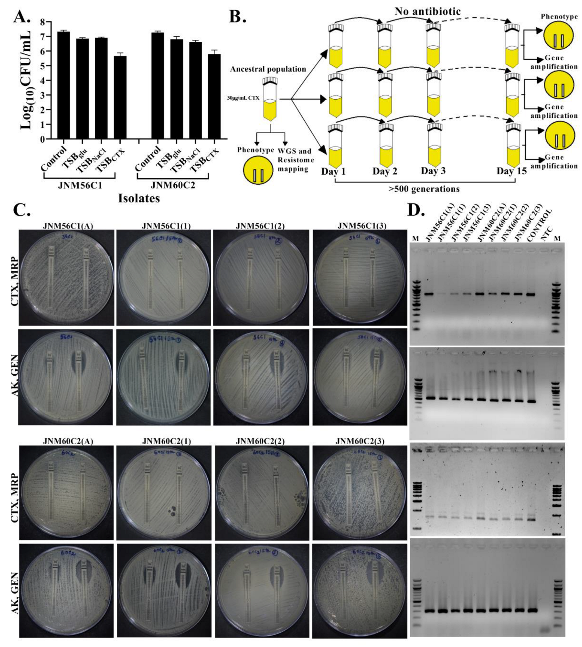
2.4. No Change in Susceptibility in the Absence of Antibiotic Selection

It has been shown in S. aureus strains using in vitro curing assays that harboring SCCmec imposes a fitness cost [32]. However, if a similar cost is attached with non-susceptibility in S. haemolyticus, a decay of non-susceptibility in the absence of antibiotic selection needs to be tested. To understand this, JNM56C1 and JNM60C2, two completely sequenced multidrug resistant S.haemolyticus populations with biofilm-forming ability (Figure 3A) and known generation time of 30 and 40 min, respectively (Supplementary Table S5), were serially passaged in triplicate for >500 generations in the absence of antibiotics (Figure 3B). Susceptibility to CTX was then compared among the six evolved clones and the two ancestral populations along with candidate resistance gene amplification. Additionally, meropenem (MRP), amikacin (AK) and gentamicin (GEN) susceptibilities were also compared. There were no differences in CTX susceptibility. Similarly, MRP, AK and GEN susceptibilities remained unchanged (Figure 3C). In all evolved populations, resistance genes blaZ, mecA and AAC(6′)-APH(2′) were retained (Figure 3D).

3. Discussion

S. haemolyticus is an emerging nosocomial pathogen known for multidrug resistance and severe outcomes among preterm neonates and immunocompromised individuals [6,33]. Blood stream and device-linked infections are commonly reported and the virulence determinants include biofilm formation along with production of modulins and rearrangements attributed to insertion sequences [34]. CTX, on the other hand, is a third-generation cephalosporin included among the first line of WHO recommended antibiotics in neonatal sepsis [35]. This study was undertaken to understand if an in vitro equivalent of the therapeutic dose of CTX (30 µg/mL) [36] could affect the physiology of resistant S. haemolyticus colonizers. Biofilm formation and presence of antibiotic-resistance genes (ARGs) mecA and blaZ reiterated the invasive phenotype of the clinical isolates included in this study [34]. We chose to delineate biofilm formation as the physiological effect as it is known to accentuate antibiotic resistance and colonization.
The sessile aggregates of biofilm-associated cells within the meshwork of extra-cellular polymeric matrix are known for enhanced resilience to antibiotics due to decreased penetration and presence of metabolically inert persister cells [37]. Thus, escalation of biofilm formation by any external agent can essentially skew a bacterial disease towards poor prognosis. Sub-inhibitory concentration antibiotics belonging to multiple classes have been reported among a number of diderms such as Acinetobacter baumannii, sensitive Campylobacter jejuni, Leptospira spp. and Pseudomonas aeruginosa [28,38,39,40].Changes in morphology with exposure to sub-optimal concentrations of penicillins have been reported as early as 1975 in the presence of intact cell division [41]. Among the monoderms, enhancement has also been reported upon the use of cell wall synthesis inhibitors including ß-lactam antibiotics among E. faecalis and S. aureus isolates with release of extracellular DNA (eDNA) [26,27]. However, reports on S. haemolyticus are lacking, though this coagulase-negative Staphylococcus species lacking the PIA operon has been shown to form biofilms in vitro under different growth conditions [42,43]. There exists only a single report where nosocomial clones were found to show enhanced biofilm formation on glass and polystyrene surfaces in the presence of one-fourth MIC of three antibiotics, namely oxacillin, vancomycin, and linezolid [7].Therefore, our study is one of the first showing increase in biofilm formation and eDNA release among MDR, nosocomial S. haemolyticus isolates in the presence of sub-inhibitory concentrations of CTX.
Evolved antibiotic resistance is a costly affair for many species of bacteria and often results in decreased competitive fitness in the absence of selection pressure [44,45]. Nevertheless, it has also been exhibited that S. aureus often tackles such trade-offs by way of mutations that compensate for changes [46]. Furthermore, in long-term evolution, experiments with E. coli populations have also shown that there is no change in susceptibility [47]. Along similar lines, our short-term evolution experiments (>500 generations) in the absence of selection pressure resulted in no change in MIC values of CTX and MRP in the evolved clones. However, we believe that a better approach would have been whole genome sequencing of the evolved clones instead of a candidate gene approach to gauge the entire repertoire of genetic changes incurred by the evolved populations.

4. Materials and Methods

4.1. Minimum Inhibitory Concentration (MIC) Determination of S. haemolyticus Isolates

The S. haemolyticus strains included in this study were isolated from nasal swab samples collected from neonates admitted after birth to the Sick Neonatal Care Unit (SNCU) of College of Medicine & JNM Hospital, Kalyani between 2017–2018 as approved by the Hospital and Institutional Ethical Committees. Consenting mothers signed a consent form prior to sample collection. The minimum inhibitory concentration (MIC) values were determined using MIC strips (Himedia Labs). The antibiotics tested were CTX, aminoglycosides, AK, GEN, carbapenem and MRP. The Kirby–Bauer disc diffusion assay was generated for the antibiotics fluoroquinolones; ciprofloxacin (CIP). Experiments were carried out according to Clinical and Laboratory Standards Institute (CLSI) guidelines [48].

4.2. Quantification of Biofilms

The biofilm-forming ability of S. haemolyticus isolates was determined by a modified crystal violet assay method as described previously [43]. Briefly, 96-well polystyrene, flat-bottom microtiter plates were filled with 180 μL of tryptic soy broth (TSB) (Himedia labs) and 20 μL bacterial cells grown to a Macfarland score of 0.5 in brain heart infusion broth (BHI) (Himedia labs) were added and incubated at 37 °C for 24 h statically. Biofilm-forming capacities of all isolates were determined in TSB, TSB with 1% glucose (TSBglu), TSB with 3% NaCl (TSBNaCl), and TSB with 30 μg/mL CTX (TSBCTX) which is an in vitro equivalent of the therapeutic dosage [36]. After 24 h, planktonic cells were removed, adherent cells were fixed with 99% methanol (Finar chemicals, Ahmedabad, India) for 10 min, and plates were washed once with 1× PBS (Sigma-Aldrich, St. Louis, MO, USA) and air-dried for 10 min. Modified crystal violet assays were performed, and the absorbance (OD) was recorded at 540 nm. The assays were performed with 6 replicates for each condition in 2 parallel runs. The isolates with an OD of ≥0.25 were considered biofilm positive. S. epidermidis ATCC 35984 was used as a positive control. The presence of living sessile cells was determined by colony-forming units (CFU) for selected isolates in the presence of TSBglu, TSBNaCl and TSBCTX in triplicate.

4.3. Extracellular DNA (eDNA) Quantification

Extracellular DNA was extracted as previously described by Kaplan et al. with modification [25]. Biofilms were grown in triplicate in TSB, TSBglu, TSBNaCl and TSBCTX in 24-well polystyrene microtiter plates, in a total volume of 1 mL per well. After 24 h of growth, the liquid was carefully removed, the plates were washed once with 1XPBS (Sigma-Aldrich, St. Louis, MO, USA) and 50 μL of Tris-EDTA buffer (10 mM Tris-HCl, 1 mM EDTA, pH 7.4) (Sigma-Aldrich, St. Louis, MO, USA) was added to each well. The biofilm-forming cells were scraped off the bottom surface of the wells and were transferred to 1.5 mL microcentrifuge tubes. The tubes were centrifuged at 13,000 rpm for 25 s and 8 μL of each of the supernatants were resolved on 0.8% agarose gels. Densitometric analyses of eDNA were carried out using Image Lab software version 6.0.1(Bio-Rad Laboratories, Hercules, CA, USA).

4.4. Genomic DNA Isolation and Whole-Genome Sequencing

Two isolates (JNM56C1 and JNM60C2) were subcultured in the presence of 30 μg/mL CTX. Total DNA from both were purified using the QIAamp DNA extraction mini kit (Qiagen, Hilden, Germany) and were subjected to paired end whole genome sequencing (2 × 300 bp) on an Illumina HiSeq2500 platform (Illumina, San Diego, CA, USA). Both de novo and reference guided assembly was carried out using Velvet and Bowtie2, respectively [49,50], to build genomes as described previously [51].

4.5. Short-Term Evolution Experiment

The isolates grown in the presence of 30 μg/mL CTX formed the ancestral population for each. Generation times for both were calculated from growth curves. The two populations were serially passaged for >500 generations in triplicate for 15 days in fresh Luria–Bertani (LB) media (Himedia labs) in the absence of antibiotic selection at a dilution of 1:100. The ancestral and evolved populations were tested for antibiotic susceptibility for β-lactam (CTX, MRP) and aminoglycoside (AK, GEN) antibiotics by Etests.

4.6. Amplification of Genes

The presence of β-lactamase genes mecA and blaZ, aminoglycoside resistance gene AAC(6′)-APH(2′), and N-acetylglucosaminyltransferase icaA gene were detected by using PCR amplification. The positive controls used were a laboratory isolate for the β-lactamase and aminoglycoside resistance gene and S. epidermidis ATCC 35984 for icaA gene amplification. A 292 basepair (bp) region of the 16S gene was amplified as an internal control. Supplementary Table S6 lists all the primers used in this study.

4.7. Statistical Analyses

Normality across datasets was evaluated using the Kolmogorov–Smirnov test. To identify significant differences among experimental conditions tested, the Wilcoxon test was performed using GraphPad Prism version 9.1.2 (GraphPad Software, La Jolla, CA, USA). A p-value of <0.05 was considered to be statistically significant.

5. Conclusions

MDR S. haemolyticus is a late-onset sepsis pathogen which has observed to be a predominant nasal colonizer among hospitalized preterm neonates. This study demonstrates that these CTX non-susceptible, MDR, biofilm-forming S. haemolyticus isolates exhibit enhanced biofilm formation upon exposure to a therapeutic dosage of a commonly used antibiotic, CTX, in vitro. Increase in eDNA release implicated in biofilm formation was significantly higher upon sub-MIC CTX usage. Further, no changes in susceptibility to both commonly used β-lactams and aminoglycosides were observed among non-susceptible isolates when grown in the absence of selection pressure in the short-term. All these findings together reiterate the need for antibiotic stewardship.

Supplementary Materials

The following supporting information can be downloaded at: https://www.mdpi.com/article/10.3390/antibiotics11030360/s1, Table S1: Summary of preterm neonate subjects and species identification ; Table S2: Summary of MIC values and resistance gene detection; Table S3: Median optical density values obtained from crystal violet assays run to determine biofilm formation; Table S4: eDNA quantitation values of 24-h biofilms; Table S5: Summary of generation time values of isolates JNM56C1 and JNM60C2; Table S6: Information on the primer sets used in this study.

Author Contributions

M.C. ran all the laboratory experiments, analyzed the data and wrote the first draft of the manuscript. T.B. ran the initial screening experiments and standardized the biofilm assays. M.B. was the clinician who was involved in specimen collection. B.B. conceptualized all the experiments, carried out all the next generation sequencing analyses, collated all the data and wrote and reviewed the final draft. All authors have read and agreed to the published version of the manuscript.

Funding

This work was supported by the Ramanujan fellowship (SB/S2/RJN-013/2014) funded by Science and Engineering Research Board, Department of Science and Technology, Government of India and intramural funding awarded to B.B.

Institutional Review Board Statement

The study was conducted according to the guidelines of the Declaration of Helsinki and approved by the College of Medicine & Jawaharlal Nehru Memorial Hospital and National Institute of Biomedical GenomicsEthicsCommittees. The hospital ethical committee reference number is F-24/Pr/COMJNMH/IEC/16/536.

Informed Consent Statement

Informed consent was obtained from all the parents of the subjects involved in the study.

Data Availability Statement

The annotated genome sequences (CP063753, CP065356) and the 16S amplicon sequences (MZ636452-MZ636490) are available at the GenBank database.

Acknowledgments

The authors acknowledge Samsiddhi Bhattacharjee, NIBMG, Kalyani, India for providing valuable inputs on statistical analyses.

Conflicts of Interest

The authors declare no conflict of interest.

References

  1. Becker, K.; Heilmann, C.; Peters, G. Coagulase-negative staphylococci. Clin. Microbiol. Rev. 2014, 27, 870–926. [Google Scholar] [CrossRef] [PubMed] [Green Version]
  2. Heilmann, C.; Ziebuhr, W.; Becker, K. Are coagulase-negative staphylococci virulent? Clin. Microbiol. Infect. 2019, 25, 1071–1080. [Google Scholar] [CrossRef] [PubMed]
  3. Salgueiro, V.C.; Seixas, M.D.L.; Guimarães, L.C.; Ferreira, D.C.; Da Cunha, D.C.; Nouér, S.A.; Dos Santos, K.R.N. High rate of neonates colonized by methicillin-resistant Staphylococcus species in an Intensive Care Unit. J. Infect. Dev. Ctries. 2019, 13, 810–816. [Google Scholar] [CrossRef] [PubMed] [Green Version]
  4. Zaha, D.C.; Zdrinca, M.M.; Vesa, C.M.; Daina, L.G.; Daina, M.C. A six-year evaluation of sepsis in neonates. Rom. Biotechnol. Lett. 2020, 25, 1892–1898. [Google Scholar] [CrossRef]
  5. Czekaj, T.; Ciszewski, M.; Szewczyk, E.M. Staphylococcus haemolyticus—An emerging threat in the twilight of the antibiotics age. Microbiology 2015, 161, 2061–2068. [Google Scholar] [CrossRef]
  6. Cavanagh, J.P.; Hjerde, E.; Holden, M.T.; Kahlke, T.; Klingenberg, C.; Flægstad, T.; Parkhill, J.; Bentley, S.D.; Sollid, J.U. Whole-genome sequencing reveals clonal expansion of multiresistant Staphylococcus haemolyticus in European hospitals. J. Antimicrob. Chemother. 2014, 69, 2920–2927. [Google Scholar] [CrossRef] [Green Version]
  7. Pereira-Ribeiro, P.M.; Sued-Karam, B.R.; Faria, Y.V.; Nogueira, B.A.; Colodette, S.S.; Fracalanzza, S.E.; Duarte, J.L.; Júnior, R.H.; Mattos-Guaraldi, A.L. Influence of antibiotics on biofilm formation by different clones of nosocomial Staphylococcus haemolyticus. Future Microbiol. 2019, 14, 789–799. [Google Scholar] [CrossRef]
  8. Giormezis, N.; Kolonitsiou, F.; Foka, A.; Drougka, E.; Liakopoulos, A.; Makri, A.; Papanastasiou, A.D.; Vogiatzi, A.; Dimitriou, G.; Marangos, M.; et al. Coagulase-negative staphylococcal bloodstream and prosthetic-device-associated infections: The role of biofilm formation and distribution of adhesin and toxin genes. J. Med. Microbiol. 2014, 63, 1500–1508. [Google Scholar] [CrossRef]
  9. Pereira, P.M.; Binatti, V.B.; Sued, B.P.; Ramos, J.N.; Peixoto, R.S.; Simões, C.; de Castro, E.A.; Duarte, J.L.; Vieira, V.V.; Hirata, R.; et al. Staphylococcus haemolyticus disseminated among neonates with bacteremia in a neonatal intensive care unit in Rio de Janeiro, Brazil. Diagn. Microbiol. Infect. Dis. 2014, 78, 85–92. [Google Scholar] [CrossRef] [Green Version]
  10. Neonatal Mortality-UNICEF Data. Available online: https://data.unicef.org/topic/child-survival/neonatal-mortality/ (accessed on 23 February 2022).
  11. Jain, K.; Sankar, M.J.; Nangia, S.; Ballambattu, V.B.; Sundaram, V.; Ramji, S.; Plakkal, N.; Kumar, P.; Jain, A.; Sivanandan, S.; et al. Causes of death in preterm neonates (<33 weeks) born in tertiary care hospitals in India: Analysis of three large prospective multicentric cohorts. J. Perinatol. 2019, 39, 13–19. [Google Scholar] [CrossRef] [Green Version]
  12. Wattal, C.; Kler, N.; Oberoi, J.K.; Fursule, A.; Kumar, A.; Thakur, A. Neonatal Sepsis: Mortality and Morbidity in Neonatal Sepsis due to Multidrug-Resistant (MDR) Organisms: Part 1. Indian J. Pediatr. 2020, 87, 117–121. [Google Scholar] [CrossRef] [PubMed]
  13. Investigators of the Delhi Neonatal Infection Study (DeNIS) Collaboration. Characterisation and antimicrobial resistance of sepsis pathogens in neonates born in tertiary care centres in Delhi, India: A cohort study. Lancet Glob. Health 2016, 4, 752–760. [Google Scholar] [CrossRef] [Green Version]
  14. Nazir, A. Neonatal sepsis due to coagulase negative Staphylococci: A study from Kashmir valley, India. Int. J. Contemp. Pediatr. 2019, 6, 650. [Google Scholar] [CrossRef]
  15. Härtel, C.; Faust, K.; Fortmann, I.; Humberg, A.; Pagel, J.; Haug, C.; Kühl, R.; Bohnhorst, B.; Pirr, S.; Viemann, D.; et al. Sepsis related mortality of extremely low gestational age newborns after the introduction of colonization screening for multi-drug resistant organisms. Antimicrob. Resist. Infect. Control 2020, 9, 144. [Google Scholar] [CrossRef] [PubMed]
  16. Kranjec, C.; Kristensen, S.S.; Bartkiewicz, K.T.; Brønner, M.; Cavanagh, J.P.; Srikantam, A.; Mathiesen, G.; Diep, D.B. A bacteriocin-based treatment option for Staphylococcus haemolyticus biofilms. Sci. Rep. 2021, 11, 13909. [Google Scholar] [CrossRef] [PubMed]
  17. Al-Mousawi, A.H.; Al-Kaabi, S.J.; Albaghdadi, A.J.H.; Almulla, A.F.; Raheem, A.; Algon, A.A.A. Effect of Black Grape Seed Extract (Vitis vinifera) on Biofilm Formation of Methicillin-Resistant Staphylococcus aureus and Staphylococcus haemolyticus. Curr. Microbiol. 2020, 77, 238–245. [Google Scholar] [CrossRef]
  18. Blair, J.M.; Webber, M.A.; Baylay, A.J.; Ogbolu, D.O.; Piddock, L.J. Molecular mechanisms of antibiotic resistance. Nat. Rev. Microbiol. 2015, 13, 42–51. [Google Scholar] [CrossRef]
  19. de Oliveira, A.; Cataneli Pereira, V.; Pinheiro, L.; Moraes Riboli, D.F.; Benini Martins, K.; Ribeiro de Souza da Cunha, M. Antimicrobial Resistance Profile of Planktonic and Biofilm Cells of Staphylococcus aureus and Coagulase-Negative Staphylococci. Int. J. Mol. Sci. 2016, 17, 1423. [Google Scholar] [CrossRef] [Green Version]
  20. Martins, K.B.; Ferreira, A.M.; Pereira, V.C.; Pinheiro, L.; de Oliveira, A.; da Cunha, M. In vitro Effects of Antimicrobial Agents on Planktonic and Biofilm Forms of Staphylococcus saprophyticus Isolated From Patients with Urinary Tract Infections. Front. Microbiol. 2019, 10, 40. [Google Scholar] [CrossRef]
  21. Alexander, H.K.; MacLean, R.C. Stochastic bacterial population dynamics restrict the establishment of antibiotic resistance from single cells. Proc. Natl. Acad. Sci. USA 2020, 117, 19455–19464. [Google Scholar] [CrossRef]
  22. Song, T.; Duperthuy, M.; Wai, S.N. Sub-Optimal Treatment of Bacterial Biofilms. Antibiotics 2016, 5, 23. [Google Scholar] [CrossRef] [PubMed] [Green Version]
  23. Wicaksono, W.A.; Erschen, S.; Krause, R.; Müller, H.; Cernava, T.; Berg, G. Enhanced survival of multi-species biofilms under stress is promoted by low-abundant but antimicrobial-resistant keystone species. J. Hazard. Mater. 2022, 422, 126836. [Google Scholar] [CrossRef] [PubMed]
  24. He, H.J.; Sun, F.J.; Wang, Q.; Liu, Y.; Xiong, L.R.; Xia, P.Y. Erythromycin resistance features and biofilm formation affected by subinhibitory erythromycin in clinical isolates of Staphylococcus epidermidis. J. Microbiol. Immunol. Infect. 2016, 49, 33–40. [Google Scholar] [CrossRef] [PubMed]
  25. Kaplan, J.B.; Izano, E.A.; Gopal, P.; Karwacki, M.T.; Kim, S.; Bose, J.L.; Bayles, K.W.; Horswill, A.R. Low levels of β-lactam antibiotics induce extracellular DNA release and biofilm formation in Staphylococcus aureus. mBio 2012, 3, e00198-12. [Google Scholar] [CrossRef] [Green Version]
  26. Ng, M.; Epstein, S.B.; Callahan, M.T.; Piotrowski, B.O.; Simon, G.L.; Roberts, A.D.; Keiser, J.F.; Kaplan, J.B. Induction of MRSA Biofilm by Low-Dose β-Lactam Antibiotics: Specificity, Prevalence and Dose-Response Effects. Dose-Response 2013, 12, 152–161. [Google Scholar] [CrossRef] [Green Version]
  27. Yu, W.; Hallinen, K.M.; Wood, K.B. Interplay between Antibiotic Efficacy and Drug-Induced Lysis Underlies Enhanced Biofilm Formation at Subinhibitory Drug Concentrations. Antimicrob. Agents Chemother. 2017, 62, e01603-17. [Google Scholar] [CrossRef] [Green Version]
  28. Tahrioui, A.; Duchesne, R.; Bouffartigues, E.; Rodrigues, S.; Maillot, O.; Tortuel, D.; Hardouin, J.; Taupin, L.; Groleau, M.C.; Dufour, A.; et al. Extracellular DNA release, quorum sensing, and PrrF1/F2 small RNAs are key players in Pseudomonas aeruginosa tobramycin-enhanced biofilm formation. NPJ Biofilms Microbiomes 2019, 5, 15. [Google Scholar] [CrossRef] [Green Version]
  29. Cadavid, E.; Robledo, S.M.; Quiñones, W.; Echeverri, F. Induction of Biofilm Formation in Klebsiella pneumoniae ATCC 13884 by Several Drugs: The Possible Role of Quorum Sensing Modulation. Antibiotics 2018, 7, 103. [Google Scholar] [CrossRef] [Green Version]
  30. Ramasamy, S.; Biswal, N.; Bethou, A.; Mathai, B. Comparison of two empiric antibiotic regimen in late onset neonatal sepsis--a randomized controlled trial. J. Trop. Pediatr. 2014, 60, 83–86. [Google Scholar] [CrossRef] [Green Version]
  31. Yen, P.; Papin, J.A. History of antibiotic adaptation influences microbial evolutionary dynamics during subsequent treatment. PLoS Biol. 2017, 15, e2001586. [Google Scholar] [CrossRef] [Green Version]
  32. Dorado-Morales, P.; Garcillán-Barcia, M.P.; Lasa, I.; Solano, C. Fitness Cost Evolution of Natural Plasmids of Staphylococcus aureus. mBio 2021, 12, e03094-20. [Google Scholar] [CrossRef] [PubMed]
  33. Cavanagh, J.P.; Klingenberg, C.; Hanssen, A.M.; Fredheim, E.A.; Francois, P.; Schrenzel, J.; Flægstad, T.; Sollid, J.E. Core genome conservation of Staphylococcus haemolyticus limits sequence based population structure analysis. J. Microbiol. Methods 2012, 89, 159–166. [Google Scholar] [CrossRef] [PubMed] [Green Version]
  34. Pain, M.; Hjerde, E.; Klingenberg, C.; Cavanagh, J.P. Comparative Genomic Analysis of Staphylococcus haemolyticus Reveals Key to Hospital Adaptation and Pathogenicity. Front. Microbiol. 2019, 10, 2096. [Google Scholar] [CrossRef] [PubMed]
  35. Chaurasia, S.; Sivanandan, S.; Agarwal, R.; Ellis, S.; Sharland, M.; Sankar, M.J. Neonatal sepsis in South Asia: Huge burden and spiralling antimicrobial resistance. BMJ 2019, 364, k5314. [Google Scholar] [CrossRef] [PubMed] [Green Version]
  36. Brochmann, P.R.; Hesketh, A.; Jana, B.; Brodersen, G.H.; Guardabassi, L. Transcriptome analysis of extended-spectrum β-lactamase-producing Escherichia coli and methicillin-resistant Staphylococcus aureus exposed to cefotaxime. Sci. Rep. 2018, 8, 16076. [Google Scholar] [CrossRef]
  37. Sharma, D.; Misba, L.; Khan, A.U. Antibiotics versus biofilm: An emerging battleground in microbial communities. Antimicrob. Resist. Infect. Control 2019, 8, 76. [Google Scholar] [CrossRef]
  38. Sato, Y.; Unno, Y.; Ubagai, T.; Ono, Y. Sub-minimum inhibitory concentrations of colistin and polymyxin B promote Acinetobacter baumannii biofilm formation. PLoS ONE 2018, 13, e0194556. [Google Scholar] [CrossRef] [Green Version]
  39. Teh, A.; Lee, S.M.; Dykes, G.A. Growth in the presence of specific antibiotics induces biofilm formation by a Campylobacter jejuni strain sensitive to them but not in resistant strains. J. Glob. Antimicrob. Resist. 2019, 18, 55–58. [Google Scholar] [CrossRef]
  40. Vinod Kumar, K.; Lall, C.; Vimal Raj, R.; Vedhagiri, K.; Sunish, I.P.; Vijayachari, P. Can Subminimal Inhibitory Concentrations of Antibiotics Induce the Formation of Biofilm in Leptospira? Microb. Drug Resist. 2018, 24, 1040–1042. [Google Scholar] [CrossRef]
  41. Lorian, V. Some effects of subinhibitory concentrations of antibiotics on bacteria. Bull. N. Y. Acad. Med. 1975, 51, 1046–1055. [Google Scholar]
  42. Panda, S.; Jena, S.; Sharma, S.; Dhawan, B.; Nath, G.; Singh, D.V. Identification of Novel Sequence Types among Staphylococcus haemolyticus Isolated from Variety of Infections in India. PLoS ONE 2016, 11, e0166193. [Google Scholar] [CrossRef] [PubMed]
  43. Panda, S.; Singh, D.V. Biofilm Formation by ica-Negative Ocular Isolates of Staphylococcus haemolyticus. Front. Microbiol. 2018, 9, 2687. [Google Scholar] [CrossRef] [PubMed]
  44. Melnyk, A.H.; Wong, A.; Kassen, R. The fitness costs of antibiotic resistance mutations. Evol. Appl. 2015, 8, 273–283. [Google Scholar] [CrossRef] [PubMed]
  45. Rasouly, A.; Shamovsky, Y.; Epshtein, V.; Tam, K.; Vasilyev, N.; Hao, Z.; Quarta, G.; Pani, B.; Li, L.; Vallin, C.; et al. Analysing the fitness cost of antibiotic resistance to identify targets for combination antimicrobials. Nat. Microbiol. 2021, 6, 1410–1423. [Google Scholar] [CrossRef]
  46. Cao, S.; Huseby, D.L.; Brandis, G.; Hughes, D. Alternative Evolutionary Pathways for Drug-Resistant Small Colony Variant Mutants in Staphylococcus aureus. mBio 2017, 8, e00358-17. [Google Scholar] [CrossRef] [Green Version]
  47. Lamrabet, O.; Martin, M.; Lenski, R.E.; Schneider, D. Changes in Intrinsic Antibiotic Susceptibility during a Long-Term Evolution Experiment with Escherichia coli. mBio 2019, 10, e00189-19. [Google Scholar] [CrossRef] [Green Version]
  48. Clinical and Laboratory Standards Institute. Performance Standards for Antimicrobial Susceptibility Testing; CLSI M100-S27; Clinical and Laboratory Standards Institute: Wayne, PA, USA, 2017. [Google Scholar]
  49. Zerbino, D.R.; Birney, E. Velvet: Algorithms for de novo short read assembly using de Bruijn graphs. Genome Res. 2008, 18, 821–829. [Google Scholar] [CrossRef] [Green Version]
  50. Langmead, B.; Salzberg, S.L. Fast gapped-read alignment with Bowtie 2. Nat. Methods 2012, 9, 357–359. [Google Scholar] [CrossRef] [Green Version]
  51. Bhattacharjee, B.; Bardhan, T.; Chakraborty, M.; Basu, M. Resistance profiles and resistome mapping of multidrug resistant carbapenem-hydrolyzing Klebsiella pneumoniae strains isolated from the nares of preterm neonates. Int. J. Antimicrob. Agents 2019, 53, 535–537. [Google Scholar] [CrossRef]
Figure 1. Antimicrobial susceptibility and gene profiles of 19 CTX non-susceptible S. haemolyticus isolates. (A). The minimum inhibitory concentration of (MIC) of the 19 non-susceptible S. haemolyticus isolates with the inset showing the MIC of the five susceptible isolates. (B). The blaZ (858 bp), mecA (533 bp) and N-acetylglucosaminyltranferase (icaA) (166 bp) amplicons were run on 1.5% agarose gels. M; 100 bp marker, control; positive control, NTC; no template control. 16S amplicon (292 bp) served as control.
Antibiotics 11 00360 g001
Figure 2. Cefotaxime (CTX), a commonly used ß-lactam antibiotic enhances biofilm formation among non-susceptible S. haemolyticus isolates in vitro. (A). Semi-quantitative determination of 24 h biofilm formation under known inducible conditions (tryptic soy broth with 1% glucose (TSBglu) and 3% sodium chloride (TSBNaCl)) and 30µg/mL CTX(TSBCTX) using crystal violet assays. The dotted line denotes the cut-off optical density value for biofilm formation (0.25). The biofilm-forming ATCC 35984 S. epidermidis strain served as a control. (B). Biofilm supernatants run on 0.8% agarose gels to determine the presence of extracellular DNA (eDNA). M; 1000 bp marker. The lanes labelled 1, 2, 3 and 4 in each case denote TSB, TSBglu, TSBNaCl and TSBCTX, respectively.
Antibiotics 11 00360 g002
Figure 3. No change in in vitro antibiotic susceptibility among MDR, biofilm-forming S. haemolyticus isolates (JNM56C1 and JNM60C2) grown in the absence of antibiotics in short-term evolution experiments. (A). The presence of living sessile cells is confirmed by log(10)CFU counts in both isolates in the presence of 30 μg/mL CTX. The error bars represent standard deviation across three technical replicates. (B). Study design of the evolution experiment. (C). Comparison of susceptibility to ß-lactam and aminoglycoside antibiotics among ancestral and evolved populations using E-tests. (D). The blaZ (858 bp, panel 1), mecA (533 bp, panel 2) and AAC(6′)-APH(2′)(1658 bp, panel 3) and 16S (292 bp, panel 4) genes were amplified from the ancestral and evolved populations and amplicons were run on 0.8–1.5% agarose gels. M; 100/1000 bp marker, control; positive control, NTC; no template control.
Antibiotics 11 00360 g003
Publisher’s Note: MDPI stays neutral with regard to jurisdictional claims in published maps and institutional affiliations.

