Article

Antimicrobial Resistance and Comparative Genomic Analysis of Elizabethkingia anophelis subsp. endophytica Isolated from Raw Milk

by
Pavel A. Andriyanov
1,*,
Pavel A. Zhurilov
1,
Daria D. Kashina
1,
Anastasia I. Tutrina
1,
Elena A. Liskova
1,
Irina V. Razheva
1,
Denis V. Kolbasov
2 and
Svetlana A. Ermolaeva
1
1
Branch in Nizhny Novgorod, Federal Research Center for Virology and Microbiology, 603950 Nizhny Novgorod, Russia
2
Federal Research Center for Virology and Microbiology, 601125 Volginsky, Russia
*
Author to whom correspondence should be addressed.
Antibiotics 2022, 11(5), 648; https://doi.org/10.3390/antibiotics11050648
Submission received: 14 April 2022 / Revised: 7 May 2022 / Accepted: 9 May 2022 / Published: 12 May 2022

Abstract

:
Elizabethkingia anophelis is an emerging multidrug-resistant pathogen that causes severe nosocomial and community-acquired infections worldwide. We report the first case of E. anophelis isolation in Russia and the first isolation from raw cow’s milk. The ML-44 demonstrated resistance to 28 antimicrobials of 33 tested in the disk-diffusion test. Whole genome-based phylogeny showed ML-44 strain clustered together with the F3201 strain isolated from a human patient in Kuwait in 1982. Both strains were a part of the “endophytica” clade. Another clade was formed by subsp. anophelis strains. Each of the E. anophelis compared genomes carried 18 to 21 antibiotic resistance determinants. The ML-44 chromosome harbored nine efflux system genes and three beta-lactamase genes, along with six other antimicrobial resistance genes. In total, 72 virulence genes were revealed. The set of virulence factors was quite similar between different E. anophelis strains and included LPS and capsule encoded genes, type IV pili, oxidative stress response genes, and genes encoding TIVSS and TVISS effectors. The particular interest caused the mip and zmp1 gene homologs, which can be essential for intracellular survival. In sum, our findings suggest that raw milk might be a source of E. anophelis harboring a set of virulence factors and a broad resistance to generally used antimicrobials.

1. Introduction

Elizabethkingia anophelis is a Gram-negative, non-motile, oxidase-, and catalase-positive aerobic bacilli [1]. Recently, it has emerged as an opportunistic pathogen that causes life-threatening nosocomial and community-acquired infections and outbreaks [2]. E. anophelis infection has high mortality rates and manifests by sepsis, meningitis, nosocomial pneumonia in immunocompromised and immunocompetent persons [3]. Outbreaks of nosocomial E. anophelis infection were registered in the Central African Republic, Singapore, Hong Kong, England, and Taiwan in 2011–2013 [4,5,6,7,8]. In 2016, an unusually large community-associated outbreak involving 65 persons and leading to 20 fatal cases was registered in the Midwestern United States (Wisconsin, Illinois, and Michigan) [9]. Several sporadic cases were also reported from Denmark and France [10,11]. To date, E. anophelis has not been reported from Russia.
E. anophelis infection is complicated by the intrinsic multidrug resistance of the pathogen. E. anophelis carries multiple chromosomally encoded determinants that provide resistance to the majority of beta-lactam and aminoglycoside antibiotics. Mainly, E. anophelis is a unique bacterium that possesses three chromosomally encoded beta-lactamase genes: blaB, blaCME, and blaGOB [12,13,14]. The spectrum of antibiotics effective against E. anophelis is restricted to minocycline, doxycycline, piperacillin–tazobactam combination, fluoroquinolones, trimethoprim–sulfamethoxazole, and rifampin [9,15,16,17].
Based on the whole genome phylogenetic analysis, E. anophelis species was divided into two subtaxons: subsp. anophelis and subsp. endophytica [18]. The first one is more frequently isolated from different sources, including human patients and clinical environments, and has confirmed pathogenicity to humans. The second one was first isolated from healthy internal stem tissues of sweet corn (Zea mays) and established as a novel species of E. endophytica, considered a plant endophyte [19]. In 2016, it was reassessed as E. anophelis subsp. endophytica [18]. To date, only a few cases of E. anophelis subsp. endophytica isolation have been reported. Besides the type strain JM-87, isolated from corn, the F3201 strain was isolated from a human host in Kuwait in 1982. The pathogenic potential of E. anophelis subsp. endophytica is still elusive.
In this study, we reported the E. anophelis subsp. endophytica strain, isolated from raw milk in Russia. We applied phenotype-based methods to assess physiological capabilities and antimicrobial susceptibility and whole-genome sequencing and comparative genomics approach to investigate genome features and phylogeny and determined similarities and differences with other E. anophelis strains.

2. Results

2.1. Isolation and Phenotypic Characterization of the E. anophelis Subsp. endophytica ML-44 Strain

We isolated E. anophelis in monitoring studies from the raw milk sample obtained from the farm at the territory of the NN region, Russia. A single colony was taken from Endo agar as a sample of lactose non-fermenting bacteria contaminating raw milk. In addition to the E. anophelis, Pseudomonas aeruginosa and Enterobacter aerogenes were isolated from this milk specimen. Identification was based on the nearly full-length 16S rRNA gene sequence using 27f and 1492r primers. The 16S RNA consensus was 1348 bp in length with 99.71% sequence identity to the sequence of E. anophelis subsp. endophytica JM-87 (NR_136481.1). The culture was designated as the E. anophelis subsp. endophytica strain ML-44.
ML-44 formed milk-white colonies of 1–2 mm in diameter with a smooth surface on Endo agar. On tryptic soy agar, the isolate formed visible yellowish colonies approximately 3–4 mm in diameter within 24 h at 37 °C. Microscopic studies showed small non-motile rods with typical Gram-negative staining (Figure S1). Biochemical properties were obtained through ENTEROtest 24 N kit. ML-44 was oxidase, catalase, ß-galactosidase, and esculin positive and hydrolyzed trehalose and mannitol. The following tested activities were negative: urease, arginine hydrolase, ornithine decarboxylase, lysine decarboxylase, hydrogen sulfide activities, Simmons citrate, and malonate utilization, ß-xylosidase activity. No acid was detected from salicine, sorbitol, melibiose, cellobiose, lactose, dulcitol, adonitol, arabitol, sucrose, inositol, raffinose (Table S1).

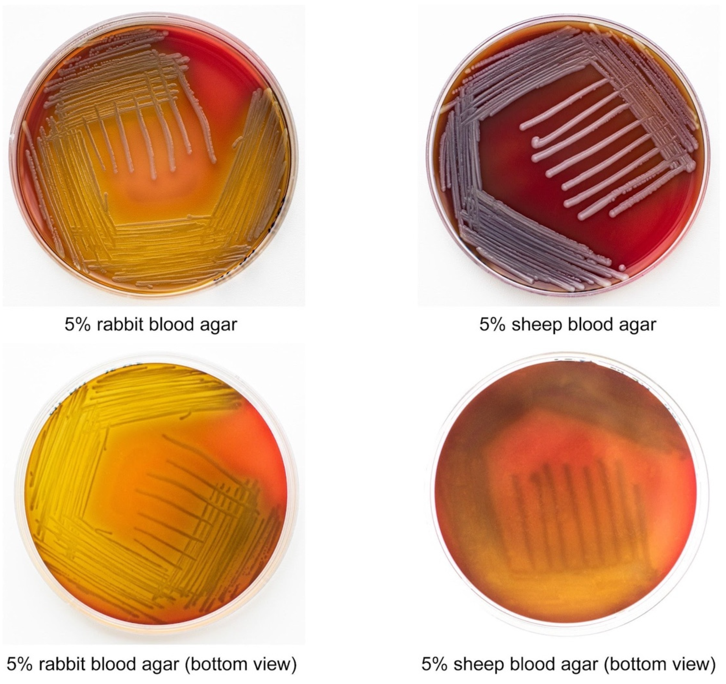
2.2. E. anophelis ML-44 Possesses an Alpha-Hemolytic Activity

Hemolytic activity is typical for many pathogenic bacteria, but it has not been described in E. anophelis subsp. endophytica yet. E. anophelis ML-44 did not exhibit yellow pigment on the blood agar; colonies were smooth and white. After 48 h of incubation, ML-44 exhibited zones of α-hemolysis. The hemolysis zones were more apparent on the agar with rabbit blood. Agar zones were more visible on the sheep blood when bacterial biomass was removed (Figure 1).

2.3. E. anophelis ML-44 Showed a Multidrug-Resistant Phenotype

E.anophelis ML-44 drug susceptibility was determined using a standard disc-diffusion method to 33 antimicrobials. Antibiotics tested, and the size of inhibition zones are listed in Table 1. ML-44 isolate was resistant to 28 antibiotics used in the clinical practice, including penicillin G, ampicillin, amoxicillin-clavulanate, aztreonam, ticarcillin, piperacillin, piperacillin-tazobactam, ticarcillin-clavulanate, ceftazidime, cefotaxime, cefepime, imipenem and meropenem, kanamycin, neomycin, tobramycin, gentamicin, amikacin, polymyxin, clindamycin, clarithromycin, erythromycin, tylosin, nalidixic acid, trimethoprim, trimethoprim-sulfamethoxazole, chloramphenicol, and tetracycline. The strain was susceptible to rifampicin, ciprofloxacin, levofloxacin, enrofloxacin, and linezolid.

2.4. Genomic Traits of E. anophelis ML-44

The assembly of the ML-44 genome contained 31 contigs with 4.03 M bp genome size and 35.4% average GC-content. In total, 3805 features were detected in the ML-44 chromosome; among them, 3758 CDS and 47 RNAs were predicted. CDS distribution among specific subsystems was predicted using the RAST web tool. Two hundred sixty subsystems were prescribed. The major subsystems were “Amino Acids and Derivatives” (264 CDSs), “Carbohydrates” (131 CDSs), “Cofactors, Vitamins, Prosthetic Groups, Pigments” (128 CDSs), and “Protein Metabolism” (126 CDSs). Notably, 36 and 11 CDSs were assigned as “Virulence, Disease and Defense” and “Phages, Prophages, Transposable elements, Plasmids” categories. “Virulence, Disease, and Defense” subsystem contained 24 CDSs related to antimicrobial and toxic compounds resistance as well as 12 CDSs involved in the invasion process and intracellular resistance.2.5. Whole Genome-Based Phylogeny and Comparative Analysis.
We used a whole genome-based phylogeny approach realized via the REALPHY web tool to determine a position of ML-44 among 14 complete sequenced E. anophelis strains (Figure 2). The ML-44 strain clustered together with the strain F3201 isolated from the human host in Kuwait in 1982. Both strains were a part of the common “endophytica” clade and formed an individual subcluster distinct from the subcluster that included the type JM-87 strain isolated from corn. Inside the “anophelis” clade, different strains formed a few subclades, including the separated CSID_3015182678-CSID_3015182681 subclade associated with the Wisconsin outbreak of 2016.
Additionally, we performed OrthoANI analysis to confirm the intraspecies level of the ML-44 strain (Figure 3). The ANI value for the ML-44-F3201 pair was the highest among all other E. anophelis strains. These findings were consistent with the results of the REALPHY phylogeny analysis. Genome-to-genome distance evaluation showed that only members of the endophytica phylogroup had a DDH value of more than 90% with the ML-44 genome (Table 2), and other strains belonged to anophelis phylogroup have DDH < 80. Based on the Genome-to-Genome Distance Calculator results, the E. anophelis ML-44 strain had the highest similarity with the JM-87 (93.5%) and F3201 (93.2%). In addition, none of the strains had a DDH level of less than 70%, indicating that all of them belong to the E. anophelis species.

2.5. Comparison of Coding Sequences

We compared the strain ML-44 with the E. anophelis strains F3201, OSUVM2, JM-87 (subsp. endophytica), and R26, CSID_3015183678 (subsp. anophelis) using the OrthoVenn2 web platform. Protein sequences formed 4074 clusters, 1090 orthologous clusters (at least two species), and 2984 single-copy clusters. In total, 3019 clusters, including 18,207 proteins, were analyzed (Figure 4). Strains F3201 and OSUVM-2 held the highest number of singletons, 308 and 263, respectively, whereas ML-44 and JM-87 had the lowest number of singletons, 145 and 213, respectively. Based on OrthoVenn2 statistical analysis, the ML-44 genome formed the most numerous cluster (3681) with R26 type strain and the smallest one with CSID_3015183678 Wisconsin strain (3654). Additionally, the latter genome demonstrated the smallest proteome amongst the studied ones. Proteomes of ML-44 and OSUVM2 were quantitatively similar, 3597 and 3544 clusters, respectively.

2.6. Comparative Resistome Analysis

We used whole-genome sequencing data to reveal a genetic basement of the ML-44 multi-drug resistance and compared genetic determinants of antibiotic resistance in ML-44 and other E. anophelis strains. According to the CARD database, genomes of 7 E. anophelis strains (ML-44, F3201, OSUVM2, JM-87, CSID_3015183678, NUHP2, R26) contain at least 29 distinct AMR genes divided into three general groups according to resistance mechanisms: factors associated with antibiotic efflux, antibiotic inactivation, and antibiotic target alteration (Table S2). Individual strains carried from 18 to 21 AMR determinants. All strains shared nine of 14 detected efflux systems, while antibiotic inactivation genes were strain-specific. The ML-44 strain carried 18 AMR genes, including 11 genes related to the antibiotic efflux, seven genes associated with antibiotic inactivation, and one gene with antibiotic target alteration. The detected AMR determinants appeared to provide ML-44 resistance to beta-lactams, aminoglycosides, macrolides, tetracycline, fluoroquinolones, and even disinfecting agents intercalating dyes resistance. Four different beta-lactamase genes were found in the ML-44 genome, including blaB-18, blaCME -12, blaGOB-41, and csp-1. The first three genes are specific to the Elizabethkingia genus and confer resistance to penams, cephalosporins, carbapenems. CSP-1 enzyme was initially isolated from Capnocytophaga sputigena in 2010. It is a metallo-beta-lactamase that confers resistance to amoxicillin, ticarcillin, narrow-spectrum cephalosporins, ceftazidime, cefotaxime, and aztreonam [20]. Aminoglycoside resistance in the ML-44 strain might be realized via the aadS gene and the ranA-ranB ABC efflux system genes [21]. Tetracycline resistance might be realized via homologs of the efflux systems txR and adeF. Multiple homologs of the efflux pump genes, including abeS, arlR, ceoB, macB, mefS, might be responsible for phenotypic resistance to macrolides. The ML-44 genome included efflux genes adeF, arlR, and ceoB that were described as conferring resistance to fluoroquinolones, while the strain did not exhibit phenotypic resistance to any of the fluoroquinolones tested. Both non-functionality of the efflux systems under conditions tested and the unsuitability of parameters used for phenotypic resistance definition could be responsible for this discrepancy. Taken together, genomic analysis data demonstrated that chromosomally encoded determinants provide the ML-44 eminent phenotypic multidrug resistance, and a noticeable part of these determinants seems to be shared by the majority of a strain belonging to the species E. anophelis.

2.7. Virulome Analysis

In sum, 92 homologs of known virulence factors (VF) were predicted among seven analyzed E. anophelis genomes (Table S3). Most of them were related to the outer membrane (LPS, O-antigen, and LOS) and capsule formation (genes of exopolysaccharide and alginate synthesis). The following VF categories were also detected: adhesion factors, including type IV pili, heme uptake, degradation, synthesis factors, LPS synthesis factors, metal ion transport proteins, stress response and stress survival factors, and several others. The ML-44 strain carried a restricted set of capsule synthesis enzymes and factors involved in the LPS synthesis that requires further studies. Preliminary results did not reveal a capsule in ML-44 (data not shown). Some virulence-associated factors recognized in E. anophelis, including heme uptake systems and anti-oxidant enzymes catalase and superoxide dismutase, are characteristic of many pathogenic bacteria. The hemolytic assay demonstrated hemolytic activity in the strain ML-44. Still, the hly gene encoding α-Hemolysin in the E. anophelis type strain JM-87 was absent from ML-44.
Among more specific virulence factors, the macrophage infectivity potentiator (Mip) homolog gene was found in all E. anophelis strains. The Mip protein that has been firstly found in Legionella pneumophila is required for macrophage infection and survival in freshwater protozoa [22,23]. A few effector proteins of the Dot-Icm type IVB secretion system (TIVSS) were found in the M-44 genome (Table S4) that could be another feature shared by E. anophelis and L. pneumophila. Still, we failed to find the structural TIVSS genes in the E. anophelis genomes. Several regulatory and auxiliary proteins of the type VI secretion system (TVISS) suggested that E. anophelis strains could carry this secretion system, too. Another enzyme that could be assigned to factors responsible for intracellular survival in ML-44 is a zinc metalloprotease 1 (Zmp1) homolog. Zmp1 is a Mycobacterium tuberculosis secreted peptidase that mediates key stages of tuberculosis disease progression [24]. Taken together, obtained data suggested that E. anophelis possesses some determinants used by intracellular parasites for infection in humans.

2.8. Prophage and CRISPR Comparative Investigation

Prophage regions with different completeness levels were detected among seven compared E. anophelis genomes. Only the strain F3201 harbored a complete prophage with 49.8 Kb sequence length, which contained 46 encoded proteins and an incomplete phage of 7.6 Kb in length. In ML-44 strain, two incomplete phages were found: 14.5 Kb and 8.3 Kb with 16 and 9 CDS. Observed CDS were annotated such as phage-like proteins, protease, fiber protein, head protein, and tail protein, but no integrase was detected among all phage CDS in the ML-44 genome. Other strains contained incomplete (score < 70) prophages with different phage protein genes except for integrases. Genomes of OSUVM2, JM-87, CSID_3015182678, NUHP2, and R26 harbored 3, 3, 1, 6, and 1 prophage regions, respectively.
In F3201, OSUVM2, CSID_3015182678, NUHP2, and R26 strains, the CRISPR-Cas system elements were not detected. Only ML-44 and JM-87 genomes had CRISPR-Cas elements. JM-87 strain had two repeats with a length of 45 bp and one spacer region, but Cas genes were not indicated. Interestingly, the ML-44 chromosome possessed a class IIC CRISPR-Cas system. Fifteen direct repeats, each with a length of 47 bp and 14 spacer regions with 30 bp in length, were identified in the ML-44 CRISPR-Cas system. In addition, this system had a complete set of Cas genes: Cas9, Cas1, and Cas2 (Table S4).

3. Discussion

E. anophelis is a recognized emerging pathogen that causes nosocomial severe and community-acquired infections among immunocompromised and immunocompetent persons globally. In addition to the severe disease infection and high mortality, this pathogen is characterized by multidrug resistance, increasing its importance for clinic practice [9,10,25,26].
Here, we report the first case of isolation of E. anophelis subsp. endophytica in Russia. Besides its novelty from the point of view of geographic distribution, the obtained isolate has other features that make it unique. Firstly, the reported E. anophelis subsp. endophytica strain ML-44 possessed hemolytic activity toward rabbit and sheep erythrocytes. To the best of our knowledge, this is the first E. anophelis subsp. endophytica strain with the hemolytic activity registered in vitro. Another essential characteristic of the strain is its food origin. E. anophelis seems to be a gut commensal of Aedes and Anopheles mosquitoes [27,28,29]. Still, there is no evidence that mosquitoes serve as a vector to transmit the bacteria to humans [9]. Isolation of the pathogen was reported from diverse environments such as hospital tap waters and sink, environmental aquatic environments, human clinical specimens, corn, and horses [4,7,19,25,30]. These data suggested that E. anophelis is a widespread environmental pathogen. We speculate that E. anophelis can resemble in its ecology such well-established environmental pathogens as Pseudomonas aeruginosa, Legionella pneumophila, Listeria monocytogenes, Yersinia pseudotuberculosis, etc. This class of pathogens is sometimes named soilborne or saprozoic human pathogens because their evolution is associated with natural soil or water ecosystems [31,32,33]. Environmental pathogens are usually characterized by polytonality, i.e., an ability to colonize a wide range of hosts, including humans, wild and domestic animals, plants, and protozoa [34,35]. Environmental pathogens often occur in farm environments, food plants, and food products [36,37].
The reported isolation of the E. anophelis strain ML-44 from raw milk aligns with the hypothesis about its belonging to the diverse group of environmental pathogens. We can only guess whether milk contamination occurred due to the dairy cow′s infection or if it occurred during milk transportation to the market. Both routes of milk contamination are possible for soilborne human and animal pathogens. There are no data that E. anophelis can cause disease or persist in cows. Still, in 2016, William L. Johnson et al. [30] reported the isolation of two E. anophelis strains from equine specimens. Phylogenetic analysis of the core genome revealed a high similarity of these equine isolates with clinical isolates that can reflect the probability of transmission of this agent among human and animal populations. Interestingly, both equine isolates formed one phylogenetic clade with the JM-87 type strain of endophytica subspecies, F3201 isolate of clinical origin, and our ML-44 strain isolated from the raw milk sample in Russia.
Obtained results demonstrated that all tested E. anophelis strains have a similar range of virulence factors (VFs) despite strains belonging to different subspecies, E. anophelis subsp. anophelis and subsp. endophytica. Notably, along with previously observed E. anophelis VF genes such as katG, IlpA, clpP, rmlA, htpB, DnaK, etc., which can be implicated in the invasion, defense, and persistence [26], we also found that all stains in our study harbor macrophage infectivity potentiator (mip) homolog gene. The Mip protein was shown to facilitate the establishment of the L. pneumophila intracellular infection cycle in free-living protozoa and macrophages [22,38]. Another virulence factor found in ML-44 and other strains belonging to both E. anophelis subspecies is a homolog of the M. tuberculosis metalloprotease Zmp1 that can provide macrophages survival. The presence of these factors and proteins of the TIVSS suggests that E. anophelis may have an intracellular stage during the infection process in humans and is in line with the hypothesis about E. anophelis as an environmental pathogen that can survive in protists during ecological existence. Taken together, obtained data demonstrated that E. anophelis belonging to both species shared a VF suggesting that both subspecies might have a similar pathogenic potential in humans. Still, these findings should be extrapolated with caution because mentioned potential virulence factors have not been studied in detail.
E. anophelis has been previously known to be highly resistant to many antibacterials. This phenomenon is caused by the possession of many resistance genes, including different antibiotic degrading enzymes and efflux pumps [30]. Our results are in line with previous findings. We found resistance to penicillins, cephalosporins, monobactam, and carbapenems. This is consistent with the fact that the ML-44 genome carries multiple resistance determinants. Four different beta-lactamase genes were found in the ML-44 genome, such as blaB-18, blaCME-12, blaGOB-41, and csp-1. The first three are specific to the Elizabethkingia genus and confer resistance to penams, cephalosporins, carbapenems. Resistance to carbapenems is of particular concern because the latter is frequently used in empirical antimicrobial therapy in acute infection cases caused by Gram-negative bacilli [39]. CSP-1 enzyme is a metallo-beta-lactamase initially isolated from Capnocytophaga sputigena in 2010. It is known to confer resistance to amoxicillin, ticarcillin, narrow-spectrum cephalosporins, ceftazidime, cefotaxime, and aztreonam in E. coli TOP10 recombinant strain [20].
There was no drug to which our strain was sensitive among five tested aminoglycoside antimicrobials. The presence of the aadS gene probably caused aminoglycoside resistance in the ML-44 strain. Interestingly, the latter gene is silent in wild-type Bacteroides, and its expression is dependent on trans-acting chromosomal mutation. The protein has significant homology to streptomycin-dependent adenyltransferase of Gram-positive bacteria [40]. Additionally, such resistance may be caused by the presence of the RanA/RanB ABC efflux homologous system. It was previously shown that this efflux confers resistance to streptomycin and amikacin and organic solvents such as dimethyl sulfoxide and some alcohols. Notably, this system is also involved in virulence in R. anatipestifer [21]. Tetracycline resistance in ML-44 can be mediated by the presence of adeF, tetB(46), and txR predicted gene homologs [41,42,43]. Particular proteins are components of efflux machinery that play a specific role in conferring tetracycline resistance. Other genes associated with efflux systems such as abeS, arlR, ceoB, macB identified in the ML-44 chromosome can be implicated in macrolide, phenicol, pleuromutilin, aminoglycoside, and even disinfecting agents and intercalating dyes resistance.
According to the disk diffusion assay, the E. anophelis ML-44 strain had no fluoroquinolone resistance phenotype. The most frequently occurring resistance mechanism to the latter one is a mutation in the DNA gyrase A enzyme subunit. Previously Lin et al. [17] have shown a significantly increased level of levofloxacin and ciprofloxacin MICs induced by Ser83Ile, Ser83Arg, or Ala709Ser substitutions in gyrA subunit of 11 clinical isolates of E. anophelis. We aligned ML-44 gyrA with another gyrase A subunits from different E. anophelis strains, including EM361-97 strain, which has Ser83Ile substitution and increased MIC to fluoroquinolones [44]. ML-44 carries Ser at 83 position, which confirms the susceptible phenotypic profile of our strain to fluoroquinolones. Notably, strains that formed anophelis and endophytica clades have a kind of reversion in amino acid sequence at 841–842 positions related to the hydrophobicity of particular positions (Figure S1). Members of anophelis clade have Ala841 and Ile842, whereas endophytica clade members like ML-44 have Val841 and Ala842. These findings also support phylogenetic differences between these two groups. Moreover, reversions of this kind can affect the MIC values of fluoroquinolones. However, this hypothesis lacks validity, and more experiments are needed to check this assumption.
Despite the found correlation between phenotypic resistance and resistance genes possessed in the ML-44 chromosome, we recognize the limitations of this study. Nowadays, criteria for E. anophelis antimicrobial susceptibility determination via disk diffusion approach are absent. Antibiotics such as ticarcillin-clavulanate, piperacillin, piperacillin-tazobactam, and cefepime, which had a zone inhibition diameter other than zero in our study, can be used to prevent infection and not be interpreted with confidence. According to Chiu C.-T. et al. [45], the disk diffusion method and Etest are not appropriate for AST assays for E. anophelis susceptibility estimating due to the high rate of errors. Only minocycline, rifampin, levofloxacin, and ceftazidime can be used in AST via the disk diffusion approach. Nonetheless, the disk diffusion method is still the most convenient, inexpensive, and widely used AST method. Criteria for antimicrobial susceptibility testing of E. anophelis as well as a clear treatment scheme are required because the mentioned microbe has high pathogenicity and ultimate resistance potential and can cause severe infections in humans. Thus, our data will be helpful for the AST criteria development for the E. anophelis disk diffusion approach.
Overall, obtained data demonstrated that the raw milk might be a source of E. anophelis strains possessing a set of virulence factors and a notable resistance to generally used antimicrobials. Further monitoring of this pathogen might be helpful to prevent its establishment as a widely spread nosocomial pathogen.

4. Materials and Methods

4.1. E. anophelis Isolation and Identification

The E. anophelis strain ML-44 was isolated via Endo agar medium (HiMedia) in raw milk monitoring studies. An unpasteurized milk sample came from a farm in the Nizhny Novgorod region of Russia. The sample was serially diluted and aseptically plated on Endo agar. After 24 h, a single colony with a smooth glittering surface and white-colored was detected. For further research, a colony was streaked onto tryptic soy agar (HiMedia).
Strain identification was performed through 16s rRNA gene sequencing as follows. A single colony was suspended in 100 µL of sterile deionized water and lysed by incubation at 95 °C for 10 min. PCR reaction was carried out with the universal bacterial primers specific for 16s rRNA gene: 27F: 5′-AGAGTTTGATCMTGGCTCAG-3′ and 1492R: 5′-TACGGYTACCTTGTTACGACTT-3′ [46]. PCR product was extracted and purified from the agarose gel to sequence using the ABI PRISM BigDye Terminator v. 3.1 reagent kit (Applied Biosystems) according to the manufacturer’s protocols, followed by an analysis of the reaction products and an automatic sequencer Applied Biosystems 3730 DNA Analyzer. The 16S rRNA nucleotide sequences were assembled using the Unipro UGENE (v39.0) software [47]. The obtained consensus was compared to other bacterial 16s rRNA genes with the EzTaxon server database [48].

4.2. Physiological and Biochemical Characterization of E. anophelis ML-44

Biochemical features were determined with the ENTEROtest 24 N kit (Erba Lachema). OXItest (Erba Lachema) was used to detect oxidase activity. The standard biochemical test with 3% hydrogen peroxide was used to detect catalase activity.

4.3. Antimicrobial Susceptibility Testing

Susceptibility to 33 antimicrobials was tested using the disk diffusion method by EUCAST 2021 (European Committee on Antimicrobial Susceptibility Testing) guidance. The following antibiotics were tested: kanamycin (30 µg), neomycin (30 µg), gentamicin (10 µg), tobramycin (10 µg), amikacin (30 µg), amoxicillin-clavulanate (20–10 µg), ampicillin (10 µg), aztreonam (30 µg), piperacillin-tazobactam (30–6 µg), ticarcillin (75 µg), piperacillin (30 µg), ticarcillin-clavulanate (75–10 µg), ceftazidime (10 µg), cefotaxime (5 µg), cefepime (30 µg), imipenem (10 µg), meropenem (10 µg), penicillin G (1 unit), clindamycin (2 µg), clarithromycin (15 µg), erythromycin (15 µg), tylosin (15 µg), levofloxacin (5 µg), enrofloxacin (5 µg), ciprofloxacin (5 µg), nalidixic acid (30 µg), trimetoprim (5 µg), rifampicin (5 µg), linezolid (30 µg), chloramphenicol (30 µg), tetracycline (30 µg), trimetoprim-sulfametoxazol (1.25–23.75 µg), and polymyxin (300 U). Susceptibility breakpoints for Pseudomonas aeruginosa were extrapolated for most antimicrobials, due to the lack of breakpoints for Elizabethkingia species. For macrolides interpretation (clarithromycin, erythromycin, tylosin) we used standard developed for Campylobacter jejuni; for clindamycin, linezolid we used Enterococcus spp. breakpoints; for rifampicin, nalidixic acid, and trimethoprim-sulfamethoxazole we used Haemophilus influenzae breakpoints, and Yersinia enterocolitica breakpoints were used for tetracycline. Mueller–Hinton agar and most antibiotic-containing disks were obtained from HiMedia; amoxicillin-clavulanate, gentamicin, polymyxin, trimethoprim-sulfamethoxazole were obtained from NICF; aztreonam, piperacillin-tazobactam, ticarcillin, piperacillin, ticarcillin-clavulanate, tobramycin, amikacin were obtained from Bioanalyse.

4.4. Hemolysis Assays

We used Columbia blood agar base (HiMedia) to evaluate hemolytic activity with 5% (v-v) rabbit blood and blood agar with 5% sheep blood. Overnight culture of the E. anophelis ML-44 strain was streaked onto the surface of the agar and incubated for 48 h at 37 °C. The positive reaction was interpreted as a clear zone of lysis of the red blood cell (beta-hemolysis) or color-changing (alpha-hemolysis). The absence of hemolytic activity was interpreted as gamma-hemolysis.

4.5. Whole-Genome Sequencing, Assembly, and Annotation

According to the manufacturer′s instructions, genomic DNA was extracted from overnight E. anophelis culture using the QIAamp DNA Kit (Qiagen, Düsseldorf, Germany). DNA concentration was estimated using the Qubit dsDNA BR Assay Kit (Fisher Scientific, Waltham, MA, USA) on a Qubit 3.0 fluorometer (Fisher Scientific). Obtained DNA sample was sequenced using an Illumina HiSeq 1500 (Illumina, San Diego, CA, USA) by 150-bp paired-end reads with coverage of 200x at Geneanalytics LLC. Reads were trimmed via Trimmomatic software (v0.4.8) with adapter removal [49]. Assembly was performed with Unicycler v0.4.8. with trimming option (Trim Galore v0.4.2) and polishing option (2 rounds of Pilon v1.23) on the PATRIC resource center (https://patricbrc.org/ (accessed on 7 October 2021).) [50]. Eventually 4,034,074 bp assembly consisting of 31 contigs with N50 = 297,287 and L50 = 5 was received. Annotation was carried out using the Rapid Annotations Subsystems Technology (RAST) server [51,52,53] and the Prokaryote Genome Annotation Pipeline. The genome sequence has been deposited at GenBank (JAJNCD000000000).

4.6. Comparative Genomic Analysis of E. anophelis Strains

A phylogenetic tree based on whole-genome sequence comparison was reconstructed using the reference sequence alignment-based phylogeny builder (REALPHY v1.13) with default parameters [54]. In sum, 14 genomes of strains with different origins were included in the phylogenetic analysis; among them, 7 are clinically pathogenic strains or human-associated, 3 are animal associated (2 with horse and 1 with fish), 3 have an environmental origin, and ML-44 strain had a dairy product (raw milk) source (Table 3). Only seven genomes were complete. All genomes were obtained from the NCBI database (www.ncbi.nlm.nih.gov) (accessed on 20 December 2021).
We assembled the panel of 7 E. anophelis genomes for a further comparative approach. Panel included the genome of our ML-44 strain and six genomes from the GenBank database: E. anophelis F3201, OSUVM2, JM87, CSID_3015182678, NUHP2, R26. Strains in the panel had a different origin from environmental to clinical.
The OrthoANI values calculation with closely related strains was performed using the Orthologous Average Nucleotide Identity Software Tool (OAT) with default parameters [55]. The heat map was visualized in TBtools [56]. Digital DNA–DNA hybridization (DDH) was calculated at the Genome-to-Genome Distance Calculator (GGDC 3.0) webserver [57]. The assembly of the ML-44 strain was hybridized against 20 publicly available genomes of E. anophelis strains of different origins. We used Formula 2 to assess DNA-DNA hybridization values (DDH), as the latter is independent of genome length and can be efficiently utilized for incomplete draft genomes. We also carried out orthologous clustering analysis with the OrthoVenn2 web server with default parameters, but E-value was set up as ≤1 × 10⁻⁵ [58]. FASTA files containing protein sequences of 6 E. anophelis strains were used to predict clusters of orthologous genes: ML-44, JM-87, OSUVM2, F3201, and R26 CSID_3015182678.
Virulence factors (VFs) and antimicrobial resistance determinants were predicted in the panel of E. anophelis strains with BLASTP (v2.12.0) search against the virulence factor database (VFDB) set B [59] and the Comprehensive Antibiotic Resistance Database (CARD) [60] databases, respectively. We used seven strains of E. anophelis to compare genes mentioned above; the following strains were used: ML-44, F3201, OSUVM2, JM-87, CSID_3015182678, NUHP2, and R26. The cut-off parameters were set up as follows: Bitscore > 100, E-value ≤ 1 × 10⁻³⁰, Identity > 40%, Query coverage > 50. Mentioned parameters were used in both searches (VDFB and CARD). Genes of the gyrA subunit were aligned using UGENE (v39.0) [47] with the ClustalW algorithm (v2.0) [61].
Prophage elements in the genome of ML-44 isolate were predicted by the PHASTER web server [62]. It implements BLAST search against custom bacteriophage databases then combines all matches in three subgroups based on score: intact (score > 90), questionable (score from 70 to 90), and incomplete (score < 70). To observe CRISPR elements in the ML-44 genome, we utilized the CRISPRCasFinder web server [63].

Supplementary Materials

The following supporting information can be downloaded at: https://www.mdpi.com/article/10.3390/antibiotics11050648/s1, Table S1: Biochemical characteristics of ML-44 strain and two Elizabethkingia anophelis type strains; Table S2: The predicted antibiotic resistance genes in genome of ML-44 strain on some other E. anophelis strains; Table S3: The predicted virulence factor genes in genome of ML-44 strain on some other E. anophelis strains; Table S4: The predicted CRISPR elements in genome of ML-44 strain on some other E. anophelis strains. Figure S1. Gram-stained smear of pure Elizabethkingia anophelis ML-44 culture.

Author Contributions

Conceptualization, P.A.A., S.A.E. and P.A.Z.; methodology, P.A.A., S.A.E. and P.A.Z.; validation, P.A.A., P.A.Z., D.D.K. and A.I.T.; formal analysis, P.A.A. and S.A.E.; investigation P.A.A., P.A.Z., D.D.K. and A.I.T.; resources, E.A.L. and I.V.R.; writing—original draft preparation, P.A.A. and S.A.E.; writing—review and editing, P.A.A., S.A.E. and D.V.K.; visualization, P.A.A.; supervision, S.A.E.; project administration, S.A.E.; funding acquisition, D.V.K. All authors have read and agreed to the published version of the manuscript.

Funding

The work was supported by the Federal Research Center for Virology and Microbiology for government assignment.

Institutional Review Board Statement

Not applicable.

Informed Consent Statement

Not applicable.

Data Availability Statement

Whole Genome Shotgun project has been deposited at DDBJ/ENA/GenBank under the accession JAJNCD000000000. BioProject (PRJNA783500), BioSample (SAMN23435286).

Conflicts of Interest

The authors declare no conflict of interest.

References

  1. Kämpfer, P.; Matthews, H.; Glaeser, S.P.; Martin, K.; Lodders, N.; Faye, I. Elizabethkingia Anophelis Sp. Nov., Isolated from the Midgut of the Mosquito Anopheles Gambiae. Int. J. Syst. Evol. Microbiol. 2011, 61, 2670–2675. [Google Scholar] [CrossRef]
  2. Breurec, S.; Criscuolo, A.; Diancourt, L.; Rendueles, O.; Vandenbogaert, M.; Passet, V.; Caro, V.; Rocha, E.P.C.; Touchon, M.; Brisse, S. Genomic Epidemiology and Global Diversity of the Emerging Bacterial Pathogen Elizabethkingia Anophelis. Sci. Rep. 2016, 6, 30379. [Google Scholar] [CrossRef] [Green Version]
  3. Janda, J.M.; Lopez, D.L. Mini Review: New Pathogen Profiles: Elizabethkingia Anophelis. Diagn. Microbiol. Infect. Dis. 2017, 88, 201–205. [Google Scholar] [CrossRef]
  4. Frank, T.; Gody, J.C.; Nguyen, L.B.L.; Berthet, N.; Le Fleche-Mateos, A.; Bata, P.; Rafaï, C.; Kazanji, M.; Breurec, S. First Case of Elizabethkingia Anophelis Meningitis in the Central African Republic. Lancet 2013, 381, 1876. [Google Scholar] [CrossRef]
  5. Teo, J. First Case of E Anophelis Outbreak in an Intensive-Care Unit. Lancet 2013, 382, 855–856. [Google Scholar] [CrossRef]
  6. Lau, S.K.P.; Chow, W.N.; Foo, C.H.; Curreem, S.O.T.; Lo, G.C.S.; Teng, J.L.L.; Chen, J.H.K.; Ng, R.H.Y.; Wu, A.K.L.; Cheung, I.Y.Y.; et al. Elizabethkingia Anophelis Bacteremia Is Associated with Clinically Significant Infections and High Mortality. Sci. Rep. 2016, 6, 26045. [Google Scholar] [CrossRef] [PubMed]
  7. Moore, L.S.P.; Owens, D.S.; Jepson, A.; Turton, J.F.; Ashworth, S.; Donaldson, H.; Holmes, A.H. Waterborne Elizabethkingia Meningoseptica in Adult Critical Care—Volume 22, Number 1—January 2016—Emerging Infectious Diseases Journal—CDC. Emerg. Infect. Dis. 2016, 22, 9–17. [Google Scholar] [CrossRef] [Green Version]
  8. Lee, Y.L.; Liu, K.M.; Chang, H.L.; Lin, J.S.; Kung, F.Y.; Ho, C.M.; Lin, K.H.; Chen, Y.T. A Dominant Strain of Elizabethkingia Anophelis Emerged from a Hospital Water System to Cause a Three-Year Outbreak in a Respiratory Care Center. J. Hosp. Infect. 2021, 108, 43–51. [Google Scholar] [CrossRef]
  9. Perrin, A.; Larsonneur, E.; Nicholson, A.C.; Edwards, D.J.; Gundlach, K.M.; Whitney, A.M.; Gulvik, C.A.; Bell, M.E.; Rendueles, O.; Cury, J.; et al. Evolutionary Dynamics and Genomic Features of the Elizabethkingia Anophelis 2015 to 2016 Wisconsin Outbreak Strain. Nat. Commun. 2017, 8, 15483. [Google Scholar] [CrossRef] [Green Version]
  10. Nielsen, H.L.; Tarpgaard, I.H.; Fuglsang-Damgaard, D.; Thomsen, P.K.; Brisse, S.; Dalager-Pedersen, M. Rare Elizabethkingia Anophelis Meningitis Case in a Danish Male. JMM Case Rep. 2018, 5, e005163. [Google Scholar] [CrossRef]
  11. Auffret, N.; Anghel, R.; Brisse, S.; Rey, B.; Schenesse, D.; Moquet, O. Elizabethkingia Anophelis Meningitis in a Traveler Returning from the Americas. Med. Mal. Infect. 2020, 51, 503–505. [Google Scholar] [CrossRef] [PubMed]
  12. González, L.J.; Vila, A.J. Carbapenem Resistance in Elizabethkingia Meningoseptica Is Mediated by Metallo-β-Lactamase BlaB. Antimicrob. Agents Chemother. 2012, 56, 1686–1692. [Google Scholar] [CrossRef] [PubMed] [Green Version]
  13. Rossolini, G.M.; Franceschini, N.; Lauretti, L.; Caravelli, B.; Riccio, M.L.; Galleni, M.; Frère, J.M.; Amicosante, G. Cloning of a Chryseobacterium (Flavobacterium) Meningosepticum Chromosomal Gene (BlaA(CME)) Encoding an Extended-Spectrum Class a β- Lactamase Related to the Bacteroides Cephalosporinases and the VEB-1 and PER β-Lactamases. Antimicrob. Agents Chemother. 1999, 43, 2193–2199. [Google Scholar] [CrossRef] [Green Version]
  14. Bellais, S.; Aubert, D.; Naas, T.; Nordmann, P. Molecular and Biochemical Heterogeneity of Class B Carbapenem- Hydrolyzing β-Lactamases in Chryseobacterium Meningosepticum. Antimicrob. Agents Chemother. 2000, 44, 1878–1886. [Google Scholar] [CrossRef] [PubMed] [Green Version]
  15. Chew, K.L.; Cheng, B.; Lin, R.T.P.; Teo, J.W.P. Elizabethkingia Anophelis Is the Dominant Elizabethkingia Species Found in Blood Cultures in Singapore. J. Clin. Microbiol. 2018, 56, e01445-17. [Google Scholar] [CrossRef] [Green Version]
  16. Han, M.S.; Kim, H.; Lee, Y.; Kim, M.; Ku, N.S.; Choi, J.Y.; Yong, D.; Jeong, S.H.; Lee, K.; Chong, Y. Relative Prevalence and Antimicrobial Susceptibility of Clinical Isolates of Elizabethkingia Species Based on 16S RRNA Gene Sequencing. J. Clin. Microbiol. 2017, 55, 274–280. [Google Scholar] [CrossRef] [Green Version]
  17. Lin, J.N.; Lai, C.H.; Yang, C.H.; Huang, Y.H.; Lin, H.H. Clinical Manifestations, Molecular Characteristics, Antimicrobial Susceptibility Patterns and Contributions of Target Gene Mutation to Fluoroquinolone Resistance in Elizabethkingia Anophelis. J. Antimicrob. Chemother. 2018, 73, 2497–2502. [Google Scholar] [CrossRef]
  18. Doijad, S.; Ghosh, H.; Glaeser, S.; Kämpfer, P.; Chakraborty, T. Taxonomic Reassessment of the Genus Elizabethkingia Using Whole-Genome Sequencing: Elizabethkingia Endophytica Kämpfer et Al. 2015 Is a Later Subjective Synonym of Elizabethkingia Anophelis Kämpfer et Al. 2011. Int. J. Syst. Evol. Microbiol. 2016, 66, 4555–4559. [Google Scholar] [CrossRef]
  19. Kämpfer, P.; Busse, H.J.; McInroy, J.A.; Glaeser, S.P. Elizabethkingia Endophytica Sp. Nov., Isolated from Zea Mays and Emended Description of Elizabethkingia Anophelis Kämpfer et Al. 2011. Int. J. Syst. Evol. Microbiol. 2015, 65, 2187–2193. [Google Scholar] [CrossRef] [Green Version]
  20. Guillon, H.; Eb, F.; Mammeri, H. Characterization of CSP-1, a Novel Extended-Spectrum β-Lactamase Produced by a Clinical Isolate of Capnocytophaga Sputigena. Antimicrob. Agents Chemother. 2010, 54, 2231–2234. [Google Scholar] [CrossRef] [Green Version]
  21. Li, S.; Chen, Q.; Gong, X.; Liu, Y.; Zheng, F. RanB, a Putative ABC-Type Multidrug Efflux Transporter Contributes to Aminoglycosides Resistance and Organic Solvents Tolerance in Riemerella Anatipestifer. Vet. Microbiol. 2020, 243, 108641. [Google Scholar] [CrossRef] [PubMed]
  22. Cianciotto, N.P.; Fields, B.S. Legionella Pneumophila Mip Gene Potentiates Intracellular Infection of Protozoa and Human Macrophages. Proc. Natl. Acad. Sci. USA 1992, 89, 5188–5191. [Google Scholar] [CrossRef] [PubMed] [Green Version]
  23. Reimer, A.; Seufert, F.; Weiwad, M.; Ebert, J.; Bzdyl, N.M.; Kahler, C.M.; Sarkar-Tyson, M.; Holzgrabe, U.; Rudel, T.; Kozjak-Pavlovic, V. Inhibitors of Macrophage Infectivity Potentiator-like PPIases Affect Neisserial and Chlamydial Pathogenicity. Int. J. Antimicrob. Agents 2016, 48, 401–408. [Google Scholar] [CrossRef] [PubMed]
  24. Liang, W.G.; Mancl, J.M.; Zhao, M.; Tang, W.J. Structural Analysis of Mycobacterium Tuberculosis M13 Metalloprotease Zmp1 Open States. Structure 2021, 29, 709–720.e3. [Google Scholar] [CrossRef] [PubMed]
  25. Burnard, D.; Gore, L.; Henderson, A.; Ranasinghe, A.; Bergh, H.; Cottrell, K.; Sarovich, D.S.; Price, E.P.; Paterson, D.L.; Harris, P.N.A. Comparative Genomics and Antimicrobial Resistance Profiling of Elizabethkingia Isolates Reveal Nosocomial Transmission and in Vitro Susceptibility to Fluoroquinolones, Tetracyclines, and Trimethoprim-Sulfamethoxazole. J. Clin. Microbiol. 2020, 58, e00730-20. [Google Scholar] [CrossRef] [PubMed]
  26. Wang, M.; Gao, H.; Lin, N.; Zhang, Y.; Huang, N.; Walker, E.D.; Ming, D.; Chen, S.; Hu, S. The Antibiotic Resistance and Pathogenicity of a Multidrug-Resistant Elizabethkingia Anophelis Isolate. Microbiologyopen 2019, 8, e804. [Google Scholar] [CrossRef] [PubMed] [Green Version]
  27. Bahia, A.C.; Dong, Y.; Blumberg, B.J.; Mlambo, G.; Tripathi, A.; Benmarzouk-Hidalgo, O.J.; Chandra, R.; Dimopoulos, G. Exploring Anopheles Gut Bacteria for Plasmodium Blocking Activity. Environ. Microbiol. 2014, 16, 2980–2994. [Google Scholar] [CrossRef] [Green Version]
  28. Terenius, O.; Lindh, J.M.; Eriksson-Gonzales, K.; Bussière, L.; Laugen, A.T.; Bergquist, H.; Titanji, K.; Faye, I. Midgut Bacterial Dynamics in Aedes Aegypti. FEMS Microbiol. Ecol. 2012, 80, 556–565. [Google Scholar] [CrossRef] [Green Version]
  29. Chen, S.; Bagdasarian, M.; Walker, E.D. Elizabethkingia Anophelis: Molecular Manipulation and Interactions with Mosquito Hosts. Appl. Environ. Microbiol. 2015, 81, 2233–2243. [Google Scholar] [CrossRef] [Green Version]
  30. Johnson, W.L.; Ramachandran, A.; Torres, N.J.; Nicholson, A.C.; Whitney, A.M.; Bell, M.; Villarma, A.; Humrighouse, B.W.; Sheth, M.; Dowd, S.E.; et al. The Draft Genomes of Elizabethkingia Anophelis of Equine Origin Are Genetically Similar to Three Isolates from Human Clinical Specimens. PLoS ONE 2018, 13, e0200731. [Google Scholar] [CrossRef]
  31. Bruehl, G.W. Soilborne Plant Pathogens; Macmillan Pub. Co.: New York, NY, USA; Collier Macmillan: London, UK, 1987; 368p. [Google Scholar]
  32. Soil Born Human Diseases—Publications Office of the EU. Available online: https://op.europa.eu/en/publication-detail/-/publication/ee3d1682-0ea4-483d-9a3d-92e83812b6c9/language-en (accessed on 9 February 2022).
  33. Adgamov, R.R.; Timchenko, N.F.; Zaitseva, E.A.; Pushkareva, V.I.; Kolbasov, D.V.; Egorova, I.Y.; Pukhovskaya, N.M.; Musatov, Y.S.; Ivanov, L.I.; Ermolaeva, S.A. Ecological and Genetic Mechanisms of Development of Epidemiologically Significant Strains of Sapronosis Causative Agents. Biol. Bull. Rev. 2013, 3, 125–138. [Google Scholar] [CrossRef]
  34. Pushkareva, V.I.; Ermolaeva, S.A. Listeria Monocytogenes Virulence Factor Listeriolysin O Favors Bacterial Growth in Co-Culture with the Ciliate Tetrahymena Pyriformis, Causes Protozoan Encystment and Promotes Bacterial Survival inside Cysts. BMC Microbiol. 2010, 10, 26. [Google Scholar] [CrossRef] [PubMed] [Green Version]
  35. Matz, C.; Kjelleberg, S. Off the Hook—How Bacteria Survive Protozoan Grazing. Trends Microbiol. 2005, 13, 302–307. [Google Scholar] [CrossRef] [PubMed]
  36. Kim, H.; Cho, J.H.; Song, M.; Cho, J.H.; Kim, S.; Kim, E.S.; Keum, G.B.; Kim, H.B.; Lee, J.H. Evaluating the Prevalence of Foodborne Pathogens in Livestock Using Metagenomics Approach. J. Microbiol. Biotechnol. 2021, 31, 1701–1708. [Google Scholar] [CrossRef] [PubMed]
  37. Franco-Frías, E.; Mercado-Guajardo, V.; Merino-Mascorro, A.; Pérez-Garza, J.; Heredia, N.; León, J.S.; Jaykus, L.A.; Dávila-Aviña, J.; García, S. Analysis of Bacterial Communities by 16S RRNA Gene Sequencing in a Melon-Producing Agro-Environment. Microb. Ecol. 2021, 82, 613–622. [Google Scholar] [CrossRef] [PubMed]
  38. Rasch, J.; Krüger, S.; Fontvieille, D.; Ünal, C.M.; Michel, R.; Labrosse, A.; Steinert, M. Legionella-Protozoa-Nematode Interactions in Aquatic Biofilms and Influence of Mip on Caenorhabditis Elegans Colonization. Int. J. Med. Microbiol. 2016, 306, 443–451. [Google Scholar] [CrossRef]
  39. Trifi, A.; Abdellatif, S.; Abdennebi, C.; Daly, F.; Nasri, R.; Touil, Y.; Ben Lakhal, S. Appropriateness of Empiric Antimicrobial Therapy with Imipenem/Colistin in Severe Septic Patients: Observational Cohort Study. Ann. Clin. Microbiol. Antimicrob. 2018, 17, 39. [Google Scholar] [CrossRef] [Green Version]
  40. Smith, C.J.; Owen, C.; Kirby, L. Activation of a Cryptic Streptomycin-resistance Gene in the Bacteroides Erm Transposon, Tn4551. Mol. Microbiol. 1992, 6, 2287–2297. [Google Scholar] [CrossRef]
  41. Coyne, S.; Rosenfeld, N.; Lambert, T.; Courvalin, P.; Périchon, B. Overexpression of Resistance-Nodulation-Cell Division Pump AdeFGH Confers Multidrug Resistance in Acinetobacter Baumannii. Antimicrob. Agents Chemother. 2010, 54, 4389–4393. [Google Scholar] [CrossRef] [Green Version]
  42. Warburton, P.J.; Ciric, L.; Lerner, A.; Seville, L.A.; Roberts, A.P.; Mullany, P.; Allan, E. TetAB(46), a Predicted Heterodimeric ABC Transporter Conferring Tetracycline Resistance in Streptococcus Australis Isolated from the Oral Cavity. J. Antimicrob. Chemother. 2013, 68, 17–22. [Google Scholar] [CrossRef] [Green Version]
  43. Teo, J.W.P.; Tan, T.M.C.; Poh, C.L. Genetic Determinants of Tetracycline Resistance in Vibrio Harveyi. Antimicrob. Agents Chemother. 2002, 46, 1038–1045. [Google Scholar] [CrossRef] [PubMed] [Green Version]
  44. Lin, J.N.; Lai, C.H.; Yang, C.H.; Huang, Y.H.; Lin, H.H. Genomic Features, Phylogenetic Relationships, and Comparative Genomics of Elizabethkingia Anophelis Strain EM361-97 Isolated in Taiwan. Sci. Rep. 2017, 7, 14317. [Google Scholar] [CrossRef] [PubMed]
  45. Chiu, C.T.; Lai, C.H.; Huang, Y.H.; Yang, C.H.; Lin, J.N. Comparative Analysis of Gradient Diffusion and Disk Diffusion with Agar Dilution for Susceptibility Testing of Elizabethkingia Anophelis. Antibiotics 2021, 10, 450. [Google Scholar] [CrossRef] [PubMed]
  46. Weisburg, W.G.; Barns, S.M.; Pelletier, D.A.; Lane, D.J. 16S Ribosomal DNA Amplification for Phylogenetic Study. J. Bacteriol. 1991, 173, 697–703. [Google Scholar] [CrossRef] [PubMed] [Green Version]
  47. Okonechnikov, K.; Golosova, O.; Fursov, M.; Varlamov, A.; Vaskin, Y.; Efremov, I.; German Grehov, O.G.; Kandrov, D.; Rasputin, K.; Syabro, M.; et al. Unipro UGENE: A Unified Bioinformatics Toolkit. Bioinformatics 2012, 28, 1166–1167. [Google Scholar] [CrossRef] [PubMed] [Green Version]
  48. Yoon, S.H.; Ha, S.M.; Kwon, S.; Lim, J.; Kim, Y.; Seo, H.; Chun, J. Introducing EzBioCloud: A Taxonomically United Database of 16S RRNA Gene Sequences and Whole-Genome Assemblies. Int. J. Syst. Evol. Microbiol. 2017, 67, 1613–1617. [Google Scholar] [CrossRef]
  49. Bolger, A.M.; Lohse, M.; Usadel, B. Trimmomatic: A Flexible Trimmer for Illumina Sequence Data. Bioinformatics 2014, 30, 2114–2120. [Google Scholar] [CrossRef] [Green Version]
  50. Davis, J.J.; Wattam, A.R.; Aziz, R.K.; Brettin, T.; Butler, R.; Butler, R.M.; Chlenski, P.; Conrad, N.; Dickerman, A.; Dietrich, E.M.; et al. The PATRIC Bioinformatics Resource Center: Expanding Data and Analysis Capabilities. Nucleic Acids Res. 2020, 48, D606–D612. [Google Scholar] [CrossRef] [Green Version]
  51. Aziz, R.K.; Bartels, D.; Best, A.; DeJongh, M.; Disz, T.; Edwards, R.A.; Formsma, K.; Gerdes, S.; Glass, E.M.; Kubal, M.; et al. The RAST Server: Rapid Annotations Using Subsystems Technology. BMC Genom. 2008, 9, 75. [Google Scholar] [CrossRef] [Green Version]
  52. Overbeek, R.; Olson, R.; Pusch, G.D.; Olsen, G.J.; Davis, J.J.; Disz, T.; Edwards, R.A.; Gerdes, S.; Parrello, B.; Shukla, M.; et al. The SEED and the Rapid Annotation of Microbial Genomes Using Subsystems Technology (RAST). Nucleic Acids Res. 2014, 42, D206–D214. [Google Scholar] [CrossRef]
  53. Brettin, T.; Davis, J.J.; Disz, T.; Edwards, R.A.; Gerdes, S.; Olsen, G.J.; Olson, R.; Overbeek, R.; Parrello, B.; Pusch, G.D.; et al. RASTtk: A Modular and Extensible Implementation of the RAST Algorithm for Building Custom Annotation Pipelines and Annotating Batches of Genomes. Sci. Rep. 2015, 5, 8365. [Google Scholar] [CrossRef] [PubMed] [Green Version]
  54. Bertels, F.; Silander, O.K.; Pachkov, M.; Rainey, P.B.; Van Nimwegen, E. Automated Reconstruction of Whole-Genome Phylogenies from Short-Sequence Reads. Mol. Biol. Evol. 2014, 31, 1077–1088. [Google Scholar] [CrossRef] [PubMed]
  55. Lee, I.; Kim, Y.O.; Park, S.C.; Chun, J. OrthoANI: An Improved Algorithm and Software for Calculating Average Nucleotide Identity. Int. J. Syst. Evol. Microbiol. 2016, 66, 1100–1103. [Google Scholar] [CrossRef] [PubMed]
  56. Chen, C.; Chen, H.; Zhang, Y.; Thomas, H.R.; Frank, M.H.; He, Y.; Xia, R. TBtools: An Integrative Toolkit Developed for Interactive Analyses of Big Biological Data. Mol. Plant 2020, 13, 1194–1202. [Google Scholar] [CrossRef]
  57. Meier-Kolthoff, J.P.; Carbasse, J.S.; Peinado-Olarte, R.L.; Göker, M. TYGS and LPSN: A Database Tandem for Fast and Reliable Genome-Based Classification and Nomenclature of Prokaryotes. Nucleic Acids Res. 2022, 50, D801–D807. [Google Scholar] [CrossRef]
  58. Xu, L.; Dong, Z.; Fang, L.; Luo, Y.; Wei, Z.; Guo, H.; Zhang, G.; Gu, Y.Q.; Coleman-Derr, D.; Xia, Q.; et al. OrthoVenn2: A Web Server for Whole-Genome Comparison and Annotation of Orthologous Clusters across Multiple Species. Nucleic Acids Res. 2019, 47, W52–W58. [Google Scholar] [CrossRef] [Green Version]
  59. Liu, B.; Zheng, D.; Jin, Q.; Chen, L.; Yang, J. VFDB 2019: A Comparative Pathogenomic Platform with an Interactive Web Interface. Nucleic Acids Res. 2019, 47, D687–D692. [Google Scholar] [CrossRef]
  60. Alcock, B.P.; Raphenya, A.R.; Lau, T.T.Y.; Tsang, K.K.; Bouchard, M.; Edalatmand, A.; Huynh, W.; Nguyen, A.L.V.; Cheng, A.A.; Liu, S.; et al. CARD 2020: Antibiotic Resistome Surveillance with the Comprehensive Antibiotic Resistance Database. Nucleic Acids Res. 2020, 48, D517–D525. [Google Scholar] [CrossRef]
  61. Larkin, M.A.; Blackshields, G.; Brown, N.P.; Chenna, R.; Mcgettigan, P.A.; McWilliam, H.; Valentin, F.; Wallace, I.M.; Wilm, A.; Lopez, R.; et al. Clustal W and Clustal X Version 2.0. Bioinformatics 2007, 23, 2947–2948. [Google Scholar] [CrossRef] [Green Version]
  62. Arndt, D.; Grant, J.R.; Marcu, A.; Sajed, T.; Pon, A.; Liang, Y.; Wishart, D.S. PHASTER: A Better, Faster Version of the PHAST Phage Search Tool. Nucleic Acids Res. 2016, 44, W16–W21. [Google Scholar] [CrossRef] [Green Version]
  63. Couvin, D.; Bernheim, A.; Toffano-Nioche, C.; Touchon, M.; Michalik, J.; Néron, B.; Rocha, E.P.C.; Vergnaud, G.; Gautheret, D.; Pourcel, C. CRISPRCasFinder, an Update of CRISRFinder, Includes a Portable Version, Enhanced Performance and Integrates Search for Cas Proteins. Nucleic Acids Res. 2018, 46, W246–W251. [Google Scholar] [CrossRef] [PubMed] [Green Version]
Figure 1. Elizabethkingia anophelis ML-44 on 5% rabbit blood agar and 5% sheep blood agar after 48 h of incubation.
Antibiotics 11 00648 g001
Figure 2. Reconstruction of the whole-genome tree formed by 14 Elizabethkingia anophelis strains. The subsp. anophelis clade is highlighted in red and the subsp. endophytica clade is highlighted in blue.
Antibiotics 11 00648 g002
Figure 3. OrthoANI analysis results: (A) Heat map based on ANI values among ML-44 strain and other 9 E. anophelis strains; (B) Cladogram based on OrthoANI calculations.
Antibiotics 11 00648 g003
Figure 4. Results of ortholog genes analysis using OrthoVenn2. The Venn diagram shows the number of shared and unique orthologous genes clusters (gene families) among 6 E. anophelis strains.
Antibiotics 11 00648 g004
Table 1. List of antibiotics tested and values of inhibition zones diameter.
ClassAntibioticInterpretationZone Diameter, mm
AminoglycosidesAmikacinR0
GentamicinR0
KanamycinR0
NeomycinR0
TobramycinR0
ß-lactams: PenicillinsPenicillinR0
AmpicillinR0
TicarcillinR0
PiperacillinR14
Amoxicillin-ClavulanateR0
Ticarcillin-ClavulanateR13
Piperacillin-TazobactamR16
ß-lactams: CephalosporinsCeftazidimeR0
CefotaximeR0
CefepimeR10
ß-lactams: CarbapenemsImipenemR0
MeropenemR0
ß-lactams: MonobactamAztreonamR0
FluoroquinolonesCiprofloxacinS32
LevofloxacinS30
EnrofloxacinI28
Nalidixic acidR18
MacrolidesClarithromycinR20
ErythromycinR19
TylosinR13
OtherLinezolidS22
RifampicinS20
TetracyclineR18
TrimethoprimR13
Trimethoprim-SulfametoxazolR0
ChloramphenicolR0
ClindamycinR17
PolymyxinR0
Table 2. Genome-to-genome distance calculator (GGDC) analysis for ML-44 strain and other related E. anophelis strains.
Query GenomeReference GenomeDDH (f2), %
ML-44JM-8793.5
ML-44F320193.2
ML-44OSUVM290.5
ML-44FDAARGOS_19879.7
ML-44CSID_301518367879.3
ML-44NUHP278.3
ML-44LDVH-AR10778
ML-44R2677.3
ML-44As177
Table 3. Elizabethkingia anophelis strains used in this study.
Strain NameSourceRegion/CountryCollection DateWGS StatusWGS GenBank Accession No.
1As1Mosquito (A. gambiae)Pennsylvania, USA2013DraftLFKT01
2CSID_3015183678Human patientWisconsin, USA2016Complete genomeCP014805.2
3CSID_3015183681Human patientWisconsin, USA2016Complete genomeCP015068.2
4F3201Human hostKuwait1982Complete genomeCP016374.1
5FDAARGOS 134Human patientWashington, D.C., USA2014Complete genomeCP014021.1
6FDAARGOS 198Human patientSwedenMissingComplete genomeCP023010.2
7JM-87Sweet corn (Zea mays)Alabama, USA2011Complete genomeCP016372.1
8LDVH-AR107Common carp (Cyprinus carpio)Montpellier, France2004DraftFTPG01
9NUH1Human patientSingapore2012DraftASYH01
10NUH4Human patientSingapore2012DraftASYI01
11NUHP2Human patientSingapore2012DraftASYF01
12OSUVM1Equine StallOklahoma, USA2016DraftPJMA01
13OSUVM2Horse (Equus caballus)Oklahoma, USA2016DraftPJLZ01
14R26Mosquito (A. gambiae)Stockholm, Sweden2005Complete genomeCP023401.1
Publisher’s Note: MDPI stays neutral with regard to jurisdictional claims in published maps and institutional affiliations.

