Article

Antimicrobial Susceptibility Profiles and Molecular Characterisation of Staphylococcus aureus from Pigs and Workers at Farms and Abattoirs in Zambia

by
Mulemba Tillika Samutela
1,2,*,
Bruno Stephen July Phiri
3,
Edgar Simulundu
4,5,
Geoffrey Kwenda
1,
Ladslav Moonga
2,
Eugene C. Bwalya
6,
Walter Muleya
7,
Therese Nyirahabimana
7,
Kaunda Yamba
4,8,
Henson Kainga
4,9,
Simegnew Adugna Kallu
4,10,
Innocent Mwape
11,
Andrew Frey
12,
Matthew Bates
13,
Hideaki Higashi
14 and
Bernard Mudenda Hang'ombe
2
1
Department of Biomedical Sciences, School of Health Sciences, University of Zambia, Lusaka 10101, Zambia
2
Department of Paraclinical Studies, School of Veterinary Medicine, University of Zambia, Lusaka 10101, Zambia
3
Central Veterinary Research Institute, Lusaka 10101, Zambia
4
Department of Disease Control, School of Veterinary Medicine, University of Zambia, Lusaka 10101, Zambia
5
Macha Research Trust, Choma P.O. Box 630166, Zambia
6
Department of Clinical Studies, School of Veterinary Medicine, University of Zambia, Lusaka 10101, Zambia
7
Department of Biomedical Sciences, School of Veterinary Medicine, University of Zambia, Lusaka 10101, Zambia
8
Department of Pathology and Microbiology, University Teaching Hospitals, Lusaka 10101, Zambia
9
Department of Veterinary Epidemiology and Public Health, Faculty of Veterinary Medicine, Lilongwe University of Agriculture and Natural Resources, Lilongwe 207203, Malawi
10
College of Veterinary Medicine, Haramaya University, Dire Dawa P.O. Box 138, Ethiopia
11
Center for Infectious Disease Research Zambia, Lusaka 10101, Zambia
12
Department of Cell Biology, Microbiology and Molecular Biology, University of South Florida, Tampa, FL 33620, USA
13
School of Life & Environmental Sciences, University of Lincoln, Lincolnshire LN6 7TS, UK
14
Division of Infection and Immunity, International Institute for Zoonosis Control, Hokkaido University, Sapporo 001-0020, Japan
*
Author to whom correspondence should be addressed.
Antibiotics 2022, 11(7), 844; https://doi.org/10.3390/antibiotics11070844
Submission received: 4 May 2022 / Revised: 30 May 2022 / Accepted: 30 May 2022 / Published: 24 June 2022

Abstract

:
Pigs have been shown to be a reservoir for recently emerging livestock-associated Staphylococcus aureus (LA-SA), including methicillin resistant strains in many countries worldwide. However, there is sparse information about LA-SA strains circulating in Zambia. This study investigated the prevalence, phenotypic and genotypic characteristics of S. aureus from pigs and workers at farms and abattoirs handling pigs in Lusaka Province of Zambia. A total of 492 nasal pig swabs, 53 hand and 53 nasal human swabs were collected from farms and abattoirs in selected districts. Standard microbiological methods were used to isolate and determine antimicrobial susceptibility patterns of S. aureus. Polymerase Chain Reaction was used to confirm the species identity and detect antimicrobial resistance and virulence genes of isolates, whereas genetic diversity was evaluated using spa typing. Overall prevalence of S. aureus was 33.1%, 37.8% for pigs and 11.8% for humans. The isolates were resistant to several antibiotics with resistance ranging from 18% to 98% but were all susceptible to vancomycin. Typical LA-SA spa types were detected. The presence of plasmid mediated resistance genes such as tetM (12.8%), other resistance determinants and immune evasion cluster genes among the isolates is of great public health concern. Thus, continuous surveillance of S. aureus using a “One health” approach is warranted to monitor S. aureus infections and spread of antimicrobial resistance.

1. Introduction

Staphylococcus aureus, a Gram-positive bacteria, is a pathobiont of humans and animals including pets, livestock and wildlife, with animal infections and reservoirs being a potential source for human infections and vice versa [1]. Currently, the epidemiology of S. aureus including methicillin resistant strains is classified into three hospital or healthcare-associated S. aureus (HA-SA and HA-MRSA, respectively), community-associated S. aureus (CA-SA) and livestock-associated S. aureus (LA-SA) [2]. Spa typing and multi-locus sequence typing (MLST) are widely used to derive spa types (t) and sequence types (STs) or clonal complexes (CCs), which have been determined globally. Some spa and ST types are commonly associated with LA-SA. Typically, LA-SA from pigs in Europe have been associated with CC398, whereas CC9 is the predominant type in Asia [3]. Spa types t011, t034, t108, t567, t571, t899, t1254, t1451, t2011 and t2510 are associated with CC398, and are among those more closely linked to LA-SA [4].
While a wide range of livestock are implicated with LA-SA, pigs are considered a major reservoir of these S. aureus strains [5]. Notably, the LA-SA have been detected in persons with occupational contact with pigs including farm and slaughterhouse workers and veterinarians [6,7]. Additionally, LA-SA has been isolated in persons without occupational contact to the animals [8]. Therefore, LA-SA are a source of concern as they can be passed on from animals to humans and from humans to humans. While LA-SA were mostly associated with colonisation and minor infections, blood stream infections (invasive infections) with livestock-associated methicillin-resistant S. aureus (LA-MRSA) have been reported in Germany and Denmark [9,10]. This is worrisome as it shows that these strains are not only circulating in the community but are entering hospitals thereby blurring the distinctions between the epidemiological groups of S. aureus. Such transmission is of major concern, because while the use of antibiotics in farm animals such as pigs may select for antimicrobial resistance, LA-SA have generally been more susceptible to antibiotics compared to the HA-SA due to excessive use of antibiotics and or poor antibiotic stewardship in hospitals. Therefore, the entry of LA-SA into health care institutions may lead to these strains acquiring resistance which could be passed back into their communities of origin as adapted strains.
While emphasis is mainly placed on MRSA, methicillin susceptible S. aureus (MSSA) strains are equally important in the evolution of S. aureus. Using whole genome sequencing, it has been shown that LA-CC398-MRSA evolved from an ancestor which was a human-adapted HA-MSSA CC398 [11]. The CC398-MSSA ancestor could have acquired resistance to methicillin and tetracycline while losing the prophage that carries the immune evasion cluster genes (IEC). The IEC genes protect S. aureus against the immune system in humans [12]. The presence or absence of the IEC genes can indicate whether S. aureus strains are human or livestock-associated, respectively. Several studies from European countries show that the LA-CC398 MSSA have emerged as a subpopulation of causative agents of invasive infections in hospitals [13,14,15,16,17]. Of interest is a subset of the CC398 MSSA which is independent of livestock but is human adapted [15]. However, there is sparse information on this clade. Therefore, more studies are warranted to further understand such lineages. Furthermore, given the role of human and animal interactions in the emergence of such lineages, it is critical to conduct such studies in a comprehensive manner using a “One Health” approach. Despite heightened interest in the epidemiology of LA-SA across the globe, there is still a paucity of data on the prevalence and characteristics of pig related LA-SA on the African continent. A recent systematic review revealed that only 19 studies specifically reported on the prevalence or incidence, antimicrobial susceptibility profiles and genetic characteristics of pig-associated S. aureus in Africa between 2000 and 2019 [18].
Pig farming is an important economic activity in Zambia, with most pig farmers being smallholder farmers in the rural areas of the country. However, commercial pig farming has over the years become more common in the more urban parts of the country especially in Lusaka Province. This shift could entail an increase in antibiotic use in pig rearing establishments, which has been shown to be a risk factor for the emergence of MRSA. Zambia, similarly to many other countries, has reported the presence of S. aureus infections in the clinical settings including multidrug resistant strains of MRSA [18,19], in pets [20] and wildlife [21]. However, there is scarce information on LA-SA. Therefore, this study aimed to determine the prevalence, phenotypic and genotypic characteristics of S. aureus in pigs and workers from farms and abattoirs handling pigs in Lusaka Province of Zambia.

2. Results

2.1. Prevalence of S. aureus in Pigs and Humans in Lusaka Province

The overall prevalence of S. aureus was 33.1% (Table 1). In pigs and workers, the prevalence was 37.8% and 11.3%, respectively. The positivity rate in both nasal and hand swabs from humans was 11.3% (Table 1). Chilanga District showed the highest (66.4%) positivity rate among the three districts studied (Table 1). Notably, some pig samples yielded more than one S. aureus isolate (n = 27) (Supplementary Materials Table S1) and these isolates were not included in the calculation of the prevalence but were further characterized. When broken down by district, the positivity rate of human samples was 30.4% (7/23), 8.7% (4/46) and 2.7% (1/37) for Chilanga, Chongwe and Lusaka districts, respectively. Therefore, positivity rate among humans was higher in Chilanga district compared with Chongwe and Lusaka districts (p = 0.005).
With respect to study sites, the overall prevalence at farms and abattoirs was 27.2% and 65.9%, respectively. The prevalence at abattoirs was comparatively higher than that of farms (Table 2). Generally, the prevalence of S. aureus in pigs was significantly higher than in humans at both the farm and abattoir levels (Table 1 and Table 2). While the prevalence of S. aureus was high for both pigs and humans at medium and large-scale facilities, the prevalence of S. aureus in humans was low in small-scale farms (Table 2).

2.2. Antimicrobial Susceptibility Profiles and Antimicrobial Resistance Genes Detected in the S. aureus Isolates

The highest resistance of the S. aureus isolates from samples collected from both pigs and humans at the farms were to penicillin (98%), whereas resistance to tetracycline, ciprofloxacin and cefoxitin was recorded at 35%, 30% and 18%, respectively (Figure 1A). In addition, these isolates were more susceptible to co-trimoxazole (92%), gentamicin (90%) and chloramphenicol (79%) (Figure 1A). Forty percent of the isolates showed intermediate susceptibility to erythromycin, whereas erythromycin-induced clindamycin resistance was detected in only one isolate. Notably, all isolates tested against vancomycin were susceptible with minimum inhibition concentrations (MICs) ranging from 1.5 μg/L to 3 μg/L (Supplementary Materials Table S2). From abattoirs, the highest resistance was recorded to penicillin at 98% followed by 35% and 25% to ciprofloxacin and tetracycline, respectively (Figure 1B). From all isolates, 100% susceptibility was observed to cefoxitin, 99% to gentamicin, 90% to co-trimoxazole and 88% to chloramphenicol. Intermediate results were highest for erythromycin, whereas no erythromycin-induced clindamycin resistance was detected.

2.2.1. Multidrug Resistance Patterns of the S. aureus Isolates

To assess whether antibiotic resistance phenotypes clustered together, antibiotic resistance patterns were assigned using designations PG + Te + CN + E + CD + Cip + C + SXT (as defined in Figure 1 legend). Isolates were grouped into 23 antibiotic resistance patterns (Table 3). A majority of the isolates were resistant to at least one or two antibiotics besides penicillin, with the predominant phenotype being P + Cip (15.7%) and P + E + CD + Cip (14.2%). Multi-drug resistance to a combination of three, four, five, and six antibiotics was observed in 17.6% of the isolates. Isolates were classified as multi-drug resistant (MDR) if, in addition to the Beta-lactams, they were resistant to 3 classes based on susceptibility to erythromycin, clindamycin, chloramphenicol, ciprofloxacin, tetracycline and co-trimoxazole. Based on this classification, 36 isolates from farms were MDR (Table 3).

2.2.2. Presence of Antimicrobial Resistance Genes in the S. aureus Isolates

The mecA and mecC genes that encode for methicillin resistance were not detected in all the isolates despite the phenotypic resistance to methicillin in some of the isolates. With respect to the genes encoding for tetracycline resistance, detection rates were 19.3% (11/57) for tetM, 12.3% (7/57) for tetK and 1.8% (1/57) for tetL. Notably, all isolates harbouring these genes were from nasal pig swabs. The tetO gene was not detected in any of the isolates tested. Only one isolate harboured both tetM and tetL genes. Farm isolates harboured more of the tetracycline resistance genes than those from the abattoirs. The ermB and ermC genes were detected in 19.2% (5/26) and 57.7% (15/26) of the isolates, respectively, whereas ermA was not detected in any of the isolates. Most of the isolates harbouring these resistance genes were from pigs sampled from farms. Only two human isolates harboured the resistance genes. Notably, all isolates resistant to erythromycin were from the same farm.

2.3. Virulence Genes Detected in the S. aureus Isolates

Neither the Panton-Valentine Leukocidin (PVL) nor the staphylococcal enterotoxin (SE) encoding genes were detected in any isolates in this study. For the IEC genes, sak, scn and chp, were detected in 7.6% (17/225), 1.3% (3/225) and 0.4% (1/225) isolates, respectively (Table 4). All these isolates were from nasal swabs of pigs, mostly from one farm (Farm 7) (Table 4).

2.4. Spa Typing of the S. aureus Isolates

All S. aureus isolates (n = 225) were positive for the spa gene by PCR and 43 representative isolates based on the most frequent resistance phenotypes were sequenced to determine the spa types (Supplementary Materials Table S3). Six spa types were detected namely, t1430 (n = 12), t034 (n = 8), t318 (n = 4), t571 (n = 1), t084 (n = 1) and t899 (n = 1). The most common spa type was t1430 (28.0%) followed by t034 (18.6%) (Table 5). Only spa type t1430 was detected in both humans and pigs, spa types t034, t318, t571 were found in pigs only while t084 and t899 were found in humans only (Table 5). A total of 16/43 (37.3%) of the isolates were of unknown spa types (Table 5). The two most common spa types, t1430 and t034, were found at both farms and abattoirs of medium and large scale from all the three districts (Supplementary Materials Table S3). Notably, t1430 was detected in most of the nasal pig swabs and one human hand swab at abattoir 1 (Supplementary Table S3). The isolates with unknown spa types were mostly from medium scale facilities (Supplementary Materials Table S3).

3. Discussion

This study aimed at determining the prevalence, phenotypic and molecular characteristics of S. aureus from pigs and workers from pig farms and abattoirs in the Lusaka Province of Zambia. This is the first report on the presence of S. aureus in pigs and workers from farms and abattoirs in Zambia. The overall prevalence rate (33.1%) of S. aureus in the present study was relatively high and is in congruence with similar studies on the African continent that have reported prevalences ranging from 0% to 55% [18]. However, specific comparisons of prevalences is difficult due to the variations in the conduct of these studies, for example, most studies have only studied isolates either from farms or abattoirs and not from both sites [1]. In addition, some studies may sample from more than one body part of the pigs [1]. A comparatively higher prevalence of S. aureus was detected in the pigs (37.8%) than in workers (11.3%), similar to the findings from a recent study in Nigeria [22]. However, the studies from Nigeria and South Africa detected more S. aureus from pigs than in our study [22,23]. The current study further showed that hand and nasal prevalence of S. aureus was the same among workers.
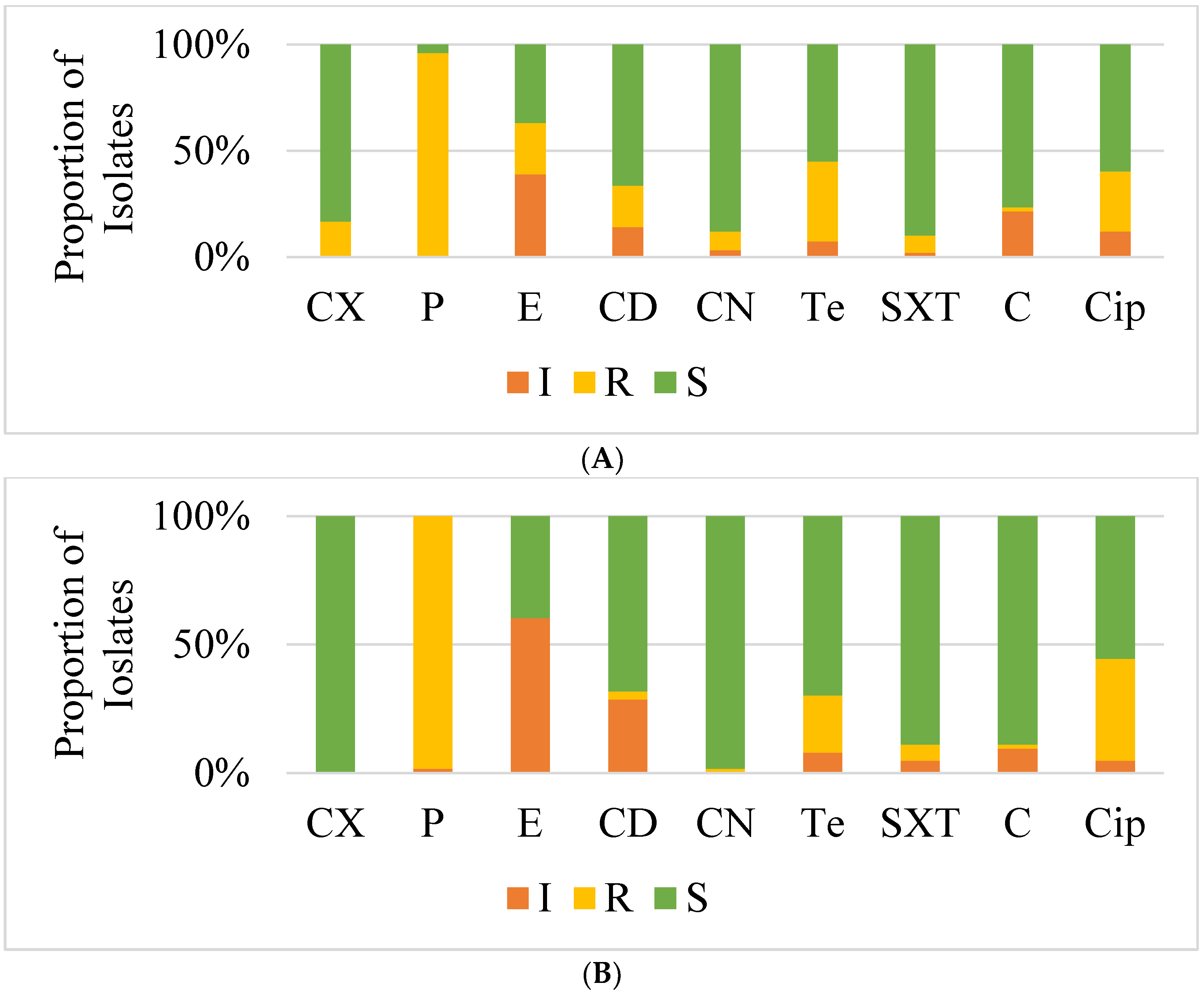
The antimicrobial susceptibility profiles of the isolates revealed that most isolates from farms and abattoirs were resistant to several antibiotics, with the highest resistance being to penicillin (98%). This finding is significantly higher than that from the study in Nigeria which reported a lower resistance to penicillin of 55% [22]. The high resistance to penicillin reflects possible overuse of the antibiotic, as penicillin is generally among most frequently used antibiotics in many farms in many countries [22,24,25]. Resistance to tetracycline, erythromycin and ciprofloxacin was also recorded in 25% to 35% of isolates in our present study. Notably, tetracycline is also commonly used to treat infections in both humans and animals and its resistance can be used as an indicative marker of LA-SA [13,26]. Only 18% of farm isolates were resistant to cefoxitin implying methicillin resistance. However, all these isolates were susceptible to vancomycin with the MICs ranging between 1.5 μg/mL to 3 μg/mL. Vancomycin is the drug of choice for MDR S. aureus infections in human health and is rarely used to treat animal infections [27]. This finding which is similar to that of a previous study that studied vancomycin susceptibility of clinical S. aureus show that vancomycin is still a viable treatment option of S. aureus infections in Zambia [28].
The isolates in the present study were more susceptible to co-trimoxazole, gentamicin and chloramphenicol ranging from 79% to 92%. Inducible resistance to macrolides, lincosamides, and group B streptogramins (MLSBi) phenotype was only detected in one isolate. MLSBi phenotype positive isolates appear to be erythromycin-resistant and clindamycin sensitive in vitro, but when given in vivo, they have constitutive erm mutations that render clindamycin ineffective [29]. A recent study at the largest referral hospital in Zambia found that none of the isolates had the MLSBi phenotype [28]. However, an earlier study at the same hospital reported a high rate of the MLSBi phenotype of 68.3% [19]. Many studies on S. aureus in animals do not report on the MLSBi phenotype probably because clindamycin is not used to treat infections in animals, however, a study from South Africa reported the MLSBi phenotype among the studied isolates from pigs [23]. Although multi-drug resistance was observed to two or more antibiotics in more than 40% of the isolates, generally our findings suggest that there are seemingly still several antibiotics that would be viable to treat infections caused by these isolates from the pig and pork production sector in Zambia.
Unexpectedly, despite the phenotypic resistance to methicillin based on resistance to oxacillin using cefoxitin disc that was detected in some of the isolates, neither the mecA nor mecC genes that encode for methicillin resistance were detected in any of the isolates. A possible explanation to the phenotypic resistance could be that the isolates are hyperproducers of penicillinases that confer some resistance to cefoxitin [30]. While the mecA is the mainstay gene responsible for methicillin resistance in clinical isolates, the mecC gene is linked to livestock associated staphylococcus especially LA-MRSA [31]. A recent study from South Africa reported the presence of the mecC in pig-associated S. aureus for the first time in Africa [32]. Relatively few countries have reported typical LA-MRSA pig-related S. aureus in Africa [18]. Studies from other parts of the world such as Europe and America state that intensive pig farming methods and heavy use of antibiotics are risk factors for the emergence and spread of methicillin resistance as well as resistance to other antibiotics in S. aureus among pigs and attending workers at farms and slaughterhouses [9,33,34]. However, none of the facilities included in the current study practises such intensive pig rearing. Markedly, MSSA cannot be overlooked as they form the reservoir from which MRSA arise [11,35]. The presence of antimicrobial resistance genes in the present study including tetM, tetK and tetL genes encoding for tetracycline resistance and ermB and ermC genes encoding resistance to erythromycin in some of the isolates indicate the need to closely monitor these strains as they may become a source of antimicrobial resistance given that some of these genes are harboured on plasmids which can be easily transferred between microorganisms [36].
Genes encoding the PVL and SEs were not detected in any of the isolates in the present study. While this was the first study to look for the presence of these genes in isolates from pigs in Zambia, the PVL has been reported in a previous study of clinical isolates howbeit only three out of 33 isolates were positive [37]. A study in Senegal on pigs and workers at commercial farm reported a high prevalence of the PVL gene [38]. The PVL is associated with skin and soft tissue infections and has a provenance for humans, but our study indicates that it is dispensible for pig colonisation. The role of SEs in Staphylococcal foodborne disease has been documented in several studies [39,40,41]. Therefore, the non-detection of SEs could indicate the relative safety of the pork and pork products on the Zambian market for consumers. Interestingly, several isolates harboured the IEC genes with the sak being the most prevalent. The staphylokinase and chemotaxis inhibitory proteins form the IEC and contribute to immune evasion in humans [12]. While IEC genes are less prevalent in livestock-adapted S. aureus lineages, they are considered good genetic markers for identification of human-associated S. aureus clones [42]. Therefore, the finding of IEC genes among S. aureus isolates from pigs in the present study potentiate the notion of possible anthropogenic nature of some of the S. aureus in Africa but could also indicate the presence of LA-SA that are well adapted to human hosts [15,18].
Our study found six spa types among which t1430 was the most prevalent followed by t304 mostly among S. aureus isolates from pigs both at the farms and abattoirs. Of significance is that t1430 and t034 are associated with CC9 and CC398 which are Livestock-associated lineages of S. aureus in Asia and Europe, respectively [3,4]. Therefore, our findings suggest that typical LA-SA lineages are present in pig and pork production facilities in Zambia. Generally, the spa types detected in pigs were different from those detected in humans in the present study, only t1430 was found in pigs and workers isolates. This would suggest distinct S. aureus lineages in the two populations. However, given that we could not identify the spa types of many isolates, we recommend further investigations into the clonal lineages using other molecular methods such as multilocus sequence typing (MLST) and whole genome sequencing (WGS) which could not be performed in the present study. Previous studies on pig related S. aureus isolates on the African continent are relatively few but show that the isolates have diverse spa types [18]. Furthermore, studies looking into the presence of LA-SA as a cause of disease among hospitalised patients in Africa are needed as this has not been reported yet but have a large impact on epidemiology of S. aureus infections.

4. Materials and Methods

4.1. Study Design and Sample Collection

The study was a cross sectional study carried out between June 2020 and September 2021 in three districts of the Lusaka Province of Zambia namely Chilanga, Lusaka and Chongwe districts (Figure 2). Lusaka Province hosts many of the commercial and semi-commercial (small and medium scale rearing of pigs meant solely for selling) pig farms in Zambia. Pig farms and abattoirs in selected districts within the province were included in the study following consent from the farm and abattoir owners. The farms and abattoirs were arbitrarily grouped into three following categories based on the number of pigs at the facility: small scale (less than 100 pigs), medium scale (100 to 500 pigs) and commercial scale (greater than 500 pigs). A total of 492 pig nasal swabs were randomly collected from 13 farms and three abattoirs by inserting a swab and gently rotating it in the anterior nares. Additionally, 53 nasal and 53 hand swabs each from humans (farm workers and abattoir workers) in close contact with the pigs were collected. The human nasal swabs were collected by inserting a swab and gently rotating it in the anterior nares, whereas a hand swab was collected by gently rubbing the swab in both palms.

4.2. S. aureus Detection and Identification

Phenotypic detection and identification of S. aureus was carried out by conventional microbiological methods [43]. Briefly, each swab was transferred into 10 mL Mueller-Hinton broth (MHB) (Oxoid, Basingstoke, UK) supplemented with 6% Sodium Chloride (NaCl) and incubated at 37 °C for 16 to 20 h. Then, a loopful of the broth was inoculated on Mannitol Salt Agar (MSA) (Oxoid, Basingstoke, UK) and the plates were then incubated for 16 to 20 h at 37 °C. Resulting yellowish colonies were then inoculated onto Baird Parker Agar (BPA) (Oxoid, Basingstoke, UK) and incubated for 16 to 20 h at 37 °C. Resulting black or greyish colonies with or without a halo on the BPA plates were then grown in Brain Heart Infusion Broth (BHIB) (Oxoid, Basingstoke, UK) for 16 to 20 h at 37 °C. Using 0.5 mL of the BHIB culture, a tube coagulase test using rabbit plasma was set up according to manufacturer’s instructions (Sigma-Aldrich, Taufkirchen, Germany) by incubating the tubes at 37 °C and reading after four (4) hours and at 24 h. All coagulase positive isolates were considered as S. aureus and stored in 20% glycerol at −20 °C until further analysis.

4.3. Determination of Antimicrobial Susceptibility Profiles

The antimicrobial susceptibility of S. aureus to antibiotic discs (Oxoid, Basingstoke, UK) of 10 µg gentamycin, 5 µg ciprofloxacin, 15 µg erythromycin, 10 µg clindamycin, 30 µg amikacin, 10 units penicillin G, 25 µg co-trimoxazole, 30 µg chloramphenicol and 30 µg tetracycline was determined using the Kirby-Baur disc diffusion method and interpreted according to the 2020 Clinical and Laboratory Standards Institute (CLSI) guidelines [44]. Methicillin resistance was detected by checking for resistance to oxacillin using the 30 µg cefoxitin discs following the 2020 CLSI guidelines [44]. The D-test using erythromycin and clindamycin discs was also used to detect inducible resistance to macrolides, lincosamides, and group B streptogramins (MLSBi) in the S. aureus isolates. Susceptibility to vancomycin was determined for the isolates resistant to methicillin using vancomycin E-strips to determine the minimum inhibition concentrations (MICs) according to the CLSI guidelines [44]. Briefly, using a swab, one to two pure colonies of the organism grown overnight on nutrient agar were suspended into 2 mLs of physiological normal saline to make a 0.5 McFarland density. These bacteria were then spread evenly on a Mueller Hinton agar plate using a sterile swab. After allowing the plate to air dry for a few minutes, antimicrobial discs were gently placed on the Mueller Hinton agar plate ensuring that discs were not closer than 24 mm from centre to centre. The plates were then incubated for 16 to 24 h at 37 °C for the other antibiotics and at 35 °C for 24 h for cefoxitin. For vancomycin, one E-strip was placed per plate of the isolate, which were incubated for 16 to 24 h at 37 °C.

4.4. Molecular Identification and Genotyping

4.4.1. DNA Extraction

Genomic DNA was prepared by thermo lysis of fresh S. aureus cells. Briefly, a loopful of S. aureus cells from Nutrient Agar (Oxoid, Basingstoke, UK) were transferred into a micro centrifuge tube containing 200 µL of 1X MiliQ water and boiled for 15 min. After cooling on ice, the DNA thermolysate were centrifuged at 14,000× g and then stored at −20 °C until required.

4.4.2. Molecular Identification of S. aureus

The species identification of the isolates was then confirmed by detection of the nuclease gene using nuc primers (Table 6) according to a previously described PCR protocol [45].

4.4.3. Detection of Methicillin Resistance Genes and Other Antimicrobial Resistance Genes

The presence of the mecA and mecC gene was checked by using previously described PCR protocols [46,47] and primers (Table 6). The erythromycin resistance encoding genes (ermA, ermB and ermC) and tetracycline resistance encoding genes (tetK, tetL, tetM and tetO) were detected using previously described protocols and primers shown in Table 6 [48,49].

4.4.4. Detection of PVL and SE Genes

PCR with gene-specific primers (Table 6) was performed according to previously described protocols to detect genes encoding several virulence factors of S. aureus including lukS-PV and lukF-PV genes encoding PVL [47], immune evasion cluster genes (IEC) sak, scn and chp [12] and staphylococcal enterotoxins: sea, seb, sec, sed, and see [52]. PCR conditions were as described in the previous protocols, respectively [12,47,52].

4.4.5. Spa Type Determination

Spa typing was performed using a previously described PCR protocol [51] with the primer sets shown in Table 6. Sequencing of the protein A gene (spa) was performed using BigDye terminator method with an ABI PRISM 3730XL DNA analyser (Applied Biosystems, Foster City, CA, USA). The DNA sequence reads were edited using the ATGC Software. The sequences obtained were then submitted to the online tool Center for Genomic Epidemiology to determine the spa types [53].

4.5. Data Analysis

Data from the study were entered into Microsoft™ excel spreadsheets, and then analysed using IBM SPSS version 25 (IBM Corp) and R. The frequencies of S. aureus in farm and abattoirs were presented as percentages and 95% confidence intervals. P values of less than 0.05 were considered statistically significant. The chromatograph sequence files of the isolates were analysed with the online tool Centre of Genomic Epidemiology (CGE) for Spa typing to determine the spa types [53].

5. Conclusions

The presence of S. aureus was high among pigs in Zambia. Furthermore, the detection of S. aureus on the hands and in nasal cavities of farm and abattoir workers is a public health concern. Although MRSA was only phenotypically detected, the significance of MSSA as a potential source from which MRSA can arise cannot be overlooked. The presence of plasmid mediated resistance genes and immune evasion genes among the isolates warrant continuous monitoring of S. aureus in this sector to combat S. aureus infections using a “One Health” approach in Zambia.

Supplementary Materials

The following supporting information can be downloaded at: https://www.mdpi.com/article/10.3390/antibiotics11070844/s1, Table S1: Samples for which more than one S. aureus colony type was isolated; Table S2: Vancomycin MICs of S. aureus; Table S3: Characteristics of S. aureus Isolates Sequenced for Spa typing. Reference [44] is cited in the supplementary materials.

Author Contributions

Conceptualization, M.T.S., E.S., G.K. and B.M.H.; Data curation, M.T.S., W.M. and H.K.; Formal analysis, M.T.S., W.M. and H.K.; Funding acquisition, M.T.S. and B.M.H.; Investigation, M.T.S., L.M., E.C.B., T.N., K.Y. and I.M.; Methodology, M.T.S. and B.S.J.P.; Project administration, M.T.S. and B.S.J.P.; Resources, M.T.S., B.S.J.P., E.S., W.M., I.M., A.F., M.B. and B.M.H.; Software, W.M. and S.A.K.; Supervision, E.S., G.K., A.F., H.H. and B.M.H.; Validation, M.T.S.; Visualization, S.A.K.; Writing—original draft, M.T.S.; Writing—review and editing, B.S.J.P., E.S., G.K., L.M., E.C.B., W.M., T.N., K.Y., H.K., S.A.K., I.M., A.F., M.B., H.H. and B.M.H. All authors have read and agreed to the published version of the manuscript.

Funding

This research was funded in part by the Africa Centre of Excellence for Infectious Diseases of Humans and Animals (ACEIDHA) project (grant # P151847) funded by the World Bank; International Foundation for Science (IFS) (grant # I3-B-6524-1) and “The APC was funded by ACEIDHA”. Authors MB and ES acknowledge support from the European and Developing Countries Clinical Trials Partnership (EDCTP2) Programme, Horizon 2020, the European Union’s Framework Programme for Research and Innovation, grants PANDORA-ID-NET, EMPIRICAL, DATURA and CANTAM.

Institutional Review Board Statement

The study was conducted in accordance with the Declaration of Helsinki and approved by the University of Zambia Biomedical Research Ethics Committee (UNZABREC) of The University of Zambia (protocol code 613-2019 approved on 20 January 2020) for studies involving humans or animals.

Informed Consent Statement

Written informed consent was obtained from all farm and abattoir workers as well as owners of the facilities involved in the study.

Data Availability Statement

Sequences for the spa types have deposited in the DDBJ.

Acknowledgments

The authors sincerely thank the farm and abattoir owners and workers, for allowing them to conduct the study at their facilities and for taking part in the study, respectively. Special thanks also go to the laboratory technicians at the University of Zambia, School of Veterinary Medicine who assisted in the data collection process.

Conflicts of Interest

The authors declare no conflict of interest. The funders had no role in the design of the study; in the collection, analyses, or interpretation of data; in the writing of the manuscript, or in the decision to publish the results.

References

  1. Lozano, C.; Gharsa, H.; Ben Slama, K.; Zarazaga, M.; Torres, C. Staphylococcus aureus in animals and food: Methicillin resistance, prevalence and population structure. A review in the african continent. Microorganisms 2016, 4, 12. [Google Scholar] [CrossRef] [PubMed] [Green Version]
  2. Köck, R.; Becker, K.; Cookson, B.; Van Gemert-Pijnen, J.E.; Harbarth, S.; Kluytmans, J.; Mielke, M.; Peters, G.; Skov, R.L.; Struelens, M.J.; et al. Methicillin-resistant Staphylococcus aureus (MRSA): Burden of disease and control challenges in Europe. Euro Surveill. 2010, 15, 19688. [Google Scholar] [CrossRef] [PubMed]
  3. Tegegne, H.A.; Koláčková, I.; Karpíšková, R. Diversity of livestock associated methicillin-resistant staphylococcus aureus. Asian Pac. J. Trop. Med. 2017, 10, 929–931. [Google Scholar] [CrossRef] [PubMed]
  4. Smith, T.C.; Pearson, N. The emergence of staphylococcus aureus ST398. Vector-Borne Zoonotic Dis. 2010, 11, 327–339. [Google Scholar] [CrossRef] [PubMed]
  5. Ye, X.; Fan, Y.; Wang, X.; Liu, W.; Yu, H.; Zhou, J.; Chen, S.; Yao, Z. Livestock-associated methicillin and multidrug resistant S. aureus in humans is associated with occupational pig contact, not pet contact. Sci. Rep. 2016, 6, 19184. [Google Scholar] [CrossRef] [Green Version]
  6. Graveland, H.; Duim, B.; van Duijkeren, E.; Heederik, D.; Wagenaar, J.A. Livestock-associated methicillin-resistant staphylococcus aureus in animals and humans. Int. J. Med Microbiol. 2011, 301, 630–634. [Google Scholar] [CrossRef]
  7. Verkade, E.; Kluytmans, J. Livestock-associated staphylococcus aureus CC398: Animal reservoirs and human infections. Infect. Genet. Evol. 2014, 21, 523–530. [Google Scholar] [CrossRef]
  8. Lekkerkerk, W.S.N.; van Wamel, W.J.B.; Snijders, S.V.; Willems, R.J.; van Duijkeren, E.; Broens, E.M.; Wagenaar, J.A.; Lindsay, J.A.; Vos, M.C. What Is the origin of livestock-associated methicillin-resistant staphylococcus aureus clonal complex 398 isolates from humans without livestock contact? An epidemiological and genetic analysis. J. Clin. Microbiol. 2015, 53, 1836–1841. [Google Scholar] [CrossRef] [Green Version]
  9. Köck, R.; Harlizius, J.; Bressan, N.; Laerberg, R.; Wieler, L.H.; Witte, W.; Deurenberg, R.H.; Voss, A.; Becker, K.; Friedrich, A.W. Prevalence and molecular characteristics of methicillin-resistant staphylococcus aureus (MRSA) among pigs on German farms and import of livestock-related MRSA into hospitals. Eur. J. Clin. Microbiol. 2009, 28, 1375–1382. [Google Scholar] [CrossRef] [Green Version]
  10. Larsen, J.; Petersen, A.; Larsen, A.R.; Sieber, R.N.; Stegger, M.; Koch, A.; Aarestrup, F.; Price, L.B.; Skov, R.L.; Johansen, H.K.; et al. Emergence of livestock-associated methicillin-resistant staphylococcus aureus bloodstream infections in Denmark. Clin. Infect. Dis. 2017, 65, 1072–1076. [Google Scholar] [CrossRef] [Green Version]
  11. Price, L.B.; Stegger, M.; Hasman, H.; Aziz, M.; Larsen, J.; Andersen, P.S.; Pearson, T.; Waters, A.E.; Foster, J.T.; Schupp, J.; et al. Staphylococcus aureus CC398: Host adaptation and emergence of methicillin resistance in livestock. mBio 2012, 3, e00305–e00311. [Google Scholar] [CrossRef] [PubMed] [Green Version]
  12. van Wamel, W.J.; Rooijakkers, S.H.; Ruyken, M.; van Kessel, K.P.; van Strijp, J.A. The innate immune modulators staphylococcal complement inhibitor and chemotaxis inhibitory protein of staphylococcus aureus are located on beta-hemolysin-converting bacteriophages. J. Bacteriol. 2006, 188, 1310–1315. [Google Scholar] [CrossRef] [PubMed] [Green Version]
  13. Benito, D.; Lozano, C.; Rezusta, A.; Ferrer, I.; Vasquez, M.A.; Ceballos, S.; Zarazaga, M.; Revillo, M.J.; Torres, C. Characterization of tetracycline and methicillin resistant staphylococcus aureus strains in a Spanish hospital: Is livestock-contact a risk factor in infections caused by MRSA CC398? Int. J. Med. Microbiol. 2014, 304, 1226–1232. [Google Scholar] [CrossRef] [PubMed]
  14. Bouiller, K.; Bertrand, X.; Hocquet, D.; Chirouze, C. Human infection of methicillin-susceptible Staphylococcus aureus CC398: A review. Microorganisms 2020, 8, 1737. [Google Scholar] [CrossRef] [PubMed]
  15. Mama, O.M.; Aspiroz, C.; Ruiz-Ripa, L.; Ceballos, S.; Iñiguez-Barrio, M.; Cercenado, E.; Azcona, J.M.; López-Cerero, L.; Seral, C.; López-Calleja, A.I.; et al. Prevalence and genetic characteristics of staphylococcus aureus cc398 isolates from invasive infections in Spanish hospitals, focusing on the livestock-independent CC398-MSSA clade. Front. Microbiol. 2021, 12, 623108. [Google Scholar] [CrossRef] [PubMed]
  16. Tavares, A.; Faria, N.A.; De Lencastre, H.; Miragaia, M. Population structure of methicillin-susceptible Staphylococcus aureus (MSSA) in Portugal over a 19-year period (1992–2011). Eur. J. Clin. Microbiol. 2013, 33, 423–432. [Google Scholar] [CrossRef]
  17. Vandendriessche, S.; Kadlec, K.; Schwarz, S.; Denis, O. Methicillin-susceptible staphylococcus aureus ST398-t571 harbouring the macrolide-lincosamide-streptogramin B resistance gene erm(T) in Belgian hospitals. J. Antimicrob. Chemother. 2011, 66, 2455–2459. [Google Scholar] [CrossRef] [Green Version]
  18. Samutela, M.T.; Kwenda, G.; Simulundu, E.; Nkhoma, P.; Higashi, H.; Frey, A.; Bates, M.; Hang’Ombe, B.M. Pigs as a potential source of emerging livestock-associated staphylococcus aureus in Africa: A systematic review. Int. J. Infect. Dis. 2021, 109, 38–49. [Google Scholar] [CrossRef]
  19. Samutela, M.T.; Mwansa, J.C.; Kalonda, A.; Mumbula, E.M.; Kaile, T.; Marimo, C.; Korolyova, L.; Hang'ombe, B.M.; Simulundu, E.; Musyani, C.; et al. Antimicrobial susceptibility profiles of methicillin resistant staphylococcus aureus isolates from the university teaching hospital, Lusaka, Zambia. J. Med. Sci. Technol. 2015, 4, 19–25. [Google Scholar]
  20. Youn, J.-H.; Park, Y.H.; Hang’Ombe, B.; Sugimoto, C. Prevalence and characterization of staphylococcus aureus and staphylococcus pseudintermedius isolated from companion animals and environment in the veterinary teaching hospital in Zambia, Africa. Comp. Immunol. Microbiol. Infect. Dis. 2014, 37, 123–130. [Google Scholar] [CrossRef]
  21. Pandey, G.S.; Nomura, Y.; Kobayashi, K.; Fujise, H.; Yamada, T. Cutaneous staphylococcal granuloma in a free living zebra (Equus burchelli) in Zambia. J. Veter-Med Sci. 1998, 60, 137–138. [Google Scholar] [CrossRef] [PubMed] [Green Version]
  22. Gaddafi, M.S.; Yakubu, Y.; Junaidu, A.U.; Bello, M.B.; Garba, B.; Bitrus, A.A.; Lawal, H. Nasal colonization of pigs and farm attendants by staphylococcus aureus and methicillin-resistant staphylococcus aureus (MRSA) in Kebbi, Northwestern Nigeria. Thai J. Vet. Med. 2021, 51, 119–124. [Google Scholar]
  23. Sineke, N.; Asante, J.; Amoako, D.G.; Abia, A.L.K.; Perrett, K.; Bester, L.A.; Essack, S.Y. Staphylococcus aureus in intensive pig production in South Africa: Antibiotic resistance, virulence determinants and clonality. Pathogens 2021, 10, 317. [Google Scholar] [CrossRef] [PubMed]
  24. Holmer, I.; Salomonsen, C.M.; Jorsal, S.E.; Astrup, L.B.; Jensen, V.F.; Høg, B.B.; Pedersen, K. Antibiotic resistance in porcine pathogenic bacteria and relation to antibiotic usage. BMC Veter-Res. 2019, 15, 449. [Google Scholar] [CrossRef] [Green Version]
  25. Lekagul, A.; Tangcharoensathien, V.; Mills, A.; Rushton, J.; Yeung, S. How antibiotics are used in pig farming: A mixed-methods study of pig farmers, feed mills and veterinarians in Thailand. BMJ Glob. Health 2020, 5, e001918. [Google Scholar] [CrossRef] [Green Version]
  26. Lozano, C.; Rezusta, A.; Gómez, P.; Gómez-Sanz, E.; Báez, N.; Martin-Saco, G.; Zarazaga, M.; Torres, C. High prevalence of spa types associated with the clonal lineage CC398 among tetracycline-resistant methicillin-resistant Staphylococcus aureus strains in a Spanish hospital. J. Antimicrob. Chemother. 2012, 67, 330–334. [Google Scholar] [CrossRef] [Green Version]
  27. Pahadi, P.C.; Shrestha, U.T.; Adhikari, N.; Shah, P.K.; Amatya, R. Growing Resistance to Vancomycin among Methicillin Resistant Staphylococcus Aureus Isolates from Different Clinical Samples. JNMA J. Nepal Med. Assoc. 2014, 52, 977–981. [Google Scholar] [CrossRef]
  28. Mutalange, M.; The University of Zambia; Yamba, K.; Kapesa, C.; Mtonga, F.; Banda, M.; Muma, J.B.; Hangombe, B.M.; Hachaambwa, L.; Bumbangi, F.N.; et al. Vancomycin resistance in staphylococcus aureus and enterococcus species isolated at the university teaching hospitals, Lusaka, Zambia: Should we be worried? Univ. Zamb. J. Agric. Biomed. Sci. 2021, 5, 18–28. [Google Scholar] [CrossRef]
  29. Prabhu, K.; Rao, S.; Rao, V. Inducible clindamycin resistance in staphylococcus aureus isolated from clinical samples. J. Lab. Physicians 2011, 3, 25–27. [Google Scholar] [CrossRef]
  30. Montanari, M.P.; Tonin, E.; Biavasco, F.; Varaldo, P.E. Further characterization of borderline methicillin-resistant staphylococcus aureus and analysis of penicillin-binding proteins. Antimicrob. Agents Chemother. 1990, 34, 911–913. [Google Scholar] [CrossRef] [Green Version]
  31. Paterson, G.K.; Larsen, A.R.; Robb, A.; Edwards, G.E.; Pennycott, T.W.; Foster, G.; Mot, D.; Hermans, K.; Baert, K.; Peacock, S.J.; et al. The newly described mecA homologue, mecALGA251, is present in methicillin-resistant staphylococcus aureus isolates from a diverse range of host species. J. Antimicrob. Chemother. 2012, 67, 2809–2813. [Google Scholar] [CrossRef] [PubMed]
  32. Dweba, C.C.; Zishiri, O.T.; El Zowalaty, M.E. Isolation and molecular identification of virulence, antimicrobial and heavy metal resistance genes in livestock-associated methicillin-resistant staphylococcus aureus. Pathogens 2019, 8, 79. [Google Scholar] [CrossRef] [PubMed] [Green Version]
  33. Fang, H.-W.; Chiang, P.-H.; Huang, Y.-C. Livestock-associated methicillin-resistant staphylococcus aureus ST9 in pigs and related personnel in Taiwan. PLoS ONE 2014, 9, e88826. [Google Scholar] [CrossRef] [PubMed] [Green Version]
  34. Van Duijkeren, E.; Ikawaty, R.; Broekhuizen-Stins, M.J.; Jansen, M.D.; Spalburg, E.C.; De Neeling, A.J.; Allaart, J.G.; Van Nes, A.; Wagenaar, J.A.; Fluit, A.C. Transmission of methicillin-resistant staphylococcus aureus strains between different kinds of pig farms. Vet. Microbiol. 2008, 126, 383–389. [Google Scholar] [CrossRef] [PubMed]
  35. Laumay, F.; Benchetrit, H.; Corvaglia, A.-R.; van der Mee-Marquet, N.; François, P. The Staphylococcus aureus CC398 lineage: An evolution driven by the acquisition of prophages and other mobile genetic elements. Genes 2021, 12, 1752. [Google Scholar] [CrossRef] [PubMed]
  36. Emaneini, M.; Bigverdi, R.; Kalantar, D.; Soroush, S.; Jabalameli, F.; Khoshgnab, B.N.; Asadollahi, P.; Taherikalani, M. Distribution of genes encoding tetracycline resistance and aminoglycoside modifying enzymes in staphylococcus aureus strains isolated from a burn center. Ann. Burn. Fire Disasters 2013, 26, 76–80. [Google Scholar]
  37. Samutela, M.T.; Kalonda, A.; Mwansa, J.; Lukwesa-Musyani, C.; Mwaba, J.; Mumbula, E.M.; Mwenya, D.; Simulundu, E.; Kwenda, G. Molecular characterisation of methicillin-resistant staphylococcus aureus (MRSA) isolated at a large referral hospital in Zambia. Pan Afr. Med, J. 2017, 26, 108. [Google Scholar]
  38. Fall, C.; Seck, A.; Richard, V.; Ndour, M.; Sembène, M.; Laurent, F.; Breurec, S. Epidemiology of staphylococcus aureus in pigs and farmers in the largest farm in dakar, senegal. Foodborne Pathog. Dis. 2012, 9, 962–965. [Google Scholar] [CrossRef] [Green Version]
  39. Bennett, R.W.; Monday, S.R. Staphylococcus aureus. In International Handbook of Foodborne Pathogens; Marcel Dekker: New York, NY, USA, 2003; pp. 41–60. [Google Scholar]
  40. Gallina, S.; Bianchi, D.M.; Bellio, A.; Nogarol, C.; Macori, G.; Zaccaria, T.; Biorci, F.; Carraro, E.; Decastelli, L. Staphylococcal poisoning foodborne outbreak: Epidemiological investigation and strain genotyping. J. Food Prot. 2013, 76, 2093–2098. [Google Scholar] [CrossRef]
  41. Kadariya, J.; Smith, T.C.; Thapaliya, D. Staphylococcus aureus and staphylococcal food-borne disease: An ongoing challenge in public health. BioMed. Res. Int. 2014, 2014, 827965. [Google Scholar] [CrossRef] [Green Version]
  42. McCarthy, A.J.; Witney, A.A.; Gould, K.A.; Moodley, A.; Guardabassi, L.; Voss, A.; Denis, O.; Broens, E.M.; Hinds, J.; Lindsay, J.A. The distribution of mobile genetic elements (MGEs) in MRSA CC398 is associated with both host and country. Genome Biol. Evol. 2011, 3, 1164–1174. [Google Scholar] [CrossRef] [PubMed] [Green Version]
  43. Winn, W.C. Koneman’s Color Atlas and Textbook of Diagnostic Microbiology; Lippincott Williams & Wilkins: Philadelphia, PA, USA, 2006. [Google Scholar]
  44. Clinical and Laboratory Standards Institute. Performance Standards for Antimicrobial Susceptibility Testing, 30th ed.; Supplement M1002020; CLSI: Tehran, Iran, 2011. [Google Scholar]
  45. Zhang, K.; Sparling, J.; Chow, B.L.; Elsayed, S.; Hussain, Z.; Church, D.L.; Gregson, D.B.; Louie, T.; Conly, J.M. New quadriplex PCR assay for detection of methicillin and mupirocin resistance and simultaneous discrimination of Staphylococcus aureus from coagulase-negative staphylococci. J. Clin. Microbiol. 2004, 42, 4947–4955. [Google Scholar] [CrossRef] [PubMed] [Green Version]
  46. Milheiriço, C.; Oliveira, D.C.; de Lencastre, H. Update to the multiplex PCR strategy for assignment of mec element types in Staphylococcus aureus. Antimicrob. Agents Chemother. 2007, 51, 3374–3377. [Google Scholar]
  47. Stegger, Á.; Andersen, P.S.; Kearns, A.; Pichon, B.; Holmes, M.A.; Edwards, G.; Laurent, F.; Teale, C.; Skov, R.; Larsen, A.R. Rapid detection, differentiation and typing of methicillin-resistant Staphylococcus aureus harbouring either mecA or the new mecA homologue mecA (LGA251). Clin. Microbiol. Infect. 2012, 18, 395–400. [Google Scholar] [CrossRef] [Green Version]
  48. Sutcliffe, J.; Grebe, T.; Tait-Kamradt, A.; Wondrack, L. Detection of erythromycin-resistant determinants by PCR. Antimicrob. Agents Chemother. 1996, 40, 2562–2566. [Google Scholar] [CrossRef] [Green Version]
  49. Aarestrup, F.M.; Agerso, Y.; Gerner–Smidt, P.; Madsen, M.; Jensen, L.B. Comparison of antimicrobial resistance phenotypes and resistance genes in enterococcus faecalis and enterococcus faecium from humans in the community, broilers, and pigs in Denmark. Diagn. Microbiol. Infect. Dis. 2000, 37, 127–137. [Google Scholar] [CrossRef]
  50. Becker, K.; Haverkämper, G.; Von Eiff, C.; Roth, R.; Peters, G. Survey of staphylococcal enterotoxin genes, exfoliative toxin genes, and toxic shock syndrome toxin 1 gene in non-staphylococcus aureus species. Eur. J. Clin. Microbiol. Infect. Dis. 2001, 20, 407–409. [Google Scholar]
  51. Shopsin, B.; Gomez, M.; Montgomery, S.O.; Smith, D.H.; Waddington, M.; Dodge, D.E.; Bost, D.A.; Riehman, M.; Naidich, S.; Kreiswirth, B.N. Evaluation of protein A gene polymorphic region DNA sequencing for typing of staphylococcus aureus strains. J. Clin. Microbiol. 1999, 37, 3556–3563. [Google Scholar] [CrossRef] [Green Version]
  52. Omoe, K.; Ishikawa, M.; Shimoda, Y.; Hu, D.L.; Ueda, S.; Shinagawa, K. Detection of seg, seh, and sei genes in staphylococcus aureus isolates and determination of the enterotoxin productivities of s. aureus isolates harboring seg, seh, or sei genes. J. Clin. Microbiol. 2002, 40, 857–862. [Google Scholar] [CrossRef] [Green Version]
  53. Bartels, M.D.; Petersen, A.; Worning, P.; Nielsen, J.B.; Larner-Svensson, H.; Johansen, H.K.; Andersen, L.P.; Jarløv, J.O.; Boye, K.; Larsen, A.R.; et al. Comparing whole-genome sequencing with sanger sequencing for spa typing of methicillin-resistant staphylococcus aureus. J. Clin. Microbiol. 2014, 52, 4305–4308. [Google Scholar] [CrossRef] [Green Version]
Figure 1. Overall Antimicrobial Susceptibility Profiles of S. aureus Isolates from pigs and workers from Farms (A) and Abattoirs (B) of Lusaka Province. Abbreviations: P = Penicillin; CN = Gentamicin; E = Erythromycin; CD = Clindamycin; Cip = Ciprofloxacin; Te = Tetracycline, SXT = Cotrimoxazole, C = Chloramphenicol, CX = Cefoxitin; I = Intermediate, R = Resistant, S = Susceptible.
Antibiotics 11 00844 g001
Figure 2. Map of the Study Sites Selected from Lusaka province in Zambia; Map was generated using the software ArcGIS version 10.3.
Antibiotics 11 00844 g002
Table 1. S. aureus positivity rates from pigs, humans and districts in Lusaka Province.
FactorCategoryn Testedn PositivesPrevalence (%)95% CI
Overall PositivityPositive59819833.129.4–37.1
HumansOverall1061211.36.2–19.3
Hand Swabs53611.34.7–23.7
Nasal Swabs53611.34.7–23.7
PigsNasal swabs49218637.833.5–42.3
DistrictsChongwe2506024.018.9–29.9
Lusaka2356326.821.4–33.0
Chilanga1137566.456.6–74.8
Abbreviations: CI = Confidence interval; n = number of samples.
Table 2. Prevalence of S. aureus in pigs and humans at farms and abattoirs.
Study SiteSpeciesType of Facility *n Testedn PositivesPrevalence (%)95% CI
FarmsCombined pigs and humansSmall531324.514.2–38.6
Medium2526124.219.1–30.1
Large2026431.725.4–38.7
Overall50713827.223.4–31.3
Pigs onlySmall451328.916.8–44.5
Medium2165726.420.8–32.9
Large1576138.931.8–47.0
Overall41813131.327.0–36.1
Humans onlyNasal3837.92.1–22.5
Hand38410.53.4–25.7
Overall7679.24.1–18.6
Human NasalSmall4000
Medium18211.12.0–36.1
Large1616.30.3–32.3
Human HandSmall4000
Medium18211.12.0–36.1
Large16212.52.2–39.6
AbattoirsCombined pigs and humansMedium204206.6–44.3
Large715678.967.3–87.3
Overall916065.955.2–75.3
Pigs onlyMedium204206.6–44.3
Large545194.483.7–98.6
Overall715577.565.7–86.2
Humans **Hand82254. 5–64.4
Nasal9333.39.0–69.1
Overall17529.411.4–56.0
* Type of facility: Small scale (less than 100 pigs), medium scale (100 to 500 pigs) and commercial scale (greater than 500 pigs; ** All human swabs from abattoirs were collected at the large facilities only. Abbreviations: CI = Confidence interval; n = number of samples.
Table 3. Antibiotic resistance patterns of S. aureus isolates from pigs and workers from pig farms and abattoirs in Lusaka Province.
Resistance PatternProportion of Isolates % (n)
Farm Isolates (n = 141)Abattoir Isolates (n = 63)
P34.8 (49)42.9 (27)
Te1.4 (2)1.6 (1)
P + Te20.6 (29)7.9 (5)
P + Cip7.1 (10)34.9 (22)
P + CD0.7 (1)3.3 (2)
P + CN + Te2.11.6 (1)
P + E + TE1.4 (2)-
P + Te + Cip0.7 (1)3.2 (2)
P + E + CD + Cip14.2 (20)-
P + E + CD + TE1.4 (2)-
P + E + C + CIP1.4 (2)-
P + CN + TE + SXT5.0 (7)-
P + E + CD + TE + SXT0.7 (1)-
P + E + CD + CN + Cip1.4 (2)-
P + E + CN + Te + CIP0.7 (1)-
P + E + CD + CN + Te + SXT0.7 (1)-
1 Other4.3 (6)4.7 (3)
Abbreviations: n = number of samples, P = Penicillin; CN = Gentamicin; E = Erythromycin; CD = Clindamycin; Cip = Ciprofloxacin; Te = Tetracycline, SXT = Cotrimoxazole, C = Chloramphenicol, - = Not detected; 1 Other = P + E, P + SXT, P + E + C, P + E + CD, P + E + SXT, P + CD + Te (farm isolates) and P + Te + SXT, P + CD + CN, P + C + Cip (Abattoir isolates). Each pattern was manifested in only one isolate.
Table 4. IEC genes distribution among the S. aureus isolates (n = 225).
IEC Gene
Source (Farm or Abattoir)Sample Typescn % (n)sak% (n)chp % (n)
Farm 1Pig nasal Swab-0.4 (1)-
Farm 2Pig nasal Swab-0.4 (1)-
Farm 4Pig nasal Swab-1.3 (3)-
Farm 5Pig nasal Swab-0.4 (1)-
Farm 6Pig nasal Swab-0.4 (1)-
Farm 7Pig nasal Swab0.9 (2)2.7 (6)-
Farm 9Pig nasal Swab-0.4 (1)-
Farm 10Pig nasal Swab-1.3 (3)-
Abattoir 1Pig nasal Swab0.4 (1)-0.4 (1)
Total1.3 (3)7.6 (17)0.4 (1)
Abbreviation: n = number of isolates; - = None detected.
Table 5. Spa type distribution among representative farm and abattoir isolates (n = 43).
Spa Type % (n)
SpeciesStudy Sitet1430t034t318t571t084t899Unknown
HumansFarms000002.3 (1)4.7 (2)
Abattoirs4.7 (2)0002.3 (1)00
PigsFarms14.0 (6)9.3 (4)9.3 (4)2.3 (1)0025.6 (11)
Abattoirs9.3 (4)9.3 (4)00007.0 (3)
Total28.0 (12)18.6 (8)9.3 (4)2.3 (1)2.3 (1)2.3 (1)37.3 (16)
Abbreviation: n = number of isolates.
Table 6. Primer sets used in the study.
Primer NameTarget GenePrimer Sequence (5′-3′)Amplicon SizeReference
Nuc1nucGCG ATT GAT GGT GAT ACG GTT279 bp[45]
Nuc2 AGC CAA GCC TTG ACG AAC TAA AGC
mecA P4 mecATCCAGATTACAACTTCACCAGG 162 bp[46]
mecA P7 CCACTTCATATCTTGTAACG
mecALGA251mecCGAAAAAAAGGCTTAGAACGCCTC138 bp[47]
mecALGA251 GAAGATCTTTTCCGTTTTCAGC
ermA-1erm[A]TCTAAAAAGCATGTAAAAGAA645 bp[48]
ermA-2 CTTCGATAGTTTATTAATATTAG
ermB-1erm[B]GAAAAGTACTCAACCAAATA639 bp[48]
ermB-2 AGTAACGGTACTTAAATTGTTTA
ermC-1erm[C]TCAAAACATAATATAGATAAA642 bp[48]
ermC-2 GCTAATATTGTTTAAATCGTCAAT
tetK-1tet[K]TTAGGTGAAGGGTTAGGTCC697 bp[49]
tetK-2 GCAAACTCATTCCAGAAGCA
tetM-1tet[M]GTTAAATAGTGTTCTTGGAG576 bp[49]
tetM-2 CTAAGATATGGCTCTAACAA
tetL-1tet[L]CATTTGGTCTTATTGGATCG456 bp[49]
tetL-2 ATTACACTTCCGATTTCGG
tetO-1tet[O]GATGGCATACAGGCACAGAC615 bp[49]
tetO-2 CAATATCACCAGAGCAGGCT
pvl-FPlukF-PVGCTGGACAAAACTTCTTGGAATAT 83[47]
pvl-RP GATAGGACACCAATAAATTCTGGATTG
SEA-3seaCCTTTGGAAACGGTTAAAACG127 bp[50]
SEA-4 TCTGAACCTTCCCATCAAAAAC
SEB-1sebTCGCATCAAACTGACAAACG477 bp[50]
SEB-4 GCAGGTACTCTATAAGTGCCTGC
SEC-3secCTCAAGAACTAGACATAAAAGCTAGG271 bp[50]
SEC-4 TCAAAATCGGATTAACATTATCC
SED-3sedCTAGTTTGGTAATATCTCCTTTAAACG319 bp[50]
SED-4 TTAATGCTATATCTTATAGGGTAAACATC
SEE-3seeCAGTACCTATAGATAAAGTTAAAACAAGC178 bp[50]
SEE-2 TAACTTACCGTGGACCCTTC
Sak-1sakAAGGCGATGACGCGAGTTAT223 bp[12]
Sak-2 GCGCTTGGATCTAATTCAAC
Chp-1chpGAAAAAGAAATTAGCAACAACAG410 bp[12]
Chp-2 CATAAGATGATTTAGACTCTCC
Scn-1scnAGCACAAGCTTGCCAACATCG258 bp[12]
Scn-2 TTAATATTTACTTTTTAGTGC
1095FspaAGACGATCCTTCGGTGAGCvariable[51]
1517R GCTTTTGCAATGTCATTTACTG
Publisher’s Note: MDPI stays neutral with regard to jurisdictional claims in published maps and institutional affiliations.

