Article

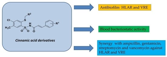
Activity of Cinnamic Acid Derivatives with 4-Chloro-2-mercaptobenzenesulfonamide Moiety against Clinical HLAR and VRE Enterococcus spp.

by
Rafał Hałasa
1,*,
Anita Bułakowska
2,
Jarosław Sławiński
2,
Magdalena Smoktunowicz
1,
Aleksandra Rapacka-Zdończyk
1 and
Urszula Mizerska
3
1
Department of Pharmaceutical Microbiology, Medical University of Gdansk, Al. Gen. J. Hallera 107, 80-416 Gdansk, Poland
2
Department of Organic Chemistry, Medical University of Gdansk, Al. Gen. J. Hallera 107, 80-416 Gdansk, Poland
3
Centre of Molecular and Macromolecular Studies, Department of Polymeric Nano-Materials, Polish Academy of Sciences, ul. Sienkiewicza 112, 90-363 Lodz, Poland
*
Author to whom correspondence should be addressed.
Antibiotics 2023, 12(12), 1691; https://doi.org/10.3390/antibiotics12121691
Submission received: 24 October 2023 / Revised: 21 November 2023 / Accepted: 22 November 2023 / Published: 2 December 2023
(This article belongs to the Special Issue Design and Synthesis of Novel Antibiotics)

Abstract

:
The rapid increase in strains that are resistant to antibiotics requires new active compounds to be found whose mechanism of action on bacteria is different to those that are currently known. Of particular interest are compounds that occur in plants as secondary metabolites. The focus of this study concerns the examination of the effects of synthetic cinnamic acid derivatives, with 4-chloro-2-mercaptobenzenesulfonamide moiety on Enterococcus spp. with HLAR (high-level aminoglycoside resistance) and VRE (vancomycin-resistant Enterococcus) mechanisms. The minimum inhibitory concentration (MIC) values of the tested compounds were determined using the serial dilution method for Enterococcus spp. groups, and the most active compounds were as follows: 16d, 17c, 16a, 16c and 16f (2–4 µg/mL). These compounds, at a concentration of 4 × MIC, inhibited the biofilm formation of HLAR strains (70 to 94%). At concentrations of 2 × MIC and 4 × MIC, they also inhibited the growth of VRE strains (42 to 96%). The best effect produced on the formed biofilm was demonstrated by compound 16f (from 62% MIC concentration to 89% 4 × MIC concentration) on the tested HLAR strains. In vitro studies, using the peripheral blood of domestic sheep, demonstrated the stable bacteriostatic activity of the tested compounds against Enterococcus spp. The compounds 16a, 16c, 16d, 16f and 17c showed synergism and additivity with ampicillin, streptomycin, gentamicin and vancomycin against resistant strains of Enterococcus spp. The tested compounds, when combined, reduce the MIC for antibiotics by 800 to 10,000 times for HLAR strains and by 8 to 10,000 times for VRE strains. The MIC of the tested compounds, in combination with antibiotics, is reduced 2–16-fold for HLAR strains and 2–32-fold for VRE strains. These studies demonstrate the potential for the therapeutic use of synthetic, cinnamic acid derivatives, with 4-chloro-2-mercaptobenzenesulfonamide moiety, to work against clinical strains of Enterococcus spp.

Graphical Abstract

1. Introduction

The genus Enterococcus comprises Gram-positive, catalase negative cocci, usually facultative anaerobic bacteria, that grow in 6.5% NaCl, 40% bile salts, 0.1% methylene blue milk, and at pH 9.6. They also grow at 10 and 45 °C and can survive for 30 min at 60 °C [1]. Enterococci belong to the phylum Firmicutes of the Enterococcaceae family, which includes many different species, and they are a natural component of the human microbiota. They colonize the lower gastrointestinal tract, oral cavity, and genitals [2]. E. faecalis and E. faecium are the main enterococci found in the human intestine. E. cecorum and E. durans are also frequently isolated, whereas E. casseliflavus, E. hirae, E. gallinarum, and E. avium are occasionally detected [3].
Enterococci can cause a wide range of clinical diseases. One of the factors causing the high infectivity of enterococci is biofilm formation ability. This property makes the treatment of the infection difficult [4]. Enterococcus spp. cause infections of the urinary tract, cardiovascular system, abdominal cavity, pelvis, central nervous system, and postoperative wounds. Asymptomatic bacteriuria is the most common clinical condition, but many of these cases are caused by colonization rather than infection. Other frequent causes of infection are bacteremia without endocarditis, which is followed by endocarditis [5]. Enterococcus spp. are frequently cited as one of the three most likely etiologies of both uncomplicated and complicated urinary tract infections (UTIs), especially healthcare-associated UTIs. Of these, the bacteria that most commonly cause these infections are E. faecalis, although E. faecium predominates among vancomycin-resistant isolates. It is usually associated with indwelling urinary catheters and instruments. The severity of the disease can range from uncomplicated cystitis to complicated cystitis, or it may cause pyelonephritis, a perinephric abscess, or prostatitis. All these cases result in the introduction of antibiotics to work against the enterococci [6].
Additionally, enterococci have the ability to obtain genetic material from other bacteria and to quickly adapt to the environment [3]. It is well established that enterococci are resistant to many antibiotics, including the following: β-lactams, aminoglycosides, trimethoprim/sulfamethoxazole, clindamycin, and some species of glycopeptides [7]. This requires combinations of glycopeptides or β-lactams with aminoglycosides to be used [5]. The application of antibiotics in hospitals, and for the treatment of animals, has led to the existence of resistant strains. Currently, drug-resistant enterococci that are isolated in hospitals include the following: high level of resistance to ampicillin; high-level aminoglycoside resistance (HLAR); resistance to glycopeptides (VRE; vancomycin and teicoplanin) and oxazolidinone resistant (LRE—linezolid-resistant Enterococcus) [3].
The World Health Organization (WHO) reports that antibiotics are becoming increasingly ineffective as drug resistance spreads around the world, thus causing difficult or ‘impossible to control’ infections. In its reports, the WHO lists microorganisms of particular concern, which require the discovery of new active compounds to work against them; among them are E. faecium VRE strains [8,9]. There is a constant need to search for new compounds with antibacterial properties.
In the light of these issues, research on new drugs to work against resistant bacteria strains is ongoing. An increasing number of studies are being conducted on combination therapies which could potentially reduce the MIC values of administered antibacterial drugs, improve pharmacokinetic/pharmacodynamic (PK/PD) parameters (e.g., bind a hydrophilic drug to a lipophilic drug), and increase activity against bacteria present in biofilm [10]. Examples of such combinations are as follows: omadacycline and oritavancin, or fosfomycin with either daptomycin or rifampicin [11,12].
Secondary plant metabolites (phytochemicals, including cinnamic acid) are a good source of active compounds, including those with antibacterial properties [13]. The broad biological activity of cinnamic acid, and its synthetic derivatives, has been demonstrated by several research teams [9,13,14,15,16,17,18]. Cinnamic acid can occur in nature in both cis and trans forms with the trans form being more stable than the cis form. Its antimicrobial activity is low against Gram-positive and Gram-negative bacteria [10]; however, synthetic derivatives (cinnamic acids, esters, amides) have a stronger antimicrobial effect on bacteria; moreover, cinnamic aldehydes inhibit the development of fungi. Cinnamic acid derivatives with phenolic groups have been classified as antioxidants. The chemical structure of cinnamic acid, which contains both a benzene ring and a carboxylic group, enables the modification and procurement of synthetic derivatives. Synthetic compounds that combine two pharmaceutical entities in one molecule may be a successful strategy since these molecules could be more effective than their individual components. Research has been conducted on the conjugation of several hydroxy- and phenyl-substituted derivatives of trans-cinnamic acids with an antimicrobial pharmacophore like carvacrol on ESKAPE pathogens [10]. Trans-cinnamaldehyde is a phenylpropanoid, which practically occurs in cinnamon essential oil, and it is Generally Recognized as Safe (GRAS) by the US Food and Drug Administration (US FDA) [19].
Sulfonamides and sulfamates are mainly known as synthetic compounds, but several were also discovered as secondary metabolites produced by actinomycetes, e.g., nucleocidin and dealanylascamycin. Sulfonamides and sulfamates constitute a diverse group of highly pharmacologically active compounds, and many clinically used drugs contain the signature sulfamoyl structural motif. Sulfonamides are widely known as synthetic “sulfa drugs”, and these were the first chemotherapeutically used antibacterial compounds. They are applied to other diseases such as Alzheimer’s disease and other central nervous system (CNS) disorders, diabetes, psychosis, and various cancers and tumors [20].
In this work, we examined the activity of synthetic derivatives of cinnamic acid with 4-chloro-2-mercaptobenzenesulfonamide moiety [17] against clinical strains of Enterococcus spp. The measure of the activity of compounds is their limit concentration value; for this purpose, we determined minimum inhibitory concentrations. From a clinical point of view, it is important to know: at what concentrations the tested compounds inhibit bacterial biofilm formation, whether they are able to penetrate the already formed biofilm and affect the bacteria or whether they are able to inhibit the further development of the existing biofilm. We examined the activity of new derivatives on enterococcal biofilm. Testing the bioavailability and pharmacodynamics of compounds in body fluids in vitro is one of the important elements showing the stable structure and activity of compounds as well as the influence of the elements of these fluids on the compounds and the impact of the compounds on the components of the higher organism. Therefore, we determined In vitro the stability and influence of blood elements on the antibacterial activity of the tested compounds. Combination therapy of two or three compounds with different targets in a bacterial cell is a common element of antibiotic therapy for infections with multi-resistant strains, including VRE HLAR strains. We investigated the interactions that may occur between the tested compounds and the antibiotics used in the antibiotic therapy of enterococcal infections.

2. Results

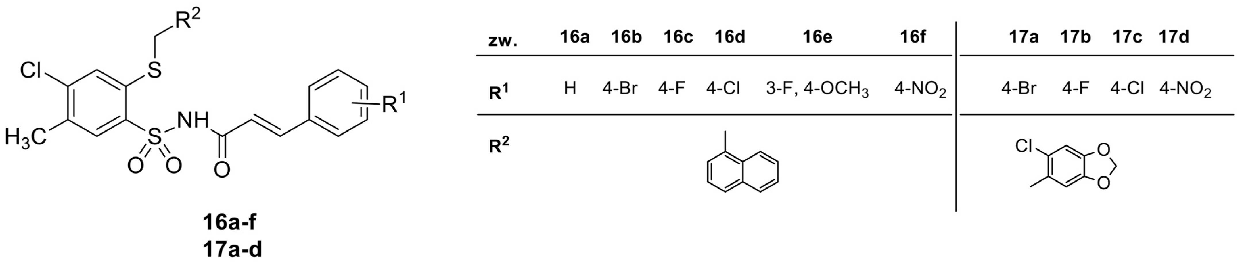
2.1. Synthesis and Chemical Characterization of Cinnamic Acid Derivatives with 4-Chloro-2-mercaptobenzenesulfonamide Moiety

The synthesis and chemical characterization of cinnamic acid derivatives with 4-chloro-2-mercaptobenzenesulfonamide moiety was described by Bułakowska et al. [18]. The structures of the tested derivatives are presented in Figure 1.

2.2. Antibacterial Activity of Cinnamic Acid Derivatives

In this work, we decided to test the activity of cinnamic acid derivatives against Enterococcus characterized by VRE. We preceded the study activity by determining the resistance pattern of all our strains. The studies were performed using the disc diffusion method. The results are presented in Table 1.
It has been observed that all the tested strains were sensitive to linezolid (inhibition zone ≥ 20 mm), and 14 out of 17 bacterial strains were susceptible to teicoplanin (inhibition zone ≥ 16 mm). In turn, 100% HLAR strains were sensitive to vancomycin (inhibition zone ≥ 12 mm). Only one strain showed sensitivity to tetracycline (inhibition zone ≥ 20 mm). However, 100% of the strains tested were resistant to streptomycin (zone of inhibition ≤ 14 mm) and gentamicin (zone of inhibition ≤ 8 mm). Susceptibility to ampicillin (susceptible strain zone ≥ 10 mm; resistant strain ≤ 8 mm), imipenem (susceptible strain zone ≥ 21; resistant strain ≤ 20 mm) and co-trimoxazole (susceptible strain zone ≥ 23 mm) was variable.
The MIC values of the cinnamic acid derivatives with 4-chloro-2-mercaptobenzenesulfonamide moiety against the Enterococcus spp. VRE group are shown in Table 2.
The most active compounds turned out to be 16d and 17c with MICs within the range of 2 ± 0.05–4 ± 0.38 µg/mL. Compounds 16a and 16c showed an MIC of 4 µg/mL. The least active compounds were 17b and 17d (MIC > 125 µg/mL). The most resistant strain turned out to be Enterococcus sp. 966351, and the most sensitive was Enterococcus sp. 885041. The majority of the compounds showed bacterial inhibitory activity, while bactericidal concentrations were equal to or greater than 125 µg/mL. The compounds 16d and 17c were the most active against Enterococcus spp. VRE.

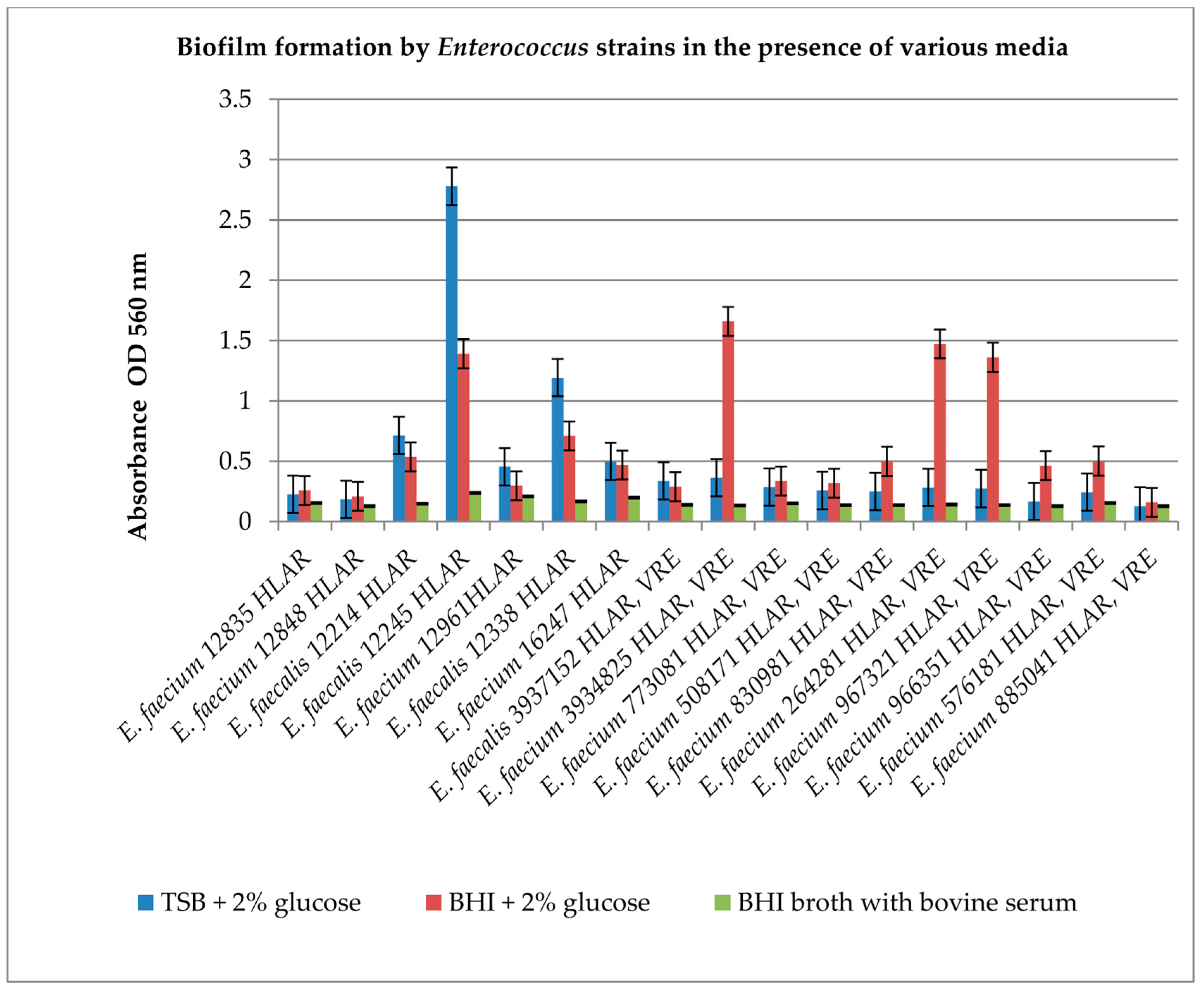
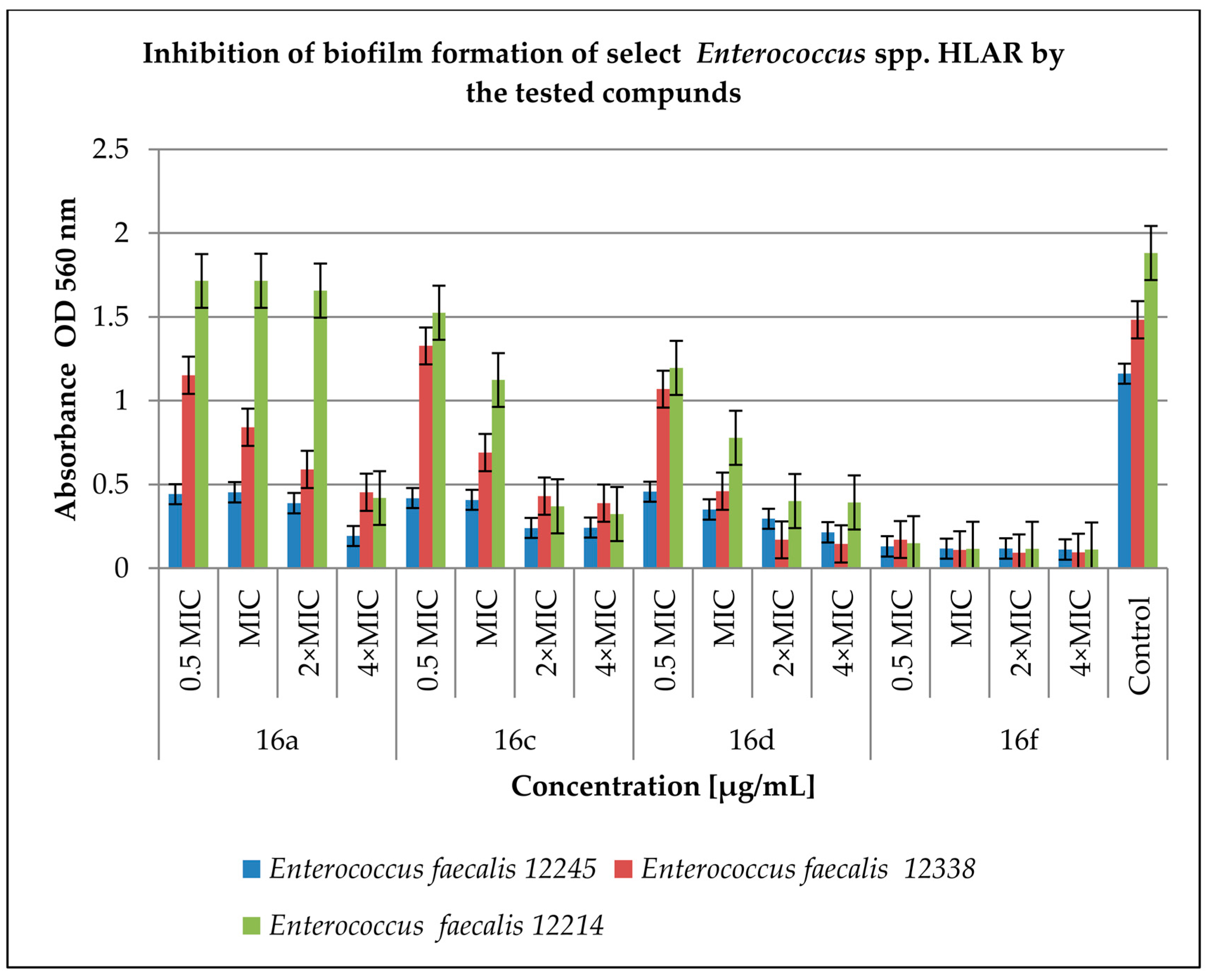
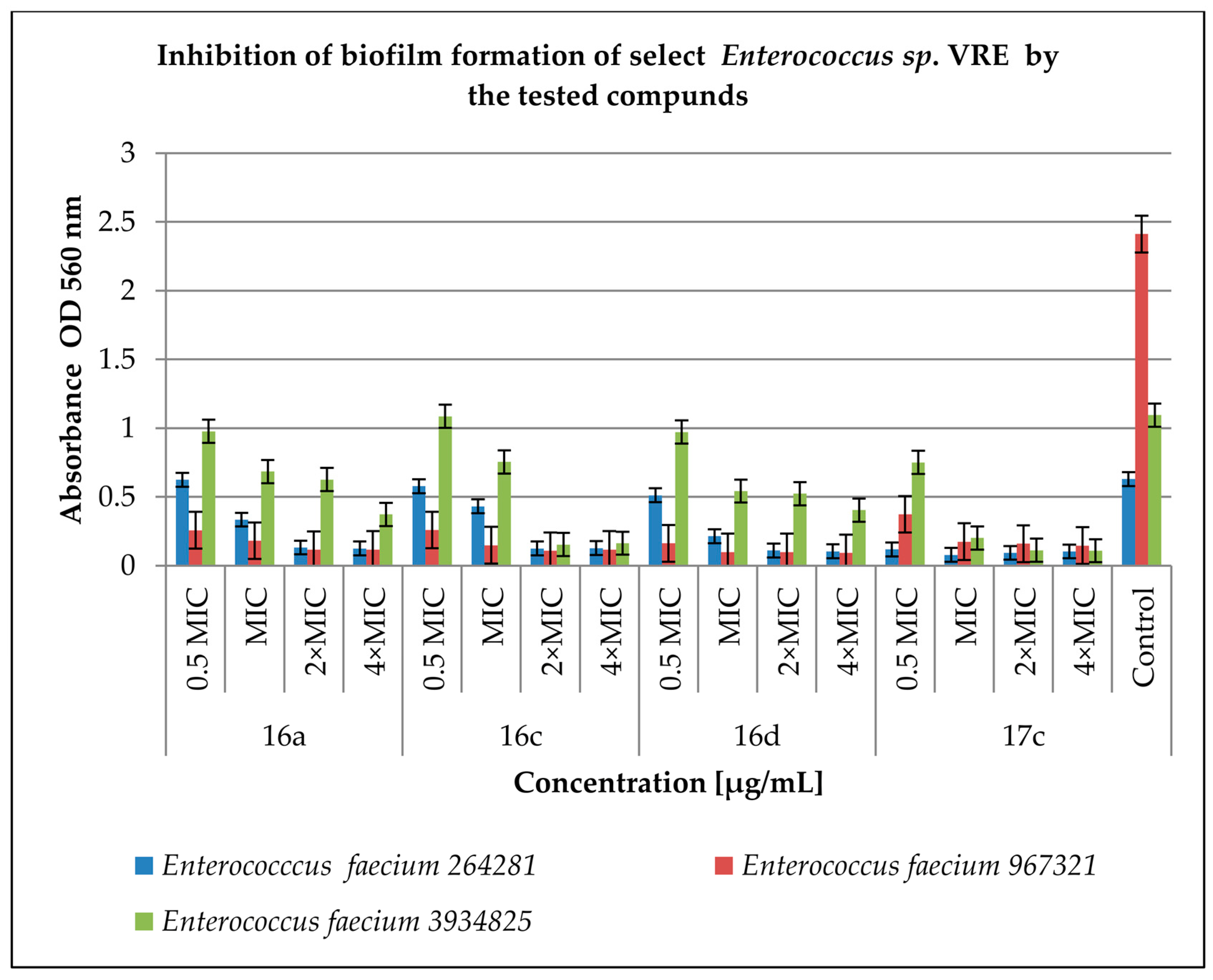
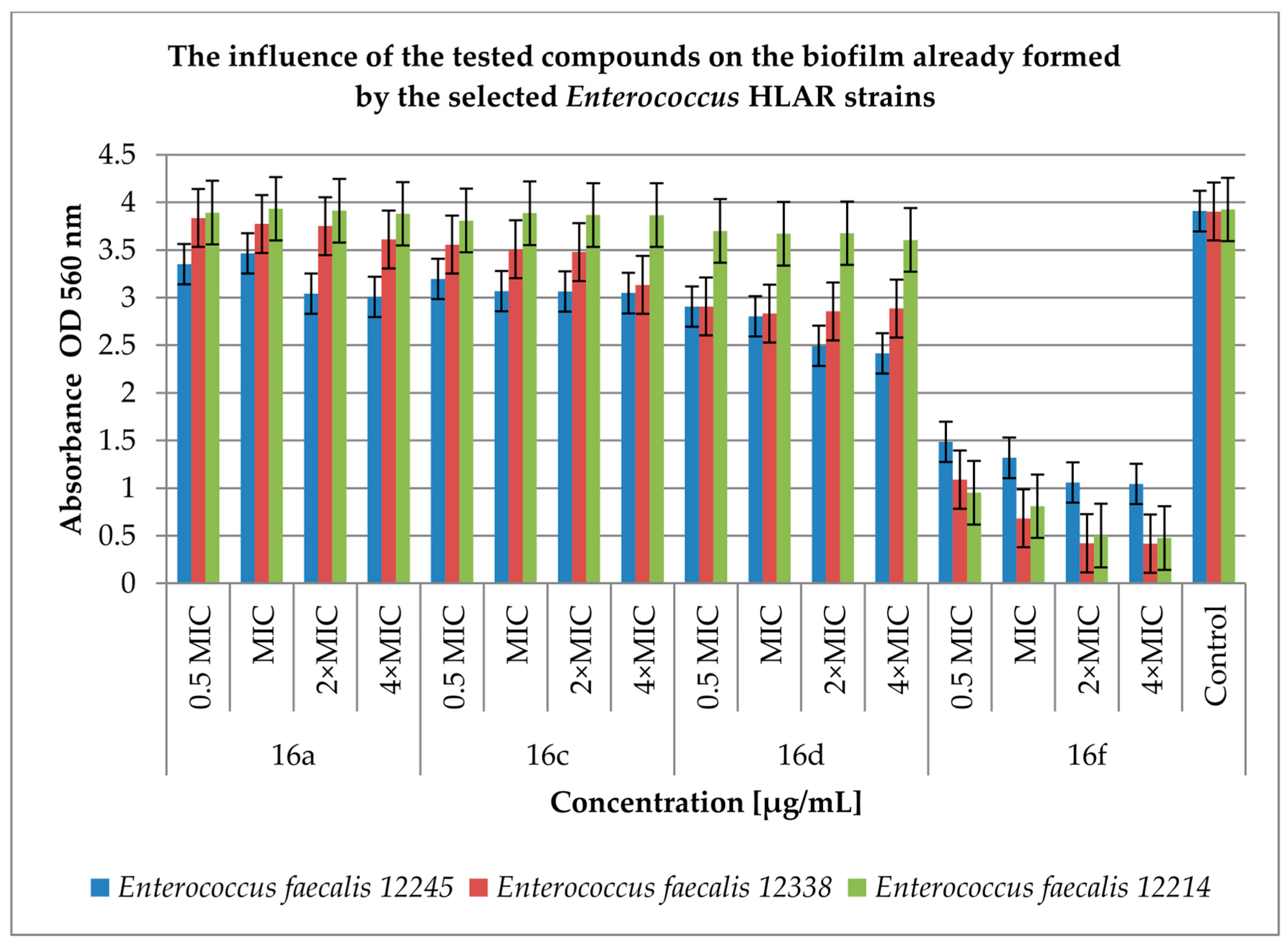
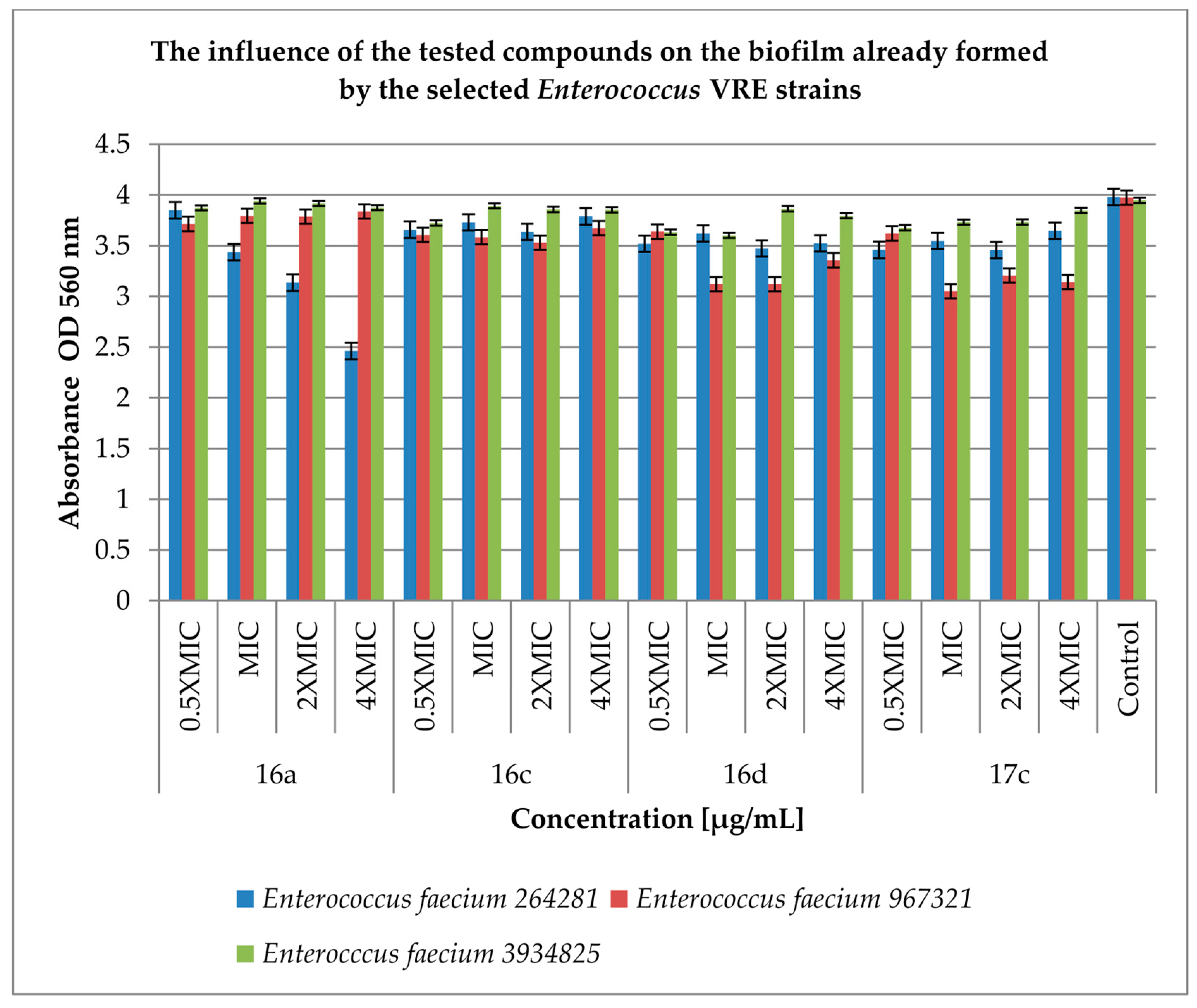
2.3. Effect of Derivatives on the Biofilm Produced by Enterococcus spp.

The next step of our study was an evaluation of whether the tested cinnamic acid derivatives act on the biofilm produced by Enterococcus spp. It is known from the literature data that Enterococcus spp. are capable of producing a biofilm, but this is not a permanent feature of the species. Therefore, we decided to verify which of the tested strains exhibit this ability. Based on the literature reports, we chose to test biofilm formation by Enterococcus spp., using the following broths: tryptic soy broth medium (TSB), brain–heart infusion broth (BHI) supplemented with 2% glucose, and BHI supplemented with 5% bovine serum. The results of the effect of substrates on biofilm formation are shown in Figure 2.
The results presented in Figure 2 show that the ability to produce biofilm is diversified in the pool of Enterococcus spp. strains used for testing in the current study. The strains used include strong biofilm producers, e.g., E. faecalis 12245 HLAR and E. faecalis 3937152 HLAR and VRE; and those that form a weak biofilm regardless of the substrate used, e.g., E. faecium 12835 HLAR and E. faecium 12848 HLAR. To the best of our knowledge, it is the first study that differentiates Enterococcus according to its ability to form biofilm in different media. The bacteria that generate the strongest biofilm in TSB supplemented with 2% glucose medium are Enterococcus spp. HLAR: E. faecalis 12214, E. faecalis 12245, and E. faecalis 12338 (p < 0.05). However, in the BHI supplemented with 2% glucose medium, the strongest biofilm is formed by Enterococcus spp. VRE: E. faecium 3934825, E. faecium 967321, and E. faecium 264281 (p < 0.05). Surprisingly, all the tested strains produced biofilm at a very low level in the presence of bovine serum in the BHI medium (p < 0.05). For further research, which concerned the activity of the tested compounds against biofilm, we chose both media and the strains producing the strongest biofilm. The most active derivatives were used in concentrations of 0.5 MIC, MIC, 2 × MIC and 4 × MIC.
Figure 3 shows the effect of the tested derivatives on biofilm formation by Enterococcus spp. HLAR strains. The most active compound was 16f. In the tested concentration range, it significantly inhibited biofilm formation by the Enterococcus spp. HLAR strains from 89 to 94% (p < 0.05) compared to the control. For the remaining compounds tested, a concentration of 4 × MIC was the most active, as this inhibited biofilm formation from 69% (compound 16a against E. faecalis 12338; p < 0.05) to 90% (compound 16d against E. faecalis 12338; p < 0.05). The concentrations of 0.5 MIC and MIC turned out to be the least active in inhibiting biofilm formation with the effectiveness of significant effects ranging from 8% (compound 16a for E. faecalis 12214; p < 0.05) to 60% (compound 16a for E. faecalis 12245; p < 0.05).
Figure 4 shows the effect of the tested derivatives on biofilm formation by Enterococcus spp. VRE strains. The anti-biofilm activity of the tested compounds varied and depended on the concentration. The inhibition levels of compounds at 0.5 MIC and MIC ranged from 0.78% (compound 16c at 0.5 MIC against E. faecium 3934825; p < 0.05) to 95% (compound 16d at MIC against E. faecium 967321; p < 0.05). The concentrations of 2 × MIC and 4 × MIC were found to be more effective and inhibited biofilm formation from 42% (compound 16a at 2 × MIC against E. faecium 3934825, p < 0.05) to 96% (compound 16d at 4 × MIC against E. faecium 967321; p < 0.05) in comparison to the control samples. Comparing the results presented in Figure 2 and Figure 3, it can be concluded that the tested derivatives inhibit the formation of biofilm by the VRE strains more efficiently than by the HLAR strains.
The next step of our work was to test the antimicrobial effect of our derivatives on already formed enterococcal biofilm. The results are presented in Figure 5 and Figure 6.
Figure 5 shows the effect of the tested compounds on biofilm already formed by Enterococcus spp. HLAR strains. Compounds 16a, 16c and 16d show a low effect on the already formed biofilm and the degree of reduction in the tested strains in biofilm ranged from 0.8% (compound 16a at a concentration of 0.5 MIC against E. faecalis 12214; p < 0.05) to 38% (compound 16d at a concentration of 4 × MIC against E. faecalis 12245; p < 0.05) when compared to the control. In contrast, compound 16f turned out to most actively reduce the number of bacteria in the biofilm from 62% (0.5 MIC concentration against E. faecalis 12245; p < 0.05) to 89% (4 × MIC concentration against E. faecalis 12338; p < 0.05) compared to the control samples.
Nevertheless, the tested compounds had no significant effect on the inhibition of further biofilm formation by the Enterococcus spp. VRE strains (Figure 6). Inhibitory activities ranged from 0.16% (compound 16a at MIC concentration against E. faecium 3934825; p < 0.05) to 23% (compound 17c at MIC concentration against E. faecium 967321; p < 0.05) compared to the control.
The most active compounds were 16f and 17c. However, the activity of the tested compounds on the already formed biofilm was low in both groups of bacteria with the exception of compound 16f acting on Enterococcus spp. HLAR, which inhibits the formation of the biofilm to about 90% when compared to the control.

2.4. Blood Bacteriostatic Activity Tests

The compounds we tested exhibited a bacteriostatic effect on Enterococcus spp. in the culture medium. We wanted to answer the question of whether this property is retained in the presence of blood components, as it was in the case of Staphylococcus spp. [18]. For this purpose, we selected two species of bacteria: E. faecium 264281 (Table 3) and E. faecalis 12245 (Table 4).
The initial number of bacteria at time t0 was (2.00 ± 0.69) × 108 CFU/mL for E. faecalis 12245 and (7.30 ± 0.67) × 108 for E. faecium 264281. Bacteria in appropriate numbers were added to blood samples containing the tested compounds in the concentration range of 0.5–16 µg/mL and then incubated for 24 h at 37 °C. Controls consisted of samples of bacteria incubated in pure blood, without supplements, the number of which after 24 h incubation was (1.44 ± 0.47) × 108 CFU/mL for E. faecalis 12245 and (1.30 ± 0.48) × 108 CFU/mL for E. faecium 264281. There is a relationship here: the higher the concentration of the active compound in the sample, the lower the number of live bacteria. In the samples with compounds 16c and 16d (16 µg/mL), there is a significant decrease in the number of viable bacteria: 16c—(7.00 ± 0.71) × 106 CFU/mL, 16d—(1.20 ± 0.55) × 106 CFU/mL (p < 0.05) compared to the control.

2.5. Study of Interactions of Cinnamic Acid Derivatives with Antibiotics on Resistant Strains of Enterococcus spp.

The most interesting interactions between antibacterial compounds relate to synergy. The synergy of compounds is based on increasing effectiveness, reducing toxicity, reducing undesirable side effects, increasing bioavailability, lowering the required dose and limiting the emergence of resistance to the tested preparations. New therapeutic combinations that include a connection with natural products have become a research priority.
To check the relationship between our derivatives and antibiotics, we conducted further studies using the common checkerboard assay. Antibiotics such as ampicillin, gentamicin, streptomycin and vancomycin as well as Enterococcus spp. selected from both tested groups were used.
In the first stage, we checked the activity of selected antibiotics and determined the limit concentrations (MIC). In the next stage, we conducted research assessing the relationship between the tested derivatives and selected antibiotics, where we used MIC and lower concentrations of the tested compounds. The antibiotics taken into consideration in the study are important preparations in the treatment of infections caused by Enterococcus spp. The results are presented in Table 5 and Table 6.
The cinnamic acid derivatives used in the research show a synergistic effect with antibiotics against selected HLAR strains. The exception is compound 16d, which presents additive properties with ampicillin and gentamicin. Compounds 16a, 16c and 16f are synergistic compounds with the tested antibiotics. These compounds may become potential preparations for clinical use in combination with currently used antibiotics against HLAR strains. However, the results obtained for selected VRE strains are not as homogeneous as for HLAR strains. Most of the tested compounds in a mixture with antibiotics show synergism. However, some compounds (16a, 16c, 16d and 17c), when combined with antibiotics (gentamicin, streptomycin and vancomycin), give additive effects. Compound 16a exhibits synergistic properties with all the tested antibiotics against selected VRE strains. In turn, all the compounds show synergy with ampicillin. The remaining tested compounds (16c, 16d and 17c) show differentiated interactions with antibiotics: synergism or additivity.

3. Discussion

In our previous study, we showed that cinnamic acid derivatives with 4-chloro-2-mercaptobenzenesulfonamide moiety were active against reference and clinical strains of Enterococcus spp. The tested enterococci strains were mostly HLAR and included both E. faecalis and E. faecium strains. Only two of the tested isolates were VRE strains. In this work, we decided to test the activity of cinnamic acid derivatives against Enterococcus characterized by VRE.
The results of our previous research conducted by Bułakowska et al. [18] showed that the most active derivatives against Enterococcus spp. HLAR were 16a, 16c, 16d and 16f. Compounds 16d and 17c were the most active against Enterococcus spp. VRE. Both derivatives contain a chlorine atom as the substituent R1 and naphthyl or piperonyl as the R2 substituent. Moreover, derivatives 16a and 16c turned out to be very active against Enterococcus spp. VRE as well as for HLAR strains. Derivative 16f was less active against VRE than against HLAR strains.
Based on the obtained results, it can be concluded that the activity of the tested derivatives against Enterococcus spp. HLAR and VRE was mainly due to the presence of the substituent R2—naphthyl, and the substituent R1 = H, F, NO2 was secondary. The high activity of compounds 16d and 17c against Enterococcus spp. of the VRE group could mean that the substituent R1 = Cl determines the main activity of the compound with the substituent R2 only in the second place. When comparing the MIC values, obtained by serial dilution, to the standard (co-trimoxazole), the tested compounds turned out to be significantly more active against the tested Enterococcus strains. This is most likely related to the structure of the compounds—the combination of sulfonamide with cinnamic acid in one molecule and a different mechanism of action on microorganisms than the composition of co-trimoxazole.
Mingoia et al. [10] investigated the antibacterial properties of several hydroxy- and phenyl-substituted trans-cinnamic acid derivatives conjugated with an antimicrobial pharmacophore such as carvacrol in order to select the best candidate for the treatment of cutaneous infections caused by pathogens. Among the list of derivatives, only DM2 and DM8 turned out to be active against the tested bacterial strains. Compound DM2 was more active against E. faecium (MIC50 32 mg/L) than E. faecalis (MIC50 256 mg/L). The authors [9] suggest that the presence of a hydroxyl group in the ortho position in the phenyl nucleus is important for the effect on the cell membrane of enterococci. Therefore, the DM8 compound turned out to be the most active against E. faecium (MIC50 = 32 mg/L). It needs to be highlighted that the authors themselves, in Table 2, presented the range of the antibacterial activity of selected derivatives, including MD8, which for E. faecalis was in the range of 32–512 mg/mL and for E. faecium was in the range of 16–512 mg/mL. Hence, the authors’ suggestion as to the mechanism is not that clear.
Guzman et al. [15] reported that cinnamic acid was active against E. faecalis and its minimum inhibitor concentration was 6.75 mM. In turn, the MICs of ferulic acid and its 4-O-acetyl derivative for E. faecalis ATCC 29212 were determined as 659 μM and 540 μM, respectively. Among natural and synthetic cinnamic esters, activity was demonstrated by 5-O-caffeoylquinic acid (MIC 181 μM) against E. faecalis OGRF1, rosmarinic acid (MIC 833 μM) and methyl rosmarinate (MIC 801 μM) against E. faecalis C159-6, and finally caffeic acid phenethyl ester (MIC 400 μM) against E. faecalis ATCC 29212. 4-tert-butylphenyl ferulate and 4-isopropylphenyl ferulate were active against E. faecalis ATCC 29212 at the concentrations of 391 μM and 51 μM, respectively. In turn, the MIC of 4-chloro-3-methylphenyl ferulate was 50 μM. Among cinnamic aldehydes, alcohols and their derivatives, the most active against E. faecalis turned out to be cinnamaldehyde at a concentration of 1.89 mM.
Many research groups are engaged in the construction of hybrid compounds using cinnamic acid. Cephem hybrid with cinnamic aldehyde gave results of activity against E. faecalis A20688 as 1.6 μM, 1.5 μM, 1.4 mM and 364 nM. Oxazolidinone hybrids are active against E. faecium ATTC 700221 (VRE; MIC 388 nM) and E. faecalis ATCC 29212 (MIC 194 nM) [15].
Gómez et al. [21] examined the antimicrobial properties of natural essential oil components, including cinnamaldehyde, against three species of bacteria: Enterococcus spp., Staphylococcus spp., and Pseudomonas spp. (10 clinical strains of each). Cinnamaldehyde (CIN) presented the highest activity for all tested bacterial strains—the MICs within the range of 10–50 mg/mL. The research was conducted in Mueller Hinton (MH) broth or cation-adjusted Mueller Hinton II broth (MH II).
Teethaisong et al. [22] investigated the effect of Stephania suberosa extract (SSE) against E. faecium strains and obtained MIC values of 0.5 mg/mL.
Biofilm consists of organized spaces, a multi-layer cluster of cells of one or several species of microorganisms, formed on biotic and abiotic surfaces. Microorganisms concentrated in biofilm are characterized by diverse metabolic activity. There are also antibiotic-resistant bacteria that “protect” the susceptible bacteria. Biofilm-forming bacteria are surrounded by a special extracellular polymeric substance (EPS) matrix. It is already known that Enterococcal surface protein (ESP) significantly enhanced E. faecalis biofilm formation in a glucose-dependent manner [22]. Biofilm is the cause of many life-threatening infections of humans and animals. Due to its complex structure, it is very difficult to remove; EPS is difficult for disinfectants and antibiotics to penetrate.
The compounds 16a, 16c, 16d, 16f and 17c were tested at concentrations of 0.5 MIC, 2 × MIC and 4 × MIC and inhibited the formation of biofilm by both groups of enterococci, but the inhibition of the VRE strains was more efficient than that of the HLAR strains. However, the activity of the tested compounds against the already formed biofilm was low in both groups of bacteria; only compound 16f was active against Enterococcus spp. HLAR and inhibited the formation of the biofilm to about 90% in comparison to the control. In our studies, we determined the amount of biofilm with crystal violet, as was previously described in [18].
Ali et al. [19], using BHI medium, examined the effect of trans-cinnamaldehyde (0.5%, 0.75% and 1%) on the biofilm formed by E. faecalis (clinical isolate) for 72 h. The authors treated the biofilm with the compounds for 5 and 15 min or 24 h and demonstrated, using 2,3-bis-(2-methoxy-4-nitro-5-sulfophenyl)-2H-tetrazolium-5-carboxanilide (XTT), that a 15-min action of cinnamaldehyde kills bacterial cells. Derivatives studied in our work had a bacteriostatic effect on planktonic bacterial cells in the tested concentration range. Due to the higher resistance of biofilm cells to chemicals than free-living cells, we chose crystal violet for biofilm determination in our studies. Crystal violet shows the size (biomass) of the biofilm presence or its absence rather than the presence of living cells, as was described in Ali et al. [19].
Akshaya et al. [23] examined the effect of cinnamaldehyde in the concentration range of 62.5–1000 μM on the biofilm formed by the E. faecalis strain (MTCC 2729) using BHI medium with 2% glucose for 72 h. The researchers used crystal violet and XTT [2,3-bis-(2-methoxy-4-nitro-5-sulfophenyl)-2H-tetrazolium-5-carboxanilide] as markers. Cinnamaldehyde prevented the growth of E. faecalis biofilm from a concentration of 62.5 μM, according to the crystal violet-staining technique. It was found that cinnamaldehyde at a concentration of 1000 µM inhibited the production of E. faecalis biofilm by 85%.
Akshaya et al. [23] studied the effect of cinnamaldehyde on the expansion and gelatinase activity of E. faecalis strains, which is important in the biofilm formation process. Gelatinase activity has been reported also to be involved in the initial steps of the generation of biofilm. In our studies, we detected phenotypically the presence of gelatinase produced by the tested strains. Unfortunately, we do not notice a correlation between the ability to form a biofilm and the presence of enzymes. The gelatinase activity was demonstrated by the following strains: E. faecalis 12245, 12214, 12338, 3937152 and E. faecium 264281, 508171, 16247; these strains formed a strong or a weak biofilm, as shown in Figure 1.
Hashem et al. [24] showed, using gelE(–) mutants, that the lack of the enzyme did not result in the lack of biofilm formation, which is consistent with our results.
The literature states [23] that the biofilm-forming activity of E. faecalis in the presence of glucose varies. Based on our results (Figure 2) (e.g., in the case of E. faecalis 12214, 12245, 12338, 12961), the presence of 2% glucose in two different media did not result in biofilm formation at the same level. There must be other factors that influence biofilm formation, depending on the medium used.
Enterococci possess in their genome the fsr locus that shares significant homology with the agr locus of staphylococci, which acts as an accessory global regulator of virulence factors and metabolism, including biofilm formation. Pillai et al. [25] showed that biofilm formation by E. faecalis OG1RF was greater in TSB supplemented with 1% glucose, but the same effect was not evident in fsr, gelE or sprE mutants. This could suggest that a glucose-dependent transcriptional regulator controlled fsr, either directly or indirectly, and that fsr exerts catabolite control over biofilm formation through GelE and SprE downstream proteases.
Kim et al. [26] examined the effect of tryptone–yeast extract broth with 0% glucose + 0% sucrose, 0.5% glucose, 1% glucose, 0.5% sucrose or 1% sucrose on Enterococcus faecalis planktonic and biofilm In vitro. They showed that the virulence-associated gene expression was the highest in broth with 1% sucrose (biofilm growth conditions) and in broth with 1% glucose (planktonic growth conditions). A higher bacteria and exopolysaccharide (EPS) bio-volume in sucrose was observed than in 0% glucose + 0% sucrose or glucose.
Interestingly, many researchers study the impact of chemical compounds on the already formed biofilm but do not check the preventive effect. Certainly, destroying the formed biofilm is a very important issue in the search for compounds that act on bacteria, but the administration of preventive preparations or coating materials used for catheterization, drainage, etc., may also be an important element in the fight against infections with resistant strains.
Caballero Gómez et al. [21], using TSB medium, examined the effect of natural essential oil components (including cinnamaldehyde) at MIC and 0.5 MIC concentrations, and their combination with EDTA (12.5–25 mM in water) and HLE (disinfectant solution—composed of 3–6% H2O2; 2.2–4.4% lactic acid) on the biofilm formed by selected strains. Crystal violet was used to determine the presence of biofilm. Cinnamaldehyde inhibited the formation of Enterococcus sp. strain M28M12 biofilm by 80%; for the other two strains, the effect was not so spectacular. However, a synergistic effect between cinnamaldehyde with HLE and EDTA was visible. CA + HLE caused 2–6 log10 reductions in CFU (colony-forming units) of enterococci and CA + EDTA 2–3 log10 reductions in CFU of enterococci in biofilm.
The presence of whole peripheral blood (Method 4.5) with its antibacterial mechanisms and the presence of phagocytic cells may have resulted in a slight decrease in the number of bacteria in the control samples. It seems that a similar mechanism occurred in the case of the lowest concentrations (0.5 µg/mL) of the tested compounds. This may be due to the fact that Enterococcus spp. do not have many virulence factors, like staphylococci, that neutralize the defense mechanisms present in the blood of mammals. In tests with higher concentrations of compounds, the number of viable bacterial cells changes significantly. These changes are a combination of the mechanisms of the immune system present in the blood and the bacteriostatic effect of the concentrations of compounds. The results obtained may be due to the influence of the tested derivatives on delays in the division of bacterial cells, which, when transferred to a solid substrate without the tested agent, have an extended doubling time and did not produce visible colonies even after 48 h of incubation. We suggested that the tested compounds have constant, stable bacteriostatic activity in whole blood, and they do not inhibit the activity of the cells of the immune system. This could be a basis for further research into potential drugs against resistant bacteria.
Doyle et al. [27] cite that trans-cinnamaldehyde has properties that limit its use: low solubility in water, sensitivity to light and air, and it is not very stable in the blood where it is converted into cinnamic acid. Present in cosmetics in concentrations above 0.005%, it causes allergies and adverse reactions. The authors emphasize the need to test aldehyde derivatives for toxicity. The derivatives we tested show toxic activity to the peripheral blood of domestic sheep at concentrations of 32 µg/mL, as we demonstrated in the publication by Bułakowska et al. [18]. These compounds present stable activity against MRSA and Enterococcus spp. strains (data above) in the tested concentration range, during 24 h of testing. However, it would be necessary to carry out tests on the stability of the antimicrobial activity of compounds in biological systems over an extended period of time and to transform the derivatives we studied into water-soluble forms.
Enterococcus spp. are bacteria that are naturally resistant to many antibiotics, and the treatment of infections is mainly based on combining several antibiotics in order to achieve a synergistic effect. This effect is intended to fight the infection caused by multidrug-resistant strains. We wanted to check whether our compounds have any interactions with conventional antibiotics.
The cinnamic acid derivatives used in the research show a synergistic effect with antibiotics against selected HLAR strains. The exception is compound 16d, which presents additive properties with ampicillin and gentamicin. It should be emphasized here that the tested compounds reduce the MIC for antibiotics by 800 to 10,000 times. However, the MIC of the tested compounds in combination with antibiotics is reduced by 2 to 16 times. Compounds 16a, 16c and 16f are compounds that act synergistically with the tested antibiotics. These compounds may become potential preparations for clinical use in combination with currently used antibiotics against HLAR-resistant strains. However, the results obtained for selected VRE strains are not as homogeneous as for HLAR strains. Most of the tested compounds in a mixture with antibiotics show synergism. However, some compounds (16a, 16c, 16d and 17c), when combined with antibiotics (gentamicin, streptomycin and vancomycin), give additive effects. The tested compounds reduce the MIC of antibiotics by 8 to 10,000 times. However, the combination of the tested derivatives with antibiotics causes a decrease in the MIC values of the compound by 2 to 32 times. Compound 16a exhibits synergistic properties with all the tested antibiotics against selected VRE strains. In turn, all the compounds show synergy with ampicillin. The next stage of further research would be to check the activity of concentrations showing synergism, how they act on bacteria over time, how they affect biofilm and whether their activity is stable in the blood.
Essential oil (from C. zeylanicum) exhibits synergistic activity with amikacin, gentamicin, imipenem and meropenem against Acinetobacter baumannii and has a positive interaction with colistin [26]. Essential oil obtained from Cinnamomum burmannii interacts with gentamicin against S. epidermidis, and Cinnamomum verum essential oil showed the ability to restore the sensitivity of Escherichia coli to piperacillin (the bacterium had the TEM-1 beta-lactamase gene) [26]. Trans-cinnamaldehyde from C. zeylanicum decreased the MIC of clindamycin for Clostridium difficile by 16-fold [26].
Doyle et al. [27] presented a report on the synergism of cinnamaldehyde in combination with ampicillin, bacitracin, clindamycin, erythromycin, novobiocin, penicillin, streptomycin, sulfamethoxazole and tetracycline against Salmonella Typhimurium SGI1 (tet A), Escherichia coli N00-666, Staphylococcus aureus blaZ, which was resistant to penicillin, and erythromycin-resistant Streptococcus pyogenes ermB.
Cinnamic acid showed synergism with amikacin, ampicillin, ciprofloxacin, erythromycin, or vancomycin against E. coli and S. aureus and a combination of cinnamic acid with ciprofloxacin against P. aeruginosa. Cinnamic acid displays synergistic effects with amikacin, clofazimine, isoniazid and rifampin against Mycobacterium tuberculosis and M. avium [27].
Teethaisong et al. [22], using the checkerboard method, determined that SSE plus ampicillin and SSE plus vancomycin combinations exhibited synergistic interactions against E. faecium isolates.

4. Materials and Methods

4.1. Materials

Sterile sheep blood was defibrinated (GrasoBiotech, Starogard Gdański, Poland). Co-trimoxazole (composition sulfamethoxazole 400 mg: trimethoprim 80 mg; WZF Polfa, Warszawa, Poland), ampicillin-Na-salt (Serva, Heidelberg, Germany), streptomycin sulfate salt (Pol-Aura, Dywity, Poland), gentamycin (40 mg/mL, solution for injection and infusion; (KRKA d.d., Novo mesto, Slovenia), amikacin disulfate salt (Pol-Aura, Dywity, Poland), norfloxacin (Pol-Aura, Dywity, Poland), levofloxacin (5 mg/mL, solution for infusion, (Pharmathen S.A, Pallini, Greece), vancomycin hydrochloride (Pol-Aura, Dywity, Poland), ciprofloxacin (10 mg/mL, solution for infusion, (KRKA d.d., Novo mesto, Slovenia), vancomycin hydrochloride (Pol-Aura, Dywity, Poland), doxycycline (20 mg/mL, solution for infusion (Polfa Tarchomin S.A., Warszawa, Poland). Brain–heart infusion broth (BHI, Becton Dickinson, Franklin Lakes, NJ, USA) was used for MIC and FICI determination, while BHI and tryptic soy broth (TSB, Becton Dickinson, Franklin Lakes, NJ, USA) medium supplemented with 2% glucose were used to culture the biofilm bacteria. Muller–Hinton agar (MH, Becton Dickinson, Franklin Lakes, NJ, USA) was used alongside the disc diffusion method. Bacterial strains and culture conditions: lists of strains tested in the publication are presented in Table 7a, b. Enterococcus faecalis ATCC 51299 and clinical strain Enterococcus spp. were cultured in an aerobic atmosphere at 37 °C for 48 h. To determine the bacterial viability, BHI blood agar plates were used.

4.2. Determination of Susceptibility to Antibiotics by the Agar Disc Diffusion Method

Disc diffusion methods for antibiotics [28] and antibiotic susceptibility were interpreted according to EUCAST clinical breakpoints (version 11.0) [29]. In our research, Muller–Hinton medium (Beckton-Dicinson, Franklin Lakes, NJ, USA) and antibiotic paper discs (BioMaxima S.A., Lublin, Poland, Country) like ampicillin 2 µg/mL, imipenem 10 µg/mL, norfloxacin 10 µg/mL, vancomycin 5 µg/mL, teicoplanin 30 µg/mL, co-trimoxazole 25 µg/mL, doxycycline 30 µg/mL, linezolid 10 µg/mL, gentamicin 120 µg/mL, and streptomycin 300 µg/mL were used.

4.3. Minimum Inhibitory Concentration Determination

The MIC determination for the reference and clinical strains was performed based on the methodology described by Bułakowska et al. [18]. The dry test samples were dissolved in dimethyl sulfoxide (DMSO) and diluted in water, resulting in a final concentration of about 500 µg/mL. Antibiotics were weighed and dissolved in water, resulting in a final concentration of ampicillin (200 mg/mL), streptomycin (200 mg/mL), amikacin (80 mg/mL), norfloxacin (40 mg/mL) and vancomycin (80 mg/mL). The other antibiotics were in concentrations: co-trimoxazole (composition sulfamethoxazole 400 mg: trimethoprim 80 mg), gentamycin (40 mg/mL), levofloxacin (5 mg/mL), ciprofloxacin (10 mg/mL), and doxycycline (20 mg/mL). These solutions were diluted and added to the first well of each microtiter line. Dilution in geometric progression was completed by transferring the mixture/dilution (100 μL) from the first to the twelfth well. An aliquot (100 μL) was discarded from the twelfth well. Then, 100 μL of bacterial suspension was added to each well. The final concentration of the synthetic compound used in the antimicrobial activity assay ranged from 125 to 0.006 µg/mL. The final concentration of the antibiotics was: ampicillin (from 10 to 0.05 mg/mL), streptomycin (from 10 to 0.05 mg/mL), gentamycin (from 2 to 0.001 mg/mL), amikacin (from 4 to 0.002 mg/mL), norfloxacin (from 2 to 0.001 mg/mL), levofloxacin (from 0.25 to 0.000125 mg/mL), ciprofloxacin (from 0.5 to 0.00025), vancomycin (from 4 to 0.002 mg/mL) and doxycycline (from 1 to 0.0005 mg/mL). Tests were incubated in adequate conditions described by Bułakowska et al. [18]. The MIC was considered the lowest concentration at which no visible growth was observed. Diluent concentration had no effect on the activity of the tested compounds. All experiments were carried out three times.

4.4. Inhibition of Biofilm (Prior to Biofilm and Post-Biofilm) Formation by the Cinnamic Acid Derivatives

Determination of the biofilm inhibitory concentration (before and after biofilm formation) was carried out according to the method described by Bułakowska et al. [18] with modification. For the determination of biofilm, we used TSB (tryptic soy broth) supplemented with 2% glucose, BHI (brain–heart infusion broth) medium supplemented with 2% glucose, and BHI medium supplemented with 5% bovine serum. We also increased the range of concentrations of the tested compounds of 0.5 × MIC, MIC, 2 × MIC and 4 × MIC. The final volume of the mixture was 200 µL. In the post-biofilm formation, biofilms were formed for 24 h at 37 °C, non-adherent cells were removed, and compounds at the concentrations of 0.5 × MIC, MIC, 2 × MIC and 4 × MIC were added into each well. After 24 h incubation at 37 °C, the contents of each well were discarded and washed 3 times with sterile deionized water in order to remove non-adherent cells. The biofilm was fixed with 2% formaldehyde (0.5 h) and then stained with 0.1% crystal violet solution for 1 h at 37 °C. Next, plates were rinsed with water until clean drops were obtained. The stained biofilm was dissolved by 96% ethanol, and growth of bacteria was quantified by measuring the OD at 560 nm using a microplate reader (Infinite® 200 PRO, Tecan, Männedorf, Switzerland). Positive control were bacteria without compounds. All experiments were carried out in three repetitions.

4.5. Blood Bacteriostatic Activity Tests

The assay was performed as previously described by Bułakowska [18] with a modification. Pure sheep blood was used for the research. Two strains of bacteria E. faecalis 12245 and E. faecium 264281 were used with an inoculum density of approximately 108 CFU/mL at time 0 (t0). The final concentration of the compounds were 0.5, 1, 2, 4, 8 and 16 µg/mL, and the final volume of the assay tube was 1 mL. Bacterial survivors were determined on BHI agar plates (CFU/mL) at 24 h after exposure. All experiments were carried out in three repetitions.

4.6. Checkerboard Arrays for Planktonic Bacteria (Fractional Inhibitory Concentration Index)

The checkerboard arrays method was described in [22]. The BHI broth was used in the studies. Antibiotics (ampicillin, gentamicin, streptomycin, vancomycin) in the range of 16 to 1 µg/mL and tested compounds in the range of 8 to 0.5 µg/mL were tested. Bacteria were cultured 48 h in aerobic conditions. After incubation, the turbidity of the samples was visually determined and the results were read. The MICs of the antibiotic and the test compound were determined and substituted into the formula to calculate the Fractional Inhibitory Concentration Index (FICI):
FICI = FICA + FICB = A/MICA + B/MICB
MICA and MICB are the MICs of drugs A and B alone, respectively. A and B are the concentrations of the drugs in combinations, respectively. FIC (index) values were interpreted as follows [30]: FIC index ≤ 0.5 indicates synergism, FIC index > 0.5 to < 1.0 shows partial synergism, FICI = 1.0 indicates addition, FIC index > 1.0 to 4.0 denotes indifference, and FIC index > 4.0 denotes antagonism [22].

4.7. Statistical Analysis

All experiments were performed at least three times. The intergroup differences were estimated by one- or two-way analysis of variance by statistical package Microsoft Excel 2010. The data are presented as mean and standard deviation (SD). A p-value was considered as statistically significant when it was less than 0.05.

5. Conclusions

In the current study, we continued the studies on cinnamic acid derivatives with 4-chloro-2-mercaptobenzenesulfonamide moiety on Gram-positive cocci—Enterococcus spp. We expanded the number of research objects; in addition to clinical strains with HLAR (MIC 16d, 17c, 16a and 16c values are published by Bułakowska et al. [18]), we added the strains with the VRE mechanism of resistance and determined the active concentrations of the tested compounds against these bacteria. The most active compounds for the group of VRE strains were 16c, 17c, 16a and 16d. Whereas, for the HLAR group, the most active turned out to be 16a, 16c, 16d and 16f. Compounds 16d and 17c contain two or three chlorine atoms per molecule, and a strong effect may result from HLAR and VRE bacterial cells. Compounds 16f, 16a, 16c and 16d were the most active and inhibited biofilm formation by the tested Enterococcus spp. HLAR strains. The biofilm formation of VRE strains was inhibited by 16a, 16c, 16d and 17a. Compounds 16a, 16c and 16d showed a low effect on the already formed biofilm of the tested strains; only compound 16f appeared to be the most effective and inhibited the biofilm. We have shown that the tested derivatives maintain stable activity, inhibiting the growth of bacteria in the peripheral blood of domestic sheep. The interactions between antibiotics and compounds 16a, 16c and 16f presented a synergistic effect with ampicillin, gentamicin, streptomycin and vancomycin against HLAR strains. The compounds 16a, 16c, 16d and 17c exhibit synergism, partial synergism or additivity against VRE strains. It should be noticed that the tested compounds reduce the MIC for antibiotics by 8 to 10,000 times. The MIC of the tested compounds in combination with antibiotics is reduced by 2 to 32 times. In summary, our results indicate that the tested cinnamic acid derivatives may be promising in the treatment of bacterial infections.

Author Contributions

Conceptualization, R.H. and A.B.; methodology, R.H.; software, R.H. and M.S.; validation, R.H. and M.S.; formal analysis, R.H.; investigation, R.H.; resources, R.H. and A.B.; data curation, R.H.; writing—original draft preparation, R.H. and M.S.; writing—review and editing, A.B., U.M., A.R.-Z. and J.S.; visualization, R.H.; supervision, A.B., U.M., A.R.-Z. and J.S.; project administration, R.H.; funding acquisition, A.R.-Z. and J.S. All authors have read and agreed to the published version of the manuscript.

Funding

This research received no external funding.

Institutional Review Board Statement

Not applicable.

Informed Consent Statement

Not applicable.

Data Availability Statement

Data are contained within the article.

Acknowledgments

The authors would like to many thanks to dr Katarzyna Turecka or her assistance, support and uplifting spirit.

Conflicts of Interest

The authors declare no conflict of interest.

References

  1. Ali, I.A.A.; Neelakantan, P. Antibiofilm activity of phytochemicals against Enterococcus faecalis: A literature review. Phytother. Res. 2022, 36, 2824–2838. [Google Scholar] [CrossRef] [PubMed]
  2. Liu, F.; Suna, Z.; Wang, F.; Liu, Y.; Zhu, Y.; Duc, L.; Wanga, D.; Xu, W. Inhibition of biofilm formation and exopolysaccharide synthesis of Enterococcus faecalis by phenyllactic acid. Food Microbiol. 2020, 86, 103344. [Google Scholar] [CrossRef] [PubMed]
  3. Krawczyk, B.; Wityk, P.; Gałecka, M.; Michalik, M. The Many Faces of Enterococcus spp.—Commensal, Probiotic and Opportunistic Pathogen. Microorganisms 2021, 9, 1900. [Google Scholar] [CrossRef] [PubMed]
  4. Woitschach, F.; Kloss, M.; Schlodder, K.; Borck, A.; Grabow, N.; Reisinger, E.C.; Sombetzki, M. Bacterial Adhesion and Biofilm Formation of Enterococcus faecalis on Zwitterionic Methylmethacrylat and Polysulfones. Front. Cell. Infect. Microbiol. 2022, 12, 868338. [Google Scholar] [CrossRef] [PubMed]
  5. De Oliveira, D.M.P.; Forde, B.M.; Kidd, T.J.; Harris, P.N.A.; Schembri, M.A.; Beatson, S.A.; Paterson, D.L.; Walkera, M.J. Antimicrobial Resistance in ESKAPE Pathogens. Clin. Microbiol. Rev. 2020, 33, e00181-19. [Google Scholar] [CrossRef] [PubMed]
  6. Cetinkaya, Y.; Falk, P.; Mayhall, C.G. Vancomycin-resistant enterococci. Clin. Microbiol. Rev. 2000, 13, 686–707. [Google Scholar] [CrossRef] [PubMed]
  7. Codelia-Anjum, A.; Lerner, L.B.; Elterman, D.; Zorn, K.C.; Bhojani, N.; Chughtai, B. Enterococcal Urinary Tract Infections: A Review of the Pathogenicity, Epidemiology, and Treatment. Antibiotics 2023, 12, 778. [Google Scholar] [CrossRef]
  8. Bag, M.A.S.; Arif, M.; Riaz, S.; Khan, M.S.R.; Islam, M.S.; Punom, S.A.; Ali, M.W.; Begum, F.; Islam, M.S.; Rahman, M.T.; et al. Antimicrobial Resistance, Virulence Profiles, and Public Health Significance of Enterococcus faecalis Isolated from Clinical Mastitis of Cattle in Bangladesh. Hindawi BioMed Res. 2022, 2022, 8101866. [Google Scholar] [CrossRef]
  9. WHO (World Health Organization). Available online: https://www.who.int/health-topics/antimicrobial-resistance (accessed on 20 May 2021).
  10. Mingoia, M.; Conte, C.; Di Rienzo, A.; Dimmito, M.P.; Marinucci, L.; Magi, G.; Turkez, H.; Cufaro, M.C.; Del Boccio, P.; Di Stefano, A.; et al. Synthesis and Biological Evaluation of Novel Cinnamic Acid-Based Antimicrobials. Pharmaceuticals 2022, 15, 228. [Google Scholar] [CrossRef]
  11. Meesters, K.; Alemayehu, T.; Benou, S.; Buonsenso, D.; Decloedt, E.H.; Pillay-Fuentes Lorente, V.; Downes, K.J.; Allegaert, K. Pharmacokinetics of Antimicrobials in Children with Emphasis on Challenges Faced by Low and Middle Income Countries, a Clinical Review. Antibiotics 2023, 12, 17. [Google Scholar] [CrossRef]
  12. Lagatolla, C.; Mehat, J.W.; La Ragione, R.M.; Luzzati, R.; Di Bella, S. In Vitro and In Vivo Studies of Oritavancin and Fosfomycin Synergism against Vancomycin-Resistant. Enterococcus faecium. Antibiotics 2022, 11, 1334. [Google Scholar] [CrossRef]
  13. Babiker, A.; Clarke, L.; Doi, Y.; Shields, R.K. Fosfomycin for Treatment of Multidrug-Resistant Pathogens Causing Urinary Tract Infection: A Real-World Perspective and Review of the Literature. Diagn. Microbiol. Infect. Dis. 2019, 95, 114856. [Google Scholar] [CrossRef] [PubMed]
  14. Malheiro, J.F.; Maillard, J.-Y.; Borges, F.; Simões, M. Evaluation of cinnamaldehyde and cinnamic acid derivatives in microbial growth control. Inter. Biodeter. Biodegrad. 2019, 141, 71–78. [Google Scholar] [CrossRef]
  15. Guzman, J.D. Natural cinnamic acids, synthetic derivatives, and hybrids with antimicrobial activity. Molecules 2014, 19, 19292–19349. [Google Scholar] [CrossRef] [PubMed]
  16. Sova, M. Antioxidant and antimicrobial activities of cinnamic acid derivatives. Mini-Rev. Med. Chem. 2012, 12, 749–767. [Google Scholar] [CrossRef] [PubMed]
  17. Ruwizhi, N.; Aderibigbe, B.A. Cinnamic acid derivatives and their biological efficacy. Int. J. Mol. Sci. 2020, 21, 5712. [Google Scholar] [CrossRef]
  18. Bułakowska, A.; Sławiński, J.; Hałasa, R.; Hering, A.; Gucwa, M.; Ochocka, J.R.; Stefanowicz-Hajduk, J. An In Vitro Antimicrobial, Anticancerand Antioxidant Activity of N–[(2–Arylmethylthio)phenylsulfonyl]cinnamamide Derivatives. Molecules 2023, 28, 3087. [Google Scholar] [CrossRef]
  19. Ali, I.A.A.; Cheung, B.P.K.; Matinlinna, J.P.; L’evesque, C.M.; Neelakanta, P. Trans-cinnamaldehyde potently kills Enterococcus faecalis biofilm cells and prevents biofilm recovery. Microb. Pathog. 2020, 149, 104482. [Google Scholar] [CrossRef]
  20. Awakawa, T.; Barra, L.; Abe, I. Biosynthesis of sulfonamide and sulfamate antibiotics in actinomycete. J. Ind. Microbiol. Biotechnol. 2021, 48, kuab001. [Google Scholar] [CrossRef]
  21. Gómez, C.N.; Manetsberger, J.; Benomar, N.; Castillo Gutiérrez, S.; Abriouel, H. Antibacterial and antibiofilm effects of essential oil components, EDTA and HLE disinfectant solution on Enterococcus, Pseudomonas and Staphylococcus sp. multiresistant strains isolated along the meat production chain. Front. Microbiol. 2022, 13, 1014169. [Google Scholar] [CrossRef]
  22. Teethaisong, Y.; Chueakwon, P.; Poolpol, K.; Ayamuang, I.; Suknasang, S.; Apinundecha, C.; Eumkeb, G. Stephania suberosa Forman extract synergistically inhibits ampicillin- and vancomycin-resistant Enterococcus faecium. Saudi J. Biol. Sci. 2023, 30, 103557. [Google Scholar] [CrossRef] [PubMed]
  23. Akshaya, B.S.; Premraj, K.; Iswarya, C.; Muthusamy, S.; Ibrahim, H.-I.M.; Khalil, H.E.; Ashokkumar, V.; Vickram, S.; Kumar, V.S.; Palanisamy, S.; et al. Cinnamaldehyde inhibits Enterococcus faecalis biofilm formation and promotes clearance of its colonization by modulation of phagocytes in vitro. Microb. Pathog. 2023, 181, 106157. [Google Scholar] [CrossRef] [PubMed]
  24. Hashem, Y.A.; Abdelrahman, K.A.; Aziz, R. K Phenotype–Genotype Correlations and Distribution of Key Virulence Factors in Enterococcus faecalis Isolated from Patients with Urinary Tract Infections. Infect. Drug Resist. 2021, 14, 1713–1723. [Google Scholar] [CrossRef] [PubMed]
  25. Pillai, S.K.; Sakoulas, G.; Eliopoulos, G.M.; Moellering, R.C., Jr.; Murray, B.E.; Inouye, R. T Effects of Glucose on fsr-Mediated Biofilm Formation in Enterococcus faecalis. J. Infect. Dis. 2004, 190, 967–970. [Google Scholar] [CrossRef]
  26. Kim, M.-A.; Rosa, V.; Min, K.-S. Characterization of Enterococcus faecalis in different culture conditions. Nat. Res. Sci. Rep. 2020, 10, 21867. [Google Scholar] [CrossRef]
  27. Doyle, A.A.; Stephens, J.C. A review of cinnamaldehyde and its derivatives as antibacterial agents. Fitoterapia 2018, 139, 104405. [Google Scholar] [CrossRef]
  28. EUCAST Disk Diffusion Test Methodology, Available online:. Available online: https://www.eucast.org/ast_of_bacteria/disk_diffusion_methodology; (accessed on 2 January 2020).
  29. Clinical Breakpoints - Breakpoints and Guidance. Available online: https://www.eucast.org/clinical_breakpoints (accessed on 2 January 2020).
  30. Turecka, K.; Chylewska, A.; Kawiak, A.; Waleron, K.F. Antifungal Activity and Mechanism of Action of the Co(III) Coordination Complexes With Diamine Chelate Ligands Against Reference and Clinical Strains of Candida spp. Front. Microbiol. 2018, 9, 1594. [Google Scholar] [CrossRef]
Figure 1. The stuctures of N-{[4-chloro-5-methyl-2-(naphthalen-1-ylmethylthio)phenyl]sulfonyl}cinnamamide derivatives (16a16f) and N-{[4-chloro-2-(6-chlorobenzo[d][1,3]dioxol-5-yl)-methylthio-5-methylphenyl]sulfonyl}cinnamamide derivatives (17a17d) [18].
Antibiotics 12 01691 g001
Figure 2. Biofilm formation by Enterococcus spp. strains in the presence of various media. The results are presented as mean values ± standard deviation (±SD) from three independent experiments. Error bars represent standard deviation. HLAR (high-level aminoglycoside resistance), VRE (vancomycin-resistant Enterococcus) TSB (tryptic soy broth) medium, BHI (brain–heart infusion broth) medium supplemented with 2% glucose and BHI supplemented 5% bovine serum. p < 0.05 was considered statistically significant.
Antibiotics 12 01691 g002
Figure 3. Inhibition of biofilm formation of the selected Enterococcus spp. HLAR strains by the tested compounds. A control group was bacterial sample without compounds. Half of the minimum inhibitory concentration—0.5 MIC, double the minimum inhibitory concentration—2 × MIC, quadruple the minimum inhibitory concentration—4 × MIC. The results are presented as mean values ± standard deviation (±SD) from three independent experiments. Error bars represent standard deviation. HLAR (high-level aminoglycoside resistance), VRE (vancomycin-resistant Enterococcus). p < 0.05 was considered as statistically significant.
Antibiotics 12 01691 g003
Figure 4. Inhibition of biofilm formation by selected Enterococcus spp. VRE by the tested compounds. A control group was bacterial sample without compounds. Half of the minimum inhibitory concentration—0.5 MIC, double the minimum inhibitory concentration—2 × MIC, quadruple the minimum inhibitory concentration—4 × MIC. The results are presented as mean values ± standard deviation (±SD) from three independent experiments. Error bars represent standard deviation. HLAR (high-level aminoglycoside resistance), VRE (vancomycin-resistant Enterococcus). p < 0.05 was considered as statistically significant.
Antibiotics 12 01691 g004
Figure 5. The effect of the tested compounds on the biofilm already formed by selected Enterococcus spp. HLAR strains. A control group was bacterial sample without compounds. Half of the minimum inhibitory concentration—0.5 MIC, double the minimum inhibitory concentration—2 × MIC, quadruple the minimum inhibitory concentration—4 × MIC. The results are presented as mean values ± standard deviation (±SD) from three independent experiments. Error bars represent standard deviation. HLAR (high-level aminoglycoside resistance), VRE (vancomycin-resistant Enterococcus. p < 0.05 was considered as statistically significant.
Antibiotics 12 01691 g005
Figure 6. The effect of the tested compounds on the biofilm already formed by selected Enterococcus spp. VRE strains. A control group was bacterial sample without compounds. Half of the minimum inhibitory concentration—0.5 MIC, double the minimum inhibitory concentration—2 × MIC, quadruple the minimum inhibitory concentration—4 × MIC. The results are presented as mean values ± standard deviation (±SD) from three independent experiments. Error bars represent standard deviation HLAR (high-level aminoglycoside resistance), VRE (vancomycin-resistant Enterococcus. p < 0.05 was considered as statistically significant.
Antibiotics 12 01691 g006
Table 1. Phenotypic characterization of resistance of Enterococcus strains. Inhibition zone diameters around antibiotic discs (mm).
Full Strain Name, and Specific Resistance MechanismAmpicillinImipenemVancomicinTeicoplaninCo-trimoxazoleNorfloxacinGentamicinStreptomycinDoxycyclineLinezolid
Enterococcus faecium 12835 HLAR6 ± 0.006 ± 0.0015 ± 0.0020 ± 0.506 ± 0.006 ± 0.006 ± 0.006 ± 0.006 ± 0.0022 ± 0.0
Enterococcus faecium 12848 HLAR6 ± 0.006 ± 0.0017 ± 0.5018 ± 0.50 6 ± 0.006 ± 0.006 ± 0.006 ± 0.006 ± 0.0025 ± 0.00
Enterococcus faecalis 12214 HLAR11 ± 0.5021 ± 0.0015 ± 0.0020 ± 0.5040 ± 1.006 ± 0.006 ± 0.006 ± 0.006 ± 0.0019 ± 0.00
Enterococcus faecalis 12245 HLAR12 ± 0.5022 ± 0.0015 ± 0.0018 ± 0.5040 ± 1.006 ± 0.006 ± 0.006 ± 0.006 ± 0.0025 ± 0.00
Enterococccus faecium 12961 HLAR6 ± 0.006 ± 0.0012 ± 0.0020 ± 0.5041 ± 1.006 ± 0.006 ± 0.006 ± 0.0020 ± 0.0026 ± 0.00
Enterococcus faecalis 12338 HLAR12 ± 0.5021 ± 0.0015 ± 0.0019 ± 0.0035 ± 1.006 ± 0.006 ± 0.006 ± 0.006 ± 0.0025 ± 0.50
nterococcus faecium 16247 HLAR6 ± 0.006 ± 0.0012 ± 0.0020 ± 0.506 ± 0.006 ± 0.006 ± 0.006 ± 0.006 ± 0.0026 ± 0.00
Enterococcus faecalis 3937152 HLAR, VRE14 ± 0.5022 ± 0.006 ± 0.0020 ± 0.006 ± 0.006 ± 0.006 ± 0.006 ± 0.006 ± 0.0025 ± 0.50
Enterococcus faecium 3934825 HLAR, VRE6 ± 0.006 ± 0.006 ± 0.0016 ± 0.006 ± 0.006 ± 0.006 ± 0.006 ± 0.006 ± 0.0025 ± 0.50
Enterococcus faecium 773081 HLAR, VRE6 ± 0.006 ± 0.006 ± 0.0017 ± 0.006 ± 0.006 ± 0.006 ± 0.006 ± 0.006 ± 0.0025 ± 0.00
Enterococcus faecium 895612 HLAR, VRE6 ± 0.006 ± 0.006 ± 0.0015 ± 0.0035 ± 0.5020 ± 0.006 ± 0.006 ± 0.0020 ± 0.0026 ± 0.00
Enterococcus faecium 508171 HLAR, VRE6 ± 0.006 ± 0.006 ± 0.0015 ± 0.0040 ± 0.506 ± 0.006 ± 0.006 ± 0.006 ± 0.0024 ± 0.00
Enterococcus faecium 830981 HLAR, VRE6 ± 0.006 ± 0.006 ± 0.006 ± 0.006 ± 0.006 ± 0.006 ± 0.006 ± 0.006 ± 0.0025 ± 0.50
Enterococcus faecium 264281 HLAR, VRE6 ± 0.006 ± 0.006 ± 0.006 ± 0.006 ± 0.006 ± 0.006 ± 0.006 ± 0.006 ± 0.0025 ± 0.00
Enterococcus faecium 967321 HLAR, VRE6 ± 0.006 ± 0.006 ± 0.0015 ± 0.0035 ± 0.506 ± 0.006 ± 0.006 ± 0.006 ± 0.0025 ± 0.00
Enterococcus faecium 966351 HLAR, VRE6 ± 0.006 ± 0.006 ± 0.0016 ± 0.006 ± 0.006 ± 0.006 ± 0.006 ± 0.006 ± 0.0026 ± 0.00
Enterococcus faecium 576181 HLAR, VRE6 ±0.006 ± 0.006 ± 0.0018 ± 0.0040 ± 0.5018 ± 0.006 ± 0.006 ± 0.006 ± 0.0025 ± 0.50
Enterococcus faecium 885041 HLAR, VRE6 ±0.006 ± 0.006 ± 0.006 ± 0.006 ± 0.006 ± 0.006 ± 0.006 ± 0.006 ± 0.0024 ± 0.00
The results are presented as mean values ± standard deviation (±SD) from three independent experiments. Error bars represent standard deviation. HLAR (high-level aminoglycoside resistance), VRE (vancomycin-resistant Enterococcus).
Table 2. MIC values of cinnamic acid derivatives against Enterococcus spp. VRE.
CompdMIC [µg/mL]
Enterococcus sp. 773081Enterococcus sp. 508171Enterococcus sp. 830981Enterococcus sp. 264281Enterococcus sp. 967321Enterococcus sp. 966351Enterococcus sp. 576181Enterococcus sp. 885041
16a4 ± 0.214 ± 0.184 ± 0.204 ± 0.154 ± 0.224 ± 0.304 ± 0.304 ± 0.33
16b8 ± 0.308 ± 0.5016 ± 0.488 ± 0.188 ± 0.254 ± 0.3016 ± 0.388 ± 0.42
16c4 ± 0.224 ± 0.384 ± 0.414 ± 0.244 ± 0.384 ± 0.314 ± 0.354 ± 0.30
16d2 ± 0.102 ± 0.102 ± 0.102 ± 0.052 ± 0.152 ± 0.102 ± 0.222 ± 0.10
16e16 ± 0.158 ± 0.5016 ± 0.5016 ± 0.5016 ± 0.5031.25 ± 0.508 ± 0.418 ± 0.50
16f8 ± 0.454 ± 0.254 ± 0.188 ± 0.504 ± 0.254 ± 0.284 ± 0.284 ± 0.35
17a8 ± 0.4516 ± 0.5016 ± 0.528 ± 0.5016 ± 0.5616 ± 0.5016 ± 0.504 ± 0.20
17b>125>125>125>125>125>125>125>125
17c2 ± 0.184 ± 0.254 ± 0.254 ± 0.192 ± 0.054 ± 0.252 ± 0.254 ± 0.25
17d>125>125>125>125>125>125>125>125
co-trimoxazole>2000:400 2000:400 ± 0.50>2000:400>2000: 400>2000:4001250:250 ± 0.50>2000:400>2000:400
The results are presented as mean values ± standard deviation (±SD).
Table 3. The results of the bacteriostatic effect of the tested compounds in different concentrations on Enterococcus faecalis 12245. Bacterial density is given in CFU/mL of sample.
Concentration
(μg/mL)
0.5124816
16a[1.08 ± 0.65] × 108[9.00 ± 0.35] × 107[7.00 ± 0.14] × 107[6.50 ± 0.10] × 107[5.00 ± 0.21] × 107[5.10 ± 0.35] × 107
16c[2.00 ± 0.35] × 108[9.30 ±0.14] × 107[8.30 ± 0.14] × 107[5.90 ± 0.21] × 107[5.90 ± 0.24] × 107[4.10 ± 0.35] × 107
16d[1.80 ± 0.10] × 108[8.20 ± 0.10] × 107[8.16 ± 0.13] × 107[4.20 ± 0.14] × 107[3.90 ± 0.42] × 107[3.50 ± 0.77] × 107
16f[1.59 ± 0.17] × 108[1.27 ± 0.30] × 108[5.70 ± 0.14] × 107[4.40 ± 0.21] × 107[2.60 ± 0.20] × 107[2.00 ± 0.45] × 107
t0 = (2.00 ± 0.69) × 108; t24 = (1.44 ± 0.47) × 108. The results are presented as mean values ± standard deviation (±SD) from three independent experiments. p < 0.05 was considered as statistically significant.
Table 4. The results of the bacteriostatic effect of the tested compounds in different concentrations on Enterococcus faecium 264281. Bacterial density is given in CFU/mL of sample.
Concentration (µg/mL)0.5124816
16a[1.80 ± 0.28] × 108[1.30 ± 0.85] × 107[1.30 ± 0.41] × 107[1.30 ± 0.70] × 107[1.20 ± 0.70]× 107[3.07 ± 0.64] × 107
16c[1.90 ± 0.23] × 108[3.00 ± 0.35] × 107[3.00 ± 0.71] × 107[3.10 ± 084] × 107[8.00 ± 0.21] × 106[7.00 ± 0.71] × 106
16d[1.50 ± 0.56] × 108[1.10 ± 0.60] × 107[7.00 ± 0.77] × 106[6.00 ± 0.71] × 106[5.40 ± 0.42] × 106[1.20 ± 0.55] × 106
17c[2.27 ± 0.16] × 108[1.59 ± 0.43] × 108[1.15 ± 0.58] × 108[4.40 ± 0.41] × 107[2.60 ± 0.35] × 107[2.00 ± 0.62] × 107
t0 = (7.30 ± 0.67) × 108; t24 = (1.30 ± 0.48) × 108. The results are presented as mean values ± standard deviation (±SD) from three independent experiments. p < 0.05 was considered as statistically significant.
Table 5. Interactions of cinnamic acid derivatives with antibiotics on resistant strains of Enterococcus spp. HLAR.
StrainsAntibioticMIC Antibiotic
(mg/mL)
Cinnamic Acid DerivativeMIC Cinnamic Acid Derivative
(mg/mL)
FICIOutcome
AloneComb.AloneComb.
Enterococcus faecalis
12214
ampicillin100.00116a0.0040.00050.126synergy
0.00116c0.0040.00050.126synergy
0.00216d0.0010.0011additivity
0.00116f0.0040.00050.126synergy
gentamycin20.00116a0.0040.0020.5synergy
0.00116c0.0040.00050.125synergy
0.00116d0.00050.00051additivity
0.00116f0.0020.00050.25synergy
streptomycin100.00116a0.0040.0020.25synergy
0.00116c0.0040.00050.126synergy
0.00116d0.0020.0010.5synergy
0.00116f0.0020.00050.25synergy
Enterococcus faecalis
12338
ampicillin0.80.00116a0.0040.00050.126synergy
0.00116c0.0040.00050.126synergy
0.00116d0.0020.00050.375synergy
0.00116f0.0040.00050.126synergy
gentamycin20.00116a0.0080.0020.25synergy
0.00116c0.0080.00050.062synergy
0.00116d0.0010.0011additivity
0.00116f0.0040.0010.25synergy
streptomycin100.00116a0.0040.0010.25synergy
0.00116c0.0020.0010.5synergy
0.00116d0.0010.00050.5synergy
0.00216f0.0020.00050.25synergy
Comb.—combination: compound + antibiotic or antibiotic + compound.
Table 6. Interactions of cinnamic acid derivatives with antibiotics on resistant strains of Enterococcus spp. VRE.
StrainsAntibioticMIC Antibiotic
(mg/mL)
Cinnamic Acid DerivativeMIC Cinnamic Acid Derivative
(mg/mL)
FICIOutcome
AloneComb.AloneComb.
E. faecium
966351 VRE
Ampicillin0.80.01616a0.0080.00050.082synergy
0.00816c0.0160.00050.041synergy
0.00216d0.0020.00050.252synergy
0.01617c0.0160.00050.082synergy
Gentamycin20.00116a0.0040.0010.25synergy
0.00116c0.0020.0010.5synergy
0.00116d0.0010.0011additivity
0.03217c0.0080.0081.016additivity
Streptomycin100.00116a0.0040.0020.5synergy
0.03216c0.0080.0081.003additivity
0.00116d0.0010.00050.5synergy
0.03217c0.0080.0081.003additivity
Vancomycin0.0080.00116a0.0040.00050.25synergy
0.00116c0.0020.0010.625partial synergism
0.00116d0.0010.0011.125additivity
0.00117c0.0080.00050.188synergy
E. faecium
830981 VRE
Ampicillin0.50.01616a0.0080.00050.082synergy
0.00816c0.0160.00050.041synergy
0.00216d0.0020.00050.252synergy
0.01617c0.0160.00050.082synergy
Gentamycin20.01616a0.0020.00050.33synergy
0.01616c0.0010.00050.58partial synergism
0.00816d0.0010.0011additivity
0.00117c0.0080.00050.063synergy
Streptomycin50.00116a0.0020.0021additivity
0.00116c0.0020.0010.5synergy
0.00116d0.0010.00050.5synergy
0.00217c0.0080.0010.125synergy
Vancomycin10.00216a0.0040.00050.126synergy
0.00216c0.0040.00050.126synergy
0.00116d0.0010.00050.5synergy
0.00117c0.0160.0040.251synergy
Comb.—combination: compound + antibiotic or antibiotic + compound.
Table 7. (a) List of full names of strains tested in our previous study [18] and in the present study. (b) List of full names of strains tested in this publication only.
(a)
List of Strains Listed in the Publication [18]Full Strain Name and Specific Resistance Mechanism
Enterococcus hirae ATCC 10541
Enterococcus faecalis ATCC 51299Enterococcus faecalis ATCC 51299 VRE
Enterococcus sp. 12835Enterococcus faecium 12835 HLAR
Enterococcus sp. 12848Enterococcus faecium 12848 HLAR
Enterococcus sp. 12214Enterococcus faecalis 12214 HLAR
Enterococcus sp. 12245Enterococcus faecalis 12245 HLAR
Enterococcus sp.12961Enterococccus faecium 12961HLAR
Enterococcus sp. 12338Enterococcus faecalis 12338 HLAR
Enterococcus sp. 16247Enterococcus faecium 16247 HLAR
Enterococcus faecalis 3937152Enterococcus faecalis 3937152 HLAR, VRE
Enterococcus faecium 3934825Enterococcus faecium 3934825 HLAR, VRE
(b)
List of Strains Listed Only in This PublicationFull Strain Name, and Specific Resistance Mechanism
Enterococcus sp. 773081Enterococcus faecium 773081 HLAR, VRE
Enterococcus sp. 508171Enterococcus faecium 508171 HLAR, VRE
Enterococcus sp. 830981Enterococcus faecium 830981 HLAR, VRE
Enterococcus sp. 264281Enterococcus faecium 264281 HLAR, VRE
Enterococcus sp. 967321Enterococcus faecium 967321 HLAR, VRE
Enterococcus sp. 966351Enterococcus faecium 966351 HLAR, VRE
Enterococcus sp. 576181Enterococcus faecium 576181 HLAR, VRE
Enterococcus sp. 885041Enterococcus faecium 885041 HLAR, VRE
HLAR (high-level aminoglycoside resistance), VRE (vancomycin-resistant Enterococcus).
Disclaimer/Publisher’s Note: The statements, opinions and data contained in all publications are solely those of the individual author(s) and contributor(s) and not of MDPI and/or the editor(s). MDPI and/or the editor(s) disclaim responsibility for any injury to people or property resulting from any ideas, methods, instructions or products referred to in the content.

