Article

Cutibacterium acnes KCTC 3314 Growth Reduction with the Combined Use of Bacteriophage PAP 1-1 and Nisin

1
Major of Biotechnology, Korea National University of Transportation, Chungju 27909, Republic of Korea
2
Atta-ur-Rahman School of Applied Biosciences (ASAB), National University of Sciences and Technology (NUST), Islamabad 44000, Pakistan
3
4D Convergence Technology Institute, Korea National University of Transportation, Chungju 27909, Republic of Korea
*
Author to whom correspondence should be addressed.
These authors contributed equally to this work.
Antibiotics 2023, 12(6), 1035; https://doi.org/10.3390/antibiotics12061035
Submission received: 17 April 2023 / Revised: 1 June 2023 / Accepted: 7 June 2023 / Published: 10 June 2023
(This article belongs to the Section Bacteriophages)

Abstract

:
Severe acne has high psychological impacts recorded worldwide, from depression to suicide. To control acne infection, bacteriophage could be used in synergy or combination with antibiotics/antimicrobials. Bacteriophages are specific to their hosts without interfering with the normal skin microbes and have the ability to lyse bacterial cells. In this current study, the bacteriophage PAP 1-1 was isolated, characterized, and tested against the pathogenic acne-causing bacterium Cutibacterium acnes. Examination under transmission electron microscopy (TEM) revealed that the newly isolated phage has a morphology typical of siphoviruses. Phylogenetic analysis, utilizing the maximum likelihood (ML) algorithm based on complete genome sequences, revealed that PAP 1-1 clustered together with bacteriophages active to Propionibacterium acnes (now known as C. acnes), forming a distinct evolutionary lineage. The genomic analysis further identified the presence of an endolysin gene in PAP 1-1, suggesting its potential to regulate the growth of C. acnes. Subsequent experiments conducted in RCM broth confirmed the ability of PAP 1-1 to effectively control the proliferation of C. acnes. In combination with bacteriocin from Lactococcus lactis CJNU 3001 and nisin, PAP 1-1 greatly decreased the viable cell counts of C. acnes in the broth.

1. Introduction

Healthy human skin harbor diverse skin microbiota, mainly belonging to the genera Corynebacterium, Staphylococcus, and Cutibacterium, many of which act as commensals or opportunistic pathogens [1,2]. A balance between members of these genera is essential to maintain healthy skin conditions [3,4]. Among the skin microbiota, some Cutibacterium acnes strains are commensal, helping to maintain skin homeostasis by avoiding the colonization of dangerous pathogens, while other strains are opportunistic pathogens, primarily responsible for producing invasive infections that result in inflammatory acne on the skin [4,5,6,7]. The global prevalence of acne infection among the human population is estimated to be approximately 9%, with infections occurring across all age groups without any specific demographic preference [8]. Notably, 85% of individuals affected by acne fall within the age range of 12 to 24 years old. Severe acne has been consistently associated with significant psychological impacts on a global scale, ranging from symptoms of depression to an increased risk of suicide. Extensive documentation exists regarding the detrimental effects on mental well-being caused by severe acne, highlighting the need for developing therapies and interventions for individuals affected by this condition [9]. Several virulence factors in different strains of C. acnes, including neuraminidase, polyunsaturated fatty acid isomerase, lipase, and heat shock and iron acquisition proteins, are involved in inflammatory potential [4]. Besides these virulence factors, C. acnes strains are involved in biofilm formation and possess resistance to clindamycin, erythromycin, and tetracycline [10,11,12]. These concerns limit the use of antibiotics for acne management and drive researchers towards finding alternative treatments for C. acnes strains causing acne infection.
Phage therapy, which involves the use of bacteriophages (viruses that infect bacteria) for the treatment of bacterial infections, has garnered significant attention among researchers as a potential adjunct to antibiotics [13]. In phage therapy, phages are used alone or in combination with other phages and/or drugs to target bacterial strains [13]. Phage therapy has demonstrated effectiveness in controlling infections caused by specific bacteria, with the advantage of phages being safe for humans and environmentally friendly [14,15]. Bacteriophages are ubiquitously present in the environment, ranging from unicellular to multicellular organisms, and play a role in regulating ecosystems and controlling bacterial populations [16,17,18]. Previous research has explored how phages help in maintaining the human microbiota against pathogenic bacteria [16,19,20,21,22]. Recently, the therapeutic potential of bacteriophages has been evaluated against specific pathogenic bacteria to control their proliferation. For instance, bacteriophages VP01 and pVa-21 have successfully reduced the growth of Vibrio alginolyticus [13]. Additionally, bacteriophages can play a crucial role in controlling outbreaks related to biofilms by infecting bacteria in the outermost regions of the matrix and spreading within the complex biofilm, where antibiotics often fail to penetrate [13]. Therefore, phages have been considered as alternatives to antibiotics, particularly in eradicating biofilms.
Apart from bacteriophages, bacteria themselves produce various other antimicrobial substances, including bacteriocins, organic acids, hydrogen peroxide, and low-molecular-weight compounds such as fatty acids and reuterin [23]. Bacteriocins are antimicrobial peptides synthesized by bacterial ribosomes and possess inhibitory activity, often exhibiting a narrow spectrum but occasionally displaying broad-spectrum effects [24]. Bacteriocins have been extensively studied as bio-preservatives in food against pathogenic bacteria, with commercially available examples such as nisin, pediocin, lacticin 3147, enterocins, plantaricins, and sakacin [25]. Each bacteriocin may have different target spectra and modes of action against specific bacteria. Pediocin is produced by Pediococcus species, such as Pediococcus acidilactici and Pediococcus pentosaceus [26]. It exhibits antimicrobial activity against a range of Gram-positive bacteria, including Listeria spp. and Clostridium perfringens. Lacticin 3147 bacteriocin produced by Lactococcus lactis subsp. lactis and has potent antimicrobial activity against several Gram-positive bacteria, including Staphylococcus aureus and Streptococcus pneumoniae [27]. Enterocins are a group of bacteriocins produced by Enterococcus species [28]. They display activity against various Gram-positive bacteria, including Enterococcus faecalis, Listeria monocytogenes, and Clostridium difficile. Plantaricins are bacteriocins produced by Lactobacillus plantarum [29]. They exhibit antimicrobial activity against a wide range of Gram-positive bacteria, including Bacillus cereus, Staphylococcus aureus, and Listeria monocytogenes. Sakacin is a bacteriocin produced by Lactobacillus sakei and has been shown to have antimicrobial activity against Listeria monocytogenes, making it valuable for food preservation [30].
Nisin is a well-known bacteriocin produced by Lactococcus lactis [31]. The bacterium produces nisin as a defense mechanism against other competing bacteria in its environment. Nisin acts by disrupting the integrity of target bacterial cell membranes, leading to cell death. It primarily targets Gram-positive bacteria, including foodborne pathogens such as Listeria monocytogenes and Staphylococcus aureus, as well as acne-causing bacteria such as C. acnes, by binding to specific lipid II molecules, a critical component in the cell wall synthesis process. This ultimately causes the formation of pores and leakage of intracellular contents. Nisin possesses several desirable properties that make it an effective antimicrobial agent. It is heat-stable, pH-resistant, and exhibits a broad spectrum of activity against a wide range of bacteria. One study in 2013 evaluated the efficacy of nisin against various strains of C. acnes. The researchers found that nisin exhibited antimicrobial activity. They concluded that nisin could be a potential alternative treatment for acne, particularly in cases where antibiotic resistance is a concern [32]. Another study in 2017 investigated the antibacterial effects of nisin against C. acnes biofilms. The researchers observed that nisin effectively reduced the viability of the biofilms, suggesting its potential as a topical treatment for acne.
In this study, we demonstrate the efficacy of phage therapy in reducing the growth of C. acnes. Our results provide compelling evidence that the phage alone, as well as in combination with nisin, exhibits high effectiveness against C. acnes KCTC 3314, which was isolated from acne lesions on human facial skin. Additionally, we present evidence of the broad spectrum activity of a crude bacteriocin derived from the L. lactis CJNU 3001 strain, as well as commercially available nisin. Both the crude bacteriocin and nisin were compared and tested against the C. acnes KCTC 3314 strain, further supporting their potential as effective treatments against C. acnes infections.

2. Results

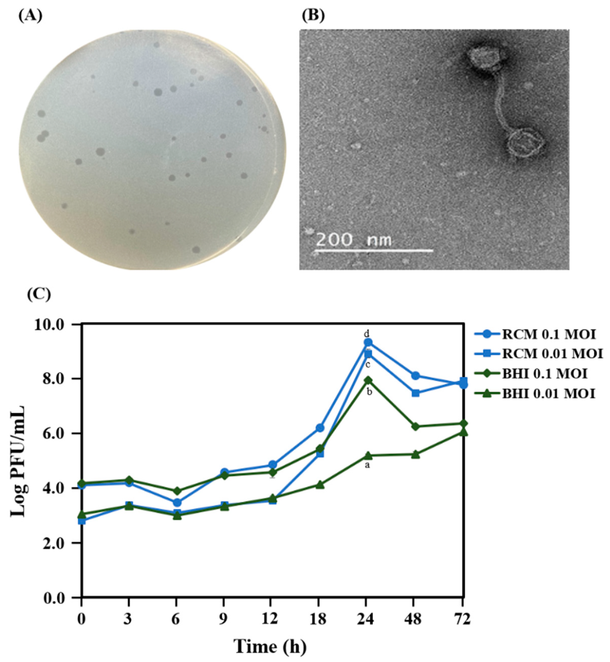
2.1. Isolation and Morphology of PAP 1-1

The morphology of phage plaques was observed on the lawn of C. acnes KCTC 3314 after 48 h of anaerobic incubation of the bacteriophage PAP 1-1 (Figure 1A). After the purification of the bacteriophage PAP 1-1, phage particle morphology was observed under a transmission electron microscope (Figure 1B). The bacteriophage PAP 1-1 had a morphology similar to that of siphovirus [7] and showed better growth on RCM than on the BHI medium (Figure 1C).

2.2. Genome Features

Using Prokka PAP 1-1, genome annotations were performed (Table 1). The de novo assembly of the genome-sequencing data of PAP 1-1 produced a single circular contig of 29,534 bp in size and consisting of a total of 40 protein-coding genes. Among them, 26 and 4 genes involved in head and tail morphogenesis and 4 genes involved in lysis were identified. Based on this single contig, the G + C content of PAP 1-1 was calculated as 54.0%. The genome sequence of PAP 1-1 was deposited in the GenBank database (accession number OP491959).

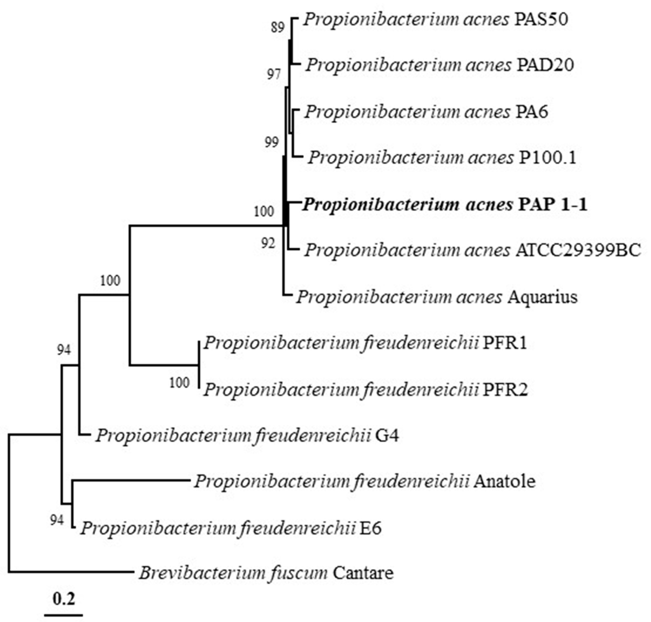
2.3. Phylogenetic Analysis and Genomic Comparison

The phylogenetic analysis using the ML algorithm based on whole-genome sequences showed that PAP 1-1 formed a close phyletic lineage with P. acnes ATCC29399BC (Figure 2) and formed a distinct phyletic lineage with other phages from Propionibacterium genus. Phylogenetic analysis results showed that PAP 1-1 belongs to bacteriophages isolated from P. acnes.

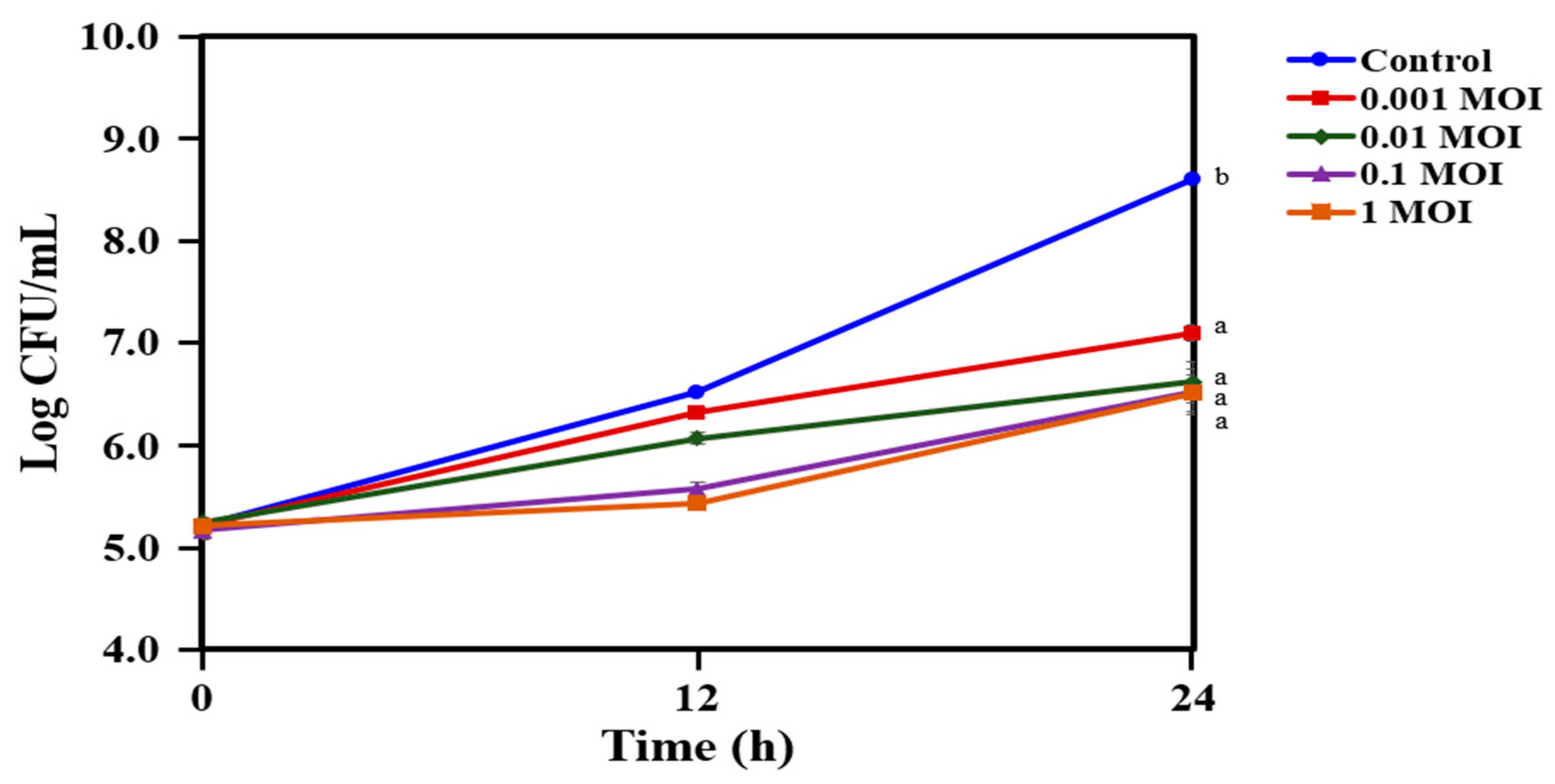
2.4. Reduction in the Growth of C. acnes KCTC 3314 with Bacteriophage PAP 1-1

The antibacterial efficacy of PAP 1-1 was assessed against C. acnes KCTC 3314 in a dose-dependent manner over a period of 12 and 24 h of incubation. The 0.001 MOI (multiplicity of infection as plaque-forming units (PFU)/CFU for host) effectively suppressed the growth of C. acnes KCTC 3314, resulting in a 1.6 log drop in CFU/mL compared with the control group (Figure 3). Notably, 0.01 MOI led to a substantial reduction in the cell population of C. acnes KCTC 3314, resulting in a two-log drop in CFU/mL. Gradually increasing the bacteriophage PAP 1-1 from 0.1 to 1 MOI effectively controlled the growth of C. acnes KCTC 3314 during the initial 12 h of incubation, which then gradually increased to 6.5 CFU/mL, similar to the count observed with 0.01 MOI after 24 h of incubation. The results show that a specific concentration of PAP 1-1 controls the growth of C. acnes KCTC 3314.

2.5. Reduction in the Growth of C. acnes KCTC 3314 Followed by Co-treatment with Bacteriophage PAP 1-1 and Bacteriocin

The antibacterial activity of the crude bacteriocin from L. lactis CJNU 3001 was tested against C. acnes KCTC 3314 alone and also in combination with 0.01 MOI in a dose-dependent manner after 12 and 24 h of incubation (Figure 4A,B). The bacteriocin treatment against C. acnes KCTC 3314 greatly reduced its growth, and cell counts gradually decreased with an increase in bacteriocin from 6.25 to 50 AU/mL (Figure 4A). The cell counts of C. acnes KCTC 3314 were reduced after treatment with bacteriocin ranging between 12.5 and 50 AU/mL, specifically with 25 AU/mL and 50 AU/mL of bacteriocin in which 5 log CFU/mL difference in growth levels were observed as compared to control. In the co-treatment of 0.01 MOI and the bacteriocin, the cell counts of C. acnes KCTC 3314 were reduced to a certain level after 24 h of incubation, and a significant reduction was observed with the co-treatment of 0.01 MOI and 50 AU/mL of bacteriocin (Figure 4B).

2.6. Reduction in the Growth of C. acnes KCTC 3314 Followed by Co-treatment with Bacteriophage PAP 1-1 and Nisin

In our previous study, the partial whole-genome sequencing of L. lactis CJNU 3001 (GenBank accession no. JAIZVR000000000.1) was found to harbor a corresponding structural gene for nisin. The activity of nisin against C. acnes KCTC 3314 was tested alone and in co-treatment with bacteriophage PAP1-1 using the same procedure as described above for the bacteriocin (Figure 5A,B). Nisin concentration range between 12.5 IU/mL and 25 IU/mL reduced the cell counts to a certain level, compared with 6.25 IU/mL and 3.125 IU/mL of nisin and control (Figure 5A). In co-treatment with 0.01 MOI, the results remained almost the same as those observed in the case of nisin alone except for the combination of 0.01 MOI and 6.25 IU/mL of nisin in which a notable reduction in the number of cells of C. acnes KCTC 3314 was observed compared with nisin alone treatment (Figure 5B). The concentration of 3.125 IU/mL was found to be ineffective against C. acnes KCTC 3314 alone and also in combination with PAP 1-1 (Figure 5A,B).

3. Discussion

The skin’s microbial community plays a crucial role in preventing pathogen invasion and the formation of biofilms. However, the exact impact of bacteriophages on modulating bacterial communities is not well understood. In this study, we focused on isolating, characterizing, and studying the bacteriophage PAP 1-1. Our research significantly enhanced our understanding of PAP 1-1’s activity against C. acnes KCTC 3314, a bacterium isolated from acne lesions on human facial skin. We examined the effects of PAP 1-1 alone and in combination with other antimicrobial compounds, including a crude bacteriocin from L. lactis CJNU 3001 and nisin. Morphological characterization using TEM revealed that PAP 1-1 exhibited siphovirus-like morphology, possessing an icosahedral head and a long tail [33]. Clear plaques against C. acnes KCTC 3314 on the RCM agar plate and the absence of lysogenic repressor genes, as determined via BLAST search, indicated that PAP 1-1 is a lytic bacteriophage [14,34]. Previous studies reported bacteriophage activity against 88% of all C. acnes strains, and approximately 18% of C. acnes strains carried bacteriophages [35].
Unlike phages from other bacterial species, the genomes of C. acnes bacteriophages show limited diversity despite their isolation from various geographical regions [25]. Marinelli et al. and Liu et al. sequenced bacteriophage genomes isolated from C. acnes and found a high degree of nucleotide identity between them [25,36]. Liu et al. also discovered identical bacteriophage strains among closely related individuals, suggesting the possibility of virus transmission from human to human [25]. In our study, we sequenced the genome of the bacteriophage PAP 1-1 isolated from C. acnes. OrthoANI analysis revealed that PAP 1-1 shares high nucleotide identity (87–93%) with other bacteriophage genomes. Phylogenetic analysis demonstrated close relatedness among bacteriophage genomes, and PAP 1-1 formed a clade with genomes isolated from C. acnes and/or P. acnes, clearly distinct from genomes of bacteriophages isolated from Propionibacterium freudenreichii strains (Figure 2). The genomic relatedness observed in the phylogenetic tree also suggests that PAP 1-1 shares common genes with bacteriophages ATCC29399BC and PAS50. C. acnes bacteriophage genomes harbor genes for endolysins, which contribute to bacterial cell wall degradation through the action of muramidases, amidases, endopeptidases, glucosaminidases, and transglycosylases [29,37,38]. Marinelli et al. proposed that these endolysins play a crucial role in using bacteriophages to treat acne infections [29]. The genes encoding these phage endolysins are highly conserved, with approximately 95% amino acid identity [36]. Phage endolysins have demonstrated efficacy in vitro and in vivo against various microbes [39]. In our study, we identified a gene encoding an endolysin through annotation using Prokka (Table 1). Future research could explore the use of C. acnes bacteriophage endolysins for treating C. acnes infections on the skin, potentially through genetic engineering, as previous studies have shown that bacteria remain sensitive to endolysins even after developing phage resistance [40].
Bacteriophage therapy has gained significant attention among scientists due to its successful treatment of infections in humans [41,42]. Bacteriophages have the ability to target both Gram-positive and Gram-negative bacteria, including multidrug-resistant strains [43,44,45,46]. Unlike antibiotics, which face increasing resistance over time, bacteriophages retain their ability to kill bacteria [47]. Moreover, since bacteriophages are host-specific, they exert minimal pressure on non-targeted microbial flora, unlike antibiotics. Several studies have investigated the specificity of bacteriophages against C. acnes [48,49]. For instance, Brown et al. isolated bacteriophages and demonstrated their ability to lyse P. acnes strains specifically, without affecting other members of the Cutibacterium genus [48]. In our present study, we also isolated the bacteriophage PAP 1-1 from acne lesions of volunteers and tested its efficacy against C. acnes KCTC 3314 in a dose-dependent manner (Figure 3). We observed that all of the tested concentrations of PAP 1-1 exhibited activity against C. acnes KCTC 3314. Among these concentrations, we selected 0.01 MOI to evaluate its combination with a crude bacteriocin from L. lactis CJNU 3001 and the commercially available nisin (Figure 4 and Figure 5). Bacteriocins have been previously reported to have antimicrobial effects and are recognized as effective bio-preservatives, such as nisin [23]. In our study, the bacteriocin treatment against C. acnes KCTC 3314 significantly reduced bacterial growth, and cell counts gradually decreased with increasing bacteriocin concentration (from 6.25 to 50 AU/mL) (Figure 4A). The co-treatment of the bacteriocin and 0.01 MOI resulted in a reduction in C. acnes KCTC 3314 cell counts to a certain level after 24 h of incubation, with a noticeable reduction observed in the co-treatment of 0.01 MOI and 50 AU/mL of the bacteriocin (Figure 4B). Similarly, regarding nisin activity, concentrations ranging between 12.5 IU/mL and 25 IU/mL reduced cell counts to a certain level compared with concentrations of 6.25 IU/mL and 3.125 IU/mL of nisin (Figure 5A). Notably, in the co-treatment of 0.01 MOI and 6.25 IU/mL of nisin, a significant reduction in the cell counts of C. acnes KCTC 3314 was observed (Figure 5B).

4. Materials and Methods

4.1. Isolation of Bacteriophage

To isolate the bacteriophage from the acne skin of volunteers, it was safely swapped twice with a sterile cotton swab, put into RCM (reinforced clostridial medium) broth inoculated to about 1 × 10⁵ CFU/mL of C. acnes KCTC (Korean Collection for Type Cultures) 3314 strain and cultured anaerobically at 37 °C for 72 h. The potential lysate was centrifuged at 10,000 rpm for 10 min at 4 °C, and the supernatant was recovered and filtered (0.45 μm syringe filter; Anylab Co., Seoul, Korea). To detect phage activity based on plaque formation, the method described by Liu et al. [33] was used. Briefly, 50 μL of the diluted supernatant was added to 3 mL of RCM soft agar (1.2% agar, w/v) where C. acnes KCTC 3314 strain had been inoculated to about 1 × 10⁵ CFU/mL, overlaid on an RCM agar plate and was incubated anaerobically at 37 °C for 48 h. After incubation, the formation of phage plaques was observed. Single phage plaque was picked up and inoculated to RCM broth where C. acnes KCTC 3314 strain had been inoculated and incubated anaerobically at 37 °C for 72 h. The culture was centrifuged at 10,000 rpm for 10 min at 4 °C to recover the supernatant, and it was filtered (0.45 μm syringe filter; Anylab Co.). To enumerate the number of plaques of the filtrate, it was decimally diluted and loaded on the RCM agar plate where C. acnes KCTC 3314 strain had been overlaid and anaerobically incubated at 37 °C for 48 h. The plaques were enumerated, and the number of plaques was represented as PFU (plaque-forming units) /mL.

4.2. Transmission Electron Microscopy of PAP 1-1 Bacteriophage

A PEG solution (20% polyethylene glycol (PEG 8000), 2 M NaCl) was treated with the filtrate harboring the PAP 1-1 bacteriophage with a ratio of 1:1 and reacted on ice for 12 h. After that, the supernatant was completely removed via centrifugation with 12,000 rpm for 30 min at 4 °C, and the precipitate was suspended in sterilized SM buffer (100 mM NaCl, 8 mM MgSO4·7H2O, 50 mM Tris-HCl (pH 7.5), 0.01% gelatin), centrifuged at 13,000 rpm for 1 min at 4 °C, filtered (0.45 μm syringe filter, Anylab Co.), and used for transmission electron microscopy (TEM). Five microliters of the concentrated bacteriophage solution were dripped onto the grid and washed 3 times with water. After staining with 2% uranyl acetate, it was analyzed using a field emission transmission electron microscope (FE-TEM; JEM 2100F, Jeol, Japan) at the Korea Basic Science Institute (Chuncheon Center, Chuncheon, Korea).

4.3. Optimal Medium for Bacteriophage Propagation

To identify the growth medium that gives the most optimal condition for phage production, two different media were tested: RCM and BHI. Specifically, the bacteriophage PAP 1-1 was incubated (anaerobic condition at 37 °C) with C. acnes KCTC 3314 strain at MOIs of 0.01 or 0.1 separately in the two testing media. Sampling was carried out at 0, 3, 6, 9, 12, 18, 24, 48, and 72 h, and the double-layer agar method was used for phage enumeration.

4.4. Genomic DNA Extraction, Sequencing, and Annotation

To analyze the whole-genome sequence, genomic DNA was extracted by using a phage DNA isolation kit (Norgen Biotek Co., Thorold, ON, Canada), according to the manufacturer’s guide. The library was developed according to the TruSeq nano DNA library prep guide (Illumina, Inc., San Diego, CA, USA). Indexed libraries were then submitted to Illumina Hiseq X-10 (Illumina), and paired-end (2 × 151 bp) sequencing was performed by the Macrogen Co. (Seoul, Korea) for whole-genome sequencing. After sequencing, FastQC (v0.11.6) [50] was performed in order to check the read quality. To mitigate biases in the analysis, an internally developed script was employed to exclude the reads where the proportion of bases below Q20 was greater than 10%. Trimmomatic (v0.36) [51] was used to remove adapter sequences. First, de novo assembly was performed using various k-mer values in Spades (v3.12) [52]. The best contig was selected based on the status of the assembled results such as the number of contigs, contigs sum, and N50. Once the assembly was completed, we employed our in-house script to verify whether the ends of the contigs overlapped and subsequently connected them to form a circular contig. For gene prediction and annotation, Prokka (v1.12b) [53] was used, and for additional annotation, the assembled sequences were searched against GenBank non-redundant (NR) database, UniProt, GO, InterPro, Pfam, CD, TIGRFAM, and EggNOG using BLAST (v2.6.0+) [54].

4.5. Phylogenetic Analysis and Genomic Comparison

For the phylogenomic analysis, we retrieved the genomes of PAP 1-1 and closely related Propionibacterium bacteriophage genomes from the EMBL public database (https://www.ebi.ac.uk/genomes/phage.html, accessed on 03 April 2023). These genome sequences were aligned using ClustalW, and a phylogenetic tree based on the entire genome sequences was constructed with bootstrap values (1000 replications). The maximum likelihood (ML) tree was constructed in the MEGA7 software, utilizing the Kimura two-parameter model, the nearest-neighbor interchange heuristic search method, and partial deletion options [55].
Based on the phylogenomic tree, we selected the genomes of P. acnes ATCC29399BC and P. acnes PAS50 for genomic comparison with the PAP 1-1 genome. To perform this comparison, we employed the Proksee server (https://beta.proksee.ca/, accessed on 03 April 2023) to create circular alignments of sequence reads to the reference genomes.

4.6. Phage Lytic Activity of Bacteriophage PAP 1-1

In order to measure the phage lytic activity, it was incubated with C. acnes KCTC 3314 strain at four different MOIs, anaerobically at 37 °C [25]. Sampling was carried out at 0, 12, and 24 h to enumerate the viable cells (CFU/mL). Specifically, serial dilutions of the incubation mixture were performed 10 times, and then 10 µL of each dilution was spread on the RCM agar plate in triplicate. After incubation at 37 °C for 24 h, colonies were counted, and the values (CFU/mL) were calculated using the formula (total colonies × dilution factor × 100).

4.7. Bacteriocin Extraction and Purification

To assess the antibacterial activity of a bacteriocin derived from L. lactis CJNU 3001, a modified acetone extraction method was employed for partial purification [56]. Specifically, the supernatant obtained from an overnight culture was subjected to centrifugation at 8000 rpm for 10 min at 4 °C. The resulting supernatant was then filtered using a 0.45 μm membrane filter (Merck Millipore Co., Tullagreen, Ireland). Subsequently, the filtrate was concentrated by a factor of 10 using a rotary vacuum concentrator. To facilitate extraction, three portions of acetone were added to the concentrate, which was then placed at −20 °C for 3 h with intermittent shaking every 30 min. After completion of the extraction, centrifugation was carried out at 9000 rpm for 20 min at 4 °C to remove the supernatant and lower layer, leaving behind the middle layer. This middle layer was subsequently diluted with water to prepare a solution containing the bacteriocin, which was then stored at −20 °C until further use.

4.8. Combination Treatment with Bacteriophage PAP 1-1 and Crude Bacteriocin from Lactococcus Lactis and Nisin

The bacteriocin activity was measured as follows: The RCM soft agar inoculated with 100 μL culture of 1 × 10⁵ CFU/mL of C. acnes KCTC 3314 strain was poured on the RCM agar plate. The two-fold serial dilution of bacteriocin suspension was carried out, and then 2 μL suspension was spotted on the plate and anaerobically cultured at 37 °C for 48 h. The antibacterial activity of the bacteriocin was quantified using arbitrary units (AUs), where the AU/mL value was determined as the reciprocal of the maximum dilution factor at which the antibacterial activity is observed, multiplied by a conversion factor for 1 mL [57]. Subsequently, the antibacterial activity of various concentrations of the bacteriocin (0, 6.25, 12.5, 25, and 50 AU/mL) was assessed. To measure the antibacterial activity, each concentration of the bacteriocin was added to RCM broth containing C. acnes KCTC 3314 strain, which had been previously inoculated to a concentration of approximately 1 × 10⁵ CFU/mL, and the number of viable cells was then measured at different time points (0, 12, and 24) h.
To the mixture of bacteriophage PAP 1-1 and the C. acnes KCTC 3314 strain at MOI of 0.01, bacteriocin was added at different final concentrations of 0, 6.25, 12.5, 25, and 50 AU/mL, and the mixture was incubated anaerobically at 37 °C. Sampling was performed at 0, 12, and 24 h to enumerate the viable cells (CFU/mL). The antibacterial activity of nisin (Galacin Nisin 101, Galactic, Hainaut, Belgium) separately, at the different concentrations (0, 3.125, 6.25, 12.5, and 25 IU/mL) and with a combination of bacteriophage PAP 1-1, was determined against the C. acnes strain of KCTC 3314 in RCM broth. The viable cells were enumerated (CFU/mL) using the method described previously.

4.9. Statistical Analysis

All experiments were repeated three times, and the results are expressed as mean ± standard deviation. For statistical analysis, SPSS ver. 25 (Statistical Package for Social Sciences, SPSS Inc., Chicago, IL, USA) was used, and significance was verified using one-way ANOVA, and it was post-tested using Duncan’s multiple range test (p < 0.05).

5. Conclusions

The bacteriophage PAP 1-1, isolated and purified from acne lesions on the facial skin of volunteers, exhibited siphovirus morphology based on TEM observation. Phylogenetic analysis revealed that PAP 1-1 formed a distinct lineage alongside other bacteriophages isolated from P. acnes species. Genomic analysis indicated the presence of genes associated with lysis, including the endolysin gene responsible for host bacterium lysis. PAP 1-1 efficiently inhibited the growth of C. acnes KCTC 3314, an acne-causing bacterium isolated from human facial skin lesions. When combined with the crude bacteriocin from L. lactis CJNU 3001 and nisin, it showed a combination effect on the control of C. acnes KCTC 3314. Thus, the findings suggest that PAP 1-1 could serve as a promising candidate for acne infection control in combination with other drugs and/or antibiotics.

Author Contributions

Conceptualization, M.-H.H. and G.-S.M.; methodology, M.-H.H. and G.-S.M.; software, M.-H.H. and S.A.K.; validation, S.A.K. and G.-S.M.; formal analysis, M.-H.H. and S.A.K.; investigation, M.-H.H.; resources, G.-S.M.; data curation, M.-H.H., S.A.K. and G.-S.M.; writing—original draft preparation, M.-H.H., S.A.K. and G.-S.M.; writing—review and editing, M.-H.H., S.A.K. and G.-S.M.; visualization, M.-H.H. and S.A.K.; supervision, G.-S.M.; project administration, G.-S.M.; funding acquisition, G.-S.M. All authors have read and agreed to the published version of the manuscript.

Funding

This research was supported by Basic Science Research Program through the National Research Foundation of Korea (NRF) funded by the Ministry of Education (No. 2021R1A6A1A03046418).

Institutional Review Board Statement

The study was performed in agreement with the Declaration of Helsinki principles and approved by the Korea National University of Transportation-Institutional Review Board (KNUT-IRB2020-19).

Informed Consent Statement

Informed consent was obtained from all subjects involved in the study.

Data Availability Statement

Data are available in a publicly accessible repository and within the article.

Conflicts of Interest

The authors declare no conflict of interest.

References

  1. Grice, E.A.; Segre, J.A. The Skin Microbiome. Nat. Rev. Microbiol. 2011, 9, 244–253. [Google Scholar] [CrossRef] [PubMed]
  2. Kao, H.J.; Wang, Y.H.; Keshari, S.; Yang, J.J.; Simbolon, S.; Chen, C.C.; Huang, C.M. Propionic acid produced by Cutibacterium acnes fermentation ameliorates ultraviolet B-induced melanin synthesis. Sci. Rep. 2021, 11, 11980. [Google Scholar] [CrossRef] [PubMed]
  3. Barnard, E.; Shi, B.; Kang, D.; Craft, N.; Li, H. The balance of metagenomic elements shapes the skin microbiome in acne and health. Sci. Rep. 2016, 6, 39491. [Google Scholar] [CrossRef] [PubMed] [Green Version]
  4. Szabó, K.; Erdei, L.; Bolla, B.S.; Tax, G.; Bíró, T.; Kemény, L. Factors shaping the composition of the cutaneous microbiota. Br. J. Dermatol. 2017, 176, 344–351. [Google Scholar] [CrossRef] [PubMed] [Green Version]
  5. Achermann, Y.; Goldstein, E.J.C.; Coenye, T.; Shirtliffa, M.E. Propionibacterium acnes: From Commensal to opportunistic biofilm-associated implant pathogen. Clin. Microbiol. Rev. 2014, 27, 419–440. [Google Scholar] [CrossRef] [PubMed] [Green Version]
  6. Dessinioti, C.; Katsambas, A.D. The role of Propionibacterium acnes in acne pathogenesis: Facts and controversies. Clin. Dermatol. 2010, 28, 2–7. [Google Scholar] [CrossRef]
  7. Grange, P.A.; Raingeaud, J.; Morelle, W.; Marcelin, A.G.; Calvez, V.; Dupin, N. Characterization of a Propionibacterium acnes Surface Protein as a Fibrinogen-Binding Protein. Sci. Rep. 2017, 7, 6428. [Google Scholar] [CrossRef] [PubMed] [Green Version]
  8. Hay, R.J.; Johns, N.E.; Williams, H.C.; Bolliger, I.W.; Dellavalle, R.P.; Margolis, D.J.; Marks, R.; Naldi, L.; Weinstock, M.A.; Wulf, S.K.; et al. The global burden of skin disease in 2010: An analysis of the prevalence and impact of skin conditions. J. Investig. Dermatol. 2014, 134, 1527–1534. [Google Scholar] [CrossRef] [Green Version]
  9. Lynn, D.; Umari, T.; Dellavalle, R.; Dunnick, C. The epidemiology of acne vulgaris in late adolescence. Adolesc. Health Med. Ther. 2016, 7, 13–25. [Google Scholar] [CrossRef] [PubMed] [Green Version]
  10. Del Rosso, J. Topical and oral antibiotics for acne vulgaris. Semin. Cutan. Med. Surg. 2016, 35, 57–61. [Google Scholar] [CrossRef] [PubMed]
  11. Dréno, B. Bacteriological resistance in acne: A call to action. Eur. J. Dermatol. 2016, 26, 127–132. [Google Scholar] [CrossRef] [PubMed]
  12. Beylot, C.; Auffret, N.; Poli, F.; Claudel, J.P.; Leccia, M.T.; Del Giudice, P.; Dreno, B. Propionibacterium acnes: An update on its role in the pathogenesis of acne. J. Eur. Acad. Dermatol. Venereol. 2014, 28, 271–278. [Google Scholar] [CrossRef] [PubMed]
  13. Eskenazi, A.; Lood, C.; Wubbolts, J.; Hites, M.; Balarjishvili, N.; Leshkasheli, L.; Askilashvili, L.; Kvachadze, L.; van Noort, V.; Wagemans, J.; et al. Combination of pre-adapted bacteriophage therapy and antibiotics for treatment of fracture-related infection due to pandrug-resistant Klebsiella pneumoniae. Nat. Comm. 2022, 13, 302. [Google Scholar] [CrossRef] [PubMed]
  14. Sharma, S.; Datta, S.; Chatterjee, S.; Dutta, M.; Samanta, J.; Vairale, M.G.; Gupta, R.; Veer, V.; Dwivedi, S.K. Isolation and characterization of a lytic bacteriophage against Pseudomonas aeruginosa. Sci. Rep. 2021, 11, 19393. [Google Scholar] [CrossRef] [PubMed]
  15. Lin, D.M.; Koskella, B.; Lin, H.C. Phage therapy: An alternative to antibiotics in the age of multi-drug resistance. World J. Gastrointest. Pharm. Ther. 2017, 8, 162–173. [Google Scholar] [CrossRef] [PubMed]
  16. Delwart, E.L. Viral metagenomics. Rev. Med. Virol. 2007, 17, 115–131. [Google Scholar] [CrossRef] [PubMed]
  17. Koonin, E.V. The wonder world of microbial viruses. Expert Rev. Anti Infect. Ther. 2010, 8, 1097–1099. [Google Scholar] [CrossRef] [PubMed] [Green Version]
  18. Suttle, C. Viruses in the sea. Nature 2005, 437, 356–361. [Google Scholar] [CrossRef]
  19. Clokie, M.R.J.; Millard, A.D.; Letarov, A.V.; Heaphy, S. Phages in nature. Bacteriophage 2011, 1, 31–45. [Google Scholar] [CrossRef] [Green Version]
  20. Young, R.; Gill, J.J. Phage therapy redux—What is to be done? Science 2015, 350, 1163–1164. [Google Scholar] [CrossRef]
  21. Salmond, G.P.C.; Fineran, P.C. A century of the phage: Past, present and future. Nature Rev. Microbiol. 2015, 13, 777–786. [Google Scholar] [CrossRef] [PubMed]
  22. Church, N.A.; McKillip, J.L. Antibiotic resistance crisis: Challenges and imperatives. Biologia 2021, 76, 1535–1550. [Google Scholar] [CrossRef]
  23. Hammami, R.; Fliss, I.; Corsetti, A. Editorial: Application of protective cultures and bacteriocins for food biopreservation. Front. Microbiol. 2019, 10, 1561. [Google Scholar] [CrossRef] [PubMed] [Green Version]
  24. Cotter, P.D.; Ross, R.P.; Hill, C. Bacteriocins-a viable alternative to antibiotics? Nature Rev. Microbiol. 2013, 11, 95–105. [Google Scholar] [CrossRef] [PubMed]
  25. Yap, P.G.; Lai, Z.W.; Tan, J.S. Bacteriocins from lactic acid bacteria: Purification strategies and applications in food and medical industries: A review. Beni-Suef Univ. J. Basic Appl. Sci. 2022, 11, 51. [Google Scholar] [CrossRef]
  26. Jiang, S.; Cai, L.; Lv, L.; Li, L. Pediococcus pentosaceus, a future additive or probiotic candidate. Microb. Cell Factories 2021, 20, 45. [Google Scholar] [CrossRef]
  27. Cotter, P.D.; Hill, C.; Ross, R.P. Bacteriocins: Developing innate immunity for food. Nat. Rev. Microbiol. 2005, 3, 777–788. [Google Scholar] [CrossRef]
  28. Yang, R.; Johnson, M.C.; Ray, B. Novel method to extract large amounts of bacteriocins from lactic acid bacteria. Appl. Environ. Microbiol. 1992, 58, 3355–3359. [Google Scholar] [CrossRef] [Green Version]
  29. Muhammad, Z.; Ramzan, R.; Abdelazez, A.; Amjad, A.; Afzaal, M.; Zhang, S.; Pan, S. Assessment of the antimicrobial potentiality and functionality of Lactobacillus plantarum strains isolated from the conventional inner Mongolian fermented cheese against foodborne pathogens. Pathogens 2019, 8, 71. [Google Scholar] [CrossRef] [Green Version]
  30. da Costa, R.J.; Voloski, F.L.; Mondadori, R.G.; Duval, E.H.; Fiorentini, Â.M. Preservation of meat products with bacteriocins produced by lactic acid bacteria isolated from meat. J. Food Qual. 2019, 2019, 4726510. [Google Scholar] [CrossRef] [Green Version]
  31. Radaic, A.; Malone, E.; Kamarajan, P.; Kapila, Y.L. Solid Lipid Nanoparticles Loaded with Nisin (SLN-Nisin) are More Effective Than Free Nisin as Antimicrobial, Antibiofilm, and Anticancer Agents. J. Biomed. Nanotechnol. 2022, 18, 1227–1235. [Google Scholar] [CrossRef] [PubMed]
  32. Barbosa, A.A.T.; de Melo, M.R.; da Silva, C.M.R.; Jain, S.; Dolabella, S.S. Nisin resistance in Gram-positive bacteria and approaches to circumvent resistance for successful therapeutic use. Crit. Rev. Microbiol. 2021, 47, 376–385. [Google Scholar] [CrossRef] [PubMed]
  33. Liu, J.; Yan, R.; Zhong, Q.; Ngo, S.; Bangayan, N.J.; Nguyen, L.; Lui, T.; Liu, M.; Erfe, M.C.; Craft, N.; et al. The diversity and host interactions of Propionibacterium acnes bacteriophages on human skin. ISME J. 2015, 9, 2078–2093. [Google Scholar] [CrossRef] [PubMed] [Green Version]
  34. Dewanggana, M.N.; Evangeline, C.; Ketty, M.D.; Waturangi, D.E.; Yogiara; Magdalena, S. Isolation, characterization, molecular analysis and application of bacteriophage DW-EC to control Enterotoxigenic Escherichia coli on various foods. Sci. Rep. 2022, 12, 495. [Google Scholar] [CrossRef] [PubMed]
  35. Castillo, D.E.; Nanda, S.; Keri, J.E. Propionibacterium (Cutibacterium) acnes Bacteriophage Therapy in Acne: Current Evidence and Future Perspectives. Dermatol. Ther. 2019, 9, 19–31. [Google Scholar] [CrossRef] [PubMed] [Green Version]
  36. Marinelli, L.J.; Fitz-Gibbon, S.; Hayes, C.; Bowman, C.; Inkeles, M.; Loncaric, A.; Russell, D.A.; Jacobs-Sera, D.; Cokus, S.; Pellegrini, M.; et al. Propionibacterium acnes bacteriophages display limited genetic diversity and broad killing activity against bacterial skin isolates. mBio 2012, 3, e00279-12. [Google Scholar] [CrossRef] [PubMed] [Green Version]
  37. Farrar, M.D.; Howson, K.M.; Bojar, R.A.; West, D.; Towler, J.C.; Parry, J.; Pelton, K.; Holland, K.T. Genome sequence and analysis of a Propionibacterium acnes bacteriophage. J. Bacteriol. 2007, 189, 4161–4167. [Google Scholar] [CrossRef] [Green Version]
  38. Schmelcher, M.; Donovan, D.M.; Loessner, M.J. Bacteriophage endolysins as novel antimicrobials. Future Microbiol. 2012, 7, 1147–1171. [Google Scholar] [CrossRef] [Green Version]
  39. Nelson, D.C.; Schmelcher, M.; Rodriguez-Rubio, L.; Klumpp, J.; Pritchard, D.G.; Dong, S.; Donovan, D.M. Endolysins as Antimicrobials. Adv. Virus Res. 2012, 83, 299–365. [Google Scholar]
  40. Jończyk-Matysiak, E.; Weber-Dabrowska, B.; Zaczek, M.; Miedzybrodzki, R.; Letkiewicz, S.; Łusiak-Szelchowska, M.; Górski, A. Prospects of phage application in the treatment of acne caused by Propionibacterium acnes. Front. Microbiol. 2017, 8, 164. [Google Scholar] [CrossRef] [Green Version]
  41. Rhoads, D.D.; Wolcott, R.D.; Kuskowski, M.A.; Wolcott, B.M.; Ward, L.S.; Sulakvelidze, A. Bacteriophage therapy of venous leg ulcers in humans: Results of a phase i safety trial. J. Wound Care 2009, 18, 237–243. [Google Scholar] [CrossRef] [PubMed]
  42. Kutter, E.; De Vos, D.; Gvasalia, G.; Alavidze, Z.; Gogokhia, L.; Kuhl, S.; Abedon, S. Phage Therapy in Clinical Practice: Treatment of Human Infections. Curr. Pharm. Biotechnol. 2010, 11, 69–86. [Google Scholar] [CrossRef] [PubMed]
  43. Merabishvili, M.; de Vos, D.; Verbeken, G.; Kropinski, A.M.; Vandenheuvel, D.; Lavigne, R.; Wattiau, P.; Mast, J.; Ragimbeau, C.; Mossong, J.; et al. Selection and Characterization of a Candidate Therapeutic Bacteriophage That Lyses the Escherichia coli O104:H4 Strain from the 2011 Outbreak in Germany. PLoS ONE 2012, 7, e52709. [Google Scholar] [CrossRef] [Green Version]
  44. Pouillot, F.; Chomton, M.; Blois, H.; Courroux, C.; Noelig, J.; Bidet, P.; Bingen, E.; Bonacorsi, S. Efficacy of bacteriophage therapy in experimental sepsis and meningitis caused by a clone O25b: H4-ST131 Escherichia coli strain producing CTX-M-15. Antimicrob. Agents Chemother. 2012, 56, 3568–3575. [Google Scholar] [CrossRef] [PubMed] [Green Version]
  45. Chhibber, S.; Kaur, T.; Kaur, S. Co-Therapy Using Lytic Bacteriophage and Linezolid: Effective Treatment in Eliminating Methicillin Resistant Staphylococcus aureus (MRSA) from Diabetic Foot Infections. PLoS ONE 2013, 8, e56022. [Google Scholar] [CrossRef] [PubMed] [Green Version]
  46. Biswas, B.; Adhya, S.; Washart, P.; Paul, B.; Trostel, A.N.; Powell, B.; Carlton, R.; Merril, C.R. Erratum: Bacteriophage therapy rescues mice bacteremic from a clinical isolate of vancomycin-resistant Enterococcus faecium (Infection and Immunity (2002) 70:1 (204–210)). Infect. Immun. 2002, 70, 1664. [Google Scholar] [CrossRef] [Green Version]
  47. Wittebole, X.; De Roock, S.; Opal, S.M. A Historical Overview of Bacteriophage Therapy as an Alternative to Antibiotics for the Treatment of Bacterial Pathogens. Virulence 2014, 5, 226–235. [Google Scholar] [CrossRef]
  48. Brown, T.L.; Petrovski, S.; Dyson, Z.A.; Seviour, R.; Tucci, J. The Formulation of Bacteriophage in a Semi Solid Preparation for Control of Propionibacterium Acnes Growth. PLoS ONE 2016, 11, e0151184. [Google Scholar] [CrossRef] [Green Version]
  49. Golembo, M.; Puttagunta, S.; Rappo, U.; Weinstock, E.; Engelstein, R.; Gahali-Sass, I.; Moses, A.; Kario, E.; Ben-Dor Cohen, E.; Nicenboim, J.; et al. Development of a topical bacteriophage gel targeting Cutibacterium acnes for acne prone skin and results of a phase 1 cosmetic randomized clinical trial. Skin Health Dis. 2022, 2, e93. [Google Scholar] [CrossRef]
  50. de Sena Brandine, G.; Smith, A.D. Falco: High-Speed FastQC Emulation for Quality Control of Sequencing Data. F1000Res 2019, 8, 1874. [Google Scholar] [CrossRef] [Green Version]
  51. Bolger, A.M.; Lohse, M.; Usadel, B. Trimmomatic: A Flexible Trimmer for Illumina Sequence Data. Bioinformatics 2014, 30, 2114–2120. [Google Scholar] [CrossRef] [PubMed] [Green Version]
  52. Bankevich, A.; Nurk, S.; Antipov, D.; Gurevich, A.A.; Dvorkin, M.; Kulikov, A.S.; Lesin, V.M.; Nikolenko, S.I.; Pham, S.; Prjibelski, A.D.; et al. SPAdes: A new genome assembly algorithm and its applications to single-cell sequencing. J. Comp. Biol. 2012, 19, 455–477. [Google Scholar] [CrossRef] [PubMed] [Green Version]
  53. Seemann, T. Prokka: Rapid Prokaryotic Genome Annotation. Bioinformatics 2014, 30, 2068–2069. [Google Scholar] [CrossRef] [PubMed] [Green Version]
  54. Camacho, C.; Coulouris, G.; Avagyan, V.; Ma, N.; Papadopoulos, J.; Bealer, K.; Madden, T.L. BLAST+: Architecture and Applications. BMC Bioinform. 2009, 10, 421. [Google Scholar] [CrossRef] [PubMed] [Green Version]
  55. Kumar, S.; Stecher, G.; Tamura, K. MEGA7: Molecular Evolutionary Genetics Analysis Version 7.0 for Bigger Datasets. Mol. Biol. Evol. 2016, 33, 1870–1874. [Google Scholar] [CrossRef] [PubMed] [Green Version]
  56. Chung, D.M.; Kim, K.E.; Jeong, S.Y.; Park, C.S.; Ahn, K.H.; Kim, D.H.; Kang, D.O.; Chun, H.K.; Yoon, B.D.; Koh, H.B.; et al. Rapid concentration of some bacteriocin-like compounds using an organic solvent. Food Sci. Biotechnol. 2011, 20, 1457–1459. [Google Scholar] [CrossRef]
  57. Kim, S.G.; Lee, Y.D.; Park, J.H.; Moon, G.S. Synergistic inhibition by bacteriocin and bacteriophage against Staphylococcus aureus. Food Sci. Anim. Resour. 2019, 39, 1015–1020. [Google Scholar] [CrossRef] [PubMed] [Green Version]
Figure 1. Formation of plaques (round, clear) in RCM agar plate (A) and phage particle morphology of the bacteriophage PAP 1-1 (B). Propagation of bacteriophage PAP 1-1 in broth (C). Data are provided as the mean value ± SD, measured in triplicate. Different letters present significant differences (p < 0.05).
Antibiotics 12 01035 g001
Figure 2. A maximum likelihood tree based on whole-genome sequences showing the phylogenetic relationships between PAP 1-1 and related bacteriophages. Bootstrap values are shown on nodes in percentages of 1000 replicates, with only values over 70%. Brevibacterium fuscum Cantare was used as an outgroup. The scale bar equals 0.2 changes per nucleotide position.
Antibiotics 12 01035 g002
Figure 3. Reduction in the growth of Cutibacterium acnes KCTC 3314 cultured with bacteriophage PAP 1-1 ranging from 0.001 to 1 MOI. Data are provided as the mean value ± SD, measured in triplicate. Different letters present significant differences (p < 0.05).
Antibiotics 12 01035 g003
Figure 4. Reduction in the growth of Cutibacterium acnes KCTC 3314 cultured with crude bacteriocin from L. lactis CJNU 3001 alone (A) and a combination of the bacteriocin and bacteriophage PAP 1-1 (B). Data are provided as the mean value ± SD, measured in triplicate.
Antibiotics 12 01035 g004
Figure 5. Reduction in the growth of Cutibacterium acnes KCTC 3314 cultured with nisin alone (A) and a combination of nisin and bacteriophage PAP 1-1 (B). Data are provided as the mean value ± SD, measured in triplicate.
Antibiotics 12 01035 g005
Table 1. Lists of genes of bacteriophage PAP1-1 identified from genomic sequences.
CDsAnnotation
PAP1-1-147373Terminase small subunit
PAP1-1-23781889Gp2 (Propionibacterium phage PA6)
PAP1-1-318863211Capsid and scaffold protein
PAP1-1-432153970Capsid maturation protease
PAP1-1-540784638Putative head assembly scaffold
PAP1-1-646455592Major capsid protein
PAP1-1-756366097Head-to-tail adaptor
PAP1-1-860996446Gp8
PAP1-1-964526742Gp9 (Propionibacterium phage PA6)
PAP1-1-1067397110Gp10 (Propionibacterium phage PAS50)
PAP1-1-1171627794Major tail protein (Propionibacterium phage P9.1)
PAP1-1-1278228118Gp12
PAP1-1-1382178504Putative tail assembly chaperone
PAP1-1-14851311,278Tape measure protein
PAP1-1-1511,29412,235Minor tail protein
PAP1-1-1612,24313,400Putative protease
PAP1-1-1713,42114,239Putative minor tail protein
PAP1-1-1814,54015,328Putative minor tail protein
PAP1-1-1915,37816,238Putative endolysin
PAP1-1-2016,25116,649Gp21
PAP1-1-2116,73517,250Uncharacterized protein
PAP1-1-2217,25617,654Helix–turn–helix DNA-binding domain protein
PAP1-1-2317,65817,942Gp25 (Propionibacterium phage PAS50)
PAP1-1-2417,95418,274Helix–turn–helix DNA-binding domain protein
PAP1-1-2518,28419,330Gp27
PAP1-1-2619,32719,521Gp28 (Propionibacterium phage PA6)
PAP1-1-2719,50520,062Gp29.1 (Propionibacterium phage PAS50)
PAP1-1-2820,05920,625Gp30 (Propionibacterium phage PA6)
PAP1-1-2920,67021,341DNA primase
PAP1-1-3021,53921,895Holliday junction resolvase
PAP1-1-3121,89522,758DNA primase/helicase
PAP1-1-3222,80423,268Gp35 (Propionibacterium phage PAS50)
PAP1-1-3323,32123,731Gp36 (Propionibacterium phage PAS50)
PAP1-1-3423,72824,675Cas4 family exonuclease (Propionibacterium phage Pirate)
PAP1-1-3524,67525,028Gp38 (Propionibacterium phage PAS50)
PAP1-1-3625,13425,337Gp39
PAP1-1-3725,33425,561Gp40 (Propionibacterium phage PAS50)
PAP1-1-3825,57826,111Gp41 (Propionibacterium phage PAS50)
PAP1-1-3926,23226,543Gp43 (Propionibacterium phage PA6)
PAP1-1-4026,57826,886Gp43 (Propionibacterium phage PA6)
Disclaimer/Publisher’s Note: The statements, opinions and data contained in all publications are solely those of the individual author(s) and contributor(s) and not of MDPI and/or the editor(s). MDPI and/or the editor(s) disclaim responsibility for any injury to people or property resulting from any ideas, methods, instructions or products referred to in the content.

