Communication

Novel Fosfomycin Resistance Mechanism in Pseudomonas entomophila Due to Atypical Pho Regulon Control of GlpT

Centro de Biotecnología y Genómica de Plantas (CBGP), Universidad Politécnica de Madrid (UPM), 28223 Madrid, Spain
*
Author to whom correspondence should be addressed.
Antibiotics 2024, 13(11), 1008; https://doi.org/10.3390/antibiotics13111008
Submission received: 7 September 2024 / Revised: 18 October 2024 / Accepted: 24 October 2024 / Published: 26 October 2024
(This article belongs to the Special Issue Antimicrobial Resistance Evolution and New Strategies to Fight It)

Abstract

:
Background/Objectives: Pseudomonas entomophila is a ubiquitous bacterium capable of killing insects of different orders and has become a model for host–pathogen studies and a promising tool for biological pest control. In the human pathogen Pseudomonas aeruginosa, spontaneous resistance to fosfomycin arises almost exclusively from mutations in the glycerol-3-phosphate transporter (GlpT), the drug’s sole entry route in this species. Here, we investigated whether this specificity is conserved in P. entomophila, as it could provide a valuable marker system for studying mutation rates and spectra and for selection in genetic engineering. Methods: We isolated 16 independent spontaneous fosfomycin-resistant mutants in P. entomophila, and studied the genetic basis of the resistance using a combination of sequencing, phenotyping and computational approaches. Results: We only found two mutants without alterations in glpT or any of its known regulatory elements. Whole-genome sequencing revealed unique inactivating mutations in phoU, a key regulator of the phosphate starvation (Pho) regulon. Computational analyses identified a PhoB binding site in the glpT promoter, and experiments showed that phoU inactivation reduced glpT expression nearly 20-fold. While placing a sugar-phosphate transporter under the Pho regulon may seem advantageous, bioinformatic analysis shows this configuration is atypical among pseudomonads. Conclusions: This atypical Pho regulon control of GlpT probably reflects the peculiarities of P. entomophila’s habitat and lifestyle; highlighting how readily regulatory evolution can lead to the rapid divergence of resistance mechanisms, even among closely related species.

1. Introduction

Pseudomonas entomophila is a strictly aerobic, motile, Gram-negative bacterium commonly found in diverse soil environments [1]. While first identified in 2005 as a pathogen of the fruit fly [2], P. entomophila can kill larvae from various insect orders, including Diptera, Lepidoptera, and Coleoptera [3]. Beyond these entomopathogenic properties, P. entomophila can also kill other organisms, such as amoebas, nematodes, and phytopathogenic fungi [3]. Consequently, P. entomophila has attracted much interest due to its potential as a biocontrol agent against a range of plant pathogens and insect pests, either as a living organism or through its isolated insecticidal effectors [3,4,5]. In addition to these practical considerations, P. entomophila has become an important model organism for studying insect–microbe interactions [6]. Its ability to establish successful infections in laboratory strains of the fruit fly provides an ideal system for exploring various aspects of host–pathogen biology, including the insect’s physiological and immune responses to infection [7,8], the dynamics of release and mechanisms of action of both antimicrobial peptides and insecticidal effectors [2,9], and the capacity of both organisms to acclimatize or even adapt to the challenges imposed by the other [10,11].
To fully realize the potential of P. entomophila as a model system, studies exploring whether the knowledge and genetic tools available for other pseudomonads are transferable to this emerging model are much needed. Here, we focus on the mechanisms of intrinsic resistance to the antibiotic fosfomycin, a spontaneous resistance marker system well characterized in Escherichia coli and Pseudomonas aeruginosa due to its clinical interest [12]. Fosfomycin is a broad-spectrum antibiotic effective against both Gram-positive and Gram-negative bacteria. It is primarily used to treat gastrointestinal and lower urinary tract infections but also in the treatment of various multidrug-resistant infections [13,14]. In E. coli, fosfomycin enters the cell through two transporters that import sugar phosphates in exchange for inorganic phosphate (Pi): the glycerol-3-phosphate transporter (GlpT) and the glucose-6-phosphate transporter (UhpT); their expression requires both the presence of their substrates and the starvation signaling molecule cAMP [15]. Once inside, fosfomycin acts as a phosphoenolpyruvate analog, irreversibly binding to enolpyruvoyl transferase MurA, an essential enzyme catalyzing the initial step of peptidoglycan biosynthesis, which ultimately results in cell lysis [16]. While spontaneous resistance can arise from mutations that modify or overexpress MurA [17,18], it is much more likely to occur through mutations that either inactivate GlpT or UhpT or suppress their expression by altering key genes involved in cAMP synthesis [15,19].
In contrast, P. aeruginosa lacks a functional uhpT homolog, leaving GlpT as the sole entry route for fosfomycin into the cells (protein identity, 83.3%) [20]. Moreover, cAMP signaling in this species is of less importance in regulating the uptake and metabolism of alternative carbon sources [21]. As a consequence, spontaneous fosfomycin resistance in P. aeruginosa is largely limited to arising from inactivating mutations in glpT, as has been demonstrated experimentally [22]. This specificity, combined with the loss-of-function nature of the mutations involved, makes fosfomycin resistance an ideal marker system for evaluating both spontaneous and induced mutation rates and spectra [23,24], adding to the relatively limited toolset available for investigating the molecular mechanisms underlying mutational processes in pseudomonads [25]. Additionally, this specificity can facilitate efficient screening schemes in genetic engineering. For instance, it can be used to either select desired mutants (e.g., by using the glpT locus as an integration site) or to counter-select undesired ones (e.g., in a glpT mutant, helping eliminate suicide vectors integrated into the genome) [26].
In principle, the above considerations should also apply to P. entomophila: no functional uhpT homolog is present in the genome, and there is no evidence that cAMP signaling differs qualitatively from what is known in P. aeruginosa. Motivated by these similarities, here we investigated the extent to which the specificity of spontaneous fosfomycin resistance observed in P. aeruginosa is conserved in P. entomophila. By characterizing independent fosfomycin-resistant mutants, we isolated resistant variants lacking alterations in glpT or any of its known regulatory elements. Experimental and computational analyses revealed a novel fosfomycin resistance mechanism in P. entomophila, arising from the placement of GlpT under the control of the Pho regulon. Finally, we used comparative genomics to show that this configuration is atypical across the diversity of pseudomonads, likely reflecting the specific demands and opportunities of P. entomophila’s habitat and lifestyle.

2. Results and Discussion

2.1. Fosfomycin Resistance in P. entomophila Involves Mechanisms Beyond glpT Inactivating Mutations

To elucidate whether the glpT gene is the primary target for fosfomycin resistance in P. entomophila, we began by conducting a standard Luria–Delbrück fluctuation assay to isolate 16 independent mutants resistant to 128 µg/mL of fosfomycin (Section 3). This concentration, a 32-fold increase over the minimal inhibitory concentration (MIC) for the wild type, was selected based on its use in previous P. aeruginosa studies [22,23]. From each isolated resistant mutant, we extracted chromosomal DNA, PCR-amplified their glpT sequences, and subjected them to Sanger sequencing (Section 3). Analysis of these sequences revealed that the majority (14/16, 87.5%) contained changes in the glpT gene, most likely representing loss-of-function mutations. This is best illustrated by the fact that out of these 14 mutations, 10 involved changes forcibly leading to substantial alterations in the resulting protein, such as insertions, deletions, or premature stop codons (Figure 1). In addition, 10/11 of the unique mutations (90.9%) mapped to transmembrane domains, representing a significant deviation from expectation since these regions account for only 29.7% of the protein sequence (binomial test, p = 1.3 × 10⁻⁴). This observation, along with the fact that these domains are known to be highly susceptible to destabilization by random mutations [27], further supports the idea that the adaptive value of these mutations lies in their likely loss-of-function nature. We also noted the presence of a prominent hotspot—absent in P. aeruginosa—that consisted of a four-base motif (GTGC) repeated three times consecutively, starting at base position 1268. Expansions and contractions of this motif caused a shift of the reading frame and were detected in five of the independently isolated mutants. Table 1 details all detected mutations in the glpT gene, including their position, molecular nature, and the corresponding amino acid and functional changes.
Next, we focused on the two mutants with no alterations along the glpT open reading frame, for which we followed a two-pronged approach (Section 3). On the one hand, we Sanger-sequenced the region approximately 100 bp upstream of glpT to search for mutations in the promoter region that might be altering gene expression. Additionally, we PCR-amplified and Sanger-sequenced the gene encoding GlpR, the local repressor of the glp regulon [28]. This regulon includes five operons involved in the uptake and metabolism of glycerol, including the glpTQ operon [29]. GlpR strongly inhibits these operons in the presence of glucose, and even in its absence, it continues to repress them unless sufficient levels of the inducers glycerol or glycerol-3-phosphate are present [30]. Our analyses revealed no alterations in any of these regions. Consequently, we subjected the two mutants to whole-genome sequencing and identified two unique mutations in phoU, a gene encoding a key negative regulator of the Pho regulon [31]. Both mutations likely result in loss of protein function: one involved a C → T substitution at position 406, leading to a premature stop codon (Q136*), and the other was a 519 bp deletion starting at position 302 (Figure 1). In E. coli, the Pho regulon includes at least eight operons that are co-regulated upon phosphate starvation, with their products involved in the uptake, transport, and metabolism of various phosphate sources [32]. Of note, glpT is not described as part of this regulon in either E. coli or P. aeruginosa.

2.2. Mutations in phoU Confer Fosfomycin Resistance in P. entomophila by Reducing GlpT Expression

Since the only two instances of fosfomycin resistance not involving changes in glpT pointed at the same regulatory gene, phoU, we hypothesized that either glpT or glpR were under the transcriptional control of the Pho regulon. The regulatory hub of the Pho regulon is the two-component signaling system PhoR/PhoB [33]. When phosphate is limited, the sensor histidine kinase PhoR acts as a kinase, transferring a phosphate group to the response regulator PhoB. Once phosphorylated, PhoB can bind to specific promoter regions, known as Pho boxes, activating the expression of the associated genes and operons [34]. However, when phosphate is abundant, PhoR primarily acts as a phosphatase, dephosphorylating PhoB and thereby preventing its role as a transcriptional activator [35]. PhoU is believed to assist PhoR in sensing the cell’s phosphate status and, depending on the availability of other micronutrients, to define PhoR’s role as a kinase or a phosphatase, and ultimately the expression of the Pho regulon genes [31,33].
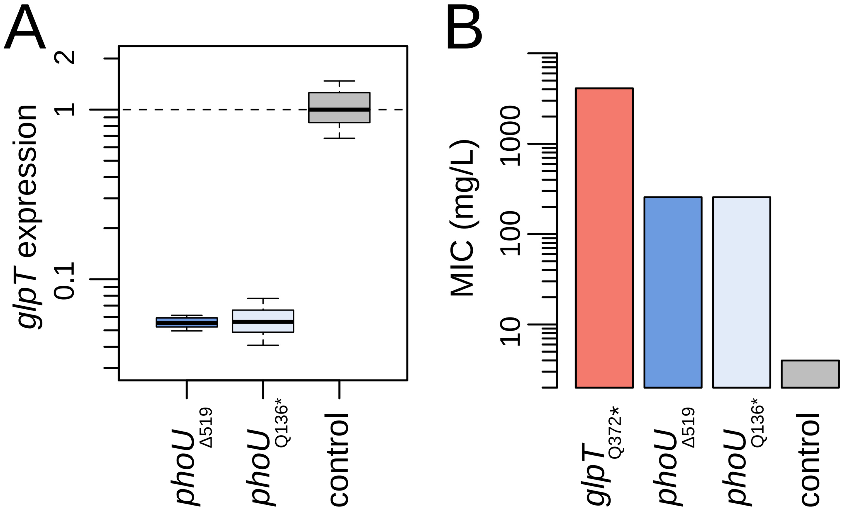
To explore the possibility that the Pho regulon in P. entomophila may include either glpT or glpR, we first turned to the widely utilized promoter prediction software BPROM to search for Pho boxes in the promoter regions of these genes (Section 3). While no hits were found for glpR, we identified a potential PhoB binding site 76 base pairs upstream of the start of glpT’s open reading frame: an ATAAAA sequence that is conserved and experimentally validated in the Pho regulon members pstS and phoE of E. coli [36]. To establish the functional significance of this finding, we next performed real-time PCR to quantify glpT expression levels in both phoU mutant backgrounds (Section 3). Figure 2A confirms that, indeed, glpT transcript levels were strongly reduced in these phoU mutants, exhibiting a nearly 20-fold decrease in expression compared to the wild-type.
We finally sought to quantify the extent to which the drastic reductions in glpT expression translate into increases in fosfomycin resistance. To this end, we used the standard broth micro-dilution method to estimate the MIC for both phoU mutants (Section 3). Besides the wild-type, we also included as a reference a glpT mutant with a nonsense mutation (Q372*). This mutant should represent the maximum resistance level achievable when no fosfomycin is transported via GlpT, a situation where all entry is expected to be limited to inefficient passive diffusion through the outer membrane porins [37]. Previous studies on fosfomycin-resistant P. aeruginosa mutants with various glpT mutations showed that nonsense, missense, and indel mutations can all lead to large elevations of the MIC, with just minor differences among these classes [22]. Figure 2B demonstrates that the phoU mutants indeed exhibited a substantial increase in resistance levels (MIC: 256 µg/mL, a 64-fold elevation over the wild type). However, this increase was of moderate magnitude compared to the presumed upper bound observed in the glpT loss-of-function mutant (MIC: 4096 µg/mL, a 1024-fold increase over the wild type), suggesting that some fosfomycin still enters the cell via GlpT, despite the reduced expression measured in the phoU mutants.

2.3. Control of GlpT by the Pho Regulon Seems Peculiar to P. entomophila

Our results indicate that, unlike in E. coli and P. aeruginosa, glpT is regulated by the Pho regulon in P. entomophila. At first glance, integrating a sugar-phosphate transporter into a phosphate starvation regulatory network may seem appropriate since the global logic of the Pho regulon is to enable the hierarchical and orderly activation of measures to acquire phosphorus from increasingly desperate sources [38]. This reasoning raises the question of why this regulatory arrangement is not observed in E. coli and, most intriguingly, in the closely related P. aeruginosa. In other words, if we look across the diversity of Pseudomonas species, is P. entomophila an outlier regarding glpT regulation, or is P. aeruginosa the exception?
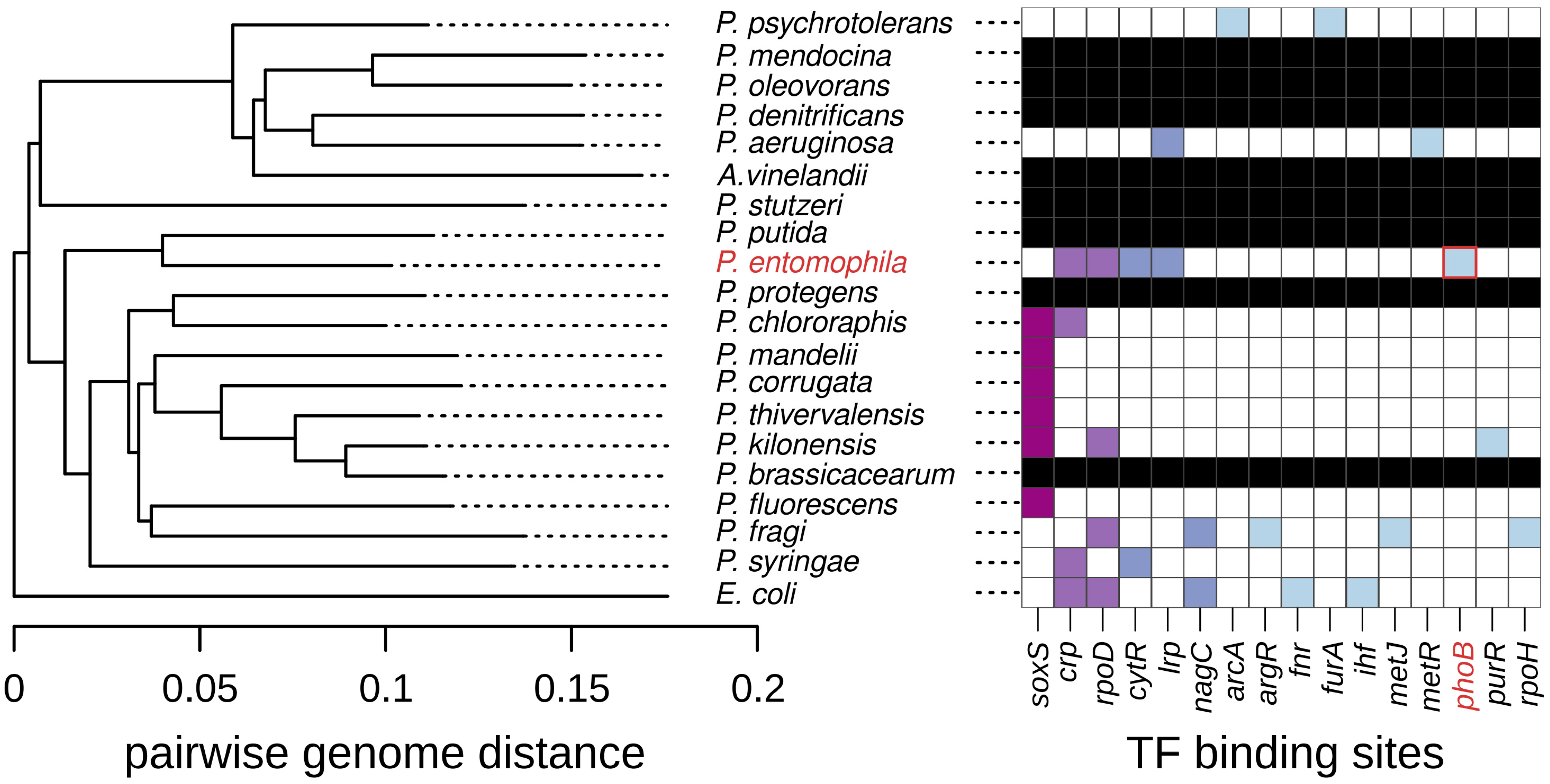
To address these questions, we compared the glpT promoter sequences across 18 representative Pseudomonas species, as well as E. coli—a standard outgroup—and Azotobacter vinelandii, a prominent member of the Pseudomonadaceae family so closely related to Pseudomonas that it is sometimes classified within this genus [39]. To this end, we began by examining the evolutionary relationships among the 20 selected species, as illustrated in Figure 3, which presents a phylogenetic tree based on pairwise genome distances (Section 3). Next, we turned to the BPROM promoter prediction software to identify transcription factor binding sites within the promoter regions of the collection of glpT homologs. Figure 3 also displays the distribution of the detected sites among species, which, for reference, are organized according to the clustering suggested by the phylogenetic tree.
Three key observations emerge from this analysis. First, 8 out of the 20 species lacked a clear glpT homolog despite the use of complete and well-annotated genomes from the MicroScope database [40]. Second, there was substantial variation in the transcription factors predicted to regulate glpT homologs across the species. Among the 16 transcription factors identified, the majority (62.5%, 10/16) were unique to a single species, and the most common transcription factor, SoxS (a transcriptional activator of the superoxide response regulon [41]), was present in only six species. We note that these results likely underestimate the true variability since BPROM has a recognition sensitivity of 83%, which is probably a compromise to minimize false positives [42]. Finally, a PhoB binding site was detected exclusively in P. entomophila, indicating that the control of GlpT by the Pho regulon is, if not unique, at least highly specific to this bacterium. Taken together, these observations suggest that the availability of external glycerol-3-phosphate varies widely across environments, a heterogeneity manifesting in at least two dimensions: first, in terms of absolute abundance, which sometimes can be virtually zero, as suggested by species lacking the transporter altogether; and second, in terms of the timing and environmental conditions associated with its presence, as suggested by the highly diverse and poorly conserved regulatory mechanisms across species.
In the specific case of P. entomophila, the suggestion would be that its ecology often features conditions where glycerol-3-phosphate is not only abundant but also frequently coincides with phosphate starvation; so it has become evolutionary worth it to integrate GlpT under the Pho regulon to utilize glycerol-3-phosphate as both a carbon and phosphate source. Of note, glycerol-3-phosphate is actively generated in insect flight muscles to facilitate NADH transport into mitochondria for oxidative phosphorylation [43], and its accumulation has been observed in butterfly larvae, where it likely serves as an energy source during this specific developmental stage [44]. Although P. entomophila is considered an opportunistic entomopathogen, and the extent of time it spends within hosts versus a free-living bacterium is uncertain [3], it is intriguing to speculate whether the availability of glycerol-3-phosphate in insects might have driven the integration of GlpT under the Pho regulon.
At a broader scale, these results highlight how readily regulatory evolution can lead to the rapid divergence of resistance mechanisms, even among closely related species. Previous studies have documented that species within the same order, such as E. coli, Salmonella enterica, and Yersinia pestis, can drastically differ in their responsiveness to environmental cues that activate resistance to the antimicrobial agent polymyxin B [45,46,47]. In contrast to our study, in these cases, the differences emerged by either the presence or absence of a key regulatory protein or by substantial molecular divergence in this protein rather than changes in promoter regions. Yet, the evolution of promoter regions, including the gain and loss of binding sites, is well-documented in both eukaryotes and prokaryotes and is believed to be a mechanism favored over changes in regulatory proteins due to its higher specificity [48,49]. Moreover, in bacteria, promoter evolution such as the one described here has been shown to occur readily through horizontal gene transfer [50,51]. In summary, our results contribute to the literature showing that regulatory circuits are in a constant state of evolution, underscoring the remarkable ability of organisms to adapt to new niches with different demands and varying stimuli patterns.

3. Materials and Methods

3.1. Isolation of Spontaneous Fosfomycin Resistant Mutants

Fifteen parallel cultures of P. entomophila L48 (obtained from Jens Rolff’s laboratory [52]) were inoculated in 3 mL of Luria broth (LB; Miller’s modification: 10 g/L of sodium chloride, 10 g/L of tryptone, 5 g/L of yeast extract; purchased in powder formulation from Condalab, Madrid, Spain) in 15 mL Falcon tubes and incubated overnight at 28 °C with shaking at 180 rpm for 48 h. The next day, 100 µL of each culture were plated onto LB-agar Petri dishes containing 128 µg/mL of fosfomycin (purchased as a disodium salt, IM, from Laboratorios Ern, Barcelona, Spain). After overnight incubation at 28 °C without shaking, a single colony from each fosfomycin plate was selected at random and re-streaked for purification under the same conditions. After further incubation, a single colony from each of these re-streaked plates was collected and stored at −80 °C for further analysis.

3.2. Antimicrobial Susceptibility Testing

The standard broth microdilution method was used to estimate the minimum inhibitory concentration (MIC) for both the wild-type and mutant strains. We followed the Clinical and Laboratory Standards Institute [53] guidelines, with the exception of using Luria broth (LB) instead of Mueller–Hinton. Briefly, strains were retrieved from −80 °C storage and inoculated into 3 mL of LB in 15 mL Falcon tubes. These cultures were incubated overnight at 28 °C with shaking at 180 rpm. After two overnight incubations, cultures were centrifuged and adjusted in saline solution to a 0.5 McFarland standard, corresponding to 1.14 ± 0.4 × 10⁷ CFU/mL (mean ± standard deviation, n = 3). Note that this value is lower than what is usually assumed for E. coli [54]. The cultures were then diluted at 1:150, and 100 µL aliquots were inoculated into microtiter plates containing a two-fold concentration gradient of fosfomycin in a final volume of 200 µL. The MIC was defined as the lowest fosfomycin concentration that completely prevented turbidity from forming after 48 h of static incubation at 28 °C.

3.3. Sanger and Whole-Genome Sequencing

Strains were retrieved from −80 °C storage and streaked on LB agar containing 128 µg/mL of fosfomycin. Genomic DNA was extracted using the DNeasy UltraClean Microbial Kit (Qiagen, Hilden, Germany) following manufacturer instructions. For Sanger sequencing, the glpT open reading frame was PCR amplified and sequenced using the forward primer 5′-CAA CAG GCC TGG GCA ATA-3′ and reverse primer 5′-CTG AAA ACA CAA AGC CCG G-3′. The glpT promoter region was amplified and sequenced using an alternative forward primer, 5′-GCT GAC GAT CCA GGT TGA CT-3′, which binds 142–123 bp upstream of glpT. The glpR open reading frame was amplified using forward primer 5′-AGC GCA ACA GAA AGC CTG TC-3′ and reverse primer 5′-GGG AAG ATC ACG ACG CCT GAT A-3′. PCR amplicons were purified using the DNeasy UltraClean Microbial Kit (Qiagen), and Sanger sequencing was conducted by Macrogen Inc. (Seoul, Republic of Korea). Mutations were identified by aligning the resultant sequences against the corresponding sequences extracted from the reference genome (downloaded from the MaGe Platform (https://mage.genoscope.cns.fr/ (accessed on 15 August 2024) [40]). These alignments were conducted using Vector NTI Advance™ 11.0 (Invitrogen, Waltham, MA, USA). For whole-genome sequencing, extracted genomic DNA was quantified using the Qubit 4 Fluorometer (Invitrogen, USA). Illumina paired-end sequencing was performed by Novogene (Cambridge, UK) at >100× coverage on the two isolates with no detected mutations in glpT, along with the isolate containing the Y407* mutation in glpT, as a control. Alignment and analysis were conducted using the computational pipeline breseq [55], using the reference genome retrieved from the MicroScope database. The raw reads were deposited in the NCBI Sequence Read Archive (PRJNA1174203).

3.4. Quantitative Real-Time PCR

RT-qPCR was performed to measure glpT gene expression using the comparative ΔΔCt method, with the ribosomal gene rpsL as the endogenous control [56,57]. Strains were retrieved from −80 °C storage and grown in 1.5 mL of Luria broth at 28 °C until the mid-log phase (OD600~0.5). After centrifugation, cells were resuspended in 100 µL of freshly prepared TE buffer (10 mM Tris-HCl, pH 8.0, 1 mM EDTA) containing 0.4 mg/mL of lysozyme (Thermo Fisher Scientific, Alcobendas, Spain) to facilitate cell lysis. RNA was extracted using the GeneJET RNA Purification Kit (Thermo Fisher Scientific, Spain) according to the manufacturer’s protocol. Extractions were performed in biological and technical triplicates. RNA concentration and purity (A260/280) were assessed using a NanoDrop® ND-1000 Spectrophotometer (NanoDrop Technologies Inc., Montchanin, DE, USA). To eliminate residual DNA, RNA samples were treated with DNase I (Thermo Fisher Scientific, Spain) before reverse transcription. cDNA was synthesized using the High-Capacity cDNA Reverse Transcription Kit (Thermo Fisher Scientific, Spain) with random hexamers, including RNase Inhibitor to prevent degradation. The qPCR reactions were conducted in triplicate using SYBR Green PCR Master Mix (Thermo Fisher Scientific, Spain) on a LightCycler® 480 system in a final volume of 20 µL per well, using alphanumerically coded white 96-well plates (Roche, Basel, Switzerland). For glpT, the following primers were used: forward, 5′-CGC CGG TCG AGC AAT ACA A-3′; reverse: 5′-CTC GTA GAA GAA GTA CGC CCA C-3′. For rpsL: forward, 5′-CCT AAC TCG GCA CTG CGT AA-3′; reverse, 5′-TGG TAA CGA ACA CCT GGC AA-3′.

3.5. Computational Methods

The genome sequences of P. entomophila L48 and all the other 19 species were retrieved from the MaGe Platform (https://mage.genoscope.cns.fr/, accessed on 15 August 2024). A list with the details of these genomes is presented in Table S1. To build the phylogenetic tree, we resorted to MaGe’s online genome clustering tool, which computes pairwise genome distances using the software Mash [58]. These distances (D) strongly correlate with the Average Nucleotide Identity (ANI), such as D ≈ 1 − ANI. Using all the pairwise distances, a tree was built dynamically using the neighbor-joining Javascript package [59]. The tree was saved in the Newick format, and visualization was conducted using the R phylogenetics package “ape” [60]. To identify putative transcription factor binding sites, the 400 bp upstream of the start site of the gene of interest (glpT or glpR) were submitted in fasta format to the promoter prediction tool BPROM (Softberry Inc., Mt Kisco, NY, USA) [42], freely available online from www.softberry.com/berry.phtml?topic=bprom&group=programs&subgroup=gfindb (accessed on 15 August 2024).

4. Conclusions

Here, we report that the single-locus specificity that spontaneous fosfomycin resistance shows in P. aeruginosa is not conserved in P. entomophila. While inactivating mutations in glpT remain the most common molecular mechanism underpinning resistance, we observed a sizable fraction of spontaneous mutants with alterations in phoU, a key regulatory element of Pho regulon. Computational evidence indicates that the observed link between phoU and glpT expression levels may be attributed to a predicted binding site for the transcription factor PhoB in the promoter region of glpT. Further research is required to elucidate the precise molecular details of this predicted interaction. Anyway, given the substantial increases in resistance conferred by any of these mutations, spontaneous fosfomycin resistance can be a valuable marker for studying mutation rates in P. entomophila. Likewise, these substantial increases in resistance can make it useful for selection and counterselection in genetic engineering. However, its effectiveness as a marker for studying mutation spectra in this species is more limited than in P. aeruginosa, as the involvement of multiple genes imposes a larger workload. Finally, our analyses indicate that glpT regulation by the Pho regulon is likely specific to the habitat and lifestyle of P. entomophila, being uncommon among other pseudomonads. This finding highlights how ecological differences can drive the rapid divergence of resistance mechanisms and suggests that future research may uncover diverse and disparate genetic elements underlying spontaneous antibiotic resistance, even among related species.

Supplementary Materials

The following supporting information can be downloaded at: https://www.mdpi.com/article/10.3390/antibiotics13111008/s1, Table S1: List of reference genomes used in this study.

Author Contributions

Conceptualization, L.S.-M. and A.C.; methodology, L.S.-M., P.G. and A.C.; investigation, L.S.-M., P.G. and A.C.; resources, A.C.; writing—original draft preparation, A.C.; writing—review and editing, L.S.-M., P.G. and A.C.; visualization, L.S.-M. and A.C.; supervision, A.C.; project administration, A.C.; funding acquisition, A.C. All authors have read and agreed to the published version of the manuscript.

Funding

This research was funded by the Agencia Estatal de Investigación (Proyectos de I + D + i, PID2019-110992GA-I00; Centros de Excelencia “Severo Ochoa”, SEV-2016-0672 and CEX2020-000999-S; FPI PhD studentship, PRE2018-086448), and a Comunidad de Madrid “Talento” Fellowship (2019-T1/BIO-12882 and 2023-5A/BIO-28940).

Institutional Review Board Statement

Not applicable.

Informed Consent Statement

Not applicable.

Data Availability Statement

All data presented in this study are contained within the article or Supplementary Materials. Raw whole-genome sequences are deposited in the NCBI Sequence Read Archive (PRJNA1174203).

Acknowledgments

We thank A. Rodríguez-Rojas and J. Rolff for sharing P. entomophila L48. We acknowledge the assistance of OpenAI’s ChatGPT-4, which was used exclusively for proofreading. We thank Jake Barber for English editing and useful feedback.

Conflicts of Interest

The authors declare no conflicts of interest. The funders had no role in the design of the study; in the collection, analyses, or interpretation of data; in the writing of the manuscript; or in the decision to publish the results.

References

  1. Vodovar, N.; Vallenet, D.; Cruveiller, S.; Rouy, Z.; Barbe, V.; Acosta, C.; Cattolico, L.; Jubin, C.; Lajus, A.; Segurens, B.; et al. Complete Genome Sequence of the Entomopathogenic and Metabolically Versatile Soil Bacterium Pseudomonas entomophila. Nat. Biotechnol. 2006, 24, 673–679. [Google Scholar] [CrossRef] [PubMed]
  2. Vodovar, N.; Vinals, M.; Liehl, P.; Basset, A.; Degrouard, J.; Spellman, P.; Boccard, F.; Lemaitre, B. Drosophila Host Defense after Oral Infection by an Entomopathogenic Pseudomonas Species. Proc. Natl. Acad. Sci. USA 2005, 102, 11414–11419. [Google Scholar] [CrossRef] [PubMed]
  3. Dieppois, G.; Opota, O.; Lalucat, J.; Lemaitre, B. Pseudomonas entomophila: A Versatile Bacterium with Entomopathogenic Properties. In Pseudomonas: Volume 7: New Aspects of Pseudomonas Biology; Ramos, J.-L., Goldberg, J.B., Filloux, A., Eds.; Springer: Dordrecht, The Netherlands, 2015; pp. 25–49. ISBN 978-94-017-9555-5. [Google Scholar]
  4. Takishita, Y.; Charron, J.-B.; Smith, D.L. Biocontrol Rhizobacterium Pseudomonas sp. 23S Induces Systemic Resistance in Tomato (Solanum lycopersicum L.) Against Bacterial Canker Clavibacter michiganensis subsp. michiganensis. Front. Microbiol. 2018, 9, 909. [Google Scholar] [CrossRef] [PubMed]
  5. Vallet-Gely, I.; Opota, O.; Boniface, A.; Novikov, A.; Lemaitre, B. A Secondary Metabolite Acting as a Signalling Molecule Controls Pseudomonas entomophila Virulence. Cell. Microbiol. 2010, 12, 1666–1679. [Google Scholar] [CrossRef] [PubMed]
  6. Martins, N.E.; Faria, V.G.; Teixeira, L.; Magalhães, S.; Sucena, É. Host Adaptation Is Contingent upon the Infection Route Taken by Pathogens. PLoS Pathog. 2013, 9, e1003601. [Google Scholar] [CrossRef]
  7. Ragheb, R.; Chuyen, A.; Torres, M.; Defaye, A.; Seyres, D.; Kremmer, L.; Fernandez-Nunez, N.; Tricoire, H.; Rihet, P.; Nguyen, C.; et al. Interplay between Trauma and Pseudomonas entomophila Infection in Flies: A Central Role of the JNK Pathway and of CrebA. Sci. Rep. 2017, 7, 16222. [Google Scholar] [CrossRef]
  8. Chakrabarti, S.; Liehl, P.; Buchon, N.; Lemaitre, B. Infection-Induced Host Translational Blockage Inhibits Immune Responses and Epithelial Renewal in the Drosophila Gut. Cell Host Microbe 2012, 12, 60–70. [Google Scholar] [CrossRef]
  9. Opota, O.; Vallet-Gély, I.; Vincentelli, R.; Kellenberger, C.; Iacovache, I.; Gonzalez, M.R.; Roussel, A.; van der Goot, F.-G.; Lemaitre, B. Monalysin, a Novel ß-Pore-Forming Toxin from the Drosophila Pathogen Pseudomonas entomophila, Contributes to Host Intestinal Damage and Lethality. PLoS Pathog. 2011, 7, e1002259. [Google Scholar] [CrossRef]
  10. Acken, K.A.; Li, B. Pseudomonas Virulence Factor Controls Expression of Virulence Genes in Pseudomonas entomophila. PLoS ONE 2023, 18, e0284907. [Google Scholar] [CrossRef]
  11. Ahlawat, N.; Geeta Arun, M.; Maggu, K.; Prasad, N.G. Enemies Make You Stronger: Coevolution between Fruit Fly Host and Bacterial Pathogen Increases Postinfection Survivorship in the Host. Ecol. Evol. 2021, 11, 9563–9574. [Google Scholar] [CrossRef]
  12. Patel, S.S.; Balfour, J.A.; Bryson, H.M. Fosfomycin Tromethamine. A Review of Its Antibacterial Activity, Pharmacokinetic Properties and Therapeutic Efficacy as a Single-Dose Oral Treatment for Acute Uncomplicated Lower Urinary Tract Infections. Drugs 1997, 53, 637–656. [Google Scholar] [CrossRef] [PubMed]
  13. Falagas, M.E.; Giannopoulou, K.P.; Kokolakis, G.N.; Rafailidis, P.I. Fosfomycin: Use beyond Urinary Tract and Gastrointestinal Infections. Clin. Infect. Dis. 2008, 46, 1069–1077. [Google Scholar] [CrossRef] [PubMed]
  14. Mirakhur, A.; Gallagher, M.J.; Ledson, M.J.; Hart, C.A.; Walshaw, M.J. Fosfomycin Therapy for Multiresistant Pseudomonas Aeruginosa in Cystic Fibrosis. J. Cyst. Fibros. 2003, 2, 19–24. [Google Scholar] [CrossRef] [PubMed]
  15. Nilsson, A.I.; Berg, O.G.; Aspevall, O.; Kahlmeter, G.; Andersson, D.I. Biological Costs and Mechanisms of Fosfomycin Resistance in Escherichia coli. Antimicrob. Agents Chemother. 2003, 47, 2850–2858. [Google Scholar] [CrossRef] [PubMed]
  16. Skarzynski, T.; Mistry, A.; Wonacott, A.; Hutchinson, S.E.; Kelly, V.A.; Duncan, K. Structure of UDP-N-Acetylglucosamine Enolpyruvyl Transferase, an Enzyme Essential for the Synthesis of Bacterial Peptidoglycan, Complexed with Substrate UDP-N-Acetylglucosamine and the Drug Fosfomycin. Structure 1996, 4, 1465–1474. [Google Scholar] [CrossRef]
  17. Couce, A.; Briales, A.; Rodríguez-Rojas, A.; Costas, C.; Pascual, A.; Blázquez, J. Genomewide Overexpression Screen for Fosfomycin Resistance in Escherichia coli: MurA Confers Clinical Resistance at Low Fitness Cost. Antimicrob. Agents Chemother. 2012, 56, 2767–2769. [Google Scholar] [CrossRef]
  18. Takahata, S.; Ida, T.; Hiraishi, T.; Sakakibara, S.; Maebashi, K.; Terada, S.; Muratani, T.; Matsumoto, T.; Nakahama, C.; Tomono, K. Molecular Mechanisms of Fosfomycin Resistance in Clinical Isolates of Escherichia coli. Int. J. Antimicrob. Agents 2010, 35, 333–337. [Google Scholar] [CrossRef]
  19. Silver, L.L. Fosfomycin: Mechanism and Resistance. Cold Spring Harb. Perspect. Med. 2017, 7, a025262. [Google Scholar] [CrossRef]
  20. Winkler, H.H. Distribution of an Inducible Hexose-Phosphate Transport System among Various Bacteria. J. Bacteriol. 1973, 116, 1079–1081. [Google Scholar] [CrossRef]
  21. Rojo, F. Carbon Catabolite Repression in Pseudomonas: Optimizing Metabolic Versatility and Interactions with the Environment. FEMS Microbiol. Rev. 2010, 34, 658–684. [Google Scholar] [CrossRef]
  22. Castañeda-García, A.; Rodríguez-Rojas, A.; Guelfo, J.R.; Blázquez, J. The Glycerol-3-Phosphate Permease GlpT Is the Only Fosfomycin Transporter in Pseudomonas Aeruginosa. J. Bacteriol. 2009, 191, 6968–6974. [Google Scholar] [CrossRef] [PubMed]
  23. Couce, A.; Rodríguez-Rojas, A.; Blázquez, J. Determinants of Genetic Diversity of Spontaneous Drug Resistance in Bacteria. Genetics 2016, 203, 1369–1380. [Google Scholar] [CrossRef] [PubMed]
  24. Rodríguez-Rojas, A.; Blázquez, J. The Pseudomonas Aeruginosa pfpI Gene Plays an Antimutator Role and Provides General Stress Protection. J. Bacteriol. 2009, 191, 844–850. [Google Scholar] [CrossRef] [PubMed]
  25. Monti, M.R.; Morero, N.R.; Miguel, V.; Argaraña, C.E. nfxB as a Novel Target for Analysis of Mutation Spectra in Pseudomonas aeruginosa. PLoS ONE 2013, 8, e66236. [Google Scholar] [CrossRef]
  26. Reyrat, J.-M.; Pelicic, V.; Gicquel, B.; Rappuoli, R. Counterselectable Markers: Untapped Tools for Bacterial Genetics and Pathogenesis. Infect. Immun. 1998, 66, 4011–4017. [Google Scholar] [CrossRef]
  27. Penn, W.D.; McKee, A.G.; Kuntz, C.P.; Woods, H.; Nash, V.; Gruenhagen, T.C.; Roushar, F.J.; Chandak, M.; Hemmerich, C.; Rusch, D.B.; et al. Probing Biophysical Sequence Constraints within the Transmembrane Domains of Rhodopsin by Deep Mutational Scanning. Sci. Adv. 2020, 6, eaay7505. [Google Scholar] [CrossRef]
  28. Schweizer, H.; Boos, W.; Larson, T.J. Repressor for the Sn-Glycerol-3-Phosphate Regulon of Escherichia coli K-12: Cloning of the glpR Gene and Identification of Its Product. J. Bacteriol. 1985, 161, 563–566. [Google Scholar] [CrossRef]
  29. Yang, B.; Gerhardt, S.G.; Larson, T.J. Action at a Distance for Glp Repressor Control of glpTQ Transcription in Escherichia coli K-12. Mol. Microbiol. 1997, 24, 511–521. [Google Scholar] [CrossRef]
  30. Lin, E.C. Glycerol Dissimilation and Its Regulation in Bacteria. Annu. Rev. Microbiol. 1976, 30, 535–578. [Google Scholar] [CrossRef]
  31. Santos-Beneit, F. The Pho Regulon: A Huge Regulatory Network in Bacteria. Front. Microbiol. 2015, 6, 402. [Google Scholar] [CrossRef]
  32. Wanner, B.L. Gene Regulation by Phosphate in Enteric Bacteria. J. Cell. Biochem. 1993, 51, 47–54. [Google Scholar] [CrossRef] [PubMed]
  33. Baek, S.; Lee, E.-J. PhoU: A Multifaceted Regulator in Microbial Signaling and Homeostasis. Curr. Opin. Microbiol. 2024, 77, 102401. [Google Scholar] [CrossRef] [PubMed]
  34. Gao, R.; Stock, A.M. Temporal Hierarchy of Gene Expression Mediated by Transcription Factor Binding Affinity and Activation Dynamics. mBio 2015, 6. [Google Scholar] [CrossRef] [PubMed]
  35. Choi, S.; Jeong, G.; Choi, E.; Lee, E.-J. A Dual Regulatory Role of the PhoU Protein in Salmonella Typhimurium. mBio 2022, 13, e00811-22. [Google Scholar] [CrossRef] [PubMed]
  36. Yang, C.; Huang, T.-W.; Wen, S.-Y.; Chang, C.-Y.; Tsai, S.-F.; Wu, W.-F.; Chang, C.-H. Genome-Wide PhoB Binding and Gene Expression Profiles Reveal the Hierarchical Gene Regulatory Network of Phosphate Starvation in Escherichia coli. PLoS ONE 2012, 7, e47314. [Google Scholar] [CrossRef]
  37. Golla, V.K.; Sans-Serramitjana, E.; Pothula, K.R.; Benier, L.; Bafna, J.A.; Winterhalter, M.; Kleinekathöfer, U. Fosfomycin Permeation through the Outer Membrane Porin OmpF. Biophys. J. 2019, 116, 258–269. [Google Scholar] [CrossRef]
  38. Bourret, R.B. Learning from Adversity? J. Bacteriol. 2017, 199, e00420-17. [Google Scholar] [CrossRef]
  39. Özen, A.I.; Ussery, D.W. Defining the Pseudomonas Genus: Where Do We Draw the Line with Azotobacter? Microb. Ecol. 2012, 63, 239–248. [Google Scholar] [CrossRef]
  40. Vallenet, D.; Labarre, L.; Rouy, Z.; Barbe, V.; Bocs, S.; Cruveiller, S.; Lajus, A.; Pascal, G.; Scarpelli, C.; Médigue, C. MaGe: A Microbial Genome Annotation System Supported by Synteny Results. Nucleic Acids Res. 2006, 34, 53–65. [Google Scholar] [CrossRef]
  41. Cabiscol, E.; Tamarit, J.; Ros, J. Oxidative Stress in Bacteria and Protein Damage by Reactive Oxygen Species. Int. Microbiol. Off. J. Span. Soc. Microbiol. 2000, 3, 3–8. [Google Scholar]
  42. Solovyev, V.; Salamov, A.; Seledtsov, I.; Vorobyev, D.; Bachinsky, A. Automatic annotation of bacterial community sequences and application to infections diagnostic. In Proceedings of the International Conference on Bioinformatics Models, Methods, and Algorithms, Rome, Italy, 26–29 January 2011; Pellegrini, M., Fred, A.L.N., Filipe, J., Gamboa, H., Eds.; SciTePress: Setúbal, Portugal, 2011; pp. 346–353. [Google Scholar]
  43. Candy, D.J.; Kilby, B.A. Insect Biochemistry and Function; Chapman and Hall; Wiley: London, UK; New York, NY, USA, 1975; ISBN 978-0-470-13347-7. [Google Scholar]
  44. Miyagi, A.; Kawai-Yamada, M.; Uchimiya, M.; Ojima, N.; Suzuki, K.; Uchimiya, H. Metabolome Analysis of Food-Chain between Plants and Insects. Metabolomics 2013, 9, 1254–1261. [Google Scholar] [CrossRef]
  45. Kato, A.; Mitrophanov, A.Y.; Groisman, E.A. A Connector of Two-Component Regulatory Systems Promotes Signal Amplification and Persistence of Expression. Proc. Natl. Acad. Sci. USA 2007, 104, 12063–12068. [Google Scholar] [CrossRef] [PubMed]
  46. Winfield, M.D.; Groisman, E.A. Phenotypic Differences between Salmonella and Escherichia coli Resulting from the Disparate Regulation of Homologous Genes. Proc. Natl. Acad. Sci. USA 2004, 101, 17162–17167. [Google Scholar] [CrossRef] [PubMed]
  47. Winfield, M.D.; Latifi, T.; Groisman, E.A. Transcriptional Regulation of the 4-Amino-4-Deoxy-L-Arabinose Biosynthetic Genes in Yersinia Pestis*. J. Biol. Chem. 2005, 280, 14765–14772. [Google Scholar] [CrossRef] [PubMed]
  48. Wray, G.A. The Evolutionary Significance of Cis-Regulatory Mutations. Nat. Rev. Genet. 2007, 8, 206–216. [Google Scholar] [CrossRef]
  49. Perez, J.C.; Groisman, E.A. Evolution of Transcriptional Regulatory Circuits in Bacteria. Cell 2009, 138, 233–244. [Google Scholar] [CrossRef]
  50. Oren, Y.; Smith, M.B.; Johns, N.I.; Kaplan Zeevi, M.; Biran, D.; Ron, E.Z.; Corander, J.; Wang, H.H.; Alm, E.J.; Pupko, T. Transfer of Noncoding DNA Drives Regulatory Rewiring in Bacteria. Proc. Natl. Acad. Sci. USA 2014, 111, 16112–16117. [Google Scholar] [CrossRef]
  51. Trouillon, J.; Sentausa, E.; Ragno, M.; Robert-Genthon, M.; Lory, S.; Attrée, I.; Elsen, S. Species-Specific Recruitment of Transcription Factors Dictates Toxin Expression. Nucleic Acids Res. 2020, 48, 2388–2400. [Google Scholar] [CrossRef]
  52. Keshavarz, M.; Zanchi, C.; Rolff, J. The Effect of Combined Knockdowns of Attacins on Survival and Bacterial Load in Tenebrio Molitor. Front. Immunol. 2023, 14, 1140627. [Google Scholar] [CrossRef]
  53. M07 Ed12|Methods for Dilution Antimicrobial Susceptibility Tests for Bacteria That Grow Aerobically, 12th Edition. Available online: https://clsi.org/standards/products/microbiology/documents/m07/ (accessed on 9 May 2024).
  54. Hombach, M.; Maurer, F.P.; Pfiffner, T.; Böttger, E.C.; Furrer, R. Standardization of Operator-Dependent Variables Affecting Precision and Accuracy of the Disk Diffusion Method for Antibiotic Susceptibility Testing. J. Clin. Microbiol. 2015, 53, 3864–3869. [Google Scholar] [CrossRef]
  55. Deatherage, D.E.; Barrick, J.E. Identification of Mutations in Laboratory-Evolved Microbes from next-Generation Sequencing Data Using Breseq. Methods Mol. Biol. 2014, 1151, 165–188. [Google Scholar] [CrossRef] [PubMed]
  56. Alqarni, B.; Colley, B.; Klebensberger, J.; McDougald, D.; Rice, S.A. Expression Stability of 13 Housekeeping Genes during Carbon Starvation of Pseudomonas Aeruginosa. J. Microbiol. Methods 2016, 127, 182–187. [Google Scholar] [CrossRef] [PubMed]
  57. Livak, K.J.; Schmittgen, T.D. Analysis of Relative Gene Expression Data Using Real-Time Quantitative PCR and the 2(-Delta Delta C(T)) Method. Methods 2001, 25, 402–408. [Google Scholar] [CrossRef] [PubMed]
  58. Ondov, B.D.; Treangen, T.J.; Melsted, P.; Mallonee, A.B.; Bergman, N.H.; Koren, S.; Phillippy, A.M. Mash: Fast Genome and Metagenome Distance Estimation Using MinHash. Genome Biol. 2016, 17, 132. [Google Scholar] [CrossRef]
  59. Simonsen, M.; Mailund, T.; Pedersen, C.N.S. Rapid Neighbour-Joining. In Algorithms in Bioinformatics; Crandall, K.A., Lagergren, J., Eds.; Springer: Berlin/Heidelberg, Germany, 2008; pp. 113–122. [Google Scholar]
  60. Paradis, E.; Claude, J.; Strimmer, K. APE: Analyses of Phylogenetics and Evolution in R Language. Bioinformatics 2004, 20, 289–290. [Google Scholar] [CrossRef]
Figure 1. Genetic features of spontaneous fosfomycin-resistant P. entomophila isolates: (A) Distribution of mutation types as detailed in Table 1, categorized by deletions, insertions, missense, nonsense, and absence of mutations. Different shades of the same color represent distinct mutations within the same category; (B) Mutation mapping along the open reading frames of the glpT and phoU genes. Bar height indicates the abundance of mutations at each position. As shown in Table 1, all mutations occur only once, except the deletion at codon 423 (which appears five times. Note that mutations are scattered along the glpT sequence, consistent with their presumed loss-of-function nature. The colors correspond to those in panel (A).
Antibiotics 13 01008 g001
Figure 2. Phenotypic features of spontaneous fosfomycin-resistant P. entomophila isolates: (A) Relative expression levels of the glpT gene in the two mutants with unique loss-of-function mutations in phoU and the wild-type strain, as measured by qRT-PCR. Colors correspond to those in Figure 1A; (B) Minimum inhibitory concentration (MIC) values in the same two phoU mutants and the wild-type strain (average of three replicates, with no observed deviation among them). For reference, a glpT mutant with a nonsense mutation (Q372*) is included, representing the upper bound for resistance when GlpT-mediated fosfomycin transport is absent. Note the y-axis logarithmic scale. The colors correspond to those in Figure 1A. Symbols follow standard convention: deletion (Δ), stop codon (*).
Antibiotics 13 01008 g002
Figure 3. Computational analysis of glpT regulation across pseudomonads and related species: the neighbor-joining phylogenetic tree on the (left) is based on pairwise distances among the genomes analyzed in this study (Table S1). The heatmap on the (right) illustrates the abundance and distribution of transcription factor binding sites predicted upstream of the glpT gene across the analyzed species. Black rows indicate species lacking a clear glpT homolog. The colors indicate the frequency of each binding site across species, with dark purple representing the highest occurrence, light blue representing a single occurrence, and white representing no hits. The labels of P. entomophila and phoB are highlighted in red for reference.
Antibiotics 13 01008 g003
Table 1. Sanger sequencing results of the glpT gene in spontaneous fosfomycin-resistant isolates of P. entomophila. Symbols follow standard convention: deletion (Δ), insertion (+), stop codon (*).
PositionBase ChangeCodonglpT MutationsEffectDomain
196Δ14 bp65Q65 Δ14 bpframeshifttransmembrane
305t → g103L102Rmissensetransmembrane
399g → a 133W133*nonsensetransmembrane
511g → a 171G171Dmissensetransmembrane
549g → t183W183Cmissensetransmembrane
808c → a 270R270Smissensetransmembrane
961+4 bp321N321 + 4 bpframeshiftIntracellular loop
1114c → t372Q372*nonsensetransmembrane
1221c → a407Y407*nonsensetransmembrane
1268+4 bp423S423 + 4 bp frameshifttransmembrane
1268Δ4 bp423S423 Δ4 bpframeshifttransmembrane
---none--
---none--
Disclaimer/Publisher’s Note: The statements, opinions and data contained in all publications are solely those of the individual author(s) and contributor(s) and not of MDPI and/or the editor(s). MDPI and/or the editor(s) disclaim responsibility for any injury to people or property resulting from any ideas, methods, instructions or products referred to in the content.