Article

The Genetic Landscape of Antimicrobial Resistance Genes in Enterococcus cecorum Broiler Isolates

1
Department of Pathobiology, Pharmacology and Zoological Medicine, Faculty of Veterinary Medicine, Ghent University, Salisburylaan 133, 9820 Merelbeke, Belgium
2
Department of Translational Physiology, Infectiology and Public Health, Faculty of Veterinary Medicine, Ghent University, Salisburylaan 133, 9820 Merelbeke, Belgium
3
PathoSense BV, 2500 Lier, Belgium
*
Authors to whom correspondence should be addressed.
These authors contributed equally to this work.
Antibiotics 2024, 13(5), 409; https://doi.org/10.3390/antibiotics13050409
Submission received: 27 March 2024 / Revised: 24 April 2024 / Accepted: 28 April 2024 / Published: 29 April 2024
(This article belongs to the Special Issue Antibiotics Resistance in Animals and the Environment)

Abstract

:
Enterococcus cecorum is associated with bacterial chondronecrosis with osteomyelitis (BCO) in broilers. Prophylactic treatment with antimicrobials is common in the poultry industry, and, in the case of outbreaks, antimicrobial treatment is needed. In this study, the minimum inhibitory concentrations (MICs) and epidemiological cutoff (ECOFF) values (COWT) for ten antimicrobials were determined in a collection of E. cecorum strains. Whole-genome sequencing data were analyzed for a selection of these E. cecorum strains to identify resistance determinants involved in the observed phenotypes. Wild-type and non-wild-type isolates were observed for the investigated antimicrobial agents. Several antimicrobial resistance genes (ARGs) were detected in the isolates, linking phenotypes with genotypes for the resistance to vancomycin, tetracycline, lincomycin, spectinomycin, and tylosin. These detected resistance genes were located on mobile genetic elements (MGEs). Point mutations were found in isolates with a non-wild-type phenotype for enrofloxacin and ampicillin/ceftiofur. Isolates showing non-wild-type phenotypes for enrofloxacin had point mutations within the GyrA, GyrB, and ParC proteins, while five amino acid changes in penicillin-binding proteins (PBP2x superfamily) were observed in non-wild-type phenotypes for the tested β-lactam antimicrobials. This study is one of the first that describes the genetic landscape of ARGs within MGEs in E. cecorum, in association with phenotypical resistance determination.

1. Introduction

Enterococcus cecorum is a facultative anaerobic, Gram-positive bacterium that is commonly found in the chicken gut. This bacterium can cause bacterial chondronecrosis with osteomyelitis (BCO), an emerging disease in broilers [1,2]. The disease is characterized by bacterial translocation from the gut to the bloodstream, followed by the hematogenous spread to internal organs, including tibiae, femora, and the caudal thoracic vertebrae [3,4]. Affected birds suffer from progressive lameness, which increases mortality due to sepsis or a lack of feed or water uptake [5].
While biosecurity and good management practices are key in preventing BCO, often, antimicrobial treatment is essential when animals develop disease. The pathogenesis is not fully understood but translocation to the bloodstream because of increased intestinal permeability seems an important first step. It has been shown that the loss of the intestinal barrier integrity, for example, due to heat stress, increases the translocation of E. cecorum [6]. Moreover, the use of lincomycin–spectinomycin on the first day post-hatching, a period of life in which the intestinal permeability is still high, can prevent BCO outbreaks [7]. When outbreaks occur, therapeutic antimicrobials are used, including tetracyclines and amoxycillin [8,9]. While high levels of resistance have been described against tetracyclines in various studies, amoxicillin resistance is generally lower. The latter antimicrobial is not a first-choice treatment for BCO, because of its importance in humans, and its frequent use may increase the risk of antimicrobial-resistant populations, including extended-spectrum β-lactamase-producing Escherichia coli (ESBL-producing E. coli) [10,11,12]. In general, only scarce data are available on antimicrobial resistance (AMR) in E. cecorum [10,11,13,14,15], and only few studies have investigated the molecular basis of the acquired resistance, i.e., the determination of related antimicrobial resistance genes (ARGs) or single nucleotide polymorphisms (SNPs) [15]. Whole-genome sequencing (WGS) has become a useful tool to identify bacterial ARGs, and SNPs in many bacterial species [15,16,17,18,19]. It can also be used to identify mobile genetic elements (MGEs) such as insertion sequences (ISs), transposons (Tns), plasmids, integrons (Ins), and integrative conjugative elements (ICEs). All playing a critical role in the processes of capturing, accumulating, and disseminating ARGs, amongst others [20,21].
In the current study, E. cecorum strains isolated from diseased and healthy chickens were selected based on their pulsotype, as previously determined [22]. On these strains, broth microdilution assays and Oxford Nanopore Technologies long-read WGS were performed. We evaluated (i) the minimum inhibitory concentration (MIC) phenotypic distributions for a selection of antimicrobials (n = 10), (ii) the relationship between these AMR phenotypes and genotypes, and (iii) the location of ARGs on MGEs.

2. Results

2.1. Determination of MIC and Epidemiological Cutoff (ECOFF) Values (COWT)

The results of the four quality control strains were within the acceptable quality control ranges as described by the Clinical and Laboratory Standards Institute (CLSI, 2023) [23]. The distributions of the MIC values for the ten tested antimicrobials are shown in Table 1 and the MIC data for all 53 E. cecorum strains are shown in Supplementary Table S1.
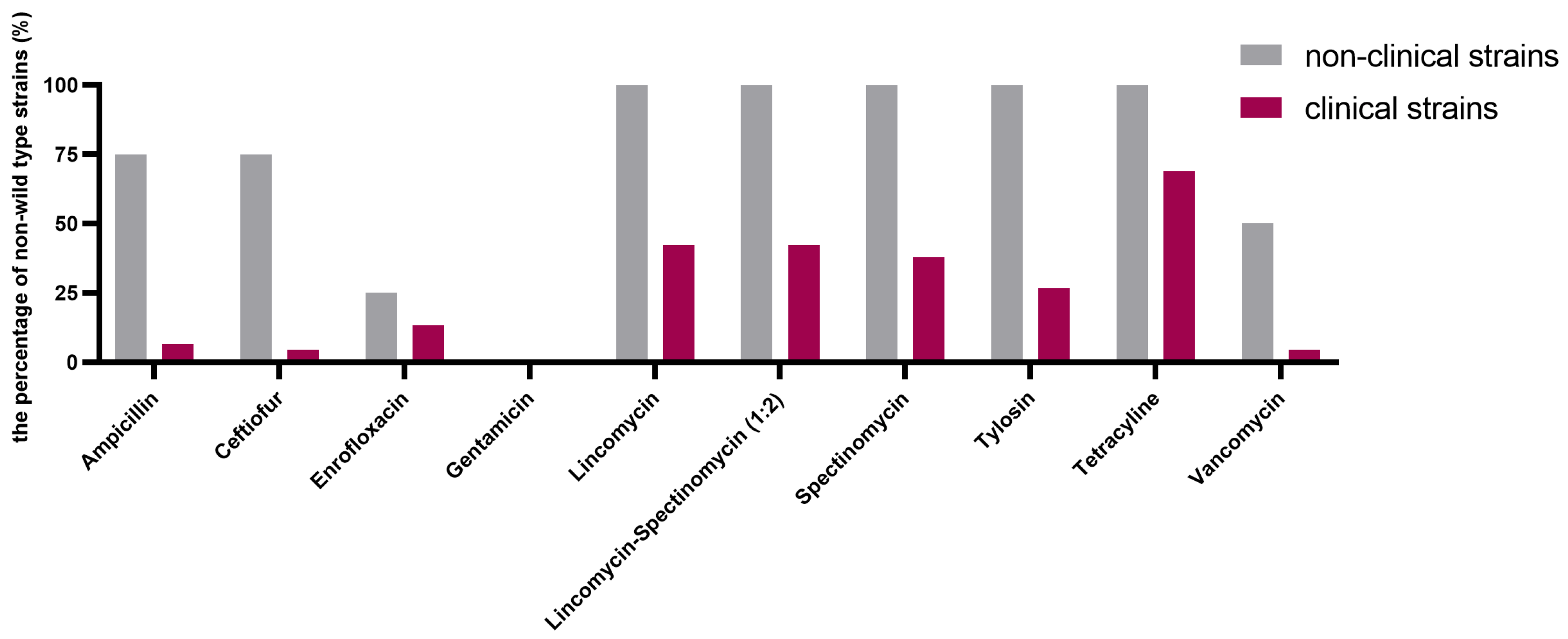
Typical bimodal or even multimodal MIC distributions were observed (Figure 1), pointing to acquired resistance against all antimicrobials, except for gentamicin. The cutoff (COWT) values were calculated for the ten antimicrobial agents based on the data from the 53 E. cecorum strains (Table 1, Figure 2, and Supplementary Table S1). A high proportion of non-wild-type strains for tetracycline was observed (68.9% for clinical strains and 100% for non-clinical strains). The proportions of non-wild-type strains for lincomycin, spectinomycin, and the lincomycin–spectinomycin combination were also high: lincomycin: 42.2% for clinical strains and 100% for non-clinical strains, spectinomycin: 37.8% for clinical strains and 100% for non-clinical strains, and lincomycin–spectinomycin (1:2): 42.2% for clinical strains and 100% for non-clinical strains. There are no indications of cross-resistance between both tested aminoglycosides (gentamicin and spectinomycin), as no gentamicin-acquired resistance was observed, in contrast to spectinomycin. Cross-resistance was observed between both tested β-lactam antimicrobials, ampicillin and ceftiofur, as all strains classified as the non-wild-type phenotype for ceftiofur also showed an acquired resistance for ampicillin. One strain showing ampicillin-acquired resistance was also borderline, categorized to belong to the wild-type phenotype for ceftiofur by the normalized resistance interpretation method (NRI). All strains showing tylosin-acquired resistance also showed lincomycin resistance, while few clinical strains showed lincomycin resistance, but no tylosin-acquired resistance. Importantly, acquired resistance was observed for the critically important antimicrobial agents enrofloxacin and vancomycin, though mainly in non-clinical strains. Relatively high MIC values (8–32 µg/mL) for strains belonging to the wild-type populations of gentamicin and spectinomycin suggest that aminoglycosides are only moderately active against E. cecorum.

2.2. Identification of Resistance Genes

A total of 14 strains were subjected to WGS using long-read nanopore sequencing. Genome assembly sizes ranged between 2,312,828 and 3,086,325 bp, with all assemblies showing a 94.34 ± 1.40% completeness based on CheckM completeness using an Enterococcus spp. database with 672 marker genes from 51 Enterococcus genomes. All were confirmed to be E. cecorum using rMLST. The genome assemblies showed 36.6 ± 0.1 GC% and 2853 ± 331 annotated coding sequences (CDSs) (Supplementary Table S2). Based on the SNP-based phylogenetic maximum-likelihood inference, clinical and non-clinical isolates clustered together (Figure 3). The ARGs were identified in the genomes of 14 strains, which showed a clear association with the observed AMR phenotypes as presented in Figure 3.
In accordance with the phenotypic susceptibility results, all selected non-clinical isolates (n = 8) carried the aminoglycoside resistance gene ant(6)-la, the macrolide (tylosin) resistance gene ermB and the lincosamide (lincomycin) resistance gene lsaE. All these strains also carried the lsaB gene, which only showed a 64.76 ± 0.15% nucleotide identity (% nt. ID). Of note, also, the genes emeA (n = 1; 66.90% nt. ID), mel (n = 4; 89.24 ± 0.12% nt. ID), and msrE (n = 4; 64.40 ± 0.03% nt. ID) were identified in some strains (Supplementary Table S3). All selected non-clinical strains also carried at least one tetracycline-associated ARG, with all of them containing the tet(M) gene and five of them containing the tet(L) gene. Four out of eight non-clinical strains additionally carried the glycopeptide (vancomycin) resistance gene operon (vanA, vanHA, vanRA, vanSA, vanXA, vanYA, and vanZA). In contrast, only two out of six clinical isolates carried ant(6)-la and ermB genes, and four out of six carried at least one tetracycline resistance gene, with four of them containing the tet(M) gene, and two of them containing the tet(L) gene and tet(O) gene. As expected, all of our resistance-gene-positive isolates were non-wild-type based on the COWT values. The lsaE gene was not detected in the two clinical strains that exhibited a non-wild-type phenotype for lincomycin resistance; however, the ermB gene, which also contributes to lincomycin resistance, was detected in these two clinical strains.

2.3. Mutations in the E. cecorum GyrA/GyrB/ParC and Genes Encoding Penicillin-Binding Proteins (PBPs) Are Associated with Enrofloxacin and β-Lactam Antimicrobial Resistance

Two out of eight non-clinical isolates were non-wild-type for enrofloxacin, while six out of eight non-clinical isolates were non-wild-type for both ampicillin and ceftiofur. While no known ARGs, as presented in the used Comprehensive Antimicrobial Resistance Database (CARD), were identified to be linked to these observed AMR phenotypes, we analyzed the genome sequences for chromosomal SNPs in the genes encoding quinolone-resistance-determining regions (QRDRs) and penicillin-binding proteins (PBPs) that may explain the resistance to fluoroquinolones and β-lactam antimicrobials, respectively (Figure 3 and Figure 4). For the enrofloxacin non-wild-type strains, mutations within the QRDRs were present in the GyrA, GyrB, and ParC protein (Ser83Tyr, Thr377Met, and Asn529Asp, respectively). A total of five putative PBPs were identified in all selected E. cecorum strains, including PBPs belonging to the PBP1a superfamily 1, PBP1a superfamily 2, PBP1a superfamily 3, PBP2 superfamily, and PBP2x superfamily as presented in Figure 4A. The data showed that all six non-wild-type strains (E. cecrum 33, 65, 66, 67, 123, and 127) for both ampicillin and ceftiofur exhibited five point mutations at positions in the active domain site of the transpeptidase of the PBP2x superfamily PBP protein, including Thr335Ile, Met346Thr, Gln366Lys, Trp377Ser, and Leu382Ile (Figure 4B). A predicted (AlphaFold) protein structure of the PBP2x superfamily highlights its PBP-like structure and the location of the amino acid changes within the transpeptidase active domain (Figure 4C).

2.4. Mobile Genetic Elements Associated with AMR

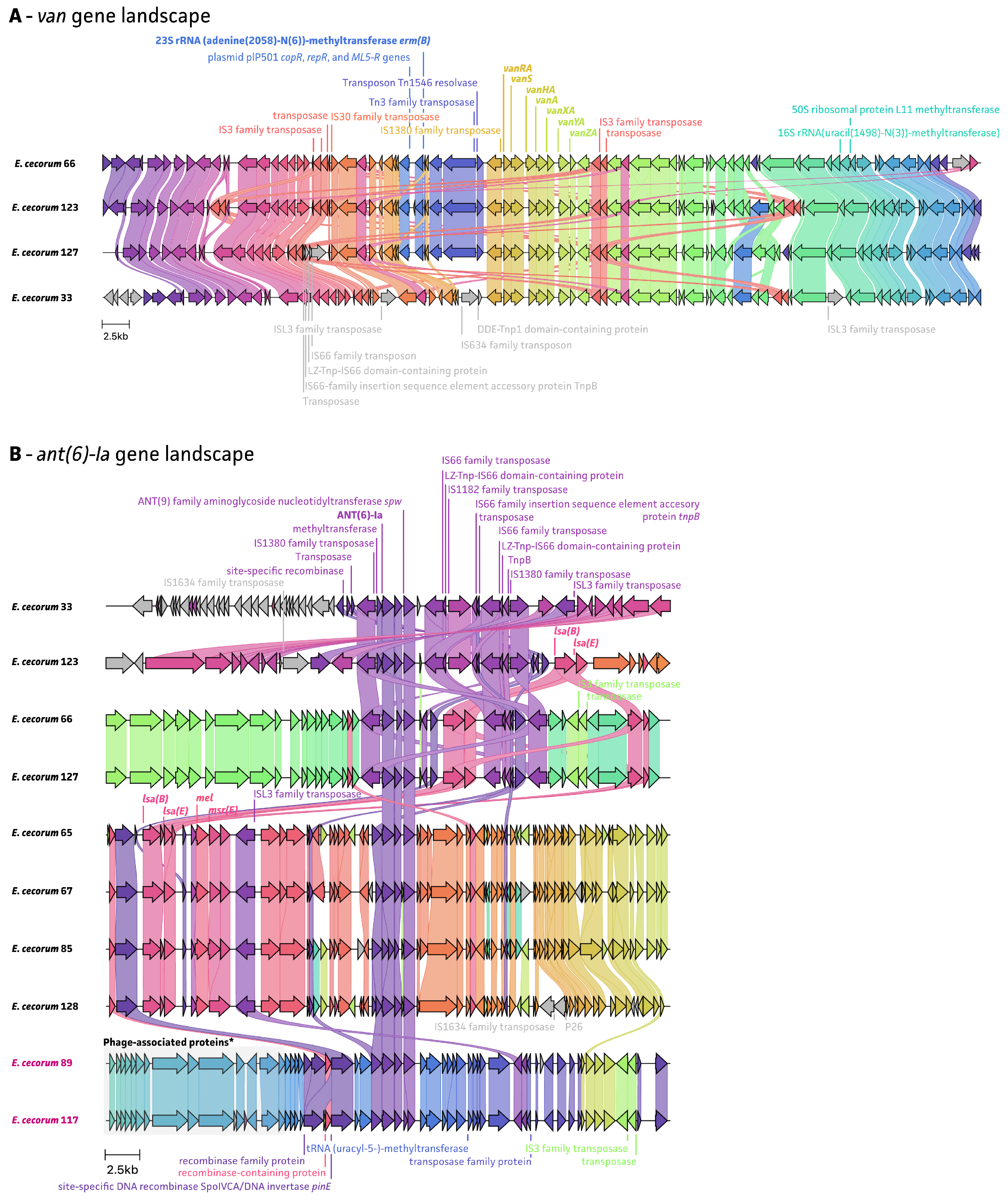
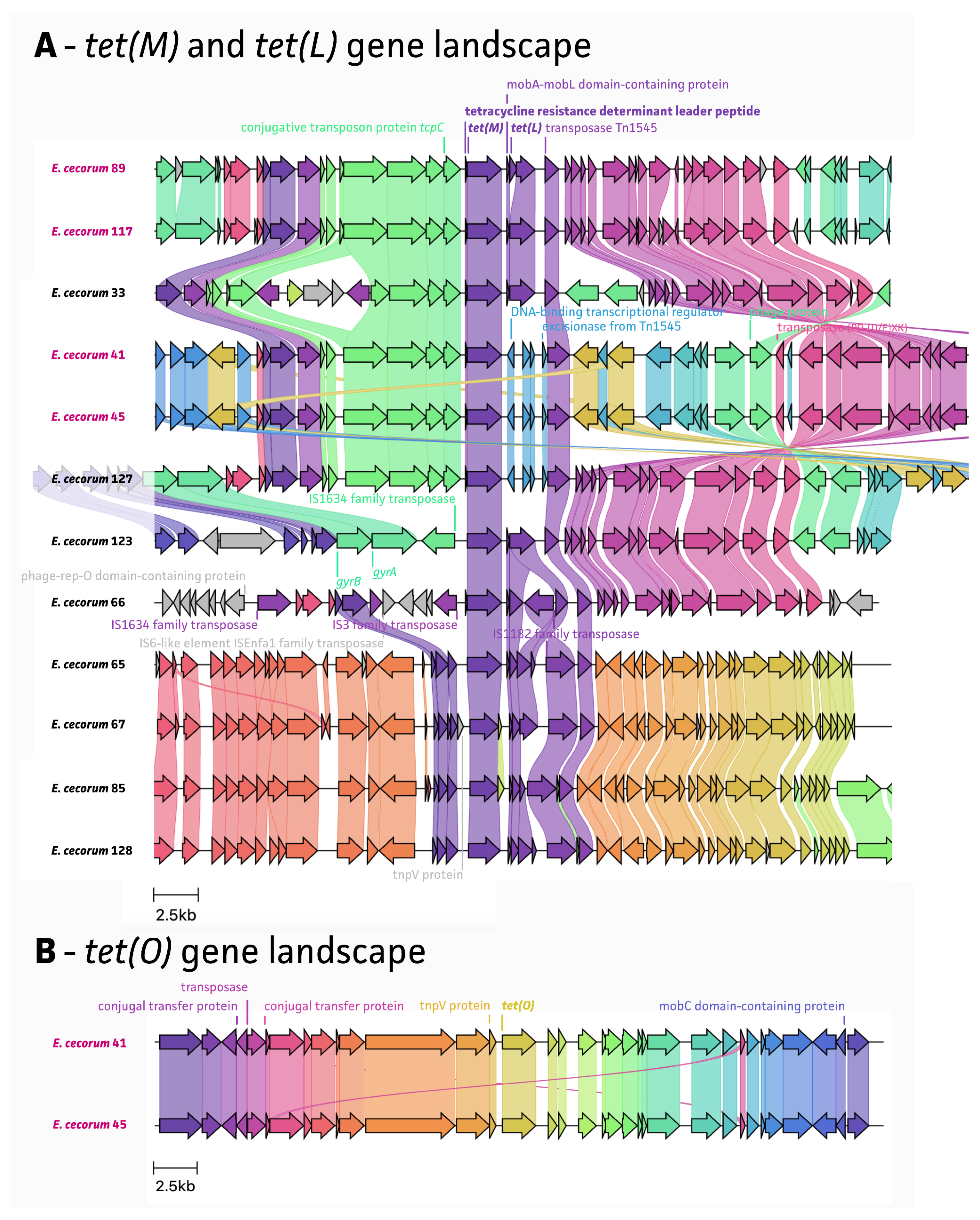
An analysis of the whole-genome sequencing data showed that all detected resistance genes are located on MGEs, including ISs and Tns (Figure 5, Figure 6 and Figure 7). The van gene landscape showed that all van operon-positive strains showed the presence of the IS1380 family transposase, located upstream of the vanRA gene, which contributes to the acquisition of ARGs (Figure 5A). The Tn1546 resolvase and Tn3 family transposase were detected in three out of four van operon-positive strains, adjacent to an IS1380 family transposase. In particular, Tn1546 has been reported as being responsible for vancomycin resistance among enterococcal species [20]. Four of the ant(6)-la-positive strains also showed the presence of an IS1380 family transposase, located upstream and downstream of the ant(6)-la gene. For the rest of ant(6)-la-positive strains, an ISL3 family transposase was detected, which was also found near the lsaE and lsaB genes (Figure 5B). Of note, E. cecorum strain 33 also harbored both lsaE and lsaB genes, which were located further upstream of the ant(6)-Ia region. The transposon Tn917 resolvase, associated with the resistance to macrolide–lincosamide–streptogramin (MSL) antimicrobials, was found in all ten ermB-positive strains, adjacent to ermB (Figure 6). The tet(M) and tet(L) gene landscape showed the presence of a Tn1545 transposase in all tetracycline-resistant strains, located downstream of the tet(M)/tet(L) gene (Figure 7). It has been previously described that the Tn1545 transposon carries the ARGs tet(M), ermB, and aphA-3 [24,25].

3. Discussion

BCO has emerged as a major concern in broilers, and unraveling the pathogenesis of this disease is crucial for resolving this problem. Antimicrobial drugs have been used prophylactically and therapeutically and concerns have been raised about the acquisition of AMR. Therefore, in this study, the AMR profile of 53 E. cecorum isolates from BCO outbreaks and healthy poultry were analyzed. Even though this collection of strains allowed an in-depth investigation of the genetic background of the phenotypically observed resistance, the relatively small number of strains, and, especially, of non-clinical strains, is a limitation of the current study, possibly affecting certain outcome parameters such as the percentages of resistance or the ECOFFs.
In our study, all non-clinical and 26.7% of clinical E. cecorum isolates exhibited a resistance to macrolides, with tylosin resistance mediated by the ermB resistance gene in the non-wild-type strains. Resistance to macrolides in enterococci from both animal and human sources has been reported [26,27,28]. In previous studies, acquired resistance to the macrolides could be attributed to the presence of one or more resistance genes, such as erythromycin resistance methylase (erm) genes (ermB, ermC, and ermG), and efflux genes responsible for the pumping of the antimicrobial from the cell (mefA, and msrD), of which the ermB gene dominated among the enterococci [15,28]. Moreover, the ermB gene confers resistance to MSLtype B, and is associated with transposons on chromosomes or plasmids [29]. A previous study showed that the dissemination of the ermB gene can occur via transposable elements on chromosomes rather than plasmids [30]. Another study demonstrated that the transposon Tn917 is the main carrier of the ermB gene in S. pneumoniae isolates [31]. Not surprisingly, all ermB gene-positive E. cecorum strains in our study harbored the transposon Tn917 located adjacent to the ermB gene, confirming the Tn917 transposon as its main carrier.
For resistance to lincomycin/spectinomycin, several studies showed that commensal isolates of E. cecorum exhibited more resistance to lincomycin and spectinomycin compared to pathogenic isolates, which was similar in our study. Of note, most studies, including our study, comprised a low number of non-clinical isolates [11,13,15]. The combinatorial treatment of lincomycin–spectinomycin in newly hatched birds has been a successful strategy in preventing E. cecorum-associated diseases. Comparing the lincomycin, spectinomycin, and the lincomycin–spectinomycin (1:2) combination MIC results, it can be concluded that E. cecorum is only moderately susceptible to spectinomycin, with the wild-type population showing MIC values in the range of 16–32 µg/mL. Adding spectinomycin does not seem to have a clear added value when used in combination with lincomycin both for strains showing wild-type or non-wild-type properties for lincomycin. Moreover, the preventive use of antimicrobials in groups of animals has been banned in the EU since 2022. In our study, all eight non-clinical and two clinical isolates were found to harbor ant(6)-la genes, along with the lsaE or ermB gene. Additionally, the lsaB gene was identified in all (n = 8) non-clinical strains, showing a low 64.76 ± 0.15% nt. ID as compared to the Mammaliicoccus sciuri lsaB reference sequence (AJ579365.1). Many of these genes are associated with MGEs located in the chromosome. As an example, the IS1380 family transposase seems to have captured various ARGs including rmtC (aminoglycoside resistance), qnrE1 (fluoroquinolone resistance), and blaOXA-204 (cephalosporin resistance), and it has been reported that the IS1380 family ISEcp1-like element is associated in the gentamicin resistance gene transfer in E. casseliflavus [20,32].
Tetracycline resistance was conferred by genes tet(M), tet(O), and tet(L) in both clinical and non-clinical E. cecorum strains. Other reports suggested that a high prevalence of tetracycline-resistant E. faecalis were found in poultry, with 35% and 55% of tetracycline-resistant E. faecalis in chicken and other poultry meat, respectively [33]. The resistance of enterococci to tetracycline has been associated with the therapeutic use of this antimicrobial on farms, and plasmids conferring this resistance have been reported [34]. In Enterococcus, ARGs linked to ribosomal protection (tet(M), tet(O), and tet(S)) and enzymatic inactivation (tet(K) and tet(L)) are commonly found antimicrobial resistance mechanisms [34]. Among these, tet(M) is most frequently observed and is usually found in close proximity to the ermB gene [15,29]. In our data, ten out of twelve tet(M)-positive E. cecorum isolates also harbored the ermB gene, supporting this. In addition, all tet(M)-positive E. cecorum isolates carried the Tn1545 transposon. According to previous research, the tet(M) gene is most frequently located on the chromosome, and this ARG can be carried by the Tn916/Tn1545 family of conjugative transposons [35]. These transposons play a crucial role in the conjugative transfer and transposition in both Gram-negative and Gram-positive bacteria, including enterococci, as the Tn916/Tn1545 family is very prevalent [36,37].
Fifty percent and 4.4% of E. cecorum isolates from non-clinical and clinical strains appear to be resistant to vancomycin, respectively. Three out of four vancomycin resistance gene clusters were found to be adjacent to a Tn1546 element [38]. In the European Union (EU) and Asia, glycopeptides were used in poultry production as a growth promoter, such as the vancomycin analog avoparcin [39,40]. However, its use as a feed additive has been banned since 1997 [41,42]. Hence, vancomycin resistance in E. cecorum isolates has been seldom found. Based on previous studies, a vanA-gene-positive E. cecorum strain was found in September 2009 [14], and, also, one vancomycin resistance broiler isolate was reported in 2016 [43]. According to the literature, E. cecorum could serve as a reservoir of vancomycin resistance genes and can be part of the vancomycin-resistant enterococci (VRE) [14,44]. Therefore, conducting surveillance on the VRE type of E. cecorum is essential, as it may pose a potential risk of AMR for humans through the food chain.
Multiple amino acid substitutions within the quinolone-resistance-determining regions (QRDRs) of both the DNA gyrase (GyrA and GyrB) and topoisomerase IV (ParC), and penicillin-binding proteins (PBPs) were identified. These mutations correlated with significantly higher MIC values compared to E. cecorum strains that did not harbor these mutations. Based on previous studies, PBP 5 and PBP 4 were often found in E. faecalis and E. faecium, and had a low affinity for penicillin [45,46,47]. However, in another study, the PBP types in E. cecorum might be more closely related to Streptococcus species rather than other Enterococcus species [48]. In our study, it was shown that all the point mutations were found in the PBP2x superfamily. This type of PBPs (PBP2x) was often found in Streptococcus pneumoniae, playing an important role in β-lactam antimicrobial resistance [49,50,51]. Here, we report five new E. cecorum PBP2x amino acid changes (Thr335Ile, Met346Thr, Gln366Lys, Trp377Ser, and Leu382Ile) within the PBP transpeptidase active domain, potentially contributing to ampicillin and ceftiofur resistance.
We also describe amino acid changes associated with enrofloxacin resistance in E. cecorum. The GyrA protein mutation (Ser83) has been previously observed in Mycoplasmopsis bovis (formerly known as Mycoplasma bovis) and Salmonella enterica, where it is associated with enrofloxacin and ciprofloxacin resistance [18,52,53]. On the other hand, the reported mutations in GyrB and ParC are not-yet-described mutations in E. cecorum associated with fluoroquinolone resistance, which might require further validation to link them with actual resistance phenotypes.
Current findings suggest that acquired resistance towards the major antimicrobial classes used to control BCO can be observed in E. cecorum. However, as there are currently no clinical breakpoints for E. cecorum infections in poultry, the clinical interpretation of these results is not straightforward. It can be assumed that strains with acquired resistance showing MIC values 10–100 times higher than the wild-type population may not be efficiently treated with these antimicrobial agents. As the observed ARGs are strongly linked with MGE, it is tempting to speculate that these resistance genes are horizontally acquired and can further spread horizontally, though it is not clear yet which bacterial species might be involved in such events.
Summarized, AMR in broiler E. cecorum isolates was relatively high, even against critically important agents such as vancomycin. Genes conferring resistance were often found on MGEs, while fluoroquinolone and β-lactam antimicrobial resistance were found to be mediated by SNPs and were characterized in detail for the first time in E. cecorum. Therefore, the current results contribute to an understanding of the genetic landscape of ARGs in E. cecorum and provide a basis for further research to understand the mechanism of the antimicrobial resistance epidemiology in E. cecorum.

4. Materials and Methods

4.1. Bacterial Strains

E. cecorum strains used in this study (n = 53) were isolated from healthy (non-clinical strains, n = 8) and BCO-affected chickens (clinical strains, n = 45) in Belgium during 2019–2020. Strain characteristics have been described previously (source of isolation, PFGE type, and presence of virulence genes) [22]. All E. cecorum isolates were cultured on Columbia sheep blood agar plates and incubated at 37 °C in a 5% CO2 atmosphere. Reference strains Enterococcus faecalis ATCC 29212, Staphylococcus aureus ATCC 29213, Escherichia coli ATCC 25922, and Streptococcus pneumoniae ATCC 49619 were used as quality control strains in the susceptibility tests and were cultured on Columbia sheep blood agar plates and incubated at 37 °C in a 5% CO2 atmosphere.

4.2. Antimicrobial Susceptibility Testing

Antimicrobial susceptibility testing was carried out for ten antimicrobial agents: ampicillin, vancomycin, lincomycin, spectinomycin, lincomycin–spectinomycin, tetracycline, tylosin, gentamicin, enrofloxacin, and ceftiofur (all obtained from Sigma-Aldrich, Darmstadt, Germany). Due to the fastidious growth characteristics of E. cecorum, that more closely resemble Streptococcus rather than Enterococcus characteristics, the broth microdilution method as described for Streptococcus spp. in the Clinical Laboratory Standards Institute (CLSI, 2023) standards was used [23]. The antimicrobials were dissolved in an appropriate solvent, and then further diluted in sterile distilled water. The Minimum Inhibitory Concentration (MIC) was determined using Mueller–Hinton П agar (Becton Dickinson, Cockeysville, MD, USA) supplemented with 5% sheep blood and incubated at 37 °C in a 5% CO2 atmosphere, containing two-fold dilutions of the antimicrobials. Concentrations of the antimicrobials ranged from 0.03 μg/mL to 128 μg/mL, except for the combination lincomycin–spectinomycin (1:2), which was tested in a range between 0.03/0.06 µg/mL and 128/256 µg/mL, respectively. Uninoculated antimicrobial free agar plates were included as sterility controls, and inoculated antimicrobial free agar plates were included as growth controls. Inoculated plates were prepared by suspending colonies from an overnight grown culture on Columbia blood agar (Thermo Fisher Scientific, Merelbeke, Belgium), in buffered saline, to a density of 0.5 McFarland standards. Approximately 1 × 10⁷ colony-forming units of each strain were inoculated on the plates. Resulting MIC values were recorded after incubation at 37 °C in a 5% CO2 atmosphere for 24 h and were defined as the lowest concentration producing no visible growth. Enterococcus faecalis ATCC 29212, Staphylococcus aureus ATCC 29213, Escherichia coli ATCC 25922, and Streptococcus pneumoniae ATCC 49619 were used as quality controls.

4.3. ECOFF Values Determination

As no EUCAST ECOFFs are available for E. cecorum, wild-type cutoff values (COWT) were determined using the “Normalized Resistance Interpretation (NRI)” method (Bioscand AB, Täby, Sweden) [54], as a proxy for the ECOFF. The outcome is limited to a tentative estimate of the ECOFF when the standard deviation of wild-type MIC values in the normal distribution exceeds 1.2 log2.

4.4. DNA Extraction and Whole-Genome Sequencing

Fourteen E. cecorum strains were selected according to the MIC results of different antimicrobial agents, ensuring strains were included to cover all types of resistance to the selected antimicrobials. All strains were grown as described above and an overnight culture was subjected to high-molecular-weight DNA extraction and native long-read DNA sequencing using the Oxford Nanopore Technologies GridION system (Oxford, UK) as described before [21,55,56]. In short, DNA was extracted using the ZymoBIOMICS DNA MiniPrep kit (ZymoResearch, Irvine, CA, USA), with the addition of a Proteinase K treatment (20 µg∙µL−1; Promega, Madison, WI, USA). Low-quality DNA samples were subjected to an extra DNA clean-up using CleanNGS (CleanNA, Waddinxveen, The Netherlands) magnetic beads at a 1:1 ratio. The SQK-RBK004 (ONT) quick library preparation was used to multiplex up to 10 strains on a single MinION flow cell. Raw data were collected in MinKNOW (ONT) and base called using the super accurate model in guppy (v6.3.9; ONT). An overview of sequencing throughput and statistics is given in Supplementary Table S2. Raw data were used to assemble genomes as described in Vereecke et al., 2023 [21] using Trycycler (v.0.5.3; [57]), minimap2 (v2.20; [58]), and medaka (v.1.7.3; ONT). All software were run at default settings as depicted on the Trycycler wiki (https://github.com/rrwick/Trycycler/wiki, accessed on 27 January 2023). Final genomes were quality checked using CheckM (v1.1.0; [59]), including 672 markers from 51 Enterococcus species, and classified up to the species level using rMLST on pubMLST [60]. An overview of assembly quality control, coverage, and accession numbers (BioProject: PRJNA1071719) can be found in Supplementary Table S2.

4.5. Phylogenetic Inference, Identification of ARGs, Mobile Genetic Elements, and Point Mutations

The resulting genome assemblies were used in an SNP-based phylogenetic maximum-likelihood (ML) inference using csi phylogeny [61] and IQtree (v.1.6.12; –bb 1000 -m GTR + R + I; [62]). Next, genome assemblies were screened for ARGs using Abricate (v.1.0.1; --minid 60 --mincov 80; https://github.com/tseemann/abricate, accessed on 27 January 2023) against the CARD database [63]. An overview of all identified ARGs is given in Supplementary Table S3. Final visualizations were made in iTOL (v.5; [64]). The characterization of MGEs was carried out by extracting flanking regions of ARGs using flanker (v.0.1.5; [65]), which were then annotated and visualized using Bakta (v.1.7.0; --db db-full --compliant --genus Enterococcus --species cecorum --gram +; [66]) and Clinker (v.0.0.26; [67]), all at default settings. Identification of point mutations was obtained by extracting target genes from the Bakta annotated genomes, aligning them with MAFFT (v.7.520; [68]), and manually annotated and/or translated in Mega11 (v.11.0.11; [69]). For PBP proteins, protein homology and structures were determined using NCBI blastP and Interpro for the (sub)classification of protein families (https://blast.ncbi.nlm.nih.gov/Blast.cgi and https://www.ebi.ac.uk/interpro/search/sequence/, accessed on 30 November 2023 with default settings). The PBP protein structure prediction was made using the Alphafold2 google colab at preset settings [70].

Supplementary Materials

The following supporting information can be downloaded at: https://www.mdpi.com/article/10.3390/antibiotics13050409/s1, Table S1: Summary of E. cecorum strains used to determine MIC data. Table S2: Statistics on QC of E. cecorum genome assemblies, including raw read statistics, genome assembly statistics, assembly quality checks, species identification (rMLST), and annotation statistics. Table S3: An overview of all identified ARGs.

Author Contributions

F.V.I., F.B. and G.A. designed the experimental work. Y.H. performed the strain collection and MIC data analysis. N.V. performed the WGS data analysis. Y.H. wrote the manuscript. All authors have read and agreed to the published version of the manuscript.

Funding

This research was funded by a fellowship from the China Scholarship Council (No. 202007650026) and Ghent University (BOF.CDV.2023.0077.01). N.V. was funded by a grant from the Flemish Agency for Innovation and Entrepreneurship (Baekeland Mandate HBC.2020.2889).

Data Availability Statement

Genomes were submitted to NCBI and are available under the BioProject PRJNA1071719 with accession numbers as presented in Supplementary Table S2.

Acknowledgments

The NRI method was used with permission from the patent holder, Bioscand AB, Täby, Sweden (European patent No. 138,3913, US Patent No. 7,465,559). The automatic and manual Excel programmes of the NRI method were made available courtesy of P. Smith, W. Finnegan, and G. Kronvall.

Conflicts of Interest

Author N.V. is a former employee of the company PathoSense BV. The remaining authors declare that the research was conducted in the absence of any commercial or financial relationships that could be construed as a potential conflict of interest.

References

  1. Wood, A.M.; MacKenzie, G.; McGiliveray, N.C.; Brown, L.; Devriese, L.A.; Baele, M. Isolation of Enterococcus cecorum from bone lesions in broiler chickens. Vet. Rec. 2002, 150, 27. [Google Scholar] [PubMed]
  2. Dolka, B.; Chrobak-Chmiel, D.; Czopowicz, M.; Szeleszczuk, P. Characterization of pathogenic Enterococcus cecorum from different poultry groups: Broiler chickens, layers, turkeys, and waterfowl. PLoS ONE 2017, 12, e0185199. [Google Scholar] [CrossRef] [PubMed]
  3. Wideman, R.F.; Prisby, R.D. Bone circulatory disturbances in the development of spontaneous bacterial chondronecrosis with osteomyelitis: A translational model for the pathogenesis of femoral head necrosis. Front. Endocrinol. 2012, 3, 183. [Google Scholar] [CrossRef] [PubMed]
  4. Borst, L.B.; Suyemoto, M.M.; Sarsour, A.H.; Harris, M.C.; Martin, M.P.; Strickland, J.D.; Oviedo, E.O.; Barnes, H.J. Pathogenesis of enterococcal Sspondylitis caused by Enterococcus cecorum in broiler chickens. Vet. Pathol. 2017, 54, 61–73. [Google Scholar] [CrossRef] [PubMed]
  5. Robbins, K.M.; Suyemoto, M.M.; Lyman, R.L.; Martin, M.P.; Barnes, H.J.; Borst, L.B. An outbreak and source investigation of enterococcal spondylitis in broilers caused by Enterococcus cecorum. Avian Dis. 2012, 56, 768–773. [Google Scholar] [CrossRef] [PubMed]
  6. Schreier, J.; Rychlik, I.; Karasova, D.; Crhanova, M.; Breves, G.; Rautenschlein, S.; Jung, A. Influence of heat stress on intestinal integrity and the caecal microbiota during Enterococcus cecorum infection in broilers. Vet. Res. 2022, 53, 110. [Google Scholar] [CrossRef] [PubMed]
  7. Schreier, J.; Karasova, D.; Crhanova, M.; Rychlik, I.; Rautenschlein, S.; Jung, A. Influence of lincomycin-spectinomycin treatment on the outcome of Enterococcus cecorum infection and on the cecal microbiota in broilers. Gut Pathog. 2022, 14, 3. [Google Scholar] [CrossRef] [PubMed]
  8. Herdt, P.; Defoort, P.; Steelant, J.; Swam, H.; Tanghe, L.; Goethem, S.; Vanrobaeys, M. Enterococcus cecorum osteomyelitis and arthritis in broiler chickens. Vlaams Diergeneeskd. Tijdschr. 2009, 78, 44–48. [Google Scholar] [CrossRef]
  9. Dolka, B.; Chrobak-Chmiel, D.; Makrai, L.; Szeleszczuk, P. Phenotypic and genotypic characterization of Enterococcus cecorum strains associated with infections in poultry. BMC Vet. Res. 2016, 12, 129. [Google Scholar] [CrossRef]
  10. Laurentie, J.; Mourand, G.; Grippon, P.; Furlan, S.; Chauvin, C.; Jouy, E.; Serror, P.; Kempf, I. Determination of Epidemiological Cutoff Values for Antimicrobial Resistance of Enterococcus cecorum. J. Clin. Microbiol. 2023, 61, e0144522. [Google Scholar] [CrossRef]
  11. Borst, L.B.; Suyemoto, M.M.; Robbins, K.M.; Lyman, R.L.; Martin, M.P.; Barnes, H.J. Molecular epidemiology of Enterococcus cecorum isolates recovered from enterococcal spondylitis outbreaks in the southeastern United States. Avian Pathol. 2012, 41, 479–485. [Google Scholar] [CrossRef] [PubMed]
  12. Authority, European Food Safety. The European Union Summary Report on antimicrobial resistance in zoonotic and indicator bacteria from humans, animals and food in 2012. Efsa J. 2014, 12, 3590. [Google Scholar] [CrossRef]
  13. Jackson, C.R.; Kariyawasam, S.; Borst, L.B.; Frye, J.G.; Barrett, J.B.; Hiott, L.M.; Woodley, T.A. Antimicrobial resistance, virulence determinants and genetic profiles of clinical and nonclinical Enterococcus cecorum from poultry. Lett. Appl. Microbiol. 2015, 60, 111–119. [Google Scholar] [CrossRef] [PubMed]
  14. Harada, T.; Kawahara, R.; Kanki, M.; Taguchi, M.; Kumeda, Y. Isolation and characterization of vanA genotype vancomycin-resistant Enterococcus cecorum from retail poultry in Japan. Int. J. Food Microbiol. 2012, 153, 372–377. [Google Scholar] [CrossRef] [PubMed]
  15. Sharma, P.; Gupta, S.K.; Barrett, J.B.; Hiott, L.M.; Woodley, T.A.; Kariyawasam, S.; Frye, J.G.; Jackson, C.R. Comparison of antimicrobial resistance and pan-genome of clinical and non-clinical Enterococcus cecorum from poultry using whole-genome sequencing. Foods 2020, 9, 686. [Google Scholar] [CrossRef] [PubMed]
  16. Ceric, O.; Tyson, G.H.; Goodman, L.B.; Mitchell, P.K.; Zhang, Y.; Prarat, M.; Cui, J.; Peak, L.; Scaria, J.; Antony, L.; et al. Enhancing the one health initiative by using whole genome sequencing to monitor antimicrobial resistance of animal pathogens: Vet-LIRN collaborative project with veterinary diagnostic laboratories in United States and Canada. BMC Vet. Res. 2019, 15, 130. [Google Scholar] [CrossRef] [PubMed]
  17. Laurentie, J.; Loux, V.; Hennequet-Antier, C.; Chambellon, E.; Deschamps, J.; Trotereau, A.; Furlan, S.; Darrigo, C.; Kempf, F.; Lao, J.; et al. Comparative genome analysis of Enterococcus cecorum reveals intercontinental spread of a lineage of clinical poultry isolates. mSphere 2023, 8, e0049522. [Google Scholar] [CrossRef] [PubMed]
  18. Bokma, J.; Vereecke, N.; Nauwynck, H.; Haesebrouck, F.; Theuns, S.; Pardon, B.; Boyen, F. Genome-wide association study reveals genetic markers for antimicrobial resistance in Mycoplasma bovis. Microbiol. Spectr. 2021, 9, e0026221. [Google Scholar] [CrossRef]
  19. Vereecke, N.; Botteldoorn, N.; Brosse, C.; Bonckaert, C.; Nauwynck, H.; Haesebrouck, F.; Boyen, F.; Maes, D.; Theuns, S. Predictive Power of Long-Read Whole-genome sequencing for rapid diagnostics of multidrug-resistant Brachyspira hyodysenteriae Strains. Microbiol. Spectr. 2023, 11, e0412322. [Google Scholar] [CrossRef]
  20. Partridge, S.R.; Kwong, S.M.; Firth, N.; Jensen, S.O. Mobile genetic elements associated with antimicrobial resistance. Clin. Microbiol. Rev. 2018, 31. [Google Scholar] [CrossRef]
  21. Vereecke, N.; Van Hoorde, S.; Sperling, D.; Theuns, S.; Devriendt, B.; Cox, E. Virotyping and genetic antimicrobial susceptibility testing of porcine ETEC/STEC strains and associated plasmid types. Front. Microbiol. 2023, 14, 1139312. [Google Scholar] [CrossRef] [PubMed]
  22. Huang, Y.; Eeckhaut, V.; Goossens, E.; Rasschaert, G.; Van Erum, J.; Roovers, G.; Ducatelle, R.; Antonissen, G.; Van Immerseel, F. Bacterial chondronecrosis with osteomyelitis related Enterococcus cecorum isolates are genetically distinct from the commensal population and are more virulent in an embryo mortality model. Vet. Res. 2023, 54, 13. [Google Scholar] [CrossRef] [PubMed]
  23. Clinical Laboratory Standards Institute. Performance Standards for Antimicrobial Disk and Dilution Susceptibility Tests for Bacteria Isolated from Animals, 6th ed.; CLSI Supplement VET01S; Clinical Laboratory Standards Institute: Wayne, PA, USA, 2023. [Google Scholar]
  24. Cochetti, I.; Tili, E.; Mingoia, M.; Varaldo, P.E.; Montanari, M.P. erm(B)-carrying elements in tetracycline-resistant pneumococci and correspondence between Tn1545 and Tn6003. Antimicrob. Agents Chemother. 2008, 52, 1285–1290. [Google Scholar] [CrossRef] [PubMed]
  25. Soge, O.O.; Beck, N.K.; White, T.M.; No, D.B.; Roberts, M.C. A novel transposon, Tn6009, composed of a Tn916 element linked with a Staphylococcus aureus mer operon. J. Antimicrob. Chemother. 2008, 62, 674–680. [Google Scholar] [CrossRef] [PubMed]
  26. Jung, A.; Chen, L.R.; Suyemoto, M.M.; Barnes, H.J.; Borst, L.B. A review of Enterococcus cecorum infection in poultry. Avian Dis. 2018, 62, 261–271. [Google Scholar] [CrossRef] [PubMed]
  27. Aarestrup, F.M.; Kruse, H.; Tast, E.; Hammerum, A.M.; Jensen, L.B. Associations between the use of antimicrobial agents for growth promotion and the occurrence of resistance among Enterococcus faecium from broilers and pigs in Denmark, Finland, and Norway. Microb. Drug Resist. 2000, 6, 63–70. [Google Scholar] [CrossRef]
  28. Jensen, L.B.; Frimodt-Moller, N.; Aarestrup, F.M. Presence of erm gene classes in gram-positive bacteria of animal and human origin in Denmark. FEMS Microbiol. Lett. 1999, 170, 151–158. [Google Scholar] [CrossRef]
  29. Roberts, M.C.; Sutcliffe, J.; Courvalin, P.; Jensen, L.B.; Rood, J.; Seppala, H. Nomenclature for macrolide and macrolide-lincosamide-streptogramin B resistance determinants. Antimicrob. Agents Chemother. 1999, 43, 2823–2830. [Google Scholar] [CrossRef] [PubMed]
  30. Jackson, C.R.; Fedorka-Cray, P.J.; Barrett, J.B.; Ladely, S.R. Effects of tylosin use on erythromycin resistance in enterococci isolated from swine. Appl. Environ. Microbiol. 2004, 70, 4205–4210. [Google Scholar] [CrossRef]
  31. Okitsu, N.; Kaieda, S.; Yano, H.; Nakano, R.; Hosaka, Y.; Okamoto, R.; Kobayashi, T.; Inoue, M. Characterization of ermB gene transposition by Tn1545 and Tn917 in macrolide-resistant Streptococcus pneumoniae isolates. J. Clin. Microbiol. 2005, 43, 168–173. [Google Scholar] [CrossRef]
  32. Chen, Y.G.; Qu, T.T.; Yu, Y.S.; Zhou, J.Y.; Li, L.J. Insertion sequence ISEcp1-like element connected with a novel aph(2″) allele [aph(2″)-Ie] conferring high-level gentamicin resistance and a novel streptomycin adenylyltransferase gene in Enterococcus. J. Med. Microbiol. 2006, 55, 1521–1525. [Google Scholar] [CrossRef]
  33. Wilcks, A.; Andersen, S.R.; Licht, T.R. Characterization of transferable tetracycline resistance genes in Enterococcus faecalis isolated from raw food. FEMS Microbiol. Lett. 2005, 243, 15–19. [Google Scholar] [CrossRef] [PubMed]
  34. Torres, C.; Alonso, C.A.; Ruiz-Ripa, L.; Leon-Sampedro, R.; Del Campo, R.; Coque, T.M. Antimicrobial resistance in Enterococcus spp. of animal origin. Microbiol. Spectr. 2018, 6. [Google Scholar] [CrossRef] [PubMed]
  35. Cauwerts, K.; Decostere, A.; De Graef, E.M.; Haesebrouck, F.; Pasmans, F. High prevalence of tetracycline resistance in Enterococcus isolates from broilers carrying the erm(B) gene. Avian Pathol. 2007, 36, 395–399. [Google Scholar] [CrossRef]
  36. Chopra, I.; Roberts, M. Tetracycline antibiotics: Mode of action, applications, molecular biology, and epidemiology of bacterial resistance. Microbiol. Mol. Biol. Rev. 2001, 65, 232–260. [Google Scholar] [CrossRef]
  37. Haack, B.J.; Andrews, R.E., Jr. Isolation of Tn916-like conjugal elements from swine lot effluent. Can. J. Microbiol. 2000, 46, 542–549. [Google Scholar] [CrossRef] [PubMed]
  38. Arthur, M.; Courvalin, P. Genetics and mechanisms of glycopeptide resistance in enterococci. Antimicrob. Agents Chemother. 1993, 37, 1563–1571. [Google Scholar] [CrossRef]
  39. Bach Knudsen, K.E. Development of antibiotic resistance and options to replace antimicrobials in animal diets. Proc. Nutr. Soc. 2001, 60, 291–299. [Google Scholar] [CrossRef]
  40. Chan, Y.Y.; Abd Nasir, M.H.; Yahaya, M.A.; Salleh, N.M.; Md Dan, A.D.; Musa, A.M.; Ravichandran, M. Low prevalence of vancomycin- and bifunctional aminoglycoside-resistant enterococci isolated from poultry farms in Malaysia. Int. J. Food Microbiol. 2008, 122, 221–226. [Google Scholar] [CrossRef]
  41. Casewell, M.; Friis, C.; Marco, E.; McMullin, P.; Phillips, I. The European ban on growth-promoting antibiotics and emerging consequences for human and animal health. J. Antimicrob. Chemother. 2003, 52, 159–161. [Google Scholar] [CrossRef]
  42. Yoshimura, H.; Ishimaru, M.; Endoh, Y.S.; Suginaka, M.; Yamatani, S. Isolation of glycopeptide-resistant enterococci from chicken in Japan. Antimicrob. Agents Chemother. 1998, 42, 3333. [Google Scholar] [CrossRef] [PubMed]
  43. Stepien-Pysniak, D.; Marek, A.; Banach, T.; Adaszek, L.; Pyzik, E.; Wilczynski, J.; Winiarczyk, S. Prevalence and antibiotic resistance of Enterococcus strains isolated from poultry. Acta Vet. Hung. 2016, 64, 148–163. [Google Scholar] [CrossRef] [PubMed]
  44. Laurentie, J.; Mourand, G.; Jouy, E.; Bougeard, S.; Keita, A.; Amelot, M.; Serror, P.; Kempf, I. Study of the effect of administration of narasin or antibiotics on in vivo selection of a narasin- and multidrug-resistant Enterococcus cecorum strain. Vet. Microbiol. 2023, 282, 109757. [Google Scholar] [CrossRef] [PubMed]
  45. Chen, H.Y.; Williams, J.D. Penicillin-binding proteins in Streptococcus faecalis and S. faecium. J. Med. Microbiol. 1987, 23, 141–147. [Google Scholar] [CrossRef] [PubMed]
  46. Fontana, R.; Canepari, P.; Lleo, M.M.; Satta, G. Mechanisms of resistance of enterococci to beta-lactam antibiotics. Eur. J. Clin. Microbiol. Infect. Dis. 1990, 9, 103–105. [Google Scholar] [CrossRef] [PubMed]
  47. Moon, T.M.; D’Andréa, É.D.; Lee, C.W.; Soares, A.; Jakoncic, J.; Desbonnet, C.; Garcia-Solache, M.; Rice, L.B.; Page, R.; Peti, W. The structures of penicillin-binding protein 4 (PBP4) and PBP5 from Enterococci provide structural insights into β-lactam resistance. J. Biol. Chem. 2018, 293, 18574–18584. [Google Scholar] [CrossRef] [PubMed]
  48. Woo, P.C.; Tam, D.M.; Lau, S.K.; Fung, A.M.; Yuen, K.Y. Enterococcus cecorum empyema thoracis successfully treated with cefotaxime. J. Clin. Microbiol. 2004, 42, 919–922. [Google Scholar] [CrossRef] [PubMed]
  49. Nagai, K.; Davies, T.A.; Jacobs, M.R.; Appelbaum, P.C. Effects of amino acid alterations in penicillin-binding proteins (PBPs) 1a, 2b, and 2x on PBP affinities of penicillin, ampicillin, amoxicillin, cefditoren, cefuroxime, cefprozil, and cefaclor in 18 clinical isolates of penicillin-susceptible, -intermediate, and -resistant pneumococci. Antimicrob. Agents Chemother. 2002, 46, 1273–1280. [Google Scholar] [CrossRef]
  50. Asahi, Y.; Takeuchi, Y.; Ubukata, K. Diversity of substitutions within or adjacent to conserved amino acid motifs of penicillin-binding protein 2X in cephalosporin-resistant Streptococcus pneumoniae isolates. Antimicrob. Agents Chemother. 1999, 43, 1252–1255. [Google Scholar] [CrossRef]
  51. Hsieh, Y.C.; Su, L.H.; Hsu, M.H.; Chiu, C.H. Alterations of penicillin-binding proteins in pneumococci with stepwise increase in beta-lactam resistance. Pathog. Dis. 2013, 67, 84–88. [Google Scholar] [CrossRef]
  52. Dahiya, S.; Kapil, A.; Lodha, R.; Kumar, R.; Das, B.K.; Sood, S.; Kabra, S.K. Induction of resistant mutants of Salmonella enterica serotype Typhi under ciprofloxacin selective pressure. Indian J. Med. Res. 2014, 139, 746–753. [Google Scholar] [PubMed]
  53. Gruger, T.; Nitiss, J.L.; Maxwell, A.; Zechiedrich, E.L.; Heisig, P.; Seeber, S.; Pommier, Y.; Strumberg, D. A mutation in Escherichia coli DNA gyrase conferring quinolone resistance results in sensitivity to drugs targeting eukaryotic topoisomerase II. Antimicrob. Agents Chemother. 2004, 48, 4495–4504. [Google Scholar] [CrossRef] [PubMed]
  54. Kronvall, G. Normalized resistance interpretation as a tool for establishing epidemiological MIC susceptibility breakpoints. J. Clin. Microbiol. 2010, 48, 4445–4452. [Google Scholar] [CrossRef] [PubMed]
  55. Reydams, H.; Toledo-Silva, B.; Mertens, K.; Piepers, S.; Vereecke, N.; Souza, F.N.; Haesebrouck, F.; De Vliegher, S. Phenotypic and genotypic assessment of iron acquisition in diverse bovine-associated non-aureus staphylococcal strains. Vet. Res. 2024, 55, 6. [Google Scholar] [CrossRef] [PubMed]
  56. Belhout, C.; Boyen, F.; Vereecke, N.; Theuns, S.; Taibi, N.; Stegger, M.; de la Fe-Rodriguez, P.Y.; Bouayad, L.; Elgroud, R.; Butaye, P. Prevalence and molecular characterization of methicillin-resistant staphylococci (MRS) and mammaliicocci (MRM) in dromedary camels from Algeria: First Detection of SCCmec-mecC Hybrid in Methicillin-Resistant Mammaliicoccus lentus. Antibiotics 2023, 12, 674. [Google Scholar] [CrossRef] [PubMed]
  57. Wick, R.R.; Judd, L.M.; Cerdeira, L.T.; Hawkey, J.; Meric, G.; Vezina, B.; Wyres, K.L.; Holt, K.E. Trycycler: Consensus long-read assemblies for bacterial genomes. Genome Biol. 2021, 22, 266. [Google Scholar] [CrossRef]
  58. Li, H. Minimap2: Pairwise alignment for nucleotide sequences. Bioinformatics 2018, 34, 3094–3100. [Google Scholar] [CrossRef] [PubMed]
  59. Parks, D.H.; Imelfort, M.; Skennerton, C.T.; Hugenholtz, P.; Tyson, G.W. CheckM: Assessing the quality of microbial genomes recovered from isolates, single cells, and metagenomes. Genome Res. 2015, 25, 1043–1055. [Google Scholar] [CrossRef] [PubMed]
  60. Jolley, K.A.; Bliss, C.M.; Bennett, J.S.; Bratcher, H.B.; Brehony, C.; Colles, F.M.; Wimalarathna, H.; Harrison, O.B.; Sheppard, S.K.; Cody, A.J.; et al. Ribosomal multilocus sequence typing: Universal characterization of bacteria from domain to strain. Microbiology 2012, 158, 1005–1015. [Google Scholar] [CrossRef]
  61. Kaas, R.S.; Leekitcharoenphon, P.; Aarestrup, F.M.; Lund, O. Solving the problem of comparing whole bacterial genomes across different sequencing platforms. PLoS ONE 2014, 9, e104984. [Google Scholar] [CrossRef]
  62. Minh, B.Q.; Schmidt, H.A.; Chernomor, O.; Schrempf, D.; Woodhams, M.D.; von Haeseler, A.; Lanfear, R. IQ-TREE 2: New models and efficient methods for phylogenetic inference in the genomic era. Mol. Biol. Evol. 2020, 37, 1530–1534. [Google Scholar] [CrossRef]
  63. Alcock, B.P.; Huynh, W.; Chalil, R.; Smith, K.W.; Raphenya, A.R.; Wlodarski, M.A.; Edalatmand, A.; Petkau, A.; Syed, S.A.; Tsang, K.K.; et al. CARD 2023: Expanded curation, support for machine learning, and resistome prediction at the Comprehensive Antibiotic Resistance Database. Nucleic Acids Res. 2023, 51, D690–D699. [Google Scholar] [CrossRef] [PubMed]
  64. Letunic, I.; Bork, P. Interactive Tree Of Life (iTOL) v5: An online tool for phylogenetic tree display and annotation. Nucleic Acids Res. 2021, 49, W293–W296. [Google Scholar] [CrossRef]
  65. Matlock, W.; Lipworth, S.; Constantinides, B.; Peto, T.E.A.; Walker, A.S.; Crook, D.; Hopkins, S.; Shaw, L.P.; Stoesser, N. Flanker: A tool for comparative genomics of gene flanking regions. Microb. Genom. 2021, 7, 000634. [Google Scholar] [CrossRef]
  66. Schwengers, O.; Jelonek, L.; Dieckmann, M.A.; Beyvers, S.; Blom, J.; Goesmann, A. Bakta: Rapid and standardized annotation of bacterial genomes via alignment-free sequence identification. Microb. Genom. 2021, 7, 000685. [Google Scholar] [CrossRef]
  67. Schmidt, B.M.; Davidson, N.M.; Hawkins, A.D.K.; Bartolo, R.; Majewski, I.J.; Ekert, P.G.; Oshlack, A. Clinker: Visualizing fusion genes detected in RNA-seq data. Gigascience 2018, 7, giy079. [Google Scholar] [CrossRef]
  68. Katoh, K.; Standley, D.M. MAFFT multiple sequence alignment software version 7: Improvements in performance and usability. Mol. Biol. Evol. 2013, 30, 772–780. [Google Scholar] [CrossRef]
  69. Tamura, K.; Stecher, G.; Kumar, S. MEGA11: Molecular evolutionary genetics analysis version 11. Mol. Biol. Evol. 2021, 38, 3022–3027. [Google Scholar] [CrossRef]
  70. Jumper, J.; Evans, R.; Pritzel, A.; Green, T.; Figurnov, M.; Ronneberger, O.; Tunyasuvunakool, K.; Bates, R.; Zidek, A.; Potapenko, A.; et al. Highly accurate protein structure prediction with AlphaFold. Nature 2021, 596, 583–589. [Google Scholar] [CrossRef]
Figure 1. Typical bimodal and multimodal MIC distributions for ten antimicrobial agents on 53 E. cecorum isolates, except for gentamicin (unimodal MIC distribution). These plots are representing the results as shown in the automated NRI software. The red line indicates the wild-type population distribution as calculated by NRI software.The blue dotted line indicates the COWT value as proxy for the ECOFF.
Antibiotics 13 00409 g001
Figure 2. The percentage of non-wild-type E. cecorum strains of non-clinical (grey, n = 8) and clinical strains (purple, n = 45) according to the COWT values for ten antimicrobials.
Antibiotics 13 00409 g002
Figure 3. Presence of antimicrobial-resistance-associated genes in 14 E. cecorum strains (6 clinical and 8 non-clinical isolates) and the association with the resistance phenotype. The bold style of antimicrobials indicates the tested antimicrobials, their range of tested concentrations [ ], and their MIC values, which are color-scaled from low (white and light pink) to high (bright pink). A complete overview of all identified ARGs is shown in Supplementary Table S3. For the enrofloxacin non-wild-type strains, mutations within the quinolone-resistance-determining regions (QRDRs) were present in the GyrA, GyrB, and ParC proteins (Ser83Tyr, Thr377Met, and Asn529Asp, respectively). The non-wild-type strains for both ampicillin and ceftiofur contains five point mutations at the PBP2x superfamily PBP protein, including Thr335Ile, Met346Thr, Gln366Lys, Trp377Ser, and Leu382Ile. *: E. cecorum 123 contains two ermB genes. Bootstrap values are represented as percentages for each node within the SNP-based maximum-likelihood phylogenetic tree.
Antibiotics 13 00409 g003
Figure 4. The putative penicillin-binding proteins (PBPs) in E. cecorum strains. (A) Five different types of PBPs were determined using blastP; (B) multiple point mutations were found in PBP2x superfamily for ampicillin- and ceftiofur-resistant isolates. The strain number of clinical isolates were colored purple; (C) a predicted (AlphaFold) protein structure of the PBP2x with five point mutations. The PASTA domain is also referred to as the penicillin-binding protein and serine/threonine kinase associated domain within PBPs.
Antibiotics 13 00409 g004
Figure 5. The landscape of antimicrobial-resistance-associated genes with MGEs. The strain number of clinical isolates were colored purple. (A) The van gene landscape for four non-clinical E. cecorum strains; (B) the ant(6)-la gene landscape for eight non-clinical E. cecorum strains and two clinical E. cecorum strains.
Antibiotics 13 00409 g005
Figure 6. The landscape of antimicrobial-resistance-associated genes with mobile genetic elements of the ermB gene for eight non-clinical E. cecorum strains and two clinical E. cecorum strains, of which E. cecorum 123 were two hits for ermB gene in genome, and E. cecorum 123* means the second ermB gene.
Antibiotics 13 00409 g006
Figure 7. The landscape of antimicrobial-resistance-associated genes with mobile genetic elements for tetracycline resistance. (A) The tet(M)/tet(L) genes landscape for eight non-clinical E. cecorum strains and four clinical E. cecorum strains. (B) The tet(O) gene landscape which was only found in two clinical E. cecorum strains.
Antibiotics 13 00409 g007
Table 1. Distribution of MIC values for ten different antimicrobial agents and the calculated ECOFF value (COWT).
Antimicrobial AgentNumber of Strains with MIC (µg/mL)
Wild-TypeNon-Wild-Type
≤0.030.060.120.250.51248163264128>128[n][%][n][%]
Ampicillin 737 35 1 4483%917%
Ceftiofur 1420101233 4585%815%
Enrofloxacin 2229121 7 4585%815%
Gentamicin 24011 53100%00
Lincomycin 719 3 1 232649%2751%
Lincomycin–Spectinomycin (1:2) 26 3 1 2212649%2751%
Spectinomycin 226 252853%2547%
Tylosin 330 203362%2038%
Tetracycline 104 19155 1426%3974%
Vancomycin 452 32 1 4789%611%
A black vertical line indicates the COWT value, as calculated by the normalized resistance. interpretation (NRI) method. For lincomycin–spectinomycin (1:2), the antimicrobial concentrations in the table indicate the concentration of lincomycin. [n]: number of E. cecorum strains.
Disclaimer/Publisher’s Note: The statements, opinions and data contained in all publications are solely those of the individual author(s) and contributor(s) and not of MDPI and/or the editor(s). MDPI and/or the editor(s) disclaim responsibility for any injury to people or property resulting from any ideas, methods, instructions or products referred to in the content.