Article

Whole-Genome Sequencing of Human and Porcine Escherichia coli Isolates on a Commercial Pig Farm in South Africa

by
Wilhelmina Strasheim
1,2,*,
Michelle Lowe
1,3,
Anthony M. Smith
4,5,
Eric M. C. Etter
2,6,7 and
Olga Perovic
1,3,*
1
Centre for Healthcare-Associated Infections, Antimicrobial Resistance and Mycoses, National Institute for Communicable Diseases (NICD), a Division of the National Health Laboratory Service (NHLS), Johannesburg 2192, South Africa
2
Department of Production Animal Studies, Faculty of Veterinary Science, University of Pretoria, Pretoria 0110, South Africa
3
Department of Clinical Microbiology and Infectious Diseases, School of Pathology, Faculty of Health Sciences, University of Witwatersrand, Johannesburg 2193, South Africa
4
Centre for Enteric Diseases, National Institute for Communicable Diseases (NICD), a Division of the National Health Laboratory Service (NHLS), Johannesburg 2192, South Africa
5
Department of Medical Microbiology, School of Medicine, Faculty of Health Sciences, University of Pretoria, Pretoria 0084, South Africa
6
CIRAD, UMR Animal, Santé, Territoires, Risque et Ecosystèmes (ASTRE), 97170 Petit-Bourg, France
7
ASTRE, University of Montpellier, CIRAD, INRAE, 34398 Montpellier, France
*
Authors to whom correspondence should be addressed.
Antibiotics 2024, 13(6), 543; https://doi.org/10.3390/antibiotics13060543
Submission received: 12 May 2024 / Revised: 2 June 2024 / Accepted: 4 June 2024 / Published: 11 June 2024

Abstract

:
Escherichia coli is an indicator micro-organism in One Health antibiotic resistance surveillance programs. The purpose of the study was to describe and compare E. coli isolates obtained from pigs and human contacts from a commercial farm in South Africa using conventional methods and whole-genome sequencing (WGS). Porcine E. coli isolates were proportionally more resistant phenotypically and harbored a richer diversity of antibiotic resistance genes as compared to human E. coli isolates. Different pathovars, namely ExPEC (12.43%, 21/169), ETEC (4.14%, 7/169), EPEC (2.96%, 5/169), EAEC (2.96%, 5/169) and STEC (1.18%, 2/169), were detected at low frequencies. Sequence type complex (STc) 10 was the most prevalent (85.51%, 59/169) among human and porcine isolates. Six STcs (STc10, STc86, STc168, STc206, STc278 and STc469) were shared at the human–livestock interface according to multilocus sequence typing (MLST). Core-genome MLST and hierarchical clustering (HC) showed that human and porcine isolates were overall genetically diverse, but some clustering at HC2–HC200 was observed. In conclusion, even though the isolates shared a spatiotemporal relationship, there were still differences in the virulence potential, antibiotic resistance profiles and cgMLST and HC according to the source of isolation.

1. Introduction

Wild-type Escherichia coli is intrinsically susceptible to most antibiotics, but this bacterium has the ability to acquire a vast range of mobile genetic elements (i.e., virulence and antibiotic resistance genes) through horizontal gene transfer, which allows it to become pathogenic and/or resistant [1,2]. It is for this reason that Escherichia coli is used as an indicator micro-organism in One Health antibiotic resistance surveillance programs, as the antibiotic susceptibility profile of this bacterium demonstrates the existing antibiotic pressure inflicted on an environment [3,4].
Certain E. coli strains can cause a wide spectrum of diseases in both humans and animals [1]. The diseases in humans caused by E. coli can broadly be classified as (i) diarrheagenic or (ii) extraintestinal diseases (i.e., urinary tract infections (UTIs), peritonitis, bacteriaemia and meningitis) [5,6]. Diarrheagenic E. coli can further be classified into seven different pathovars, namely (i) enteropathogenic E. coli (EPEC), (ii) Shiga-toxin-producing E. coli (STEC), (iii) enterotoxigenic E. coli (ETEC), (iv) enteroaggregative E. coli (EAEC), (v) enteroinvasive E. coli (EIEC), (vi) diffusely adherent E. coli (DAEC) and adherent invasive E. coli (AIEC) [6,7].
Previously, the classification of E. coli pathovars relied on the presence or absence of group-specific virulence genes detected by a polymerase chain reaction (PCR) assay [8]. However, this method can only detect a limited number of virulence genes simultaneously [7]. This has changed in the era of whole-genome sequencing (WGS), as it is now possible to detect multiple virulence genes simultaneously without having any prior knowledge of the potential pathovar under investigation [6,7,8]. Whole-genome sequencing had shown that specific virulence factors, previously thought to be pathovar-specific, can be shared among different pathovars [6,7]. It is therefore important to define the epidemiological and clinical significance of different E. coli pathovars using WGS [6].
The primary ecological niche of E. coli is the gastro-intestinal tract of its vertebra host, where it normally exists as a commensal [6,9]. Escherichia coli may also be found in secondary habitats, such as water and soil sediments, and is used as an indicator of environmental fecal contamination [9,10]. The overlapping ecological niches of E. coli between humans, animals and the environment present the opportunity for the transmission of virulence and antibiotic-resistant genes, either (i) directly between humans and animals due to close proximity or (ii) indirectly through an intermediary vehicle, such as through the food chain or insect vectors [1,11]. Furthermore, virulence genes and antibiotic resistance genes are often located on mobile genetic elements, which can be transferred between groups of unrelated bacteria [2].
It is important to understand the transmission of antibiotic resistance and virulence factors, within a bacterium’s phylogenetic background, to develop interventions to reduce the burden of disease and antibiotic resistance [7,12]. However, the spread of antibiotic resistance at the human–livestock interface is somewhat controversial [12,13]. Earlier studies, using multilocus sequence typing (MLST) and pulsed-field gel electrophoresis, found the same sequence types (STs) and pulsotypes circulating in pigs and farmer workers [14]. However, MLST is based on seven different housekeeping genes and has a lower resolution than WGS [15]. Nowadays, WGS is considered as the gold standard, but there is still conflicting evidence of transmission at the human–livestock interface using a higher resolution technique [12,15].
A study conducted by Leekitcharoephon and colleagues (2021) investigated the genetic relatedness among 627 poultry, porcine and veal E. coli isolates from different farms in six European countries during 2014–2015 using WGS [16]. Escherichia coli isolated from the same farm, as well as different farms within the same country, showed some level of clonality but were genetically diverse between different animal species and different countries [16]. In addition, a study by Ludden and colleagues (2019) found limited evidence of clonality within a large collection of E. coli isolated from livestock and the food chain compared to isolates implicated in bloodstream infections in eastern England using WGS [17]. However, the isolates from the different sources were not isolated during the same time, and the majority of clinical isolates were healthcare-associated, which could be the potential reasons for the distinct lineages detected and limited evidence of transmission at the human–livestock interface in this setting [13,17].
The use of WGS combined with the appropriate study design can overcome these limitations and increase our understanding of complex transmission pathways [12]. Multiple studies on antibiotic resistance in E. coli have been conducted in South Africa, across various provinces, focusing on different production animals, humans, various stages in the food chain and the environment [18,19,20,21,22,23,24,25,26,27,28,29,30,31,32]. However, only a limited number of studies used WGS as a typing technique to investigate potential transmission events, and no studies were conducted on animal and human E. coli isolates that shared a direct spatiotemporal relationship at the farm level [18,33,34]. The study aimed to describe and compare E. coli isolates obtained from pigs and farm workers in close contact on a commercial pig farm in South Africa using conventional methods and WGS to investigate antibiotic resistance, virulence potential and phylogeny to identify potential transmission events.

2. Materials and Methods

2.1. Study Setting

Farm recruitment has been described previously [35]. The farm is situated in the North West province of South Africa and has a population of more than 1000 sows. The farm produces approximately 3000 metric tons of pork meat per year and has 25 production houses. Pigs are housed in four operational stages, namely (i) breeding, (ii) farrowing, (iii) weaning and (iv) growing.

2.2. Participant Recruitment, Sample Collection and Transportation

Sample collection was performed in December 2019. All farm employees (age ≥ 18 years) were invited to participate in the study after the completion of informed consent. Participants self-collected a dual-tip rectal swab (BD BBL CultureSwab EZ Swab, Franklin Lakes, NJ, USA) and completed a study questionnaire to collect the following variables: (i) age, (ii) sex and (iii) routine farm duties (i.e., animal handler, market transportation, routine maintenance and housekeeping or other). Four to five fresh, undistributed pig fecal droppings, weighing approximately 10 g, were collected aseptically from randomly selected pens per production house for all production stages. All samples were transported by road and processed within 48 h of collection at the Centre for Healthcare-Associated Infections, Antimicrobial Resistance and Mycoses (CHARM), National Institute for Communicable Diseases (NICD), a division of the National Health Laboratory Service (NHLS).

2.3. Isolation, Identification and Antibiotic Susceptibility Testing (AST) of E. coli

A primary inoculum was performed with the self-collected rectal swab on a MacConkey agar plate (Diagnostic Media Products, Johannesburg, South Africa). The inoculum was streaked for single colonies and incubated aerobically at 35 °C (±2 °C) for 18–24 h. Ten grams (10 g) of the pig fecal droppings was added to 90 mL of buffered peptone water (BPW) (Oxoid, Thermo Fisher Microbiology, Basingstoke, Hampshire, United Kingdom) and vortexed to homogenize the sample. The inoculated BPW was incubated at 35 °C (±2 °C) for 18–24 h. The next day, 10 µL of the BPW was plated onto MacConkey agar (Diagnostic Media Products, Johannesburg, South Africa) and incubated under the same conditions.
The identity of all (both human rectal swabs and pig fecal droppings) presumptive E. coli colonies was confirmed with matrix-assisted laser desorption/ionization time of flight mass spectrometry (MALDI-TOF MS) (Bruker Daltonics, Billerica, MA, USA). The MicroScan WalkAway plus (Beckman Coulter, Brea, CA, USA) system was used for antibiotic susceptibility testing using the NM44 panel. The minimum inhibitory concentration (MIC) of colistin was determined with the Sensititre system using custom-made plates (FRCOL) (Thermo Fisher Scientific, Waltham, MA, USA). The MIC for ciprofloxacin was determined using a gradient diffusion method (Etest®, BioMerieux SA, Marcy l’Etoile, France) to detect low-level ciprofloxacin resistance (from 0.06 µg/mL to 0.5 µg/mL). The gradient diffusion method (Etest®, BioMerieux SA, Marcy l’Etoile, France) was also used to detect the MIC of streptomycin, as this antibiotic was not included in the commercial NM44 panel. The Clinical and Laboratory Standards Institute (CLSI) (M100, 2020) guidelines were used for the interpretation of MIC for all antibiotics, except for streptomycin, colistin and tigecycline [36]. The epidemiological cut-off (ECOFF) value of ≥32 µg/mL, as established by the US National Antimicrobial Resistance Monitoring System (NARMS), was used for streptomycin, whereas the European Committee on Antimicrobial Susceptibility Testing (EUCAST) guidelines (v.13.0, 2023) were used for colistin and tigecycline [37,38].

2.4. Total Genomic DNA Extractions and Whole-Genome Sequencing (WGS)

A purity plate prepared during AST was used to inoculate Brain Heart Infusion (BHI) broth (Diagnostic Media Products, Johannesburg, South Africa) for each E. coli isolate. The BHI broth was incubated at 35 °C (±2 °C) for 18 h to serve as the starting material for DNA extractions. Total genomic DNA extractions were performed using the QIAamp DNA Minikit (Qiagen, Hilden, Germany) according to the manufacturer’s instructions. The DNA extracts were submitted to the Sequencing Core Facility (SCF), NICD, for WGS. The Nextera DNA Flex library prep kit (Illumina, San Diego, CA, USA) was used for library preparation, with the inclusion of a gBlock Gene Fragment (Integrated DNA technologies, Coralville, IA, USA) as a quality control measure. The Illumina NextSeq 550 instrument (Illumina, USA) was used for sequencing at 100× coverage, using 2 × 150 base pairs (bp) paired-end sequencing for each flow cell, with the addition of a PhiX v.3 (Illumina, San Diego, CA, USA) to serve as a cluster generation and sequencing control.

2.5. Bioinformatics Analysis

The JEKESA pipeline (v 1.0) was used in part for the processing of sequencing reads [39]. In short, quality control was performed using FastQC (v.0.11.9) (Available online: https://www.bioinformatics.babraham.ac.uk/projects/fastqc/, accessed on 23 October 2021), and paired-end reads were trimmed with Trim Galore! (v.0.6.7) (Available online: https://github.com/FelixKrueger/TrimGalore, accessed on 5 August 2021), with the Q-score and read length parameters set at >30 bp and >50 bp, respectively. The presence of contaminating sequences other than E. coli was assessed with Kraken 2 [40]. The assembly was performed using SPAdes (v.3.14.1) and polished in shovill (v.1.1.0) (Available online: https://github.com/tseemann/shovill, accessed on 5 August 2021) [41]. The final read assembly was assessed using QUAST [42].
Annotation of the assembled genomes was performed using VirulenceFinder 2.0 and ResFinder 4.1 hosted on the Centre for Genomic Epidemiology server, with the pre-set parameters [43,44,45,46,47]. Enteroaggregative E. coli was defined based on the definition proposed by Boison and colleagues (2020), which states that an E. coli strain should contain AggR (aggR together with its Agg-R-activator regulator protein encoded by the aar gene) and complete aggregative adherence fimbriae (AAF) (I–V) or CS22 colonization factor gene cluster [48]. Enteropathogenic E. coli was defined based on the presence of the locus of enterocyte effacement (LEE) pathogenicity island, as well as the presence of non-LEE effectors [49]. Enterotoxigenic E. coli isolates were defined based on the virulence factors listed by Duan et al., 2012 and Pakbin et al., 2021 [50,51]. Extraintestinal pathogenic E. coli isolates were defined based on the presence of Group II or Group III capsules, which protect ExPEC from phagocytosis and complement-mediated killing by the host’s immune system [52,53]. An E. coli isolate had to harbor at least two of the above-mentioned virulence factors simultaneously to be classified as ExPEC. Shiga-toxin-producing E. coli was defined as any isolate that harbored the stx genes.
Raw sequencing data (FastQ files for paired-end reads) were uploaded to the EnteroBase platform (Available online: https://enterobase.warwick.ac.uk/species/index/ecoli, accessed on 23 October 2021), where various tools were used to investigate phylogeny, namely (i) MLST, (ii) Clermont phylogroups and (iii) core-genome MLST (cgMLST) and hierarchical clustering (HC) among human and porcine E. coli isolates [54,55,56,57]. The genomic relationships were visualized using GrapeTree with the MSTree V2 algorithm based on the cgMLST V1 + HierCC V1 scheme [58]. All sequencing reads were deposited in the National Centre for Biotechnology Information (NCBI)’s GenBank under BioProject number: PRJNA994298.

2.6. Statistical Analysis

Data were captured in Microsoft Excel 2016. Data cleaning and analysis were performed in R (v4.0.2) using the janitor, dplyr and rstatix packages [59,60,61]. The categorical variables were summarized as numbers and percentages. Pearson’s chi-squared test with Yates’ continuity correction was used to compare the differences between human and porcine E. coli isolates. A p-value of 0.05 was considered significant.

3. Results

3.1. Participant Demographics and the Isolation Rate of E. coli

Sixty-four (64) farm workers were recruited, of whom the majority were male (76.56%, 49/64). The participants were on average 40 years old (range: 22 years–67 years). The routine farm duties were unknown for six participants (9.38%, 6/64). The remaining participants were involved in various duties, which included animal handling (56.90%, 33/58), maintenance and housekeeping (31.03%, 18/58), transportation of pigs to the abattoir (8.62%, 5/58) and working in the feeding mill (3.45%, 2/58). Each participant (n = 64) self-collected a rectal swab, and E. coli was isolated from 78.13% (50/64) of the swabs. Different colony morphologies of E. coli isolates were observed from the same rectal swab in thirteen instances, and two isolates were therefore processed. Thus, a total of 63 human E. coli isolates obtained from 50 rectal swabs underwent further testing.
A total of 113 pig fecal droppings were collected from 23 production houses. Escherichia coli was not isolated from every pig fecal dropping (detection rate: 88.5%, 100/113), but it was isolated from every production house (100%, 23/23) across all production phases (100%, 4/4). Escherichia coli isolates with different colony morphologies were also observed in five pig fecal droppings (two different colony morphologies in four droppings and three different colony morphologies in a single dropping). Thus, a total of 106 porcine E. coli isolates obtained from 100 pig fecal droppings underwent further testing.

3.2. Phenotypic Antibiotic Resistance Testing

The phenotypic resistance rates for human and porcine E. coli isolates are shown in Table 1. Overall, high levels of resistance were observed for ampicillin (71.60%, 121/169) and tetracycline (71.01%, 120/170), whereas moderate levels of resistance were observed for trimethoprim-sulfamethoxazole (32.54%, 55/169), streptomycin (26.04%, 44/169), chloramphenicol (24.26%, 41/169) and ciprofloxacin (10.65%, 18/169). Low levels of resistance toward cephems (0.59%, 1/169), monobactams (0.59%, 1/169), fosfomycin (1.18%, 2/169) and gentamicin (4.14%, 7/169) were observed. Colistin and carbapenem resistance was not detected. A single porcine E. coli isolate (P74) had a MIC breakpoint of >2 µg/mL for tigecycline. Porcine E. coli isolates were more resistant than human isolates toward ampicillin, piperacillin, ampicillin-sulbactam, tetracycline, ciprofloxacin and chloramphenicol (p < 0.05).

3.3. Genotypic Antibiotic Resistance Profiles

The genotypic resistance rates, as determined by ResFinder (v4.1) for human and porcine E. coli isolates, are shown in Table 2.
No acquired antibiotic resistance genes (i.e., no hits found) were detected for fosfomycin, fusidic acid, nitroimidazoles, oxazolidinones and glycopeptides. A single porcine isolate (P42) harbored a point mutation (V161G) in the pmrB gene but was not phenotypically resistant to colistin. The antibiotic resistance genes for aminoglycosides, ß-lactams, quinolones and tetracyclines are further summarized below.

3.3.1. Aminoglycoside-Modifying Enzymes (AMEs)

All three classes of aminoglycoside-modifying enzymes (AMEs), namely acetyltransferases (ACC), nucleotidyltransferases (ANT) and phosphotransferases (APH), were detected in human and porcine E. coli isolates. In addition, multiple combinations of APHs, which encode resistance toward streptomycin and kanamycin, were detected. A richer diversity of APHs and ANTs was detected in porcine E. coli isolates compared to human E. coli isolates (p < 0.05).

3.3.2. ß-Lactam Resistance Genes

Beta-lactam resistance was predominantly mediated by different variants of the blaTEM gene in both human and porcine E. coli isolates (64.50%, 109/169), with the exception of the detection of blaOXA-1 in combination with blaTEM-1B in 5.66% (6/106) of the porcine E. coli isolates. Sixty-eight percent (68.25%, 43/63) of human E. coli isolates did not harbor a ß-lactam antibiotic resistance gene, whereas the majority of the porcine E. coli isolates harbored a resistance gene associated with ampicillin resistance (89.62%, 95/106) (p < 0.05).

3.3.3. Quinolone Resistance Genes

Quinolone resistance was mediated by chromosomal point mutations, as well as antibiotic resistance determinants residing on plasmids. One was more likely to detect both the S83L point mutation in DNA gyrase (gyrA) gene and OqxAB efflux pump in porcine than in human E. coli isolates (p < 0.05). Human E. coli isolates were more likely not to harbor any plasmid-mediated antibiotic-resistant determinants for fluoroquinolones than porcine E. coli isolates (p < 0.05).

3.3.4. Tetracycline Resistance Genes

A total of 71.43% (45/63) of human E. coli isolates did not harbor a tetracycline resistance gene, in contrast to the 4.72% (5/106) of porcine E. coli isolates without a tetracycline resistance determinant (p < 0.05). Overall, tetracycline resistance was mediated by three genes, namely tetA, tetB and tetM, of which tetA occurred with the highest frequency (36.69%, 62/169), followed by tetB (34.32%, 58/169). The tetM gene was not detected in human isolates and was always detected in combination with either tetA (4.72%, 5/106) or tetB (11.32%, 12/106) in porcine isolates. A single porcine E. coli isolate harbored the tetA and tetB genes simultaneously.

3.4. Virulence Potential

Overall, 103 different types of virulence genes were detected. Thirteen virulence genes were only detected in porcine E. coli isolates (12.62%, 13/103), whereas forty-seven different virulence genes (45.63%, 47/103) were only detected in human E. coli isolates (p < 0.05). All isolates (100%, 169/169) harbored the tellurite resistance (terC) gene. The occurrence of virulence genes was further interpreted based on the virulence gene combinations that grouped into gene clusters, pathogenicity islands and pathotypes (Table 3).
The following pathovars were detected at low frequencies and is described in more detail below: (i) extraintestinal E. coli (ExPEC) (12.43%, 21/169), (ii) ETEC (4.14%, 7/169), (iii) EAEC (2.96%, 5/169), (iv) EPEC (2.96%, 5/169) and (v) STEC (1.18%, 2/169). Overlaps between the ETEC and ExPEC pathovars in three porcine isolates, as well as in EAEC and ExPEC pathovars in two human isolates, were observed. The majority of ETEC (85.71%, 6/7), EPEC (80.00%, 4/5) and all STEC (100.00%, 2/2) pathovars were isolated from pigs, whereas all EAEC (100.00%, 5/5) pathovars and the majority of ExPEC (80.95%, 17/21) pathovars were isolated from healthy human volunteers.

3.4.1. Enteroaggregative E. coli (EAEC)

The complete molecular gene signatures associated with EAEC were only detected in three human E. coli isolates (H24-2, H31 and H59-2) and not in any of the porcine isolates. These three human isolates also harbored the aaiC gene cluster with the putative proteins ORF3 and ORF4, as well as the dispersin (aap) and the dispersin transporter protein (aatA) genes simultaneously. Two human E. coli isolates (H21 and H54) harbored a complete AFA-III gene cluster (H21: afaABCDE; and H54-1: afaABCDE8), but the AggR transcriptional activator was not detected. Various other genes (sat and sigA) previously reported to be associated with EAEC were also mostly detected in the human E. coli isolates (p < 0.05).

3.4.2. Enteropathogenic E. coli (EPEC)

A total of five E. coli isolates were classified as EPEC, of which a single isolate originated from a human (H27-2), and four isolates (P6-2, P75, P105 and P107) originated from pigs. All porcine EPEC isolates were detected in different production houses but originated from two production stages, namely the weaning phase (P6-2 and P75) and the growing phase (P105 and P107).

3.4.3. Enterotoxigenic E. coli (ETEC)

The virulence factors associated with ETEC detected in this study included astA, ltcA, mbcA, F18 fimbriae (fedAF) and STb toxin (stb). An E. coli isolate was defined as ETEC in this study if it harbored at least two of the above-mentioned virulence factors simultaneously. A total of six porcine E. coli isolates (P10-2, P91, P102, P97, P98-1 and P35) and a single human E. coli isolate (H29) harbored the following gene combinations: (i) astA-stb (P10-2, P102, P97 and P98-1), (ii) astA-mcbA (P91), (iii) astA-fedAF (P35) and (iv) astA-ltcA (H29).

3.4.4. Extraintestinal Pathogenic E. coli (ExPEC)

Human ExPEC harbored different Group II capsules, namely K1, K5, K23 and K52. Four pig isolates (P60, P97, P98-1 and P102) harbored the kpsMII capsule, whereas two porcine isolates (P68 and P113) only harbored the kpsE transporter protein. Three porcine isolates (P97, P98-1 and P102) also simultaneously harbored genes associated with ETEC, whereas two human isolates (H21 and H54-1) also simultaneously harbored genes associated with EAEC.

3.4.5. Shiga-Toxin-Producing E. coli (STEC)

Two porcine isolates [P92 (ST162, phylogroup B1, O8:H28) and P109 (ST23, phylogroup B1, O8:H9)] from the growing production phase were classified as STEC. These isolates harbored the Shiga toxin type 2 (Stx2) (stx2AB) and were detected in different production houses (House 19 and House 22).

3.4.6. Other Virulence Genes Detected That Are Not Associated with a Specific Pathovar

Multiple other virulence genes that overlap between the different pathovars or that are not associated with a specific pathovar were also detected. These virulence factors were further grouped according to function and are available in the Supplementary Material (Table S1). Multiple virulence factors encoding bacteriocins were detected. In addition, genes associated with iron acquisition, colonization and toxins (hlyA and toxB) were also detected.

3.5. Phylogeny

3.5.1. Clermont Phylogroups

Overall, most E. coli isolates were assigned to Clermont phylogroup A (63.31%, 107/169), followed by phylogroup B1 (23.08%, 39/169), whereas phylogroups B2, C, D, E and F were detected at low frequencies (Table 4).
Clermont phylogroup A was more frequently detected in porcine E. coli isolates than in human isolates (p < 0.05), whereas Clermont phylogroup D was more frequently detected in human isolates than in porcine isolates (p < 0.05).

3.5.2. Multilocus Sequence Typing—Sequence Type Complexes

The sequence type complex (STc) could not be assigned for 36.09% (61/169) of E. coli isolates. Among the remaining isolates, 18 different STcs were detected overall, of which 4 STcs (STc23, STc32, STc165 and STc467) were only detected in porcine isolates, and 8 STcs (STc69, STc95, STc101, STc155, STc156, STc394, STc399 and STc522) were only detected in human isolates (Table 5).
Six different STcs (STc10, STc86, STc168, STc206, STc278 and STc469) were shared between human and porcine isolates. There was no statistically significant difference in the distribution between STs among human and porcine E. coli isolates, except for STc 69, which was more frequently detected in human E. coli isolates (p < 0.05).
The composition of each STc with the different STs is listed in Supplementary Table S2. Sequence type 10 (22.49%, 38/169), followed by ST542 (5.92%, 10/169), were detected with the highest frequency. Different STs were detected for human and porcine isolates within the same STc (i.e., STc168, STc206 and STc278). For example, within STc168, ST93 was only detected in porcine E. coli isolates, whereas ST484 was detected in a single human E. coli isolate.

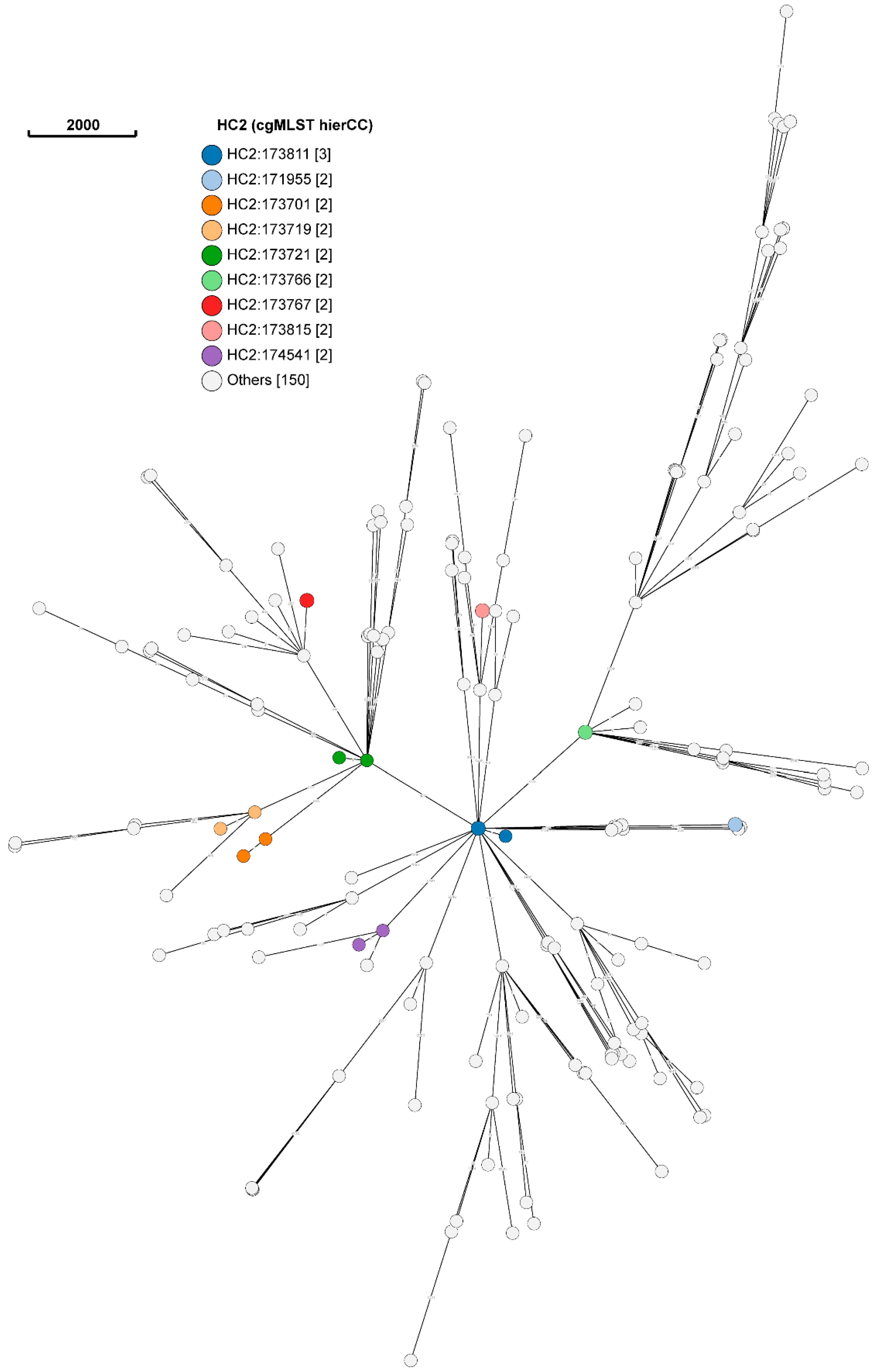
3.5.3. Core-Genome Multilocus Sequence Typing (cgMLST) and Hierarchical Clustering (HC)

The overall phylogeny of E. coli isolates is shown in the minimum spanning tree (Figure 1). Five clusters of genetically indistinguishable isolates were detected at HC level 0 (HC0), namely HC0:ST171955, HC0:ST173766, HC0:ST173767, HC0:ST173811 and HC0:ST173815. Each cluster consisted of two isolates. HC0:171955 was only detected in humans, whereas HC0:ST173767 originated from the same pig but was morphologically distinct. The three other STs (i.e., HC0:ST173766, HC0:ST173811 and HC0:ST173815) originated from the same production stage (i.e., weaning), but each cluster of weaner E. coli isolates originated from a different production house (House 2, House 12 and House 16).
A total of nine clusters were detected at HC level 2 (HC2), namely HC2:173881, HC2:171955, HC2:173701, HC2:173719, HC2: 173721, HC2:173766, HC2:173767, HC2:173815 and HC2:174541 (Figure 2).
Each cluster consisted of two isolates, with the exception of HC2:173811, which consisted of three isolates. Three clusters (i.e., HC2:171955, HC2:173701 and HC2:173719) consisted of only human isolates, whereas five clusters (i.e., HC2:173766, HC2:17367, HC2:173811, HC2:173815 and HC2:174541) consisted of only porcine isolates. Hierarchical cluster 2:173721 (HC2:173721) consisted of a human (H47) and porcine (P101) E. coli isolate, which provides some evidence for the transmission and subsequent spread of antibiotic resistance genes between humans and animals.
Additional evidence of the clustering of human and porcine isolates was revealed at HC10, with the detection of HC10:173841. This HC consisted of a single human E. coli isolate (H15-1) isolated from a female animal handler working in the weaning production stage and a porcine E. coli isolate (P52) isolated from a dry sow at the same site (i.e., Site B) on the farm. A total of three clusters of human and porcine isolates were detected at HC level 200 [i.e., HC200:3556, consisting of human E. coli (H27-1) and porcine E. coli (P4) isolate; HC200:41988, previously HC10:173841, but with an additional porcine E. coli P53 isolate; and HC200:32433, previously HC2:173721], whereas a total of seven clusters of human and porcine E. coli isolates were detected at HC level 400 (i.e., HC400:13, HC400:37, HC400:82, HC400:4483, HC400:4993, HC400:5951 and HC400:31574).

4. Discussion

Escherichia coli serves as an indicator micro-organism in antibiotic resistance surveillance programs [4]. Whole-genome sequencing can concurrently provide information on an isolate’s antibiotic resistance profile, its virulence potential and its genetic relationship with other E. coli isolates [62]. This can provide insights into the spread of antibiotic resistance genes among the One Health continuum [62]. The study found that porcine E. coli isolates were more resistant and harbored a richer diversity of antibiotic resistance genes than E. coli isolated from close human contacts on the same farm, specifically for penicillins (i.e., blaTEM-1), tetracyclines (i.e., tetA and tetB), ciprofloxacin (i.e., S83L point mutation in gyrA and OqxAB efflux pump) and phenicols (p < 0.05). On the other hand, a richer diversity of virulence genes associated with the specific pathotypes was detected in human E. coli isolates (p < 0.05). Although the same STcs were circulating in both pigs and close human contacts, only a single set of human and porcine E. coli isolates showed clonality at HC2, which is an indication of a recent transmission event.
The high rates of tetracycline and ampicillin resistance described in this study are in line with global antibiotic resistance rates described in various food-producing animals [63]. The antibiotic resistance rates in this study were also similar to a study conducted in an intensive pig production system in KwaZulu Natal, South Africa, where the highest rate of resistance detected was toward tetracyclines, and the lowest rate of resistance detected was toward carbapenems [64]. It is well established that tetracycline resistance in the South African agricultural setting is high, which may be a reflection of the antibiotic practices in South Africa [34,65,66]. Tetracycline can be purchased over the counter and used at the farmer’s own discretion [67]. A previous study by the authors (2022) showed that tetracyclines were the antibiotic class used in the highest quantity (i.e., 453.65 ± 35.49 kg or 135.16 mg/kg ± 3.31 mg/kg) on the same farm, which will explain why tetracycline resistance was high (95.28%, 101/113) in porcine isolates and was predominantly mediated by the tetA and tetB genes [35].
Tetracycline and ampicillin resistance determinants are often harbored on the same plasmid, which may explain why ampicillin and tetracycline resistance was equivalent in the porcine E. coli isolates [68]. Ampicillin resistance was predominantly mediated by the blaTEM-1 gene, which has previously been reported in non-O157 E. coli isolates in cattle farming from the same province [34]. The level of ampicillin resistance in this study was also higher compared to a study performed on swine farms in southern Brazil [11]. Brisola and colleagues (2019) found that the level of ampicillin resistance was 11.76% (8/103) compared to 94.34% (100/113) found in the porcine isolates in this study [11]. The high levels of ampicillin resistance are again potentially a reflection of the antibiotic practices on the farm, where ampicillin is used as metaphylaxis for post-weaning diarrhea [35].
Multiple porcine E. coli isolates harbored the multidrug efflux pump, OqxAB, which confers resistance to a veterinary growth promotor, olaquindox, but also to chloramphenicol, tigecycline, nitrofurantoin, fluoroquinolones, detergents and disinfectants [69,70]. This efflux pump is located on a plasmid and flanked by two insertion sequence (IS26) elements, which means it can easily be acquired by other bacteria, such as Klebsiella pneumoniae, through horizontal gene transfer [70,71]. Strasheim and colleagues (2022) previously reported that olaquindox was the antibiotic class used in the second highest quantity (i.e., 258.33 ± 8.04 kg or 77.07 mg/kg ± 3.93 mg/kg) on the farm [35]. Antibiotic growth promotors are not banned in South Africa, and their use has well-documented consequences of co-selection of resistance toward other antibiotics, which may lead to co-transmission of multiple antibiotic resistance genes [67,70]. The use of olaquindox as a growth promotor should be reconsidered in the South African context, taking food security, economic productivity, animal welfare and the emergence of antibiotic resistance into account. Plasmid-mediated resistance toward colistin was not detected in human or porcine E. coli isolates collected from this farm. This may be linked to the ban on colistin use in livestock by the South African Veterinary Council after detection of the mcr-1 gene in South Africa in 2015 by Perreten and colleagues (2016) in animals and by Coetzee and colleagues (2016) in humans [72,73,74].
A study by Founou and colleagues (2022) investigated five extended-spectrum β-lactams-producing (ESBL) porcine E. coli isolates obtained from two abattoirs in South Africa [75]. Isolates from the same abattoir were closely related and harbored the blaCTX-M-1 and 15 genes, together with multiple other resistance determinants for fluoroquinolones, aminoglycosides, tetracyclines and trimethoprim-sulfametaoxazole [75]. In this study, ESBL E. coli isolates were not detected, potentially because the isolates were grown on non-selective media. It is well known that media supplemented with antibiotics lead to different isolation rates compared to non-selective media [4]. Some authors state that the use of non-selective media may underestimate the true prevalence of ESBL producers [76,77], whereas others state that it is important to monitor both dominant (non-selective media) and subdominant (selective media) microflora to accurately define the true extent of antibiotic resistance rates [78]. In future, media used for the isolation of bacteria in antibiotic resistance monitoring should be standardized to enhance the comparability of antibiotic resistance rates.
The majority of human and porcine E. coli isolates were non-pathogenic in this study, but different pathovars, namely EAEC, EPEC, ETEC, ExPEC and STEC, were detected at low frequencies. Enteroaggregative E. coli was only detected in humans, whereas EPEC, ETEC and STEC were predominantly isolated from pigs. Overlaps of pathovar-specific genes were also observed, as evidenced by the detection of the astA gene, which encodes the enteroaggregative E. coli heat-stable enterotoxin (EAST1). This gene has previously been associated with EAEC in humans, but it is also reported as an important virulence factor of ETEC in piglets [48,79]. In this study, the astA gene was also detected in the porcine EPEC isolates, as well as human EAEC isolates. In addition, the astA gene was more frequently detected in porcine E. coli isolates than in human E. coli isolates. This suggests that the astA gene may play a different role in disease based on the host species.
Sequence type complex 10 was the most dominant STc in both human and porcine E. coli isolates based on Achtman’s 7-gene MLST scheme. A study by Peng and colleagues (2022) sequenced 1871 E. coli isolates obtained from pigs and their immediate environment from 31 provinces in China and compared these genomes with publicly available human E. coli genomes [80]. The authors also found STc10 to be the most predominant ST, as in this study.
Core-genome MLST and HC showed that human and porcine E. coli isolates were overall genetically diverse in this study, but there was some evidence (albeit very little) of HC of isolates at levels HC:2–HC:200. This indicates (i) the transmission between pigs from different production houses, phases and sites; (ii) the transmission between pigs and humans, potentially due to proximity; and (iii) the transmission between humans, potentially due to shared facilities [13]. These findings are in line with a previous study by Muloi and colleagues (2022) in Nairobi, Kenya, which showed that the transmission of E. coli between humans and animals can occur, but it remains an infrequent event [81]. The findings are also in agreement with a longitudinal study conducted by Dohmen and colleagues (2023) on Dutch pigs and farm workers over six months [82]. Dohmen and colleagues found evidence of antibiotic resistance gene transmission among workers and pigs, either vertically or horizontally, in 11 of the 39 pig farms studied [82]. More transmission events could have potentially occurred on the farm in this study, but antibiotic resistance transmission routes based on plasmid type through the food chain and the environment were not investigated.
The study has some limitations, as only a single pig farm was included, which has implications for the generalizability of the findings. African swine fever was in circulation in South Africa at the time of sample collection, and multiple farms could therefore not be visited due to the fear of spreading disease. In addition, shortly after the first farm visit, the COVID-19 pandemic emerged. Although only a single recent transmission event between a human and porcine E. coli isolate was detected on this farm, it remains important to strengthen the antibiotic stewardship practices and reduce antibiotic use for growth promotion and metaphylaxis to mitigate the risk of transmission and spread of resistance linked to antibiotic usage. In future, this study can serve as a blueprint for implementing One Health antibiotic resistance surveillance programs in South Africa on a broader scale.

Supplementary Materials

The following supporting information can be downloaded at: https://www.mdpi.com/article/10.3390/antibiotics13060543/s1, Table S1: Other virulence genes detected in human and porcine E. coli isolates; Table S2: Sequence type complex composition with the different STs detected in human and porcine E. coli isolates.

Author Contributions

Conceptualization, W.S., O.P. and E.M.C.E.; methodology, W.S., M.L., O.P. and A.M.S.; validation, W.S. and M.L.; formal analysis, W.S.; investigation, W.S. and M.L.; resources, O.P. and A.M.S.; data curation, W.S. and M.L.; writing—original draft preparation, W.S.; writing—review and editing, W.S., M.L., E.M.C.E., A.M.S. and O.P.; visualization, W.S.; supervision, O.P., A.M.S. and E.M.C.E.; project administration, W.S. and M.L.; funding acquisition, O.P. and A.M.S. All authors have read and agreed to the published version of the manuscript.

Funding

The South African Medical Research Council (SAMRC) funded this research via a sub-grant received from the Bill and Melinda Gates Foundation (Grant number: 96086). The contents and findings reported/illustrated are the sole deduction, view and responsibility of the researcher and do not reflect the official position and sentiments of the SAMRC or the Bill and Melinda Gates Foundation. Whole-genome sequencing was made possible through support from the SEQAFRICA project, which is funded by the Department of Health and Social Care’s Fleming Fund using UK aid. The views expressed in this publication are those of the authors and not necessarily those of the UK Department of Health and Social Care or its Management Agent, Mott MacDonald. W.S. was also supported by a Fogarty International Center Global Infectious Disease research-training grant, National Institutes of Health, to the University of Pittsburgh and the National Institute for Communicable Diseases (D43TW011255).

Institutional Review Board Statement

The study was conducted in accordance with the Declaration of Helsinki and approved by different human and animal health research ethics committees (HREC/AREC) from three statutory bodies, namely (i) The University of the Witwatersrand (Medical HREC M190244; 10 May 2019), (ii) the University of Pretoria (AREC of Faculty of Veterinary Science REC0055-20; 6 July 2020; REC of Faculty of Humanities HUM027/0620; 25 June 2020; and HREC of Faculty of Health Sciences 406/2020; 12 August 2020) and (iii) the National Institute for Communicable Diseases (NICD) (AEC003-19; 23 June 2019). In addition, a Section 20 clearance certificate (project reference number: 12/11/1/1/13; 23 April 2019) was issued by the Department of Agriculture, Land Reform and Rural Development of the Republic of South Africa. Informed consent was obtained from all subjects involved in the study.

Informed Consent Statement

Informed consent was obtained from all subjects involved in the study.

Data Availability Statement

Sequencing data are available at the NCBI’s GenBank under Bioproject number: PRJNA994298 (https://www.ncbi.nlm.nih.gov/sra/PRJNA994298). Additional data generated during this study were placed in an online repository (https://doi.org/10.6084/m9.figshare.23677266.v2). The accession numbers are listed in the Supplementary Material, together with the isolate’s unique identifier.

Acknowledgments

The authors would hereby like to acknowledge the farmer and his veterinarian in charge for providing access to the farm. The authors would also like to thank Ashika Singh-Moodley, Marshagne Smith, Naseema Bulbulia, Rosah Mabokachaba, Nompumelelo Shezi and Nokuthula Linda for their assistance in processing the isolates in the laboratory. Else Marais is hereby acknowledged for initiating the overall project idea. The Sequencing Core Facility at the National Institute for Communicable Diseases is also acknowledged for providing whole-genome sequencing services.

Conflicts of Interest

The authors declare no conflicts of interest. The funders had no role in the design of the study; in the collection, analyses, or interpretation of data; in the writing of the manuscript; or in the decision to publish the results.

References

  1. Poirel, L.; Madec, J.Y.; Lupo, A.; Schink, A.K.; Kieffer, N.; Nordmann, P.; Schwarz, S. Antimicrobial resistance in Escherichia coli. Microbiol. Spectr. 2018, 6. [Google Scholar] [CrossRef]
  2. Braz, V.S.; Melchior, K.; Moreira, C.G. Escherichia coli as a multifaceted pathogenic and versatile bacterium. Front. Cell. Infect. Microbiol. 2020, 10, 548492. [Google Scholar] [CrossRef]
  3. Hesp, A.; Veldman, K.; van der Goot, J.; Mevius, D.; van Schaik, G. Monitoring antimicrobial resistance trends in commensal Escherichia coli from livestock, the Netherlands, 1998 to 2016. Eurosurveillamce 2019, 24, 1800438-26. [Google Scholar] [CrossRef]
  4. WHO. Integrated Surveillance of Antimicrobial Resistance in Foodborne Bacteria: Application of a One Health Approach. Available online: https://apps.who.int/iris/bitstream/handle/10665/255747/9789241512411-eng.pdf?sequence=1 (accessed on 25 May 2021).
  5. Croxen, M.A.; Finlay, B.B. Molecular mechanisms of Escherichia coli pathogenicity. Nat. Rev. Microbiol. 2010, 8, 26–38. [Google Scholar] [CrossRef]
  6. Robins-Browne, R.M.; Holt, K.E.; Ingle, D.J.; Hocking, D.M.; Yang, J.; Tauschek, M. Are Escherichia coli pathotypes still relevant in the era of whole-genome sequencing? Front. Cell. Infect. Microbiol. 2016, 6, 141. [Google Scholar] [CrossRef]
  7. Denamur, E.; Clermont, O.; Bonacorsi, S.; Gordon, D. The population genetics of pathogenic Escherichia coli. Nat. Rev. Microbiol. 2021, 19, 37–54. [Google Scholar] [CrossRef]
  8. Riley, L.W. Distinguishing pathovars from nonpathovars: Escherichia coli. Microbiol. Spectr. 2020, 8. [Google Scholar] [CrossRef]
  9. Tenaillon, O.; Skurnik, D.; Picard, B.; Denamur, E. The population genetics of commensal Escherichia coli. Nat. Rev. Microbiol. 2010, 8, 207–217. [Google Scholar] [CrossRef]
  10. Jang, J.; Hur, H.G.; Sadowsky, M.J.; Byappanahalli, M.N.; Yan, T.; Ishii, S. Environmental Escherichia coli: Ecology and public health implications—A review. J. Appl. Microbiol. 2017, 123, 570–581. [Google Scholar] [CrossRef]
  11. Brisola, M.C.; Crecencio, R.B.; Bitner, D.S.; Frigo, A.; Rampazzo, L.; Stefani, L.M.; Faria, G.A. Escherichia coli used as a biomarker of antimicrobial resistance in pig farms of Southern Brazil. Sci. Total Environ. 2019, 647, 362–368. [Google Scholar] [CrossRef]
  12. Wee, B.A.; Muloi, D.M.; van Bunnik, B.A.D. Quantifying the transmission of antimicrobial resistance at the human and livestock interface with genomics. Clin. Microbiol. Infect. 2020, 26, 1612–1616. [Google Scholar] [CrossRef] [PubMed]
  13. Hanage, W.P. Two health or not two health? That is the question. mBio 2019, 10, e00550-00510. [Google Scholar] [CrossRef]
  14. Hammerum, A.M.; Larsen, J.; Andersen, V.D.; Lester, C.H.; Skovgaard Skytte, T.S.; Hansen, F.; Olsen, S.S.; Mordhorst, H.; Skov, R.L.; Aarestrup, F.M.; et al. Characterization of extended-spectrum beta-lactamase (ESBL)-producing Escherichia coli obtained from Danish pigs, pig farmers and their families from farms with high or no consumption of third- or fourth-generation cephalosporins. J. Antimicrob. Chemother. 2014, 69, 2650–2657. [Google Scholar] [CrossRef] [PubMed]
  15. Uelze, L.; Becker, N.; Borowiak, M.; Busch, U.; Dangel, A.; Deneke, C.; Fischer, J.; Flieger, A.; Hepner, S.; Huber, I.; et al. Toward an integrated genome-based surveillance of Salmonella enterica in Germany. Front. Microbiol. 2021, 12, 626941. [Google Scholar] [CrossRef] [PubMed]
  16. Leekitcharoenphon, P.; Johansson, M.H.K.; Munk, P.; Malorny, B.; Skarzynska, M.; Wadepohl, K.; Moyano, G.; Hesp, A.; Veldman, K.T.; Bossers, A.; et al. Genomic evolution of antimicrobial resistance in Escherichia coli. Sci. Rep. 2021, 11, 15108. [Google Scholar] [CrossRef] [PubMed]
  17. Ludden, C.; Raven, K.E.; Jamrozy, D.; Gouliouris, T.; Blane, B.; Coll, F.; de Goffau, M.; Naydenova, P.; Horner, C.; Hernandez-Garcia, J.; et al. One Health genomic surveillance of Escherichia coli demonstrates distinct lineages and mobile genetic elements in isolates from humans versus livestock. mBio 2019, 10, e02693-18. [Google Scholar] [CrossRef] [PubMed]
  18. Ramatla, T.; Tawana, M.; Lekota, K.E.; Thekisoe, O. Antimicrobial resistance genes of Escherichia coli, a bacterium of “One Health” importance in South Africa: Systematic review and meta-analysis. AIMS Microbiol. 2023, 9, 75–89. [Google Scholar] [CrossRef] [PubMed]
  19. Theobald, S.; Etter, E.M.C.; Gerber, D.; Abolnik, C. Antimicrobial resistance trends in Escherichia coli in South African poultry: 2009–2015. Foodborne Pathog. Dis. 2019, 16, 652–660. [Google Scholar] [CrossRef] [PubMed]
  20. Iwu, C.J.; Iweriebor, B.C.; Obi, L.C.; Okoh, A.I. Occurrence of non-O157 Shiga toxin-producing Escherichia coli in two commercial swine farms in the Eastern Cape province, South Africa. Comp. Immunol. Microbiol. Infect. Dis. 2016, 44, 48–53. [Google Scholar] [CrossRef]
  21. Iwu, C.J.; Jaja, I.F.; Iweriebor, B.C.; Obi, L.C.; Okoh, A.I. Antibiotic resistance profiles of Escherichia coli O26, O145, and O157:H7 isolated from swine in the Eastern Cape province, South Africa. Asian Pac. J. Trop. Dis. 2017, 7, 553–559. [Google Scholar] [CrossRef]
  22. McIver, K.S.; Amoako, D.G.; Abia, A.L.K.; Bester, L.A.; Chenia, H.Y.; Essack, S.Y. Molecular epidemiology of antibiotic-resistant Escherichia coli from farm-to-fork in intensive poultry production in KwaZulu-Natal, South Africa. Antibiotics 2020, 9, 850. [Google Scholar] [CrossRef] [PubMed]
  23. Kanengoni, A.T.; Thomas, R.; Gelaw, A.K.; Madoroba, E. Epidemiology and characterization of Escherichia coli outbreak on a pig farm in South Africa. FEMS Microbiol. Lett. 2017, 364, fnx010. [Google Scholar] [CrossRef] [PubMed]
  24. Mbelle, N.M.; Feldman, C.; Osei Sekyere, J.; Maningi, N.E.; Modipane, L.; Essack, S.Y. The resistome, mobilome, virulome and phylogenomics of multidrug-resistant Escherichia coli clinical isolates from Pretoria, South Africa. Sci. Rep. 2019, 9, 16457. [Google Scholar] [CrossRef] [PubMed]
  25. DeFrancesco, A.S.; Tanih, N.F.; Samie, A.; Guerrant, R.L.; Bessong, P.O. Antibiotic resistance patterns and beta-lactamase identification in Escherichia coli isolated from young children in rural Limpopo Province, South Africa: The MAL-ED cohort. S. Afr. Med. J. 2017, 107, 205–214. [Google Scholar] [CrossRef] [PubMed]
  26. Genthe, B.; Ndlela, L.; Madlala, T. Antimicrobial resistance screening and profiles: A glimpse from the South African perspective. J. Water Health 2020, 18, 925–936. [Google Scholar] [CrossRef] [PubMed]
  27. Iwu, C.D.; Nontongana, N.; Iwu-Jaja, C.J.; Anyanwu, B.O.; du Plessis, E.; Korsten, L.; Okoh, A.I. Spatial diarrheal disease risks and antibiogram diversity of diarrheagenic Escherichia coli in selected access points of the Buffalo River, South Africa. PLoS ONE 2023, 18, e0288809. [Google Scholar] [CrossRef] [PubMed]
  28. Ratshilingano, M.T.; du Plessis, E.M.; Duvenage, S.; Korsten, L. Characterization of multidrug-resistant Escherichia coli isolated from two commercial lettuce and spinach supply chains. J. Food Prot. 2022, 85, 122–132. [Google Scholar] [CrossRef] [PubMed]
  29. Richter, L.; du Plessis, E.M.; Duvenage, S.; Korsten, L. Occurrence, identification, and antimicrobial resistance profiles of extended-spectrum and AmpC β-lactamase-producing Enterobacteriaceae from fresh vegetables retailed in Gauteng province, South Africa. Foodborne Pathog. Dis. 2019, 16, 421–427. [Google Scholar] [CrossRef] [PubMed]
  30. Mbanga, J.; Amoako, D.G.; Abia, A.L.K.; Allam, M.; Ismail, A.; Essack, S.Y. Genomic insights of multidrug-resistant Escherichia coli from wastewater sources and their association with clinical pathogens in South Africa. Front. Vet. Sci. 2021, 8, 636715. [Google Scholar] [CrossRef]
  31. Mbanga, J.; Abia, A.L.K.; Amoako, D.G.; Essack, S.Y. Longitudinal surveillance of antibiotic resistance in Escherichia coli and Enterococcus spp. from a wastewater treatment plant and its associated waters in KwaZulu-Natal, South Africa. Microb. Drug Resist. 2021, 27, 904–918. [Google Scholar] [CrossRef]
  32. Baloyi, T.; Duvenage, S.; Du Plessis, E.; Villamizar-Rodríguez, G.; Korsten, L. Multidrug resistant Escherichia coli from fresh produce sold by street vendors in South African informal settlements. Int. J. Environ. Health Res. 2022, 32, 1513–1528. [Google Scholar] [CrossRef] [PubMed]
  33. Founou, L.L.; Founou, R.C.; Ntshobeni, N.; Govinden, U.; Bester, L.A.; Chenia, H.Y.; Djoko, C.F.; Essack, S.Y. Emergence and spread of extended spectrum beta-lactamase producing Enterobacteriaceae (ESBL-PE) in pigs and exposed Workers: A multicentre comparative study between Cameroon and South Africa. Pathogens 2019, 8, 10. [Google Scholar] [CrossRef] [PubMed]
  34. Bumunang, E.W.; McAllister, T.A.; Zaheer, R.; Ortega Polo, R.; Stanford, K.; King, R.; Niu, Y.D.; Ateba, C.N. Characterization of non-O157 Escherichia coli from cattle faecal samples in the North-West province of South Africa. Microorganisms 2019, 7, 272. [Google Scholar] [CrossRef] [PubMed]
  35. Strasheim, W.; Etter, E.M.C.; Lowe, M.; Perovic, O. Method to assess farm-level vaccine and antibiotic usage utilizing financial documentation: A pilot study in a commercial pig farm in South Africa from 2016 to 2018. Front. Vet. Sci. 2022, 9, 856729. [Google Scholar] [CrossRef] [PubMed]
  36. CLSI. Performance Standards for Antimicrobial Susceptibility Testing, 30th ed.; CLSI Supplement M100; Clinical and Laboratory Standards Institute: Wayne, PA, USA, 2020. [Google Scholar]
  37. The European Committee on Antimicrobial Susceptibility Testing. Breakpoint Tables for Interpretation of MICs and Zone Diameters. Version 13.0, 2023. Available online: http://eucast.org (accessed on 8 May 2023).
  38. National Antimicrobial Resistance Monitoring System for Enteric Bacteria (NARMS). Antimicrobial Agents Used for Susceptibility Testing for E. coli Isolates. 2023. Available online: https://www.cdc.gov/narms/antibiotics-tested.html (accessed on 8 May 2023).
  39. Kwenda, S.; Allam, M.; Khumualo, Z.T.H.; Mtshali, S.; Mnyameni, F.; Ismail, A. Jekesa: An Automated Easy-to-Use Pipeline for Bacterial Whole Genome Typing. Available online: https://github.com/stanikae/jekesa (accessed on 8 May 2023).
  40. Wood, D.E.; Lu, J.; Langmead, B. Improved metagenomic analysis with Kraken 2. Genome Biol. 2019, 20, 257. [Google Scholar] [CrossRef] [PubMed]
  41. Bankevich, A.; Nurk, S.; Antipov, D.; Gurevich, A.A.; Dvorkin, M.; Kulikov, A.S.; Lesin, V.M.; Nikolenko, S.I.; Pham, S.; Prjibelski, A.D.; et al. SPAdes: A new genome assembly algorithm and its applications to single-cell sequencing. J. Comput. Biol. 2012, 19, 455–477. [Google Scholar] [CrossRef] [PubMed]
  42. Gurevich, A.; Saveliev, V.; Vyahhi, N.; Tesler, G. QUAST: Quality assessment tool for genome assemblies. Bioinformatics 2013, 29, 1072–1075. [Google Scholar] [CrossRef] [PubMed]
  43. Joensen, K.G.; Scheutz, F.; Lund, O.; Hasman, H.; Kaas, R.S.; Nielsen, E.M.; Aarestrup, F.M. Real-time whole-genome sequencing for routine typing, surveillance, and outbreak detection of verotoxigenic Escherichia coli. J. Clin. Microbiol. 2014, 52, 1501–1510. [Google Scholar] [CrossRef] [PubMed]
  44. Malberg Tetzschner, A.M.; Johnson, J.R.; Johnston, B.D.; Lund, O.; Scheutz, F. In silico genotyping of Escherichia coli isolates for extraintestinal virulence genes by use of whole-genome sequencing data. J. Clin. Microbiol. 2020, 58, e01269-01220. [Google Scholar] [CrossRef]
  45. Camacho, C.; Coulouris, G.; Avagyan, V.; Ma, N.; Papadopoulos, J.; Bealer, K.; Madden, T.L. BLAST+: Architecture and applications. BMC Bioinform. 2009, 10, 421. [Google Scholar] [CrossRef]
  46. Zankari, E.; Allesoe, R.; Joensen, K.G.; Cavaco, L.M.; Lund, O.; Aarestrup, F.M. PointFinder: A novel web tool for WGS-based detection of antimicrobial resistance associated with chromosomal point mutations in bacterial pathogens. J. Antimicrob. Chemother. 2017, 72, 2764–2768. [Google Scholar] [CrossRef] [PubMed]
  47. Bortolaia, V.; Kaas, R.S.; Ruppe, E.; Roberts, M.C.; Schwarz, S.; Cattoir, V.; Philippon, A.; Allesoe, R.L.; Rebelo, A.R.; Florensa, A.F.; et al. ResFinder 4.0 for predictions of phenotypes from genotypes. J. Antimicrob. Chemother. 2020, 75, 3491–3500. [Google Scholar] [CrossRef]
  48. Boisen, N.; Osterlund, M.T.; Joensen, K.G.; Santiago, A.E.; Mandomando, I.; Cravioto, A.; Chattaway, M.A.; Gonyar, L.A.; Overballe-Petersen, S.; Stine, O.C.; et al. Redefining enteroaggregative Escherichia coli (EAEC): Genomic characterization of epidemiological EAEC strains. PLoS Negl. Trop. Dis. 2020, 14, e0008613. [Google Scholar] [CrossRef] [PubMed]
  49. Gaytan, M.O.; Martinez-Santos, V.I.; Soto, E.; Gonzalez-Pedrajo, B. Type three secretion system in attaching and effacing pathogens. Front. Cell. Infect. Micro. 2016, 6, 129. [Google Scholar] [CrossRef] [PubMed]
  50. Duan, Q.; Yao, F.; Zhu, G. Major virulence factors of enterotoxigenic Escherichia coli in pigs. Ann. Microbiol. 2011, 62, 7–14. [Google Scholar] [CrossRef]
  51. Pakbin, B.; Bruck, W.M.; Rossen, J.W.A. Virulence factors of enteric pathogenic Escherichia coli: A review. Int. J. Mol. Sci. 2021, 22, 9922. [Google Scholar] [CrossRef] [PubMed]
  52. Johnson, J.R.; Russo, T.A. Molecular epidemiology of extraintestinal pathogenic Escherichia coli. EcoSal Plus 2018, 8. [Google Scholar] [CrossRef]
  53. Aldawood, E.; Roberts, I.S. Regulation of Escherichia coli group 2 capsule gene expression: A mini review and update. Front. Microbiol. 2022, 13, 858767. [Google Scholar] [CrossRef]
  54. Roer, L.; Tchesnokova, V.; Allesoe, R.; Muradova, M.; Chattopadhyay, S.; Ahrenfeldt, J.; Thomsen, M.C.F.; Lund, O.; Hansen, F.; Hammerum, A.M.; et al. Development of a web tool for Escherichia coli subtyping based on fimH alleles. J. Clin. Microbiol. 2017, 55, 2538–2543. [Google Scholar] [CrossRef]
  55. Beghain, J.; Bridier-Nahmias, A.; Le Nagard, H.; Denamur, E.; Clermont, O. ClermonTyping: An easy-to-use and accurate in silico method for Escherichia genus strain phylotyping. Microb. Genom. 2018, 4, e000192. [Google Scholar] [CrossRef]
  56. Waters, N.R.; Abram, F.; Brennan, F.; Holmes, A.; Pritchard, L. Easy phylotyping of Escherichia coli via the EzClermont web app and command-line tool. Access Microbiol. 2020, 2, acmi000143. [Google Scholar] [CrossRef]
  57. Zhou, Z.; Alikhan, N.F.; Mohamed, K.; Fan, Y.; Agama Study, G.; Achtman, M. The EnteroBase user’s guide, with case studies on Salmonella transmissions, Yersinia pestis phylogeny, and Escherichia core genomic diversity. Genome Res. 2020, 30, 138–152. [Google Scholar] [CrossRef]
  58. Zhou, Z.; Alikhan, N.F.; Sergeant, M.J.; Luhmann, N.; Vaz, C.; Francisco, A.P.; Carrico, J.A.; Achtman, M. GrapeTree: Visualization of core genomic relationships among 100,000 bacterial pathogens. Genome Res. 2018, 28, 1395–1404. [Google Scholar] [CrossRef]
  59. Wickham, H.; François, R.; Henry, L.; Müller, K. dplyr: A Grammar of Data Manipulation, R Package Version 1.0.6; 2021. Available online: https://CRAN.R-project.org/package=dplyr (accessed on 8 May 2023).
  60. Kassambara, A. Rstatix: Pipe-Friendly Framework for Basic Statistical Tests, R Package Version 0.7.2.; 2023. Available online: https://cran.r-project.org/web/packages/rstatix/index.html (accessed on 8 May 2023).
  61. Firke, S. Janitor: Simple Tools for Examining and Cleaning Dirty Data., R Package Version 2.2.0.; 2023. Available online: https://cran.r-project.org/web/packages/janitor/index.html (accessed on 8 May 2023).
  62. Al-Mustapha, A.I.; Raufu, I.A.; Ogundijo, O.A.; Odetokun, I.A.; Tiwari, A.; Brouwer, M.S.M.; Adetunji, V.; Heikinheimo, A. Antibiotic resistance genes, mobile elements, virulence genes, and phages in cultivated ESBL-producing Escherichia coli of poultry origin in Kwara State, North Central Nigeria. Int. J. Food Microbiol. 2023, 389, 110086. [Google Scholar] [CrossRef]
  63. Silva, A.; Silva, V.; Pereira, J.E.; Maltez, L.; Igrejas, G.; Valentao, P.; Falco, V.; Poeta, P. Antimicrobial resistance and clonal lineages of Escherichia coli from food-producing animals. Antibiotics 2023, 12, 1061. [Google Scholar] [CrossRef]
  64. Abdalla, S.E.; Abia, A.L.K.; Amoako, D.G.; Perrett, K.; Bester, L.A.; Essack, S.Y. From farm-to-fork: E. coli from an intensive pig production system in South Africa shows high resistance to critically important antibiotics for human and animal use. Antibiotics 2021, 10, 178. [Google Scholar] [CrossRef] [PubMed]
  65. Ateba, C.N.; Bezuidenhout, C.C. Characterisation of Escherichia coli O157 strains from humans, cattle and pigs in the North-West province, South Africa. Int. J. Food Microbiol. 2008, 128, 181–188. [Google Scholar] [CrossRef] [PubMed]
  66. Iweriebor, B.C.; Iwu, C.J.; Obi, L.C.; Nwodo, U.U.; Okoh, A.I. Multiple antibiotic resistances among shiga-toxin producing Escherichia coli O157 in feces of dairy cattle farms in Eastern Cape of South Africa. BMC Microbiol. 2015, 15, 213. [Google Scholar] [CrossRef] [PubMed]
  67. Eagar, H.; Naidoo, V. Veterinary antimicrobial stewardship in South Africa. Int. Biol. Rev. 2017, 1, 1–14. [Google Scholar]
  68. Rozwandowicz, M.; Brouwer, M.S.M.; Fischer, J.; Wagenaar, J.A.; Gonzalez-Zorn, B.; Guerra, B.; Mevius, D.J.; Hordijk, J. Plasmids carrying antimicrobial resistance genes in Enterobacteriaceae. J. Antimicrob. Chemother. 2018, 73, 1121–1137. [Google Scholar] [CrossRef]
  69. Poirel, L.; Cattoir, V.; Nordmann, P. Plasmid-mediated quinolone resistance; interactions between human, animal, and environmental ecologies. Front. Microbiol. 2012, 3, 24. [Google Scholar] [CrossRef] [PubMed]
  70. Li, J.; Zhang, H.; Ning, J.; Sajid, A.; Cheng, G.; Yuan, Z.; Hao, H. The nature and epidemiology of OqxAB, a multidrug efflux pump. Antimicrob. Resist. Infect. Control 2019, 8, 44. [Google Scholar] [CrossRef] [PubMed]
  71. Perez, F.; Rudin, S.D.; Marshall, S.H.; Coakley, P.; Chen, L.; Kreiswirth, B.N.; Rather, P.N.; Hujer, A.M.; Toltzis, P.; van Duin, D.; et al. OqxAB, a quinolone and olaquindox efflux pump, is widely distributed among multidrug-resistant Klebsiella pneumoniae isolates of human origin. Antimicrob. Agents Chemother. 2013, 57, 4602–4603. [Google Scholar] [CrossRef] [PubMed]
  72. Perreten, V.; Strauss, C.; Collaud, A.; Gerber, D. Colistin resistance gene mcr-1 in avian-pathogenic Escherichia coli in South Africa. Antimicrob. Agents Chemother. 2016, 60, 4414–4415. [Google Scholar] [CrossRef] [PubMed]
  73. Mendelson, M.; Brink, A.; Gouws, J.; Mbelle, N.; Naidoo, V.; Pople, T.; Schellack, N.; van Vuuren, M.; Rees, H. The One Health stewardship of colistin as an antibiotic of last resort for human health in South Africa. Lancet Infect. Dis. 2018, 18, e288–e294. [Google Scholar] [CrossRef] [PubMed]
  74. Coetzee, J.; Corcoran, C.; Prentice, E.; Moodley, M.; Mendelson, M.; Poirel, L.; Nordmann, P.; Brink, A.J. Emergence of plasmid-mediated colistin resistance (MCR-1) among Escherichia coli isolated from South African patients. S. Afr. Med. J. 2016, 106, 35–36. [Google Scholar] [CrossRef] [PubMed]
  75. Founou, L.L.; Founou, R.C.; Allam, M.; Ismail, A.; Essack, S.Y. Genome analysis of ESBL-producing Escherichia coli isolated from pigs. Pathogens 2022, 11, 776. [Google Scholar] [CrossRef] [PubMed]
  76. Ghosh, K.; Janecko, N.; Agunos, A.; Deckert, A.; Reid-Smith, R.; Gow, S.; Rubin, J. Evaluation of selective media in antimicrobial surveillance programs capturing broad-spectrum beta-lactamase producing Escherichia coli from chickens at slaughter. Can. Vet. J. 2021, 62, 608–610. [Google Scholar] [PubMed]
  77. Duggett, N.; AbuOun, M.; Randall, L.; Horton, R.; Lemma, F.; Rogers, J.; Crook, D.; Teale, C.; Anjum, M.F. The importance of using whole genome sequencing and extended spectrum beta-lactamase selective media when monitoring antimicrobial resistance. Sci. Rep. 2020, 10, 19880. [Google Scholar] [CrossRef]
  78. Gay, E.; Bour, M.; Cazeau, G.; Jarrige, N.; Martineau, C.; Madec, J.Y.; Haenni, M. Antimicrobial usages and antimicrobial resistance in commensal Escherichia coli from veal calves in France: Evolution during the fattening process. Front. Microbiol. 2019, 10, 792. [Google Scholar] [CrossRef]
  79. Mohlatlole, R.P.; Madoroba, E.; Muchadeyi, F.C.; Chimonyo, M.; Kanengoni, A.T.; Dzomba, E.F. Virulence profiles of enterotoxigenic, shiga toxin and enteroaggregative Escherichia coli in South African pigs. Trop. Anim. Health Prod. 2013, 45, 1399–1405. [Google Scholar] [CrossRef] [PubMed]
  80. Peng, Z.; Hu, Z.; Li, Z.; Zhang, X.; Jia, C.; Li, T.; Dai, M.; Tan, C.; Xu, Z.; Wu, B.; et al. Antimicrobial resistance and population genomics of multidrug-resistant Escherichia coli in pig farms in mainland China. Nat. Commun. 2022, 13, 1116. [Google Scholar] [CrossRef] [PubMed]
  81. Muloi, D.M.; Wee, B.A.; McClean, D.M.H.; Ward, M.J.; Pankhurst, L.; Phan, H.; Ivens, A.C.; Kivali, V.; Kiyong’a, A.; Ndinda, C.; et al. Population genomics of Escherichia coli in livestock-keeping households across a rapidly developing urban landscape. Nat. Microbiol. 2022, 7, 581–589. [Google Scholar] [CrossRef]
  82. Dohmen, W.; Liakopoulos, A.; Bonten, M.J.M.; Mevius, D.J.; Heederika, D.J.J. Longitudinal study of dynamic epidemiology of extended spectrum beta-lactamase-producing Escherichia coli in pigs and humans living and/or working on pig farms. Microbiol. Spectr. 2023, 11, e0294722. [Google Scholar] [CrossRef]
Figure 1. Minimum spanning tree based on the core-genome MLST and hierarchical clustering (HC) of human (yellow dots) and porcine (pink dots) E. coli isolates on EnteroBase, visualized using GrapeTree.
Antibiotics 13 00543 g001
Figure 2. Minimum spanning tree of human and porcine E. coli isolates at hierarchical level 2 on EnteroBase, visualized using GrapeTree.
Antibiotics 13 00543 g002
Table 1. Phenotypic antibiotic resistance rates of human and porcine E. coli isolates.
Antibiotic ClassR MIC Breakpoint (µg/mL)Human
% (n = 63)
Pigs
% (n =106)
Total
% (n = 169)
p-Value
Penicillins
Ampicillin≥3233.33 (21)94.34 (100)71.60 (121)<0.05
Piperacillin≥12831.75 (20)84.91 (90)65.09 (110)<0.05
ß-lactam combination agents
Amoxicillin-clavulanate≥32/160.0 (0)0.0 (0)0.0 (0)NS
Ampicillin-sulbactam≥32/169.52 (6)40.57 (43)28.99 (49)<0.05
Piperacillin-tazobactam≥128/40.0 (0)0.00 (0)0.0 (0)NS
Cephems
Cefepime≥161.59 (1)0.0 (0)0.59 (1)NS
Cefotaxime≥40.0 (0)0.0 (0)0.0 (0)NS
Cefotaxime-clavulante >0.50.0 (0)0.0 (0)0.0 (0)NS
Cefoxitin≥320.0 (0)0.0 (0)0.0 (0)NS
Ceftazidime≥161.59 (1)0.0 (0)0.59 (1)NS
Ceftazidime-clavulante>0.250.0 (0)0.0 (0)0.0 (0)NS
Cefuroxime≥321.59 (1)0.0 (0)0.59 (1)NS
Monobactams
Aztreonam≥161.59 (1)0.0 (0)0.59 (1)NS
Carbapenems
Doripenem≥40.0 (0)0.0 (0)0.0 (0)NS
Ertapenem≥20.0 (0)0.0 (0)0.0 (0)NS
Imipenem≥40.0 (0)0.0 (0)0.0 (0)NS
Meropenem≥40.0 (0)0.0 (0)0.0 (0)NS
Aminoglycosides
Amikacin≥640.0 (0)0.0 (0)0.0 (0)NS
Gentamicin≥160.0 (0)6.6 (7)4.14 (7)NS
Streptomycin #≥3222.22 (14)28.3 (30)26.04 (44)NS
Tobramycin≥160.0 (0)3.77 (4)2.37 (4)NS
Tetracyclines
Tetracycline≥1630.16 (19)95.28 (101)71.01 (120)<0.05
Quinolones
Ciprofloxacin *≥11.59 (1)16.04 (17)10.65 (18)<0.05
Levofloxacin≥20.0 (0)4.72 (5)2.96 (5)NS
Other
Chloramphenicol≥324.76 (3)35.85 (38)24.26 (41)<0.05
Colistin #$>20.0 (0)0.0 (0)0.0 (0)NS
Fosfomycin≥2560.0 (0)1.89 (2)1.18 (2)NS
Tigecycline #0.50.0 (0)0.94 (1)0.59 (1)NS
Trimethoprim-sulfamethoxazole≥4/7639.68 (25)28.3 (30)32.54 (55)NS
MIC = Minimum inhibitory concentration; R = Resistant; # Epidemiological cut-off values (ECOFF) reported and interpreted according to EUCAST guidelines (2023) NARMS; * As determined by the commercial broth microdilution method; $ MIC for colistin was determined using Sensititre. NS = Not statistically significant.
Table 2. Antibiotic resistance genes detected in human and porcine E. coli isolates using ResFinder (v.4.1).
Antibiotic Resistance Gene ClassHuman
% (n = 63)
Pigs
% (n =106)
Total
% (n = 169)
p-Value
Aminoglycoside-modifying enzymes
 Acetyltransferases
ACC(3)-IId0.0 (0)0.94 (1)0.59 (1)NS
ACC(3)-IId; ACC-Ib-cr0.0 (0)5.66 (6)3.55 (6)NS
Not detected100.00 (63)61.11 (99)95.86 (162)NS
 Nucleotidyltransferases
ANT(3″)-Ia19.05 (12)50.0 (53)38.46 (65)<0.05
Not detected80.95 (51)50.0 (53)61.54 (104)<0.05
 Phosphotransferases
APH(3′)-Ia0.0 (0)12.26 (13)7.69 (13)<0.05
APH(3″)-Ib; APH(6)-Id30.16 (19)22.64 (24)25.44 (43)NS
APH(3′)-Ia; APH(3″)-Ib; APH(6)-Id0.0 (0)11.32 (12)7.10 (12)<0.05
Not detected69.84 (44)53.77 (57)59.76 (101)NS
ß-lactam resistance genes
blaTEM-1A1.59 (1)2.83 (3)2.37 (4)NS
blaTEM-1B28.57 (18)76.42 (81)58.58 (99)<0.05
blaTEM-1C0.0 (0)2.83 (3)1.78 (3)NS
ß-lactam resistance genes
blaTEM-1B and blaOXA-10.0 (0)5.66 (6)3.55 (6)NS
Multiple blaTEM variants1.59 (1)1.89 (2)1.78 (3)NS
Not detected68.25 (43)10.38 (11)31.95 (54)<0.05
Colistin
pmrB(V161G)0.0 (0)0.94 (1)0.59 (1)NS
Macrolides
mdfA95.24 (60)97.17 (103)96.45 (163)NS
mdfA, mphA4.76 (3)2.83 (6)3.55 (6)NS
Phenicols
catA10.00 (0)1.89 (2)1.18 (2)NS
cmlA11.59 (1)4.72 (5)3.55 (6)NS
cmlA1, catB30.0 (0)0.94 (1)0.59 (1)NS
cmlA1, catB4, floR0.0 (0)4.72 (5)2.96 (5)NS
floR0.0 (0)1.89 (2)1.18 (2)NS
Not detected98.41 (62)85.85 (91)90.53 (153)<0.05
Quinolones
 Chromosomal mutations
gyrA (S83A)4.76 (3)5.66 (6)5.33 (9)NS
gyrA (S83L)6.35 (4)23.58 (25)17.16 (29)<0.05
parC (A56T)1.59 (1)2.83 (3)2.37 (4)NS
parC (S80I) 1.59 (1)0.94 (1)1.18 (2)NS
parE (I355T)1.59 (1)0.94 (1)1.18 (2)NS
 Plasmid-mediated
OqxAB3.17 (2)32.08 (34)21.30 (36)<0.05
OqxAB, acc-(6′)-Ib-cr, qnrS20.0 (0)4.72 (5)2.96 (5)NS
OqxAB, qnrS10.0 (0)6.60 (7)4.14 (7)NS
OqxAB, qnrS20.0 (0)0.94 (1)0.59 (1)NS
qnrS17.94 (5)6.60 (7)7.10 (12)NS
Not detected88.89 (56)49.06 (52)63.91 (108)<0.05
Rifampicin
arr-30.0 (0)5.66 (6)3.55 (6)NS
Not detected100.00 (63)94.34 (100)96.45 (163)NS
Sulphonamides
sul14.76 (3)12.26 (13)9.47 (16)NS
sul1, sul211.11 (7)3.77 (4)6.51 (11)NS
sul1, sul30.0 (0)0.94 (1)0.59 (1)NS
sul222.22 (14)10.38 (11)14.79 (25)NS
sul2, sul30.0 (0)1.89 (2)1.18 (2)NS
sul31.59 (1)10.38 (11)7.10 (12)NS
Not detected60.32 (38)60.38 (64)60.36 (102)NS
Tetracyclines
tetA15.87 (10)43.40 (46)33.14 (56)<0.05
tetA, tetM0.0 (0)4.72 (5)2.96 (5)NS
tetB12.70 (8)34.91 (37)26.63 (45)<0.05
tetB, tetM0.0 (0)11.32 (12)7.10 (12)<0.05
tetA, tetB 0.0 (0)0.94 (1)0.59 (1)NS
Not detected71.43 (45)4.72 (5)29.59 (50)<0.05
Trimethoprim
drfA19.52 (6)2.83 (3)5.33 (9)NS
drfA1, drfA141.59 (1)0.0 (0)0.59 (1)NS
drfA123.17 (2)25.47 (27)17.16 (29)<0.05
drfA12, drfA210.0 (0)3.77 (4)2.37 (4)NS
drfA1412.70 (8)4.72 (5)7.69 (13)NS
drfA174.76 (3)2.83 (3)3.55 (6)NS
drfA210.0 (0)0.94 (1)0.59 (1)NS
drfA51.59 (1)0.0 (0)0.59 (1)NS
drfA74.76 (3)0.0 (0)1.78 (3)NS
drfA7, drfA141.59 (1)0.0 (0)0.59 (1)NS
Not detected60.32 (38)59.43 (63)59.76 (101)NS
NS = Not statistically significant.
Table 3. Virulence factors grouped according to pathotype in human and porcine E. coli isolates.
Virulence Gene CombinationsHuman
% (n = 63)
Pigs
% (n =106)
Total
% (n = 169)
p-Value
Enteroaggregative E. coli (EAEC)
Dispersin (aap)12.70 (8)0.0 (0)4.73 (8)<0.05
Dispersin transporter protein (aatA)9.52 (6)0.0 (0)3.55 (6)<0.05
aaiC, ORF4 and ORF44.76 (3)0.0 (0)1.78 (3)NS
aaiC0.0 (0)0.94 (1)0.59 (1)NS
 Biogenesis of AFA-III
afaABCDE1.59 (1)0.0 (0)0.59 (1)NS
afaABCDE81.59 (1)0.0 (0)0.59 (1)NS
afaD1.59 (1)0.0 (0)0.59 (1)NS
 Biogenesis of AAF-I
aggACD3.17 (2)0.0 (0)1.18 (2)NS
aggABCD1.59 (1)0.0 (0)0.59 (1)NS
 AggR transcriptional activation
aggR and aar4.76 (3)0.0 (0)1.78 (3)NS
 Other genes associated with EAEC
air7.94 (5)0.94 (1)3.55 (6)NS
pet1.59 (1)0.0 (0)0.59 (1)NS
pic4.76 (3)0.0 (0)1.78 (3)NS
sat15.87 (10)0.0 (0)5.92 (10)<0.05
sigA14.29 (9)0.0 (0)5.33 (9)<0.05
Enteropathogenic E. coli (EPEC)
 Genes harbored on LEE pathogenicity island
eae1.59 (1)3.77 (4)2.96 (5)NS
espB0.0 (0)1.89 (2)1.18 (2)NS
espA1.59 (1)3.77 (4)2.96 (5)NS
espF1.59 (1)0.0 (0)0.59 (1)NS
tccP0.00 (0)0.94 (1)0.59 (1)NS
tir1.59 (1)3.77 (4)2.96 (5)NS
 Non-LEE effectors
cif1.59 (1)3.77 (4)2.96 (5)NS
espJ1.59 (1)3.77 (4)2.96 (5)NS
nleA1.59 (1)3.77 (4)2.96 (5)NS
nleB1.59 (1)3.77 (4)2.96 (5)NS
Enterotoxigenic E. coli (ETEC)
astA11.11 (7)17.92 (19)15.38 (26)NS
F18 fimbriae (fedAF)0.0 (0)0.94 (1)0.59 (1)NS
ltcA1.59 (1)0.0 (0)0.59 (1)NS
mcbA1.59 (1)0.94 (1)1.18 (2)NS
STb toxin (stb)0.00 (0)3.77 (4)2.37 (4)NS
Extraintestinal E. coli (ExPEC)
 Group II capsule
Only kpsE1.59 (1)1.89 (2)1.78 (3)NS
kpsM1.59 (1)0.0 (0)0.59 (1)NS
kpsMII1.59 (1)3.77 (4)2.96 (5)NS
K17.94 (5)0.0 (0)2.96 (5)NS
K57.94 (5)0.0 (0)2.96 (5)NS
K231.59 (1)0.0 (0)0.59 (1)NS
K524.76 (3)0.0 (0)1.78 (3)NS
neuC15.87 (10)0.0 (0)5.92 (10)<0.05
 Group III capsule
K961.59 (1)0.0 (0)0.59 (1)NS
K981.59 (1)0.0 (0)0.59 (1)NS
Shiga-toxin-producing E. coli (STEC)
Stx-2 (stx2AB)0.00 (0)1.89 (2)1.18 (2)NS
AFA = Afimbrial adhesion; AAF = Aggregative adherence fimbria; LEE = Locus of enterocyte effacement pathogenicity island. NS = Not statistically significant.
Table 4. Phylogroups in human and porcine E. coli isolates according to Clermont typing by EnteroBase.
PhylogroupsHumanPigsTotalp-Value
% (n = 63)% (n = 106)% (n =169)
A46.03 (29)73.58 (78)63.31 (107)<0.05
B131.75 (20)17.92 (19)23.08 (39)NS
B24.76 (3)0.0 (0)1.78 (3)NS
C0.0 (0)0.94 (1)0.59 (1)NS
Cryptic1.59 (1)0.94 (1)1.18 (2)NS
D11.11 (7)1.89 (2)5.33 (9)<0.05
E1.59 (1)1.89 (2)1.78 (3)NS
F1.59 (1)0.94 (1)1.18 (2)NS
U/cryptic1.59 (1)1.89 (2)1.78 (3)NS
NS = Not statistically significant.
Table 5. Sequence type complexes in human and porcine E. coli isolates according to MLST.
STc *Human
% (n = 63)
Pigs
% (n = 106)
Total
% (n = 169)
p-Value
10 *30.16 (19)37.74 (40)34.91 (59)NS
23 (ST23)0.0 (0)0.94 (1)0.59 (1)NS
32 (ST137)0.0 (0)1.89 (2)1.18 (2)NS
69 (ST69)6.35 (4)0.0 (0)2.37 (4)<0.05
86 (ST453; ST641; ST877) *6.35 (4)6.60 (7)6.51 (11)NS
95 (ST95; ST12411) *3.17 (2)0.0 (0)1.18 (2)NS
101 (ST101)1.59 (1)0.0 (0)0.59 (1)NS
155 (ST155)3.17 (2)0.0 (0)1.18 (2)NS
156 (ST12350)1.59 (1)0.0 (0)0.59 (1)NS
165 (ST165; ST1114; ST1178; ST5455)0.0 (0)6.60 (7)4.14 (7)NS
168 (ST93; ST484)1.59 (1)2.83 (3)2.37 (4)NS
206 (ST793; ST4995) *1.59 (1)0.94 (1)1.18 (2)NS
278 (ST336; ST795) *1.59 (1)1.89 (2)1.78 (3)NS
394 (ST394)1.59 (1)0.0 (0)0.59 (1)NS
399 (ST399)3.17 (2)0.0 (0)1.18 (2)NS
467 (ST480; ST2325) *0.0 (0)2.83 (3)1.78 (3)NS
469 (ST162)1.59 (1)0.94 (1)1.18 (2)NS
522 (ST3075)1.59 (1)0.0 (0)0.59 (1)NS
Complex not assigned *34.92 (22)36.79 (39)36.09 (61)NS
* STc = Sequence type complex. ST = Sequence type. The individual STs constituting the STc are indicated in brackets if no more than four STs were detected. The distribution of the individual STs within each STc is shown in the Supplementary Material if more than four different STs were detected in a complex (Supplementary Table S2). NS = Not statistically significant.
Disclaimer/Publisher’s Note: The statements, opinions and data contained in all publications are solely those of the individual author(s) and contributor(s) and not of MDPI and/or the editor(s). MDPI and/or the editor(s) disclaim responsibility for any injury to people or property resulting from any ideas, methods, instructions or products referred to in the content.

