Article

Fighting Emerging Caspofungin-Resistant Candida Species: Mitigating Fks1-Mediated Resistance and Enhancing Caspofungin Efficacy by Chitosan

1
Faculty of Veterinary Medicine, Zagazig University, Zagazig 44511, Egypt
2
Department of Microbiology, Faculty of Veterinary Medicine, Zagazig University, Zagazig 44511, Egypt
3
Department of Biotechnology, Faculty of Bioengineering of Animal Resources, University of Life Sciences “King Mihai I” from Timisoara, 300645 Timisoara, Romania
4
Faculty of Veterinary Medicine, Badr University in Cairo (BUC), Badr City 11829, Egypt
*
Authors to whom correspondence should be addressed.
These authors contributed equally to this work.
Antibiotics 2024, 13(7), 578; https://doi.org/10.3390/antibiotics13070578
Submission received: 22 May 2024 / Revised: 10 June 2024 / Accepted: 18 June 2024 / Published: 22 June 2024

Abstract

:
Invasive candidiasis poses a worldwide threat because of the rising prevalence of antifungal resistance, resulting in higher rates of morbidity and mortality. Additionally, Candida species, which are opportunistic infections, have significant medical and economic consequences for immunocompromised individuals. This study explores the antifungal potential of chitosan to mitigate caspofungin resistance in caspofungin-resistant Candida albicans, C. krusei, and C. tropicalis isolates originating from human and animal sources using agar well diffusion, broth microdilution tests, and transmission electron microscope (TEM) analysis of treated Candida cells. Reverse transcriptase quantitative polymerase chain reaction (RT-qPCR) was performed to assess the expression of SAGA complex genes (GCN5 and ADA2) and the caspofungin resistance gene (FKS) in Candida species isolates after chitosan treatment. The highest resistance rate was observed to ketoconazole (80%) followed by clotrimazole (62.7%), fluconazole (60%), terbinafine (58%), itraconazole (57%), miconazole (54.2%), amphotericin B (51.4%), voriconazole (34.28%), and caspofungin (25.7%). Nine unique FKS mutations were detected, including S645P (n = 3 isolates), S645F, L644F, S645Y, L688M, E663G, and F641S (one isolate in each). The caspofungin minimum inhibitory concentration (MIC) and minimum fungicidal concentration (MFC) values before chitosan treatment ranged from 2 to 8 µg/mL and 4 to 16 µg/mL, respectively. However, the MIC and MFC values were decreased after chitosan treatment (0.0625–1 µg/mL) and (0.125–2 µg/mL), respectively. Caspofungin MIC was significantly decreased (p = 0.0007) threefold following chitosan treatment compared with the MIC values before treatment. TEM analysis revealed that 0.5% chitosan disrupted the integrity of the cell surface, causing irregular morphologies and obvious aberrant changes in cell wall thickness in caspofungin-resistant and sensitive Candida isolates. The cell wall thickness of untreated isolates was 0.145 μm in caspofungin-resistant isolate and 0.125 μm in sensitive isolate, while it was significantly lower in chitosan-treated isolates, ranging from 0.05 to 0.08 μm when compared with the cell wall thickness of sensitive isolate (0.03 to 0.06 μm). Moreover, RT-qPCR demonstrated a significant (p < 0.05) decrease in the expression levels of histone acetyltransferase genes (GCN5 and ADA2) and FKS gene of caspofungin-resistant Candida species isolates treated with 0.5% chitosan when compared with before treatment (fold change values ranged from 0.001 to 0.0473 for GCN5, 1.028 to 4.856 for ADA2, and 2.713 to 12.38 for FKS gene). A comparison of the expression levels of cell wall-related genes (ADA2 and GCN5) between caspofungin-resistant and -sensitive isolates demonstrated a significant decrease following chitosan treatment (p < 0.001). The antifungal potential of chitosan enhances the efficacy of caspofungin against various caspofungin-resistant Candida species isolates and prevents the development of further antifungal resistance. The results of this study contribute to the progress in repurposing caspofungin and inform a development strategy to enhance its efficacy, appropriate antifungal activity against Candida species, and mitigate resistance. Consequently, chitosan could be used in combination with caspofungin for the treatment of candidiasis.

1. Introduction

Life-threatening invasive candidiasis is a global health concern that has become more common in recent years, especially in hospital settings [1]. The most isolated Candida species are Candida albicans and C. krusei, which account for 70–75% of all cases of candidiasis in humans and animals [2]. Candida species are opportunistic yeasts that result in infections of the mucosal membranes and deep tissues by colonizing the mucosa of the mouth, esophagus, vagina, and gastrointestinal tract. Candidiasis is frequently associated with HIV infection, organ transplantation, cancer, and diabetes, and has a mortality rate of up to 60% [3]. Candida species frequently form a biofilm on implanted medical equipment [4]. The necessity for innovative antifungal agents to treat infections caused by these resistant Candida species is urgent due to the high mortality rates, high rate of resistance, drug-induced toxicity, and limited number of antifungals.
Azoles, polyenes, and echinocandins are the three major types of antifungals that have been licensed to treat candidiasis [5]. Resistance has emerged with the overuse of antifungals [6].
Echinocandins are lipopeptide antifungals that are structurally distinguished by a cyclic hexapeptide core that is bound to a variety of lipid side chains. It prevents the activity of the glucan synthase enzyme, which is responsible for the production of glucan, a critical polysaccharide component of the fungal cell wall. A plasma membrane-bound catalytic subunit (encoded by the FKS gene) and an activating subunit (encoded by the RHO1 gene) constitute the 1–3 glucan synthase complex [7]. Because echinocandins are more effective and offer a wider antifungal spectrum than azoles against Candida spp., they have replaced azoles as the primary antifungal treatment. The emergence of echinocandin-resistant C. albicans, C. krusei, and C. glabrata has been linked to increased use of these antifungal agents [8]. During treatment, echinocandin resistance is typically acquired in various species of Candida. Amino acid modifications in hot-spot regions of the FKS gene of glucan synthase are responsible for the mechanism of resistance, which diminishes the enzyme’s drug sensitivity [9].
In fact, several studies have shown that the morphological phenotypic change may be regulated epigenetically by chromatin structure modifiers and well-known transcription factors in signaling pathways. Posttranslational histone changes significantly influence the control of this fungus’ pathogenicity, including the yeast-to-hyphae transition, the white-opaque switching, biofilm formation, and drug efflux activity [10]. Epigenetic regulatory systems are now acknowledged as essential regulators of the phenotypic plasticity of C. albicans. A rapid and reversible change in the expression of genes and adaptability to adverse environments may result from epigenetic regulation’s detection of environmental changes [11]. Histone acetyltransferases (HATs) and histone deacetylases (HDACs) are opposing enzymes that control histone acetylation and deacetylation, which are important processes in chromatin dynamics [12]. Candida can adjust to changes in the environment as well as the stress that is faced when interacting with host cells. This adaptability and pathogenicity both depend on histone deacetylation-mediated chromatin remodeling [13]. The discovery of the ADA2 (alteration/deficiency in activation gene) was made in Saccharomyces cerevisiae [14]. ADA2 and GCN5 are components of the Spt-Ada-Gcn5-acetyltransferase (SAGA) complex, which is a critical participant in histone acetylation and the regulation of several genes. The histone acetyltransferase (HAT) activity of Gcn5 enables it to acetylate the N-terminal lysines of histones. ADA3 (Ngg1) and ADA2 are also essential for the histone acetylation mechanism on nucleosomes [15]. Since C. albicans have high-similarity homologs of S. cerevisiae SAGA complex components, the ADA2 gene is crucial for histone acetylation [16].
Chitosan, a linear polysaccharide that is naturally occurring, biodegradable, and non-toxic, is produced from deacetylated chitin [17]. Chitosan, with a higher molecular weight, has the potential to produce a polymer barrier on the cell surface, which may prevent the entry of nutrients into the cell. The calcineurin pathway enzyme Crz1p, the stress-response factor Cin5p, and the transcription factor Rlm1p, which is crucial for maintaining cell wall integrity, are activated in chitosan-treated cells [18]. In addition, S. cerevisiae treated with chitosan displayed increased resistance to the cell wall-degrading enzyme β-1,3-glucanase, indicating that chitosan-induced plasma membrane stress can be compensated for at the cell wall level through the activation of cell wall integrity transcription pathways [19].
This study investigates for the first time the antifungal potential of chitosan to mitigate caspofungin resistance in C. albicans, C. krusei, and C. tropicalis isolates originated from human and animal sources. Furthermore, the effect of chitosan on the cell architecture and cell wall thickness of caspofungin-resistant and -sensitive clinical isolates as well as its capacity on antagonizing SAGA complex and FKS genes expression were examined.

2. Results

2.1. Antifungal Susceptibility of Candida Species Isolates

The highest resistance rate was observed for KTZ (80%), followed by CLT (62.7%), FLZ (60%), TERB (58%), ITC (57%), MCZ (54.2%), AmB (51.4%), VRZ (34.28%), and CAS (25.7%) (Table 1). The mean of the inhibition zone diameters was observed for AmB (7.54 ± 1.22 mm), CLT (9.06 ± 1.25 mm), ITC (8.84 ± 1.47 mm), FLZ (11.56 ± 1.04 mm), KTZ (9.40 ± 1.57 mm), MCZ (7.26 ± 1.24 mm), TERB (6.74 ± 1.1 mm), VRC (8.67 ± 1.32 mm), and CAS (11.74 ± 1.16 mm).
C. albicans isolates were resistant to KTZ (40%), followed by ITC and CLT (28.5%, each), TERB (25.7%), AmB and MCZ (22.8%, each), FLZ (20%), and CAS (11.43%). Different resistance rates were observed in C. krusei isolates as the highest resistance was found for FLZ and KTZ (40%, each), then CLT and TERB (34.2%, each), MCZ (31.4%), AmB and ITC (28.5%), and CAS (11.43%) (Table 1).
Correlation between different antifungals revealed that there were high positive correlation coefficients between the resistance to CLT and KTZ (r = 0.56) and FLZ (r = 0.11). Also, high coefficients were detected between CAS resistance and CLT (r = 0.31) and VRC (r = 0.16) (Supplementary Figure S1).

2.2. Detection of Mutations in FKS Genes

Caspofungin resistance in Candida species is due to mutations in “hot spot” regions of FKS-encoded subunits of glucan synthase, which decrease the sensitivity of the enzyme to the drug, resulting in higher MIC values. Nine isolates with CAS MICs at the intermediate or resistance range (Table 1) were screened for mutations in FKS1. Nine unique mutations were detected in FKS1 HS1 (Table 2), including S645P (n = 3 isolates), S645F, L644F, S645Y, L688M, E663G, and F641S (one isolate for each amino acid change). No mutations were detected in FKS1 HS2.

2.3. Antifungal Activity of Chitosan

The antifungal potential of chitosan against caspofungin-sensitive and -resistant isolates was assessed by the agar-well diffusion assay and broth micro-dilution test. The antifungal activity of chitosan was concentration-dependent (Table S2 and Supplementary Figure S2) as there was a high significant difference (p < 0.0001) between inhibition zone diameters of Candida species when tested with different concentrations of chitosan, with 0.5% being the most effective one. At 0.5% chitosan, the inhibition zone diameter (IZD) ranged from 10 to 23 mm (the mean value of IZD was 13.485 ± 1.27 mm). At 0.25% concentration, the IZD ranged from 7 to 15 mm (mean value was 8.02 ± 1.12 mm). At 0.125% concentration, the IZD ranged from 7 to 14 mm (mean value was 6.13 ± 1.08), and at 0.0625% concentration, the highest IZD was 10 mm (mean value was 3.26 ± 0.93). After chitosan treatment, the isolates grew as uniform, relatively small colonies when compared with the colony morphology before chitosan treatment.

2.4. Chitosan Increases the Susceptibility of Candida Species to Caspofungin

To further confirm the screening results, the caspofungin-resistant and -sensitive isolates were tested for their sensitivity to different concentrations of chitosan (0.5, 0.25, 0.125, and 0.0625) and then for their susceptibility to caspofungin. After treatment with different concentrations of chitosan, caspofungin-resistant isolates showed an IZD ranging from 0.8 to 20 mm when compared with the IZD for caspofungin before chitosan treatment (Table S3). Caspofungin-sensitive isolates showed a higher IZD ranging from 10 to 25 mm when compared with its zone diameters before chitosan treatment. Caspofungin MIC and MFC values ranged from 2 to 8 µg/mL and 4–16 µg/mL, respectively (Table 2). However, the MIC and MFC values after chitosan treatment became 0.0625–1 µg/mL and 0.125–2 µg/mL, respectively. The MIC of caspofungin was significantly decreased (p = 0.0007) threefold following chitosan treatment compared with the MIC values before treatment.

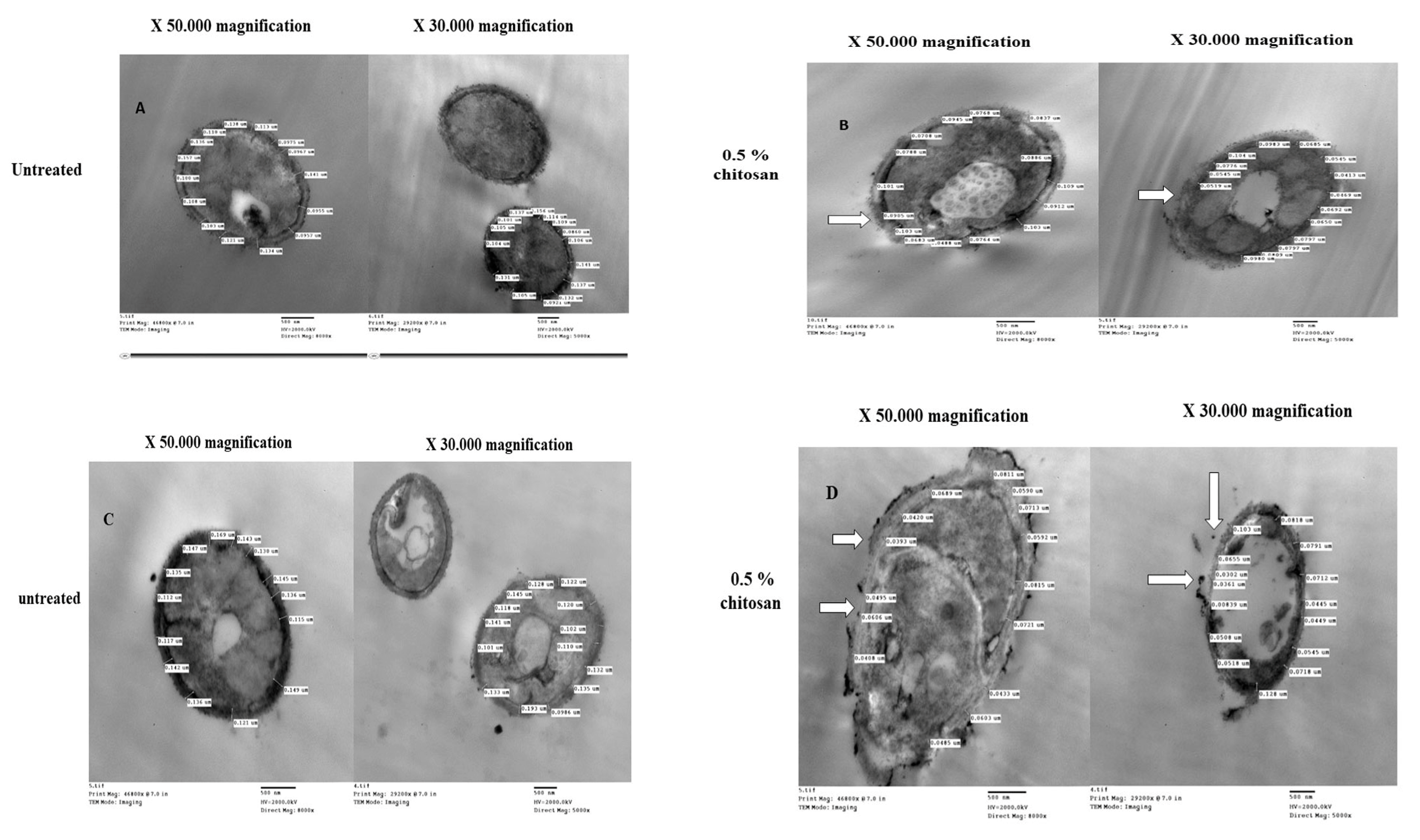
2.5. Chitosan-Induced Changes in Cell Wall Thickness of Caspofungin-Resistant Candida Species

TEM examination of control (untreated) C. albicans cells showed typical morphology of Candida with a uniform density, typically structured nucleus, and cytoplasm with several elements of an endomembrane system enveloped by a regular, intact cell wall (Figure 1A,B). After 24 h treatment with 0.5% chitosan, the cells exhibited notable alterations in the cell membrane and the cell wall, causing irregular morphologies that were occasionally visible, especially in sensitive isolates. Chitosan principally interacts with the yeast cell wall, causing severe swelling and asymmetric rough shapes, and subsequent cell wall lysis. More significantly, there was an alteration in the morphology of the cells; the integrity of the cell walls was unclear, and it was unclear where the cell wall and cell membrane separated. Comparison between cell wall thickness and cell membrane revealed that C. albicans cells exhibited an extremely thin cell wall for resistant (Figure 1A,B) and sensitive isolates (Figure 1C,D). Three cell wall thicknesses were measured in twenty sites around the circumference of each cell. The quantitative analysis revealed that the untreated isolate had a cell wall thickness of 0.145 μm, measured between the plasma membrane and the cell wall. In contrast, the cell wall thickness of the chitosan-treated isolate was significantly lower (p < 0.05), ranging from 0.05 to 0.08 (Figure 2).
Interestingly, chitosan-treated cells for both isolates appear transparent with evidence of a loss of cell morphology, potentially resulting from an efflux of intracellular material following penetration of the cell by chitosan.

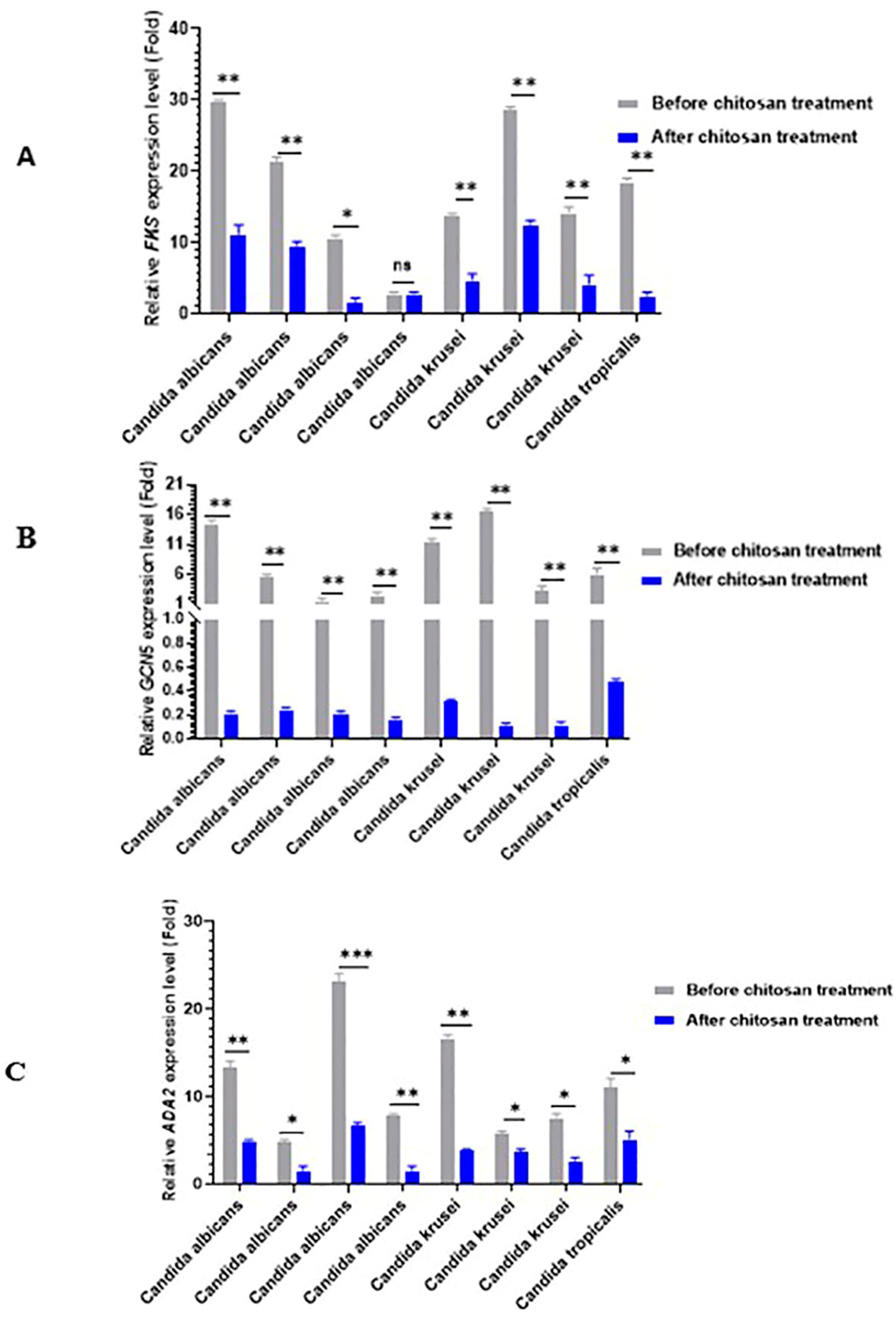
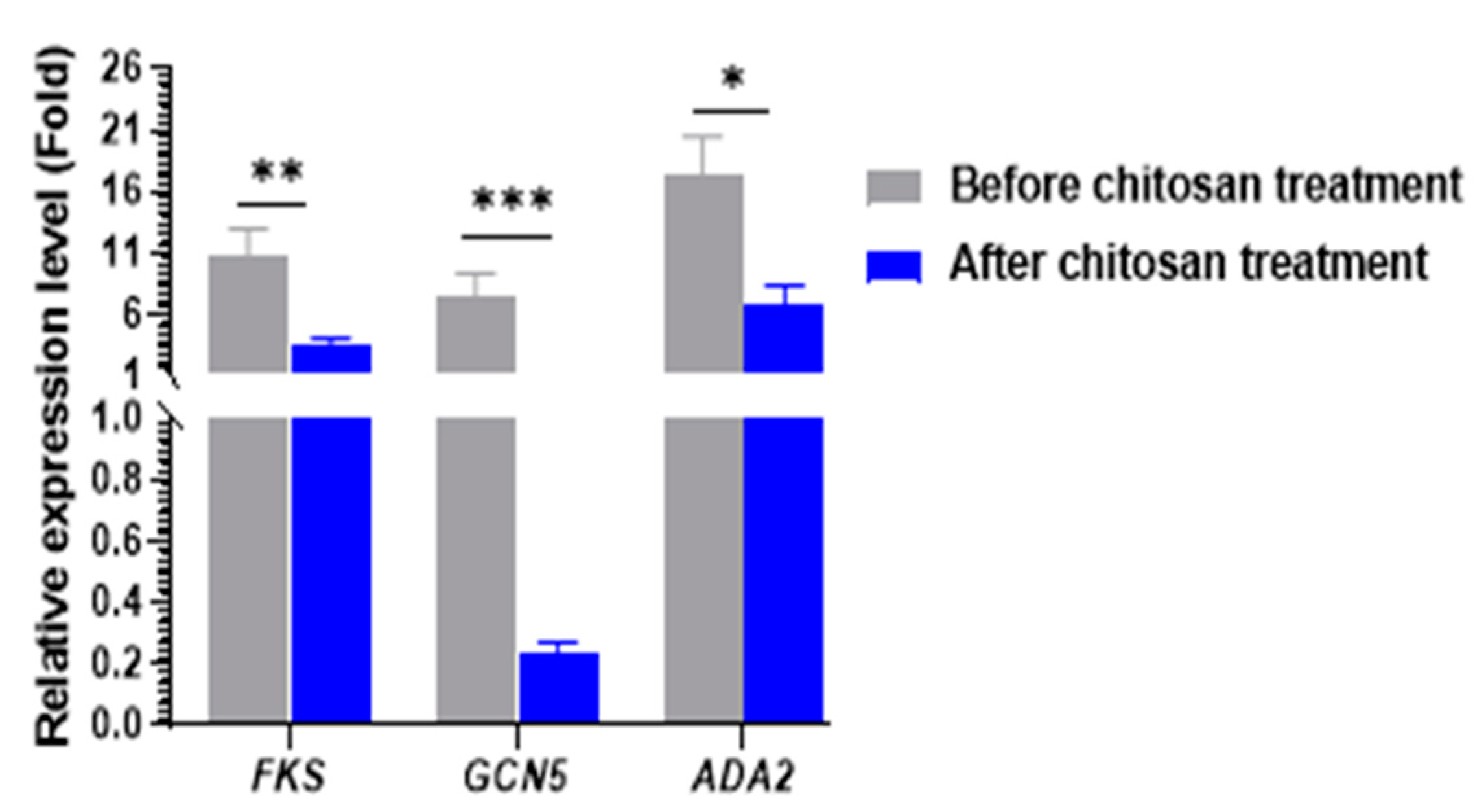
2.6. Real-Time RT-PCR for Gene Expression Analysis

To further confirm the inhibitory effects of chitosan on histone acetyltransferase genes (GCN5 and ADA2) and caspofungin resistance gene (FKS) in Candida species isolates, the transcript levels of GCN5, ADA2, and FKS genes were determined by reverse transcriptase quantitative PCR (RT-qPCR). Candida species isolates showed low transcript levels of the histone acetyltransferase genes, GCN5 (fold change range = 0.001–0.0473 and ADA2 (fold change range = 1.028–4.856), and FKS gene (fold change range = 2.713–12.38 after chitosan treatments when compared with the untreated isolates. Notable significant differences were observed in the expression levels of the two histone acetyltransferase genes (GCN5-ADA2 SAGA complex (p < 0.05) in caspofungin- resistant isolates (Figure 3A–C). As presented in Figure 4, comparison of the expression levels of cell wall-related genes between caspofungin-resistant and -sensitive isolates treated with and without chitosan revealed a significant decrease following chitosan treatment (p < 0.001). The expression level was normalized to the ACT1 gene.

3. Discussion

Invasive fungal infections pose a substantial infection risk for individuals with underlying immunosuppression. Serious fungal diseases are becoming increasingly prevalent, resulting in the deaths of over 1.5 million individuals annually. This issue is further complicated by the emergence of drug-resistant and/or multidrug-resistant species, particularly in Candida species [20,21]. The classification of echinocandins as fungicidal or fungistatic is dependent upon the species and strain of the fungus. Anidulafungin, micafungin, and caspofungin were the most successful echinocandins. These drugs are suitable for use as first-line treatments due to their potent activity, wide range, high safety, excellent pharmacokinetics and pharmacodynamics, and minimal drug-drug interactions, particularly in the context of nosocomial candidemia [22]. Recently, there has been a significant increase in the focus on the bioactivity of natural compounds and their practical applications in pharmaceutical research. Despite the fact that the majority of antibacterial compounds extracted from marine sources have not been sufficiently efficacious to compete with conventional antibiotics derived from microbes, the majority of marine creatures have not yet been investigated to identify valuable antibiotics [21,22,23]. The aim of this study was to investigate the role of chitosan as a biological factor for the mitigation of caspofungin resistance and enhancing its efficacy against resistant C. albicans, C. krusei, and C. tropicalis isolated from human and animal samples and its effect on the FKS gene and histone acetyltransferase genes.
Candida species frequently exhibit resistance to a wide variety of antifungals [4]. In this study, C. albicans isolates were resistant to ketoconazole (40%) followed by itraconazole and clotrimazole (28.5%, each), terbinafine (25.7%), amphotericin B and miconazole (22.8%, each), fluconazole (20%), and caspofungin (11.43%). However, it has been reported that C. albicans isolates exhibited high resistance rate to itraconazole (64.3%), clotrimazole (50%), fluconazole (46.4%), and ketoconazole (39.3%) [4]. Moreover, Badiee et al. [24] declared that C. albicans strains were resistant to itraconazole (33.7%), fluconazole (10.7%), ketoconazole (9.4%), amphotericin B (7%), and caspofungin (1.8%). Nevertheless, C. krusei strains were resistant to fluconazole (66.6%), amphotericin B (3.1%), ketoconazole (10.4%), itraconazole (86.5%), and caspofungin (4.2%). Here, different resistance rates were observed in C. krusei isolates and the highest resistance was found for fluconazole and ketoconazole (40%, each), then clotrimazole and terbinafine (34.2%, each), miconazole (31.4%), amphotericin B and itraconazole (28.5%), and caspofungin (11.43%). Resistance rates are dependent upon the species, antifungal agents, and the duration of fungistatic drug use, which may result in the pathogenic yeasts developing resistance, thereby diminishing the drug’s efficacy.
At present, the only clinically significant acquired resistance of Candida species to echinocandins has been linked to single amino acid substitutions that result from mutations within conserved (“hot spot”) sequences of the FKS1 and FKS2 genes in C. glabrata, which encode for glucan synthase. FKS mutations were the sole significant predictor of clinical echinocandin treatment failure in multivariate analyses instead of echinocandin MIC. For this reason, the most precise method of classifying echinocandin-resistant isolates may be the molecular detection of FKS mutations. Currently, there are no commercially available assays for FKS genotyping, which restricts molecular testing of resistance in reference laboratories [25]. Our findings correspond with the findings conducted by Coste et al. [26] that the point mutation S645P is predominant in C. albicans strains. All FKS-mutant isolates exhibited caspofungin MICs that were interpreted as non-susceptible according to the CLSI breakpoints and were similar to those reported in France from 2004 to 2010 that eight caspofungin-resistant C. albicans have different types of mutations in the HS1 region, while two C. krusei isolates had two different mutations in the HS1 region [27].
We investigated the antifungal properties of chitosan against all isolates of Candida species. There was a range of 10 to 23 mm in the IZD at 0.5% chitosan. At a concentration of 0.25%, the IZD varied from 7 to 15 mm. This was in accordance with the study conducted by Yadav et al., which determined that the IZD of 0.5% chitosan against C. albicans was 21 mm [28]. Nevertheless, 15 mm IZD has been achieved for Candida species using 1.5% low molecular weight (LMW) chitosan [29].
Chitosan converts caspofungin-resistant isolates to caspofungin-sensitive isolates (IZD of 10 to 25 mm). Prior to chitosan treatment, the caspofungin MIC and MFC values were 2–8 µg/mL and 4–16 µg/mL, respectively. Nevertheless, the caspofungin MIC and MFC values decreased following chitosan treatment (range: 0.0625 to 1 µg/mL) and (0.125 to 2 µg/mL), respectively. Our results were inconsistent with the findings of Abdelatti et al. [30] that the effects of chitosan-amphotericin B and chitosan-caspofungin as combinations against C. albicans and C. tropicalis isolates were indifferent. Nevertheless, the inhibitory effects of the combinations of chitosan-amphotericin B and chitosan-caspofungin were not observed, even though C. albicans SC5314 was highly susceptible to amphotericin B (MIC: 1.0 μg/mL), fluconazole (0.125 μg/mL), and caspofungin (0.25 μg/mL).
The antibacterial action of chitosan is closely linked to the degree of deacetylation and pH [31]. Particularly, the antimicrobial activity of chitosan is enhanced by increased deacetylation. Increased antimicrobial activity is associated with lower pH values [32]. Under acidic conditions, the amine groups of chitosan are protonated, resulting in the chitosan molecules becoming polycationic. The biological activity arises from the positive charge of protonated amine groups. These groups enable the interaction between protonated chitosan molecules and negatively charged proteins, fatty acids, lipids, and nucleic acids present in the fungal cell. Consequently, this process causes the breakdown of fungal cell membranes, confinement of cell components, and the resulting antifungal activity. Chitosan exhibits activity exclusively in an acidic environment [33]. Nevertheless, it has been noted that the restricted level of activity observed at pH 6 may be due to the low solubility [34]. The activity of chitosan against Staphylococcus aureus isolates was equal at pH 5.5 and 7.2 (MIC = 256 μg/mL). Therefore, when the negatively charged microbial cell surface interacts with the positively charged chitosan, an inhibitory effect may result from the disruption of the anion-cation balance [35]. Therefore, the antifungal properties are associated with the interaction of chitosan with the cell wall or cell membrane. However, the molecular weight (MW) and level of deacetylation of chitosan, the pH of the solvent, and the type of fungus being targeted are all strongly correlated with the MICs of chitosan against fungi [36].
Several studies [37,38] declared that the antifungal activity of chitosan can be influenced by its MW. Variations in MW can impact the properties of chitosan in two ways: First, living cells are susceptible to the penetration of LMW chitosan, which can block several enzymes and impair protein synthesis, hence interfering with mRNA synthesis. Second, there is an increased adsorption of high molecular weight (HMW) chitosan on cell walls, which causes cell walls to be covered, the membrane to weaken and break, and cells to leak. Furthermore, chitosan chelates essential minerals and metals to stop the growth of bacteria [37] and fungi [39]. Electrostatic interactions between the protonated NH3+ groups and the negative residues are responsible for the mode of action [40], presumably by competing with Ca2+ for electronegative regions on the surface membrane [41]. Furthermore, the chitosan susceptibility may be positively correlated with the unsaturated fatty acid content of the cell membrane. This is due to the fact that a higher content of unsaturated fatty acids results in improved membrane fluidity, which in turn leads to a more negative charge on the cell membrane [42]. According to the screening results, the chitosan employed in this investigation possessed prospective intracellular action in addition to extracellular interaction capabilities. Some fungal pathogens, including Cryptococcus neoformans, employ chitosan to preserve the integrity of their cell walls during the vegetative phase [43].
Indeed, TEM analysis verified that chitosan, albeit at a concentration of 0.5%, induced obvious aberrant changes in cell wall thickness in caspofungin-resistant and sensitive isolates. The quantitative results indicated that the untreated isolates had a cell wall thickness of 0.145 μm for the resistant isolate and 0.125 μm for the sensitive isolate, as measured between the plasma membrane and the cell wall. In contrast, the cell wall thickness of chitosan-treated isolates was significantly lower, ranging from 0.07 to 0.08 and 0.05 to 0.06 for the resistant isolate, respectively, when compared with the cell wall thickness of the sensitive isolate, which was 0.03 to 0.04 μm and 0.05 to 0.06 μm. This is in accordance with the results reported by Shih et al. that the cell wall thickness of the untreated strain was 0.125 μm, whereas the chitosan-treated strains had a substantially lower cell wall thickness, ranging from 0.04 to 0.05 μm and 0.06 to 0.07 μm, respectively [39]. This may be attributed to the antimicrobial activity of chitosan, which targets the cell surface, suggesting that chitosan is a potential alternative approach to antifungal agents against resistant fungi [44]. The cell wall of susceptible isolates is widened, in contrast to that of isolates with reduced susceptibility. This was linked to elevated levels of β-1,3-glucan in the cell wall, with no noticeable modifications in chitin content. The proteomic analysis revealed modifications in the repertoire of proteins involved in cell wall organization and maintenance in drug-resistant strains in comparison to susceptible strains following incubation with caspofungin [45].
In the current study, the RT-qPCR assay was employed to investigate the expressions of the GCN5, ADA2, and FKS genes both before and after chitosan treatment. Compared with the non-treated isolates, the relative expression of GCN5 and ADA2 genes was significantly reduced in chitosan-treated isolates. This could be attributed to the inhibition of SAGA complex expression by chitosan, which is one of the biocidal mechanisms of chitosan against C. albicans. This hypothesis was additionally confirmed by Shih et al.‘s findings that chitosan 0.2% inhibited ADA2 expression. Additionally, chitosan induces the repression of the GCN5 histone acetyltransferase [16]. Neurospora crassa genes, such as gel-1 (involved in glucan elongation) and β-1,3-glucan synthesis (FKS), were significantly reduced by chitosan. The downregulation was observed in the presence of caspofungin and at all concentrations of chitosan (10 and 30 μg /mL) [46]. The results suggest that the chitosan treatment of Candida cells either reduces the levels of chitin and β-glucan or modifies the ultrastructure of the cell wall and cell membrane by inhibiting the expression of SAGA complex components. As a result, tangible evidence to substantiate our hypothesis can be obtained by examining the composition and organization of the cell wall in both chitosan-treated and untreated cells. Additionally, it is probable that the outcome arises from a combination of indirect mechanisms, where multiple signaling pathways are necessary for the response to chitosan. This can result in a decrease in cell wall strength and changes in gene expression [47].
In budding yeast and Candida species, the ADA2-ADA3-GCN5 SAGA complex is sufficient for the induction or repression of the expression of specific genes, resulting in robust HAT activity [48]. In C. albicans, the ADA2 gene is crucial for histone acetylation; strains with ADA2 mutations had less H3K9 acetylation. The recruitment of ADA2, its ability to bind to 200 gene promoters, and its role in mediating the expression of several genes, including those involved in glycolysis, pyruvate metabolism, oxidative stress, drug responses, and cell wall responses, were further confirmed by chromatin immunoprecipitation assays [49]. Gcn5 may acetylate N-terminal lysines on histones and demonstrates HAT activity. Moreover, Ada3 (Ngg1) and ADA2 are required for the histone acetylation process on nucleosomes [50].
Consequently, the reversal of caspofungin resistance to sensitivity is most likely the result of altered cell wall composition and dysregulated glucan synthesis enzymes, such as FKS1, FKS2, and FKS3, as GCN5-mediated regulation affects signaling pathways that are required for the regulation of morphogenetic changes, including filamentation and virulence [51]. Conversely, previous research indicated that 3 mg/mL of chitosan, in both solution and nanoparticle form, are required to inhibit 90% of C. albicans, Aspergillus niger, and Fusarium solani. The natural antifungal activity of chitosan was, therefore, less potent than that of synthetic antifungal agents [52]. Thus, chitosan in combination with caspofungin may increase its antifungal activity. Moreover, chitosan could also inhibit the FKS gene, which leads to a rise in the efficacy of antifungals against resistant isolates.

4. Materials and Methods

4.1. Isolates

Thirty-five clinical isolates were included in this study. The isolates were recovered from different human and animal samples between 2022 and 2023. Twenty-five clinical cases were admitted at the clinics of different hospitals. From the clinical samples tested and considered in this study, fourteen cases were from human urine samples, three were from sputum, five were from onychomycosis cases, and three were from vaginitis cases in pregnant women. In addition, ten samples from diarrhea in calves were included. Sabouraud dextrose agar with chloramphenicol (SDA, Oxoid Ltd., Cambridge, UK) was used for culture. The isolates were identified based on their characteristics on HiCrome Candida differential agar medium (Himedia Laboratories, Mumbai, India), micromorphology on rice agar with Tween 80, and the germ tube test. Matrix-assisted laser desorption/ionization- time of flight mass spectrometry (MALDI-TOF MS)-based identification of all isolates to the species level was performed according to Bruker Daltonics (Biotyper RTC software, v 3.0 (Bruker Daltonics, Bremen, Germany) using the ethanol (EtOH)/formic acid (FA) extraction protocol [53].

4.2. Antifungal Susceptibility Testing

Disk diffusion testing was performed according to the Clinical and Laboratory Standards Institute (CLSI) protocol, as outlined in the CLSI M44-Ed3 [54]. The clinical Candida species isolates were tested for their susceptibility to various antifungal agents including caspofungin (CAS; 5 μg), amphotericin B (AmB; 10 μg), fluconazole (FLZ; 25 μg), voriconazole (VRZ; 1 μg), terbinafine (TERB; 30 mg), itraconazole (ITC; 10 μg), ketoconazole (KTZ; 15 μg), miconazole (MCZ; 10 mg), and clotrimazole (CLT; 10 μg). All disks were purchased from Sigma-Aldrich Quimica, Madrid, Spain. The suspension of each isolate was adjusted to McFarland No. 0.5 and plated on Mueller-Hinton agar (SSI Diagnostica, Hillerd, Denmark) enriched with 2% dextrose and 0.5 μg/mL methylene blue. The plates were incubated at 37 °C for 24 h. The inhibition zones diameters (IZD) were measured and analyzed according to the CLSI M44-ED3 guidelines.

4.3. Determination of Minimum Inhibitory Concentrations

The minimum inhibitory concentration (MIC) of CAS was determined using the broth microdilution method according to the CLSI M27M44S guidelines [55] standard. CAS (Cancidas®, Merck & Co. Inc., Rahway, NJ, USA) was dissolved in sterile water and prepared at a concentration range of 0.015 to 4 μg/mL. The isolates were cultured on SDA at 35 °C for 24 h, then the inoculum (1–5 × 10⁶ cells/mL) was prepared in 5 mL of sterile water using the 0.5 McFarland standard. Afterwards, a 1:50 dilution, followed by a final dilution of 1:20 in RPMI 1640 supplemented with 3-(N-morpholino) propanesulfonic acid (MOPS) (Sigma-Aldrich, St. Louis, MO, USA) were performed to reach a final concentration of 1–5 × 10³ cells/mL. The inoculum (100 µL) was transferred to 100 µL of medium in a 96-well microplate. The CAS was twofold serially diluted to a concentration range of 0.016 to 8 μg/mL. Growth control (wells containing RPMI-MOPS and inoculum) and sterility control (wells containing RPMI-MOPS), and quality control strains were included in the test. Plates were then incubated at 35 °C for 24 h. The MIC of CAS was reported as the lowest concentration that inhibits 50% of the growth compared with the growth controls. Twenty microliters from each well were plated on SDA and incubated at 35 °C for 24 h to confirm the colony counts. The MFC was defined as the lowest concentration that prevented visible growth according to CLSI document M27-A2 [56]. To ensure rigorous and reproducible data, each experiment was performed in triplicate.

4.4. Sequencing of FKs Gene for Detection of Mutations

Genomic DNA from fresh cultures of caspofungin-resistant isolates was extracted using a Qiagen DNA kit (QIAGEN, Hilden, Germany) following the manufacturer’s instructions. The oligonucleotide primers listed in Supplementary Table S1 were used for the amplification of the FKS gene and sequencing [57]. The NanoDrop™ 2000/2000c spectrophotometers (Thermo Fisher Scientific, Waltham, MA, USA) were employed to quantify the DNA concentrations. A 25 µL reaction mixture containing 12.5 µL of EmeraldAmp Max PCR Master Mix (Takara, Shigi-no-higashi, Joto-ku, Osaka, Japan), 1 µL of each primer (20 pmol; Biobasic, Toronto, ON, Canada), 4.5 µL of nuclease-free water, and 6 µL of DNA template was used to conduct PCR assays in a T3 thermal cycler (Thermo Scientific, Waltham, MA, USA). The cycling conditions were as follows: initial denaturation at 98 °C for 30 s, followed by 30 cycles of 98 °C for 10 s, 51 °C for 10 s, 72 °C for 30 s, and a final extension step at 72 °C for 5 min. PCR products were electrophoresed on a 1.5% agarose gel (Applichem GmbH, Armstadt, Germany), and an Alpha Innotech gel documentation system (Biometra GmbH, Göttingen, Germany) was used for imaging the gel.
The PCR products were purified by using PureLink PCR Purification kits (ThermoFisher Scientific, Waltham, MA, USA) according to the manufacturer’s instructions. A BigDye Terminator v3.1 cycle sequencing kit (Applied Biosystems, Tokyo, Japan) was used for DNA sequencing in the ABI 3100 Genetic Analyzer (Applied Biosystems, Foster City, CA, USA). Nucleotide sequences were compared with the available sequences in the NCBI database using the Basic Local Alignment Search Tool (www.ncbi.nlm.nih.gov/BLAST/, accessed on 13 April 2024). The amino acid sequences were deduced with the standard genetic code using the Expert Protein Analysis System Translate Tool (ExPASy; https://www.expasy.org/, accessed on 13 April 2024). For detection of mutation, nucleotide and amino acid sequences were aligned with the reference strains C. albicans SC 5341, C. krusei ATCC 6258, and C. tropicalis ATCC 750 using the BioEdit program 7.2 (BioEdit.exe) (Ibis Therapeutics, Carlsbad, CA, USA). DNA sequences were deposited into the GenBank database (accession numbers PP663620–PP663628).

4.5. Screening of Antifungal Activity of Chitosan against Candida species Isolates

The antifungal activity of chitosan was determined by the well diffusion test [58]. Chitosan with molecular weight (MW) 50,000–190,000 and 75–85% deacetylation (Sigma Aldrich, St. Louis, MO, USA) was dissolved in 1% acetic acid (Sigma-Aldrich Quimica, Madrid, Spain) to obtain different concentrations (0.5, 0.25, 0.125, and 0.0625). Yeast suspensions were prepared by suspending single colonies in sterile 0.85% saline. The turbidity of each suspension was adjusted to 1.5 × 10⁶ CFU/mL. The suspensions were evenly spread and inoculated on SDA plates and SDA agar containing 1% acetic acid (solvent control group). Six-millimeter diameter wells were made in the agar plate using a sterile cork borer and filled with 100 µL of each concentration of chitosan (0.5, 0.25, 0.125, and 0.0625). The plates were left for 2 h at 4 °C for diffusion and then incubated at 37 °C for 24 h. Growth of the isolates was observed, and the IZD was recorded. The experiment was repeated three times, and the mean values were calculated.

4.6. Antifungal Susceptibility Testing for Caspofungin after Chitosan Treatment

Following chitosan treatment, disk diffusion testing was performed using Mueller-Hinton agar (SSI Diagnostica, Hillerd, Denmark) supplemented with 2% dextrose and 0.5 μg/mL methylene blue according to the CLSI M44-Ed3 protocol [54]. The IZD were measured after 24 h of incubation, and the results were recorded. Candida species isolates were tested twice for CAS MIC (before and after chitosan treatment).
To confirm the inhibitory influence of chitosan on various Candida species isolates, the broth microdilution test was conducted for each isolate following chitosan treatment. The MIC was established by preparing four concentrations of chitosan (0.5%, 0.25%, 0.125%, and 0.0625%) in 1% acetic acid and adding them to RPMI-1640. The plates were then incubated for 24 h before being washed with phosphate buffer saline (PBS) twice and plated onto SDA. Afterward, fresh culture colonies were selected, and the CAS MIC was determined according to the M27M44S protocol of the CLSI guidelines [55].

4.7. Transmission Electron Microscopy Analysis

Both caspofungin resistant- and -sensitive isolates were cultured in RPMI 1640 medium as [untreated group] and RPMI 1640 medium supplemented with 0.5% chitosan as a [treated group] at 30 °C for 20 min. They were then rinsed three times with sterile water. The cleaned samples were first fixed with 2.5% glutaraldehyde (Sangon Biotech Co. Ltd., Shanghai, China) for 80 min at room temperature, followed by three 15 min washes with 0.1 M PBS. After fixation for 1 h at room temperature with 1% osmium tetroxide (Sigma-Aldrich Quimica, Madrid, Spain), the samples were washed three times for 15 min using a 0.1 M phosphate buffer. Afterward, the samples were dehydrated by immersing them in ethanol at progressively higher concentrations (30%, 50%, 70%, 90%, and 100%) for 15 min each. Additionally, they were subjected to additional dehydration twice in 100% acetone for 30 min each. The samples were embedded for 4 h in different ratios of acetone Spurr’s resin. This was followed by embedding in pure Spurr’s resin for 48 h at 70 °C [59]. Thin slices of 50–70 nm thickness were cut from the blocks of samples using an ultramicrotome. Images at magnifications of 10,000, 30,000, and 50,000 were acquired using JEOL, JEM-1400 (Tokyo, Japan) to assess the thickness of cell walls. Digital microscopy software e J 1.45s software1493 was used to quantify the measures of the cell wall thickness of Candida cells.

4.8. Quantitative Reverse Transcription Polymerase Chain Reaction

Two hundred microliters of Sabouraud broth containing Candida cells, which had been grown overnight, was transferred to 10 mL of fresh Sabouraud broth. The cells were then centrifuged at 3000 rpm for 10 min, after which they were washed with sterile water three times. Subsequently, the cells were treated with RPMI 1640 medium and RPMI 1640 medium supplemented with 0.5% chitosan at 30 °C for 20 min. Following this treatment, the cells were once again centrifuged at 3000 rpm for 10 min and washed with sterile water three times [60]. Total RNAs were then extracted using a RNeasy Mini Kit (Qiagen, Germany) following the manufacturer’s instructions. The whole procedure was performed under DNAse-free conditions to avoid DNA contamination with 5 μL of Turbo DNase buffer and 1 μL of Turbo DNase enzyme (Thermo Fisher Scientific, Waltham, MA, USA). The real-time PCR mixture (25 µL) consisted of 3 µL of template RNA, 0.25 µL of RevertAid Reverse Transcriptase (Thermo Fisher Scientific, Waltham, MA, USA), 12.5 µL of 2× QuantiTect SYBR Green PCR Master Mix (Invitrogen, Paisley, UK), 0.5 µL of forward and reverse primers (20 pmol) of each gene (ADA2, GCN5, and FKS1) (Table S1), and 8.25 microliters of nuclease-free water. Three separate sets of samples from each strain for each treatment were examined three times using a Stratagene MX3005P qPCR System. The cycling conditions were as follows: 95 °C for 20 s, followed by 40 cycles of 95 °C for 1 s and 60 °C for 20 s. The data were processed using Stratagene MX3005P Mxpro Mx3005p V4 10 software (Stratagene, Amsterdam, The Netherlands). The normalization of gene expression was performed using the ACT1 housekeeping gene, following the 2−ΔΔCT method [61]. The fold changes were determined as the mean normalized expression of treated strains relative to the mean normalized expression of the untreated one.

4.9. Data Analysis

Data were processed in Microsoft Excel (Microsoft Corporation, Redmond, WA, USA) provided by the SPSS software V18 (Chicago. IL, USA). A Shapiro-Wilk test was used to assess normality, as outlined by Razali and Wah [62]. The Student’s t-test was used for testing the difference between the pre- and post-chitosan treatments on the cell wall-related genes and cell wall thickness (SAS Institute Inc., Cary, NC, USA) [63], setting the level of significance at α = 0.05. The correlation matrix between different antifungals was fitted according to the Corr procedure. The differences between different treatments on IZD were examined according to one-way Anova. Results were expressed as means ± SE. Figures were fitted by the GraphPad Prism software 9.0 (GraphPad, Boston, MA, USA).

5. Conclusions

The antifungal potential of chitosan enhances the efficacy of caspofungin against various caspofungin-resistant Candida species isolates and prevents the development of further antifungal resistance. The results of this study contribute to the progress in repurposing caspofungin and to an informed development strategy to enhance its efficacy, ensure appropriate antifungal activity against Candida species, and mitigate resistance. Consequently, chitosan could be used in combination with caspofungin for the treatment of candidiasis. Future research may examine the efficacy of combinational therapy by combining chitosan with caspofungin in a clinical trial or with other combinations that could result in novel drug therapies against drug-resistant Candida species in medical settings.

Supplementary Materials

The following supporting information can be downloaded at: https://www.mdpi.com/article/10.3390/antibiotics13070578/s1, Figure S1: Correlation matrix between different antifungals showing high positive correlation coefficients between the resistance to CLT and both KTZ and FLZ. High coefficients were detected between CAS and both CLT and VRZ; Figure S2: Antifungal potential of different chitosan concentrations against Candida species using agar well diffusion test. A high significant difference (p < 0.0001) was found between inhibition zone diameters of Candida species when tested with different concentrations of chitosan, with 0.5% being the most effective one. Table S1: Primers used in the study [16,52,64,65]; Table S2: Antifungal activity of chitosan (inhibition zone diameter mm) against Candida species; Table S3: Caspofungin inhibition zone diameters (mm) after treatment of Candida species with different concentrations of chitosan.

Author Contributions

Conceptualization, M.N.H., Y.H.T. and A.T.; methodology, Y.H.T., M.N.H. and A.T.; validation, M.N.H., Y.H.T. and A.T.; formal analysis, Y.H.T. and A.T.; investigation, Y.H.T., M.N.H., A.T., A.A., I.P. and M.A.; data curation, Y.H.T., M.N.H., A.T., A.A., I.P. and M.A.; writing—original draft preparation, A.T. and Y.H.T.; writing—review and editing, Y.H.T. All authors have read and agreed to the published version of the manuscript.

Funding

This research received no external funding.

Institutional Review Board Statement

Not applicable.

Informed Consent Statement

Not applicable.

Data Availability Statement

The datasets generated or analyzed during the current study are available from the corresponding author upon reasonable request.

Conflicts of Interest

The authors declare no conflicts of interest.

References

  1. Vélez, N.; Argel, A.; Kissmann, A.K.; Alpízar-Pedraza, D.; Escandón, P.; Rosenau, F.; Ständker, L.; Firacative, C. Pore-forming peptide C14R exhibits potent antifungal activity against clinical isolates of Candida albicans and Candida auris. Front. Cell Infect. Microbiol. 2024, 14, 1389020. [Google Scholar] [CrossRef]
  2. Mboussou, F.; Ndumbi, P.; Ngom, R.; Kassamali, Z.; Ogundiran, O.; Van Beek, J.; Williams, G.; Okot, C.; Hamblion, E.; Impouma, B. Infectious disease outbreaks in the African region: Overview of events reported to the World Health Organization. Epidemiol. Infect. 2019, 147, 299–305. [Google Scholar] [CrossRef]
  3. Okiro, E.A.; Yahaya, A.A.; Stephen, M.; Bonkoungou, B.; Musa, E.O.; Minkoulou, E.M.; Okeibunor, J.; Impouma, B.; Djingarey, H.M. Spatial and temporal distribution of infectious disease epidemics, disasters and other potential public health emergencies in the World Health Organisation Africa region, 2016. Glob. Health 2020, 16, 1–12. [Google Scholar]
  4. Tartor, Y.H.; Elmowalid, G.A.; Hassan, M.N.; Shaker, A.; Ashour, D.F.; Saber, T. Promising Anti-Biofilm Agents and Phagocytes Enhancers for the Treatment of Candida albicans Biofilm-Associated Infections. Front. Cell Infect. Microbiol. 2022, 12, 72–80. [Google Scholar] [CrossRef]
  5. Arastehfar, A.; Gabaldón, T.; Garcia-Rubio, R.; Jenks, J.D.; Hoenigl, M.; Salzer, H.J.F.; Ilkit, M.; Lass-Flörl, C.; Perlin, D.S. Drug-Resistant Fungi: An Emerging Challenge Threatening Our Limited Antifungal Armamentarium. Antibiotics 2020, 9, 877. [Google Scholar] [CrossRef]
  6. Campoy, S.; Adrio, J.L. Antifungals. Biochem. Pharmacol. 2017, 133, 86–96. [Google Scholar] [CrossRef]
  7. Szymański, M.; Chmielewska, S.; Czyżewska, U.; Malinowska, M.; Tylicki, A. Echinocandins—Structure, mechanism of action and use in antifungal therapy. J. Enzyme Inhib. Med. Chem. 2022, 37, 876–894. [Google Scholar] [CrossRef]
  8. Perlin, D.S. Echinocandin Resistance in Candida. Clin. Infect. Dis. 2015, 61 (Suppl. S6), 612–617. [Google Scholar] [CrossRef]
  9. Perlin, D.S. Mechanisms of echinocandin antifungal drug resistance. Ann. N. Y. Acad. Sci. 2015, 1354, 1–11. [Google Scholar] [CrossRef]
  10. Frazer, C.; Staples, M.I.; Kim, Y.; Hirakawa, M.; Dowell, M.A.; Johnson, N.V.; Hernday, A.D.; Ryan, V.H.; Fawzi, N.L.; Finkelstein, I.J.; et al. Epigenetic cell fate in Candida albicans is controlled by transcription factor condensates acting at super-enhancer-like elements. Nat. Microbiol. 2020, 5, 1374–1378. [Google Scholar] [CrossRef]
  11. Mallick, E.M.; Bergeron, A.C.; Jones, S.K.; Newman, Z.R.; Brothers, K.M.; Creton, R.; Wheeler, R.T.; Bennett, R.J. Phenotypic Plasticity Regulates Candida albicans Interactions and Virulence in the Vertebrate Host. Front. Microbiol. 2016, 7, 780. [Google Scholar] [CrossRef]
  12. Conte, M.; Eletto, D.; Pannetta, M.; Petrone, A.M.; Monti, M.C.; Cassiano, C.; Giurato, G.; Rizzo, F.; Tessarz, P.; Petrella, A.; et al. Effects of Hst3p inhibition in Candida albicans: A genome-wide H3K56 acetylation analysis. Front. Cell Infect. Microbiol. 2022, 12, 1031814. [Google Scholar] [CrossRef]
  13. Yu, S.; Paderu, P.; Lee, A.; Eirekat, S.; Healey, K.; Chen, L.; Perlin, D.S.; Zhao, Y. Histone Acetylation Regulator Gcn5 Mediates Drug Resistance and Virulence of Candida glabrata. Microbiol. Spectr. 2022, 10, e0096322. [Google Scholar] [CrossRef]
  14. Eberharter, A.; Sterner, D.E.; Schieltz, D.; Hassan, A.; Yates, J.R., 3rd; Berger, S.L.; Workman, J.L. The ADA complex is a distinct histone acetyltransferase complex in Saccharomyces cerevisiae. Mol. Cell Biol. 1999, 19, 6621–6631. [Google Scholar] [CrossRef]
  15. Helmlinger, D.; Papai, G.; Devys, D.; Tora, L. What do the structures of GCN5-containing complexes teach us about their function? BBA J. 2021, 1864, 194614. [Google Scholar] [CrossRef]
  16. Shih, P.Y.; Liao, Y.T.; Tseng, Y.K.; Deng, F.S.; Lin, C.H. A Potential Antifungal Effect of Chitosan Against Candida albicans Is Mediated via the Inhibition of SAGA Complex Component Expression and the Subsequent Alteration of Cell Surface Integrity. Front. Microbiol. 2019, 10, 602. [Google Scholar] [CrossRef]
  17. Iracane, E.; Vega-Estévez, S.; Buscaino, A. On and Off: Epigenetic Regulation of C. albicans Morphological Switches. Pathogens 2021, 10, 1463. [Google Scholar] [CrossRef]
  18. Afroz, M.M.; Kashem, M.N.H.; Piash, K.; Islam, N. Saccharomyces Cerevisiae as an Untapped Source of Fungal Chitosan for Antimicrobial Action. Appl. Biochem. Biotechnol. 2021, 193, 3765–3786. [Google Scholar] [CrossRef]
  19. Zakrzewska, A.; Boorsma, A.; Brul, S.; Hellingwerf, K.J.; Klis, F.M. Transcriptional response of Saccharomyces cerevisiae to the plasma membrane-perturbing compound chitosan. Eukaryot. Cell 2005, 4, 703–715. [Google Scholar] [CrossRef]
  20. Shields, R.K.; Nguyen, M.H.; Press, E.G.; Kwa, A.L.; Cheng, S.; Du, C.; Clancy, C.J. The presence of an FKS mutation rather than MIC is an independent risk factor for failure of echinocandin therapy among patients with invasive candidiasis due to Candida glabrata. Antimicrob. Agents Chemother. 2012, 56, 4862–4869. [Google Scholar]
  21. Pogurschi, E.N.; Petcu, C.D.; Mizeranschi, A.E.; Zugravu, C.A.; Cirnatu, D.; Pet, I.; Ghimpețeanu, O.M. Knowledge, Attitudes and Practices Regarding Antibiotic Use and Antibiotic Resistance: A Latent Class Analysis of a Romanian Population. Int. J. Environ. Res. Public Health 2022, 19, 7263. [Google Scholar] [CrossRef] [PubMed]
  22. de Prado, R.S.; Alves, R.J.; Oliveira, C.M.A.d.; Kato, L.; Silva, R.A.d.; Quintino, G.O.; do Desterro Cunha, S.; de Almeida Soares, C.M.; Pereira, M. Inhibition of Paracoccidioides lutzii Pb 01 isocitrate lyase by the natural compound argentilactone and its semi-synthetic derivatives. PLoS ONE 2014, 9, 94832–94838. [Google Scholar] [CrossRef] [PubMed]
  23. Rajendran, N.K.; Ramakrishnan, J. In vitro evaluation of antimicrobial activity of crude extracts of medicinal plants against multi drug resistant pathogens. Biyol. Bilim. Araptyrma Derg. 2009, 2, 97–101. [Google Scholar]
  24. Badiee, P.; Alborzi, A. Susceptibility of clinical Candida species isolates to antifungal agents by E-test, Southern Iran: A five-year study. Iran. J. Microbiol. 2011, 3, 183–188. [Google Scholar] [PubMed]
  25. Ben-Ami, R. Treatment of Invasive Candidiasis: A Narrative Review. J. Fungi 2018, 4, 97. [Google Scholar] [CrossRef] [PubMed]
  26. Coste, A.T.; Kritikos, A.; Li, J.; Khanna, N.; Goldenberger, D.; Garzoni, C.; Zehnder, C.; Boggian, K.; Neofytos, D.; Riat, A.; et al. Emerging echinocandin-resistant Candida albicans and glabrata in Switzerland. Infection 2020, 48, 761–766. [Google Scholar] [CrossRef]
  27. Dannaoui, E.; Desnos-Ollivier, M.; Garcia-Hermoso, D.; Grenouillet, F.; Cassaing, S.; Baixench, M.T.; Bretagne, S.; Dromer, F.; Lortholary, O. Candida spp. with acquired echinocandin resistance, France, 2004. Emerg. Infect. Dis. 2012, 18, 86–90. [Google Scholar] [CrossRef] [PubMed]
  28. Yadav, P.; Chaudhary, S.; Saxena, R.K.; Talwar, S.; Yadav, S. Evaluation of Antimicrobial and Antifungal efficacy of Chitosan as endodontic irrigant against Enterococcus faecalis and Candida albicans Biofilm formed on tooth substrate. J. Clin. Exp. Dent. 2017, 9, e361–e367. [Google Scholar] [CrossRef] [PubMed]
  29. Ewais, A.H.; Raslan, M.; Hassanien, R.A.; Zedan, I. Nanoparticles and irradiated chitosan impact on the quality of sweet green bell pepper (Capsicum annuum L.) under cold storage conditions. Arab J. Nucl. Sci. Appl. 2022, 30, 1–12. [Google Scholar]
  30. Lo, W.H.; Deng, F.S.; Chang, C.J.; Lin, C.H. Synergistic Antifungal Activity of Chitosan with Fluconazole against Candida albicans, Candida tropicalis, and Fluconazole-Resistant Strains. Molecules 2020, 25, 5114. [Google Scholar] [CrossRef]
  31. Cheung, R.C.F.; Ng, T.B.; Wong, J.H.; Chan, W.Y. Chitosan: An update on potential biomedical and pharmaceutical applications. Mar. Drugs 2015, 13, 5156–5186. [Google Scholar] [CrossRef]
  32. Kong, M.; Chen, X.G.; Xing, K.; Park, H.J. Antimicrobial properties of chitosan and mode of action: A state of the art review. Int. J. Food Microbiol. 2010, 144, 51–63. [Google Scholar] [CrossRef]
  33. Poznanski, P.; Hameed, A.; Orczyk, W. Chitosan and chitosan nanoparticles: Parameters enhancing antifungal activity. Molecules 2023, 28, 2996. [Google Scholar] [CrossRef] [PubMed]
  34. Rabea, E.I.; Badawy, M.E.; Stevens, C.V.; Smagghe, G.; Steurbaut, W. Chitosan as antimicrobial agent: Applications and mode of action. J. Biol. Macromol. 2003, 4, 1457–1465. [Google Scholar] [CrossRef]
  35. Rathinam, S.; Solodova, S.; Kristjánsdóttir, I.; Hjálmarsdóttir, M.; Másson, M. The antibacterial structure-activity relationship for common chitosan derivatives. Int. J. Biol. Macromol. 2020, 165, 1686–1693. [Google Scholar] [CrossRef]
  36. Younes, I.; Sellimi, S.; Rinaudo, M.; Jellouli, K.; Nasri, M. Influence of acetylation degree and molecular weight of homogeneous chitosans on antibacterial and antifungal activities. Int. J. Food Microbiol. 2014, 185, 57–63. [Google Scholar] [CrossRef] [PubMed]
  37. Garcia, L.G.S.; Guedes, G.M.d.M.; da Silva, M.L.Q.; Castelo-Branco, D.S.C.M.; Sidrim, J.J.C.; Cordeiro, R.d.A.; Rocha, M.F.G.; Vieira, R.S.; Brilhante, R.S.N. Effect of the molecular weight of chitosan on its antifungal activity against Candida spp. in planktonic cells and biofilm. Carbohydr. Polym. 2018, 195, 662–669. [Google Scholar] [CrossRef] [PubMed]
  38. Garcia, L.G.S.; de Melo Guedes, G.M.; Fonseca, X.M.Q.C.; Pereira-Neto, W.A.; Castelo-Branco, D.S.C.M.; Sidrim, J.J.C.; de Aguiar Cordeiro, R.; Rocha, M.F.G.; Vieira, R.S.; Brilhante, R.S.N. Antifungal activity of different molecular weight chitosans against planktonic cells and biofilm of Sporothrix brasiliensis. Int. J. Biol. Macromol. 2020, 143, 341–348. [Google Scholar] [CrossRef]
  39. Yuan, G.; Lv, H.; Tang, W.; Zhang, X.; Sun, H. Effect of chitosan coating combined with pomegranate peel extract on the quality of Pacific white shrimp during iced storage. Food Control. 2016, 59, 818–823. [Google Scholar] [CrossRef]
  40. Tsai, G.-J.; Su, W.-H. Antibacterial Activity of Shrimp Chitosan against Escherichia coli. J. Food Prot. 1999, 62, 239–243. [Google Scholar] [CrossRef]
  41. Másson, M.; Holappa, J.; Hjálmarsdóttir, M.; Rúnarsson, Ö.V.; Nevalainen, T.; Järvinen, T. Antimicrobial activity of piperazine derivatives of chitosan. Carbohydr. Polym. 2008, 74, 566–571. [Google Scholar] [CrossRef]
  42. Ke, C.L.; Deng, F.S.; Chuang, C.Y.; Lin, C.H. Antimicrobial actions and applications of chitosan. Polymers 2021, 13, 904. [Google Scholar] [CrossRef] [PubMed]
  43. Baker, L.G.; Specht, C.A.; Donlin, M.J.; Lodge, J.K. Chitosan, the deacetylated form of chitin, is necessary for cell wall integrity in Cryptococcus neoformans. Eukaryot. Cell 2007, 6, 855–867. [Google Scholar] [CrossRef] [PubMed]
  44. Peña, A.; Sánchez, N.S.; Calahorra, M. Effects of chitosan on Candida albicans: Conditions for its antifungal activity. Biomed. Res. Int. 2013, 2013, 5–15. [Google Scholar] [CrossRef]
  45. De Cesare, G.B.; Hafez, A.; Stead, D.; Llorens, C.; Munro, C.A. Biomarkers of caspofungin resistance in Candida albicans isolates: A proteomic approach. Virulence 2022, 13, 1005–1018. [Google Scholar] [CrossRef] [PubMed]
  46. Aranda-Martinez, A.; Lopez-Moya, F.; Lopez-Llorca, L.V. Cell wall composition plays a key role on sensitivity of filamentous fungi to chitosan. J. Basic. Microbiol. 2016, 56, 1059–1070. [Google Scholar] [CrossRef] [PubMed]
  47. Verlee, A.; Mincke, S.; Stevens, C.V. Recent developments in antibacterial and antifungal chitosan and its derivatives. Carbohydr. Polym. 2017, 164, 268–283. [Google Scholar] [CrossRef]
  48. Näär, A.M.; Lemon, B.D.; Tjian, R. Transcriptional coactivator complexes. Annu. Rev. Biochem. 2001, 70, 475–501. [Google Scholar] [CrossRef]
  49. Sellam, A.; Askew, C.; Epp, E.; Lavoie, H.; Whiteway, M.; Nantel, A. Genome-wide mapping of the coactivator AdA2p yields insight into the functional roles of SAGA/ADA complex in Candida albicans. J. Mol. Cell Biol. 2009, 20, 2389–2400. [Google Scholar] [CrossRef]
  50. Marcus, G.A.; Silverman, N.; Berger, S.L.; Horiuchi, J.; Guarente, L. Functional similarity and physical association between GCN5 and ADA2: Putative transcriptional adaptors. EMBO J. 1994, 13, 4807–4815. [Google Scholar]
  51. Yien, L.; Zin, N.M.; Sarwar, A.; Katas, H. Antifungal activity of chitosan nanoparticles and correlation with their physical properties. Int. J. Biomater. 2012, 632698. [Google Scholar] [CrossRef]
  52. Shivarathri, R.; Tscherner, M.; Zwolanek, F.; Singh, N.K.; Chauhan, N.; Kuchler, K. The fungal histone acetyl transferase Gcn5 controls virulence of the human pathogen Candida albicans through multiple pathways. Sci. Rep. 2019, 9, 9445–9450. [Google Scholar] [CrossRef] [PubMed]
  53. Bader, O.; Weig, M.; Taverne-Ghadwal, L.; Lugert, R.; Gross, U.; Kuhns, M. Improved clinical laboratory identification of human pathogenic yeasts by matrix-assisted laser desorption ionization time-of-flight mass spectrometry. Clin. Microbiol. Infect. 2011, 17, 1359–1365. [Google Scholar] [CrossRef] [PubMed]
  54. CLSI. Method for Antifungal Disk Diffusion Susceptibility Testing of Yeasts, (M44-ED3), 3rd ed.; Clinical and Laboratory Standards Institute: Wayne, PA, USA, 2018. [Google Scholar]
  55. CLSI. Performance Standards for Antifungal Susceptibility Testing of Yeasts, 3rd ed.; Clinical and Laboratory Standards Institute: Wayne, PA, USA, 2022. [Google Scholar]
  56. NCCLS. Reference Method for Broth Dilution Antifungal Susceptibility Testing of Yeasts, 2nd ed.; NCCLS: Wayne, PA, USA, 2002. [Google Scholar]
  57. Thompson, G.R., III; Wiederhold, N.P.; Vallor, A.C.; Villareal, N.C.; Lewis, J.S.; Patterson, T.F. Development of caspofungin resistance following prolonged therapy for invasive candidiasis secondary to Candida glabrata infection. Antimicrob. Agents Chemother. 2008, 52, 3783–3785. [Google Scholar] [CrossRef] [PubMed]
  58. Kumar, D.; Bhattacharyya, S.; Gupta, P.; Banerjee, G.; Singh, M. Comparative Analysis of Disc Diffusion and E-test with Broth Micro-dilution for Susceptibility Testing of Clinical Candida Isolates Against Amphotericin B, Fluconazole, Voriconazole and Caspofungin. J. Clin. Diagn. Res. 2015, 9, Dc01-4. [Google Scholar] [CrossRef] [PubMed]
  59. Vazquez-Muñoz, R.; Avalos-Borja, M.; Castro-Longoria, E. Ultrastructural analysis of Candida albicans when exposed to silver nanoparticles. PLoS ONE 2014, 9, e108876. [Google Scholar]
  60. Chang, W.-H.; Liang, S.-H.; Deng, F.-S.; Lin, C.-H. The conserved dual phosphorylation sites of the Candida albicans Hog1 protein are crucial for white–opaque switching, mating, and pheromone-stimulated cell adhesion. Sabouraudia 2016, 54, 628–640. [Google Scholar] [CrossRef] [PubMed]
  61. Livak, K.J.; Schmittgen, T.D. Analysis of relative gene expression data using real-time quantitative PCR and the 2(-Delta Delta C(T)) Method. Methods 2001, 25, 402–408. [Google Scholar] [CrossRef] [PubMed]
  62. Kim, H.Y. Statistical notes for clinical researchers: Assessing normal distribution (1). Restor. Dent. Endod. 2012, 37, 245–248. [Google Scholar] [CrossRef]
  63. Cary, N. SAS/STAT statistics user’s guide. In Statistical Analytical System, 5th ed.; SAS Institute Inc.: Cary, NC, USA, 2012; pp. 1–10. [Google Scholar]
  64. Desnos-Ollivier, M.; Bretagne, S.; Raoux, D.; Hoinard, D.; Dromer, F.; Dannaoui, E. Mutations in the fks1 gene in Candida albicans, C. tropicalis, and C. krusei correlate with elevated caspofungin MICs uncovered in AM3 medium using the method of the European Committee on Antibiotic Susceptibility Testing. Antimicrob. Agents Chemother. 2008, 52, 3092–3098. [Google Scholar] [CrossRef]
  65. Khan, Z.; Ahmad, S.; Mokaddas, E.; Meis, J.F.; Joseph, L.; Abdullah, A.; Vayalil, S. Development of Echinocandin Resistance in Candida tropicalis following Short-Term Exposure to Caspofungin for Empiric Therapy. Antimicrob. Agents Chemother. 2018, 62, 201–211. [Google Scholar] [CrossRef] [PubMed]
Figure 1. TEM images for individual cells of caspofungin-resistant isolates (A,B) and caspofungin-sensitive isolates (C,D) that were obtained at ‘’50,000’’ ‘’X’’ and ‘’30,000’’ ‘’X’’ magnifications, randomly selected before chitosan treatment and their cell wall thickness was measured. Twenty sites around the circumference of each selected cell were measured. Individual cells were randomly selected after chitosan (0.5%) treatment and their cell wall thickness was measured. The integrity of the Candida cell wall and cell membrane was disrupted and exhibited slightly irregular cell morphologies after chitosan treatment. White arrows identify disruption of C. albicans cell walls after chitosan 0.5% treatment.
Antibiotics 13 00578 g001
Figure 2. (A) Cell wall thickness of caspofungin-resistant isolates was significantly reduced after 0.5% chitosan treatment compared with those before-treatment at magnification powers ‘’10,000’’, ‘’30,000’’and ‘’50,000’’. The values are the means ± standard errors (SE). *** p < 0.001; * p < 0.05. Statistical significance was determined using the Student’s t-test. (B) The cell wall thickness of caspofungin-sensitive isolates was significantly reduced after chitosan treatment compared with before treatment at magnification powers ‘’10,000’’ and ‘’50,000’’. However, non-significant differences were observed at magnification power ‘’30,000”. The values are the means ± SE. *** p < 0.001; ** p < 0.01; ns p > 0.05. Statistical significance was determined using the Student’s t-test.
Antibiotics 13 00578 g002
Figure 3. The expression levels of cell wall-related genes. (A) FKS, (B) GCN5, and (C) ADA2 genes in caspofungin-resistant isolates. All examined isolates showed a significant decrease following treatment with chitosan 0.5% compared with the control group. The mean values ± standard errors (SE) are provided. * p < 0.05; ** p < 0.001, *** p < 0.001, and ns non-significant. Statistical significance was determined using the Student’s t-test.
Antibiotics 13 00578 g003
Figure 4. The expression levels of cell wall-related genes in C. albicans, C. krusei, and C. tropicalis, specifically FKs, GCN5, and ADA2 genes showed a significant decrease following treatment with chitosan when compared with the untreated isolates. The mean values ± standard errors (SE) are provided. * p < 0.05; ** p < 0.001, *** p < 0.001. Statistical significance was determined using the Student’s t-test. The expression values were compared between caspofungin-resistant and -sensitive isolates treated with and without chitosan. The expression was normalized to the ACT1 gene.
Antibiotics 13 00578 g004
Table 1. Antifungal resistance pattern of Candida species isolates and caspofungin minimum inhibitory concentration values before and after chitosan treatment.
Isolate No.SpeciesSourceResistance PatternCaspofungin MIC (µg/mL)Caspofungin MIC after 0.5% Chitosan Treatment (µg/mL)
C1C. albicansCalf, DiarrheaFLZ, CLT, TERB, KTZ, ITC, CAS20.25
C2C. kruseiUrine, HumanFLZ, CLT, KTZ, ITC, TERB, MCZ VRZ, CAS40.5
C3C. kruseiOnychomycosis, HumanFLZ, CLT, ITC, AMB, MCZ, VRZ, TERB, CAS80.25
C4C. albicansUrine, HumanFLZ, CLT, AMB, TERB, KTZ, ITC, MCZ, VRZ, CAS21
C5C. kruseiUrine, HumanFLZ, AMB, ITC, TERB, VRZ, CAS80.125
C6C. albicansCalf, diarrheaFLZ, CLT, TERB, KTZ, ITC, VRZ, CAS20.0625
C7C. kruseiCalf, diarrheaFLZ, CLT, KTZ, CAS20.25
C8C. albicansUrine, HumanFLZ, CLT, AMB, KTZ, TERB, VRZ, MCZ, CASP40.25
C9C. tropicalisSputum, HumanCLT, AMB, TERB, KTZ, ITC, VRZ, MCZ, CAS80.125
C10C. albicansUrine, HumanFLZ, CLT, KTZ, ITC, VRZ, MCZ0.250.125
C11C. albicansOnychomycosis, HumanFLZ, CLT, TERB, KTZ, AMB0.1250.0625
C12C. kruseiUrine, HumanFLZ, CLT, VRZ, MCZ, AMB, TERB0.250.125
C13C. kruseiUrine, HumanFLZ, CLT, TERB, KTZ, ITC, VRZ, MCZ0.250.125
C14C. kruseiUrine, HumanFLZ, TERB, KTZ, MCZ0.1250.0625
C15C. kruseiOnychomycosis, HumanFLZ, CLT, KTZ, TERB, MCZ0.250.125
C16C. kruseiVaginal swab, HumanFLZ, CLT, KTZ0.1250.065
C17C. kruseiUrine, HumanFLZ, CLT, TERB, KTZ, ITC, MCZ0.250.125
C18C. albicansCalf, diarrheaCLT, AMB, KTZ, ITC, MCZ0.06250.03125
C19C. kruseiSputum, HumanAMB, KTZ, MCZ0.1250.0625
C20C. kruseiCalf, diarrheaAMB, ITC, MCZ0.06250.03125
C21C. kruseiCalf, diarrheaFLZ, AMB, KTZ0.1250.03125
C22C. kruseiHuman, UrineFLZ, CLT, TERB, KT, VRZ0.250.03125
C23C. albicansUrine, HumanCLT, AMB, TERB, KTZ, ITC0.250.0625
C24C. albicansVaginal swab, HumanFLZ, AMB, KTZ, ITC, VRZ0.1250.0625
C25C. kruseiUrine, HumanCLT, TERB, KTZ, ITC0.250.0625
C26C. kruseiOnychomycosis, HumanFLZ, CLT, KTZ, ITC0.250.125
C27C. albicansOnychomycosis, HumanKTZ, ITC, VRZ, MCZ0.1250.03125
C28C. kruseiUrine, HumanCLT, AmB, KTZ, ITC, MCZ0.06250.03125
C29C. kruseiCalf, DiarrheaAmB, TERB0.250.0625
C30C. albicansCalf, DiarrheaAmB, KTZ, MCZ0.1250.03125
C31C. albicansCalf, DiarrheaKTZ, TERB, MCZ0.250.0625
C32C. albicansCalf, DiarrheaTERB, AMP, KTZ0.250.0625
C33C. albicansVaginal swab, HumanAmB, TERB, KTZ, ITC0.06250.03125
C34C. kruseiSputum, HumanAmB, TERB, KTZ, ITC0.250.125
C35C. kruseiUrine, HumanFLZ, MCZ, AmB0.1250.0325
ITC: itraconazole, KTZ: ketoconazole, MCZ: miconazole, CLT: clotriconazole, FLZ: fluconazole, VRZ: voriconazole, TERB: terbinafine, CAS: caspofungin, and AmB: amphotericin B.
Table 2. FKS1 mutations in caspofungin-resistant Candida species.
Isolates No.SpeciesCaspofungin MIC (µg/mL)Caspofungin MIC after 0.5% Chitosan TreatmentMutation (s) in FKS1Amino Acid ChangeAccession No.
C1C. albicans20.25A1929T
T1933C
S645PPP663620
C2C. krusei40.5C1934TS645FPP663621
C3C. krusei80.25A1929T
A1988G
E663GPP663622
C4C. albicans21T1922CF641SPP663623
C5C. krusei80.125A1929T
T2062A, A2064G
L688MPP663624
C6C. albicans20.0625C1934AS645YPP663625
C7C. krusei20.25T1933CS645PPP663626
C8C. albicans40.25T1933CS645PPP663627
C9C. tropicalis80.125G1932TL644FPP663628
Disclaimer/Publisher’s Note: The statements, opinions and data contained in all publications are solely those of the individual author(s) and contributor(s) and not of MDPI and/or the editor(s). MDPI and/or the editor(s) disclaim responsibility for any injury to people or property resulting from any ideas, methods, instructions or products referred to in the content.

