Article

Unraveling the Microbial Symphony: Impact of Antibiotics and Probiotics on Infant Gut Ecology and Antibiotic Resistance in the First Six Months of Life

1
Department of Epidemiology and Biostatistics, School of Public Health, Xi’an Jiaotong University Health Science Center, Xi’an 710061, China
2
Experimental Teaching Center, School of Public Health, Xi’an Jiaotong University Health Science Center, Xi’an 710061, China
3
Department of Health, Northwest Women’s and Children’s Hospital, Xi’an 710003, China
4
Center for Chronic Disease Control and Prevention, Global Health Institution, Xi’an Jiaotong University, Xi’an 710061, China
5
Key Laboratory for Disease Prevention and Control and Health Promotion of Shaanxi Province, Xi’an 710061, China
*
Authors to whom correspondence should be addressed.
Antibiotics 2024, 13(7), 602; https://doi.org/10.3390/antibiotics13070602
Submission received: 16 April 2024 / Revised: 24 May 2024 / Accepted: 5 June 2024 / Published: 27 June 2024
(This article belongs to the Special Issue Antibiotics and Probiotics: What Is the Effect on the Gut?)

Abstract

:
We aimed to examine the effects of antibiotic and probiotic usage on the gut microbiota structure and the presence of antibiotic-resistance genes (ARGs) in infants during the first six months of life. Questionnaires and fecal samples were collected within three days of birth, two months, and six months to assess antibiotic and probiotic exposure. Gut microbiotas were sequenced via 16S rRNA, and ARGs were conducted by qPCR, including beta-lactam (mecA, blaTEM), tetracycline (tetM), fluoroquinolone (qnrS), aminoglycoside (aac(6′)-Ib), and macrolide (ermB). Infants were categorized by antibiotic and probiotic usage and stratified by delivery mode, microbial composition, and ARG abundances were compared, and potential correlations were explored. A total of 189 fecal samples were analyzed in this study. The gut microbiota diversity (Chao1 index) was significantly lower in the “only probiotics” (PRO) group compared to the “neither antibiotics nor probiotics” (CON) group at six months for the CS stratification (p = 0.029). Compositionally, the abundance of core genus Bifidobacterium_pseudocatenulatum was less abundant for the antibiotic during delivery (IAP) group than that in the CON group within the first three days (p = 0.009), while core genus Enterococcus_faecium was more abundant in the PRO than that in the CON group (p = 0.021) at two months. ARGs were highly detected, with Enterococcus hosting tetM and Escherichia associated with blaTEM within three days of birth, though no correlation was found between Bifidobacterium and ARGs. These findings emphasized the critical importance of carefully managing antibiotic and probiotic exposures in early life, with implications for promoting lifelong health through preserving a healthy infant gut ecosystem.

1. Introduction

Gut microbiota plays a crucial role in infant health and development. During the first six months of life, the gut microbiota undergoes highly dynamic changes, reaching a stable, adult-like state by 2–3 years of age [1,2]. Disturbances to early life gut microbiota have been associated with increased risks of chronic conditions, including obesity, metabolic syndrome, diabetes, and immune system disorders [3,4,5]. As such, promoting healthy gut microbiota development in infancy may be crucial for lifelong well-being. Researchers have increasingly focused on how antibiotics and probiotics impact the infant’s gut microbiota [6,7]. Understanding these interventions may provide valuable insights for mitigating disease risks in childhood and adulthood.
Antibiotics are commonly prescribed to infants to treat bacterial infections, beginning at the time of delivery. Specifically, intrapartum antibiotic prophylaxis (IAP) is a clinical measure used in more than 30% of deliveries to prevent group B Streptococcus (GBS) infection that may be administered before or during a cesarean section (CS) to prevent infection [8,9]. However, researchers have revealed adverse effects of IAP, including delayed microbial maturation and altered microbiota establishment [10,11]. Furthermore, children are highly prescribed antibiotics in China (43.5%) at a rate higher than those reported in many countries, such as France (26.1%) and Australia (23%) [12]. Antibiotic exposure during early life has been linked to disrupted microbial colonization, decreased microbial diversity and stability, increased risk of gut dysbiosis and chronic diseases, and elevated levels of antibiotic resistance genes (ARGs) [13,14,15].
Studies have shown that probiotics have potential significance in modulating and clinically counteracting antibiotics’ adverse effects on the gut microbiota [16,17]. However, probiotic co-prescription rates remain low, particularly in the Asia–Pacific region [18]. Controlled trials in infants have shown that probiotic interventions increase the relative abundance of Bifidobacterium infantis and Lactobacillus, restore microbial diversity, decrease the relative abundance of pathogenic commensal bacteria, and reduce antibiotic resistance in gut microbiota, indicating better restoration of the child [17,19,20]. Yet, the efficacy of probiotics in treating microbiota dysbiosis in infants varies in clinical practice. Specific probiotic strains have demonstrated no significant benefit over the placebo in eradicating antibiotic-resistant colonization, and probiotic genome analyses have identified potential risks of spreading antibiotic resistance [21,22].
Previous studies have often examined antibiotics and probiotics separately, using cross-sectional or retrospective designs with limited samples and durations. This highlights the need for research on their combined effects on gut microbiota and antibiotic resistance in infants under six months, particularly in rural areas with high rates of antibiotic misuse. This study aimed to investigate the distinct impacts of antibiotic and probiotic administration on the composition and structure of gut microbiota and the prevalence of six commonly used ARGs in infants within the initial six months of life based on a well-characterized birth cohort.

2. Results

2.1. Sociodemographic Information of Mothers and Infants

A total of 77 newborns were enrolled in our study; of these, 55 and 57 infants were followed up at two and six months of age, respectively, so that 189 fecal samples were ultimately analyzed. A total of 70.1% of the neonates were delivered vaginally. During delivery, 23.4% of the neonates’ mothers used IAP, but none used probiotics. The usage of antibiotics and probiotics by infants in the first three days, at two months, and at six months is shown in Table 1. We defined the group that used antibiotics during delivery as IAP, those infants who used only antibiotics as ABX, those who used only probiotics as PRO, those who used both antibiotics and probiotics as ABX + PRO, and those who used neither antibiotics nor probiotics as CON.

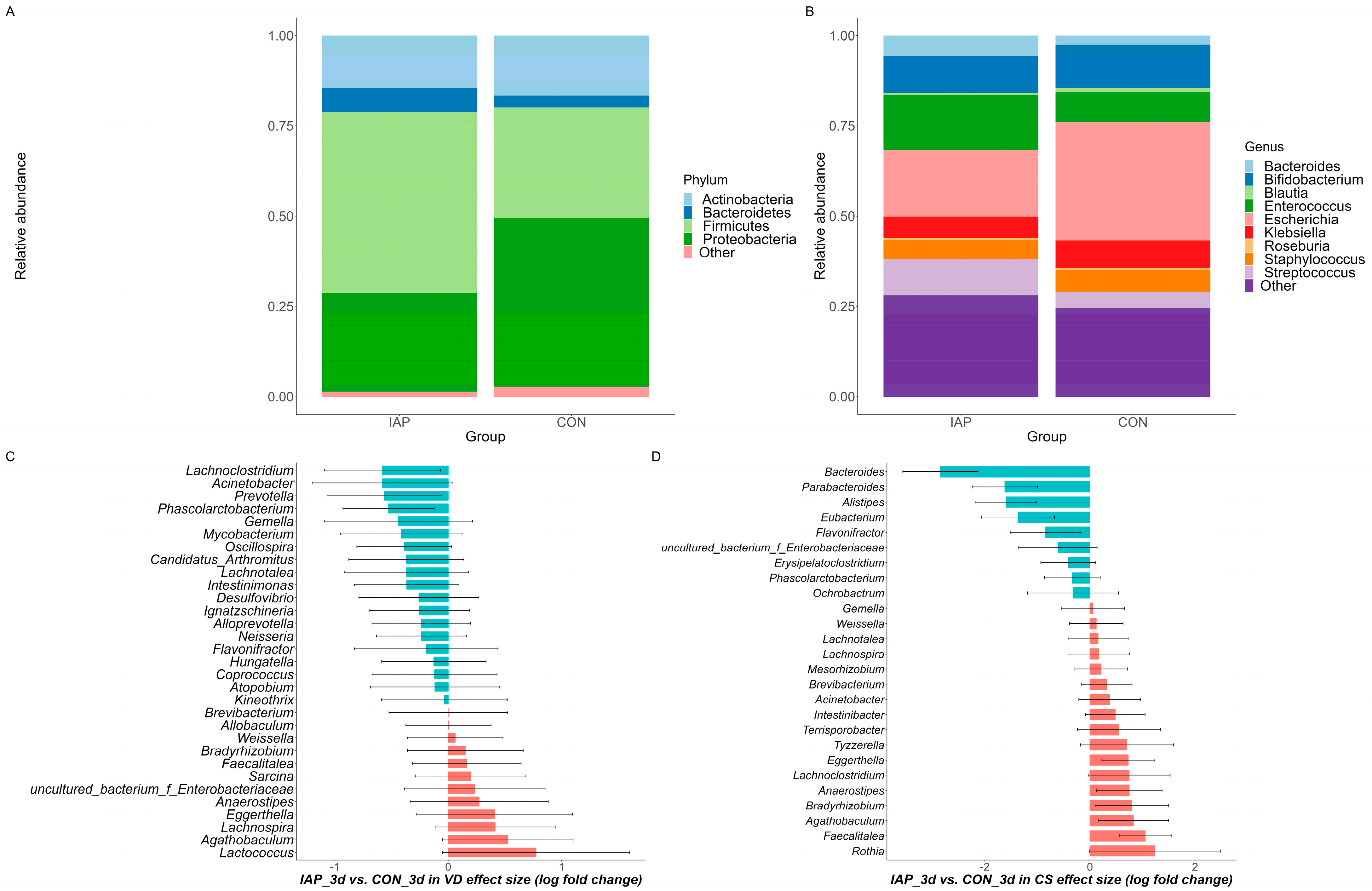
2.2. Overview of Microbial Communities in Infants

After quality control and denoising procedures were carried out, the sequencing process resulted in a total of 13,197,758 high-quality reads with eligible sequence lengths. The number of reads per sequenced sample ranged from 38,802 to 84,787, with an average of 69,829 (SD 10,700). These reads were subsequently classified into 821 amplicon sequence variants (ASVs), which were used for further analysis.
To gain a clear picture of communities of infant gut microbiota, we carried out analyses at the phylum and genus levels. We found that the phyla Actinobacteria, Bacteroidetes, Firmicutes, and Proteobacteria were present at levels exceeding 0.01% in at least 75% of the samples taken in the first three days of life. In both stratification groups, Firmicutes and Proteobacteria were the dominant phyla, comprising over 80% of all phyla present (Figure 1A). At the genus level, we identified and filtered out rare species whose relative abundance was less than 0.01% while retaining species that were present in at least 80% of samples, as shown in Figure 1B. ANCOM-BC results revealed that, in the vaginal-delivery stratification, genera such as Lachnoclostridium, Acinetobacter, and Prevotella exhibited upregulation in the IAP group, while Lachnospira, Agathobaculum, and Lactococcus showed upregulation in the CON group. In the cesarean section stratification, Bacteroides, Parabacteroides, and Alistipes were the top three genera upregulated in the IAP group, while Agathobaculum, Faecalitalea, and Rothia were the top three genera upregulated in the CON group (Figure 1C,D). The complete list is presented in Supplementary Table S1. Correspondingly, microbial community compositions at two months and six months of age are shown in Supplementary Figure S1.

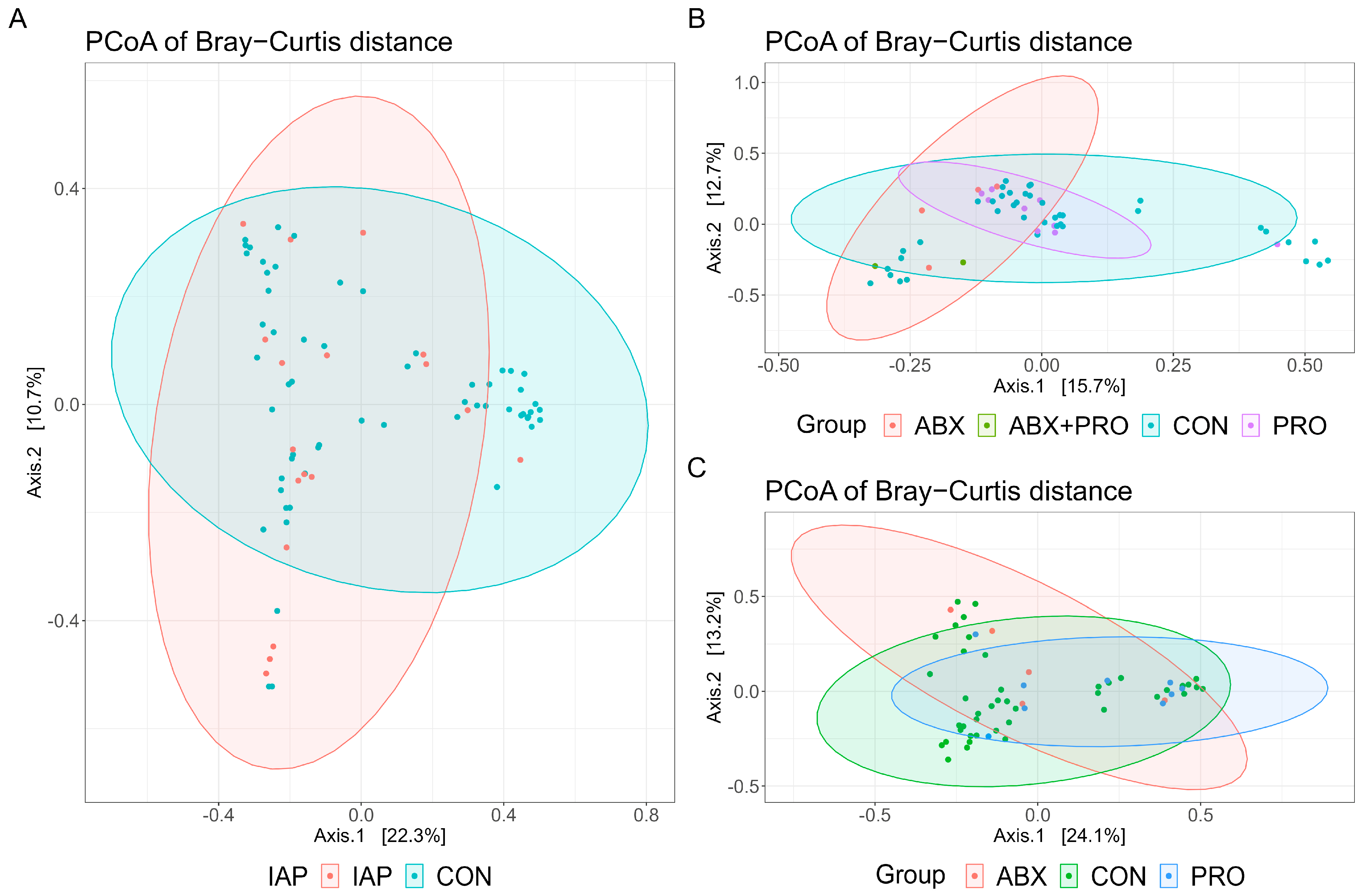
2.3. Impact of Antibiotics and Probiotics on Diversity of Gut Microbiota in Infants

We found no significant differences in the alpha diversities of gut microbiota in infants aged within three days or in those aged two months, with or without stratification (Table 2 and Table 3). For infants aged six months in the CS stratification, the Chao1 index showed a significant difference among groups (p = 0.049), and in the PRO group was significantly lower than that in the CON group (p = 0.029), see Table 3. However, there were no significant differences in the beta diversity of the microbial community structure among the groups of infants at any of the three time points (Figure 2, all p > 0.05). The results of the permutational multivariate analysis of variance (PERMANOVA) are shown in Table S2.

2.4. Core Genus Differences Resulting from Usage of Antibiotics and Probiotics

The analysis of 77 neonatal fecal samples identified 6 core gut microbiota genera, with Escherichia_coli as the dominant taxon, followed by Bifidobacterium_pseudocatenulatum and Bifidobacterium_longum (Table 4). Notably, the relative abundance of Bifidobacterium_pseudocatenulatum (ASV2) in the IAP group was significantly lower than that in the CON group (p = 0.009), which was the same as the CS stratification (p = 0.035). In samples taken from two-month-old infants, the ABX + PRO group was not shown due to the small sample size (n = 2). The abundance of Enterococcus_faecium (ASV6) was found to differ significantly among groups (p = 0.008), and the abundance in the PRO group was significantly higher than in the CON group (p = 0.021), which remained the same in the VD stratification (p = 0.019) (Table 5). In samples taken from infants at six months, the abundances of Bifidobacterium_pseudocatenulatum (ASV2), Blautia_obeum (ASV9), and Erysipelatoclostridium_ramosum (ASV34) were found to differ significantly among the groups (p = 0.004, p = 0.013, p = 0.026, respectively) (Table 6). After pairwise comparisons, it was found that the abundances of the three genera in the PRO group were significantly lower than that in the CON group (p = 0.002, p = 0.007, p = 0.011, respectively), which remained the same after stratification.

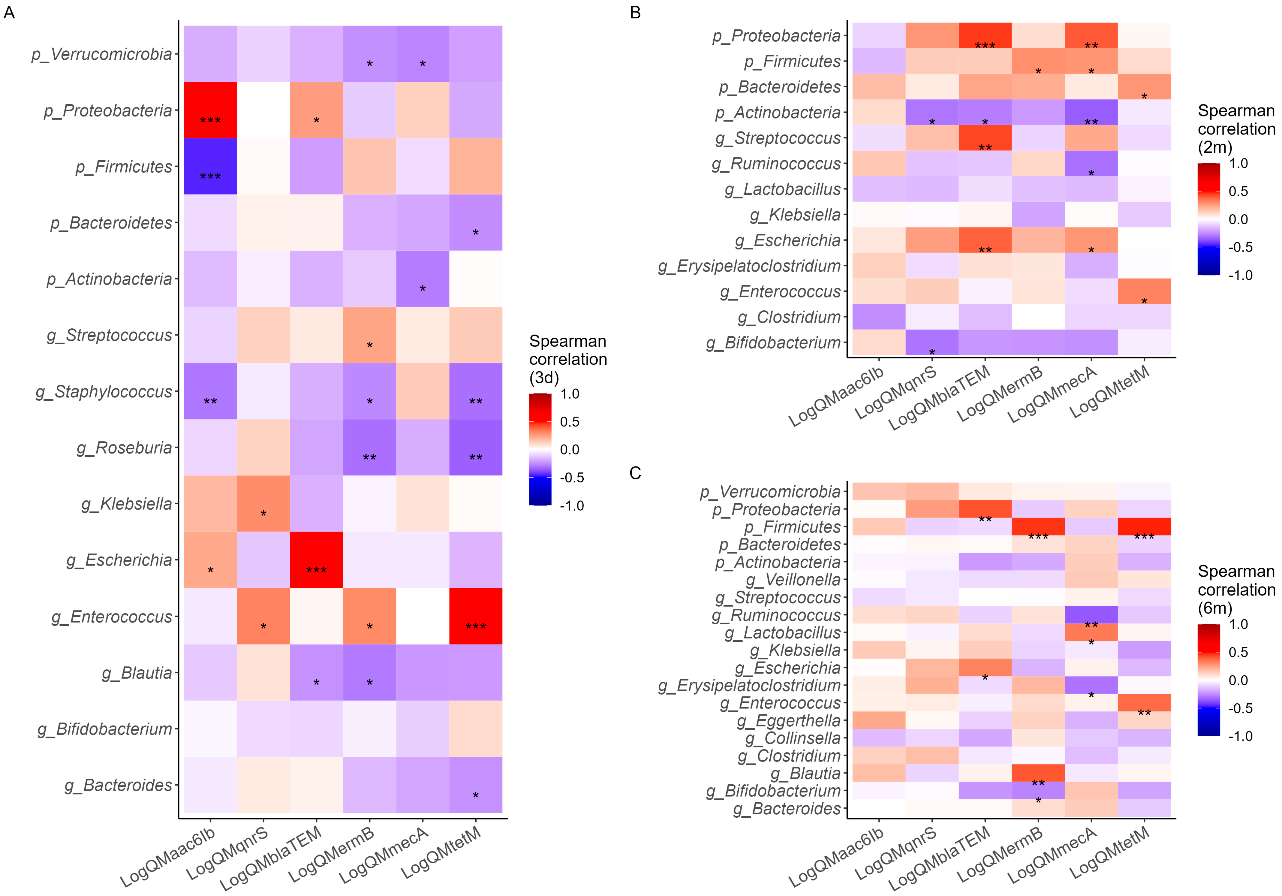
2.5. Effects of Antibiotic and Probiotic Use on Antibiotic Resistance Genes

As is shown in Table S3, the aac(6′)-Ib gene was detected in all samples, but the ermB and tetM genes were only detected at two months and six months, while the qnrS gene was only detected at six months. Overall, the detection rate of ARGs was high. In the unstratified analysis, no statistically significant differences in ARGs were observed among the groups. However, after stratification, in samples taken during the first three days of life, there was a higher trend in the relative abundance of the qnrS gene in the IAP subgroup (p = 0.052) and a lower trend in the relative abundance of the blaTEM gene in the IAP subgroup compared with the CON group (p = 0.051). At six months, there were significant differences in the absolute abundances of aac(6′)-Ib and tetM genes among the groups of infants born vaginally (p = 0.057, p = 0.059). Pairwise comparisons between groups revealed that the relative abundance of the aac(6′)-Ib gene was lower in the ABX group compared with the CON group (p = 0.008), while the relative abundance of the tetM gene was higher in the ABX group, compared with the CON group (p = 0.013).

2.6. Correlation of Antibiotic Resistance Gens with Gut Microbiota Communities

A correlation heatmap was used to identify the specific microbial hosts of the ARGs (Figure 3). In infants within three days of age, the abundances of aac(6′)-Ib were significantly correlated with the phyla Proteobacteria and Firmicutes, suggesting that these taxa may serve as potential hosts for the aac(6′)-Ib gene. Similarly, the genus Enterococcus was identified as a potential host for tetM, while the genus Escherichia was associated with the blaTEM gene. Notably, no significant correlation was observed between the genus Bifidobacterium and the ARGs examined. Correspondingly, at two months, the phylum Proteobacteria was associated with the blaTEM gene, while at six months, the phylum Firmicutes was associated with the ermB and tetM genes. R-values and p-values are shown in Tables S4–S6.

3. Discussion

In this study, we characterized the composition of gut microbiota and the presence of ARGs in infants at three time points in the first six months of life. Due to loss to follow-up at two and six months of age, lost respondents were distributed across antibiotics and probiotics use groups; they were not statistically different from follow-up respondents in terms of mother and infant characteristics and, thus, would have no impact on the results. By exploring correlations between gut microbial communities, ARGs, and exposure to antibiotics and probiotics, we sought to elucidate potential interactions among these factors. Our findings revealed that the use of antibiotics and probiotics can significantly impact the core genera, and while antibiotic exposure was associated with altered ARG abundances, probiotic intake did not exhibit such effects. Additionally, we were able to identify putative microbial hosts for specific ARGs. These results enhance our understanding of the complex interplay between clinical interventions, the developing gut microbiota, and the emergence of antimicrobial resistance in early life, providing insights to guide evidence-based strategies for optimizing infant gut health and mitigating resistance risks.
The postnatal colonization and assembly of the infant gut microbiota is a highly dynamic process, and antibiotic exposure during this critical window can disrupt gut homeostasis, leading to the depletion of keystone taxa, diminished taxonomic diversity, altered metabolic functions, and the potential proliferation of pathogenic organisms [23]. In the present study, the predominant phyla in infants were found to be Actinobacteria, Bacteroidetes, Firmicutes, and Proteobacteria, which mirrored compositions reported previously [24]. Interestingly, further stratification by delivery mode revealed an upregulation of the genus Lactococcus associated with the CON group under the VD subgroup. Notably, a previous study reported a Lactococcus strain isolated from the maternal vagina that exhibited probiotic properties [25], which implies that antibiotic exposure during delivery may disrupt the vertical transmission of beneficial taxa like Lactococcus from the maternal to infant gut microbiota. We did not observe notable disparities in alpha and beta diversity in the first three days of life or at the two-month time point. This may be attributable to the uniform colonization and development characteristics of this early period. Alternatively, the effects of antibiotics or probiotics may not yet be manifested in such young infants, as prior research has demonstrated limited impacts of early-life probiotic exposure on gut microbiota diversity [26,27]. The results of the present study align with these previous findings.
Core microbiome genera are widely distributed and abundant across samples. Consistent with prior research [28,29,30], we found that exposure to IAP reduced the abundance of the core genus Bifidobacterium in infants within three days of age. Conversely, in healthy full-term infants without IAP exposure, Bifidobacterium dominated the gut. Furthermore, IAP has been shown to decrease Bifidobacterium over time while increasing opportunistic pathogens like Clostridium difficile [10,31]. This Bifidobacterium depletion may promote gut dysbiosis, including an elevated pH and proliferation of spore-forming bacteria [32]. Importantly, we observed a higher Enterococcus_faecium abundance in the probiotic-supplemented group at two months. As Enterococcus is commonly used clinically as a probiotic [33], this likely reflects the exogenous supplementation. By four to six months, as solid foods are introduced, genera like Helicobacter and Clostridium are typically established [34,35]. Interestingly, probiotic intake may competitively inhibit or modulate the gut environment to reduce these genera. Further mechanistic research is warranted to fully elucidate probiotic impacts on the infant gut microbiota.
The present study found a high prevalence of antibiotic resistance genes (ARGs) in the infant gut microbiota, which is consistent with previous longitudinal research detecting ARGs for aminoglycosides, beta-lactams, macrolides, and tetracyclines across the first year of life [36]. These ARG variations may stem from resistant bacterial strains present harboring specific ARGs that can spread to other strains. Additionally, infant exposure to environmental antibiotic residues may enhance the competitive advantage of resistant microbes. Notably, we did not find evidence that probiotic supplementation reduced ARG abundance, aligning with prior reports [37]. This may be explained by the variable antibacterial properties and intrinsic resistance profiles of different probiotic strains [38,39,40]. Thus, when selecting probiotics, it is critical to not only identify the species but also characterize their resistance determinants.
Previous work has postulated that ARGs could potentially find host organisms within microbial communities based on significant correlations between ARG genes and similarities of abundances, which were observed across various samples (p < 0.01; r > 0.6) [41]. Consistent with this, we observed associations between ARGs and the gut microbiota in the present study, suggesting that the microbiota harbors antibiotic resistance. In recent years, correlation analysis has been widely conducted to infer ARG hosts in fecal samples [42]. To build on these observations, future research should integrate sequencing technologies with functional metagenomics or genome assembly approaches to further validate ARG–microbe linkages.
In summary, the present study provides preliminary evidence linking antibiotics, probiotics, gut microbiota, and ARGs. This finding may be particularly relevant in regions with high antibiotic usage, highlighting the need for the appropriate regulation of these interventions. The longitudinal design enabled the observation and analysis of the microbial composition and antibiotic resistance within the first six months of infant life. The comprehensive dataset, including high-throughput sequencing, usage records, and resistance gene detection, offered insights into the effects of antibiotics and probiotics on the gut microbiota. However, there were some limitations to our study. First, the infant numbers in the antibiotics and probiotics groups were relatively small, and the group that used both antibiotics and probiotics was not shown due to the limited number in the follow-up samples. It is necessary to increase the number of findings to provide insights for future studies with larger sample sizes to elucidate the effects of combining antibiotics and probiotics for intervention. Second, qPCR methods have limitations compared to metagenomic sequencing for characterizing the resistome and microbiota, but this targeted and cost-effective technique provides insights into our future utilization of metagenomic sequencing to determine antibiotic resistance profiles. Third, though infants within six months have a relatively simple environment, potential confounding factors in the study may not be adequately controlled and can affect the interpretation of results, so it is difficult to establish causality. Furthermore, this study considered the cross-sectional exposure of antibiotics and probiotics without residual effects alongside the consideration of earlier exposures, which need to be analyzed further in a larger sample. Lastly, according to the WHO classification, combining Access (penicillins and cefazolin) and Watch (cefotiam) antibiotics in the analysis led to different attributable risks due to neglecting the prevalence of usage. Specifying the AWaRe category and analyzing it separately is crucial to highlight their appropriate use and public health significance. Therefore, further research is needed to better elucidate the mechanisms by which antibiotics and probiotics shape the developing gut microbiota and antibiotic resistance, emphasizing their public health implications.

4. Materials and Methods

4.1. Study Design and Participant Enrollment

This study was based on a birth cohort study conducted in northwest China from January 2018 to June 2019. Comprehensive details regarding the cohort are described elsewhere [43]. First, we collected 77 neonatal fecal samples; then, during follow-up visits at two months and six months of age, we collected 55 and 57 samples, respectively, due to loss of contact or the parents going out of town for work. IAP is recommended within 30-60 min prior to skin incision, with no difference in reducing the incidence of infectious morbidity after CS for single and multiple-dose regimens, but there are concerns about transmitting antibiotics and promoting antibiotic resistance in neonates via the umbilical cord [44]. Some providers withhold antimicrobials until after cord clamping. We considered IAP exposure as receiving intravenous antimicrobials during labor [45] and no-IAP exposure as no antimicrobial exposure, emergency CS, or exposure only after delivery. Follow-up antibiotic or probiotic exposure was assessed within seven days prior to sample collection. In total, 189 fecal samples were analyzed for microbiome composition and ARGs.
The inclusion criteria were as follows: (1) women with a full-term singleton fetus; (2) infants with complete antibiotic and probiotic records of delivery and follow-up visits; (3) and women who did not have any diagnosed gestational complications. The exclusion criteria covered women who could not complete the required investigation and follow-up visits.
During recruitment, the women were provided with a comprehensive explanation of the study and oral and written consent was obtained from all participants. The study received approval from the Institutional Review Board (IRB) of Xi’an Jiaotong University Health Science Center (No. 2018-293).

4.2. Collection of Sociodemographic Information

Birth outcome data, including maternal antibiotic and probiotic use, were obtained from hospital records. During follow-up visits, questionnaires collected information on sociodemographics, feeding patterns (including nutritional supplements and addition of complementary foods), disease and treatment history (focusing on the use of antibiotics and probiotics), and maternal medication use during breastfeeding. Maternal exposure to antibiotics during the perinatal period predominantly included cephalosporins and penicillin, with an initial dose of 3.2 million units of penicillin every 12 h or cefazolin/cefotiam every 6 h. Common reasons for infant antibiotic use included upper respiratory tract infections, pneumonia, and abdominal pain, with penicillin and cephalosporins as the predominant medications, and very few used macrolide antibiotics. According to the WHO AWaRe classification [46], penicillin and cefazolin are Access antibiotics, while cefotiam is a Watch antibiotic. However, due to sample size limitations, we conducted a combined analysis based on their similar mechanisms of action despite differences in their antibiotic accessibility. Moreover, probiotic products containing Bacillus, Bifidobacterium, Lactobacillus, and Enterococcus were commonly used.

4.3. Collection of Fecal Samples

After delivery, neonate fecal samples were obtained from diapers, either by parents or investigators, within the first three days of the infant’s life. Most of the samples were from either the first or second defecation. At follow-up time points, fecal samples were collected by parents and investigators and notified within 8 h so that samples could be collected. To ensure proper preservation, the fecal samples were carefully packaged, labeled, and promptly stored in a −20 °C refrigerator for short-term storage. Later, they were transferred to the laboratory and stored in a −80 °C freezer until further analysis.

4.4. DNA Extraction and High-Throughput 16s rRNA Gene Amplicon Sequencing

The gut microbiota composition was determined by means of the high-throughput sequencing of 16S rRNA gene amplicons. PCR amplification was carried out using barcoded bacterial primers (341F: 5′-CCTAYGGGRBGCASCAG-3′ and 806R: 5′-GGACTACNNGGGTATCTAAT-3′ [47]), targeting the V3-V4 variable region of the 16S rRNA gene. Then, a standard protocol was followed to generate sequencing libraries alongside paired-end reads using an Illumina HiSeq 2500 platform provided by Biomarker Technologies Co., Ltd. (Beijing, China). The sequencing platform generated FASTQ format files containing read information and sequence quality. Next, the raw data were processed; this involved filtering using Trimmomatic v0.33 [48] and the removal of primer sequences using cutadapt 1.9.1 [49] so that high-quality reads without primers were obtained. Denoising and the removal of chimeric sequences were then carried out using DATA2 [50] in QIIME 2020.6 [51], resulting in non-chimeric reads. Silva 16S rRNA was used to conduct taxonomic annotations of feature sequences [52].

4.5. Detection of Antibiotic-Resistant Genes Using qPCR

The study utilized cost-effective quantitative PCR (qPCR) analysis to quantify a panel of six clinically relevant antibiotic resistance genes (ARGs) across different antibiotic classes, reflecting both the environmental and antibiotic exposures seen in rural and urban infant populations [53,54]. The ARGs included beta-lactam (mecA, blaTEM), tetracycline (tetM), fluoroquinolone (qnrS), aminoglycoside (aac(6′)-Ib), and macrolide (ermB). Each sample was analyzed with three replicates for data accuracy.

4.6. Bioinformatic and Statistical Analysis

Statistical analysis and visualization were conducted using Stata 15.0 (Stata Corp., College Station, TX, USA) and R (version 4.1.0). Normally distributed continuous data were reported as the mean ± standard deviation, while non-normally distributed data were presented as the median and quartile (P25, P75). Count data were expressed as frequency (n) and percentage (%). Group comparisons were performed using the Chi-square test (χ2) or Fisher’s exact test for categorical variables. The significance level was defined as p < 0.05.
For microbiota analysis in R, the Phyloseq package [55] was employed to create an object comprising the ASV table, sample variables, and a taxonomy table. Then, we retained the sequencing depth of each sample without rarefaction [56]. Furthermore, to address potential issues, such as low biomass in neonate fecal samples and spurious taxa caused by sequencing errors, we applied the filter_taxa function to filter the ASV data. Only those ASVs present with a minimum count of 2 in at least 10% of fecal samples were retained.
Community composition and alpha diversity indices, including Chao1, Shannon, and Simpson diversity, were calculated using the filtered ASV table and the microbiome package [57]. Differences in the alpha diversity indices were assessed using the Mann–Whitney U test. To further address taxonomic differences in the gut microbiota, the abundant microbial communities were identified using the analysis of compositions of microbiomes with bias correction (ANCOM-BC) (v1.2.2) [58]. Adjustments were made for confounding factors, and correction values obtained from the models were adjusted using the Bonferroni method (q < 0.05). Taxa whose proportion of zeroes exceeded 90% were excluded from the analysis. Beta diversity analysis was conducted using the vegan package [59], and PERMANOVA with 9999 permutations was performed to investigate the impact of antibiotics and probiotics on the gut microbiota community. To achieve the visualization of beta diversity, we carried out principal coordinate analysis (PCoA) using the vegan and ggplot2 packages [59,60]. Furthermore, we identified the core genera that were consistently present in the majority of infant fecal samples; this was achieved using the core_members function of the microbiome package. To determine the core genera, a threshold of a 0.01% presence in 75% of infant fecal samples was applied, and the result was transformed compositionally and visualized using ggplot2.
The raw data of antibiotic resistance genes were preprocessed and subjected to logarithmic transformation. Due to the three duplications of each sample, an average value of the sample copies was calculated before the transformation so that a better conformity to the assumption of normal distribution could be obtained. Then, the processed sample data were subjected to statistical analysis, including t-tests, analysis of variance (ANOVA), and Spearman correlation analysis with microbial communities.

5. Conclusions

This study demonstrates that in the first six months of life, exposure to antibiotics and probiotics significantly impacts the composition and structure of the infant’s gut microbiota. Antibiotics decrease the abundance of key taxa and increase the prevalence of ARGs, while probiotic intake does not exhibit such effects. Importantly, certain dominant bacterial taxa, such as Enterococcus and Escherichia, were identified as potential reservoirs for ARGs in the infant gut. This study highlights the critical need to optimize antibiotic and probiotic use in early life to protect the infant’ gut microbiome and mitigate antimicrobial resistance, with the expectation that further research on the gut microbiome–resistance relationship will guide evidence-based strategies to promote lifelong health through preserving a healthy infant gut ecosystem.

Supplementary Materials

The following supporting information can be downloaded at https://www.mdpi.com/article/10.3390/antibiotics13070602/s1, Figure S1: Relative abundance of gut microbial communities at phylum level and genus level at two months (A,B) and at six months of age (C,D); Table S1: ANCOM-BC analysis of different bacterial genera in infant fecal samples with and without IAP use stratified by delivery mode; Table S2: PERMANOVA analysis of beta diversity in infant fecal samples with antibiotics/probiotics in the first three days, at two months, and at six months; Table S3: Absolute abundance of six antibiotic resistance genes in infant fecal samples in the first three days, at two months, and at six months (log copies/µL) (log copies/µL); Table S4: Spearman correlation result of ARGs and microbial communities in the first three days; Table S5: Spearman correlation result of ARGs and microbial communities at two months of age; Table S6: Spearman correlation result of ARGs and microbial communities at six months of age.

Author Contributions

Conceptualization, L.Z.; methodology, L.Z. and Q.Q.; software, Q.Q.; validation, L.Z. and Z.Z.; formal analysis, Q.Q.; investigation, Q.Q., S.L., L.W. and Y.Z.; resources, Q.Q. and S.L.; data curation, Q.Q., S.L., M.A.G. and B.W.; writing—original draft preparation, Q.Q.; writing—review and editing, L.W., Y.Z., M.A.G., S.L., B.W., Z.Z. and L.Z.; visualization, Q.Q.; supervision, L.Z.; project administration, L.Z.; funding acquisition, L.Z., Z.Z. and Q.Q. All authors have read and agreed to the published version of the manuscript.

Funding

This research was funded by the National Natural Science Foundation of China, grant number 81872633; the Shaanxi Provincial Innovation Capability Support Plan, grant number 2023-CX-PT-47; the General Projects of the Key Research and Development Program of Shaanxi Province, grant number 2024SF-YBXM-310; and the China Scholarship Council, grant number 202006280219.

Institutional Review Board Statement

The study was conducted in accordance with the Declaration of Helsinki and approved by the Institutional Review Board (IRB) of Xi’an Jiaotong University Health Science Centre (No. 2018-293, date 2018-3-8).

Informed Consent Statement

Informed consent was obtained from all pregnant women and their families included in the study.

Data Availability Statement

The data that support the findings of this study are available upon request from the corresponding author upon reasonable request.

Acknowledgments

The authors would like to thank local doctors, nurses, and medical staff for the recruitment, sample, and questionnaire collection, as well as all the women and their families in the study.

Conflicts of Interest

The authors declare no conflicts of interest. The funders had no role in the design of the study; in the collection, analyses, or interpretation of data; in the writing of the manuscript; or in the decision to publish the results.

References

  1. Kundu, P.; Blacher, E.; Elinav, E.; Pettersson, S. Our Gut Microbiome: The Evolving Inner Self. Cell 2017, 171, 1481–1493. [Google Scholar] [CrossRef] [PubMed]
  2. Lim, E.S.; Wang, D.; Holtz, L.R. The Bacterial Microbiome and Virome Milestones of Infant Development. Trends Microbiol. 2016, 24, 801–810. [Google Scholar] [CrossRef] [PubMed]
  3. Derrien, M.; Alvarez, A.-S.; Vos, W.M. de The Gut Microbiota in the First Decade of Life. Trends Microbiol. 2019, 27, 997–1010. [Google Scholar] [CrossRef] [PubMed]
  4. Selma-Royo, M.; Tarrazó, M.; García-Mantrana, I.; Gómez-Gallego, C.; Salminen, S.; Collado, M.C. Shaping Microbiota During the First 1000 Days of Life. Adv. Exp. Med. Biol. 2019, 1125, 3–24. [Google Scholar] [PubMed]
  5. Milani, C.; Duranti, S.; Bottacini, F.; Casey, E.; Turroni, F.; Mahony, J.; Belzer, C.; Delgado Palacio, S.; Arboleya Montes, S.; Mancabelli, L.; et al. The First Microbial Colonizers of the Human Gut: Composition, Activities, and Health Implications of the Infant Gut Microbiota. Microbiol. Mol. Biol. Rev. 2017, 81, e00036-17. [Google Scholar] [CrossRef] [PubMed]
  6. Lu, Y.; Wang, Y.; Wang, J.; Lowe, A.J.; Grzeskowiak, L.E.; Hu, Y.J. Early-Life Antibiotic Exposure and Childhood Asthma Trajectories: A National Population-Based Birth Cohort. Antibiotics 2023, 12, 314. [Google Scholar] [CrossRef] [PubMed]
  7. Korpela, K.; Salonen, A.; Vepsäläinen, O.; Suomalainen, M.; Kolmeder, C.; Varjosalo, M.; Miettinen, S.; Kukkonen, K.; Savilahti, E.; Kuitunen, M.; et al. Probiotic Supplementation Restores Normal Microbiota Composition and Function in Antibiotic-Treated and in Caesarean-Born Infants. Microbiome 2018, 6, 182. [Google Scholar] [CrossRef] [PubMed]
  8. Azad, M.; Konya, T.; Persaud, R.; Guttman, D.; Chari, R.; Field, C.; Sears, M.; Mandhane, P.; Turvey, S.; Subbarao, P.; et al. Impact of Maternal Intrapartum Antibiotics, Method of Birth and Breastfeeding on Gut Microbiota during the First Year of Life: A Prospective Cohort Study. BJOG Int. J. Obstet. Gynaecol. 2016, 123, 983–993. [Google Scholar] [CrossRef] [PubMed]
  9. Montoya-Williams, D.; Lemas, D.J.; Spiryda, L.; Patel, K.; Carney, O.O.; Neu, J.; Carson, T.L. The Neonatal Microbiome and Its Partial Role in Mediating the Association between Birth by Cesarean Section and Adverse Pediatric Outcomes. Neonatology 2018, 114, 103–111. [Google Scholar] [CrossRef] [PubMed]
  10. Stearns, J.C.; Simioni, J.; Gunn, E.; McDonald, H.; Holloway, A.C.; Thabane, L.; Mousseau, A.; Schertzer, J.D.; Ratcliffe, E.M.; Rossi, L.; et al. Intrapartum Antibiotics for GBS Prophylaxis Alter Colonization Patterns in the Early Infant Gut Microbiome of Low Risk Infants. Sci. Rep. 2017, 7, 16527. [Google Scholar] [CrossRef] [PubMed]
  11. Nogacka, A.; Salazar, N.; Suárez, M.; Milani, C.; Arboleya, S.; Solís, G.; Fernández, N.; Alaez, L.; Hernández-Barranco, A.M.; de Los Reyes-Gavilán, C.G.; et al. Impact of Intrapartum Antimicrobial Prophylaxis upon the Intestinal Microbiota and the Prevalence of Antibiotic Resistance Genes in Vaginally Delivered Full-Term Neonates. Microbiome 2017, 5, 93. [Google Scholar] [CrossRef] [PubMed]
  12. Wushouer, H.; Du, K.; Yu, J.; Zhang, W.; Hu, L.; Ko, W.; Fu, M.; Zheng, B.; Shi, L.; Guan, X. Outpatient Antibiotic Prescribing Patterns in Children among Primary Healthcare Institutions in China: A Nationwide Retrospective Study, 2017–2019. Antibiotics 2024, 13, 70. [Google Scholar] [CrossRef] [PubMed]
  13. Aversa, Z.; Atkinson, E.J.; Schafer, M.J.; Theiler, R.N.; Rocca, W.A.; Blaser, M.J.; LeBrasseur, N.K. Association of Infant Antibiotic Exposure With Childhood Health Outcomes. Mayo Clin. Proc. 2021, 96, 66–77. [Google Scholar] [CrossRef] [PubMed]
  14. Aires, J. First 1000 Days of Life: Consequences of Antibiotics on Gut Microbiota. Front. Microbiol. 2021, 12, 681427. [Google Scholar] [CrossRef] [PubMed]
  15. Cotten, C.M. Adverse Consequences of Neonatal Antibiotic Exposure. Curr. Opin. Pediatr. 2016, 28, 141–149. [Google Scholar] [CrossRef] [PubMed]
  16. Rueda-Robles, A.; Rodríguez-Lara, A.; Meyers, M.S.; Sáez-Lara, M.J.; Álvarez-Mercado, A.I. Effect of Probiotics on Host-Microbiota in Bacterial Infections. Pathogens 2022, 11, 986. [Google Scholar] [CrossRef] [PubMed]
  17. Esaiassen, E.; Hjerde, E.; Cavanagh, J.P.; Pedersen, T.; Andresen, J.H.; Rettedal, S.I.; Støen, R.; Nakstad, B.; Willassen, N.P.; Klingenberg, C. Effects of Probiotic Supplementation on the Gut Microbiota and Antibiotic Resistome Development in Preterm Infants. Front. Pediatr. 2018, 6, 347. [Google Scholar] [CrossRef] [PubMed]
  18. Ghoshal, U.C.; Gwee, K.-A.; Holtmann, G.; Li, Y.; Park, S.J.; Simadibrata, M.; Sugano, K.; Cohen, H.; Quigley, E.M.M. Physician Perceptions on the Use of Antibiotics and Probiotics in Adults: An International Survey in the Asia-Pacific Area. Front. Cell Infect. Microbiol. 2021, 11, 722700. [Google Scholar] [CrossRef] [PubMed]
  19. Barratt, M.J.; Nuzhat, S.; Ahsan, K.; Frese, S.A.; Arzamasov, A.A.; Sarker, S.A.; Islam, M.M.; Palit, P.; Islam, M.R.; Hibberd, M.C.; et al. Bifidobacterium Infantis Treatment Promotes Weight Gain in Bangladeshi Infants with Severe Acute Malnutrition. Sci. Transl. Med. 2022, 14, eabk1107. [Google Scholar] [CrossRef] [PubMed]
  20. Alcon-Giner, C.; Dalby, M.J.; Caim, S.; Ketskemety, J.; Shaw, A.; Sim, K.; Lawson, M.A.E.; Kiu, R.; Leclaire, C.; Chalklen, L.; et al. Microbiota Supplementation with Bifidobacterium and Lactobacillus Modifies the Preterm Infant Gut Microbiota and Metabolome: An Observational Study. Cell Rep. Med. 2020, 1, 100077. [Google Scholar] [CrossRef] [PubMed]
  21. Ljungquist, O.; Kampmann, C.; Resman, F.; Riesbeck, K.; Tham, J. Probiotics for Intestinal Decolonization of ESBL-Producing Enterobacteriaceae: A Randomized, Placebo-Controlled Clinical Trial. Clin. Microbiol. Infect. 2020, 26, 456–462. [Google Scholar] [CrossRef] [PubMed]
  22. Tóth, A.G.; Judge, M.F.; Nagy, S.Á.; Papp, M.; Solymosi, N. A Survey on Antimicrobial Resistance Genes of Frequently Used Probiotic Bacteria, 1901 to 2022. Eurosurveillance 2023, 28, 2200272. [Google Scholar] [CrossRef] [PubMed]
  23. Vangay, P.; Ward, T.; Gerber, J.S.; Knights, D. Antibiotics, Pediatric Dysbiosis, and Disease. Cell Host Microbe 2015, 17, 553–564. [Google Scholar] [CrossRef] [PubMed]
  24. Chang, H.-Y.; Chiang Chiau, J.-S.; Chang, J.-H.; Hsu, C.-H.; Lin, C.-Y.; Ko, M.H.-J.; Lee, H.-C. Characteristics of Gut Microbiota in Small for Gestational Age Infants with Very Low Birth Weight. Nutrients 2022, 14, 5158. [Google Scholar] [CrossRef] [PubMed]
  25. Gao, Y.; Lu, Y.; Teng, K.-L.; Chen, M.-L.; Zheng, H.-J.; Zhu, Y.-Q.; Zhong, J. Complete Genome Sequence of Lactococcus Lactis Subsp. Lactis CV56, a Probiotic Strain Isolated from the Vaginas of Healthy Women. J. Bacteriol. 2011, 193, 2886–2887. [Google Scholar] [CrossRef] [PubMed]
  26. Murphy, R.; Morgan, X.C.; Wang, X.Y.; Wickens, K.; Purdie, G.; Fitzharris, P.; Otal, A.; Lawley, B.; Stanley, T.; Barthow, C.; et al. Eczema-Protective Probiotic Alters Infant Gut Microbiome Functional Capacity but Not Composition: Sub-Sample Analysis from a RCT. Benef. Microbes 2019, 10, 5–17. [Google Scholar] [CrossRef] [PubMed]
  27. Zaidi, A.Z.; Moore, S.E.; Okala, S.G. Impact of Maternal Nutritional Supplementation during Pregnancy and Lactation on the Infant Gut or Breastmilk Microbiota: A Systematic Review. Nutrients 2021, 13, 1137. [Google Scholar] [CrossRef] [PubMed]
  28. Saturio, S.; Suárez, M.; Mancabelli, L.; Fernández, N.; Mantecón, L.; de los Reyes-Gavilán, C.G.; Ventura, M.; Gueimonde, M.; Arboleya, S.; Solís, G. Effect of Intrapartum Antibiotics Prophylaxis on the Bifidobacterial Establishment within the Neonatal Gut. Microorganisms 2021, 9, 1867. [Google Scholar] [CrossRef] [PubMed]
  29. Stinson, L.F.; Payne, M.S.; Keelan, J.A. A Critical Review of the Bacterial Baptism Hypothesis and the Impact of Cesarean Delivery on the Infant Microbiome. Front. Med. 2018, 5, 135. [Google Scholar] [CrossRef] [PubMed]
  30. Mazzola, G.; Murphy, K.; Ross, R.P.; Di Gioia, D.; Biavati, B.; Corvaglia, L.T.; Faldella, G.; Stanton, C. Early Gut Microbiota Perturbations Following Intrapartum Antibiotic Prophylaxis to Prevent Group B Streptococcal Disease. PLoS ONE 2016, 11, e0157527. [Google Scholar] [CrossRef] [PubMed]
  31. Dierikx, T.; Berkhout, D.; Eck, A.; Tims, S.; van Limbergen, J.; Visser, D.; de Boer, M.; de Boer, N.; Touw, D.; Benninga, M.; et al. Influence of Timing of Maternal Antibiotic Administration during Caesarean Section on Infant Microbial Colonisation: A Randomised Controlled Trial. Gut 2022, 71, 1803–1811. [Google Scholar] [CrossRef] [PubMed]
  32. Ren, J.; Li, H.; Zeng, G.; Pang, B.; Wang, Q.; Wei, J. Gut Microbiome-Mediated Mechanisms in Aging-Related Diseases: Are Probiotics Ready for Prime Time? Front. Pharmacol. 2023, 14, 1178596. [Google Scholar] [CrossRef] [PubMed]
  33. Fu, X.; Lyu, L.; Wang, Y.; Zhang, Y.; Guo, X.; Chen, Q.; Liu, C. Safety Assessment and Probiotic Characteristics of Enterococcus Lactis JDM1. Microb. Pathog. 2022, 163, 105380. [Google Scholar] [CrossRef] [PubMed]
  34. Arrieta, M.-C.; Stiemsma, L.T.; Amenyogbe, N.; Brown, E.M.; Finlay, B. The Intestinal Microbiome in Early Life: Health and Disease. Front. Immunol. 2014, 5, 427. [Google Scholar] [CrossRef] [PubMed]
  35. Nance, C.L.; Deniskin, R.; Diaz, V.C.; Paul, M.; Anvari, S.; Anagnostou, A. The Role of the Microbiome in Food Allergy: A Review. Children 2020, 7, 50. [Google Scholar] [CrossRef] [PubMed]
  36. Loo, E.X.L.; Zain, A.; Yap, G.C.; Purbojati, R.W.; Drautz-Moses, D.I.; Koh, Y.Q.; Chong, Y.S.; Tan, K.H.; Gluckman, P.D.; Yap, F.; et al. Longitudinal Assessment of Antibiotic Resistance Gene Profiles in Gut Microbiomes of Infants at Risk of Eczema. BMC Infect. Dis. 2020, 20, 312. [Google Scholar] [CrossRef] [PubMed]
  37. Ouwehand, A.C.; Forssten, S.; Hibberd, A.A.; Lyra, A.; Stahl, B. Probiotic Approach to Prevent Antibiotic Resistance. Ann. Med. 2016, 48, 246–255. [Google Scholar] [CrossRef] [PubMed]
  38. Pieniz, S.; Andreazza, R.; Anghinoni, T.; Camargo, F.; Brandelli, A. Probiotic Potential, Antimicrobial and Antioxidant Activities of Enterococcus Durans Strain LAB18s. Food Control 2014, 37, 251–256. [Google Scholar] [CrossRef]
  39. Neut, C.; Mahieux, S.; Dubreuil, L.J. Antibiotic Susceptibility of Probiotic Strains: Is It Reasonable to Combine Probiotics with Antibiotics? Médecine Mal. Infect. 2017, 47, 477–483. [Google Scholar] [CrossRef] [PubMed]
  40. Hollenbeck, B.L.; Rice, L.B. Intrinsic and Acquired Resistance Mechanisms in Enterococcus. Virulence 2012, 3, 421–433. [Google Scholar] [CrossRef] [PubMed]
  41. Li, B.; Yang, Y.; Ma, L.; Ju, F.; Guo, F.; Tiedje, J.M.; Zhang, T. Metagenomic and Network Analysis Reveal Wide Distribution and Co-Occurrence of Environmental Antibiotic Resistance Genes. ISME J. 2015, 9, 2490–2502. [Google Scholar] [CrossRef]
  42. Wu, G.; Zhang, C.; Wang, J.; Zhang, F.; Wang, R.; Shen, J.; Wang, L.; Pang, X.; Zhang, X.; Zhao, L.; et al. Diminution of the Gut Resistome after a Gut Microbiota-Targeted Dietary Intervention in Obese Children. Sci. Rep. 2016, 6, 24030. [Google Scholar] [CrossRef] [PubMed]
  43. Qi, Q.; Wang, L.; Gebremedhin, M.A.; Li, S.; Wang, X.; Shen, J.; Zhu, Y.; Andegiorgish, A.K.; Cheng, Y.; Shi, L.; et al. The Impact of Early-Life Antibiotics and Probiotics on Gut Microbial Ecology and Infant Health Outcomes: A Pregnancy and Birth Cohort in Northwest China (PBCC) Study Protocol. BMC Pediatr. 2022, 22, 738. [Google Scholar] [CrossRef] [PubMed]
  44. Pinto-Lopes, R.; Sousa-Pinto, B.; Azevedo, L. Single Dose versus Multiple Dose of Antibiotic Prophylaxis in Caesarean Section: A Systematic Review and Meta-Analysis. BJOG Int. J. Obstet. Gynaecol. 2017, 124, 595–605. [Google Scholar] [CrossRef] [PubMed]
  45. Russell, N.J.; Seale, A.C.; O’Sullivan, C.; Le Doare, K.; Heath, P.T.; Lawn, J.E.; Bartlett, L.; Cutland, C.; Gravett, M.; Ip, M.; et al. Risk of Early-Onset Neonatal Group B Streptococcal Disease with Maternal Colonization Worldwide: Systematic Review and Meta-Analyses. Clin. Infect. Dis. 2017, 65, S152–S159. [Google Scholar] [CrossRef] [PubMed]
  46. World Health Organization. WHO Antibiotic Categorization. Available online: https://aware.essentialmeds.org/list (accessed on 4 June 2024).
  47. Liu, H.; Hou, C.; Li, N.; Zhang, X.; Zhang, G.; Yang, F.; Zeng, X.; Liu, Z.; Qiao, S. Microbial and Metabolic Alterations in Gut Microbiota of Sows during Pregnancy and Lactation. FASEB J. 2019, 33, 4490–4501. [Google Scholar] [CrossRef] [PubMed]
  48. Bolger, A.M.; Lohse, M.; Usadel, B. Trimmomatic: A Flexible Trimmer for Illumina Sequence Data. Bioinformatics 2014, 30, 2114–2120. [Google Scholar] [CrossRef] [PubMed]
  49. Martin, M. Cutadapt Removes Adapter Sequences from High-Throughput Sequencing Reads. EMBnet J. 2011, 17, 10–12. [Google Scholar] [CrossRef]
  50. Callahan, B.J.; McMurdie, P.J.; Rosen, M.J.; Han, A.W.; Johnson, A.J.A.; Holmes, S.P. DADA2: High-Resolution Sample Inference from Illumina Amplicon Data. Nat. Methods 2016, 13, 581–583. [Google Scholar] [CrossRef] [PubMed]
  51. Bolyen, E.; Rideout, J.R.; Dillon, M.R.; Bokulich, N.A.; Abnet, C.C.; Al-Ghalith, G.A.; Alexander, H.; Alm, E.J.; Arumugam, M.; Asnicar, F.; et al. Reproducible, Interactive, Scalable and Extensible Microbiome Data Science Using QIIME 2. Nat. Biotechnol. 2019, 37, 852–857. [Google Scholar] [CrossRef]
  52. Quast, C.; Pruesse, E.; Yilmaz, P.; Gerken, J.; Schweer, T.; Yarza, P.; Peplies, J.; Glöckner, F.O. The SILVA Ribosomal RNA Gene Database Project: Improved Data Processing and Web-Based Tools. Nucleic Acids Res. 2013, 41, D590–D596. [Google Scholar] [CrossRef] [PubMed]
  53. Gosalbes, M.J.; Vallès, Y.; Jiménez-Hernández, N.; Balle, C.; Riva, P.; Miravet-Verde, S.; de Vries, L.E.; Llop, S.; Agersø, Y.; Sørensen, S.J.; et al. High Frequencies of Antibiotic Resistance Genes in Infants’ Meconium and Early Fecal Samples. J. Dev. Orig. Health Dis. 2016, 7, 35–44. [Google Scholar] [CrossRef] [PubMed]
  54. Liu, H.; Wang, H.H. Impact of Microbiota Transplant on Resistome of Gut Microbiota in Gnotobiotic Piglets and Human Subjects. Front. Microbiol. 2020, 11, 932. [Google Scholar] [CrossRef] [PubMed]
  55. McMurdie, P.J.; Holmes, S. Phyloseq: An R Package for Reproducible Interactive Analysis and Graphics of Microbiome Census Data. PLoS ONE 2013, 8, e61217. [Google Scholar] [CrossRef] [PubMed]
  56. McMurdie, P.J.; Holmes, S. Waste Not, Want Not: Why Rarefying Microbiome Data Is Inadmissible. PLoS Comput. Biol. 2014, 10, e1003531. [Google Scholar] [CrossRef] [PubMed]
  57. Lahti, L.; Shetty, S. Introduction to the Microbiome R Package. Available online: https://microbiome.github.io/tutorials/ (accessed on 4 June 2024).
  58. Lin, H.; Peddada, S.D. Analysis of Compositions of Microbiomes with Bias Correction. Nat. Commun. 2020, 11, 3514. [Google Scholar] [CrossRef] [PubMed]
  59. Oksanen, J.; Blanchet, F.G.; Friendly, M.; Kindt, R.; Legendre, P.; McGlinn, D.; Minchin, P.R. Vegan: Community Ecology Package. R Package Version 2.5-6. Available online: https://github.com/vegandevs/vegan (accessed on 4 June 2024).
  60. Wickham, H. Mastering the Grammar. In ggplot2: Elegant Graphics for Data Analysis; Springer: New York, NY, USA, 2016; pp. 27–40. [Google Scholar]
Figure 1. Relative abundance of gut microbial communities at the phylum level (A) and genus level (B) in the first three days. Waterfall plot of differentially abundant genus in the neonate microbiota derived from the ANCOM-BC model, representing beta values (log-fold change) by (C) vaginal delivery (VD) and (D) cesarean section (CS) after birth. The X-axis represents the log-fold change in beta values in the differential abundance of taxa in the IAP group versus the CON group, while the Y-axis represents differentially abundant taxa at the genus level. All effect sizes were adjusted by the Bonferroni method (q < 0.05). Taxa represented by blue bars are abundant in the IAP group, while those represented by red bars are abundant in the CON group. Statistical significance was determined at the p < 0.05 and p < 0.001 levels. VD, vaginal delivery; CS, cesarean section; IAP, intrapartum antibiotic prophylaxis; CON, use neither antibiotics nor probiotics.
Antibiotics 13 00602 g001
Figure 2. PCoA analysis of Bray–Curtis distance regarding the difference in the microbial community composition in the first three days (A), at two months (B), and at six months (C) of age (PERMANOVA with 9999 permutations, all p > 0.05). PERMANOVA, permutational multivariate analysis of variance.
Antibiotics 13 00602 g002
Figure 3. Correlation heatmaps between the log quality mean value of six ARGs and top microbiota at the phylum and genus level in the first three days (A), at two months (B), and at six months of age (C). The R–value is depicted using distinct colors. Additionally, the significance level is denoted by asterisks (*, **, and ***) to indicate that the correlation is significant at the 0.05, 0.01, and 0.001 levels, respectively.
Antibiotics 13 00602 g003
Table 1. Sociodemographic characteristics of mothers and infants in the first three days, at two months, and at six months.
VariableFirst 3d2 m6 m
n = 189 775557
Mother
Age (Year)Mean ± SD27.4 ± 3.626.8 ± 3.627.6 ± 3.3
<2521 (27.3)20 (36.4)12 (21.1)
≥2556 (72.7)35 (63.6)45 (78.9)
Education (Year) aMean ± SD10.6 ± 2.710.7 ± 2.710.7 ± 3.1
<942 (55.3)30 (55.6)32 (57.1)
≥934 (44.7)24 (44.4)24 (42.9)
Occupation b
Farmer59 (77.6)41 (75.9)44 (78.6)
Non-farmer17 (22.4)13 (24.1)12 (21.4)
Height (m)Mean ± SD1.6 ± 0.051.6 ± 0.051.6 ± 0.05
Weight (kg)Mean ± SD56.0 ± 7.755.1 ± 7.554.4 ± 8.0
Pre-pregnancy BMI (kg/m2)Mean ± SD21.8 ± 3.221.4 ± 2.921.4 ± 3.2
Delivery mode
VD54 (70.1)42 (76.4)41 (71.9)
CS23 (29.9)13 (23.6)16 (28.1)
Infant
Gender
Male36 (46.7)22 (40.0)26 (45.6)
Female41 (53.3)33 (60.0)31 (54.4)
Gestational age (Week)Median (P25, P75)39.2 (39.0, 40.1)39.2 (39.0, 40.1)39.2 (39.0, 40.1)
Birth weight (g)Mean ± SD3230.5 ± 386.83183.6 ± 411.53186.0 ± 377.3
Birth length (cm) cMean ± SD51.6 ± 1.851.6 ± 2.051.3 ± 1.7
IAP
Yes18 (23.4)
No59 (76.6)
Only antibiotics (ABX) 4 (7.3)5 (8.8)
Only probiotics (PRO) 9 (16.4)9 (15.8)
Both antibiotics and probiotics (ABX + PRO) 2 (3.6)0
Neither antibiotics nor probiotics (CON) 40 (72.7)43 (75.4)
a “Education” data missing (n = 1); b “Occupation” data missing (n = 1); c “Birth length” data missing (n = 1); VD, vaginal delivery; CS, cesarean section; IAP, intrapartum antibiotic prophylaxis; ABX, use only antibiotics; PRO, use only probiotics; ABX + PRO, use both antibiotics and probiotics; CON, use neither antibiotics nor probiotics.
Table 2. Alpha diversity of infant gut microbiota in the first three days, median (P25, P75).
α Diversity IndicesIAP (n = 18)CON (n = 59)p Value
Chao1
General35.5 (23.0, 80.0)60.0 (30.0, 86.0)0.233
VD52.0 (26.0, 89.0)58.0 (30.0, 85.0)0.871
CS25.0 (23.0, 57.0)67.5 (40.0, 95.0)0.186
Shannon
General1.8 (0.9, 2.3)1.7 (0.8, 3.0)0.895
VD2.2 (1.1, 2.3)1.5 (0.7, 2.5)0.494
CS1.8 (0.5, 2.2)2.0 (1.2, 3.3)0.208
Simpson
General0.8 (0.4, 0.8)0.7 (0.3, 0.9)0.914
VD0.8 (0.6, 0.8)0.6 (0.3, 0.8)0.523
CS0.7 (0.2, 0.8)0.8 (0.5, 0.9)0.186
Table 3. Alpha diversity of infant gut microbiota at two months and six months of age, median (P25, P75).
α Diversity IndicesABXPROABX + PROCONp Value a
2m (n = 55)49240
Chao1
General85.5 (57.5, 90.0)55.0 (34.0, 87.0)43.5 (40.0, 47.0)42.5 (34.0, 73.5)0.405
VD83.0 (32.0, 88.0)47.0 (34.0, 87.0)43.5 (40.0, 47.0)43.0 (34.0, 73.0)0.793
CS92.0 (92.0, 92.0)84.0 (28.0, 89.0)-39.0 (34.0, 77.0)0.242
Shannon
General2.2 (2.1, 2.2)2.4 (1.7, 2.5)2.0 (1.8, 2.2)1.9 (1.5, 2.3)0.484
VD2.1 (2.1, 2.2)2.5 (2.2, 2.6)2.0 (1.8, 2.2)1.9 (1.5, 2.3)0.309
CS2.2 (2.2, 2.2)1.7 (1.2, 2.4)-1.6 (1.2, 2.3)0.724
Simpson
General0.8 (0.8, 0.8)0.9 (0.7, 0.9)0.8 (0.7, 0.8)0.8 (0.6, 0.8)0.426
VD0.8 (0.8, 0.8)0.9 (0.9, 0.9)0.8 (0.7, 0.8)0.8 (0.7, 0.8)0.239
CS0.8 (0.8, 0.8)0.7 (0.6, 0.9)-0.6 (0.4, 0.8)0.724
6m (n = 57)59043
Chao1
General85.0 (65.0, 90.0)59.0 (40.0, 74.0)-81.0 (44.0, 93.0)0.345
VD75.0 (50.5, 87.5)74.0 (62.0, 91.0)-79.0 (44.0, 91.0)0.807
CS91.0 (91.0, 91.0)34.5 (26.0, 42.5)-82.0 (79.0, 101.0)0.049
Shannon
General1.7 (1.6, 2.0)1.8 (1.7, 2.3)-2.0 (1.7, 2.4)0.428
VD1.7 (1.1, 1.9)1.8 (1.7, 2.7)-2.0 (1.7, 2.4)0.286
CS2.1 (2.1, 2.1)1.8 (1.3, 2.1)-2.0 (1.7, 2.2)0.726
Simpson
General0.6 (0.6, 0.7)0.7 (0.7, 0.8)-0.8 (0.7, 0.8)0.261
VD0.6 (0.4, 0.7)0.7 (0.7, 0.9)-0.8 (0.7, 0.8)0.130
CS0.8 (0.8, 0.8)0.8 (0.5, 0.8)-0.7 (0.6, 0.8)0.820
a The p-values were derived from nonparametric comparisons between the four groups.
Table 4. Core genus abundance of infant gut microbiota in the first three days of age, median (P25, P75).
Core GenusGeneral (n = 77)IAP (n = 18)CON (n = 59)p Value a
ASV1: Escherichia. Escherichia_coli
General50.1 (13.2, 91.3)26.6 (2.1, 88.8)52.5 (23, 92)0.102
VD66.8 (26.5, 96.7)56.7 (13.3, 91.3)69.3 (31.4, 97.1)0.372
CS22.5 (0.4, 50.1)15.3 (2.1, 33.6)32.9 (0.4, 50.1)0.659
ASV2: Bifidobacterium. Bifidobacterium_pseudocatenulatum
General1.6 (0.2, 13.9)0.2 (0, 3.7)4.2 (0.6, 17.3)0.009
VD2.3 (0.4, 11)0.3 (0, 4.5)3.6 (0.6, 11)0.147
CS1 (0, 14.8)0 (0, 3.6)10.6 (0.3, 18.3)0.035
ASV3: Bifidobacterium. Bifidobacterium_longum
General1.5 (0.1, 13.5)0.5 (0, 17.3)2.2 (0.1, 13.5)0.709
VD1.5 (0.2, 13.4)0.5 (0.2, 18.2)2.1 (0.3, 12.4)0.991
CS1.5 (0.1, 17.2)0.5 (0, 9.5)5.9 (0.1, 17.2)0.571
ASV6: Enterococcus. Enterococcus_faecium
General1.2 (0.1, 6.1)2.3 (0.5, 8.2)0.7 (0, 6.1)0.152
VD0.3 (0, 3.6)1.2 (0.5, 5.5)0.3 (0, 2.8)0.104
CS6.9 (1.2, 88.7)3.4 (0.8, 8.2)13.3 (3.8, 88.7)0.314
ASV7: Streptococcus. Streptococcus_salivarius
General1 (0, 5.1)1.3 (0.1, 6.5)0.4 (0, 4.9)0.229
VD0.4 (0, 2.9)1.2 (0.1, 5.9)0.4 (0, 2.2)0.384
CS3.2 (0.1, 8.7)1.4 (0.2, 6.5)4 (0, 8.7)0.801
ASV30: Staphylococcus. Staphylococcus_epidermidis
General0.3 (0, 1.7)0.5 (0, 4.6)0.2 (0, 1.7)0.596
VD0.1 (0, 1.7)0.3 (0.1, 1.4)0.1 (0, 1.7)0.935
CS0.7 (0, 29.5)1.6 (0, 34.3)0.5 (0.1, 1.7)0.488
a The p-values were derived from nonparametric comparisons between the IAP and CON groups.
Table 5. Core genus abundance of infant gut microbiota at two months of age, median (P25, P75) a.
Core GenusGeneral (n = 55)ABX (n = 4)PRO (n = 9)CON (n = 40)p Value b
ASV1: Escherichia. Escherichia_coli
General8.8 (1.7, 26.2)7.6 (4.5, 23)7.1 (5.7, 13.4)11.7 (2.4, 35.7)0.168
VD10 (2.7, 34.1)7.3 (1.7, 38)6.7 (5.7, 13.4)13.7 (5.5, 41)0.123
CS7.1 (1.6, 15)8 (8, 8)7.1 (1.6, 15)2.2 (1.1, 17.2)0.943
ASV2: Bifidobacterium. Bifidobacterium_pseudocatenulatum
General1.8 (0.2, 11.2)13 (7.7, 32.1)17.1 (1.1, 33.1)1.4 (0.2, 6.5)0.064
VD1.8 (0.4, 10.6)10.6 (4.8, 48.6)25.1 (1.2, 41.3)1.4 (0.4, 6.7)0.057
CS1.1 (0.2, 15.5)15.5 (15.5, 15.5)1.1 (0, 33.1)0.6 (0.2, 5.7)0.709
ASV3: Bifidobacterium. Bifidobacterium_longum
General8.7 (1.7, 27)31.9 (15.9, 38)9.4 (1.4, 19)7.4 (1.5, 27)0.429
VD11.8 (3.8, 28.3)29.9 (1.9, 33.8)14.2 (1.9, 24.6)10.8 (3.8, 29.8)0.855
CS2.2 (1, 13.3)42.2 (42.2, 42.2)1.4 (1.1, 13.3)2.2 (0, 8.7)0.268
ASV4: Bifidobacterium. Bifidobacterium_longum
General2.2 (1, 10.5)0.7 (0.3, 0.9)1.7 (1.2, 1.9)3.3 (1.3, 22.4)0.072
VD1.9 (0.8, 8.8)0.6 (0, 1)1.3 (0, 1.9)2.6 (1.1, 20.8)0.121
CS4.1 (1.8, 70.5)0.8 (0.8, 0.8)1.8 (1.7, 73.1)4.3 (3.1, 70.5)0.314
ASV5: Bifidobacterium. Bifidobacterium_breve
General1.7 (0.5, 13.2)18.2 (6.3, 53.1)1.7 (0.1, 2.5)1.4 (0.5, 12.4)0.120
VD2.3 (0.3, 13.6)12 (0.6, 81.8)0.9 (0, 1.9)2.7 (0.3, 13.6)0.150
CS1.4 (0.9, 2.5)24.4 (24.4, 24.4)2.5 (1.3, 2.5)1.1 (0.9, 1.6)0.273
ASV6: Enterococcus. Enterococcus_faecium
General0.8 (0.2, 2.8)4.6 (1.9, 9.2)4.8 (1.1, 14.3)0.5 (0.1, 1.5)0.008
VD0.8 (0.2, 2.1)2.8 (1.1, 11.9)3.5 (1.1, 9.7)0.5 (0.1, 1.3)0.019
CS1.3 (0.5, 14.3)6.5 (6.5, 6.5)14.3 (0.7, 18.9)0.5 (0.2, 4.7)0.380
ASV7: Streptococcus. Streptococcus_salivarius
General3.1 (1.1, 9)0.9 (0.4, 1.7)2.7 (1.3, 6.8)3.7 (1.2, 13.6)0.117
VD3.1 (1.3, 9)1.4 (0.3, 2.1)4.7 (1.3, 8.5)3.5 (1.3, 13.1)0.228
CS1.6 (0.6, 8.9)0.4 (0.4, 0.4)1.6 (0.3, 3.1)3.9 (0.6, 22.7)0.392
ASV13: Bifidobacterium. Bifidobacterium_bifidum
General1.9 (0.4, 6.7)1.4 (0.5, 3.8)1.9 (0.8, 14.4)2.2 (0.4, 6.4)0.737
VD2 (0.5, 6.9)0.9 (0, 5.6)2.8 (1.8, 14.4)2.1 (0.4, 6.9)0.652
CS1.9 (0.4, 3)1.9 (1.9, 1.9)0.8 (0.2, 14.8)2.3 (0.4, 3)0.999
ASV18: Ruminococcus. Ruminococcus_gnavus
General0.3 (0.1, 1.3)0.2 (0.1, 0.6)0.2 (0.1, 0.9)0.3 (0.1, 1.7)0.834
VD0.5 (0.1, 1.6)0.1 (0.1, 0.9)0.4 (0, 0.9)0.5 (0.1, 2.3)0.658
CS0.1 (0.1, 0.3)0.3 (0.3, 0.3)0.1 (0.1, 88.1)0.1 (0, 0.3)0.590
ASV34: Erysipelatoclostridium. Erysipelatoclostridium_ramosum
General0.1 (0, 0.5)0.1 (0, 0.1)0.1 (0.1, 0.3)0.1 (0, 0.6)0.781
VD0.1 (0, 0.6)0.1 (0, 0.1)0.3 (0.1, 1.8)0.1 (0, 0.6)0.456
CS0 (0, 0)0.1 (0.1, 0.1)0.1 (0, 0.1)0 (0, 0.2)0.693
a The group that uses both antibiotics and probiotics (ABX + PRO group) is not shown due to the small sample size (n = 2). b The p-values were derived from nonparametric comparisons between the three groups.
Table 6. Core genus abundance of infant gut microbiota at six months of age, median (P25, P75).
Core GenusGeneral (n = 57)ABX (n = 5)PRO (n = 9)CON (n = 43)p Value a
ASV1: Escherichia. Escherichia_coli
General9.6 (2.4, 23.8)15.8 (1.5, 23.8)12.6 (4.3, 26.9)8.2 (2.4, 21.8)0.911
VD6.3 (2, 21.8)12.6 (0.9, 45.2)17.4 (12.3, 26.9)5.1 (2, 17.2)0.336
CS13.9 (6.2, 28.6)15.8 (15.8, 15.8)7.2 (1.2, 20.5)15.2 (8, 30)0.474
ASV2: Bifidobacterium. Bifidobacterium_pseudocatenulatum
General1.3 (0.7, 10.8)1.1 (0.7, 1.2)0.7 (0, 1)2.4 (0.8, 22.4)0.004
VD2.4 (1, 19.6)1.2 (0.5, 1.8)1 (1, 1)5.5 (1.1, 26.9)0.048
CS0.8 (0.3, 1.5)0.7 (0.7, 0.7)0 (0, 0.3)1.3 (0.7, 1.8)0.041
ASV3: Bifidobacterium. Bifidobacterium_longum
General6.7 (1.4, 19.3)1.8 (1.6, 6.7)1.2 (0.8, 7.1)8.8 (1.7, 19.5)0.125
VD6.9 (1.6, 19.5)1.7 (1.1, 4.2)1 (0.8, 7.1)9.6 (2.2, 22.5)0.114
CS2.1 (1.1, 15)7.7 (7.7, 7.7)1.4 (0.6, 19.5)2.5 (1, 19.3)0.717
ASV4: Bifidobacterium. Bifidobacterium_longum
General3 (0.9, 52.8)0.6 (0.2, 13)52.8 (1.6, 60)2.7 (1, 48)0.393
VD1.8 (0.4, 28.8)0.4 (0.1, 31)58.3 (4.2, 60)1.8 (0.4, 13.9)0.197
CS31.6 (2.1, 60.7)13 (13, 13)27.2 (0.8, 69.9)48 (2.7, 68.1)0.870
ASV5: Bifidobacterium. Bifidobacterium_breve
General1.7 (0.7, 14.1)1.5 (1.1, 52.6)0.5 (0.2, 0.9)2.1 (0.9, 14.8)0.106
VD1.8 (0.8, 19.4)1.3 (0.8, 39.9)0.5 (0.4, 0.9)4.3 (0.9, 31.1)0.276
CS1.6 (0.6, 11.4)52.6 (52.6, 52.6)0.4 (0.1, 26.9)1.6 (1.2, 10)0.221
ASV6: Enterococcus. Enterococcus_faecium
General1 (0.2, 2.9)0.4 (0.4, 24.4)1 (0.2, 1.8)1.1 (0.2, 2.9)0.938
VD0.6 (0.2, 2.5)12.4 (0.2, 59.7)0.4 (0.1, 1.8)0.7 (0.2, 2.4)0.701
CS1.1 (0.3, 3.5)0.4 (0.4, 0.4)1 (0.6, 27.3)1.9 (0.2, 3.6)0.750
ASV7: Streptococcus. Streptococcus_salivarius
General1 (0.3, 3.2)0.5 (0.4, 2.3)3.1 (0.7, 4.5)0.9 (0.3, 2.2)0.655
VD0.6 (0.3, 2.2)0.5 (0.3, 4)3.1 (0.3, 5.3)0.9 (0.3, 2)0.804
CS1.5 (0.4, 4.3)2.3 (2.3, 2.3)2.5 (0.8, 4.3)0.9 (0.3, 8.4)0.762
ASV9: Blautia. Blautia_obeum
General0.3 (0.1, 0.9)0.3 (0.2, 0.3)0.2 (0, 0.3)0.6 (0.2, 1.6)0.013
VD0.6 (0.2, 1.4)0.3 (0.1, 0.6)0.3 (0.2, 0.3)0.7 (0.2, 2.6)0.161
CS0.2 (0, 0.4)0.2 (0.2, 0.2)0 (0, 0)0.3 (0.1, 0.5)0.047
ASV12: Collinsella. Collinsella_aerofaciens
General0.1 (0.1, 0.2)0.1 (0.1, 0.1)0.2 (0, 0.2)0.2 (0.1, 0.3)0.684
VD0.2 (0, 0.9)0.1 (0, 0.5)0.2 (0.2, 0.2)0.2 (0.1, 1.4)0.644
CS0.07 (0, 0.1)0.1 (0.1, 0.1)0 (0, 0.1)0.1 (0.1, 0.1)0.275
ASV13: Bifidobacterium. Bifidobacterium_bifidum
General2 (0.2, 4.5)0.2 (0.1, 1.8)0.6 (0.2, 2)2.4 (0.3, 5.9)0.217
VD1.8 (0.2, 4.2)0.2 (0, 1)0.3 (0.2, 0.8)2.2 (0.3, 5.8)0.082
CS2.5 (0.5, 6.1)6.4 (6.4, 6.4)1.3 (0.3, 6.3)2.6 (0.4, 6.1)0.447
ASV18: Ruminococcus. Ruminococcus_gnavus
General0.3 (0.1, 1.6)0.2 (0.1, 0.2)0.1 (0, 0.9)0.5 (0.2, 2)0.256
VD0.3 (0.1, 2)0.2 (0.1, 3.8)0.3 (0.1, 0.9)0.5 (0.1, 2.1)0.876
CS0.3 (0.1, 1.2)0.2 (0.2, 0.2)0 (0, 0.6)0.5 (0.2, 1.6)0.178
ASV19: Akkermansia. Akkermansia_muciniphila
General0.2 (0, 0.5)0.2 (0.1, 0.2)0.1 (0, 0.2)0.2 (0, 0.5)0.682
VD0.2 (0, 0.5)0.2 (0, 0.3)0.2 (0.1, 0.2)0.2 (0, 0.6)0.869
CS0.2 (0, 6.4)0.2 (0.2, 0.2)0 (0, 16)0.2 (0.1, 0.4)0.531
ASV34: Erysipelatoclostridium. Erysipelatoclostridium_ramosum
General0.1 (0, 0.8)0.1 (0, 0.1)0 (0, 0.1)0.2 (0.1, 1.1)0.026
VD0.2 (0, 1.1)0.1 (0, 3.4)0 (0, 0.1)0.2 (0.1, 1.3)0.276
CS0 (0, 0.3)0.1 (0.1, 0.1)0 (0, 0)0.1 (0.1, 0.8)0.117
ASV39: Streptococcus. Streptococcus_oralis
General0.1 (0, 0.1)0.1 (0.1, 0.1)0.1 (0, 0.2)0.1 (0, 0.1)0.596
VD0 (0, 0.1)0.1 (0, 0.1)0.1 (0.1, 0.1)0.1 (0, 0.1)0.696
CS0 (0, 0.1)0.2 (0.2, 0.2)0.1 (0, 3.8)0 (0, 0.1)0.376
a The p-values were derived from nonparametric comparisons between the three groups.
Disclaimer/Publisher’s Note: The statements, opinions and data contained in all publications are solely those of the individual author(s) and contributor(s) and not of MDPI and/or the editor(s). MDPI and/or the editor(s) disclaim responsibility for any injury to people or property resulting from any ideas, methods, instructions or products referred to in the content.

