Article

Antimicrobial Activity of Water-Soluble Silver Complexes Bearing C-Scorpionate Ligands

by
Abdallah G. Mahmoud
1,2,
Sílvia A. Sousa
3,
M. Fátima C. Guedes da Silva
1,4,*,
Luísa M. D. R. S. Martins
1,4,* and
Jorge H. Leitão
3,*
1
Centro de Química Estrutural, Institute of Molecular Sciences, Instituto Superior Técnico, Universidade de Lisboa, Av. Rovisco Pais, 1049-001 Lisbon, Portugal
2
Department of Chemistry, Faculty of Science, Helwan University, Ain Helwan, Cairo 11795, Egypt
3
Department of Bioengineering (DBE), Institute for Bioengineering and Biosciences (iBB), The Associate Laboratory Institute for Health and Bioeconomy (i4HB), Instituto Superior Técnico (IST), Universidade de Lisboa, 1049-001 Lisboa, Portugal
4
Departamento de Engenharia Química, Instituto Superior Técnico, Universidade de Lisboa, Av. Rovisco Pais, 1049-001 Lisboa, Portugal
*
Authors to whom correspondence should be addressed.
Antibiotics 2024, 13(7), 647; https://doi.org/10.3390/antibiotics13070647
Submission received: 16 April 2024 / Revised: 2 July 2024 / Accepted: 10 July 2024 / Published: 13 July 2024

Abstract

:
The novel hydrosoluble silver coordination polymer [Ag(NO3)(μ-1κN;2κN′,N″-TPMOH)]n (1) (TPMOH = tris(1H-pyrazol-1-yl)ethanol) was obtained and characterized. While single crystal X-ray diffraction analysis of compound 1 disclosed an infinite 1D helical chain structure in the solid state, NMR analysis in polar solvents confirmed the mononuclear nature of compound 1 in solution. Compound 1 and the analogue [Ag(μ-1κN;2κN′,N″-TPMS)]n (2) (TPMS = tris(1H-pyrazol-1-yl)methane sulfonate) were evaluated with regard to their antimicrobial activities towards the Gram-negative Escherichia coli, Pseudomonas aeruginosa, and Burkholderia contaminans, the Gram-positive Staphylococcus aureus, and the fungal species Candida albicans and Candida glabrata. Compound 1 exhibited minimal inhibitory concentration (MIC) values ranging from 2 to 7.7 µg/mL towards the tested Gram-negative bacteria, 18 µg/mL towards the Gram-positive S. aureus, and 15 and 31 µg/mL towards C. albicans and C. glabrata, respectively. Compound 2 was less effective towards the tested bacteria, with MIC values ranging from 15 to 19.6 µg/mL towards the Gram-negative bacteria and 51 µg/mL towards S. aureus; however, it was more effective against C. albicans and C. glabrata, with MIC values of about 6 µg/mL towards these fungal species. The toxicity of compounds 1 and 2 was assessed by evaluating the survival of the Caenorhabditis elegans model organism to concentrations of up to 100 µg/mL. The value of 50% lethality (LD50) could only be estimated as 73.2 µg/mL for compound 1 at 72 h, otherwise LD50 was >100 µg/mL for both compounds 1 and 2. These results indicate compounds 1 and 2 as novel silver complexes with interesting antimicrobial properties towards bacterial and fungal pathogens.

1. Introduction

The global rise in antimicrobial-resistant human pathogens has become a critical concern worldwide. This emergence of antimicrobial resistance stems from several factors, including the misuse and abuse of antimicrobials, allied with a decreased investment by pharma on the development of novel antimicrobials [1]. In 2017, the World Health Organization (WHO) issued the first list of antimicrobial-resistant bacterial pathogens, prioritized as of critical, high, and medium urgency [2]. The WHO list was prepared for the guidance and promotion of research and development of new antimicrobials to surpass the worldwide crisis of antimicrobial resistance. The shortage of new antimicrobials coming into market, together with the increasing resistance, led to the search for novel antimicrobials, an effort mainly carried out by the academia [2]. This effort led to the discovery of novel molecules with antimicrobial activity, including peptides [3], extracts from plants, herbs and spices [4], polymeric materials modified with molecules known for their antimicrobial activities [5], organic molecules, and metal-based complexes [6].
Silver complexes have emerged as promising antibacterial agents in various therapeutic protocols due to their potent antimicrobial properties coupled with low human toxicity [7,8,9]. They possess broad-spectrum antimicrobial activity, effectively targeting both Gram-positive and Gram-negative strains, along with resistance mitigation capabilities and the ability to disrupt bacterial biofilms [10,11]. With their favorable safety profile, silver complexes facilitate wound healing and exhibit synergistic interactions with other antimicrobial agents to optimize therapeutic outcomes [12,13]. Although the precise mechanism of action of silver-based pharmaceuticals remains incompletely understood, their efficacy involves the release of Ag(I) ions, which can penetrate cell membranes and disrupt their functionality [14]. This disruption is thought to result from non-specific mechanisms like DNA and protein binding, generation of reactive oxygen species, interfering with membrane functions and energy generation processes, as well as with efflux pumps [15,16]. Consequently, it is essential to utilize ligands that can efficiently coordinate with the active Ag(I) ions to enhance their therapeutic potential [17]. In this context, several classes of ligands have been studied, including N-heterocyclic carbenes [18,19,20], carboxylates [21,22,23], phosphines [24,25,26], and N-ligands [27,28,29,30]. Meeting the growing need for effective therapeutic agents, the development of water-soluble Ag complexes possessing antiproliferative or antibacterial activity is increasing.
Tris(1H-pyrazol-1-yl)methane (TPM, Figure 1) and its derivatives, known as C-homoscorpionates, or just C-scorpionates, represent an important type of N-donor tridentate class of ligands that have garnered significant attention in the field of coordination chemistry [31,32,33,34]. The water-soluble TPM derivatives, tris(1H-pyrazol-1-yl)methane sulfonate (TPMS) and tris(1H-pyrazol-1-yl)ethanol (TPMOH), have been utilized to obtain hydrophilic metal complexes for biological applications or catalytic transformations in an aqueous medium (Figure 1 top) [35]. The high versatility of these compounds is attributed to the possibility of interchanging between the N,N-bipodal and the N,N,N- or N,N,O-tripodal modes of coordination (Figure 1 bottom). Additionally, they address the limitations of poly(pyrazol-1-yl)borates, which are unstable towards hydrolysis and, despite their ionic nature, insoluble in water [35].
While the coordination chemistry of hydrosoluble C-scorpionates with group 11 transition metals has been extensively focused on copper [35,36], that of silver is limited to [Ag(μ-1κN;2κN′,N″-TPMS)]n, [Ag(TPMS)(P)](BF4), and [AgP4]·4TPMS·BF4 examples (P = phosphine ligand) [30,37,38]. Silver complexes based on TPMOH have not been reported yet; therefore, filling this gap constitutes one of the objectives of the current study.
Despite their high-water solubility and stability against hydrolysis, investigations on the antiproliferative and antibacterial activities of metal complexes bearing hydrophilic C-scorpionates have been restricted to a few studies. Silver(I)-TPMS complexes have displayed significant antibacterial and antifungal activities against a set of microbial pathogens, including Candida albicans, Pseudomonas aeruginosa, Staphylococcus aureus, Enterococcus faecalis, Escherichia coli, Streptococcus pneumoniae, Streptococcus pyogenes, Streptococcus mutans, and Streptococcus sanguinis [29,30]. The antiproliferative activity of [Ag(TPMS)] has been studied against two human tumor cell lines, A2780 and HCT116 [37]. The compound exhibited a significantly stronger effect on A2780 cells, with an IC50 of 0.04 μM, compared to HCT116 cells and normal fibroblasts. Concerning the other metal complexes based on TPMOH, only the water-soluble cobalt(II) complex, [Co(TPMOH)2]·[Co(TPMOH)(H2O)3]2(Cl)6, has been tested, exhibiting moderate in vitro cytotoxic activity against the human HepG2 hepatocellular carcinoma and HCT116 colorectal cell lines [39].
Herein, we report the synthesis and characterization of the novel silver coordination polymer bearing the TPMOH ligand, [Ag(NO3)(μ-1κN;2κN′,N″-TPMOH)]n (1). The antimicrobial activity of the silver–TPMS and –TPMOH complexes was investigated against representative strains of the Gram-negative bacteria, E. coli, P. aeruginosa, and Burkholderia contaminans, the Gram-positive S. aureus, and the fungal species, C. albicans and Candida glabrata. The toxicity of both compounds was also assessed using living Caenorhabditis elegans by means of evaluating L4 larvae survival in the presence of concentrations of up to 100 µg/mL of each compound.

2. Results and Discussion

Reacting equimolar amounts of TPMOH and AgNO3 in methanol resulted in the formation of complex [Ag(NO3)(μ-1κN;2κN′,N″-TPMOH)]n (1) as colorless crystals, which were stable in air and soluble in water, alcohols, and dimethylsulfoxide (DMSO). Complex [Ag(TPMS)]n (2) was obtained by reacting the lithium salt of TPMS with AgNO3 in methanol at room temperature, following our published procedure [37]. The solid-state molecular structure of compound 1 was established using single crystal X-ray diffraction (SCXRD) analysis.
Compound 1 crystallized in the monoclinic space group Cc, with the asymmetric unit comprising one TPMOH, a silver cation, a nitrate anion, and a crystallization water molecule. The infinite 1D helical chain of the compound was revealed upon symmetry expansion, disclosing the TPMOH ligand bridging two silver cations by acting as an NN-donor to one metal and as an N-donor to another (Figure 2 and Figure 3) thus forming the (type 1) coordination polymer (base vector [101]; Figure S1). Each Ag metal center in 1 adopted a distorted N3O tetrahedral geometry, with Ag–N bond lengths falling within the range of 2.231(6) to 2.452(4) Å, an Ag–O bond length of 2.57(4) Å, and an intrachain Ag···Ag distance of 5.0830(6) Å. Hydrogen bond interactions with graph set D 1 1 2 (Figure S2) involved the TPMOH hydroxyl group as a donor towards the crystallization water molecule which, in turn, donated to the nitrate ligand. The consequent formation of the C 2 2 (12) and C 2 2 (6) graph sets explained the expansion of the structure to a (type 3) 3D framework (base vectors: [100], [001], [010]).
The high solubility of compound 1 in polar solvents, such as water and DMSO, suggests the potential dissociation of its polymeric structure upon dissolution. This hypothesis can be substantiated through NMR measurements in DMSO-d6 (Figures S3–S7). Both 1H and 13C NMR spectra of compound 1 exhibit a set of resonances like those of TPMOH pro-ligand. The 1H NMR spectrum of compound 1 reveals that all pyrazolyl protons are equivalent and downfield shifted when compared to those of free TPMOH (Figure 4) due its coordination to silver. Similarly, the 13C NMR spectrum shows a slight downfield shift of all pyrazolyl carbons upon coordination (Figure S8). Therefore, the NMR analysis suggests that compound 1 in solution exhibits a monomeric complex structure where the scorpionate ligand adopts an NNN coordination mode (Figure 5). The same behavior is also observed and reported for 2, which exhibits a polymeric structure of [Ag(μ-1κN;2κN′,N″-TPMS)]n in the solid state and dissociates in polar solvents [37].
Compounds 1 and 2 showed a different efficacy towards the bacterial and fungal strains tested (Table 1), with compound 1 showing the highest antimicrobial activity towards bacteria than 2. On the other hand, compound 2 was more active towards the two Candida strains tested (Table 1). The MIC of compound 1 towards P. aeruginosa (2.0 μg/mL = 4.6 μM) was much lower than the MIC found for 2 (19.5 μg/mL = 48.6 μM) and other silver compounds containing TPMS, viz. [AgP4]·4TPMS·BF4 (16–32 μM) and [Ag(TPMS)P]·BF4 (128 μM) [30]. Concerning the other bacterial pathogens, compound 1 was still more active than 2 as shown, respectively, by the following values (in μM): S. aureus Newman, 34.5 and 128.1; E. coli, 17.8 and 36.1; and B. contaminans, 12.7 and 48.9. The activity of compound 2 towards the fungal strains C. albicans SC5134 and C. glabrata CBS138 (6.3 and 5.7 μg/mL = 15.7 μM and 14.2, respectively) is intermediate between those previously found for [AgP4]·4TPMS·BF4 (8 μM) and [Ag(TPMS)P]·BF4 (32 μM) [30]. Considering that integrity is retained in solution for the mononuclear [Ag(TPMOH)(NO3)] (obtained from 1) and [Ag(TPMS)] (obtained from 2), both neutral entities, the observed overall distinct biological behavior is conceivably related to steric effects, apart from the electronic ones. Under the assumption that the molecules of compound 1 are larger than those of 2, based on the molecular masses of the compounds, the observed differences in MIC values towards Gram-negative bacteria cannot be explained based on the relative size of the compounds since the former should have greater difficulties in the crossing of the cell walls. Without doubt, compound 2 is less effective on bacteria than on fungi.
The toxicity of compounds 1 and 2 was assessed using the nematode C. elegans, widely used to assess the toxicity of various compounds, as recently reviewed by Li et al. [40]. For this purpose, larvae of the C. elegans strain BN2 at the larval stage L4 were placed in M9 buffer containing heat-killed E. coli and supplemented with concentrations of compounds 1 or 2 up to 100 µg/mL, and the number of surviving worms was then registered after 24, 48, and 72 h (Figure 6). Results shown in Figure 6 show that after 24 h, 95 and 90% of the worms remained alive in the presence of 100 µM of compounds 1 or 2, respectively. After 74 h of incubation in the presence of 100 µM of compounds 1 or 2, about 40 and 70% of the worms were alive, respectively. Results from these experiments were also used to determine the concentrations of compounds 1 and 2 that killed 50% of the nematodes, LD50, at 48h and 72 h. Only for compound 1 at 72 h, the LD50 could be estimated as 73.2 µg/L, otherwise the LD50 was > 100 µg/L. Although further toxicity tests need to be performed in more complex animal models, these results are promising in showing a therapeutic window that can be exploited to use compounds 1 and 2 as antimicrobials.
Hirshfeld analyses [41] were performed on 1 and 2, as well as on [AgP4].4TPMS.BF4, in order to explore intermolecular interactions with the aim of recognizing the structural features that may be related to the MIC activity of the compounds, without excluding the fact that TPMS in the silver phosphine complex do not act as ligands but as counter-anions instead. Crystal Explorer version 3.1 was used for the calculations [42]. The 2D fingerprint plots relating the distance from the surfaces to the nearest atom exterior (de) and the interior (di) to the surface are displayed in Figures S9 and S10. They enabled us (Figure 7, left) to identify the type of non-covalent interactions that mostly contribute to the surfaces, including H···H (33.4–53.4%), followed by O···H contact, at least for compounds 1 and 2 (over 30%, against only 5.6% for [AgP4].4TPMS.BF4. Contributions of Ag···N contacts are present only in compounds 1 (5.2%) and 2 (6.5%). Based on these general features, no possible explanation can be envisioned for the biological behavior of the compounds. However, believing that the biological behavior of the compounds is grounded in non-covalent interactions, the distances from the surfaces to the nearest atom exterior to the surface (de) may play a role. If so, the longer such distances, the higher the availability of the compound and its activity. Actually, the trend in O···H distances (Figure 7, right) follows the one observed for the activity against S. aureus, E. coli, and P. aeruginosa, where 1 > [AgP4].4TPMS.BF4 > 2 (see above). For C. albicans, however, it seems that the N···H contacts are the central ones. Despite the limited number of compounds of this study, such relationships are tempting, and they will conceivably inspire and guide future analyses on these matters.
Although structural features influence the antimicrobial effect of compounds 1 and 2, their specific targets and molecular mechanisms of action remain to be studied. Future work on the unveiling of the mechanisms of action should be performed, including the evaluation of the effects of the compounds on oxidative stress including lipid peroxidation, analysis of membrane integrity, and electron transfer chain functioning, and thiol group oxidation, general mechanisms already described as underlying silver antimicrobial activity.

3. Materials and Methods

3.1. General Procedures

Reagents and solvents were obtained from commercial suppliers (AgNO3 ≥ 99%, Aldrich; methanol ≥ 99.8%, Fisher Chemical; DMSO, Fisher Chemical, Hampton, NH, USA) and used without further purification. All reactions were carried out in air. Compounds Li(TPMS), TPMOH, and [Ag(TPMS)] (2) were obtained and characterized following the reported procedures [37,43,44]. Infrared spectra (4000–400 cm−1) were recorded using the Cary 630 FTIR Spectrometer (Agilent Technologies, Santa Clara, CA, USA). The elemental analyses (C, N and H) were carried out by the Microanalytical Service (at Laboratório de Análises) at Instituto Superior Técnico (IST). NMR analysis was performed using the Bruker Advance (Bruker, Billerica, MA, USA) 300 MHz spectrometer at ambient temperature (23 °C). The chemical shifts were internally referenced to residual protio-solvent resonance and reported in ppm relative to tetramethyl silane.

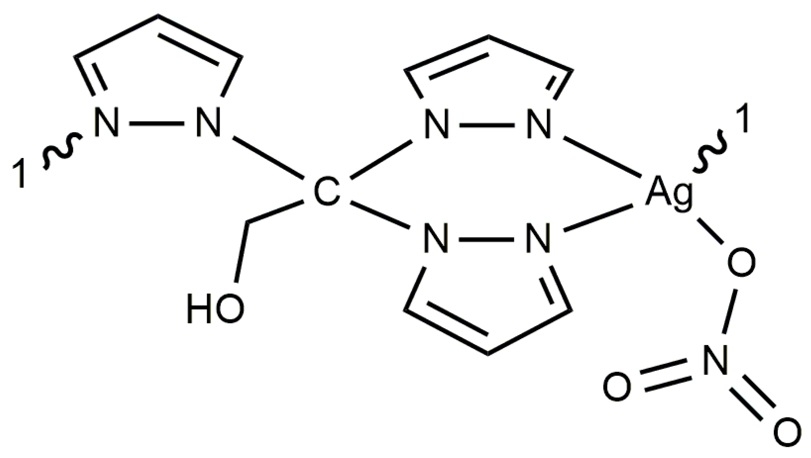
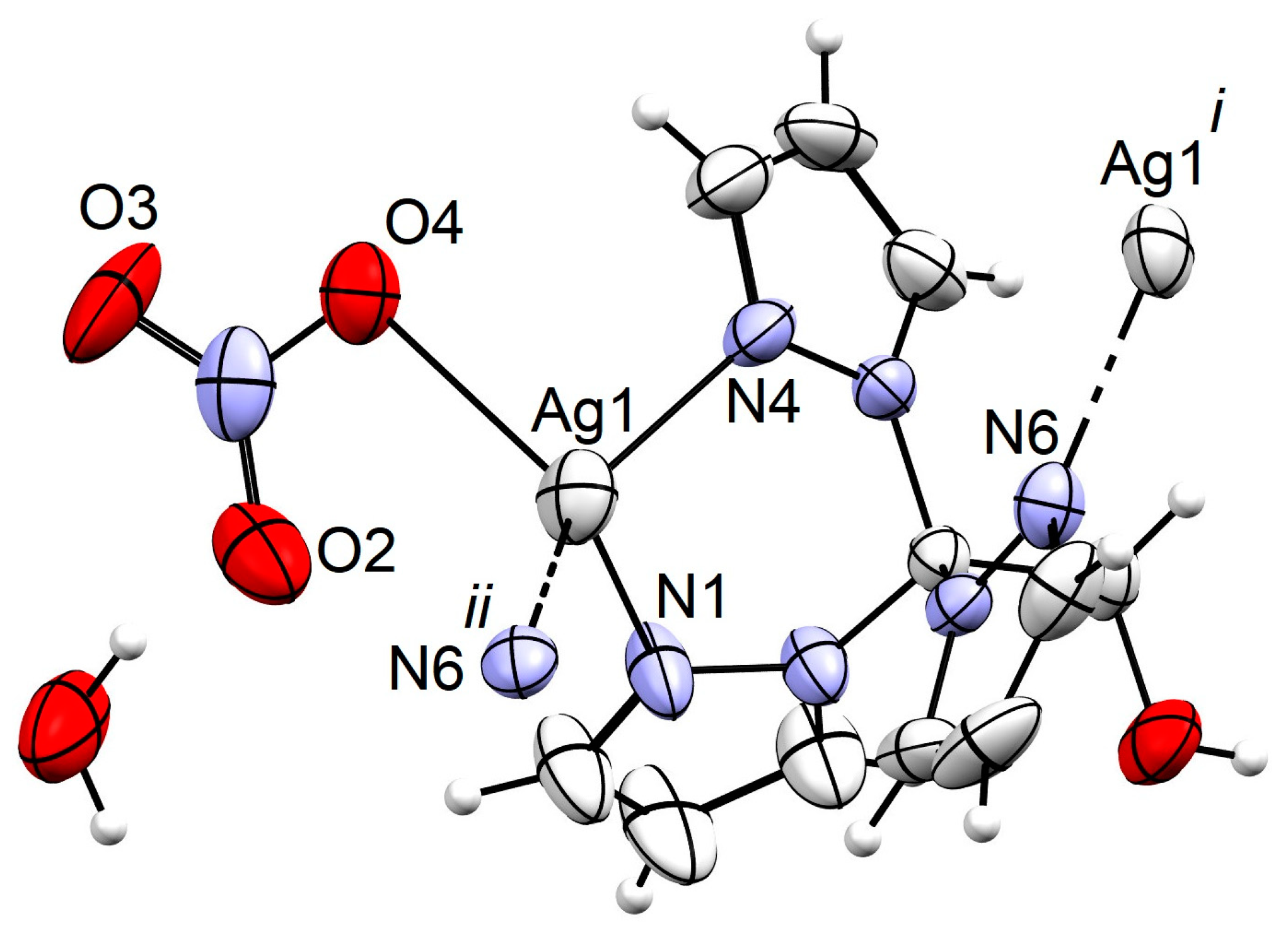
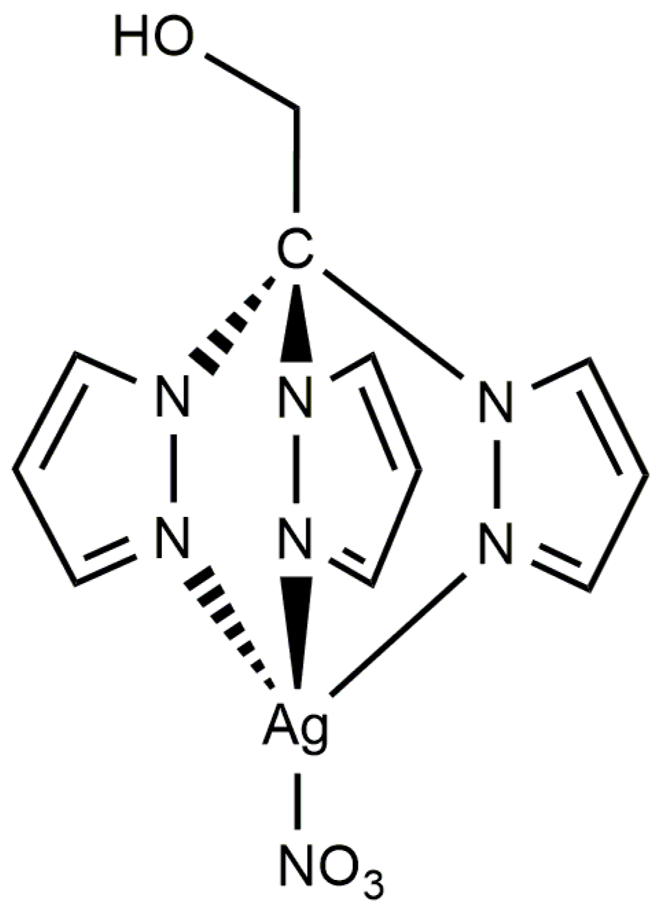
3.2. Synthesis of [Ag(NO3)(μ-1κN;2κN′,N″-TPMOH)]n (1)

In a round bottom flask, a solution of TPMOH (100 mg, 0.4 mmol) in 10 mL of methanol was added to a solution of AgNO3 (68 mg, 0.4 mmol) in 10 mL of methanol. The obtained colorless solution underwent overnight stirring at room temperature, followed by evaporation in open air to obtain compound 1 as colorless crystals that were suitable for SCXRD analysis.
Yield = 58%, based on silver. The elemental analysis calculated (%) for C11H12AgN7O4·H2O (432.14 g/mol) was as follows: C 30.57, H 3.27, N 22.69; found: C 30.32, H 3.19, N 22.43. FTIR (KBr): ν (cm−1) = 3153 w, 1521 m, 1395 s, 1338 m, 1294 s, 1205 m, 1093 s, 1062 m, 1031 m, 983 w, 954 m, 916 m, 872 s, 822 w, 773 s, 748 s, 662 m, 620 s, 604 s, 505 m. 1H NMR (300 MHz, DMSO-d6, δ): 7.67 (d, J = 1.2 Hz, 3H, 5-H-pz), 7.52 (d, J = 2.4 Hz, 3H, 3-H-pz), 6.41 (dd, J1 = 2.3 Hz, J2 = 2.1 Hz, 3H, 4-H-pz), 6.00 (t, J = 5.7 Hz, 1H, OH), 4.96 (d, J = 6.0 Hz, 2H, CH2). 13C{1H} NMR (300 MHz, DMSO-d6, δ): 141.13 (3-C-pz), 131.15 (5-C-pz), 106.47 (4-C-pz), 89.85 (CCH2), 65.38 (CH2).

3.3. X-ray Structure Determination

The crystals of compound 1 were immersed in cryo-oil, mounted in a nylon loop. X-ray diffraction intensity data were then collected by a Bruker SMART APEX-II CCD area detector (Bruker, Billerica, MA, USA), using graphite monochromated MoKα radiation (λ = 0.71073 Å) at 296 K. SADABS was used for absorption corrections [45,46]. The structure was elucidated through direct methods and refined on F2 by the full-matrix least-squares method, employing Bruker’s SHELXTL-97 (Bruker, Billerica, MA, USA) [47]. Anisotropic refinement was performed for all non-hydrogen atoms. Comprehensive crystallographic data are summarized in Table S1. The pertinent structural information has been deposited at the Cambridge Crystallographic Data Centre [CCDC 2332207]. Interested parties can obtain a complimentary copy of these data from the Director, CCDC, 12 Union Road, Cambridge CB2 1EZ, UK (Fax: (+44) 1223-336033; E-mail: [email protected] or www.ccdc.cam.ac.uk/data_request/cif, accessed on 1 July 2024.

3.4. Bacterial and Fungal Strains and Determination of Antimicrobial Activity

The bacterial strains, S. aureus Newman, E. coli ATCC25922, B. contaminans IST408 and P. aeruginosa 477, as well as the fungal strains, C. glabrata CBS138 and C. albicans SC5134, were used in this work. Bacterial and fungal strains were maintained, respectively, in Lennox Broth (LB) solid medium (10 g/L tryptone, 5 g/L yeast extract, 5 g/L NaCl and 15 g/L agar) and Yeast Extract–Peptone–Dextrose (YPD) solid medium (20 g/L glucose, 20 g/L peptone, 10 g/L yeast extract and 15 g/L agar).
Stock solutions of 1, 2, and TPMOH (10.0, 5.0, and 10.0 mg/mL, respectively) were prepared in 100% DMSO.
Measurement of the antibacterial activity of the compounds was carried out with the standardized microdilution method recommended by EUCAST (European Committee on Antimicrobial Susceptibility Testing) [48]. Briefly, 96-well polystyrene microtiter plates (Greiner Bio-One, Kremsmünster, Austria) were filled with 100 μL of Mueller–Hinton (MH) broth (Fluka Analytical, Darmstadt, Germany). Sequential 1:2 dilutions of each compound from the stock solutions were performed to obtain final concentrations ranging from 250 µg/mL to 0.49 µg/mL. Bacterial cultures that had been grown for five hours (carried out in MHB at 37 °C and 250 rev·min−1) were diluted with MHB fresh medium and added to the 96-well plates at a final inoculum of 5 × 10⁵ CFU/mL. The microplates were then incubated for 22 h at 37 °C. After incubation, the wells were examined for turbidity (growth), resuspended by pipetting, and their optical density was measured in a SPECTROstar Nano microplate reader (BMG Labtech, Ortenberg, Germany) at 640 nm.
Antifungal susceptibility testing was carried out according to the standardized microdilution method recommended by EUCAST (European Committee on Antimicrobial Susceptibility Testing) for Candida spp. [49]. Briefly, the 96-well microtiter plates (Greiner Bio-One) were filled with 100 μL of RPMI-1640 liquid medium (SIGMA, Burlington, MA, USA) buffered to pH 7.0 with 0.165 M morpholine propanesulfonic acid (MOPS; SIGMA). Sequential 1:2 dilutions of each compound stock solution were carried out in order to obtain final concentrations ranging from 125 µg/mL to 0.49 µg/mL. Overnight grown fungal cultures (carried out in YPD broth at 30 °C and 250 rev.min−1) were diluted with RPMI-1640 fresh liquid medium to a final optical density of 0.025, measured at 530 nm (OD530) in a Hitachi U-2000 UV/Vis spectrophotometer (Hitachi, Tokyo, Japan). The wells were then inoculated with the addition of 100 μL of the fungal suspensions (C. glabrata CBS138 or C. albicans SC5134) and incubated for 24 h at 35 °C. After incubation, the wells were examined for turbidity (growth), resuspended, and their optical density was measured in a SPECTROstar Nano microplate reader (BMG Labtech) at 530 nm.
Adequate quality controls were performed using ampicillin for the bacterial strains and fluconazole and auranofin for the fungal strains, respectively.
All compounds were tested in at least three independent experiments and in duplicate wells. Minimum inhibitory concentration (MIC) was estimated after data fitting of the OD640 or OD530 mean values using a modified Gompertz equation, using the GraphPad Prism software (version 6.07) [50]. In each experiment, positive (without compound) and negative controls (no organism inoculum) were carried out. The effect of 5% (v/v) or 2.5% (v/v) DMSO on bacterial or fungal growth, respectively, was also assessed, and no effects were detected.

3.5. Toxicity of Complexes to the Nematode Caenorhabditis Elegans

The nematode C. elegans Bristol strain N2 used in the present work was obtained from the Caenorhabditis Genetics Center (Minneapolis, MN, USA), which is supported by the National Institutes of Health, Office of Research Infrastructure Programs (P40 OD010440).
The nematode strain was maintained at 20 °C on Nematode Growth Medium (NGM) agar plates seeded with E. coli OP50 and synchronized using standard protocols and as previously described [51]. The NGM was composed of 25 mM NaCl, 1.7% (w/v) agar, 2.5 mg/mL peptone, 5 μg/mL cholesterol, 1 mM CaCl2, 1 mM MgSO4, and 50 mM KH2PO4 (pH 6.0). The uracil-deficient E. coli OP50 was used as the nematode food source. L4 synchronized worms were exposed to complexes final concentrations of 0, 10, 25, 50, 75, or 100 µg/mL in M9 buffer (pH 6), supplemented with 0.2% (w/v) of heat-killed E. coli OP50. Nematode appearance and viability were followed for 72 h at 20 °C, in a final volume of 500 µL contained in the wells of a 24-well plate. The total number of living and dead worms per well was assessed at 24, 48, and 72 h timepoints with the aid of a Zeiss Stemi 2000-C stereomicroscope (Zeiss, Jena, Germany). The survival of live worms was computed by tapping the plate and counting the moving worms. At least three independent experiments were performed using a total of 198 ± 28 L4 worms per each experimental condition tested. Kaplan–Meier survival curves were drawn using GraphPad Prism (GraphPad Software, San Diego, CA, USA). Concentrations that resulted in the death of 50% of the tested organisms (lethal concentration—LC50) were determined using the LC50 calculator (https://www.aatbio.com/tools/lc50-calculator, accessed on 1 July 2024), using a four-parameter logistic model with background correction and normalization.

4. Conclusions

The novel hydrosoluble silver complex [Ag(NO3)(μ-1κN;2κN′,N″-TPMOH)]n (1) was synthesized and characterized. SCXRD analysis proved compound 1 as a coordination polymer with bridging TPMOH ligands in the solid state. The solubility of compound 1 in polar solvents and the observed NMR spectra indicate a monomeric complex in solution. The antimicrobial activities of compound 1 and of the [Ag(μ-1κN;2κN′,N″-TPMS)]n (2) analogue were evaluated against a set of Gram-negative and Gram-positive bacterial pathogens, and the two important fungal pathogens, C. albicans and C. glabrata. Compound 1 showed remarkably low MIC values towards the Gram-negative strains tested (particularly in the case of P. aeruginosa 477 (4.6 μM)), in comparison to 2 and the other TPMS-containing silver species; however, it had a lower activity towards the Gram-positive S. aureus and the fungal species C. albicans and C. glabrata. Remarkably high LC50 values were obtained using living C. elegans, highlighting the potential therapeutic utility of compounds 1 and 2. Previous work from our research group has highlighted silver camphorimine complexes as a group of novel compounds with antimicrobial activities against pathogenic bacteria and fungi [52,53,54]. The present work adds silver C-scorpionate complexes to the silver complexes with antimicrobial activities of potential therapeutic use in medicine.

Supplementary Materials

The following supporting information can be downloaded at: https://www.mdpi.com/article/10.3390/antibiotics13070647/s1, Table S1: crystallographic data and structure refinement details for 1; Figure S1: Structural fragment representing the 1D polymer 1; Figure S2: Hydrogen bond interactions ( D 1 1 2 graph sets) in the structure of 1 (distances in Å). Symmetry operations to generate equivalent atoms: (i) −1 + x, −y, −1.5 + z; (ii) x, −y, −1/2 + z; Figure S3: 1H NMR spectrum of complex 1 in DMSO-d6 (300 MHz); Figure S4: 13C NMR spectrum of complex 1 in DMSO-d6 (300 MHz); Figure S5: DEPT NMR spectrum of complex 1 in DMSO-d6 (300 MHz); Figure S6: HSQC NMR spectrum of complex 1 in DMSO-d6 (300 MHz); Figure S7: COSY NMR spectrum of complex 1 in DMSO-d6 (300 MHz); Figure S8: 13C NMR spectrum of TpmOH (Top) and complex 1 (bottom) in DMSO-d6.

Author Contributions

Conceptualization, L.M.D.R.S.M. and J.H.L.; methodology, A.G.M. and L.M.D.R.S.M.; validation, M.F.C.G.d.S., L.M.D.R.S.M., J.H.L. and S.A.S.; investigation, A.G.M. and S.A.S.; resources, M.F.C.G.d.S., L.M.D.R.S.M. and J.H.L.; data curation, S.A.S. and J.H.L.; writing—original draft preparation, A.G.M. and J.H.L.; writing—review and editing, M.F.C.G.d.S., L.M.D.R.S.M., S.A.S. and J.H.L.; supervision, M.F.C.G.d.S., L.M.D.R.S.M. and J.H.L.; funding acquisition, M.F.C.G.d.S., L.M.D.R.S.M. and J.H.L. All authors have read and agreed to the published version of the manuscript.

Funding

This work has been supported by the Fundação para a Ciência e a Tecnologia (FCT) (Portugal), projects UIDB/00100/2020 (DOI: 10.54499/UIDB/00100/2020), UIDP/00100/2020 (DOI: 10.54499/UIDP/00100/2020) and LA/P/0056/2020 (DOI: 10.54499/LA/P/0056/2020) of Centro de Química Estrutural, IBB—Institute for Bioengineering and Biosciences for projects UIDB/04565/2020 and UIDP/04565/2020, and i4HB for project LA/P/0140/2020. AGM is grateful to Associação do Instituto Superior Técnico para Investigação e Desenvolvimento for his research and post-doctoral fellowships through grant no. BL133/2021-IST-ID.

Institutional Review Board Statement

Not applicable.

Informed Consent Statement

Not applicable.

Data Availability Statement

The data presented in this study are available in article and Supplementary Materials.

Conflicts of Interest

The authors declare no conflicts of interest.

References

  1. Theuretzbacher, U. Future Antibiotics Scenarios: Is the Tide Starting to Turn? Int. J. Antimicrob. Agents 2009, 34, 15–20. [Google Scholar] [CrossRef] [PubMed]
  2. WHO Publishes List of Bacteria for Which New Antibiotics Are Urgently Needed. Available online: https://www.who.int/en/news-room/detail/27-02-2017-who-publishes-list-of-bacteria-for-which-new-antibiotics-are-urgently-needed (accessed on 7 February 2024).
  3. Ghosh, C.; Sarkar, P.; Issa, R.; Haldar, J. Alternatives to Conventional Antibiotics in the Era of Antimicrobial Resistance. Trends Microbiol. 2019, 27, 323–338. [Google Scholar] [CrossRef]
  4. Singh, P.A.; Desai, S.D.; Singh, J. A Review on Plant Antimicrobials of Past Decade. Curr. Top. Med. Chem. 2018, 18, 812–833. [Google Scholar] [CrossRef]
  5. Kenawy, E.R.; Worley, S.D.; Broughton, R. The Chemistry and Applications of Antimicrobial Polymers: A State-of-the-Art Review. Biomacromolecules 2007, 8, 1359–1384. [Google Scholar] [CrossRef]
  6. Lemire, J.A.; Harrison, J.J.; Turner, R.J. Antimicrobial Activity of Metals: Mechanisms, Molecular Targets and Applications. Nat. Rev. Microbiol. 2013, 11, 371–384. [Google Scholar] [CrossRef] [PubMed]
  7. Turner, R.J. The Good, the Bad, and the Ugly of Metals as Antimicrobials. BioMetals 2023, 2023, 545–559. [Google Scholar] [CrossRef] [PubMed]
  8. Tang, X.; Li, L.; You, G.; Li, X.; Kang, J. Metallic Elements Combine with Herbal Compounds Upload in Microneedles to Promote Wound Healing: A Review. Front. Bioeng. Biotechnol. 2023, 11, 1283771. [Google Scholar] [CrossRef]
  9. Żyro, D.; Sikora, J.; Szynkowska-Jóźwik, M.I.; Ochocki, J. Silver, Its Salts and Application in Medicine and Pharmacy. Int. J. Mol. Sci. 2023, 24, 15723. [Google Scholar] [CrossRef]
  10. Hamad, A.; Khashan, K.S.; Hadi, A. Silver Nanoparticles and Silver Ions as Potential Antibacterial Agents. J. Inorg. Organomet. Polym. Mater. 2020, 30, 4811–4828. [Google Scholar] [CrossRef]
  11. Frei, A.; Verderosa, A.D.; Elliott, A.G.; Zuegg, J.; Blaskovich, M.A.T. Metals to Combat Antimicrobial Resistance. Nat. Rev. Chem. 2023, 7, 202–224. [Google Scholar] [CrossRef]
  12. Tortella, G.R.; Rubilar, O.; Durán, N.; Diez, M.C.; Martínez, M.; Parada, J.; Seabra, A.B. Silver Nanoparticles: Toxicity in Model Organisms as an Overview of Its Hazard for Human Health and the Environment. J. Hazard. Mater. 2020, 390, 121974. [Google Scholar] [CrossRef] [PubMed]
  13. Rajan, R.; Huo, P.P.; Chandran, K.; Manickam Dakshinamoorthi, B.; Yun, S.I.; Liu, B. A Review on the Toxicity of Silver Nanoparticles against Different Biosystems. Chemosphere 2022, 292, 133397. [Google Scholar] [CrossRef] [PubMed]
  14. Hartinger, C.G.; Dyson, P.J. Bioorganometallic Chemistry—From Teaching Paradigms to Medicinal Applications. Chem. Soc. Rev. 2009, 38, 391–401. [Google Scholar] [CrossRef] [PubMed]
  15. Silver, S. Bacterial silver resistance: Molecular biology and uses and misuses of silver compounds. FEMS Microbiol. Rev. 2003, 27, 341–353. [Google Scholar] [CrossRef] [PubMed]
  16. Yan, X.; He, B.; Liu, L.; Qu, G.; Shi, J.; Hu, L.; Jiang, G. Antibacterial mechanism of silver nanoparticles in Pseudomonas aeruginosa: Proteomics approach. Metallomics 2018, 10, 557–564. [Google Scholar] [CrossRef] [PubMed]
  17. Liang, X.; Luan, S.; Yin, Z.; He, M.; He, C.; Yin, L.; Zou, Y.; Yuan, Z.; Li, L.; Song, X.; et al. Recent Advances in the Medical Use of Silver Complex. Eur. J. Med. Chem. 2018, 157, 62–80. [Google Scholar] [CrossRef] [PubMed]
  18. Prencipe, F.; Zanfardino, A.; Di Napoli, M.; Rossi, F.; D’errico, S.; Piccialli, G.; Mangiatordi, G.F.; Saviano, M.; Ronga, L.; Varcamonti, M.; et al. Silver (I) N-Heterocyclic Carbene Complexes: A Winning and Broad Spectrum of Antimicrobial Properties. Int. J. Mol. Sci. 2021, 22, 2497. [Google Scholar] [CrossRef] [PubMed]
  19. Kaloğlu, M.; Kaloğlu, N.; Günal, S.; Özdemir, İ. Synthesis of N-Heterocyclic Carbene-Based Silver Complexes and Their Antimicrobial Properties against Bacteria and Fungi. J. Coord. Chem. 2022, 74, 3031–3047. [Google Scholar] [CrossRef]
  20. Karci, H.; Dündar, M.; Nawaz, Z.; Özdemir, İ.; Gürbüz, N.; Koç, A.; Özdemir, İ.; Mansour, L.; Hamdi, N. Sythesis, Characterisation, Anticancer and Antimicrobial Activity of Ag-N-Heterocyclic Carbene Complexes Containing Benzimidazole Derivatives. Inorg. Chim. Acta 2024, 565, 121992. [Google Scholar] [CrossRef]
  21. Carvalho, M.A.; De Paiva, R.E.F.; Bergamini, F.R.G.; Gomes, A.F.; Gozzo, F.C.; Lustri, W.R.; Formiga, A.L.B.; Shishido, S.M.; Ferreira, C.V.; Corbi, P.P. A Silver Complex with Tryptophan: Synthesis, Structural Characterization, DFT Studies and Antibacterial and Antitumor Assays in Vitro. J. Mol. Struct. 2013, 1031, 125–131. [Google Scholar] [CrossRef]
  22. Stathopoulou, M.E.K.; Banti, C.N.; Kourkoumelis, N.; Hatzidimitriou, A.G.; Kalampounias, A.G.; Hadjikakou, S.K. Silver Complex of Salicylic Acid and Its Hydrogel-Cream in Wound Healing Chemotherapy. J. Inorg. Biochem. 2018, 181, 41–55. [Google Scholar] [CrossRef] [PubMed]
  23. Xie, B.P.; Chai, J.W.; Fan, C.; Ouyang, J.H.; Duan, W.J.; Sun, B.; Chen, J.; Yuan, L.X.; Xu, X.Q.; Chen, J.X. Water-Stable Silver-Based Metal-Organic Frameworks of Quaternized Carboxylates and Their Antimicrobial Activity. ACS Appl. Bio Mater. 2020, 3, 8525–8531. [Google Scholar] [CrossRef] [PubMed]
  24. Kuchar, J.; Rust, J.; Lehmann, C.W.; Mohr, F. Silver(I) Complexes with Camphorsulfonato and Phosphine Ligands: Structural Diversity and Antibacterial Activity. Inorg. Chem. 2020, 59, 10557–10568. [Google Scholar] [CrossRef] [PubMed]
  25. Jaros, S.W.; Król, J.; Bazanów, B.; Poradowski, D.; Chrószcz, A.; Nesterov, D.S.; Kirillov, A.M.; Smoleński, P. Antiviral, Antibacterial, Antifungal, and Cytotoxic Silver(I) BioMOF Assembled from 1,3,5-Triaza-7-Phoshaadamantane and Pyromellitic Acid. Molecules 2020, 25, 2119. [Google Scholar] [CrossRef] [PubMed]
  26. Jaros, S.W.; Florek, M.; Bażanów, B.; Panek, J.; Krogul-Sobczak, A.; Oliveira, M.C.; Król, J.; Śliwińska-Hill, U.; Nesterov, D.S.; Kirillov, A.M.; et al. Silver Coordination Polymers Driven by Adamantoid Blocks for Advanced Antiviral and Antibacterial Biomaterials. ACS Appl. Mater. Interfaces 2024, 16, 13411–13421. [Google Scholar] [CrossRef]
  27. Pandurangan, K.; Gallagher, S.; Morgan, G.G.; Müller-Bunz, H.; Paradisi, F. Structure and Antibacterial Activity of the Silver(i) Complex of 2-Aminophenoxazine-3-One. Metallomics 2010, 2, 530–534. [Google Scholar] [CrossRef] [PubMed]
  28. Stenger-Smith, J.; Chakraborty, I.; Sameera, W.M.C.; Mascharak, P.K. Antimicrobial Silver (I) Complexes Derived from Aryl-Benzothiazoles as Turn-on Sensors: Syntheses, Properties and Density Functional Studies. Inorg. Chim. Acta 2018, 471, 326–335. [Google Scholar] [CrossRef]
  29. Pettinari, C.; Marchetti, F.; Lupidi, G.; Quassinti, L.; Bramucci, M.; Petrelli, D.; Vitali, L.A.; Guedes Da Silva, M.F.C.; Martins, L.M.D.R.S.; Smoleński, P.; et al. Synthesis, Antimicrobial and Antiproliferative Activity of Novel Silver(I) Tris(Pyrazolyl)Methanesulfonate and 1,3,5-Triaza-7-Phosphadamantane Complexes. Inorg. Chem. 2011, 50, 11173–11183. [Google Scholar] [CrossRef]
  30. Smoleński, P.; Pettinari, C.; Marchetti, F.; Guedes Da Silva, M.F.C.; Lupidi, G.; Badillo Patzmay, G.V.; Petrelli, D.; Vitali, L.A.; Pombeiro, A.J.L. Syntheses, Structures, and Antimicrobial Activity of New Remarkably Light-Stable and Water-Soluble Tris(Pyrazolyl)Methanesulfonate Silver(I) Derivatives of N-Methyl-1,3,5-Triaza-7-Phosphaadamantane Salt—[MPTA]BF4. Inorg. Chem. 2015, 54, 434–440. [Google Scholar] [CrossRef]
  31. Martins, L.M.D.R.S.; Pombeiro, A.J.L. Tris(Pyrazol-1-Yl)Methane Metal Complexes for Catalytic Mild Oxidative Functionalizations of Alkanes, Alkenes and Ketones. Coord. Chem. Rev. 2014, 265, 74–88. [Google Scholar] [CrossRef]
  32. Alkorta, I.; Claramunt, R.M.; Díez-Barra, E.; Elguero, J.; de la Hoz, A.; López, C. The Organic Chemistry of Poly(1H-Pyrazol-1-Yl)Methanes. Coord. Chem. Rev. 2017, 339, 153–182. [Google Scholar] [CrossRef]
  33. Silva, F.; Fernandes, C.; Campello, M.P.C.; Paulo, A. Metal Complexes of Tridentate Tripod Ligands in Medical Imaging and Therapy. Polyhedron 2017, 125, 186–205. [Google Scholar] [CrossRef]
  34. Mahmoud, A.G.; Martins, L.M.D.R.S.; Guedes da Silva, M.F.C.; Pombeiro, A.J.L. Copper Complexes Bearing C-Scorpionate Ligands: Synthesis, Characterization and Catalytic Activity for Azide-Alkyne Cycloaddition in Aqueous Medium. Inorg. Chim. Acta 2018, 483, 371–378. [Google Scholar] [CrossRef]
  35. Martins, L.M.D.R.S.; Pombeiro, A.J.L. Water-Soluble C-Scorpionate Complexes—Catalytic and Biological Applications. Eur. J. Inorg. Chem. 2016, 2016, 2236–2252. [Google Scholar] [CrossRef]
  36. Mahmoud, A.G.; Martins, L.M.D.R.S.; Guedes da Silva, M.F.C.; Pombeiro, A.J.L. Hydrosoluble Complexes Bearing Tris(Pyrazolyl)Methane Sulfonate Ligand: Synthesis, Characterization and Catalytic Activity for Henry Reaction. Catalysts 2019, 9, 611. [Google Scholar] [CrossRef]
  37. Almeida, J.; Roma-Rodrigues, C.; Mahmoud, A.G.; Guedes da Silva, M.F.C.; Pombeiro, A.J.L.; Martins, L.M.D.R.S.; Baptista, P.V.; Fernandes, A.R. Structural Characterization and Biological Properties of Silver(I) Tris(Pyrazolyl)Methane Sulfonate. J. Inorg. Biochem. 2019, 199, 110789. [Google Scholar] [CrossRef] [PubMed]
  38. Mahmoud, A.G.; Guedes da Silva, M.F.C.; Pombeiro, A.J.L. A New Amido-Phosphane as Ligand for Copper and Silver Complexes. Synthesis, Characterization and Catalytic Application for Azide–Alkyne Cycloaddition in Glycerol. Dalton Trans. 2021, 50, 6109–6125. [Google Scholar] [CrossRef]
  39. Silva, T.F.S.; Martins, L.M.D.R.S.; Guedes Da Silva, M.F.C.; Fernandes, A.R.; Silva, A.; Borralho, P.M.; Santos, S.; Rodrigues, C.M.P.; Pombeiro, A.J.L. Cobalt Complexes Bearing Scorpionate Ligands: Synthesis, Characterization, Cytotoxicity and DNA Cleavage. Dalton Trans. 2012, 41, 12888–12897. [Google Scholar] [CrossRef] [PubMed]
  40. Li, H.; Zeng, L.; Wang, C.; Shi, C.; Li, Y.; Peng, Y.; Chen, H.; Zhang, J.; Cheng, B.; Chen, C.; et al. Review of the toxicity and potential molecular mechanisms of parental or successive exposure to environmental pollutants in the model organism Caenorhabditis elegans. Environ. Pollut. 2022, 311, 119927. [Google Scholar] [CrossRef]
  41. Spackman, M.A.; McKinnon, J.J. Fingerprinting Intermolecular Interactions in Molecular Crystals. CrystEngComm 2002, 4, 378–392. [Google Scholar] [CrossRef]
  42. Spackman, P.R.; Turner, M.J.; McKinnon, J.J.; Wolff, S.K.; Grimwood, D.J.; Jayatilaka, D.; Spackman, M.A. CrystalExplorer: A Program for Hirshfeld Surface Analysis, Visualization and Quantitative Analysis of Molecular Crystals. J. Appl. Crystallogr. 2021, 54, 1006–1011. [Google Scholar] [CrossRef] [PubMed]
  43. Kläui, W.; Berghahn, M.; Rheinwald, G.; Lang, H. Tris(Pyrazolyl)Methanesulfonates: A Novel Class of Water-Soluble Ligands. Angew. Chemie Int. Ed. 2000, 39, 2464–2466. [Google Scholar] [CrossRef]
  44. Reger, D.L.; Grattan, T.C.; Brown, K.J.; Little, C.A.; Lamba, J.J.S.; Rheingold, A.L.; Sommer, R.D. Syntheses of Tris(Pyrazolyl)Methane Ligands and {[Tris(Pyrazolyl)Methane]Mn(CO)3}SO3CF3 Complexes: Comparison of Ligand Donor Properties. J. Organomet. Chem. 2000, 607, 120–128. [Google Scholar] [CrossRef]
  45. SMART & SAINT Software Reference Manuals, Version 6.22; Bruker AXS Analytic X-Ray Systems, Inc.: Madison, WI, USA, 2000.
  46. Sheldrick, G.M. SADABS. Program for Empirical Absorption Correction; University of Gottingen: Gottingen, Germany, 2000. [Google Scholar]
  47. Sheldrick, G.M. SHELXTL V5.1, Software Reference Manual; Bruker AXS Inc.: Madison, WI, USA, 1997. [Google Scholar]
  48. The European Committee on Antimicrobial Susceptibility Testing. MIC Determination of Non-Fastidious and Fastidious Organisms. Available online: https://www.eucast.org/ast_of_bacteria/mic_determination (accessed on 1 July 2024).
  49. The European Committee on Antimicrobial Susceptibility Testing. Susceptibility Testing of Yeasts. Available online: https://www.eucast.org/astoffungi/methodsinantifungalsusceptibilitytesting/susceptibility_testing_of_yeasts (accessed on 1 July 2024).
  50. Lambert, R.J.W.; Pearson, J. Susceptibility Testing: Accurate and Reproducible Minimum Inhibitory Concentration (MIC) and Non-inhibitory Concentration (NIC) Values. J. Appl. Microbiol. 2000, 88, 784–790. [Google Scholar] [CrossRef] [PubMed]
  51. Costa, J.P.; Pinheiro, T.; Martins, M.S.; Carvalho, M.F.N.N.; Feliciano, J.R.; Leitão, J.H.; Silva, R.A.L.; Guerreiro, J.F.; Alves, L.M.C.; Custódio, I.; et al. Tuning the Biological Activity of Camphorimine Complexes through Metal Selection. Antibiotics 2022, 11, 1010. [Google Scholar] [CrossRef] [PubMed]
  52. Costa, J.P.; Sousa, S.A.; Galvão, A.M.; Mata, J.M.; Leitão, J.H.; Carvalho, M.F.N.N. Key Parameters on the Antibacterial Activity of Silver Camphor Complexes. Antibiotics 2021, 10, 135. [Google Scholar] [CrossRef] [PubMed]
  53. Carvalho, M.F.N.N.; Leite, S.; Costa, J.P.; Galvão, A.M.; Leitão, J.H. Ag(I) Camphor Complexes: Antimicrobial Activity by Design. J. Inorg. Biochem. 2019, 199, 110791. [Google Scholar] [CrossRef]
  54. Leitão, J.H.; Sousa, S.A.; Leite, S.A.; Carvalho, M.F.N.N. Silver Camphor Imine Complexes: Novel Antibacterial Compounds from Old Medicines. Antibiotics 2018, 7, 65. [Google Scholar] [CrossRef]
Figure 1. (Top): schematic representation of TPM, TPMS, and TPMOH; (Bottom): bipodal and tripodal coordination ability of TPMS.
Antibiotics 13 00647 g001
Figure 2. Polymeric structure of compound 1 in solid state.
Antibiotics 13 00647 g002
Figure 3. ORTEP diagram of compound 1, drawn at a 40% probability level, with partial atom labelling scheme. Selected bond distances (Å) and angles (°): Ag1–N1 2.452(4), Ag1–N4 2.264(6), Ag1–N6ii 2.231(6), Ag1–O4A 2.57(4), Ag1···Ag1i 5.0830(6), N1–Ag1–N6ii 108.5(2), N4–Ag1–N6ii 149.3(2), N1–Ag1–N4 77.4(2), O4A–Ag1–N6ii 121.5(9), O4A–Ag1–N1 103.9(7) and O4A–Ag1–N4 84.3(7). Symmetry operations to generate the equivalent atoms: (i) −1/2 + x,1/2 − y, −1/2 + z; (ii) 1/2 + x,1/2 − y,1/2 + z.
Antibiotics 13 00647 g003
Figure 4. 1H NMR spectrum of TPMOH (top) and complex 1 (bottom) in DMSO-d6.
Antibiotics 13 00647 g004
Figure 5. Structure of complex 1 in solution.
Antibiotics 13 00647 g005
Figure 6. Percentage of surviving worms after incubation in M9 buffer containing 0.2% (w/v) of heat-killed E. coli OP50 as food source and supplemented with the indicated concentrations of compounds 1 (A) or 2 (B). Controls without compounds 1 and 2 or with DMSO were used as controls. Results are the means of at least three independent experiments using a total of 198 ± 28 worms at the L4 stage of development per each experimental condition tested. Differences compared to the control were considered as significant, with an * indicating a p < 0.0165, and **** indicating a p < 0.000001, determined using the log-rank (Mantel–Cox) test.
Antibiotics 13 00647 g006
Figure 7. (Left): Percentage contribution to the Hirshfeld surface of compounds 1, 2, and [AgP4].4TPMS.BF4. (Right): Shortest de dimensions (in Å) in the most relevant non-covalent interactions detected in compounds 1, 2 and [AgP4].4TPMS.BF4.
Antibiotics 13 00647 g007
Table 1. Minimal inhibitory concentration (MIC) for compounds 1, 2, and TPMOH towards the indicated bacterial and fungal strains. Results are presented as the means ± SD of at least three experiments carried out in duplicate.
MIC (μg/mL)
12TPMOH
E. coli ATCC259227.7 ± 0.214.5 ± 4.3>250
P. aeruginosa 4772.0 ± 0.119.5 ± 0.9>250
B. contaminans IST4085.5 ± 1.319.6 ± 4.0>250
S. aureus Newman17.9 ± 3.251.4 ± 8.7>250
C. albicans SC513415.4 ± 0.46.3 ± 1.0>125
C. glabrata CBS13831.5 ± 0.15.7 ± 0.2>125
Disclaimer/Publisher’s Note: The statements, opinions and data contained in all publications are solely those of the individual author(s) and contributor(s) and not of MDPI and/or the editor(s). MDPI and/or the editor(s) disclaim responsibility for any injury to people or property resulting from any ideas, methods, instructions or products referred to in the content.

