
Comparative Analysis of Methicillin-Resistant Staphylococcus pseudintermedius Prevalence and Resistance Patterns in Canine and Feline Clinical Samples: Insights from a Three-Year Study in Germany
 , ,
, ,
Abstract
:1. Introduction
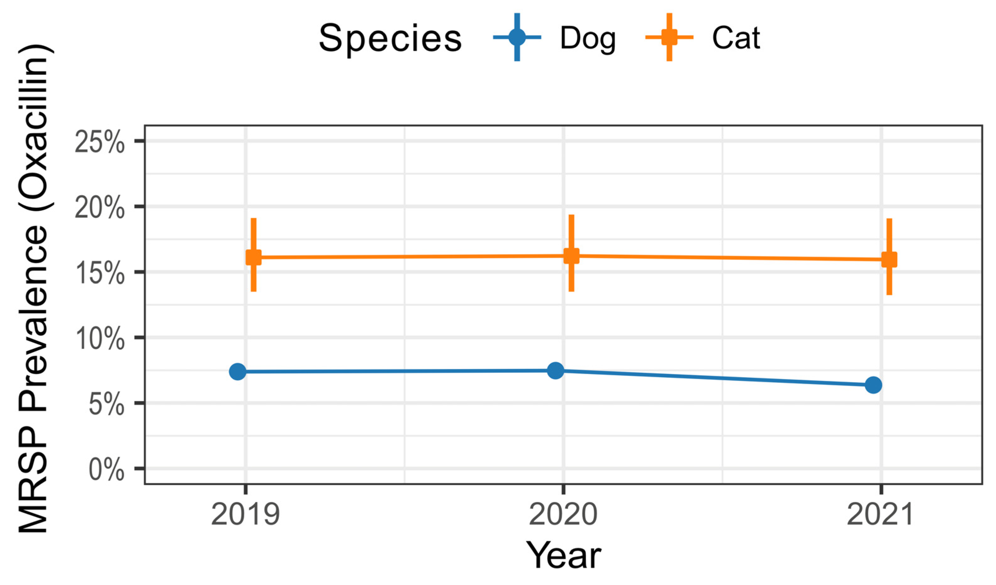
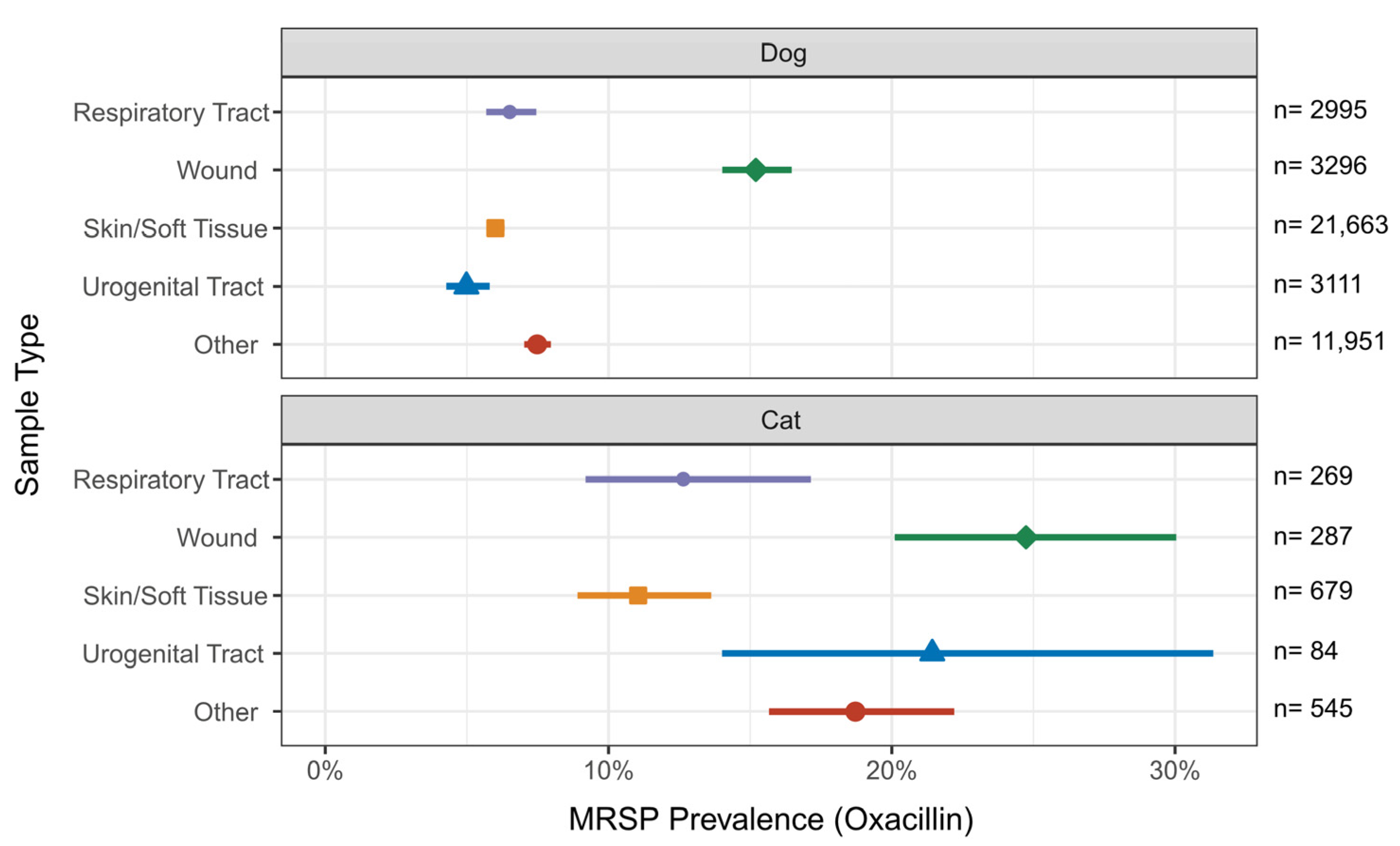
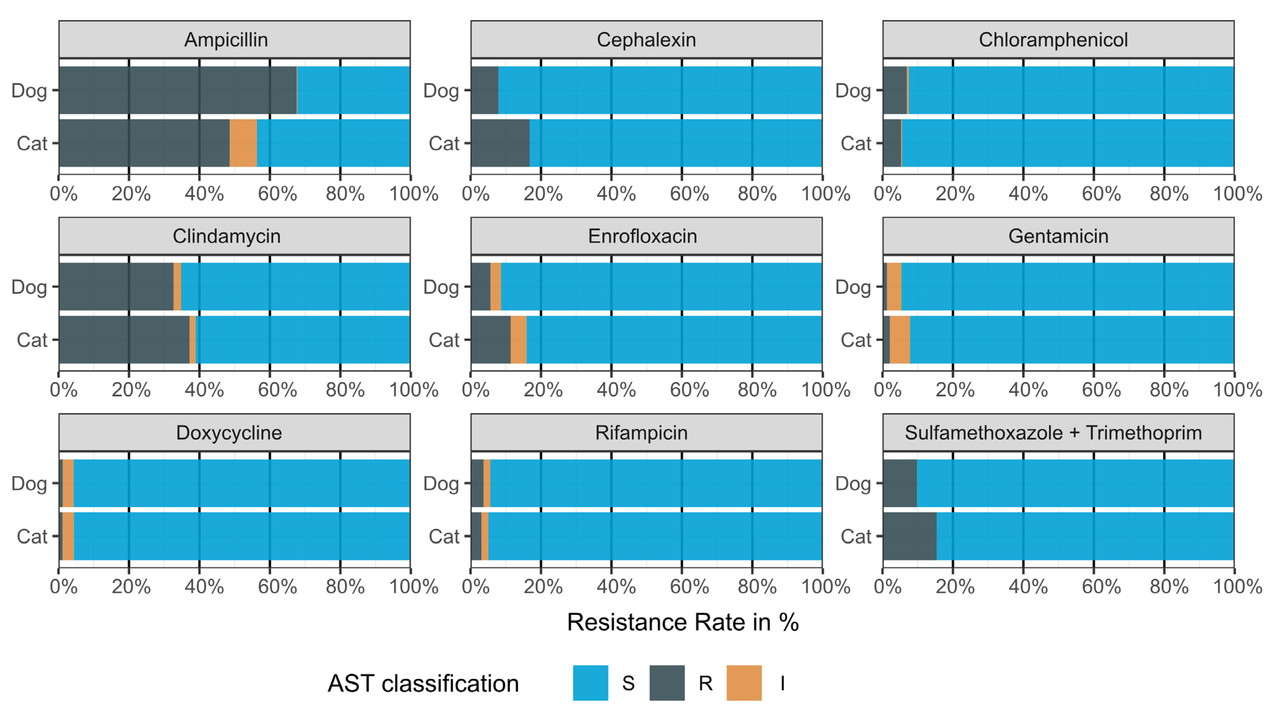
2. Results
3. Discussion
3.1. MRSP Prevalence in Cats and Dogs
3.2. Feline and Canine Sample Types
3.3. Resistance Patterns in Feline and Canine S. pseudintermedius
3.4. AMR Monitoring
3.5. Limitations
4. Materials and Methods
4.1. Samples
4.2. Bacterial Identification and Antimicrobial Susceptibility Testing (AST) of S. pseudintermedius
4.3. AST Classification
4.4. Data Processing and Statistical Analysis
- Skin and soft tissue (e.g., ear swabs)
- Wounds (e.g., swabs from surgery wounds and abscesses)
- Respiratory tract (e.g., nasal swabs and bronchoalveolar lavage)
- Urogenital tract (e.g., urinary samples and vaginal swabs)
- Other (e.g., unknown and gastrointestinal swabs)
Supplementary Materials
Author Contributions
Funding
Institutional Review Board Statement
Data Availability Statement
Conflicts of Interest
References
- EFSA Panel on Animal Health and Welfare (AHAW); Nielsen, S.S.; Bicout, D.J.; Calistri, P.; Canali, E.; Drewe, J.A.; Garin-Bastuji, B.; Rojas, J.L.G.; Schmidt, C.G.; Herskin, M.; et al. Scientific Opinion on the assessment of animal diseases caused by bacteria resistant to antimicrobials: Dogs and cats. EFSA J. 2021, 19, e06680. [Google Scholar] [CrossRef]
- Bannoehr, J.; Guardabassi, L. Staphylococcus pseudintermedius in the dog: Taxonomy, diagnostics, ecology, epidemiology and pathogenicity. Vet. Dermatol. 2012, 23, 253-e52. [Google Scholar] [CrossRef] [PubMed]
- Sykes, J.E. Staphylococcus Infections. In Canine and Feline Infectious Diseases; Elsevier: Amsterdam, The Netherlands, 2014; pp. 347–354. ISBN 978-1-4377-0795-3. [Google Scholar]
- van Duijkeren, E.; Catry, B.; Greko, C.; Moreno, M.A.; Pomba, M.C.; Pyörälä, S.; RužAuskas, M.; Sanders, P.; Threlfall, E.J.; Torren-Edo, J.; et al. Review on methicillin-resistant Staphylococcus pseudintermedius. J. Antimicrob. Chemother. 2011, 66, 2705–2714. [Google Scholar] [CrossRef] [PubMed]
- Holmes, A.H.; Moore, L.S.P.; Sundsfjord, A.; Steinbakk, M.; Regmi, S.; Karkey, A.; Guerin, P.J.; Piddock, L.J.V. Understanding the mechanisms and drivers of antimicrobial resistance. Lancet 2016, 387, 176–187. [Google Scholar] [CrossRef] [PubMed]
- World Health Organization. Global Action Plan on Antimicrobial Resistance; World Health Organization: Geneva, Switzerland, 2015; p. 28. ISBN 9789241509763.
- Morris, D.O.; Loeffler, A.; Davis, M.F.; Guardabassi, L.; Weese, J.S. Recommendations for approaches to methicillin-resistant staphylococcal infections of small animals: Diagnosis, therapeutic considerations and preventative measures. Veter. Dermatol. 2017, 28, 304-e69. [Google Scholar] [CrossRef] [PubMed]
- Lord, J.; Millis, N.; Jones, R.D.; Johnson, B.; Kania, S.A.; Odoi, A. An epidemiological study of the predictors of multidrug resistance and methicillin resistance among Staphylococcus spp. isolated from canine specimens submitted to a diagnostic laboratory in Tennessee, USA. PeerJ 2023, 11, e15012. [Google Scholar] [CrossRef] [PubMed]
- Lynch, S.A.; Helbig, K.J. The Complex Diseases of Staphylococcus pseudintermedius in Canines: Where to Next? Veter. Sci. 2021, 8, 11. [Google Scholar] [CrossRef] [PubMed]
- Weese, J.S.; van Duijkeren, E. Methicillin-Resistant Staphylococcus aureus and Staphylococcus pseudintermedius in Veterinary Medicine. Veter. Microbiol. 2010, 140, 418–429. [Google Scholar] [CrossRef] [PubMed]
- Bundesministerium der Justiz. Zweite Verordnung zur Änderung der Verordnung über tierärztliche Hausapotheken (TÄHAV). 2018. Available online: https://www.buzer.de/gesetz/12981/index.htm (accessed on 20 June 2024).
- Commission Implementing Regulation EU 2022/1255 of 19 July 2022 Designating Antimicrobials or Groups of Antimicrobials Reserved for Treatment of Certain Infections in Humans, in Accordance with Regulation (EU) 2019/6 of the European Parliament and of the Council (Text with EEA Relevance). 2022. Available online: https://eur-lex.europa.eu/eli/reg_impl/2022/1255/oj (accessed on 20 June 2024).
- Allerton, F.; Russell, J. Antimicrobial stewardship in veterinary medicine: A review of online resources. JAC-Antimicrobial Resist. 2023, 5, dlad058. [Google Scholar] [CrossRef]
- Schmerold, I.; van Geijlswijk, I.; Gehring, R. European regulations on the use of antibiotics in veterinary medicine. Eur. J. Pharm. Sci. 2023, 189, 106473. [Google Scholar] [CrossRef]
- Lagrange, J.; Amat, J.-P.; Ballesteros, C.; Damborg, P.; Grönthal, T.; Haenni, M.; Jouy, E.; Kaspar, H.; Kenny, K.; Klein, B.; et al. Pilot testing the EARS-Vet surveillance network for antibiotic resistance in bacterial pathogens from animals in the EU/EEA. Front. Microbiol. 2023, 14. [Google Scholar] [CrossRef]
- Bundesamt für Verbraucherschutz und Lebensmittelsicherheit (BVL) BVL-Report·17.6 Bericht Zur Resistenzmonitoringstudie 2021-Resistenzsituation Bei Klinisch Wichtigen Tierpathogenen Bakterien; Berlin. Available online: https://www.bvl.bund.de/SharedDocs/Berichte/07_Resistenzmonitoringstudie/Bericht_Resistenzmonitoring_2021.pdf (accessed on 20 June 2024).
- Loeffler, A.; Beever, L.; Chang, Y.; Klein, B.; Kostka, V.; Meyer, C.; Müller, E.; Weis, J.; Wildermuth, B.; Fishwick, J.; et al. Intervention with impact: Reduced isolation of methicillin-resistant Staphylococcus pseudintermedius from dogs following the introduction of antimicrobial prescribing legislation in Germany. Veter. Rec. 2023, 194, e3714. [Google Scholar] [CrossRef]
- Sobkowich, K.E.; Weese, J.S.; Poljak, Z.; Plum, A.; Szlosek, D.; Bernardo, T.M. Epidemiology of companion animal AMR in the United States of America: Filling a gap in the one health approach. Front. Public Health 2023, 11, 1161950. [Google Scholar] [CrossRef]
- Bierowiec, K.; Miszczak, M.; Korzeniowska-Kowal, A.; Wzorek, A.; Płókarz, D.; Gamian, A. Epidemiology of Staphylococcus pseudintermedius in cats in Poland. Sci. Rep. 2021, 11, 18898. [Google Scholar] [CrossRef]
- Clinical and Laboratory Standards Institute Performance Standards for Antimicrobial Disk and Dilution Sus-Ceptibility Tests for Bacteria Isolated from Animals, 6th ed.; CLSI Guideline Vet01S; CLSI: Wayne, PA, USA, 2023.
- Clinical and Laboratory Standards Institute Performance Standards for Antimicrobial Susceptibility Testing, 33rd ed.; CLSI Guideline M100; CLSI: Wayne, PA, USA, 2023.
- Feuer, L.; Frenzer, S.K.; Merle, R.; Leistner, R.; Wolfgang, B.; Bethe, A.; Lübke-Becker, A.; Klein, B.; Bartel, A. Prevalence of MRSA in Canine and Feline Clinical Samples from One Third of Veterinary Practices in Germany from 2019–2021. J. Antimicrob. Chemother. 2024, dkae225. [Google Scholar] [CrossRef]
- Bundestierärztekammer e. V. Statistik 2021: Tierärzteschaft in Der Bundesrepublik Deutschland. Dtsch. Tierärzteblatt. 2022, 70, 762–772. [Google Scholar]
- Menandro, M.L.; Dotto, G.; Mondin, A.; Martini, M.; Ceglie, L.; Pasotto, D. Prevalence and characterization of methicillin-resistant Staphylococcus pseudintermedius from symptomatic companion animals in Northern Italy: Clonal diversity and novel sequence types. Comp. Immunol. Microbiol. Infect. Dis. 2019, 66, 101331. [Google Scholar] [CrossRef]
- Miszczak, M.; Korzeniowska-Kowal, A.; Wzorek, A.; Gamian, A.; Rypuła, K.; Bierowiec, K. Colonization of methicillin-resistant Staphylococcus species in healthy and sick pets: Prevalence and risk factors. BMC Veter. Res. 2023, 19, 85. [Google Scholar] [CrossRef]
- Burke, M.; Santoro, D. Prevalence of multidrug-resistant coagulase-positive staphylococci in canine and feline dermatological patients over a 10-year period: A retrospective study. Microbiology 2023, 169, 001300. [Google Scholar] [CrossRef] [PubMed]
- Ruscher, C.; Lübke-Becker, A.; Wleklinski, C.-G.; Şoba, A.; Wieler, L.H.; Walther, B. Prevalence of Methicillin-resistant Staphylococcus pseudintermedius isolated from clinical samples of companion animals and equidaes. Veter. Microbiol. 2008, 136, 197–201. [Google Scholar] [CrossRef] [PubMed]
- Lehner, G.; Linek, M.; Bond, R.; Lloyd, D.H.; Prenger-Berninghoff, E.; Thom, N.; Straube, I.; Verheyen, K.; Loeffler, A. Case–control risk factor study of methicillin-resistant Staphylococcus pseudintermedius (MRSP) infection in dogs and cats in Germany. Veter. Microbiol. 2014, 168, 154–160. [Google Scholar] [CrossRef]
- Feßler, A.T.; Scholtzek, A.D.; Schug, A.R.; Kohn, B.; Weingart, C.; Schink, A.-K.; Bethe, A.; Lübke-Becker, A.; Schwarz, S. Antimicrobial and Biocide Resistance among Feline and Canine Staphylococcus aureus and Staphylococcus pseudintermedius Isolates from Diagnostic Submissions. Antibiotics 2022, 11, 127. [Google Scholar] [CrossRef]
- Bundesamt für Verbraucherschutz und Lebensmittelsicherheit (BVL) BVL-Report·16.6 Bericht Zur Resistenzmonitoringstudie 2020-Resistenzsituation Bei Klinisch Wichtigen Tierpathogenen Bakterien; Berlin. 2022. Available online: https://www.bvl.bund.de/SharedDocs/Berichte/07_Resistenzmonitoringstudie/Bericht_Resistenzmonitoring_2020.pdf (accessed on 20 June 2024).
- Lenart-Boroń, A.; Stankiewicz, K.; Czernecka, N.; Ratajewicz, A.; Bulanda, K.; Heliasz, M.; Sosińska, D.; Dworak, K.; Ciesielska, D.; Siemińska, I.; et al. Wounds of Companion Animals as a Habitat of Antibiotic-Resistant Bacteria That Are Potentially Harmful to Humans—Phenotypic, Proteomic and Molecular Detection. Int. J. Mol. Sci. 2024, 25, 3121. [Google Scholar] [CrossRef]
- Windahl, U.; Bengtsson, B.; Nyman, A.-K.; Holst, B.S. The distribution of pathogens and their antimicrobial susceptibility patterns among canine surgical wound infections in Sweden in relation to different risk factors. Acta Veter. Scand. 2015, 57, 11. [Google Scholar] [CrossRef]
- Viegas, F.M.; Santana, J.A.; Silva, B.A.; Xavier, R.G.C.; Bonisson, C.T.; Câmara, J.L.S.; Rennó, M.C.; Cunha, J.L.R.; Figueiredo, H.C.P.; Lobato, F.C.F.; et al. Occurrence and characterization of methicillin-resistant Staphylococcus spp. in diseased dogs in Brazil. PLoS ONE 2022, 17, e0269422. [Google Scholar] [CrossRef]
- Quinn, P.J.; Markey, B.K.; Leonard, F.C.; FitzPatrick, E.S.; Fanning, S. Concise Review of Veterinary Microbiology, 2nd ed.; John Wiley & Sons, Inc.: West Sussex, UK; Ames, Iowa, 2016; ISBN 978-1-118-80270-0. [Google Scholar]
- Carroll, K.C.; Burnham, C.-A.D.; Westblade, L.F. From canines to humans: Clinical importance of Staphylococcus pseudintermedius. PLOS Pathog. 2021, 17, e1009961. [Google Scholar] [CrossRef]
- MacFadyen, A.C.; Paterson, G.K. Methicillin resistance in Staphylococcus pseudintermedius encoded within novel staphylococcal cassette chromosome mec (SCCmec) variants. J. Antimicrob. Chemother. 2024, 79, 1303–1308. [Google Scholar] [CrossRef]
- Moerer, M.; Merle, R.; Bäumer, W. A Cross-Sectional Study of Veterinarians in Germany on the Impact of the TÄHAV Amendment 2018 on Antimicrobial Use and Development of Antimicrobial Resistance in Dogs and Cats. Antibiotics 2022, 11, 484. [Google Scholar] [CrossRef]
- Iyori, K.; Shishikura, T.; Shimoike, K.; Minoshima, K.; Imanishi, I.; Toyoda, Y. Influence of hospital size on antimicrobial resistance and advantages of restricting antimicrobial use based on cumulative antibiograms in dogs with Staphylococcus pseudintermedius infections in Japan. Veter. Dermatol. 2021, 32, 668-e178. [Google Scholar] [CrossRef]
- Servia-Dopazo, M.; Taracido-Trunk, M.; Figueiras, A. Non-Clinical Factors Determining the Prescription of Antibiotics by Veterinarians: A Systematic Review. Antibiotics 2021, 10, 133. [Google Scholar] [CrossRef]
- Ruzante, J.M.; Harris, B.; Plummer, P.; Raineri, R.R.; Loy, J.D.; Jacob, M.; Sahin, O.; Kreuder, A.J. Surveillance of antimicrobial resistance in veterinary medicine in the United States: Current efforts, challenges, and opportunities. Front. Veter. Sci. 2022, 9, 1068406. [Google Scholar] [CrossRef]
- Berends, M.S.; Luz, C.F.; Friedrich, A.W.; Sinha, B.N.M.; Albers, C.J.; Glasner, C. AMR: An R Package for Working with Antimicrobial Resistance Data. J. Stat. Softw. 2022, 104, 1–31. [Google Scholar] [CrossRef]

| Overall | Dog | Cat | |
|---|---|---|---|
| Samples | 175,171 | 122,831 | 52,340 |
| Samples with S. pseudintermedius isolated | 44,880 | 43,016 | 1864 |
| Year (%) | 44,880 (100) | ||
| 2019 | 16,145 (36.0) | 15,487 (36.0) | 658 (35.3) |
| 2020 | 13,944 (31.1) | 13,340 (31.0) | 604 (32.4) |
| 2021 | 14,791 (32.9) | 14,189 (33.0) | 602 (32.3) |
| Sample type (%) | 44,880 (100) | ||
| Skin/soft tissue | 22,342 (49.8) | 21,663 (50.4) | 679 (36.4) |
| Wound | 3583 (8.0) | 3296 (7.7) | 287 (15.4) |
| Respiratory tract | 3264 (7.3) | 2995 (7.0) | 269 (14.4) |
| Urogenital tract | 3195 (7.1) | 3111 (7.2) | 84 (4.5) |
| Other | 12,496 (27.8) | 11,951 (27.7) | 545 (29.3) |
Disclaimer/Publisher’s Note: The statements, opinions and data contained in all publications are solely those of the individual author(s) and contributor(s) and not of MDPI and/or the editor(s). MDPI and/or the editor(s) disclaim responsibility for any injury to people or property resulting from any ideas, methods, instructions or products referred to in the content. |
© 2024 by the authors. Licensee MDPI, Basel, Switzerland. This article is an open access article distributed under the terms and conditions of the Creative Commons Attribution (CC BY) license (https://creativecommons.org/licenses/by/4.0/).
Share and Cite
Feuer, L.; Frenzer, S.K.; Merle, R.; Bäumer, W.; Lübke-Becker, A.; Klein, B.; Bartel, A. Comparative Analysis of Methicillin-Resistant Staphylococcus pseudintermedius Prevalence and Resistance Patterns in Canine and Feline Clinical Samples: Insights from a Three-Year Study in Germany. Antibiotics 2024, 13, 660. https://doi.org/10.3390/antibiotics13070660
Feuer L, Frenzer SK, Merle R, Bäumer W, Lübke-Becker A, Klein B, Bartel A. Comparative Analysis of Methicillin-Resistant Staphylococcus pseudintermedius Prevalence and Resistance Patterns in Canine and Feline Clinical Samples: Insights from a Three-Year Study in Germany. Antibiotics. 2024; 13(7):660. https://doi.org/10.3390/antibiotics13070660
Chicago/Turabian StyleFeuer, Leonie, Stefanie Katharina Frenzer, Roswitha Merle, Wolfgang Bäumer, Antina Lübke-Becker, Babette Klein, and Alexander Bartel. 2024. "Comparative Analysis of Methicillin-Resistant Staphylococcus pseudintermedius Prevalence and Resistance Patterns in Canine and Feline Clinical Samples: Insights from a Three-Year Study in Germany" Antibiotics 13, no. 7: 660. https://doi.org/10.3390/antibiotics13070660