Article

In Vitro and In Silico Studies of the Antimicrobial Activity of Prenylated Phenylpropanoids of Green Propolis and Their Derivatives against Oral Bacteria

by
Tatiana M. Vieira
1,†,
Julia G. Barco
1,†,
Sara L. de Souza
2,†,
Anna L. O. Santos
2,†,
Ismail Daoud
3,4,†,
Seyfeddine Rahli
5,†,
Noureddine Amdouni
6,†,
Jairo K. Bastos
7,
Carlos H. G. Martins
2,
Ridha Ben Said
5,6,* and
Antônio E. M. Crotti
1,*
1
Department of Chemistry, Faculty of Philosophy, Science and Letters at Ribeirão Preto, University of São Paulo, Ribeirão Preto 14040-901, SP, Brazil
2
Department of Microbiology, Institute of Biomedical Sciences, Federal University of Uberlândia, Uberlândia 38405320, MG, Brazil
3
Department of Matter Sciences, University Mohamed Khider, BP 145 RP, Biskra 07000, Algeria
4
Laboratory of Natural and Bio-Active Substances, Faculty of Science, Tlemcen University, Tlemcen P.O. Box 119, Algeria
5
Department of Chemistry, College of Science, Qassim University, Qassim 51452, Saudi Arabia
6
Laboratoire de Caractérisations, Applications et Modélisations des Matériaux, Faculté des Sciences de Tunis, Université Tunis El Manar, Tunis 1068, Tunisia
7
School of Pharmaceutical Sciences of Ribeirão Preto, University of São Paulo, Ribeirão Preto 14040-903, SP, Brazil
*
Authors to whom correspondence should be addressed.
These authors contributed equally to this work.
Antibiotics 2024, 13(8), 787; https://doi.org/10.3390/antibiotics13080787 (registering DOI)
Submission received: 8 July 2024 / Revised: 14 August 2024 / Accepted: 17 August 2024 / Published: 22 August 2024

Abstract

:
Artepillin C, drupanin, and plicatin B are prenylated phenylpropanoids that naturally occur in Brazilian green propolis. In this study, these compounds and eleven of their derivatives were synthesized and evaluated for their in vitro antimicrobial activity against a representative panel of oral bacteria in terms of their minimum inhibitory concentration (MIC) and minimum bactericidal concentration (MBC) values. Plicatin B (2) and its hydrogenated derivative 8 (2′,3′,7,8-tetrahydro-plicatin B) were the most active compounds. Plicatin B (2) displayed strong activity against all the bacteria tested, with an MIC of 31.2 μg/mL against Streptococcus mutans, S. sanguinis, and S. mitis. On the other hand, compound 8 displayed strong activity against S. mutans, S. salivarius, S. sobrinus, Lactobacillus paracasei (MIC = 62.5 μg/mL), and S. mitis (MIC = 31.2 μg/mL), as well as moderate activity against Enterococcus faecalis and S. sanguinis (MIC = 125 μg/mL). Compounds 2 and 8 displayed bactericidal effects (MBC: MIC ≤ 4) against all the tested bacteria. In silico studies showed that the complexes formed by compounds 2 and 8 with the S. mitis, S. sanguinis, and S. mutans targets (3LE0, 4N82, and 3AIC, respectively) had energy score values similar to those of the native S. mitis, S. sanguinis, and S. mutans ligands due to the formation of strong hydrogen bonds. Moreover, all the estimated physicochemical parameters satisfied the drug-likeness criteria without violating the Lipinski, Veber, and Egan rules, so these compounds are not expected to cause problems with oral bioavailability and pharmacokinetics. Compounds 2 and 8 also had suitable ADMET parameters, as the online server pkCSM calculates. These results make compounds 2 and 8 good candidates as antibacterial agents against oral bacteria.

1. Introduction

Oral pathogens comprise a group of microorganisms that adhere to and colonize the oral cavity, causing periodontal inflammatory diseases, caries, or infective endocarditis [1]. Oral bacteria are responsible for dental caries, an infectious disease that affects people worldwide, especially in low-income regions of developing countries [2]. These bacteria adhere to the mature acquired salivary pellicle [3,4], which provides specific receptors for bacteria to attach and initiate colonization of the tooth surface [5]. The initial colonizers that adhere to acquired salivary pellicles are usually Streptococcus species and Actinomyces species [6,7]. Streptococcus mutans, one of the primary bacteria associated with dental caries, can establish a strongly acidic microenvironment (pH below 5.0) due to acidic by-products of the bacterial fermentation of different types of carbohydrates from the diet [8], thereby forming biofilms on the tooth surface (i.e., the dental plaque), which culminates in teeth cavitation (i.e., caries) and the onset of tooth decay [9]. However, oral diseases generally have a polymicrobial origin, and together with S. mutans, other acid-resistant bacteria can cause cavities, including S. sobrinus and S. sanguinis [10].
The most effective method of preventing dental biofilm is through mechanical removal by brushing and flossing [11]. However, in most cases, this mechanical method is performed improperly, especially in hard-to-reach places, such as sub-gums and fissures, which end up favoring biofilm accumulation. Thus, the use of chemical methods, such as mouthwashes with antimicrobial action, becomes necessary to promote the reduction of the adhesion of bacteria to the dental surface and inhibit cariogenic bacteria growth and proliferation [12,13]. Chlorhexidine is the gold standard anti-cariogenic agent used in mouthwash formulations to reduce plaque [13,14]. However, despite its effectiveness, chlorhexidine has shown adverse effects, especially in terms of its daily use, in addition to the emergence of resistant strains [15,16]. In this scenario, the interest in new compounds with antimicrobial action as effective as chlorhexidine in controlling biofilm but with fewer adverse effects has grown in the last few years [17,18].
Propolis is a resinous product formed by plant material collected and deposited in hives by bees. The chemical composition of propolis depends on the botanical sources of each region [19]. Brazilian green propolis (BGP) is produced by Apis mellifera using shoot apices of Baccharis dracucunlifolia (Asteraceae) in the country’s southeast region [20]. Because of its wide diversity of biological and pharmacological activities [21,22,23,24,25,26,27,28], BGP has also been used in folk medicine and exported to several countries (e.g., Japan, China, Russia, France, and Germany) as sprays, toothpaste, soaps, ointments, and creams for skin [29]. The antimicrobial properties of BGP are of particular interest and include activity against several Gram-positive (Staphylococcus aureus [30,31,32,33], Staphylococcus saprophyticus [30], Listeria monocytogenes [30], Enterococcus faecalis [30]) and Gram-negative bacteria (Enterobacter aerogenes [30], Salmonella typhimurium [30], Shigella flexneri [30], Escherichia coli [30,33], Pseudomonas aeruginosa [30,33,34], Providencia rettgeri [30], and Klebsiella pneumoniae [30]) and Candida albicans [33]. BGP also has bacteriostatic effects on Streptococcus mutans cultures [35] and is a potent antibacterial agent against S. mutans [36].
Chemically, Brazilian green propolis is rich in prenylated compounds [37,38], mainly phenylpropanoids like drupanin (I), and artepillin C (II, Figure 1). These phenolic acids are commonly detected and identified in Brazilian green propolis [32,39,40,41]. In the literature, many studies have reported the biological and pharmacological activities of artepillin C [32,39,42,43] and drupanin [40,41,44]. However, data on the antibacterial activity of these prenylated compounds against oral bacteria are still scarce [45].
In this study, we investigated the antibacterial activity of a series of synthetic prenylated phenylpropanoids, including the naturally occurring compounds artepillin C and drupanin and their derivatives, against a representative panel of oral bacteria. In silico studies with the most active compounds aiming to understand the target–compound interactions and to estimate the physicochemical, drug-likeness, and ADMET/pharmacokinetic properties were also performed.

2. Results

2.1. In Vitro Antibacterial Activity of Compounds 114

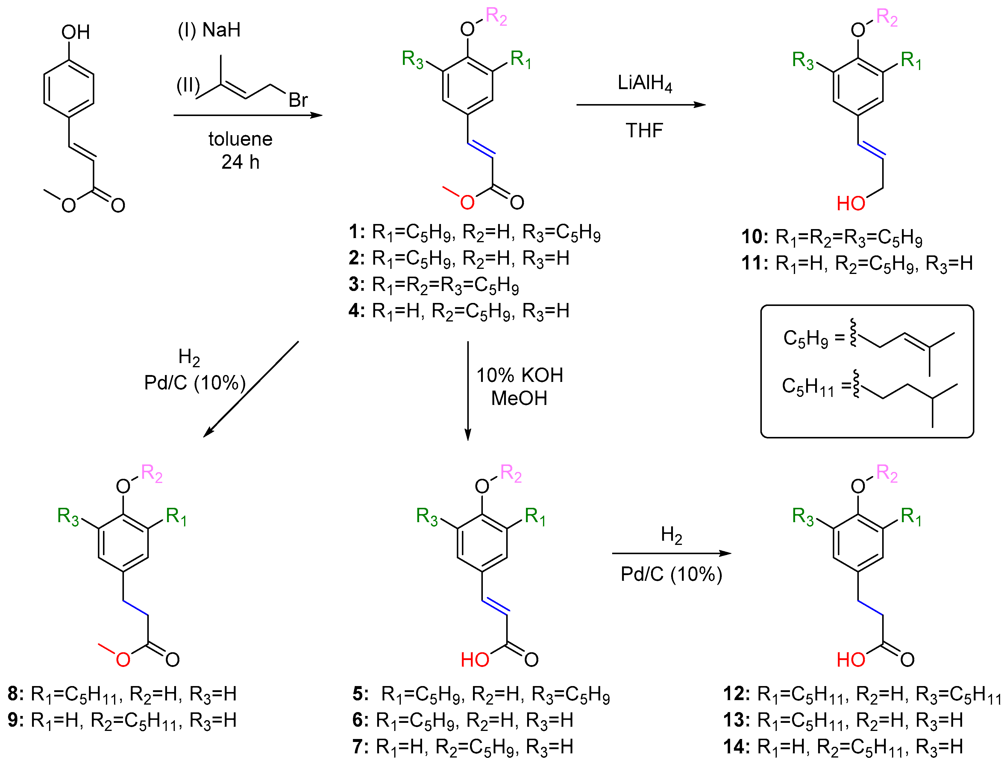
Artepillin C (5), drupanin (6), and compounds 714 were obtained by hydrolysis, reduction with LiAlH4, and/or catalytic hydrogenation of prenylation products of methyl p-coumarate 14, as shown in Scheme 1. Table 1 summarizes the results of the antimicrobial assays with the fourteen compounds against a representative panel of oral bacteria regarding their minimum inhibitory concentration (MIC) and minimum bactericidal concentration (MBC). The lowest MIC values (31.2 µg/mL) were observed for compound 2 (plicatin B) against Streptococcus mutans (S. mutans), Streptococcus mitis (S. mitis), and Streptococcus sanguinis (S. sanguinis) and for compound 8 against S. mitis.

2.2. In Silico Studies on the Antibacterial Activity of 2 and 8

2.2.1. Target–Compound Interactions

Some information related to the crystal structures of the S. mitis (PDB ID: 3LE0) [46], S. sanguinis (PDB ID: 4N82) [47], and S. mutans (PDB ID: 3AIC) [48] targets are given in Table 2. The docking simulation results for compounds 2 and 8, the most active compounds against the selected oral bacteria, along with both X-ray crystals of the studied targets, are listed in Table 3, Tables S1 and S2.
The complexes formed by compounds 2 and 8 with the active site residue of S. mitis (3LE0) have low energy values (−4.228 and −4.476 kcal/mol, respectively, Table 3) compared to the native ligand glycerol (GOL, −3.655 kcal/mol). Four strong hydrogen bonds between compound 8 and the active site residue of the S. mitis (3LE0) target were observed: one conventional H-bond type (H/HIS85(A)-NE2/bond distance = 2.23 Å) and three other carbon H-bonds (H/ASP114(A)-OD1/bond distance = 2.87 Å, H/ASP77(A)-OD2/bond distance = 2.90 Å), and H/ASP114(A)-OD1/bond distance = 2.95 Å). One electrostatic interaction between compound 8 and ARG112(A) was also formed. On the other hand, this compound formed one hydrophobic interaction with VAL117(A) (Table 3 and Figure 2b). In addition, the native ligand (GOL) formed three strong hydrogen bonds, two conventional hydrogen bonds with CYS241 and one carbon hydrogen bond with HIS85(A) (Figure 2c). Noteworthily, the residues ARG120(A), ARG112(A), and HIS85(A) were found in the binding site of the S. mitis target and they were involved in different interactions in the compounds 2, 8, and native ligand (GOL) (Figure 2a–c).
In the case of the S. sanguinis (4N82) target, the complex formed by compound 2 gave a high negative energy value (−6.156 kcal/mol) as compared to compound 8 (Table S1). In addition, the score value of compound 2 (−6.156 kcal/mol) was very close to the native ligand FMN (−6.671 kcal/mol) (Table S1). The docked conformation of compound 2 with the S. sanguinis (4N82) target is shown in Figure 3a. It can be noted that this compound makes two strong carbon H-bonds (O/ASN104(A)-H/bond distance = 2.37 Å, and O/GLY103(A)-HA3/bond distance = 2.93 Å), besides one Pi–lone pair interaction (with TYR63(A)) and four hydrophobic interactions with residues PHE107(A), MET132(A), PRO62(A), and TYR64(A) (Table S1 and Figure 3a). Moreover, the native ligand (flavin mononucleotide, or FMN) forms ten strong hydrogen bonds with the following residues: LEU11(A), SER12(A), GLY13(A), ASN14(A), THR15(A), TYR64(A), and SER102(A) (Figure 3c). In addition, the native ligand (FMN) established six hydrophobic interactions with two residues: PHE107(A), and LEU65(A) (Table S1 and Figure 3c). Lastly, it is noteworthy that (Figure 3a–c) the residues PHE107(A), and LEU65(A) appeared in the pocket site of the S. sanguinis target and were involved in different interactions in compounds 2, 8, and the native ligand (FMN) (Figure 2a–c).
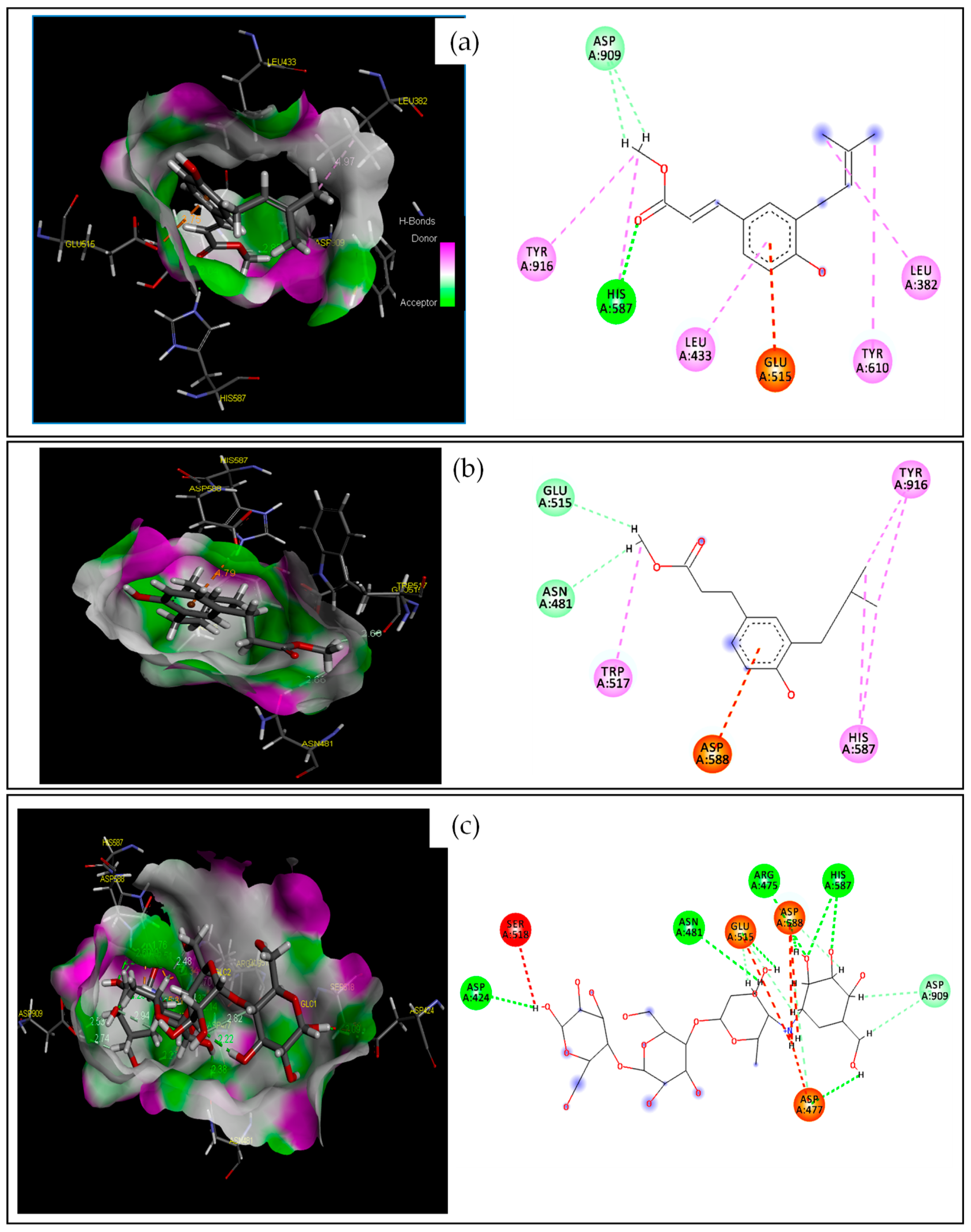
The complexes formed by compounds 2 and 8 with the S. mutans (3AIC) target had low energy values of −5.049 and −5.042 kcal/mol, respectively, which were similar to those of the native ligand, acarbose (ACA, −6.674 kcal/mol, Table S2). Furthermore, three strong hydrogen bonds between compound 2 and the active site residue of the S. mutans (3AIC) target were observed: one conventional H-bond type (O/HIS587(A)-HE2/bond distance = 2.04 Å) and two other carbon H-bonds (H/ASP909(A)-OD1/bond distance = 2.85 Å, and H/ASP909(A)-OD1/bond distance = 2.74 Å). An electrostatic interaction with GLU515(A) was also observed. In contrast, this compound formed five hydrophobic interactions with the following active site residues: LEU382(A), HIS587(A), TYR610(A), TYR916(A), and LEU433(A) (Table S2 and Figure 4a). This result was supported by several recent studies [49,50,51].

2.2.2. Molecular Dynamics Simulation

Figure 5 presents the 3D diagrams of the best pose for compounds 2 and 8 with the active binding sites 3LE0, 4N82, and 3AIC, as determined by molecular docking (green) and molecular dynamics (yellow) studies. As shown in Figure 6, a fluctuation in all the curves was initially observed during the first 100 picoseconds. A slight variation in the potential energy was observed between 100 and 800 ps in curve (a), and between 100 and 400 ps in curve (e), and between 100 and 900 ps in curve (c). Finally, the 3LE0–compound 8, 4N82–compound 2, and 3AIC–compound 2 complexes retained their stabilities in the last intervals (between 800 and 1500 ps, between 400 and 1500 ps, and between 900 and 1500 ps, respectively) (Figure 6a–c).

2.2.3. Drug-Likeness Prediction and ADMET Properties

Drug-Likeness Evaluation

Different physicochemical properties were calculated for compounds 2 and 8, aiming to verify the drug-likeness rules using the SwissADME online server. As shown in Table 4, it is apparent that compounds 2 and 8 have hydrogen bond donors < 5 (n-HD: 0~7) and hydrogen bond acceptors < 10 (n-HA: 0~10). In addition, the molecular weight values of these compounds belong to the interval 100~500 g/mol, and their MLogP and WLogP values are <5. Also, the nROTB values are <11, which denotes the flexibility of these compounds. Moreover, the TPSA values obtained for both compounds are less than 140 Å.

ADMET Properties

The absorption, distribution, metabolism, excretion, and toxicity (ADMET) were calculated for compounds 2 and 8 using the online server pkCSM (Table 5). The Caco-2 values obtained for 2 and 8 are 1.178 and 1.417, respectively, whereas the human intestinal absorption (HIA) values are 99.207% and 97.682%, respectively. In terms of the distribution, the logPS values of 2 and 8 are 1.800 and −1.692, respectively, while their logBB values are 0.311 and 0.030, respectively (Table 5). Compounds 2 and 8 are CYP1A2 inhibitors, but not CYP2C19 and CYP2D6 inhibitors. In addition, these compounds are not CYP2D6 and CYP3A4 substrates and are likely not OCT2 substrates. These compounds have a total clearance of 0.744 and 0.780, respectively. Furthermore, compounds 2 and 8 are neither hERG I nor hERG II inhibitors.

3. Discussion

3.1. In Vitro Antibacterial Activity of Compounds 114

The antimicrobial activity of compounds 114 was classified based on the MIC (minimum inhibitory concentration) values as follows: MIC values lower than 10 μg/mL, between 11 and 100 μg/mL, between 101 and 500 μg/mL, and between 501 and 1000 μg/mL correspond to very strong, strong, moderate, and weak activities, respectively, while MIC values higher than 1000 μg/mL denote inactivity [1,52,53,54]. Based on these criteria, compounds 2 and 8 displayed strong or moderate activity against all the tested bacteria (Table 1). However, the lowest MIC value was observed for 2 (plicatin B), which showed strong activity against S. mutans, S. mitis, and S. sanguinis (MIC = 31.2 µg/mL). This compound also showed strong activity against S. salivarius and S. sobrinus (MIC = 62.5 µg/mL). The strong activity of compound 2 against S. mutans is an auspicious result because it is one of the leading agents causing dental caries [1]. Plicatin B (2) has been isolated from Brazilian propolis samples, even though its occurrence is less commonly reported [55]. However, data on the antimicrobial activity of plicatin B are still scarce in the literature [56]. On the other hand, artepillin C (5) and drupanin (6), which are among the most common prenylated compounds found in Brazilian green propolis, showed moderate or weak activity against most of the tested bacteria (MIC between 125 and 1000 µg/mL), with drupanin displaying MIC values slightly lower than artepillin C. Compounds 1, 10, and 14 had no activity against any of the oral bacteria tested, with MIC values higher than 1000 µg/mL.
According to the literature, compounds with an MBC:MIC ratio ≤ 4, and 4 < MBC:MIC ≤ 32 display bactericidal and bacteriostatic effects, respectively, whereas MBC:MIC values > 32 denote a bacteria-resistant effect [57]. Based on these criteria, compounds 2 and 8 are considered to display bactericidal activity against all the oral bacteria tested in this study.
We compared our data on the antibacterial activity of compounds 114 with the relevant literature to derive insights into the potential structure–activity relationships. Based on the comparison of the antimicrobial activities of artepillin C (5), drupanin (6), and 4-hydroxycinnamic acid, Aga and coworkers considered that the antimicrobial activity of 4-hydroxycinnamic acid derivatives may be boosted by an increasing number of prenyl groups in the structure [58]. However, in this study, the mono-prenylated compound 2 (plicatin B) and its corresponding carboxylic acid 6 (drupanin) were more active against the tested oral bacteria than the di-prenylated compounds 1 and 5 (artepillin C), respectively. These results indicated clearly that other structural features than the prenyl groups also play a key role in the antibacterial activity of compounds 114. For example, the phenolic hydroxyl is a crucial structural feature in relation to the antibacterial activity, as evidenced by the comparison between the MIC values of the isomers 2 (a C-prenylated compound) and 4 (an O-prenylated compound). The role played by phenolic hydroxyl is likely due to its capability to form a hydrogen bond to the active binding sites of the bacteria enzymes, thus inhibiting microbial enzymes and simultaneously increasing the affinity with cytoplasmic membranes [59]. This is reinforced by the higher activity of 2 (a mono-C-prenylated ester) compared to compound 3 (a tri-prenylated ester that contains a prenyl group at the oxygen at C4), despite the higher lipophilicity of 3. Indeed, docking studies confirmed the formation of a hydrogen bond between the phenolic hydroxyl of compounds 2 and 8 with the residues HIS 85(A), and ASN 104(A) in S. mitis, and S sanguinis, respectively.
The nature of the oxygenated function at C9 and the type of covalent bond between C7 and C8 were also found to affect the antibacterial activity of compounds 114; however, the effects of these structural features vary according to the number and position of the prenyl groups. For example, the mono-C-prenylated methyl ester 2 (plicatin B) and its hydrogenated derivative 8 are more active than their corresponding carboxylic acids 7 (drupanin) and 13, indicating that the presence of an ester function at C9 may potentialize the antibacterial activity of mono-C-prenylated compounds. However, in the case of compounds di-C-prenylated 5 (artepillin C) and O-prenylated 7, the carboxyl group at C9 increases the antibacterial activity as compared to the corresponding methyl esters 1 and 4. Similarly, the presence of a single bond between C7 and C8 in the structure of mono-C-prenylated compounds 8 (a methyl ester) and 13 (a carboxylic acid) decreases the antibacterial activity as compared to compounds 2 and 6, which display a double bond between C7 and C8. On the other hand, for compounds di- and tri-prenylated 12 and 9, a single bond between C7 and C8 increases the antibacterial activity as compared to 5 and 3, respectively.
The important role played by the number and position of the prenyl groups in terms of the antibacterial activity of compounds 114 can be better understood based on the studies reported by Kubo and coworkers. According to those authors, the mechanism by which phenolic acids enter the molecular structure of the bacteria membrane involves the orientation of the phenol hydroxyl into the aqueous phase by hydrogen bonding, and the non-polar carbon chain alignment into the lipid phase by dispersion forces. The activity tends to disappear when the hydrophilic force exceeds the hydrophobic one [60]. Therefore, based on the literature [60], the antibacterial activity of compounds 114 can be understood in terms of the balance between hydrophilicity/lipophilicity. In this sense, compounds 2 and 8 can be inferred to achieve the best lipophilicity/hydrophilicity balance among the tested compounds. Hydrolysis of plicatin B (2) produces drupanin (6), which has a carboxyl group at C9. Although this group can anchor the compound to the lipid bilayer [61], it also increases the hydrophilicity of drupanin (6) as compared to plicatin B (2) and consequently decreases its antibacterial activity. On the other hand, the presence of two C-prenyl groups in the structure of artepillin C (5) increases lipophilicity as compared to 2 and 6, thus decreasing its antibacterial activity. Finally, the presence of an α,β-unsaturated carbonyl conjugated with the aromatic ring of phenylpropanoids 17 confers conformational and electronic characteristics that are strongly influenced by the phenol-OH group in the para position [62]. In principle, because of the rotation around the single bond between C7 and C8, compounds 8, 9, 12, 13, and 14 can assume a wider variety of conformations as compared to 17, 10, and 11, which could improve their intermolecular interactions with a potential target [63]. However, data from this study suggest a combined effect of the nature of the bond between C7 and C8 and the number and position of the prenyl groups on the molecular shape of compounds 114 and, consequently, on their antibacterial activity.

3.2. Molecular Docking and Molecular Dynamics

The antibacterial activity of compounds 2 and 8 against S. mitis, S. sanguinis, and S. mutans was further investigated using molecular docking. Molecular docking is a computational technique that predicts the precise positioning of a ligand molecule over a receptor protein molecule to create a stable complex [64]. This orientation is essential for predicting the binding affinity and strength of the ligand–protein connection through the use of a scoring function [65]. The interaction between a drug and its receptor reduces the overall free energy of the system, providing insights into the molecule’s affinity and activity, which is crucial for drug design and discovery [66]. Docking studies can both describe the protein and the ligand as complementary surfaces [67,68] and provide the ligand–protein pairwise interaction energies [69].
The protein targets in S. mitis (3LE0) [46], S. sanguinis (4N82) [47], and S. mutans (3AIC) [48] were selected on the basis of their relevance to the microorganism virulence and the availability of their resolved crystallographic structures in the Protein Data Bank (PDB) [70]. Based on the binding score energy values, compounds 2 (−4.228 kcal/mol) and 8 (−4.476 kcal/mol) were found to inhibit S. mitis (3LE0) more effectively than the native ligand glycerol (GOL) (−3.655 kcal/mol). However, compound 8 was predicted to be a stronger S. mitis (3LE0) target binder compared to compound 2 due to the formation of a more stable complex, as indicated by its negative score energy of −4.476 kcal/mol. These results are in full agreement with the lower MIC value of compound 8 (0.12 mM) compared to compound 2 (0.13 mM) (Table 2). Furthermore, these results suggest that compound 8 has a high affinity with the S. mitis (3LE0) pocket, which is confirmed by establishing four strong hydrogen bonds (a conventional H-bond type and three carbon H-bonds) [71,72]. Of particular interest is the electrostatic interaction with ARG112(A). Recent studies have revealed that residues ARG112(A) and HIS85(A) play an essential role in the inhibition of the S. mitis (3LE0) target [73,74].
Conversely, compound 2 is more active against S. sanguinis (MIC = 31.2 µg/mL, 0.13 mM) than compound 8 (MIC = 62.5 µg/mL, 0.25 mM) (Table 1). Indeed, molecular docking with these compounds revealed the higher stability of the complex formed by compound 2 (score energy of −6.156 kcal/mol) with the protein target in S. sanguinis (4N82) compared to that of the complex formed by compound 4 (score energy of −5.575 kcal/mol). This stability is similar to that of the flavin mononucleotide (FMN), the native ligand (score energy of −6.671 kcal/mol, Table 3). This higher stability can be due to the two strong carbon H-bonds (O/ASN104(A)-H and O/GLY103(A)-HA3), one Pi–lone pair interaction (with TYR63(A)), and four hydrophobic interactions between compound 2 and residues PHE107(A), MET132(A), PRO62(A), and TYR64(A) of the S. sanguinis protein target (Table 3 and Figure 3A). Indeed, the role played by these residues in the stability of the formed complex has been reported [47,73].
The score energies of the complexes formed between the S. mutans target (3AIC) and compounds 2 (−5.049 kcal/mol) and 8 (−5.042 kcal/mol) are similar to those of acarbose (−6.674 kcal/mol, Table 3), the native ligand. Furthermore, compound 2 fits nicely into the S. mutans (3AIC) pocket due to the formation of three strong hydrogen bonds [71,72] with active site residue of the S. mutans (3AIC) target: one conventional H-bond type and two other carbon H-bonds. An electrostatic interaction with GLU515(A) was also observed. In contrast, this compound formed five hydrophobic interactions with the following active site residues: LEU382(A), HIS587(A), TYR610(A), TYR916(A), and LEU433(A) (Table 3, Figure 4A). This result was supported by several recent studies [49,50,51].
MD simulations were also performed to investigate the stability of the best complexes obtained through molecular-docking calculations for compounds 2 and 8 with the protein targets. Based on a generic model of the physics driving interatomic interactions, MD simulations predict the movements of individual atoms inside proteins and other molecular systems across time [75]. These simulations display the locations of all the atoms with femtosecond temporal precision, and they can capture a wide range of significant biomolecular processes, such as conformational change, ligand binding, and protein folding [76]. Crucially, these simulations can also forecast, down to the atomic level, how biomolecules will react to various perturbations like mutation, phosphorylation, protonation, or ligand addition or removal [77]. In this study, the stability of the best complexes obtained through molecular-docking calculations for compound 2 (4N82–compound 2 and 3AIC–compound 2) and 8 (3LE0–compound 8) with the protein targets was investigated by MD simulations (Figure 5). The energy potential fluctuation observed in all the curves in the first 100 picoseconds (ps) (Figure 5) can be justified based on the variation in the potential energy of three complexes: 3LE0–compound 8 (Figure 5a); 4N82–compound 2 (Figure 5b); and 3AIC–compound 2 (Figure 5c). However, all the studied complexes exhibit high stability, as confirmed by the MD simulations, because they maintained almost the same types of interactions compared to the molecular-docking studies. This was also confirmed by the stability of the potential energy as a function of time (Figure 5a–c), as reported in previous papers [78,79].

3.3. Drug-Likeness and ADMET Properties

We also estimated the drug-likeness and ADMET properties of compounds 2 and 8. The drug-likeness considers how simple physicochemical properties impact molecular behavior in vivo [80]. Lipinski’s “rule-of-five” predicts the drug-likeness of an oral therapeutic agent based on its physicochemical properties [81]. This depicts the connection between physicochemical and pharmacokinetics indices [82]. According to this rule, an orally active drug-like compound should not have more than one violation of the following criteria: hydrogen bond donors not greater than 5, hydrogen bond acceptors not greater than 10, molecular weight not greater than 500 Da, and an octanol–water partition coefficient (log P) not greater than 5 [81,82]. As shown in Table 4, all the physicochemical properties of compounds 2 and 8 follow Lipinsky’s rule.
The ADMET (absorption, distribution, metabolism, excretion, and toxicity) properties play a critical role in sorting ligands in drug discovery programs, as they account for the failure of 60% of drug candidates during the drug development process [83]. In this study, the Caco-2 values obtained for 2 and 8 are higher than −5.15 (>−5.15 cm/s), which confirms that these compounds have good human intestinal permeability. Moreover, both compounds have HIA values higher than 30%, which means that compounds 2 and 8, when administered orally, can be absorbed from the gastrointestinal system into the human body’s bloodstream. The logPS values (−3< logPS <−2) indicate that compounds 2 and 8 cannot penetrate the CNS. Additionally, the logBB values of compounds 2 (0.311) and 8 (0.030) indicate that compound 2 is expected to readily cross the blood–brain barrier, whereas compound 8 is poorly distributed in the brain. Compounds 2 and 8 are CYP1A2 inhibitors, but not CYP2C19 and CYP2D6 inhibitors. In addition, these compounds are not CYP2D6 and CYP3A4 substrates and are not likely OCT2 substrates. These compounds are also expected to have a low clearance (<5 mL/min/kg). Furthermore, compounds 2 and 8 are neither hERG I nor hERG II inhibitors, showing no hepatotoxicity risk. Therefore, compounds 2 and 8 satisfy the drug-likeness criteria without violating the Lipinski [81], Veber [84], and Egan rules. Furthermore, based on these results, these compounds are not expected to cause problems with regard to oral bioavailability and pharmacokinetic parameters.

4. Materials and Methods

4.1. Synthesis of Compounds 114

Compounds 14 were synthesized by alkylation of methyl p-coumarate according to the methodology employed by Patra and coworkers [85] with modifications (Scheme 1). In this procedure, methyl p-coumarate (0.5 mmol) was added to a 25 mL flask equipped with a magnetic stirrer bar. Next, 15 mL toluene was added. The mixture was cooled to 0 °C, and 1.5 mmol NaH was added in portions. After 15 min, 1.5 mmol (153 µL) of prenyl bromide was added dropwise. The reaction progress was monitored through TLC using a Hex:EtOAc 8:2 (v/v) solution as the eluent. After 24 h, the solvent was removed under reduced pressure on a rotary evaporator, and the reaction mixture was then extracted with EtOAc (3 × 15 mL). The organic phase was washed with saturated NaCl solution (15 mL), dried over MgSO4, and filtered off. The solvent was removed by evaporation at reduced pressure on a rotaevaporator. The compounds were isolated by column chromatography with gradient elution, starting with 100% hexane and changing to Hex:EtOAc 9.8:0.2 (v/v) after the separation of the first compounds. The resulting solids were dried under a vacuum to yield compounds 1 (32% yield), 2 (18% yield), 3 (15% yield), and 4 (35% yield) (Scheme 1).
Compounds 5, 6, and 7 were obtained from a hydrolysis reaction according to the methodology described by Uto and co-workers [86]. In this procedure, a solution of KOH (15 mL of a 10% aqueous) was added to a solution of the methyl esters 1, 2, and 4 in MeOH (15 mL) (Scheme 1). The mixture was heated under reflux for 1 h, cooled to 0–5 °C, and acidified with 1 mol/L HCl. After removing MeOH under reduced pressure, the aqueous residue was extracted with EtOAc (3 × 15 mL). The organic phase was washed with saturated NH4Cl and brine, dried over MgSO4, and evaporated under reduced pressure. The resulting white solids were dried under a vacuum to afford compounds 5, 6, and 7 in a 100% yield.
Compounds 8, 9, 12, 13, and 14 were obtained from 2, 4, 5, 6, and 7 by catalytic hydrogenation. In this procedure, compounds 2, 4, 5, 6, and 7 and Pd/C (catalyst) were wholly dissolved in HPLC-grade EtOAc and transferred to a high-pressure reactor under stirring and kept under an H2 atmosphere and 400 psi at room temperature for 1–2 h (Scheme 1). The resulting oil was dried under a vacuum to yield compounds 8, 9, 12, 13, and 14 in a 100% yield.
Compounds 10 and 11 were synthesized according to the methodology reported by Kantee and co-workers [87] with some modifications (Scheme 1). In this procedure, LiAlH4 (0.68 mmol) was quickly added to a solution of compounds 3 and 4 (0.34 mmol) in THF (5.5 mL) at 0 °C. The reaction mixture was stirred under an N2 atmosphere at 0 °C for 1 h. After one hour, the mixture was stirred at room temperature for 9–10 h. The reaction mixture was then augmented with H2O, conc. HCl and extracted with EtOAc (x3). The combined organic layers were washed with brine, dried over MgSO4, and concentrated under a vacuum. Purification of the crude residue by column chromatography with isocratic elution with Hex/EtOAc 9.8:0.2 (v/v) gave the corresponding alcohol derivative. The resulting compounds were dried under a vacuum to yield compounds 10 (65% yield) and 11 (89% yield).

4.2. Antibacterial Assays

The in vitro antimicrobial action of artepillin C (5), drupanin (6) and derivatives 14, and 714 was evaluated in terms of their minimum inhibitory concentration (MIC) values [88], which were interpreted as the lowest concentrations that inhibited bacterial growth. To this end, Streptococcus mutans (ATCC 25175), Streptococcus mitis (ATCC 49456), Streptococcus salivarius (ATCC 25975), Streptococcus sanguinis (ATCC 10556), Streptococcus sobrinus (ATCC 33478), Enterococcus faecalis (ATCC 4082), and Lactobacillus paracasei (ATCC 11578) were assayed by the broth microdilution method in 96-well microplates. The bacterial colonies were cultured at 37 °C for 24 h in blood agar (Difco Labs, Detroit, MI, USA). Further standardization of the inoculum quantity was accomplished on a Femto spectrophotometer (São Paulo, Brazil) operating at a wavelength of 625 nm to match 0.5 on the McFarland scale (1.5 × 10⁸ CFU/mL). The microorganism suspensions were diluted to a final concentration of 5 × 10⁵ CFU/mL. Samples of compounds 114 were dissolved in DMSO (Merck, Darmstadt, Germany) and tryptic soy broth (TSB, Difco) to obtain final concentrations varying from 0.98 to 2000 μg/mL. Inoculated microplate wells containing DMSO (1%) and TSB (1:5 v/v and 100%) were employed as the negative control. A non-inoculated well was also added to ensure the medium sterility. Chlorhexidine (Sigma-Aldrich, St. Louis, MO, USA) was used as the positive control at concentrations ranging from 0.115 to 59 μg/mL in TSB (Difco). The microplates were sealed with plastic film and incubated at 37 °C for 24 h. Next, 30 μL of revealing 0.02% resazurin (Sigma-Aldrich, St. Louis, MO, USA) was added to each microplate well to indicate the microbial viability. Before resazurin was added and to determine the MBC, a 10 µL aliquot of the inoculum was aseptically removed from each well and plated onto blood agar (Difco). The plates were incubated as described previously. The minimum bactericidal concentration (MBC) was determined as the lowest compound concentration that killed > 99.9% of the initial bacteria population, at which point no visible bacterial growth occurred [89]. The MIC values were assessed by analysis of the color change of the resazurin solution from blue (without metabolic activity) to pink (with metabolic activity) [90]. The MIC and MBC were determined in triplicate for each microorganism, and the results are presented as the mean of three replicates.

4.3. Computational Methodology

4.3.1. Ligands and Targets Preparation

The 3D structures of the most active compounds 2 and 8 (Scheme 1) were optimized using the semi-empirical method AM1 [91], which was implemented in Hyperchem 8.0.8 software (Hypercube Inc., Gainesville, FL, USA). Next, the 3D structures were converted into .mdb format for use as input in the MOE-docking.
The crystal structures of S. mitis (3LE0) [46], S. sanguinis (4N82) [47], and S. mutans (3AIC) [48] were selected as antibacterial targets, which were downloaded from the Protein Data Bank (http://www.rcsb.org/, accessed on 8 June 2024). Some information related to the target structures is provided in Table 1.

4.3.2. Docking Method Protocol and Validation

The molecular-docking studies were carried out to identify the binding interactions of the most active compounds 2 and 8 within the binding site residue of the targets by using MOE software [92], and the docking protocol steps were followed and detailed in our previous research [93,94] by using the following default parameters: placement: Triangle Matcher; rescoring 1: London dG scoring function. The visualization of all the possible interactions that have been formed between the compounds and receptor active site residues was generated by the BIOVIA DS visualize package (Dassault Systèmes BIOVIA, Discovery Studio Modeling Environment, 2020).
The re-docking of all the native ligands to their targets was conducted using “Dock Option” implemented in the MOE software [92] to validate the used method. The RSMD values of the obtained complexes (targets–crystallized ligands) were less than 2.50 Å [95], meaning that the docking method is accurate and successful.

4.3.3. MD Simulations

MD simulations were carried out using the “Compute/Simulations/Dynamics” options in the MOE 2014.0901 software [92] to study the stability of the two best-formed complexes. The following default parameters were used: the Nose–Poincaré–Andersen (NPA) algorithm to search the interactions of different residues in each system, and the MMFF94x force field for the energy minimization step of these complexes [96,97]. The MD protocol was settled for 1500 ps (an equilibrium period of 100 ps, followed by a production period of 1400 ps, at a constant temperature of 310 K). Finally, the plots of the potential energies U (kcal/mol) variations as a function of time t (ps) were produced using OriginPro 9.1.software [98].

4.3.4. ADMET Evaluation

The drug-likeness rules, namely Lipinski, Veber, and Ghose, were verified by calculating the different physicochemical parameters (total polar surface area (TPSA); number of rotatable bonds (nROTB), molecular weight (MW), lipophilicity (LogP), number of hydrogen bond acceptors (nHA), and number of hydrogen bond donors (nHD)) using the SwissADME server (http://www.swissadme.ch/, accessed on 8 June 2024) [99].
The pkCSM server (http://biosig.unimelb.edu.au/pkcsm/prediction, accessed on 8 June 2024) [100] was used for the analysis of the ADMET profiles by calculating the following parameters: the absorption (Caco-2: colon adenocarcinoma, HIA: human intestinal absorption), distribution (CNS: central nervous system permeability, BBB: blood–brain barrier permeability), metabolism (CYP1A2 inhibitor, CYP2C19 inhibitor, CYP2D6 inhibitor), excretion (renal OCT2 substrate: organic cation transporter 2, total clearance), and toxicity (hERG: human ether-a-go-go-related gene, hepatotoxicity).

5. Conclusions

Plicatin B (2) was identified as a compound with promising activity against oral bacteria, including S. mutans, one of the main causative agents of bacteria caries. Moreover, this study revealed plicatin B to be more active than artepillin C, which has been highlighted as the main factor responsible for the antimicrobial activity of the Brazilian green propolis. However, among all the tested compounds, the most remarkable activity against S. mutans was found for compound 8, which was obtained by hydrogenation of plicatin B.
The molecular-docking/dynamics simulation results proved that compounds 2 and 8 have high binding affinities against three targets, S. sanguinis (4N82) and S. mitis (3LE0), as confirmed by the low energy values and various interactions with the active site residues of these targets. The ADMET properties were predicted for all the candidates to validate the pharmacodynamics and pharmacokinetics profiles, and these compounds verified the three rules, Lipinski, Veber, and Egan. In summary, the results of the present research revealed that the most active compounds 2 and 8 should be further investigated to explore their anti-cariogenic properties, including evaluation of their cytotoxicity, shelf life, anti-demineralization, remineralization, and cariostatic properties. However, the initial efforts must be focused on optimizing the synthesis of compounds 14, aiming to reduce the reaction time and to make the isolation of the prenylated compounds 1, 2, 3, and 4 from the reaction mixture simpler and faster.

Supplementary Materials

The following supporting information can be downloaded at https://www.mdpi.com/article/10.3390/antibiotics13080787/s1, Tables S1 and S2: Docking results of compounds 2 and 8 docked into the S. sanguinis (4N82) and S. mutans (3AIC) targets. Figures S1–S56: 1H and 13C NMR spectra, DEPT 135 spectra, and ESI-TOF-MS spectra of compounds 114.

Author Contributions

Conceptualization, A.E.M.C., J.K.B. and R.B.S.; methodology, A.E.M.C., C.H.G.M. and R.B.S.; software, R.B.S.; investigation, T.M.V., J.G.B., S.L.d.S. and A.L.O.S.; validation, formal analysis, I.D., S.R. and N.A.; resources, J.K.B.; writing—original draft preparation, A.E.M.C. and R.B.S.; writing—review and editing, A.E.M.C. and R.B.S. All authors have read and agreed to the published version of the manuscript.

Funding

This research was funded by the São Paulo Research Foundation (FAPESP, grants 19/11700-0 and 17/04138-8) and the National Council for Scientific and Technological Development (CNPq, proc. 301417/2019-9 and 310648/2022-0).

Institutional Review Board Statement

Not applicable.

Informed Consent Statement

Not applicable.

Data Availability Statement

A draft of this paper is available as a preprint at https://chemrxiv.org/engage/chemrxiv/article-details/65a42dbe9138d23161e85cb4 (accessed on 17 January 2024).

Acknowledgments

The authors thank the São Paulo Research Foundation (FAPESP) for the financial support and scholarships, and the National Council for Scientific and Technological Development (CNPq) for fellowships.

Conflicts of Interest

The authors declare no conflicts of interest.

References

  1. Oliveira, T.A.S.; Santiago, M.B.; Santos, V.H.P.; Silva, E.O.; Martins, C.H.G.; Crotti, A.E.M. Antibacterial activity of essential oils against oral pathogens. Chem. Biodiv. 2022, 19, e202200097. [Google Scholar] [CrossRef]
  2. Teshome, A.; Muche, A.; Girma, B. Prevalence of dental caries and associated factors in East Africa, 2000–2020: Systematic review and meta-analysis. Front. Public Health 2021, 9, 645091. [Google Scholar] [CrossRef] [PubMed]
  3. Lendenmann, U.; Grogan, J.; Oppenheim, F. Saliva and dental pellicle-a review. Adv. Dent. Res. 2000, 14, 22–24. [Google Scholar] [CrossRef] [PubMed]
  4. Chawhuaveang, D.D.; Yu, O.Y.; Yin, I.X.; Lam, W.Y.-H.; Mei, M.L.; Chu, C.-H. Acquired salivary pellicle and oral diseases: A literature review. J. Dent. Sci. 2021, 16, 523–529. [Google Scholar] [CrossRef] [PubMed]
  5. Reich, M.; Kümmerer, K.; Al-Áhmad, A.; Hannig, C. Fatty acid profile of the initial oral biofilm (pellicle): An in-situ study. Lipids 2013, 48, 929–937. [Google Scholar] [CrossRef] [PubMed]
  6. Siqueira, W.; Custodio, W.; McDonald, E. New insights into the composition and functions of the acquired enamel pellicle. J. Dent. Res. 2012, 91, 1110–1118. [Google Scholar] [CrossRef]
  7. Teixeira, E.W.; Napimoga, M.; Carneiro, V. In vitro inhibition of Streptococci binding to enamel acquired pellicle by plant lectins. J. Appl. Microbiol. 2006, 101, 111–116. [Google Scholar] [CrossRef]
  8. Castro, P.; Tovar, J.A.; Jaramilo, L. Adhesion of Streptococcus mutans to salivary proteins in caries-free and caries-susceptible individuals. Acta Odontol. Latinoam. 2006, 19, 59–66. [Google Scholar]
  9. Selwitz, R.H.; Ismail, A.I.; Pitts, N.B. Dental caries. Lancet 2007, 369, 51–59. [Google Scholar] [CrossRef]
  10. Cavalcanti, I.M.; Del Bel Cury, A.A.; Jenkinson, H.F.; Nobbs, A.H. Interactions between Streptococcus oralis, Actinomyces oris, and Candida albicans in the development of multispecies oral microbial biofilms on salivary pellicle. Mol. Oral Microbiol. 2017, 32, 60–73. [Google Scholar] [CrossRef]
  11. Sahoo, S.R.; Nandini, D.B.; Basandi, P.S.; Selvamani, M.; Donoghue, M. A Comparison of pre- and postbreakfast tooth brushing in caries prevention through the estimation of Streptococcus mutans counts: A prospective clinical and microbiological study. J. Microsc. Ultrastruct. 2022, 10, 168–173. [Google Scholar] [CrossRef] [PubMed]
  12. Kamal, D.; Hassanein, H.; Akah, M.; Abdelkawy, M.A.; Hamza, H. Caries preventive and antibacterial effects of two natural mouthwashes vs chlorhexidine in high caries-risk patients: A randomized clinical trial. J. Contemp. Dent. Pract. 2020, 21, 1316–1324. [Google Scholar] [CrossRef]
  13. Fibryanto, E.; Santoso, L. Mouthwashes: A review on its efficacy in preventing dental caries. JKGT 2023, 5, 91–96. [Google Scholar] [CrossRef]
  14. Masapu, A.; Kumar, S.S.M.; Ashok, K.P.; Anusha, G.; Sangineedy, J.; Ashok, S. Evaluation of the antiplaque efficacy of chlorhexidine mouthwash immediately and 30 min after brushing with fluoride toothpaste—A pilot study. RGUHS J. Dent. Sci. 2019, 11, 17–21. [Google Scholar] [CrossRef] [PubMed]
  15. Busxer, S. Has resistance to chlorhexidine increased among clinically-relevant bacteria? A systematic review of time course and subpopulation data. PLoS ONE 2021, 16, e0256336. [Google Scholar] [CrossRef]
  16. Cieplik, F.; Jakubovics, N.S.; Buchalla, W.; Maisch, T.; Hellwig, E.; Al-Ahmad, A. Resistance toward chlorhexidine in oral bacteria—Is there cause for concern? Front. Microbiol. 2019, 10, 587. [Google Scholar] [CrossRef]
  17. Fahim, A.; Himratul-Aznita, W.H.; Abdul-Rahman, P.S. Allium-sativum and bakuchiol combination: A natural alternative to Chlorhexidine for oral infections? Pakistan J. Med. Sci. 2020, 36, 271–275. [Google Scholar] [CrossRef]
  18. Soares, K.H.; Firoozi, P.; Souza, G.M.; Martins, B.L.; Falci, S.G.M.; Santos, C.R.R. Efficacy of probiotics compared to chlorhexidine mouthwash in improving periodontal status: A systematic review and meta-analysis. Int. J. Dent. 2023, 2023, 4013004. [Google Scholar] [CrossRef]
  19. Hossain, R.; Quispe, C.; Khan, R.A.; Saikat, A.S.M.; Ray, P.; Ongalbek, D.; Yeskaliyeva, B.; Jain, D.; Smeriglio, A.; Trombetta, D.; et al. Propolis: An update on its chemistry and pharmacological applications. Chin. Med. 2022, 17, 651. [Google Scholar] [CrossRef]
  20. Teixeira, E.W.; Negri, G.; Meira, R.M.S.A.; Message, D.; Salatino, A. Plant origin of green propolis: Bee behavior, plant anatomy and chemistry. Evid. Based Complement. Altern. Med. 2005, 2, 85–92. [Google Scholar] [CrossRef]
  21. Costa, P.; Somensi, L.B.; da Silva, R.; Mariano, L.N.B.; Boeing, T.; Longo, B.; Perfoll, E.; de Souza, P.; Gushiken, L.F.S.; Pellizzon, C.H.; et al. Role of the antioxidant properties in the gastroprotective and gastric healing activity promoted by Brazilian green propolis and the healing efficacy of Artepillin C. Inflammopharmacology 2020, 28, 1009–1025. [Google Scholar] [CrossRef] [PubMed]
  22. de Oliveira, P.F.; Lima, I.M.D.; Munari, C.C.; Bastos, J.K.; da Silva, A.A.; Tavares, D.C. Comparative evaluation of antiproliferative effects of Brazilian green propolis, its main source Baccharis dracunculifolia, and their major constituents artepillin c and baccharin. Planta Med. 2014, 80, 490–492. [Google Scholar] [CrossRef] [PubMed]
  23. Ding, J.L.; Matsumiya, T.; Hayakari, R.; Shiba, Y.; Kawaguchi, S.; Seya, K.; Ueno, K.; Imaizumi, T. Daily Brazilian green propolis intake elevates blood artepillin C levels in humans. J. Sci. Food Agric. 2021, 101, 4855–4861. [Google Scholar] [CrossRef]
  24. Kimoto, T.; Koya-Miyata, S.; Hino, K.; Micallef, M.J.; Hanaya, T.; Arai, S.; Ikeda, M.; Kurimoto, M. Pulmonary carcinogenesis induced by ferric nitrilotriacetate in mice and protection from it by Brazilian propolis and artepillin C. Virchows Archiv. Int. J. Phatol. 2001, 438, 259–270. [Google Scholar] [CrossRef]
  25. Klosek, M.; Sedek, L.; Lewandowska, H.; Czuba, Z.P. The effect of ethanolic extract of Brazilian green propolis and artepillin C on aFGF-1, Eselectin, and CD40L secreted by human gingival fibroblasts. Cent. Eur. J. Immunol. 2021, 46, 438–445. [Google Scholar] [CrossRef]
  26. Naramoto, K.; Kato, M.; Ichihara, K. Effects of an ethanol extract of Brazilian green propolis on human cytochrome P450 enzyme activities in vitro. J. Agric. Food Chem. 2014, 62, 11296–11302. [Google Scholar] [CrossRef]
  27. Paulino, N.; Abreu, S.R.L.; Uto, Y.; Koyama, D.; Nagasawa, H.; Hori, H.; Dirsch, V.M.; Vollmar, A.M.; Scremin, A.; Bretz, W.A. Anti-inflammatory effects of a bioavailable compound, Artepillin C, in Brazilian propolis. Eur. J. Pharmacol. 2008, 587, 296–301. [Google Scholar] [CrossRef]
  28. Ahn, M.R.; Kunimasa, K.; Ohta, T.; Kumazawa, S.; Kamihira, M.; Kaji, K.; Uto, Y.; Hori, H.; Nagasawa, H.; Nakayama, T. Suppression of tumor-induced angiogenesis by Brazilian propolis: Major component artepillin C inhibits in vitro tube formation and endothelial cell proliferation. Cancer Lett. 2007, 252, 235–243. [Google Scholar] [CrossRef]
  29. Sawaya, A.C.H.F.; Cunha, I.B.S.; Marcucci, M.C. Analytical methods applied to diverse types of Brazilian propolis. Chem. Cent. J. 2011, 5, 27. [Google Scholar] [CrossRef]
  30. Seibert, J.B.; Bautista-Silva, J.P.; Amparo, T.R.; Petit, A.; Pervier, P.; Almeida, J.C.D.; Azevedo, M.C.; Silveira, B.M.; Brandao, G.C.; de Souza, G.H.B.; et al. Development of propolis nanoemulsion with antioxidant and antimicrobial activity for use as a potential natural preservative. Food Chem. 2019, 287, 61–67. [Google Scholar] [CrossRef]
  31. Souza, R.M.; de Souza, M.C.; Patitucci, M.L.; Silva, J.F.M. Evaluation of antioxidant and antimicrobial activities and characterization of bioactive components of two Brazilian propolis samples using a pK(a)-guided fractionation. Z. Naturforsch. C 2007, 62, 801–807. [Google Scholar] [CrossRef]
  32. Veiga, R.S.; Mendonça, S.D.; Mendes, P.B.; Paulino, N.; Mimica, M.J.; Lagareiro Netto, A.A.; Lira, I.S.; López, B.G.C.; Negrão, V.; Marcucci, M.C. Artepillin C and phenolic compounds responsible for antimicrobial and antioxidant activity of green propolis and Baccharis dracunculifolia DC. J. Appl. Microbiol. 2017, 122, 911–920. [Google Scholar] [CrossRef] [PubMed]
  33. Jorge, R.; Furtado, N.A.J.C.; Sousa, J.P.B.; da Silva, A.A.; Gregorio, L.E.; Martins, C.H.G.; Soares, A.E.E.; Bastos, J.K.; Cunha, W.R.; Silva, M.L.A. Brazilian Propolis: Seasonal variation of the prenylated p-coumaric acids and antimicrobial activity. Pharm. Biol. 2008, 46, 889–893. [Google Scholar] [CrossRef]
  34. Meto, A.; Colombari, B.; Meto, A.; Boaretto, G.; Pinetti, D.; Marchetti, L.; Benvenuti, S.; Pellati, F.; Blasi, E. Propolis affects Pseudomonas aeruginosa growth, biofilm formation, eDNA release and phenazine production: Potential involvement of polyphenols. Microorganisms 2020, 8, 243. [Google Scholar] [CrossRef] [PubMed]
  35. Leitão, D.P.S.; Silva Filho, A.A.; Polizello, A.C.M.; Bastos, J.K.; Spadaro, A.C.C. Comparative evaluation of in-vitro effects of Brazilian green propolis and Baccharis dracunculifolia extracts on cariogenic factors of Streptococcus mutans. Biol. Pharm. Bull. 2004, 27, 1834–1839. [Google Scholar] [CrossRef] [PubMed]
  36. Cardoso, J.G.; Iorio, N.L.P.; Rodrigues, L.F.; Couri, M.L.B.; Farah, A.; Maia, L.C.; Antonio, A.G. Influence of a Brazilian wild green propolis on the enamel mineral loss and Streptococcus mutans’ count in dental biofilm. Arch. Oral Biol. 2016, 65, 77–81. [Google Scholar] [CrossRef]
  37. Righi, A.A.; Negri, G.; Salatino, A. Comparative chemistry of propolis from eight Brazilian localities. Evid. Based Complement. Altern. Med. 2013, 2013, 267878. [Google Scholar] [CrossRef]
  38. Salatino, A.; Teixeira, E.W.; Negri, G.; Message, D. Origin and chemical variation of Brazilian propolis. Evid. Based Complement. Altern. Med. 2005, 2, 33–38. [Google Scholar] [CrossRef]
  39. Shahinozzama, M.; Basak, B.; Emran, R.; Rozario, P.; Obanda, D.N. Artepillin C: A comprehensive review of its chemistry, bioavailability, and pharmacological properties. Fitoterapia 2020, 147, 104775. [Google Scholar] [CrossRef]
  40. Costa, P.; Almeida, M.O.; Lemos, M.; Arruda, C.; Casoti, R.; Somensi, L.B.; Boeing, T.; Mariott, M.; da Silva, R.; Stein, B.D.; et al. Artepillin C, drupanin, aromadendrin-4′-O-methyl-ether and kaempferide from Brazilian green propolis promote gastroprotective action by diversified mode of action. J. Ethnopharmacol. 2018, 226, 82–89. [Google Scholar] [CrossRef]
  41. Mishima, S.; Ono, Y.; Araki, Y.; Akao, Y.; Nozawa, Y. Two related cinnamic acid derivatives from Brazilian honey bee propolis, baccharin and drupanin, induce growth inhibition in allografted sarcoma S-180 in mice. Biol. Pharm. Bull. 2005, 28, 1025–1030. [Google Scholar] [CrossRef]
  42. Antunes, O.A.C.; da Silva, J.F.M.; Estrada, G.O.D. Artepillin C: A review. Lett. Drug Des. Discov. 2008, 5, 88–92. [Google Scholar] [CrossRef]
  43. Kano, Y.; Horie, N.; Doi, S.; Aramaki, F.; Maeda, H.; Hiragami, F.; Kawamura, K.; Motoda, H.; Koike, Y.; Akiyama, J.; et al. Artepillin C derived from propolis induces neurite outgrowth in PC12m3 cells via ERK and p38 MAPK pathways. Neurochem. Res. 2008, 33, 1795–1803. [Google Scholar] [CrossRef] [PubMed]
  44. Rodrigues, D.M.; Portapilla, G.B.; Silva, G.M.; Duarte, A.; Rotta, C.G.; Silva, C.; de Albuquerque, S.; Bastos, J.K.; Campo, V.L. Synthesis, antitumor activity and in silico analyses of amino acid derivatives of artepillin C, drupanin and baccharin from green propolis. Bioorg. Med. Chem. 2021, 47, 116372. [Google Scholar] [CrossRef] [PubMed]
  45. Tiveron, A.P.; Rosalen, P.L.; Franchin, M.; Lacerda, R.C.C.; Bueno-Silva, B.; Benso, B.; Denny, C.; Ikegaki, M.; Alencar, S.M. Chemical characterization and antioxidant, antimicrobial, and anti-inflammatory activities of South Brazilian organic propolis. PLoS ONE 2016, 11, e0165588. [Google Scholar] [CrossRef]
  46. Feil, S.C.; Lawerence, S.; Mulhern, T.D.; Holien, J.K.; Hotze, E.M.; Farrand, S.; Tweten, R.K.; Parker, M.W. Structure of the lectin regulatory domain of the cholesterol-dependent cytolysin lectinolysin reveals the basis for its lewis antigen specificity. Structure 2012, 20, 248–258. [Google Scholar] [CrossRef]
  47. Makhlynets, O.; Boal, A.K.; Rhodes, D.V.; Kitten, T.; Rosenzweig, A.C.; Stubbe, J. Streptococcus sanguinis class Ib ribonucleotide reductase: High activity with both iron and manganese cofactors and structural insights. J. Biol. Chem. 2014, 289, 6259–6272. [Google Scholar] [CrossRef] [PubMed]
  48. Ito, K.; Ito, S.; Shimamura, T.; Weyand, S.; Kawarasaki, Y.; Miwaka, T.; Abe, K.; Kobayashi, T.; Cameron, A.D.; Iwata, S. Crystal structure of glucansucrase from the dental caries pathogen Streptococcus mutans. J. Mol. Biol. 2011, 408, 177–186. [Google Scholar] [CrossRef]
  49. Nijampatnam, B.; Zhang, H.; Cai, X.; Michalek, S.M.; Wu, H.; Velu, S.E. Inhibition of Streptococcus mutans biofilms by the natural stilbene piceatannol through the inhibition of glucosyltransferases. ACS Omega 2018, 3, 8378–8385. [Google Scholar] [CrossRef]
  50. Atta, L.; Khalil, R.; Khan, K.M.; Zehra, M.; Saleem, F.; Nur-e-Alam, M.; Ul-Haq, Z. Virtual screening, synthesis and biological evaluation of Streptococcus mutans mediated biofilm inhibitors. Molecules 2022, 27, 1455. [Google Scholar] [CrossRef]
  51. Liao, Y.; Zhang, M.; Lin, X.; Yan, F. Diaryl urea derivative molecule inhibits cariogenic Streptococcus mutans by affecting exopolysaccharide synthesis, stress response, and nitrogen metabolism. Front. Cell. Infect. Microbiol. 2022, 12, 904488. [Google Scholar] [CrossRef] [PubMed]
  52. Rios, J.L.; Recio, M.C. Medicinal plants and antimicrobial activity. J. Ethnopharmacol. 2005, 100, 80–84. [Google Scholar] [CrossRef]
  53. Saleem, M.; Nazir, M.; Ali, M.S.; Hussain, H.; Lee, Y.S.; Riaz, N.; Jabbar, A. Antimicrobial natural products: An update on future antibiotic drug candidates. Nat. Prod. Rep. 2010, 27, 238–254. [Google Scholar] [CrossRef] [PubMed]
  54. Abdallah, E.M.; Alhatlani, B.Y.; Menezes, R.P.; Martins, C.H.G. Back to nature: Medicinal plants as promising sources for antibacterial drugs in the post-antibiotic era. Plants 2023, 12, 3077. [Google Scholar] [CrossRef] [PubMed]
  55. Tazawa, S.; Warashina, T.; Noro, T.; Miyase, T. Studies on the constituents of Brazilian Propolis. Chem. Pharm. Bull. 1998, 46, 1477–1479. [Google Scholar] [CrossRef]
  56. Schmitt, A.; Telikepalli, H.; Mitscher, L.A. Plicatin B, the antimicrobial principle of Psoralea juncea. Phytochemistry 1991, 30, 3569–3570. [Google Scholar] [CrossRef]
  57. Makade, C.S.; Shenoi, P.R.; Bhongade, B.A.; Shingane, S.A.; Ambulkar, P.C.; Schewale, A.M.; Shewale, A.M. Estimation of MBC: MIC ratio of herbal extracts against common endodontic pathogens. J. Pharm. Bioall. Sci. 2024, 16, S1414–S1416. [Google Scholar] [CrossRef]
  58. Aga, H.; Shibuya, T.; Sugimoto, T.; Kurimoto, M.; Nakajima, S. Isolation and identification of antimicrobial compounds in Brazilian propolis. Biosci. Biotech. Biochem. 1994, 58, 945–946. [Google Scholar] [CrossRef]
  59. Liu, X.L.; Xu, Y.J.; Go, M.L. Functionalized chalcones with basic functionalities have antibacterial activity against drug sensitive Staphylococcus aureus. Eur. J. Med. Chem. 2008, 43, 1681–1687. [Google Scholar] [CrossRef]
  60. Kubo, I.; Muroi, H.; Kubo, A. Structural functions of antimicrobial long-chain alcohols and phenols. Bioorg. Med. Chem. 1995, 3, 873–880. [Google Scholar] [CrossRef]
  61. Roleira, F.M.F.; Siquet, C.; Orrù, E.; Garrido, E.M.; Garrido, J.; Milhazes, N.; Podda, G.; Paiva-Martins, F.; Reis, S.; Carvalho, R.A. Lipophilic phenolic antioxidants: Correlation between antioxidant profile, partition coefficients and redox properties. Bioorg. Med. Chem. 2010, 18, 5816–5825. [Google Scholar] [CrossRef]
  62. Oufensou, S.; Casalini, S.; Balmas, V.; Carta, P.; Chtioui, W.; Dettori, M.A.; Fabbri, D.; Migheli, Q.; Delogu, G. Prenylated trans-cinnamic esters and ethers against clinical Fusarium spp.: Repositioning of natural compounds in antimicrobial discovery. Molecules 2021, 26, 658. [Google Scholar] [CrossRef]
  63. Araya-Cloutier, C.; Vincken, J.-P.; van de Schans, M.G.M.; Hageman, J.; Schafternaar, G.; den Besten, H.M.W.; Gruppen, H. QSAR-based molecular signatures of prenylated (iso)flavonoids underlying antimicrobial potency against and membrane-disruption in Gram positive and Gram negative bacteria. Sci. Rep. 2018, 8, 9267. [Google Scholar] [CrossRef] [PubMed]
  64. Agu, P.C.; Afiukwa, C.A.; Orji, O.U.; Ezeh, E.M.; Ofoke, I.H.; Ogbu, C.O.; Ugwaja, E.I.; Aja, P.M. Molecular docking as a tool for the discovery of molecular targets of nutraceuticals in diseases management. Sci. Rep. 2023, 13, 13398. [Google Scholar] [CrossRef]
  65. Yunta, M.J.R. Docking and ligand binding affinity: Uses and pitfalls. Am. J. Model. Optim. 2016, 4, 74–114. [Google Scholar] [CrossRef]
  66. Bhagat, R.T.; Butle, S.R.; Khobragade, D.S.; Wankhede, S.B.; Prasad, C.C.; Mahure, D.S.; Armarkar, A.V. Molecular Docking in Drug Discovery. J. Pharm. Res. Int. 2021, 33, 46–58. [Google Scholar] [CrossRef]
  67. Meng, E.C.; Shoichet, B.K.; Kuntz, I.D. Automated docking with grid-based energy evaluation. J. Comput. Chem. 2004, 13, 505–524. [Google Scholar] [CrossRef]
  68. Morris, G.M.; Goodsell, D.S.; Halliday, R.S.; Huey, R.; Hart, W.E.; Belew, R.K.; Olson, A.J. Automated docking using a Lamarckian genetic algorithm and an empirical binding free energy function. J. Comp. Chem. 1998, 19, 1639–1662. [Google Scholar] [CrossRef]
  69. Feig, M.; Onufriev, A.; Lee, M.S.; Im, W.; Case, D.A.; Brooks, C.L. Performance comparison of generalized born and Poisson methods in the calculation of electrostatic solvation energies for protein structures. J. Comput. Chem. 2004, 25, 265–284. [Google Scholar] [CrossRef]
  70. Silva, D.R.; Deps, T.D.; Sakaguchi, O.A.S.; Costa, E.M.M.B.; Santos, C.A.O.; Silva, J.P.R.; Silva, B.D.; Ribeiro, F.F.; Mendonça-Júnior, F.J.B.; Silva, A.C.B. Molecular docking of phytochemicals against Streptococcus mutans virulence targets: A proteomic insight into drug plannin. In Oral Health Care—An Important Issue of the Modern Society; Ardelean, L.C., Rusu, L.C., Eds.; IntechOpen: London, UK, 2022; pp. 1–20. [Google Scholar]
  71. Imberty, A.; Hardman, K.D.; Carver, J.P.; Perez, S. Molecular modelling of protein-carbohydrate interactions. Docking of monosaccharides in the binding site of concanavalin A. Glycobiology 1991, 1, 631–642. [Google Scholar] [CrossRef]
  72. Wade, R.C.; Goodford, P.J. The role of hydrogen-bonds in drug binding. Progr. Clin. Biol. Res. 1989, 289, 433–444. [Google Scholar]
  73. Fernandes, L.P.; Silva, J.M.B.; Martins, D.O.S.; Santiago, M.B.; Martins, C.H.G.; Jardim, A.C.G.; Oliveira, G.S.; Pivatto, M.; Souza, R.A.C.; Franca, E.F.; et al. Fragmentation study, dual anti-bactericidal and anti-viral effects and molecular docking of cobalt (III) complexes. Int. J. Mol. Sci. 2020, 21, 8355. [Google Scholar] [CrossRef] [PubMed]
  74. Velarde, J.J.; Piai, A.; Lichtenstein, I.J.; Lynskey, N.N.; Chou, J.J.; Wessels, M.R. Structure of the Streptococcus pyogenes NAD+ Glycohydrolase translocation domain and its essential role in toxin binding to oropharyngeal keratinocytes. J. Bacteriol. 2022, 204, e0036621. [Google Scholar] [CrossRef] [PubMed]
  75. Hollingsworth, S.A.; Dror, R.O. Molecular dynamics simulation for all. Neuron 2018, 99, 1129–1143. [Google Scholar] [CrossRef]
  76. De Vivo, M.; Masetti, M.; Bottegoni, G.; Cavalli, A. Role of molecular dynamics and related methods in drug discovery. J. Med. Chem. 2016, 59, 4035–4061. [Google Scholar] [CrossRef]
  77. Salo-Ahen, O.M.; Alanko, I.; Bhadane, R.; Bonvin, A.M.J.J.; Honorato, R.V.; Hossain, S.; Juffer, A.H.; Kabedev, A.; Lahtela-Kakkonen, M.; Larsen, A.S.; et al. Molecular Dynamics simulations in drug discovery and pharmaceutical development. Processes 2021, 9, 71. [Google Scholar] [CrossRef]
  78. Roy, N.; Ghosh, B.; Roy, D.; Bhaumik, B.; Roy, M.N. Exploring the inclusion complex of a drug (umbelliferone) with α-cyclodextrin optimized by molecular docking and increasing bioavailability with minimizing the doses in human body. ACS Omega 2020, 5, 30243–30251. [Google Scholar] [CrossRef] [PubMed]
  79. Djebaili, R.; Kenouche, S.; Daoud, I.; Melkemi, N.; Belkadi, A.; Mesli, F. Investigation of [3H]diazepam derivatives as allosteric modulators of GABAA receptor α1β2γ2 subtypes: Combination of molecular docking/dynamic simulations, pharmacokinetics/drug-likeness prediction, and QSAR analysis. Struct. Chem. 2022, 34, 791–823. [Google Scholar] [CrossRef] [PubMed]
  80. Bickerton, G.R.; Paolini, G.V.; Besnard, J.; Muresan, S.; Hopkins, A.L. Quantifying the chemical beauty of drugs. Nat. Chem. 2012, 4, 90–98. [Google Scholar] [CrossRef] [PubMed]
  81. Lipinski, C.A.; Lombardo, F.; Dominy, B.W.; Feeney, P.J. Experimental and computational approaches to estimate solubility and permeability in drug discovery and development settings. Adv. Drug. Deliv. Rev. 2001, 46, 3–26. [Google Scholar] [CrossRef]
  82. Lipinski, C.A. Drug-like properties and the causes of poor solubility and poor permeability. J. Pharmacol. Toxicol. Methods 2000, 44, 235–249. [Google Scholar] [CrossRef]
  83. Pant, S.; Verma, S.; Pathak, R.K.; Singh, D.B. Structure-based drug designing. In Bioinformatics: Methods and Applications; Academic Press: Cambridge, MA, USA, 2022; pp. 219–231. [Google Scholar]
  84. Veber, D.F.; Johnson, S.R.; Cheng, H.Y.; Smith, B.R.; Ward, K.W.; Kopple, K.D. Molecular properties that influence the oral bioavailability of drug candidates. J. Med. Chem. 2002, 45, 2615–2623. [Google Scholar] [CrossRef] [PubMed]
  85. Patra, T.; Bag, S.; Kancherla, R.; Mondal, A.; Dey, A.; Pimparkar, S.; Agasti, S.; Modak, A.; Debabrata Maiti, D. Palladium-catalyzed directed para C-H functionalization of phenols. Angew. Chem. Int. Ed. 2016, 55, 7751–7755. [Google Scholar] [CrossRef]
  86. Uto, Y.; Hirata, A.; Fujita, T.; Takubo, S.; Nagasawa, H.; Hori, H. First total synthesis of artepillin C established by o,o’-diprenylation of p-halophenols in water. J. Org. Chem. 2002, 67, 2355–2357. [Google Scholar] [CrossRef] [PubMed]
  87. Kantee, K.; Rukachaisirikul, V.; Tadpetch, K. Synthesis of tetrahydropyranyl diarylheptanoids from Dioscorea villosa. Tetrahedron Lett. 2016, 57, 3505–3509. [Google Scholar] [CrossRef]
  88. CLSI. Methods for Dilution Antimicrobial Susceptibility Tests for Bacteria That Grow Aerobically, 11th ed.; Approved Standard M07; Clinical and Laboratory Standards Institute: Wayne, PA, USA, 2018. [Google Scholar]
  89. Duarte, E.A.; Santiago, M.B.; Silva, N.B.S.; Martins, C.H.G.; Gatto, C.C. Crystal design, spectroscopic analyses and antibacterial study of new carbazate ligands and their Cu(II) complexes. Inorg. Chim. Acta 2023, 549, 121421. [Google Scholar] [CrossRef]
  90. Sarker, S.D.; Nahar, L.; Kumarasamy, Y. Microtitre plate-based antibacterial assay incorporating resazurin as an indicator of cell growth, and its application in the in vitro antibacterial screening of phytochemicals. Methods 2007, 42, 321–324. [Google Scholar] [CrossRef]
  91. Stewart, J.M.P. Optimization of parameters for semi-empirical methods V: Modification of NDDO approximations and application to 70 elements. J. Mol. Model. 2007, 13, 1173–1213. [Google Scholar] [CrossRef]
  92. MOE. Molecular Operating Environment (MOE), 2013.08; Chemical Computing Group Inc.: Montreal, QC, Canada, 2014. [Google Scholar]
  93. Daoud, I.; Melkemi, N.; Salah, T.; Ghalem, S. Combined QSAR, molecular docking and molecular dynamics study on new Acetylcholinesterase and Butyrylcholinesterase inhibitors. Comput. Biol. Chem. 2018, 74, 304–326. [Google Scholar] [CrossRef]
  94. Daoud, I.; Mesli, F.; Melkemi, N.; Ghalem, S.; Salah, T. Discovery of potential SARS-CoV 3CL protease inhibitors from approved antiviral drugs using: Virtual screening, molecular docking, pharmacophore mapping evaluation and dynamics simulation. J. Biomol. Struct. Dyn. 2021, 40, 12574–12591. [Google Scholar] [CrossRef]
  95. Bajda, M.; Więckowska, A.; Hebda, M.; Guzior, N.; Sotriffer, C.A.; Malawska, B. Structure-based search for new inhibitors of cholinesterases. Int. J. Mol. Sci. 2013, 14, 5608–5632. [Google Scholar] [CrossRef] [PubMed]
  96. Bond, S.D.; Leimkuhler, B.J.; Laird, B.B. The Nosé-Poincaré method for constant temperature molecular dynamics. J. Comp. Phys. 1999, 151, 114–134. [Google Scholar] [CrossRef]
  97. Parikesit, A.A.; Zahroh, H.; Nugroho, A.S.; Hapsari, A.; Tambunan, U.S.F. The computation of cyclic peptide with prolin-prolin bond as fusion inhibitor of DENV envelope protein through molecular docking and molecular dynamics simulation. arXiv-bio 2015, arXiv:1511.01388. [Google Scholar] [CrossRef]
  98. OriginLab. OriginPro 9.1; OriginLab Corporation: Northampton, MA, USA, 2014. [Google Scholar]
  99. Daina, A.; Michielin, O.; Zoete, V. SwissADME: A free web tool to evaluate pharmacokinetics, drug-likeness and medicinal chemistry friendliness of small molecules. Sci. Rep. 2017, 7, 42717. [Google Scholar] [CrossRef]
  100. Pires, D.E.; Blundell, T.L.; Ascher, D.B. pkCSM: Predicting small-molecule pharmacokinetic and toxicity properties using graph-based signatures. J. Med. Chem. 2015, 58, 4066–4072. [Google Scholar] [CrossRef]
Figure 1. Chemical structures of drupanin (I) and artepillin C (II).
Antibiotics 13 00787 g001
Scheme 1. Synthesis of compounds 114.
Antibiotics 13 00787 sch001
Figure 2. Three- and two-dimensional diagrams of the interaction between the S. mitis target (3LE0) and compound 2 (a), compound 8 (b), and the native ligand (GOL) (c).
Antibiotics 13 00787 g002
Figure 3. Three- and two-dimensional diagrams of the interactions between the S. sanguinis target (4N82) and compound 2 (a), compound 8 (b), and the native ligand (FMN) (c).
Antibiotics 13 00787 g003
Figure 4. Three- and two-dimensional diagrams of the interactions between the S. mutans target (4N82) and compound 2 (a), compound 8 (b), and the native ligand (FMN) (c).
Antibiotics 13 00787 g004
Figure 5. Three-dimensional diagrams of the best pose found by molecular docking (green) and molecular dynamics (yellow) for 3LE0–compound 8 (a), 4N82–compound 2 (b), and 3AIC–compound 2 (c).
Antibiotics 13 00787 g005
Figure 6. Potential energy (U) as a function of time for three complexes; (a) 3LE0–compound 8, (b) 4N82–compound 2, and (c) 3AIC–compound 2.
Antibiotics 13 00787 g006
Table 1. Minimum inhibitory concentration (MIC) and minimum bactericidal concentration (MBC) values (µg/mL) of compounds 29, and 1113 *.
CompoundEnterococcus
Faecalis
ATCC 4082
Lactobacillus
Paracasei
ATCC 11578
Streptococcus Salivarius
ATCC 25975
Streptococcus Sobrinus ATCC 33478Streptococcus Mitis
ATCC 49456
Streptococcus Sanguinis ATCC 10556Streptococcus Mutans
ATCC 25175
2500 (2.03)
500 (2.03)
500 (2.03)
1000 (4.06)
62.5 (0.25)
250 (1.01)
62.5 (0.25)
62.5 (0.25)
31.2 (0.13)
31.2 (0.13)
31.2 (0.13)
31.2 (0.13)
31.2 (0.13)
31.2 (0.51)
3>2000 (>5.23)
>2000 (>5.23)
>2000 (>5.23)
>2000 (>5.23)
2000 (5.23) >2000 (>5.23)2000 (5.23) >2000 (>5.23500 (1.31)
1000 (2.61)
1000 (2.61) 2000 (5.23)>2000 (5.23) >2000 (5.23)
4>2000 (8.12) >2000 (>8.12)>2000 (8.12) >2000 (>8.12)>2000 (8.12) >2000 (>8.12)>2000 (8.12) >2000 (>8.12)1000 (4.06) 2000 (8.12)>2000 (8.12) >2000 (>8.12)>2000 (8.12) >2000 (>8.12)
5>2000 (>6.66) >2000 (>6.66)500 (1.66)
1000 (3.33)
1000 (3.33) 2000 (6.66)500 (1.66)
2000 (6.66)
125 (0.41)
1000 (3.33)
500 (1.66)
1000 (3.33)
500 (1.66)
2000 (6.66)
61000 (3.09)
1000 (3.09)
500 (1.55)
500 (1.55)
250 (0.77)
500 (1.55)
125 (0.39)
125 (0.39)
250 (0.77)
250 (0.77)
250 (0.77)
250 (0.77)
125 (0.39)
250 (0.77)
7500 (2.15)
>2000 (>8.61)
1000 (4.31) 2000 (8.61)1000 (4.31) 2000 (8.61)1000 (4.31) 1000 (4.31)500 (2.15)
500 (2.15)
500 (2.15)
500 (2.15)
1000 (4.31) 2000 (8.61)
8125 (0.50)
125 (0.50)
62.5 (0.25)
250 (1.0)
62.5 (0.25)
62.5 (0.25)
125 (0.50)
125 (0.50)
31.25 (0.12) 31.25 (0.12)62.5 (0.25)
62.5 (0.25)
62.5 (0.25)
62.5 (0.25)
9>2000 (8.0) >2000 (8.0)>2000 (8.0) >2000 (8.0)1000 (4.0)
2000 (8.0)
1000 (4.0)
1000 (4.0)
500 (2.0)
1000 (4.0)
1000 (4.0)
1000 (4.0)
2000 (8.0)
2000 (8.0)
112000 (9.16) >2000 (>9.1)2000 (9.16) >2000 (>9.1)62.5 (0.29)
125 (0.57)
2000 (9.16) 2000 (9.16)500 (2.29)
1000 (4.58)
1000 (4.58) 2000 (9.16)1000 (4.58) 2000 (9.16)
121000 (3.26)
2000 (6.53)
500 (1.63)
1000 (3.26)
500 (1.63)
500 (1.63)
250 (0.82)
500 (1.63)
62.5 (0.20)
125 (0.41)
500 (1.63)
1000 (3.26)
500 (1.63)
500 (1.63)
13>2000 (8.46) >2000 (8.46)>2000 (8.46) >2000 (8.46)>2000 (8.46) >2000 (8.46)>2000 (8.46) >2000 (8.46)250 (1.06)
1000 (4.23)
1000 (4.23) 1000 (4.23)>2000 (8.46)
>2000 (8.46)
CHD3.69 (7.30)
3.69 (7.30)
1.84 (3.64)
1.84 (3.64)
0.92 (1.82)
0.92 (1.82)
1.84 (3.64)
1.84 (3.64)
3.69 (7.30)
3.69 (7.30)
3.69 (7.30)
3.69 (7.30)
0.92 (1.82)
0.92 (1.82)
* Compounds 1, 10, and 14 were inactive (MIC > 1000 µg/mL) against all the tested bacteria and were not included). CHD: chlorhexidine. Values between parenthesis are expressed in mM. MBC values are given in blue.
Table 2. Some information related to the studied targets.
3LE04N823AIC
MethodX-ray diffractionX-ray diffractionX-ray diffraction
MicroorganismS. mitisS. sanguinisS. mutans
ChainAA, B, C, D, E, FA, B, C, D, E
Sequence length153178488
Resolution (Å)1.911.883.11
Native ligandsGOLFMNACA
GOL: glycerol; FMN: flavin mononucleotide; ACA: acarbose.
Table 3. Docking results of compounds 2 and 8 when docked into the S. mitis target (3LE0).
S-Score (kcal/mol)RMSD
(Å)
Bonds between Atoms of Compounds and Active Site Residues
Atom of CompoundInvolved Receptor AtomsInvolved Receptor ResiduesCategoryType of
Interaction
2−4.2282.997OHH11ARG120(A)HBConventional HB
HNE2HIS85(A)HBConventional HB
/NH2ARG112(A)ElectrostaticPi-Cation
C/TYR62(A)HydrophobicPi-Pi T-shaped
C/VAL117(A)HydrophobicAlkyl
8−4.4762.932HNE2HIS85(A)HBConventional HB
HOD1ASP114(A)HBCarbon HB
HOD2ASP77(A)HBCarbon HB
HOD1ASP114(A)HBCarbon HB
/NH2ARG112(A)ElectrostaticPi-Cation
C/VAL117(A)HydrophobicAlkyl
GOL−3.6552.149O1HH11ARG120(A)HBConventional HB
O3HH11ARG112(A)HBConventional HB
H12NE2HIS85(A)HBCarbon H-Bond
HB: hydrogen bond. GOL: glycerol.
Table 4. Physicochemical properties and drug-likeness predictions of compounds 2 and 8.
Physicochemical PropertiesDrug-Likeness Rules
TPSA (Å2)n-ROTMW (g/mol)MLogPn-HAn-HDLipinskiVeberEgan
WLogP
(0~140)(0~11)(100~500)(0~5)(0~12)(0~7)
239.422225.251.4930AcceptedAcceptedAccepted
2.40
830.191223.272.3320AcceptedAcceptedAccepted
3.01
TPSA: topological polar surface area; n-ROT: number of rotatable; MW: molecular weight; Log P: logarithm of partition coefficient of compound between n-octanol and water; n-HA: number of hydrogen bond acceptors; n-HD: number of hydrogen bonds donors.
Table 5. ADMET/pharmacokinetic properties of compounds 2 and 8.
ADMETParametersCompounds
28
AbsorptionCaco2 (Log Papp × 10−6 cm/s)1.1781.417
HIA (%)99.20797.682
DistributionCNS (logPS)−1.800−1.692
BBB (logBB)0.3110.030
Metabolism CYP1A2 inhibitorYesYes
CYP2C19 InhibitorNoNo
CYP2D6 inhibitorNoNo
CYP2D6 substrateNoNo
CYP3A4 substrateNoNo
ExcretionRenal OCT2 substrateNoNo
Total clearance (log mL/min/kg)0.7440.780
ToxicityhERG I and II inhibitorsNoNo
HepatotoxicityNoNo
Caco-2: colon adenocarcinoma, HIA: human intestinal absorption, CNS: central nervous system permeability, BBB: blood–brain barrier permeability, Renal OCT2 substrate: organic cation transporter 2, hERG: human ether-a-go-go-related gene.
Disclaimer/Publisher’s Note: The statements, opinions and data contained in all publications are solely those of the individual author(s) and contributor(s) and not of MDPI and/or the editor(s). MDPI and/or the editor(s) disclaim responsibility for any injury to people or property resulting from any ideas, methods, instructions or products referred to in the content.