Article

Murepavadin Enhances the Killing Efficacy of Ciprofloxacin against Pseudomonas aeruginosa by Inhibiting Drug Efflux

1
State Key Laboratory of Medicinal Chemical Biology, Key Laboratory of Molecular Microbiology and Technology of the Ministry of Education, Department of Microbiology, College of Life Sciences, Nankai University, Tianjin 300071, China
2
Department of Laboratory Medicine, 5th Medical Center of PLA General Hospital, Beijing 100071, China
*
Author to whom correspondence should be addressed.
Antibiotics 2024, 13(9), 810; https://doi.org/10.3390/antibiotics13090810
Submission received: 11 July 2024 / Revised: 20 August 2024 / Accepted: 23 August 2024 / Published: 26 August 2024

Abstract

:
Pseudomonas aeruginosa is a multidrug-resistant Gram-negative pathogen and one of the leading causes of ventilator-associated pneumonia and infections in patients with chronic obstructive pulmonary disease and cystic fibrosis. Murepavadin is a peptidomimetic that specifically targets outer-membrane lipopolysaccharide transport protein LptD of P. aeruginosa. In this study, we find that murepavadin enhances the bactericidal efficacy of ciprofloxacin. We further demonstrate that murepavadin increases intracellular accumulation of ciprofloxacin by suppressing drug efflux. In addition, the murepavadin–ciprofloxacin combination exhibits a synergistic bactericidal effect in an acute murine pneumonia model. In conclusion, our results identify an effective drug combination for the treatment of P. aeruginosa infections.

1. Introduction

Antimicrobial resistance (AMR) has become a major challenge in global public health. The continuous evolution of antimicrobial resistance in bacterial pathogens has caused significant global impacts on clinics [1,2]. Presently, 700,000 people die each year worldwide due to antimicrobial-resistant bacteria. It is predicted that there will be more than 10 million deaths per year attributed to antimicrobial resistance by 2050, with a global cost of around USD 100 trillion [3]. Meanwhile, the development rate of new antibiotics is declining, leaving fewer options for treating multidrug-resistant bacterial infections. To deal with this challenge, new antibiotics are needed to ensure that effective therapeutic strategies are sustained [4].
Pseudomonas aeruginosa is a ubiquitous Gram-negative pathogen that is responsible for high morbidity and mortality in patients with cystic fibrosis (CF) as well as community-acquired and nosocomial infections [5]. Data from China Antimicrobial Surveillance Network (CHINET) revealed that among 445,199 clinical bacterial isolates in 2023, P. aeruginosa was the fifth prevalent nosocomial pathogen, following Escherichia coli, Klebsiella pneumoniae, Staphylococcus aureus, and Acinetobacter baumannii. P. aeruginosa displays resistance to a variety of antibiotics [6]. The intrinsic resistance mechanisms include low membrane permeability, multidrug efflux pumps, chromosomally encoded β–lactamase, and biofilm formation. Mutation and acquisition of antibiotic resistance genes through horizontal transfer further increase its resistance [7,8,9]. In May 2024, the World Health Organization (WHO) updated its bacterial priority pathogens list, in which P. aeruginosa is listed in the high-priority group that requires development of new treatments (https://www.who.int/publications/i/item/9789240093461, accessed on 24 June 2024).
Murepavadin (also known as POL7080) is the first peptide-based inhibitor of LptD in P. aeruginosa [10]. LptD is a component of the LptABCDEFG complex, which is responsible for the transport and insertion of lipopolysaccharide (LPS) into the outer membrane [1,11]. By binding to LptD, murepavadin inhibits the assembly of LPS, ultimately leading to cell death. In preclinical studies, murepavadin showed no cross-resistance with current standard-of-care antibiotics and was highly specific and potent against P. aeruginosa in vitro and in vivo [9,12]. In addition to its direct antibacterial activity, murepavadin may also enhance the host’s immune response by activating MRGPRX2 and induce degranulation in murine mast cells, thereby contributing to bacterial clearance and wound healing [13]. A phase III trial of intravenous murepavadin for the treatment of pneumonia was withdrawn due to severe nephrotoxicity [14]. On the contrary, aerosol administration of murepavadin displays low systemic exposure and limited toxicity, resulting in good tolerability [14,15]. Inhaled murepavadin is under a clinical trial to treat P. aeruginosa lung infections in cystic fibrosis patients (https://spexisbio.com/impv/, accessed on 20 June 2024).
Ciprofloxacin is a fluoroquinolone antibiotic that is used to treat P. aeruginosa infections, but rising resistance frequently precludes its utilization [16,17,18]. It inhibits the activity of DNA gyrase, leading to DNA damage and cell death. In response to ciprofloxacin-induced DNA damage, RecA is activated, which triggers the cleavage of LexA and PrtR, leading to activation of the SOS response and the production of pyocins, respectively [19,20]. A novel ciprofloxacin–azithromycin sinus stent maintained a uniform coating and sustained delivery with anti-biofilm activity against P. aeruginosa [21]. A phase III clinical trial in non-cystic fibrosis bronchiectasis patients demonstrated that ciprofloxacin dry powder for inhalation has the potential to be an effective treatment for P. aeruginosa infections [22]. Another phase III clinical trial in non-cystic fibrosis bronchiectasis patients demonstrated trends towards clinical benefit with ciprofloxacin dry powder for inhalation. However, primary end-points were not met [23]. In addition, inhaled liposomal ciprofloxacin significantly delayed the median time to first pulmonary exacerbation in a phase III clinical trial in non-cystic fibrosis bronchiectasis patients with chronic P. aeruginosa lung infection, but not in another trial with similar settings [24].
Combination therapy may achieve synergistic effects and reduce the likelihood of resistance emergence and side effects [25,26]. Drug combinations may produce pharmacokinetic potentiation, where the therapeutic activity of one drug is enhanced through the influences on absorption, distribution, or metabolism by another drug [27]. For example, the combination of colistin with rifampicin was able to significantly improve the therapeutic outcomes in patients infected by carbapenemase-producing Enterobacteriaceae strains [28]. Previously, we demonstrated that murepavadin enhances the efficacy of β-lactam antibiotics against P. aeruginosa by disrupting outer-membrane integrity [29]. Since both inhaled ciprofloxacin and murepavadin have been evaluated in clinical trials, here, we examined the combinatorial bactericidal effects of the two antibiotics. Our results demonstrate that murepavadin enhances the cellular accumulation of ciprofloxacin and hence generates synergistic effects in vitro and in vivo.

2. Results

2.1. Combination of Murepavadin with Ciprofloxacin Increases the Killing Efficacy against P. aeruginosa

We first examined the bactericidal activity of murepavadin and ciprofloxacin against a reference strain, PA14. Murepavadin was used at 0.5 μg/mL, which has been shown to inhibit the growth of 99.1% of tested clinical isolates from the United States, Europe, and China [30]. The concentration of ciprofloxacin was used at 0.25 μg/mL, which is below the clinical breakpoint (0.5 μg/mL) [31]. Murepavadin and ciprofloxacin alone reduced the live bacterial numbers by approximately 50 and 500 times, respectively, eight hours after treatment. The combination further reduced the bacterial number by 1.51 × 10⁴ and 1.50 × 10³ times, respectively. Afterwards, bacteria treated with the single antibiotic started to grow, whereas the live bacterial number kept decreasing in the presence of the murepavadin–ciprofloxacin combination, resulting in approximately 4.50 × 10⁸- and 9.90 × 10⁷-fold fewer bacteria compared with each antibiotic alone after twenty four hours (Figure 1, Table S2).

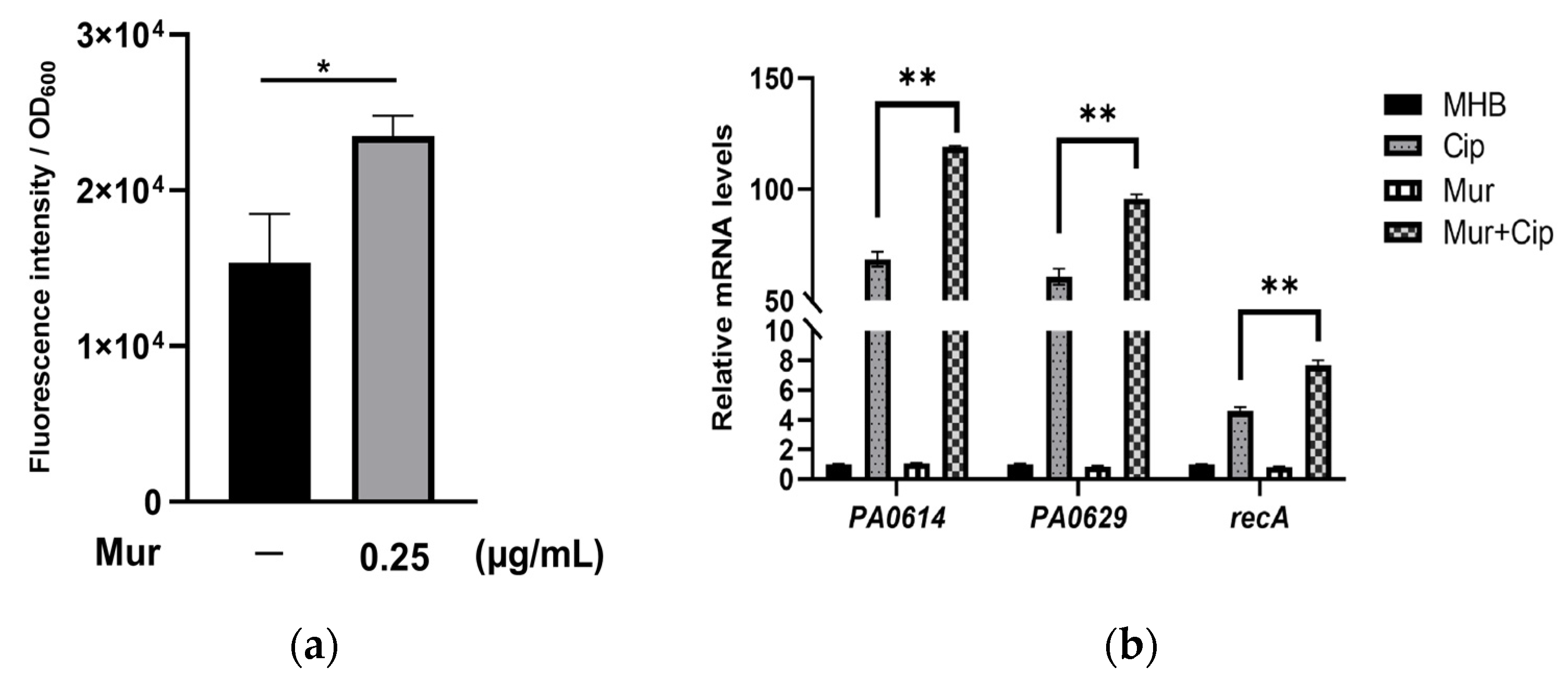
2.2. Murepavadin Enhances Intracellular Accumulation of Ciprofloxacin by Suppressing Drug Efflux

To understand the mechanism of the enhanced bactericidal effect, we examined the intracellular accumulation of ciprofloxacin. Treatment with 0.25 μg/mL murepavadin increased the intracellular level of ciprofloxacin (Figure 2a). Consistent with the ciprofloxacin accumulation results, the presence of murepavadin enhanced the ciprofloxacin-induced upregulation of recA and the pyocin-related genes PA0614 and PA0629. Collectively, these results demonstrate that murepavadin enhances the bacterial uptake of ciprofloxacin (Figure 2b).
In P. aeruginosa, efflux pumps play an important role in bacterial resistance to ciprofloxacin by reducing intracellular drug concentration [17]. To examine whether murepavadin affects drug efflux, we treated bacteria with an efflux pump inhibitor, carbonyl cyanide m-chlorophenylhydrazine (CCCP) [32]. The addition of CCCP increased the intracellular ciprofloxacin amounts to the same level in the presence and absence of murepavadin (Figure 3a), indicating that murepavadin mainly increases intracellular ciprofloxacin by suppressing drug efflux. We then performed an ethidium bromide accumulation assay, which has been used to measure efflux pump activity in bacteria [33]. The presence of murepavadin increased the intracellular accumulation of ethidium bromide (Figure 3b). The addition of CCCP enhanced the intracellular ethidium bromide levels and diminished the difference in cells with or without murepavadin treatment (Figure 3b). Collectively, these results suggest that murepavadin inhibits drug efflux, which subsequently promotes the intracellular accumulation of ciprofloxacin.

2.3. Murepavadin Enhances Bactericidal Effects of Ciprofloxacin against P. aeruginosa Clinical Isolates

We then assessed the bactericidal effects of murepavadin in combination with ciprofloxacin against twelve clinical isolates. The strains were isolated from various infection sites and are susceptible to both of the antibiotics (Table S3). Murepavadin or ciprofloxacin alone reduced the live bacterial number of ten strains by 14.92–1.43 × 10⁴ times eight hours after the treatment (Figure 4a–g,j–l). Afterwards, the bacteria started to grow (Figure 4a–g,j–l). Strains #26 and #123 started to grow four and six hours after the treatment of ciprofloxacin, respectively (Figure 4h,i). The combination of murepavadin and ciprofloxacin resulted in the 1.83 × 10⁵–4.03 × 10⁶-fold killing of all of the strains eight hours after the treatment, which resulted in 4.00 × 10²–1.78 × 10⁵-fold fewer bacteria than with each of the antibiotics alone (Figure 4). Eight hours after treatment with the combination, three out of the twelve strains started to grow (Figure 4j–l). Nevertheless, the combination reduced the bacterial number by 2.19 × 10³ to 4.65 × 10⁸ times compared with either antibiotic alone after twenty four hours (Figure 4). Collectively, these results demonstrate a synergistic bactericidal effect of the murepavadin–ciprofloxacin combination.

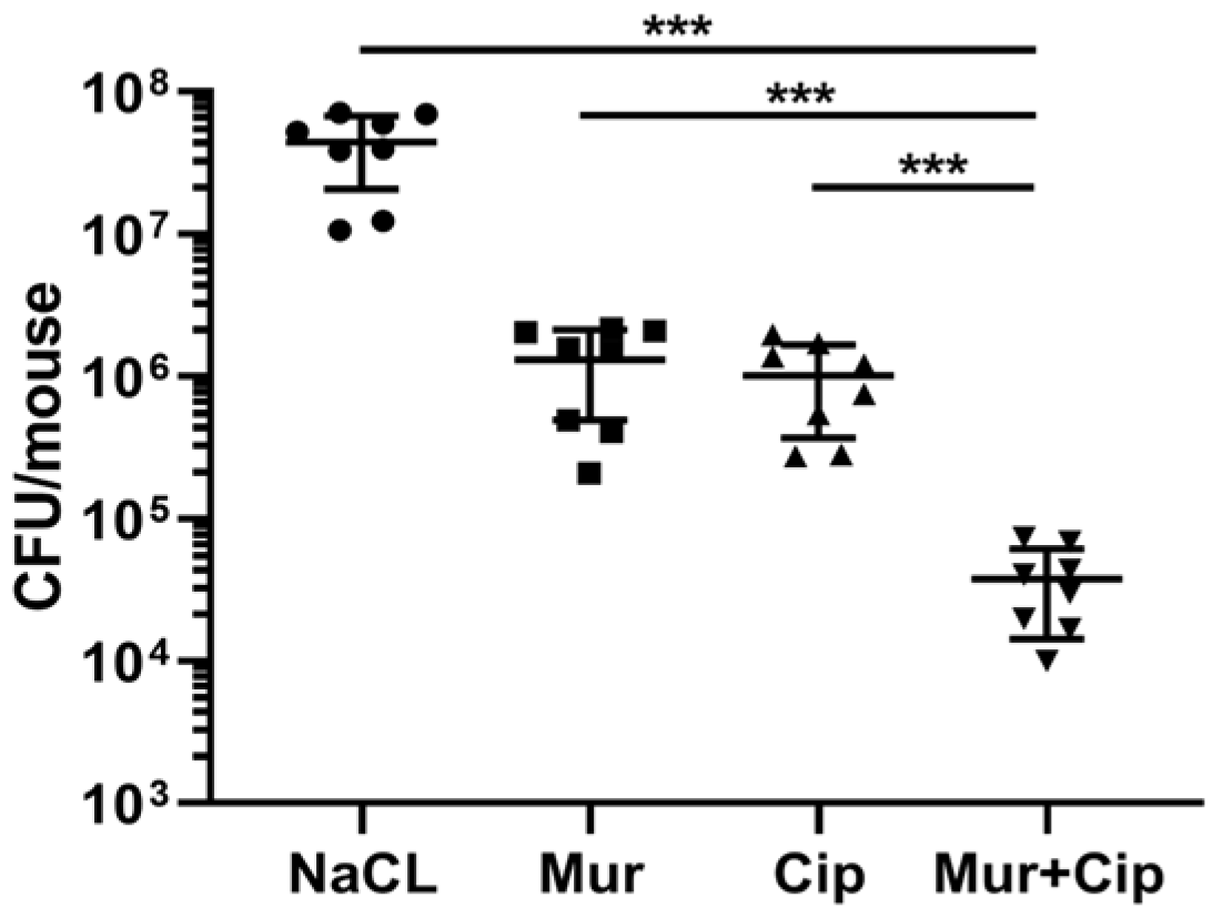
2.4. The Murepavadin–Ciprofloxacin Combination Displays a Synergistic Therapeutic Effect In Vivo

We next evaluated the in vivo efficacy of the murepavadin–ciprofloxacin combination against PA14 in a murine pneumonia model [34,35]. The dose of murepavadin (0.25 mg/kg) was used as previously reported in a murine pneumonia model [36]. For ciprofloxacin, the recommended daily dose for adult patients with nosocomial pneumonia is 400 mg intravenously injected every eight hours (https://reference.medscape.com/drug/cipro-xr-ciprofloxacin-342530, accessed on 18 June 2024). According to the Meeh–Rubner equation [37], the equivalent dose for intravenous injection of ciprofloxacin in mice is 52 mg/kg ciprofloxacin. In a study involving CF patients receiving a single dose of ciprofloxacin dry powder for inhalation (32.5 mg), the drug acquired a Cmax (mg/L) of 34.9 in sputum and 0.0790 in plasma [38]. In our assay, ciprofloxacin and murepavadin were administered intranasally. To minimize potential side effects, the ciprofloxacin dose was set to 0.5 mg/kg, which resulted in a similar CFU reduction to murepavadin. Treatment with murepavadin and ciprofloxacin alone decreased the bacterial loads by 33 times and 43 times, respectively, whereas the combined treatment reduced the mean bacterial load by 1171 times (Figure 5), indicating a synergistic bactericidal effect.

3. Discussion

ESKAPE pathogens (Enterococcus faecium, S. aureus, K. pneumoniae, A. baumannii, P. aeruginosa, and Enterobacter species) are highly antibiotic-resistant and impose a global threat to human health [39]. P. aeruginosa causes lung infections in patients with mechanical ventilation, cystic fibrosis, chronic obstructive pulmonary disorder (COPD), and immunodeficiency [6,40]. Nebulized antibiotics, such as tobramycin and amikacin, have been used to treat P. aeruginosa infections [41].
Murepavadin is a novel cyclic peptide antibiotic that specifically targets P. aeruginosa LptD, leading to the disruption of the asymmetry of the outer membrane [1]. LptD is a component of the LptA-G machinery that transports LPS across the periplasm and integrates it into the outer leaflet of the outer membrane. LPS is synthesized in the cytoplasm and on the inner membrane. The LptFGB complex extracts LPS from the inner membrane, followed by transportation by the LptA bridge to the outer-membrane complex, which is composed of the lipoprotein LptE and the integral β-barrel membrane protein LptD. Then, the LptD/E complex inserts LPS into the outer leaflet of the outer membrane. It has been shown that murepavadin binds to the periplasmic segment of LptD, which contains an N-terminal insert domain and a β-jellyroll domain. The binding of the antibiotic blocks LPS transport and insertion into the outer membrane [11]. In clinical trials, patients receiving inhaled murepavadin demonstrated a notable improvement in clinical symptoms and a significant reduction in bacterial burden compared with the control group [42]. The targeted delivery via inhalation facilitated direct action against the lung-colonizing bacteria, minimizing systemic toxicity. Additionally, the drug was well tolerated, with few reported adverse events. These findings underscore the potential of inhaled murepavadin as an effective and safe treatment option for patients with P. aeruginosa pneumonia, including those infected with drug-resistant strains. The fluoroquinolone antibiotic ciprofloxacin is commonly used to treat P. aeruginosa infections [17]. Clinical trials with inhaled ciprofloxacin produced promising outcomes in treating P. aeruginosa lung infections. Patients administered with inhaled ciprofloxacin experienced a marked reduction in bacterial load and improved clinical symptoms [43].
Low membrane permeability and multidrug efflux pumps play important roles in the bacterial resistance to ciprofloxacin [17]. We previously reported that murepavadin increases outer-membrane permeability, presumably owing to disruption of the LPS in the outer leaflet and alteration of the outer-membrane protein profile [29]. Here, we found that murepavadin promotes the bactericidal effect of ciprofloxacin against P. aeruginosa. The combination of murepavadin and ciprofloxacin synergistically reduced the bacterial load in a murine acute pneumonia model. In the ciprofloxacin uptake and ethidium bromide influx assay, the addition of CCCP diminished the effect of murepavadin, thus indicating a suppressive role of murepavadin on the multidrug efflux pumps. P. aeruginosa harbors at least twelve resistance-nodulation-cell division (RND) multidrug efflux pumps, which are composed of an inner-membrane protein, an outer-membrane protein, and a periplasmic protein that connects the inner- and outer-membrane proteins [44]. In P. aeruginosa, the pumps MexAB-OprM, MexCD-OprJ, MexEF-OprN, and MexXY-OprM play crucial roles in the resistance to fluoroquinolones [45]. Efflux through the RND pumps depends on the proton motive force [46]. We previously found that murepavadin increases the potential of the inner membrane [47]; thus, it might not affect the energy source of the pumps. It has been demonstrated that treatment with murepavadin results in the accumulation of LPS in the inner membrane [36], which might interfere with the assembly of RND pumps. Further experiments are needed to examine the expression and assembly of the pumps. In addition, increased outer-membrane permeability might also contribute to the murepavadin-promoted bactericidal effect of ciprofloxacin. Treatment with murepavadin might increase membrane permeability while reducing the efflux systems, which may sensitize bacteria to ciprofloxacin or other antibiotics. Thus, sequential treatment with murepavadin and ciprofloxacin might further increase bactericidal efficacy. Further experiments are warranted to evaluate the therapeutic effect.
In this study, we used the PA14 strain to elucidate the mechanism of synergy and evaluate in vivo efficacy. PA14 is a burn wound isolate and has attracted much attention due to its high toxicity in plants and animals [48]. It has been wildly used as a P. aeruginosa reference strain [49]. Since its genome sequence is available, PA14 has been used in numerous mechanistic studies and animal models. The clinical strains were isolated from various infection sites. The MICs of ciprofloxacin for the strains are comparable to wild type PA14 (within a 2-fold difference). Thus, the uptake and efflux of ciprofloxacin of those strains might be similar to wild type PA14. Clinical isolates are different in genotypes, leading to variations in bacterial pathogenesis and responses to antibiotics. However, the genotypes of the stains were not examined. Since the therapeutic effect is also affected by bacterial virulence, biofilm formation, etc., the phenotypes and the in vivo treatment efficacy warrant further evaluation in the future to verify the effectivity of the combination.
We investigated the synergistic effects of the murepavadin–ciprofloxacin combination against ciprofloxacin-susceptible strains. Additionally, we tested strains that are resistant to ciprofloxacin (with MIC higher than the break point, 2 μg/mL). No synergistic effect was observed with the combination. In clinical isolates, one of the major resistance mechanisms is mutation in the DNA gyrase gene gyrA, which reduces the drug binding affinity. Additionally, the upregulation of efflux systems contributes to resistance [44,50]. Although murepavadin interferes with the efflux systems, the increased accumulation of intracellular ciprofloxacin might not be able to overcome the reduced affinity, rendering no synergistic effect. Therefore, it is likely that the ciprofloxacin-resistant strain we tested harbors mutation in the gyrA gene. Further experiments are needed to elucidate the resistance mechanism of the strain. Other antibiotics can be used in combination with murepavadin to test treatment efficacy.
The combination of β-lactam and β-lactase inhibitors is a common combination with antipseudomonal activity, such as ceftolozane–tazobactam and ceftazidime–avibactam [51,52]. Chronic infection of P. aeruginosa in cystic fibrosis patients is usually treated with nebulized tobramycin or aztreonam, which improves lung function and reduces acute exacerbation of CF [53]. Compared with this clinical practice, murepavadin exhibits high effectivity against a wide range of P. aeruginosa clinical isolates, including carbapenem-resistant strains [1]. A phase I study of inhaled murepavadin has been initiated (https://spexisbio.com/impv/, accessed on 24 May 2024), and this is of great significance for managing chronic lung infections, especially P. aeruginosa infections in CF patients. Clinical trials have shown that inhaled ciprofloxacin is effective in treating chronic respiratory infections caused by P. aeruginosa [22,23]. Due to the potential formation of persistent bacteria in chronic infections that are difficult to eradicate, there is an urgent need for inhaled antibiotic strategies to achieve high airway concentrations and minimal systemic toxicity, in order to prevent further lung damage [42]. At present, the options of inhaled antibiotics on the market are limited, and the successful development and application of murepavadin and ciprofloxacin may provide new treatment options for such patients.
A phase III clinical trial of intravenous injection of murepavadin was halted due to its nephrotoxicity, and nebulized antibiotics are still in clinical trials [14]. It has been reported that ciprofloxacin caused acute renal failure, demonstrating potential nephrotoxicity [54,55]. Therefore, the safety of the murepavadin–ciprofloxacin combination needs to be carefully evaluated.
Overall, our results suggest that the combination of nebulized murepavadin and ciprofloxacin might be beneficial for the treatment of human lung infections caused by P. aeruginosa.

4. Materials and Methods

4.1. Bacterial Strains and Growth Conditions

The bacterial strains and primers utilized in this study are shown in Supplementary Table S1. P. aeruginosa strains were cultivated in Mueller–Hinton broth (MHB) or Lysogeny broth (LB) medium at 37 °C with under 200 rpm shaking.

4.2. Minimal Inhibitory Concentration (MIC) Determination

Bacteria were cultured in LB to an OD600 of 1.0. The Clinical and Laboratory Standards Institute (CLSI) guidelines were followed to measure the minimal inhibitory concentrations (MICs) of the indicated antibiotics in Cation-adjusted Mueller–Hinton Broth (CA-MHB, QDRS Biotec, Qingdao, China) [31]. The MICs were examined in triplicate. Murepavadin trifluoroacetic acid and ciprofloxacin were purchased from MCE (MedChemExpress, Shanghai, China) and Sangon Biotech (Shanghai, China), respectively.

4.3. Time-Dependent Killing Assays

Bacteria were cultured to an OD600 of 1.0 in LB. After being diluted ten-fold into 2 mL of fresh MHB, the bacteria were treated with the specified antibiotics at 37 °C with shaking. At indicated intervals (0, 2, 4, 6, 8, and 24 h), viable bacterial counts were determined by plating on LB agar. All experiments were conducted in triplicate.

4.4. Intracellular Accumulation of Ciprofloxacin

The uptake of ciprofloxacin was measured as described previously, with slight modifications [56]. Briefly, PA14 was cultured in MHB at 37 °C with shaking until the OD600 reached 1.0. For each individual bacterial aliquot (2.5 mL), a final concentration of 10 μg/mL ciprofloxacin was added with or without 0.25 µg/mL murepavadin and 40 µg/mL CCCP, followed by incubation with shaking for 15 min. Then, 300 µL of each sample was collected for OD600 measurement. The remaining bacteria were collected by centrifugation at 3000× g at 4 °C for 7 min and washed three times with cold PBS to remove extracellular ciprofloxacin. The bacterial cells were then resuspended in 0.1 M glycine–HCl buffer (pH 3.0). The samples were kept overnight in the dark to enable bacterial lysis and ciprofloxacin release. After centrifugation at 20,000× g for 7 min, the fluorescence of ciprofloxacin in the supernatant was examined by utilizing a microplate reader (BioTek Synergy H1, Agilent, Santa Clara, CA, USA) with excitation at 275 nm and emission at 450 nm. The intracellular ciprofloxacin levels were normalized to OD600.

4.5. Ethidium Bromide Influx Assay

The ethidium bromide influx assay was carried out as described previously, with slight modifications [57,58]. Bacteria were cultured in LB to an OD600 of 0.5. A total of 1 mL of the bacterial culture was collected with centrifugation and resuspended in an equal volume of PBS. The bacteria were incubated at 37 °C for 30 min with 0.25 μg/mL murepavadin and 40 µg/mL CCCP alone or in combination, in the presence of 2 μg/mL ethidium bromide. Subsequently, the bacteria were rinsed twice and resuspended in 0.2 mL of PBS. The fluorescence of ethidium bromide (excitation wavelength of 530 nm and emission wavelength of 585 nm) was detected with a microplate reader (Biotek Synergy H1, Agilent, Santa Clara, CA, USA). The relative uptake of ethidium bromide was normalized to the OD600 of each sample.

4.6. RNA Isolation and Quantitative Real-Time PCR (qRT-PCR)

The overnight bacterial cultures were diluted 100-fold into MHB and incubated with murepavadin (0.03125 μg/mL) and ciprofloxacin (0.025 μg/mL), either alone or in combination, at 37 °C until the OD600 reached 1.0. The bacteria were harvested by centrifugation. The total bacterial RNA was extracted with a Bacteria Total RNA Kit (Zoman Biotec, Beijing, China). cDNA was synthesized from 1 μg of RNA with PrimeScript Reverse Transcriptase (TransGen biotech, Beijing, China) in a volume of 20 μL. Afterwards, the cDNA was mixed with SYBR Premix (Vazyme, Nanjing, China) and specific primers (Supplementary Table S1). The rpsL gene that encodes the 30S ribosomal protein was used as an internal reference.

4.7. Murine Lung Infection Model

The mouse acute pneumonia model was conducted as previously described [34,35]. PA14 was cultivated to an OD600 of 1.0 in LB. The bacteria were washed twice with 0.9% NaCl and resuspended to a concentration of 2 × 10⁸ CFU/mL. Four groups of eight female BALB/c mice (aged 6–8 weeks) (Vital River) were anesthetized via intraperitoneal injection with 90 μL of 7.5% chloral hydrate. A total of 20 μL of the bacterial suspension was inoculated intranasally to each mouse. Three hours post-infection (hpi), the mice were anesthetized again and treated intranasally with either 20 μL of NaCl alone or NaCl containing murepavadin (0.25 mg/kg), ciprofloxacin (0.5 mg/kg), or a combination of both antibiotics at the concentrations used for monotreatment. After 13 h, the mice were sacrificed by CO2 inhalation. The lungs of the mice were homogenized in sterile 1% proteose peptone (Solarbio, Beijing, China). The bacterial burdens were quantified through serial dilution and plating.

Supplementary Materials

The following supporting information can be downloaded at: https://www.mdpi.com/article/10.3390/antibiotics13090810/s1, Table S1: Bacterial strains, plasmids, and primers used in this study [59]; Table S2: MICs (mg/L) of indicated antibiotics for PA14, Table S3: Clinical isolates used in this study and MICs (mg/L) of indicated antibiotics.

Author Contributions

Conceptualization, W.W., S.J. and X.W.; methodology, X.W., D.Z., S.C. and C.X.; validation, W.W., S.J., X.W., Y.J. and Z.C.; formal analysis, W.W. and X.W.; investigation, X.W., D.Z., C.X., S.C. and P.C.; resources, S.C. and W.W.; data curation, X.W. and D.Z.; writing—original draft preparation, X.W. and W.W.; writing—review and editing, S.J., Y.J. and Z.C.; supervision, W.W.; project administration, W.W.; funding acquisition, W.W. All authors have read and agreed to the published version of the manuscript.

Funding

This research was supported by National Key R&D Program of China, grant number 2021YFE0101700; National Science Foundation of China, grants number 32170177 and 32170199; and Nankai University Tianjin Applied and Fundamental Research Project, grant number 22JCZDJC0041.

Institutional Review Board Statement

The animal study protocol was approved by the Animal Care and Use Committee of the College of Life Sciences, Nankai University (permission number: NK-04-2392012). Animal infection experiments were conducted according to the Chinese rules for the use of animals.

Informed Consent Statement

Not applicable.

Data Availability Statement

The data that support the findings of this study are available from the corresponding author upon reasonable request.

Acknowledgments

We would like to thank Liqun Jia from Tianjin Medical University General Hospital for providing clinical isolates.

Conflicts of Interest

The authors declare no conflicts of interest. The funders had no role in the design of the study; in the collection, analyses, or interpretation of data; in the writing of the manuscript; or in the decision to publish the results.

References

  1. Martin-Loeches, I.; Dale, G.E.; Torres, A. Murepavadin: A new antibiotic class in the pipeline. Expert. Rev. Anti Infect. Ther. 2018, 16, 259–268. [Google Scholar] [CrossRef]
  2. Zheng, M.; Lupoli, T.J. Counteracting antibiotic resistance enzymes and efflux pumps. Curr. Opin. Microbiol. 2023, 75, 102334. [Google Scholar] [CrossRef] [PubMed]
  3. Shang, Z.; Chan, S.Y.; Song, Q.; Li, P.; Huang, W. The Strategies of Pathogen-Oriented Therapy on Circumventing Antimicrobial Resistance. Research 2020, 2020, 2016201. [Google Scholar] [CrossRef]
  4. Zhu, M.; Tse, M.W.; Weller, J.; Chen, J.; Blainey, P.C. The future of antibiotics begins with discovering new combinations. Ann. N. Y. Acad. Sci. 2021, 1496, 82–96. [Google Scholar] [CrossRef] [PubMed]
  5. Silby, M.W.; Winstanley, C.; Godfrey, S.A.; Levy, S.B.; Jackson, R.W. Pseudomonas genomes: Diverse and adaptable. FEMS Microbiol. Rev. 2011, 35, 652–680. [Google Scholar] [CrossRef]
  6. Qin, S.; Xiao, W.; Zhou, C.; Pu, Q.; Deng, X.; Lan, L.; Liang, H.; Song, X.; Wu, M. Pseudomonas aeruginosa: Pathogenesis, virulence factors, antibiotic resistance, interaction with host, technology advances and emerging therapeutics. Signal Transduct. Target. Ther. 2022, 7, 199. [Google Scholar] [CrossRef] [PubMed]
  7. Malhotra, S.; Hayes, D., Jr.; Wozniak, D.J. Cystic Fibrosis and Pseudomonas aeruginosa: The Host-Microbe Interface. Clin. Microbiol. Rev. 2019, 32, e00138-18. [Google Scholar] [CrossRef] [PubMed]
  8. Sathe, N.; Beech, P.; Croft, L.; Suphioglu, C.; Kapat, A.; Athan, E. Pseudomonas aeruginosa: Infections and novel approaches to treatment “Knowing the enemy” the threat of Pseudomonas aeruginosa and exploring novel approaches to treatment. Infect. Med. 2023, 2, 178–194. [Google Scholar] [CrossRef]
  9. Moradali, M.F.; Ghods, S.; Rehm, B.H. Pseudomonas aeruginosa Lifestyle: A Paradigm for Adaptation, Survival, and Persistence. Front. Cell Infect. Microbiol. 2017, 7, 39. [Google Scholar] [CrossRef]
  10. Srinivas, N.; Jetter, P.; Ueberbacher, B.J.; Werneburg, M.; Zerbe, K.; Steinmann, J.; Van der Meijden, B.; Bernardini, F.; Lederer, A.; Dias, R.L.; et al. Peptidomimetic antibiotics target outer-membrane biogenesis in Pseudomonas aeruginosa. Science 2010, 327, 1010–1013. [Google Scholar] [CrossRef]
  11. Andolina, G.; Bencze, L.C.; Zerbe, K.; Muller, M.; Steinmann, J.; Kocherla, H.; Mondal, M.; Sobek, J.; Moehle, K.; Malojcic, G.; et al. A Peptidomimetic Antibiotic Interacts with the Periplasmic Domain of LptD from Pseudomonas aeruginosa. ACS Chem. Biol. 2018, 13, 666–675. [Google Scholar] [CrossRef] [PubMed]
  12. Garcia-Gros, J.; Cajal, Y.; Marques, A.M.; Rabanal, F. Synthesis of the Antimicrobial Peptide Murepavadin Using Novel Coupling Agents. Biomolecules 2024, 14, 526. [Google Scholar] [CrossRef] [PubMed]
  13. Amponnawarat, A.; Chompunud Na Ayudhya, C.; Ali, H. Murepavadin, a Small Molecule Host Defense Peptide Mimetic, Activates Mast Cells via MRGPRX2 and MrgprB2. Front. Immunol. 2021, 12, 689410. [Google Scholar] [CrossRef]
  14. Diez-Aguilar, M.; Hernandez-Garcia, M.; Morosini, M.I.; Fluit, A.; Tunney, M.M.; Huertas, N.; Del Campo, R.; Obrecht, D.; Bernardini, F.; Ekkelenkamp, M.; et al. Murepavadin antimicrobial activity against and resistance development in cystic fibrosis Pseudomonas aeruginosa isolates. J. Antimicrob. Chemother. 2021, 76, 984–992. [Google Scholar] [CrossRef] [PubMed]
  15. Butler, M.S.; Henderson, I.R.; Capon, R.J.; Blaskovich, M.A.T. Antibiotics in the clinical pipeline as of December 2022. J. Antibiot. 2023, 76, 431–473. [Google Scholar] [CrossRef]
  16. Kelly, K.R.; Brooks, B.W. Global Aquatic Hazard Assessment of Ciprofloxacin: Exceedances of Antibiotic Resistance Development and Ecotoxicological Thresholds. Prog. Mol. Biol. Transl. Sci. 2018, 159, 59–77. [Google Scholar]
  17. Rehman, A.; Patrick, W.M.; Lamont, I.L. Mechanisms of ciprofloxacin resistance in Pseudomonas aeruginosa: New approaches to an old problem. J. Med. Microbiol. 2019, 68, 1–10. [Google Scholar] [CrossRef]
  18. Gales, A.C.; Jones, R.N.; Turnidge, J.; Rennie, R.; Ramphal, R. Characterization of Pseudomonas aeruginosa isolates: Occurrence rates, antimicrobial susceptibility patterns, and molecular typing in the global SENTRY Antimicrobial Surveillance Program, 1997–1999. Clin. Infect. Dis. 2001, 32 (Suppl. S2), S146–S155. [Google Scholar] [CrossRef] [PubMed]
  19. Podlesek, Z.; Zgur Bertok, D. The DNA Damage Inducible SOS Response Is a Key Player in the Generation of Bacterial Persister Cells and Population Wide Tolerance. Front. Microbiol. 2020, 11, 1785. [Google Scholar] [CrossRef]
  20. Long, Y.; Fu, W.; Wang, S.; Deng, X.; Jin, Y.; Bai, F.; Cheng, Z.; Wu, W. Fis Contributes to Resistance of Pseudomonas aeruginosa to Ciprofloxacin by Regulating Pyocin Synthesis. J. Bacteriol. 2020, 202, e00064-20. [Google Scholar] [CrossRef]
  21. Lim, D.J.; Skinner, D.; McLemore, J.; Rivers, N.; Elder, J.B.; Allen, M.; Koch, C.; West, J.; Zhang, S.; Thompson, H.M.; et al. In-vitro evaluation of a ciprofloxacin and azithromycin sinus stent for Pseudomonas aeruginosa biofilms. Int. Forum Allergy Rhinol. 2020, 10, 121–127. [Google Scholar] [CrossRef] [PubMed]
  22. De Soyza, A.; Aksamit, T.; Bandel, T.J.; Criollo, M.; Elborn, J.S.; Operschall, E.; Polverino, E.; Roth, K.; Winthrop, K.L.; Wilson, R. RESPIRE 1: A phase III placebo-controlled randomised trial of ciprofloxacin dry powder for inhalation in non-cystic fibrosis bronchiectasis. Eur. Respir. J. 2018, 51, 1702052. [Google Scholar] [CrossRef]
  23. Aksamit, T.; De Soyza, A.; Bandel, T.J.; Criollo, M.; Elborn, J.S.; Operschall, E.; Polverino, E.; Roth, K.; Winthrop, K.L.; Wilson, R. RESPIRE 2: A phase III placebo-controlled randomised trial of ciprofloxacin dry powder for inhalation in non-cystic fibrosis bronchiectasis. Eur. Respir. J. 2018, 51, 1702053. [Google Scholar] [CrossRef] [PubMed]
  24. Haworth, C.S.; Bilton, D.; Chalmers, J.D.; Davis, A.M.; Froehlich, J.; Gonda, I.; Thompson, B.; Wanner, A.; O’Donnell, A.E. Inhaled liposomal ciprofloxacin in patients with non-cystic fibrosis bronchiectasis and chronic lung infection with Pseudomonas aeruginosa (ORBIT-3 and ORBIT-4): Two phase 3, randomised controlled trials. Lancet Respir. Med. 2019, 7, 213–226. [Google Scholar] [CrossRef] [PubMed]
  25. Coates, A.R.M.; Hu, Y.; Holt, J.; Yeh, P. Antibiotic combination therapy against resistant bacterial infections: Synergy, rejuvenation and resistance reduction. Expert. Rev. Anti Infect. Ther. 2020, 18, 5–15. [Google Scholar] [CrossRef]
  26. Fischbach, M.A. Combination therapies for combating antimicrobial resistance. Curr. Opin. Microbiol. 2011, 14, 519–523. [Google Scholar] [CrossRef]
  27. Chou, T.C. Theoretical basis, experimental design, and computerized simulation of synergism and antagonism in drug combination studies. Pharmacol. Rev. 2006, 58, 621–681. [Google Scholar] [CrossRef]
  28. Zhou, C.; Wang, Q.; Jin, L.; Wang, R.; Yin, Y.; Sun, S.; Zhang, J.; Wang, H. In vitro Synergistic Activity of Antimicrobial Combinations Against bla (KPC) and bla (NDM)-Producing Enterobacterales With bla (IMP) or mcr Genes. Front. Microbiol. 2020, 11, 533209. [Google Scholar] [CrossRef]
  29. Wei, X.; Gao, J.; Xu, C.; Pan, X.; Jin, Y.; Bai, F.; Cheng, Z.; Lamont, I.L.; Pletzer, D.; Wu, W. Murepavadin induces envelope stress response and enhances the killing efficacies of beta-lactam antibiotics by impairing the outer membrane integrity of Pseudomonas aeruginosa. Microbiol. Spectr. 2023, 11, e0125723. [Google Scholar] [CrossRef]
  30. Sader, H.S.; Dale, G.E.; Rhomberg, P.R.; Flamm, R.K. Antimicrobial Activity of Murepavadin Tested against Clinical Isolates of Pseudomonas aeruginosa from the United States, Europe, and China. Antimicrob. Agents Chemother. 2018, 62, e00311–e00318. [Google Scholar] [CrossRef]
  31. CLSI. Performance Standards for Antimicrobial Susceptibility Testing, M100, 33rd ed.; Clinical and Laboratory Standards Institute: Wayne, PA, USA, 2023. [Google Scholar]
  32. Bhattacharyya, T.; Sharma, A.; Akhter, J.; Pathania, R. The small molecule IITR08027 restores the antibacterial activity of fluoroquinolones against multidrug-resistant Acinetobacter baumannii by efflux inhibition. Int. J. Antimicrob. Agents 2017, 50, 219–226. [Google Scholar] [CrossRef] [PubMed]
  33. Blair, J.M.; Piddock, L.J. How to Measure Export via Bacterial Multidrug Resistance Efflux Pumps. mBio 2016, 7, e00840-16. [Google Scholar] [CrossRef]
  34. Pan, X.; Wu, W. Murine Acute Pneumonia Model of Pseudomonas aeruginosa Lung Infection. Bio Protoc. 2020, 10, e3805. [Google Scholar] [CrossRef] [PubMed]
  35. Ren, H.; Liu, Y.; Zhou, J.; Long, Y.; Liu, C.; Xia, B.; Shi, J.; Fan, Z.; Liang, Y.; Chen, S.; et al. Combination of Azithromycin and Gentamicin for Efficient Treatment of Pseudomonas aeruginosa Infections. J. Infect. Dis. 2019, 220, 1667–1678. [Google Scholar] [CrossRef]
  36. Sabnis, A.; Hagart, K.L.; Klockner, A.; Becce, M.; Evans, L.E.; Furniss, R.C.D.; Mavridou, D.A.; Murphy, R.; Stevens, M.M.; Davies, J.C.; et al. Colistin kills bacteria by targeting lipopolysaccharide in the cytoplasmic membrane. Elife 2021, 10, e65836. [Google Scholar] [CrossRef]
  37. Spiers, D.E.; Candas, V. Relationship of skin surface area to body mass in the immature rat: A reexamination. J. Appl. Physiol. Respir. Environ. Exerc. Physiol. 1984, 56, 240–243. [Google Scholar] [CrossRef]
  38. Panthi, V.K.; Fairfull-Smith, K.E.; Islam, N. Ciprofloxacin-Loaded Inhalable Formulations against Lower Respiratory Tract Infections: Challenges, Recent Advances, and Future Perspectives. Pharmaceutics 2024, 16, 648. [Google Scholar] [CrossRef]
  39. De Oliveira, D.M.P.; Forde, B.M.; Kidd, T.J.; Harris, P.N.A.; Schembri, M.A.; Beatson, S.A.; Paterson, D.L.; Walker, M.J. Antimicrobial Resistance in ESKAPE Pathogens. Clin. Microbiol. Rev. 2020, 33, e00181-19. [Google Scholar] [CrossRef] [PubMed]
  40. Ibrahim, D.; Jabbour, J.F.; Kanj, S.S. Current choices of antibiotic treatment for Pseudomonas aeruginosa infections. Curr. Opin. Infect. Dis. 2020, 33, 464–473. [Google Scholar] [CrossRef]
  41. Gorham, J.; Taccone, F.S.; Hites, M. How to Use Nebulized Antibiotics in Severe Respiratory Infections. Antibiotics 2023, 12, 267. [Google Scholar] [CrossRef] [PubMed]
  42. Li, D.; Schneider-Futschik, E.K. Current and Emerging Inhaled Antibiotics for Chronic Pulmonary Pseudomonas aeruginosa and Staphylococcus aureus Infections in Cystic Fibrosis. Antibiotics 2023, 12, 484. [Google Scholar] [CrossRef] [PubMed]
  43. Tejada, S.; Ramirez-Estrada, S.; Forero, C.G.; Gallego, M.; Soriano, J.B.; Cardinal-Fernandez, P.A.; Ehrmann, S.; Rello, J. Safety and Efficacy of Devices Delivering Inhaled Antibiotics among Adults with Non-Cystic Fibrosis Bronchiectasis: A Systematic Review and a Network Meta-Analysis. Antibiotics 2022, 11, 275. [Google Scholar] [CrossRef]
  44. Lorusso, A.B.; Carrara, J.A.; Barroso, C.D.N.; Tuon, F.F.; Faoro, H. Role of Efflux Pumps on Antimicrobial Resistance in Pseudomonas aeruginosa. Int. J. Mol. Sci. 2022, 23, 15779. [Google Scholar] [CrossRef] [PubMed]
  45. Aeschlimann, J.R. The role of multidrug efflux pumps in the antibiotic resistance of Pseudomonas aeruginosa and other gram-negative bacteria. Insights from the Society of Infectious Diseases Pharmacists. Pharmacotherapy 2003, 23, 916–924. [Google Scholar] [CrossRef]
  46. Blair, J.M.; Piddock, L.J. Structure, function and inhibition of RND efflux pumps in Gram-negative bacteria: An update. Curr. Opin. Microbiol. 2009, 12, 512–519. [Google Scholar] [CrossRef] [PubMed]
  47. Wei, X.; Gao, J.; Zhou, D.; Xu, C.; Chen, P.; Chen, S.; Zhang, Y.; Liu, X.; Li, G.; Zhu, G.; et al. Murepavadin promotes the killing efficacies of aminoglycoside antibiotics against Pseudomonas aeruginosa by enhancing membrane potential. Antimicrob. Agents Chemother. 2024, 68, e0153923. [Google Scholar] [CrossRef]
  48. Mathee, K. Forensic investigation into the origin of Pseudomonas aeruginosa PA14-old but not lost. J. Med. Microbiol. 2018, 67, 1019–1021. [Google Scholar] [CrossRef]
  49. Grace, A.; Sahu, R.; Owen, D.R.; Dennis, V.A. Pseudomonas aeruginosa reference strains PAO1 and PA14: A genomic, phenotypic, and therapeutic review. Front. Microbiol. 2022, 13, 1023523. [Google Scholar] [CrossRef]
  50. Rehman, A.; Jeukens, J.; Levesque, R.C.; Lamont, I.L. Gene-Gene Interactions Dictate Ciprofloxacin Resistance in Pseudomonas aeruginosa and Facilitate Prediction of Resistance Phenotype from Genome Sequence Data. Antimicrob. Agents Chemother. 2021, 65, e0269620. [Google Scholar] [CrossRef]
  51. Haidar, G.; Philips, N.J.; Shields, R.K.; Snyder, D.; Cheng, S.; Potoski, B.A.; Doi, Y.; Hao, B.; Press, E.G.; Cooper, V.S.; et al. Ceftolozane-Tazobactam for the Treatment of Multidrug-Resistant Pseudomonas aeruginosa Infections: Clinical Effectiveness and Evolution of Resistance. Clin. Infect. Dis. 2017, 65, 110–120. [Google Scholar] [CrossRef]
  52. Torres, A.; Zhong, N.; Pachl, J.; Timsit, J.F.; Kollef, M.; Chen, Z.; Song, J.; Taylor, D.; Laud, P.J.; Stone, G.G.; et al. Ceftazidime-avibactam versus meropenem in nosocomial pneumonia, including ventilator-associated pneumonia (REPROVE): A randomised, double-blind, phase 3 non-inferiority trial. Lancet Infect. Dis. 2018, 18, 285–295. [Google Scholar] [CrossRef]
  53. Reynolds, D.; Kollef, M. The Epidemiology and Pathogenesis and Treatment of Pseudomonas aeruginosa Infections: An Update. Drugs 2021, 81, 2117–2131. [Google Scholar] [CrossRef] [PubMed]
  54. Hajji, M.; Jebali, H.; Mrad, A.; Blel, Y.; Brahmi, N.; Kheder, R.; Beji, S.; Fatma, L.B.; Smaoui, W.; Krid, M.; et al. Nephrotoxicity of Ciprofloxacin: Five Cases and a Review of the Literature. Drug Saf. Case Rep. 2018, 5, 17. [Google Scholar] [CrossRef]
  55. Campbell, R.E.; Chen, C.H.; Edelstein, C.L. Overview of Antibiotic-Induced Nephrotoxicity. Kidney Int. Rep. 2023, 8, 2211–2225. [Google Scholar] [CrossRef] [PubMed]
  56. Viveiros, M.; Rodrigues, L.; Martins, M.; Couto, I.; Spengler, G.; Martins, A.; Amaral, L. Evaluation of efflux activity of bacteria by a semi-automated fluorometric system. Methods Mol. Biol. 2010, 642, 159–172. [Google Scholar]
  57. Maurya, A.; Dwivedi, G.R.; Darokar, M.P.; Srivastava, S.K. Antibacterial and synergy of clavine alkaloid lysergol and its derivatives against nalidixic acid-resistant Escherichia coli. Chem. Biol. Drug Des. 2013, 81, 484–490. [Google Scholar] [CrossRef]
  58. Moo, C.L.; Osman, M.A.; Yang, S.K.; Yap, W.S.; Ismail, S.; Lim, S.H.; Chong, C.M.; Lai, K.S. Antimicrobial activity and mode of action of 1,8-cineol against carbapenemase-producing Klebsiella pneumoniae. Sci. Rep. 2021, 11, 20824. [Google Scholar] [CrossRef] [PubMed]
  59. Liberati, N.T.; Urbach, J.M.; Miyata, S.; Lee, D.G.; Drenkard, E.; Wu, G.; Villanueva, J.; Wei, T.; Ausubel, F.M. An ordered, nonredundant library of Pseudomonas aeruginosa strain PA14 transposon insertion mutants. Proc. Natl Acad. Sci. USA 2006, 103, 2833–2838. [Google Scholar] [CrossRef]
Figure 1. The time–killing curves of murepavadin (0.5 μg/mL) and ciprofloxacin (0.25 μg/mL) alone and in combination against PA14. *, p < 0.05; **, p < 0.01 compared with the bacteria treated with the single antibiotic at the corresponding time points by Student’s t-test. The data are the means ± SD of three independent samples. Mur, murepavadin; Cip, ciprofloxacin.
Antibiotics 13 00810 g001
Figure 2. Murepavadin increases the intracellular accumulation of ciprofloxacin. (a) Wild type PA14 was treated with 10 µg/mL ciprofloxacin with or without 0.25 µg/mL murepavadin in Mueller–Hinton Broth (MHB) for 15 min, followed by measurement of fluorescence of intracellular ciprofloxacin. The relative fluorescence was normalized by the OD600. (b) PA14 cells were treated with or without murepavadin (0.03125 μg/mL) and ciprofloxacin (0.025 μg/mL), individually or in combination and grown to OD600 = 1 at 37 °C. The mRNA levels of PA0614, PA0629, and recA were determined by qRT-PCR. The data are the means ± SD of three independent samples. *, p < 0.05; **, p < 0.01 by Student’s t-test. Mur, murepavadin; Cip, ciprofloxacin.
Antibiotics 13 00810 g002
Figure 3. Effects of CCCP on intracellular accumulation of ciprofloxacin and ethidium bromide. (a) Wild type PA14 was treated with 10 µg/mL ciprofloxacin with or without 0.25 µg/mL murepavadin and 40 µg/mL CCCP in MHB for 15 min, followed by measurement of the fluorescence of intracellular ciprofloxacin. (b) Wild type PA14 was incubated with or without 0.25 µg/mL murepavadin, 40 µg/mL CCCP and 2 μg/mL ethidium bromide in PBS for 30 min at 37 °C. The relative fluorescence was normalized by the OD600. ns, not significant; *, p < 0.05; ***, p < 0.001 by Student’s t-test. Mur, murepavadin.
Antibiotics 13 00810 g003
Figure 4. Bactericidal effects of murepavadin in combination with ciprofloxacin against clinical strains. (al) The bacteria were treated with murepavadin (0.5 μg/mL) and ciprofloxacin (0.25 μg/mL) individually or in combination at 37 °C with shaking. At the indicated time points, the live bacteria numbers were determined by plating. The strain numbers were shown in each of the figures. *, p < 0.05; **, p < 0.01; ***, p < 0.001 compared with the bacteria treated with the single antibiotic at the corresponding time points by Student’s t-test. The data are the means ± SD of three independent samples. Mur, murepavadin; Cip, ciprofloxacin.
Antibiotics 13 00810 g004
Figure 5. The murepavadin–ciprofloxacin combination displayed synergistic effect in vivo. Mice were inoculated intranasally with 4 × 10⁶ CFU of wild type PA14. Three hours later, NaCl, murepavadin, ciprofloxacin, or the murepavadin–ciprofloxacin combination were administered intranasally. Sixteen hours post-infection, the bacterial loads in the lungs were determined. ***, p < 0.001 by Student’s t-test. Mur, murepavadin; Cip, ciprofloxacin.
Antibiotics 13 00810 g005
Disclaimer/Publisher’s Note: The statements, opinions and data contained in all publications are solely those of the individual author(s) and contributor(s) and not of MDPI and/or the editor(s). MDPI and/or the editor(s) disclaim responsibility for any injury to people or property resulting from any ideas, methods, instructions or products referred to in the content.

