Article

Genomic Analysis of Enterobacter Species Isolated from Patients in United States Hospitals

by
Fred C. Tenover
1 and
Isabella A. Tickler
2,*
1
College of Arts and Sciences, University of Dayton, Dayton, OH 45469, USA
2
Cepheid, Sunnyvale, CA 94089, USA
*
Author to whom correspondence should be addressed.
Antibiotics 2024, 13(9), 865; https://doi.org/10.3390/antibiotics13090865
Submission received: 29 July 2024 / Revised: 3 September 2024 / Accepted: 5 September 2024 / Published: 10 September 2024

Abstract

:
We analyzed the whole genome sequences (WGS) and antibiograms of 35 Enterobacter isolates, including E. hormaechei and E. asburiae, and the recently described E. bugandensis, E. kobei, E. ludwigii, and E. roggenkampii species. Isolates were obtained from human blood and urinary tract infections in patients in the United States. Our goal was to understand the genetic diversity of antimicrobial resistance genes and virulence factors among the various species. Thirty-four of 35 isolates contained an AmpC class blaACT allele; however, the E. roggenkampii isolate contained blaMIR-5. Of the six Enterobacter isolates resistant to ertapenem, imipenem, and meropenem, four harbored a carbapenemase gene, including blaKPC or blaNDM. All four isolates were mCIM-positive. The remaining two isolates had alterations in ompC genes that may have contributed to the resistance phenotype. Interpretations of cefepime test results were variable when disk diffusion and automated broth microdilution results were compared due to the Clinical Laboratory and Standards Institute use of the “susceptible dose-dependent” classification. The diversity of the blaACT alleles paralleled species identifications, as did the presence of various virulence genes. The classification of recently described Enterobacter species is consistent with their resistance gene and virulence gene profiles.

1. Introduction

The genus Enterobacter consists of human, animal, and plant pathogens as well as environmental species [1]. Enterobacter species are included among the “ESKAPE” pathogens, i.e., as key causes of healthcare-associated infections that are often multidrug-resistant [1,2,3]. The taxonomy of the genus Enterobacter is complex and fluid, consisting of multiple individual species, subspecies, and species complexes. Several longstanding human pathogens, e.g., the former species Enterobacter aerogenes and Enterobacter sakazakii, have been reclassified as Klebsiella aerogenes and Cronobacter sakazakii, respectively [4,5,6]. Enterobacter species typically produce potent AmpC beta-lactamases capable of hydrolyzing most extended-spectrum cephalosporins, with the exception of the fourth-generation cephalosporin, cefepime. AmpC beta-lactamases, together with changes in the outer membrane porins OmpF and OmpC, have been reported to mediate carbapenem resistance in Klebsiella aerogenes, and likely in Enterobacter species as well [1,7]. Several Enterobacter species have been reported to produce carbapenemases, including NDM, KPC, and OXA-48, in addition to less common carbapenemases, such as NMC/IMI [3,8]. The AmpC enzymes of Enterobacter species may give false positive results for some phenotypic susceptibility testing methods, such as Modified Hodge tests to detect carbapenemase production [9]. The antimicrobial resistance profiles of Enterobacter species often include resistance to multiple classes of antimicrobial agents, including aminoglycosides and fluoroquinolones, which can make the treatment of human infections with these organisms difficult [10].
Our preliminary analysis of a recent collection of Enterobacter isolates from the United States revealed discrepancies between the species identifications produced by whole genome sequencing (WGS) methods and by MALDI-TOF. However, newer databases for both WGS data and MALTI-TOF have become available. Thus, we wanted to determine whether the newer databases would resolve the discrepancies in species identification. Furthermore, we sought to determine whether the newer species identifications would contain novel AmpC enzymes and virulence factors. Consequently, the goals of this study were to analyze the WGS data and antibiograms of the Enterobacter isolates obtained from human infections in patients in the United States to determine (1) how sequence-based species identification results differed from identifications by MALDI-TOF, (2) how AmpC-type beta-lactamases varied by species, (3) whether differences among the AmpC enzymes were reflected in varying susceptibilities to beta-lactam antibiotics, and (4) whether virulence factors were consistent among the different Enterobacter species.

2. Materials and Methods

Bacterial strains: A convenience sample of 35 isolates of a variety of Enterobacter species from patients in U.S. hospitals in 11 different states was selected from our reference collection for the study. Selection criteria included isolates that were non-susceptible to cefotaxime, ceftriaxone, ceftazidime, ertapenem, meropenem, or imipenem, as previously described [11].
Bacterial identification: Organisms were identified originally using MALDI-TOF MS (Bruker Daltonics GmbH, Bremen, Germany) RUO Library revisions G (2020) and H (2021), and by WGS data analysis using K-mer spectra analysis (the kmer database for species identification was downloaded on 24 May 2023) and the NCBI average nucleotide identity (ANI) workflows [11,12]. Identifications were subsequently repeated using updated versions of the two databases, specifically the MALDI-TOF MBT Compass (RUO) Library revision K (2022) and the new K-mer spectra analysis database (downloaded on 24 May 2024).
Antimicrobial Susceptibility Testing: Antimicrobial susceptibility testing (AST) was performed using an automated broth microdilution system (ABMD) with the MicroScan Walkaway Neg MIC 56 panels (Beckman Coulter Inc., West Sacramento, CA, USA) and by disk diffusion (DD) following Clinical Laboratory and Standards Institute (CLSI) guidance and interpreted using CLSI criteria. Both CLSI and European Committee on Antimicrobial Susceptibility Testing (EUCAST) interpretative criteria were used for cefepime results when discrepancies in interpretations specifically for this antimicrobial agent became apparent [13,14,15]. Carbapenemase activity was detected using the mCIM and eCIM tests as described in the CLSI M100 document [14].
Whole genome sequencing: Nucleic acid extraction, sequence assembly, and whole genome sequencing analysis were performed as previously described using the CLC Genomics Workbench version 22.0.2 and CLC Microbial Genomics Module version 22.1.1 (QIAGEN Bioinformatics, Aarhus, Denmark) [11]. Multi-locus sequence typing (MLST, carried out using the 7-locus scheme), k-mer based prediction of species, and the detection of acquired antimicrobial resistance genes were performed using workflows from the CLC Microbial Genomics Module version 22.1.1 (QIAGEN Bioinformatics). The ResFinder database (downloaded on 1 March 2024) was used for the detection of acquired resistance genes in the CLC Microbial Genomics Module workflow. A phylogenetic tree of the Enterobacter spp. strains in this study was inferred using CSIPhylogeny, the single nucleotide polymorphisms (SNP) analysis tool created by the Center for Genomic Epidemiology (https://www.genomicepidemiology.org/services/ accessed on 24 April 2024) [16]. Nucleic acid sequence data from this study have been deposited in GenBank under BioProject Accession # PRJNA981469.
Analysis of Enterobacter hormaechei from Pathogen Detection collection: E. hormaechei sequences were downloaded from the NCBI Pathogen Detection collection (https://www.ncbi.nlm.nih.gov/pathogens/isolates accessed on 30 August 2024) to compare sequence types and beta-lactamase genes with the dataset from the current study. For a closer comparison, the query was restricted to a creation date of 2021-2022, United States-only, and isolation source blood and urine. A total of 48 assemblies were downloaded. Multi-locus sequence typing (MLST, carried out using the 7-locus scheme) and the detection of acquired antimicrobial resistance genes were performed using workflows from the CLC Microbial Genomics Module version 22.1.1 (QIAGEN Bioinformatics). The ResFinder database (downloaded on 1 March 2024) was used for the detection of acquired resistance genes in the CLC Microbial Genomics Module workflow.

3. Results

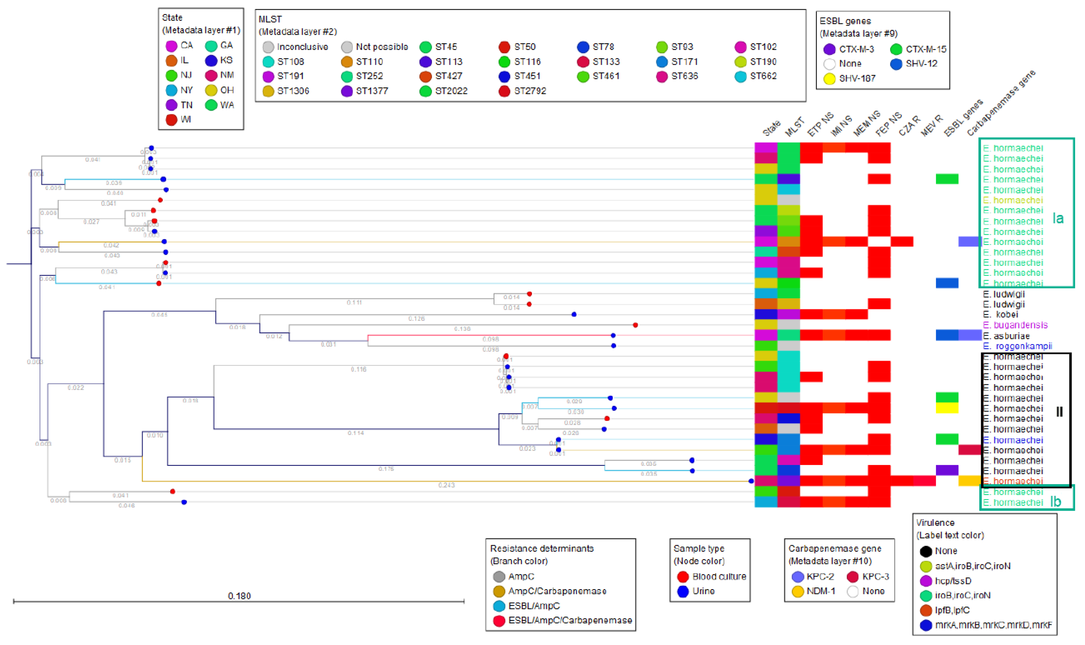
3.1. Enterobacter Species Identification and MLST Typing

The initial identification of the isolates by MALDI-TOF yielded one isolate of E. asburiae, one Enterobacter bugandensis, two Enterobacter hormaechei, three Enterobacter xiangfangensis, and twenty-eight belonging to the Enterobacter cloacae complex. A comparison of the results of the three identification methods, two based on WGS (ANI and kmer) and the updated MALDI-TOF database, showed significant changes in the identifications and indicates agreement to the species level among all methods used for the 35 isolates (Table 1). However, the k-mer spectra WGS method added subspecies identifications to 28 of the E. hormaechei isolates.
Overall, 23 distinct MLST types were identified among all isolates. Among the 29 isolates now identified as E. hormachei, there were 19 different MLST types, with an additional 3 isolates showing inconclusive MLST results (Figure 1). Among the E. hormaechei isolates, small clusters of related MLST types could be identified, such as two isolates belonging to the successful ST171 lineage, typically associated with E. hormaechei subsp. xianfangensis, one from New Jersey and the other from Kansas (Figure 1 and Table S1). A cluster of four strains belonging to ST108 were obtained from three geographically distinct hospitals, but all carried blaACT-55. Lastly, two groups of genetically related isolates (three ST45 and two ST636) were again from geographically distinct hospitals, suggesting that these clones are widely dispersed in the U.S. (Figure 1 and Table S1).
The Enterobacter spp. phylogenetic tree inferred with the CGE SNP tree method showed the 29 E. hormaechei isolates separated into two major clusters (named I and II) with distinct characteristics in their virulence genes (Figure 2). Cluster I was further subdivided into clusters Ia and Ib based on sequence divergence (Figure 2). This divergence and clustering within E. hormaechei are also evident in the Minimum Spanning Tree constructed with MLST data (Figure 1).

3.2. Phenotypic Antimicrobial Susceptibility Testing Results for Beta-Lactams

All of the isolates were resistant with ABMD to ampicillin, cefazolin, cefoxitin, and amoxicillin–clavulanic acid, with the exception of one isolate that was intermediate to the latter drug (MIC = 16/8 µg/mL). Thus, there were no unique phenotypes apparent among the Enterobacter species with regard to these antimicrobial agents. Overall, the interpretations of the ABMD results were consistent with DD results, except for cefepime. Cefepime showed the greatest variation in interpretations of AST results among the antimicrobial agents tested, in part due to the susceptible dose-dependent (SDD) designation used exclusively by CLSI. Thus, for cefepime, we also compared the categorical results using both CLSI and EUCAST criteria (Table S1). Twelve isolates were susceptible to cefepime according to all four criteria (ABMD and DD results using both CLSI and EUCAST criteria). Thirteen isolates were resistant to cefepime by ABMD testing using both CLSI and EUCAST criteria; all thirteen but one (that was intermediate) were also resistant using EUCAST DD criteria (CLSI and EUCAST used the same concentration of cefepime in the disk). However, of those cefepime-resistant or intermediate isolates by DD, six were called SDD by CLSI. The remaining 10 isolates (out of 35 Enterobacter spp.) tested by ABMD were called SDD by CLSI criteria but interpreted as resistant or intermediate by EUCAST criteria. Four of the ten were interpreted as susceptible by CLSI DD and just one was susceptible according to EUCAST DD, while the rest were either resistant or susceptible. The ABMD and DD results were interpreted as SDD by CLSI criteria, but R by EUCAST breakpoints included an E. hormaechei harboring blaKPC-2. Overall, EUCAST criteria tended to show more resistant results than CLSI.

3.3. Phenotypic and Genotypic Results for Aminoglycosides and Fluoroquinolones

Nine isolates (eight E. hormaechei and one E. asburiae) were tobramycin-resistant, which was consistent with the presence of aac(6′)-Ib-cr gene. Only three isolates were gentamicin-non-susceptible (two resistant and one intermediate), which correlated with the presence of the ant(2′)-Ia, aac(3)-IIa, and aac(3)-IVa genes.
Ciprofloxacin resistance was seen in ten isolates, including six with aac(6′)-Ib-cr with or without a qnr gene, two with only qnrS1, and two with no acquired fluoroquinolone resistance but with well-described alterations in the chromosomal gyrA at positions S83F and D87A, and in parC at position S80I. All the mutations have previously been associated with fluoroquinolone resistance [17]. Eight of the ten isolates were also levofloxacin-resistant, while one was intermediate and one, which was carrying the qnrS1 gene, was susceptible (Table S1). Both aminoglycoside and fluoroquinolone resistance appeared more frequently in the E. hormaechei isolates of cluster II of the SNP tree (Figure 2). There was no correlation with specific beta-lactamase resistance gene carriage and the cefepime resistance phenotype.

3.4. AmpC Diversity

AmpC beta-lactamase genes were identified in all of the Enterobacter isolates. Thirty-four of thirty-five isolates contained a blaACT allele; however, the E. roggenkampii isolate (17,310) contained a different AmpC-type beta-lactamase, blaMIR-5 (with 99.48% sequence identity to the blaMIR-5 reference gene). A BLAST search of the contig containing the genomic environment surrounding the blaMIR-5 gene of isolate 17,310 also returned a 99% match to the chromosome of a published E. roggenkampii strain (GenBank accession CP056737.1). Eighteen different blaACT genes were detected among the thirty-four isolates (Figure 3). Among the 29 isolates identified as E. hormaechei by ANI, 14 different blaACT genes were detected, although many clustered in highly related branches (Figure 3). The two E. ludwigii isolates had different but highly related blaACT alleles (blaACT-12 and blaACT-117; Figure 3). The blaACT-80 from E. bugandensis and the blaACT-52 from E. kobei were on distinct branches of the blaACT phylogenetic tree, while the blaACT-3 from E. asburiae, while also on a distinct branch, was more closely linked to other blaACT alleles (Figure 3). Thus, the presence of specific blaACT alleles was aligned with their Enterobacter species host.

3.5. ESBLs and Other Beta Lactamases

Fifteen isolates contained beta-lactamase genes in addition to the blaACT genes. This included seven isolates with narrow-spectrum beta-lactamase genes (blaTEM-1B, blaOXA-1, blaOXA-9, or blaOXA-10), two of which also carried a carbapenemase gene, seven isolates with ESBL genes (blaCTX-M-3, blaCTX-M-15, blaSHV-7 and blaSHV-12), plus or minus a narrow-spectrum beta-lactamase or a carbapenemase gene, and one isolate with a blaKPC-2 in addition to the blaACT-27 gene. Of the seven isolates with ESBLs and AmpC beta-lactamases, four were identified as ESBLs by cefotaxime-clavulanic acid testing, ceftazidime-clavulanic acid testing, or both. Four isolates that did not contain an ESBL but did have a blaACT allele were falsely positive with a clavulanic acid combination test. One isolate with blaKPC was also identified as positive by clavulanic acid combination testing (Table S1).

3.6. Carbapenem Resistance Phenotypes and Genotypes

There were 16 isolates that were resistant to ertapenem according to ABMD (15 E. hormaechei and 1 E. asburiae), and 1 additional isolate that was intermediate (E. kobei). All 17 isolates were also resistant or intermediate to ertapenem according to DD testing. Six of the isolates (five E. hormaechei and one E. asburiae) were also resistant to meropenem and imipenem according to ABMD and DD, and two additional isolates were intermediate to meropenem. However, only one E. hormaechei was resistant to the meropenem–vaborbactam combination. Of the six Enterobacter isolates resistant to ertapenem, imipenem, and meropenem, four harbored a carbapenemase gene. The carbapenemase genes detected by WGS included two blaKPC-2 (one in E. hormachei and one in E. asburiae), one blaKPC-3 (in E. hormaechei), and one blaNDM-1 (in E. hormaechei). All four isolates were mCIM-positive, and the positive eCIM result for the blaNDM-1-containing isolate was consistent with a metallo-beta-lactamase. Both blaKPC-2 producers harbored a pKPC-CAV1193 plasmid and were from the same hospital, although the genes were from different Enterobacter species, suggesting that the plasmid was transmissible. The two carbapenem-resistant E. hormaechei isolates that did not carry a carbapenemase gene had either a single amino acid substitution or an amino acid deletion in ompC, which was not found among the other isolates. Reduced carbapenem susceptibility mediated by non-enzymatic mechanisms such as porin alterations has been described in Enterobacter spp. [18,19].

3.7. Virulence Genes

Virulence genes were identified by WGS to better understand the genetic diversity of the various Enterobacter species. The siderophore-encoding gene cluster iroB/iroC/iroN was detected in 16 out of 35 isolates (45.7%), all of which were E. hormaechei, and all of which belonged to both phylogenetic clusters, Ia and Ib (Figure 2). One of the isolates also carried the astA gene, which encodes a heat-stable enterotoxin. The E. bugandensis carried type VI secretion system (T6SS)-associated genes hcp/tssD [20,21]. Among phylogenetic cluster II, only two isolates carried known virulence genes. One E. hormaechei isolate, belonging to ST1377, harbored the lpfB, lpfC genes, which encode long polar fimbriae. The mrk gene cluster (mrkA, mrkB, mrkC, mrkD, mrkF), encoding type 3 fimbrial adhesins, was found in one E. hormaechei and one E. roggenkampii [22] (Figure 2).

3.8. Pathogen Detection of E. hormaechei

The NCBI Pathogen Detection (PD) query and typing results are listed in Supplementary Table S2. Twenty-six unique sequence types were identified out of the forty-eight sequences of E. hormaechei isolates obtained from blood and urine clinical samples. ST190 was the most frequent sequence type, representing 14.6 % (7/48) of all strains, followed by ST461 and ST78 (both 8.3%, 4/48). These sequence types were also identified in the current study. However, ST108, the most frequent MLST observed in our study, was not identified in the PD dataset. Similarly, there were 27 sequence types that did not overlap between the two collections of E. hormaechei. Overall, 15 blaACT subtypes were identified, with blaACT-73 representing 35.4% (17/48) of the sequences in the dataset, followed by blaACT-56 (14.6%, 7/48). In the current study, blaACT-73 is also the predominant subtype, and was harbored by 20.7% of all E. hormaechei (6/29), while blaACT-56 was carried by 10.3% (3/29). Carbapenemase genes were identified in 43.7% (21/48) of the PD strains, as opposed to the 10.3% (3/29) observed among E. hormaechei in the current study (Table S1 and Table S2).

4. Discussion

We sequenced 35 Enterobacter isolates from human blood and urinary tract infections to understand the genetic diversity of antimicrobial resistance genes and virulence factors, especially among the recently described Enterobacter species. Sutton et al. reported that many bacterial isolates identified as E. cloacae complex (ECC) by non-sequence-based methods are often incorrectly classified [4]. Most of these isolates are E. hormaechei or subspecies of E. hormaechei when identified by WGS methods, such as those we characterized in this study [4]. Interestingly, we also noted that many of our isolates, originally identified as ECC by MALDI-TOF, changed to E. hormaechei when retested with a newer MALDI-TOF database. According to the MALDI Biotyper Update Release notes (Revision K, 2022. Ref. 1829023), a new set of 48 fully characterized ECCs was added to the library, resulting in 69 ECCs now being identified at the species level as opposed to the complex level (e.g., E. roggenkampii). This illustrates both the confusion that microbiologists and physicians often face when dealing with infection caused by Enterobacter species and the importance of updating databases frequently [23,24]. Even when sequence-based methods are used, species identification can vary by method, as we have noted here. The biggest difference between the WGS-based identifications was the addition of subspecies names to all but one of the E. hormaechei isolates by the Kmer method, as opposed to the simpler identification of E. hormaechei given by ANI predictions. As noted by Sutton and colleagues, when using the nomenclature established by Hoffman et al., “…genomes named E. xiangfangensis in GenBank fell within the E. hormaechei subsp. steigerwaltii cluster rather than a separate cluster. Moreover, most of the genomes in these clusters were mistakenly identified as E. cloacae when they were submitted to GenBank.” [4]. The polyphyletic nature of Enterobacter described by Chavda et al. was evident in our small collection of isolates, as shown by the SNP phylogenetic tree, where E. hormaechei isolates clearly diverge into three branches [25]. Three genetic clusters of E. hormaechei, one with higher divergence and two more closely related, but all still assigned to the same species, had originally been described by Hoffmann et al. [26]. Identifications of Enterobacter isolates at the species or subspecies level will no doubt continue to be a challenge in the future.
The diversity of the MLST profiles, especially among E. hormachei isolates, was surprising. Ironically, while several well-known MLST types were present in multiple hospitals, the strains were apparently not associated with outbreaks of infections within those hospitals. Thus, the epidemiology of Enterobacter infections indicates a broad dispersion of strain types but few clusters of infections. This rich diversity of MLST types was observed in the similar dataset (same year, country, and sample types) downloaded from Pathogen Detection, where not only were common sequence types observed between the current study and the PD dataset, but also over 20 unique sequence types.
The diversity of the chromosomal AmpC beta-lactamases, which in 34 of 35 isolates were blaACT enzymes, was also surprising. The sole E. roggenkampii isolate carried a blaMIR-5 but no blaACT. Although blaMIR-1 was initially reported to have been derived from an E. cloacae AmpC gene [27], a subsequent study concluded that it more likely originated from E. roggenkampii, which is consistent with our data [28]. The phylogenetic tree of the blaACT alleles, annotated with the corresponding Enterobacter species identified in our study, showed that the blaACT subtypes tended to be species-specific, as previously suggested by Dong et al. [29]. This was also confirmed by the comparison with the Pathogen Detection dataset, which showed similar blaACT diversity and dominant subtypes among E. hormaechei.
While 16 isolates in our study were resistant to ertapenem, only 6 isolates were additionally resistant to both imipenem and meropenem. Of those six, only four isolates (all from hospitals in different U.S. states) carried carbapenemases; there were three blaKPC and one blaNDM. The other two isolates, which only carried a blaACT enzyme and a blaACT and blaSHV-7, respectively, also harbored altered OmpC proteins that may explain the carbapenem resistance in the absence of carbapenemase genes. There were no unique susceptibility profiles associated with specific AmpC enzymes, diminishing the effectiveness of phenotypic AST to elucidate possible genetic mechanisms.
Analysis of the extended antibiograms of the various Enterobacter species showed a high correlation of ABMD and DD results, except for cefepime. The classification of SDD (susceptible dose-dependent) by CLSI for cefepime was reported in multiple isolates that were resistant according to EUCAST breakpoints and included a carbapenemase-producing isolate. This discrepancy in cefepime susceptibility reports, particularly among carbapenemase-producing Enterobacterales, has been reported extensively, culminating with a recommendation in the 34th edition of CLSI document M100 to suppress the resistant cefepime S/SDD results for carbapenemase producers or report them as resistant [14,30]. We did not observe any unique antimicrobial resistance patterns among other drug classes, such as aminoglycosides or fluoroquinolones.
ESBL testing with cefotaxime and ceftazidime with and without clavulanic acid using the disk diffusion method was not a reliable indicator of ESBLs in all Enterobacter species, especially with the ceftazidime/clavulanic acid combination. This phenomenon has been previously reported for organisms harboring blaKPC and/or plasmid-mediated AmpC beta-lactamases, as clavulanic acid may induce AmpC beta-lactamases [31].
The most common virulence genes found among the Enterobacter isolates belonged to the iroBCDEN gene group, which encodes siderophores also known as salmochelins [32]. Strains carrying this cluster also appeared to be phylogenetically more closely related. One ST1377 E. hormaechei isolate harbored lpfB, lpfC genes encoding long polar fimbriae. The same virulome was also reported in an E. hormaechei ST1377 with the same MLST in a study from Nepal [33]. The presence in two urine isolates of the mrk gene cluster, encoding type 3 fimbrial adhesins, which aid in colonization and biofilm formation, further confirms the flexibility and adaptability of the Enterobacter genus [22].
This study had several limitations, in particular the relatively small sample size. This limitation was in part mitigated by the fact that the bacterial isolates came from 11 distinct and geographically separated laboratories and by being able to compare and corroborate results with genomic sequences from NCBI Pathogen Detection. It would be interesting to repeat the study in the future to monitor phenotypic and genotypic changes in Enterobacter species isolated from urine and bloodstream infections.

5. Conclusions

In summary, our study further confirms the polyphyletic and dynamic nature of Enterobacter species and the challenges of identifying this organism at the species and subspecies level. A strong correlation of species identifications by WGS-based methods and MALDI-TOF was only achieved after updating all databases, although phylogenetic analysis revealed distinct clusters within E. hormaechei, suggesting that more species or subspecies-level taxonomic reclassifications may be coming. The diversity of the blaACT alleles paralleled species identifications, as did the presence of various virulence genes. Thus, the classification of recently described Enterobacter species within the E. cloacae complex is consistent with their resistance gene and virulence gene profiles.

Supplementary Materials

The following supporting information can be downloaded at: https://www.mdpi.com/article/10.3390/antibiotics13090865/s1, Supplementary Table S1: SNP tree cluster, sample type, MLST, and beta-lactamase genes detected. Supplementary Table S2: NCBI Pathogen Detection E. hormaechei sequences, MLST, and beta-lactamase genes detected.

Author Contributions

Conceptualization, F.C.T. and I.A.T.; methodology, F.C.T. and I.A.T.; software, I.A.T.; formal analysis, F.C.T. and I.A.T.; investigation, F.C.T. and I.A.T.; resources, F.C.T.; data curation, I.A.T.; writing—original draft preparation, F.C.T. and I.A.T.; writing—review and editing, F.C.T. and I.A.T.; visualization, I.A.T.; supervision, F.C.T.; funding acquisition, F.C.T. Both authors have equally contributed to the manuscript. All authors have read and agreed to the published version of the manuscript.

Funding

This research was funded by Cepheid.

Institutional Review Board Statement

Not applicable.

Informed Consent Statement

Not applicable.

Data Availability Statement

The data presented in this study are available in this article and in the NCBI BioProject database (https://www.ncbi.nlm.nih.gov/bioproject/ Submission date 8 June 2023) with accession # PRJNA981469.

Acknowledgments

We thank Stephanie Mitchell for reviewing the manuscript and for her helpful and thoughtful suggestions.

Conflicts of Interest

Isabella A. Tickler is an employee of Cepheid and Fred C. Tenover is a former employee of Cepheid.

References

  1. Davin-Regli, A.; Lavigne, J.-P.; Pagès, J.-M. Enterobacter spp.: Update on Taxonomy, Clinical Aspects, and Emerging Antimicrobial Resistance. Clin. Microbiol. Rev. 2019, 32, 10–1128. [Google Scholar] [CrossRef] [PubMed]
  2. Jadimurthy, R.; Mayegowda, S.B.; Nayak, S.; Mohan, C.D.; Rangappa, K.S. Escaping mechanisms of ESKAPE pathogens from antibiotics and their targeting by natural compounds. Biotechnol. Rep. 2022, 34, e00728. [Google Scholar] [CrossRef] [PubMed]
  3. Liu, S.; Fang, R.; Zhang, Y.; Chen, L.; Huang, N.; Yu, K.; Zhou, C.; Cao, J.; Zhou, T. Characterization of resistance mechanisms of Enterobacter cloacae Complex co-resistant to carbapenem and colistin. BMC Microbiol. 2021, 21, 208. [Google Scholar] [CrossRef] [PubMed]
  4. Sutton, G.G.; Brinkac, L.M.; Clarke, T.H.; Fouts, D.E. Enterobacter hormaechei subsp. hoffmannii subsp. nov., Enterobacter hormaechei subsp. xiangfangensis comb. nov., Enterobacter roggenkampii sp. nov., and Enterobacter muelleri is a later heterotypic synonym of Enterobacter asburiae based on computational analysis of sequenced Enterobacter genomes. F1000Research 2018, 7, 521. [Google Scholar] [CrossRef]
  5. Iversen, C.; Lehner, A.; Mullane, N.; Bidlas, E.; Cleenwerck, I.; Marugg, J.; Fanning, S.; Stephan, R.; Joosten, H. The taxonomy of Enterobacter sakazakii: Proposal of a new genus Cronobacter gen. nov. and descriptions of Cronobacter sakazakii comb. nov. Cronobacter sakazakii subsp. sakazakii, comb. nov., Cronobacter sakazakii subsp. malonaticus subsp. nov., Cronobacter turicensis sp. nov., Cronobacter muytjensii sp. nov., Cronobacter dublinensis sp. nov. and Cronobacter genomospecies 1. BMC Evol. Biol. 2007, 7, 64. [Google Scholar] [CrossRef]
  6. Horinouchi, N.; Shiota, S.; Takakura, T.; Yoshida, A.; Kikuchi, K.; Nishizono, A.; Miyazaki, E. Bacteremia caused by Enterobacter asburiae misidentified biochemically as Cronobacter sakazakii and accurately identified by matrix-assisted laser desorption/ionization time-of-flight mass spectrometry: A case report. J. Med. Case Rep. 2022, 16, 19. [Google Scholar] [CrossRef]
  7. Yigit, H.; Anderson, G.J.; Biddle, J.W.; Steward, C.D.; Rasheed, J.K.; Valera, L.L.; McGowan, J.E.; Tenover, F.C. Carbapenem resistance in a clinical isolate of Enterobacter aerogenes is associated with decreased expression of OmpF and OmpC porin analogs. Antimicrob. Agents Chemother. 2002, 46, 3817–3822. [Google Scholar] [CrossRef]
  8. Annavajhala, M.K.; Gomez-Simmonds, A.; Uhlemann, A.-C. Multidrug-Resistant Enterobacter cloacae Complex Emerging as a Global, Diversifying Threat. Front. Microbiol. 2019, 10, 44. [Google Scholar] [CrossRef]
  9. Pasteran, F.; Mendez, T.; Rapoport, M.; Guerriero, L.; Corso, A. Controlling false-positive results obtained with the Hodge and Masuda assays for detection of class a carbapenemase in species of enterobacteriaceae by incorporating boronic Acid. J. Clin. Microbiol. 2010, 48, 1323–1332. [Google Scholar] [CrossRef]
  10. Intra, J.; Carcione, D.; Sala, R.M.; Siracusa, C.; Brambilla, P.; Leoni, V. Antimicrobial Resistance Patterns of Enterobacter cloacae and Klebsiella aerogenes Strains Isolated from Clinical Specimens: A Twenty-Year Surveillance Study. Antibiotics 2023, 12, 775. [Google Scholar] [CrossRef]
  11. Tickler, I.A.; Kawa, D.; Obradovich, A.E.; Fang, F.C.; Tenover, F.C. The Healthcare Associated Infections Consortium. Characterization of Carbapenemase- and ESBL-Producing Gram-Negative Bacilli Isolated from Patients with Urinary Tract and Bloodstream Infections. Antibiotics. 2023, 12, 1386. [Google Scholar] [CrossRef] [PubMed]
  12. Ciufo, S.; Kannan, S.; Sharma, S.; Badretdin, A.; Clark, K.; Turner, S.; Brover, S.; Schoch, C.L.; Kimchi, A.; DiCuccio, M. Using average nucleotide identity to improve taxonomic assignments in prokaryotic genomes at the NCBI. Int. J. Syst. Evol. Microbiol. 2018, 68, 2386–2392. [Google Scholar] [CrossRef] [PubMed]
  13. CLSI Standard M02; Performance Standards for Antimicrobial Disk Susceptibility Tests. 13th ed. Clinical and Laboratory Standards Institute: Wayne, PA, USA, 2018.
  14. CLSI Supplement M100; Performance Standards for Antimicrobial Susceptibility Testing. 34th ed. Clinical and Laboratory Standards Institute: Wayne, PA, USA, 2024.
  15. The European Committee on Antimicrobial Susceptibility Testing. EUCAST: Clinical Breakpoints and Dosing of Antibiotics 2024. Available online: https://www.eucast.org/clinical_breakpoints/ (accessed on 8 April 2024).
  16. Kaas, R.S.; Leekitcharoenphon, P.; Aarestrup, F.M.; Lund, O. Solving the problem of comparing whole bacterial genomes across different sequencing platforms. PLoS ONE 2014, 9, e104984. [Google Scholar] [CrossRef] [PubMed]
  17. Minarini, L.A.R.; Darini, A.L.C. Mutations in the quinolone resistance-determining regions of gyrA and parC in Enterobacteriaceae isolates from Brazil. Braz. J. Microbiol. 2012, 43, 1309–1314. [Google Scholar] [CrossRef]
  18. Sarangi, J.; Matsuo, N.; Nonogaki, R.; Hayashi, M.; Kawamura, K.; Suzuki, M.; Jin, W.; Tamai, K.; Ogawa, M.; Wachino, J.-I.; et al. Molecular Epidemiology of Enterobacter cloacae Complex Isolates with Reduced Carbapenem Susceptibility Recovered by Blood Culture. Jpn. J. Infect. Dis. 2022, 75, 41–48. [Google Scholar] [CrossRef]
  19. Flury, B.B.; Ellington, M.J.; Hopkins, K.L.; Turton, J.F.; Doumith, M.; Loy, R.; Staves, P.; Hinic, V.; Frei, R.; Woodford, N. Association of Novel Nonsynonymous Single Nucleotide Polymorphisms in AMPD with Cephalosporin Resistance and Phylogenetic Variations in ampC, ampR, ompF, and ompC in Enterobacter cloacae Isolates That Are Highly Resistant to Carbapenems. Antimicrob. Agents Chemother. 2016, 60, 2383–2390. [Google Scholar] [CrossRef]
  20. Anderson, A.J.G.; Morrell, B.; Campos, G.L.; Valvano, M.A. Distribution and diversity of type VI secretion system clusters in Enterobacter bugandensis and Enterobacter cloacae. Microb. Genom. 2023, 9, 001148. [Google Scholar] [CrossRef] [PubMed]
  21. Chen, C.; Yang, X.; Shen, X. Confirmed and potential roles of bacterial T6SSs in the intestinal ecosystem. Front. Microbiol. 2019, 10, 1484. [Google Scholar] [CrossRef]
  22. Khonsari, M.S.; Behzadi, P.; Foroohi, F. The prevalence of type 3 fimbriae in Uropathogenic Escherichia coli isolated from clinical urine samples. Meta Gene 2021, 28, 100881. [Google Scholar] [CrossRef]
  23. Morhart, P.; Gerlach, R.G.; Kunz, C.; Held, J.; Valenza, G.; Wölfle, J.; Reutter, H.; Hanslik, G.J.; Fahlbusch, F.B. Application of Next-Generation Sequencing to Enterobacter hormaechei Subspecies Analysis during a Neonatal Intensive Care Unit Outbreak. Children 2023, 10, 1696. [Google Scholar] [CrossRef]
  24. Candela, A.; Guerrero-López, A.; Mateos, M.; Gómez-Asenjo, A.; Arroyo, M.J.; Hernandez-García, M.; del Campo, R.; Cercenado, E.; Cuénod, A.; Méndez, G.; et al. Automatic Discrimination of Species within the Enterobacter cloacae Complex Using Matrix-Assisted Laser Desorption Ionization-Time of Flight Mass Spectrometry and Supervised Algorithms. J. Clin. Microbiol. 2023, 61, e0104922. [Google Scholar] [CrossRef] [PubMed]
  25. Chavda, K.D.; Chen, L.; Fouts, D.E.; Sutton, G.; Brinkac, L.; Jenkins, S.G.; Bonomo, R.A.; Adams, M.D.; Kreiswirth, B.N. Comprehensive Genome Analysis of Carbapenemase-Producing Enterobacter spp.: New Insights into Phylogeny, Population Structure, and Resistance Mechanisms. MBio 2016, 7, e02093-16. [Google Scholar] [CrossRef] [PubMed]
  26. Hoffmann, H.; Stindl, S.; Ludwig, W.; Stumpf, A.; Mehlen, A.; Monget, D.; Pierard, D.; Ziesing, S.; Heesemann, J.; Roggenkamp, A.; et al. Enterobacter hormaechei subsp. oharae subsp. nov., E. hormaechei subsp. hormaechei comb. nov., and E. hormaechei subsp. steigerwaltii subsp. nov., three new subspecies of clinical importance. J. Clin. Microbiol. 2005, 43, 3297–3303. [Google Scholar] [CrossRef] [PubMed]
  27. Papanicolaou, G.A.; Medeiros, A.A.; Jacoby, G.A. Novel plasmid-mediated beta-lactamase (MIR-1) conferring resistance to oxyimino- and alpha-methoxy beta-lactams in clinical isolates of Klebsiella pneumoniae. Antimicrob. Agents Chemother. 1990, 34, 2200–2209. [Google Scholar] [CrossRef] [PubMed]
  28. Feng, Y.; Hu, Y.; Zong, Z. Reexamining the Association of AmpC Variants with Enterobacter Species in the Context of Updated Taxonomy. Antimicrob. Agents Chemother. 2021, 65, e0159621. [Google Scholar] [CrossRef] [PubMed]
  29. Dong, X.; Zhu, M.; Li, Y.; Huang, D.; Wang, L.; Yan, C.; Zhang, L.; Dong, F.; Lu, J.; Lin, X.; et al. Whole-Genome Sequencing-Based Species Classification, Multilocus Sequence Typing, and Antimicrobial Resistance Mechanism Analysis of the Enterobacter cloacae Complex in Southern China. Microbiol. Spectr. 2022, 10, e0216022. [Google Scholar] [CrossRef]
  30. Fouad, A.; Simner, P.J.; Nicolau, D.P.; Asempa, T.E. Comparison of BD Phoenix and disk diffusion to broth microdilution for determining cefepime susceptibility among carbapenem-resistant Enterobacterales. J. Clin. Microbiol. 2024, 62, e0152023. [Google Scholar] [CrossRef]
  31. Thomson, K.S. Extended-spectrum-beta-lactamase, AmpC, and Carbapenemase issues. J. Clin. Microbiol. 2010, 48, 1019–1025. [Google Scholar] [CrossRef] [PubMed]
  32. Zhu, Z.; Xie, X.; Yu, H.; Jia, W.; Shan, B.; Huang, B.; Qu, F.; Niu, S.; Lv, J.; Gao, Q.; et al. Epidemiological characteristics and molecular features of carbapenem-resistant Enterobacter strains in China: A multicenter genomic study. Emerg. Microbes Infect. 2023, 12, 2148562. [Google Scholar] [CrossRef] [PubMed]
  33. Manandhar, S.; Nguyen, Q.; Nguyen, T.N.T.; Pham, D.T.; Rabaa, M.A.; Dongol, S.; Basnyat, B.; Dixit, S.M.; Baker, S.; Karkey, A. Genomic epidemiology, antimicrobial resistance and virulence factors of Enterobacter cloacae complex causing potential community-onset bloodstream infections in a tertiary care hospital of Nepal. JAC Antimicrob. Resist. 2022, 4, dlac050. [Google Scholar] [CrossRef]
Figure 1. Minimum Spanning Tree based on MLST of Enterobacter spp. Node color indicates the sequence type (ST) identified. Squares, circles, and arrows indicate species identified by ANI. Reference strain (REF) is Enterobacter hormaechei subsp. steigerwaltii strain VKH10 (Accession ASM2421883v1).
Antibiotics 13 00865 g001
Figure 2. SNP tree constructed using the CGE CSI Phylogeny tool. Reference strain used was Enterobacter hormaechei spp. steigerwaltii ASM2421883v1. The three clusters of E. hormaechei designated as Ia and Ib (green) and II (black) are shown.
Antibiotics 13 00865 g002
Figure 3. Maximum Likelihood Phylogeny of all blaACT genes. The blaACT genes identified in the study and the species carrying them are distinguished by colored bands.
Antibiotics 13 00865 g003
Table 1. Identification of the 35 Enterobacter spp. isolates by WGS and MALDI-TOF methods.
IsolateIsolated fromOrganism by K-mer SpectraIdentification by ANI (GenBank)Identification by MALDI-TOF *
17193Blood cultureEnterobacter hormaechei subsp. steigerwaltiiEnterobacter hormaecheiEnterobacter hormaechei *
17317Blood cultureEnterobacter hormaechei subsp. steigerwaltiiEnterobacter hormaecheiEnterobacter hormaechei *
17461Blood cultureEnterobacter ludwigiiEnterobacter ludwigiiEnterobacter ludwigii *
17536Blood cultureEnterobacter hormaechei subsp. steigerwaltiiEnterobacter hormaecheiEnterobacter hormaechei **
17571Blood cultureEnterobacter hormaechei subsp. steigerwaltiiEnterobacter hormaecheiEnterobacter hormaechei **
17597Blood cultureEnterobacter hormaechei subsp. steigerwaltiiEnterobacter hormaecheiEnterobacter hormaechei *
17616Blood cultureEnterobacter hormaechei subsp. steigerwaltiiEnterobacter hormaecheiEnterobacter hormaechei **
17618Blood cultureEnterobacter hormaechei subsp. steigerwaltiiEnterobacter hormaecheiEnterobacter hormaechei *
17623Blood cultureEnterobacter hormaechei subsp. steigerwaltiiEnterobacter hormaecheiEnterobacter hormaechei *
17627Blood cultureEnterobacter bugandensisEnterobacter bugandensisEnterobacter bugandensis
17825Blood cultureEnterobacter ludwigiiEnterobacter ludwigiiEnterobacter ludwigii *
17180UrineEnterobacter hormaecheiEnterobacter hormaecheiEnterobacter hormaechei *
17181UrineEnterobacter asburiaeEnterobacter asburiaeEnterobacter asburiae *
17208UrineEnterobacter hormaechei subsp. steigerwaltiiEnterobacter hormaecheiEnterobacter hormaechei *
17246UrineEnterobacter kobeiEnterobacter kobeiEnterobacter kobei *
17307UrineEnterobacter hormaechei subsp. steigerwaltiiEnterobacter hormaecheiEnterobacter hormaechei *
17308UrineEnterobacter hormaechei subsp. steigerwaltiiEnterobacter hormaecheiEnterobacter hormaechei *
17310UrineEnterobacter roggenkampiiEnterobacter roggenkampiiEnterobacter roggenkampii
17328UrineEnterobacter hormaechei subsp. steigerwaltiiEnterobacter hormaecheiEnterobacter hormaechei *
17437UrineEnterobacter hormaechei subsp. steigerwaltiiEnterobacter hormaecheiEnterobacter hormaechei *
17440UrineEnterobacter hormaechei subsp. steigerwaltiiEnterobacter hormaecheiEnterobacter hormaechei *
17525UrineEnterobacter hormaechei subsp. steigerwaltiiEnterobacter hormaecheiEnterobacter hormaechei *
17526UrineEnterobacter hormaechei subsp. steigerwaltiiEnterobacter hormaecheiEnterobacter hormaechei *
17529UrineEnterobacter hormaechei subsp. steigerwaltiiEnterobacter hormaecheiEnterobacter hormaechei *
17534UrineEnterobacter hormaechei subsp. steigerwaltiiEnterobacter hormaecheiEnterobacter hormaechei *
17549UrineEnterobacter hormaechei subsp. steigerwaltiiEnterobacter hormaecheiEnterobacter hormaechei *
17550UrineEnterobacter hormaechei subsp. steigerwaltiiEnterobacter hormaecheiEnterobacter hormaechei *
17559UrineEnterobacter hormaechei subsp. steigerwaltiiEnterobacter hormaecheiEnterobacter hormaechei *
17600UrineEnterobacter hormaechei subsp. steigerwaltiiEnterobacter hormaecheiEnterobacter hormaechei *
17603UrineEnterobacter hormaechei subsp. steigerwaltiiEnterobacter hormaecheiEnterobacter hormaechei *
17609UrineEnterobacter hormaechei subsp. steigerwaltiiEnterobacter hormaecheiEnterobacter hormaechei *
17714UrineEnterobacter hormaechei subsp. steigerwaltiiEnterobacter hormaecheiEnterobacter hormaechei *
17738UrineEnterobacter hormaechei subsp. steigerwaltiiEnterobacter hormaecheiEnterobacter hormaechei *
17816UrineEnterobacter hormaechei subsp. steigerwaltiiEnterobacter hormaecheiEnterobacter hormaechei
17844UrineEnterobacter hormaechei subsp. steigerwaltiiEnterobacter hormaecheiEnterobacter hormaechei
* Indicates an organism identified as E. cloacae complex by the previous version of the MTB library. ** Indicates E. hormaechei identified as E. xianfangensis by the previous version of the MTB library.
Disclaimer/Publisher’s Note: The statements, opinions and data contained in all publications are solely those of the individual author(s) and contributor(s) and not of MDPI and/or the editor(s). MDPI and/or the editor(s) disclaim responsibility for any injury to people or property resulting from any ideas, methods, instructions or products referred to in the content.

