Communication

Participation of S. Typhimurium cysJIH Operon in the H2S-mediated Ciprofloxacin Resistance in Presence of Sulfate as Sulfur Source

1
Laboratorio de Genética y Patogénesis Bacteriana, Departamento de Ciencias Biológicas, Facultad de Ciencias Biológicas, Universidad Andres Bello, República 217, Santiago 8370146, Chile
2
Center for Integrative Medicine and Innovative Science, Facultad de Medicina, Universidad Andres Bello, Echaurren 183, Santiago 8370071, Chile
3
Laboratorio de Microbiología, Departamento de Ciencias Biológicas, Facultad de Ciencias Biológicas, Universidad Andres Bello, República 217, Santiago 8370146, Chile
4
Gut Microbiota and Clostridia Research Group, Departamento de Ciencias Biológicas, Facultad de Ciencias Biológicas, Universidad Andres Bello, República 217, Santiago 837014, Chile
*
Author to whom correspondence should be addressed.
Antibiotics 2015, 4(3), 321-328; https://doi.org/10.3390/antibiotics4030321
Submission received: 18 June 2015 / Revised: 22 July 2015 / Accepted: 27 July 2015 / Published: 31 July 2015

Abstract

:
H2S production has been proposed as a mechanism to explain bacterial resistance to antibiotics. In this work, we present evidence for the role of the cysJIH operon in resistance to ciprofloxacin mediated by H2S production with different sulfate as the only sulfur source. We found that the products of the cysJIH operon are involved in ciprofloxacin resistance by increasing both, the levels of H2S and reduced thiols apparently counteracting antimicrobial-induced reactive oxygen species (ROS). This protective effect was observed only when bacteria were cultured in the presence of sulfate, but not with cysteine, as the sole sulfur source.

1. Introduction

In prokaryotes, sulfur can be assimilated into sulfur-containing amino acids through enzymatic fixation from inorganic sources such as sulfate [1], or from organic sources such as cysteine [2,3,4]. Since H2S is considered a gasotransmitter that protects neurons and cardiac muscle from oxidative stress [5,6,7], it has been hypothesized that bacterial H2S likewise acts as a cellular protector. In this sense, bacteria with mutations that suppress H2S production are sensitive to several antimicrobial compounds that exert their bactericidal activity via oxidative stress, like β-lactam antibiotics [8,9,10].
The genes of the cysJIH operon encode enzymes that participate in the last step of H2S synthesis in the sulfate assimilation pathway [11]. In Salmonella enterica serovar Typhimurium (S. Typhimurium), the role of the cysJIH operon in the protection against reactive oxygen species (ROS) induced by antibiotics or other compounds was demonstrated in our last work, where the sulfur source was determinant in the protection against two ROS-generating compounds (ceftriaxone and menadione) [12]. In the present study, we examined the role of cysJIH in the resistance to the antimicrobial activity of ciprofloxacin (CIP), a quinolone. We found that the products of the cysJIH operon are involved in CIP-resistance by increasing both the levels of H2S and reduced thiols, apparently counteracting the ROS induced by this kind of antimicrobial agents. This protective effect was observed only when bacteria were cultured in sulfate, but not with cysteine, as the sole sulfur source.

2. Materials and Methods

2.1. Bacterial Strains and Growth Conditions

S. Typhimurium ATCC 14028s, and ΔcysJIH and WT/pBADcysJIH derivatives were described previously in Álvarez et al. [12]. Very briefly, ΔcysJIH strain was obtained using cysJIHwannerF (5'-TTACTGGAACATAACGACGCATGACGACACCGGCTCCACTTGTAGGCTGGAGCTGCTTCG-3') and cysJIHwannerR (5'-ATCATACCGCGTAAGGACAATTACCCTTCATGCAGCCCGCCATATGAATATCCTCCTTAG-3') to perform the Red-Swap technique [13]. The pBADcysJIH plasmid, containing the S. Typhimurium cysJIH operon under the Para promoter, was obtained as previously described [11] by cloning the cysJIH operon obtained by PCR using pBADcysJIHF (5'-ATGACGACACCGGCTCCACTGACTG-3') and pBADcysJIHR (5'-CCCTTCATGCAGCCCGCACTCGCGC-3') primers.
The strains were grown routinely at 37 °C in Luria Bertani broth (LB) with shaking to OD600 0.45, washed 3 times with sterile PBS, and changed when required to glucose 0.4% minimal medium 9 (M9) supplemented either with sulfate (MgSO4 2 mM) or cysteine (0.5 mM) as the sulfur source. When necessary, media were supplemented with sub-lethal concentrations of CIP (0.91 μM) according to MIC determination for strains in all culture media used in this work.

2.2. Determination of the Minimal Inhibitory Concentration (MIC) of CIP

S. Typhimurium ATCC 14028s, ΔcysJIH and WT/pBADcysJIH, were grown routinely at 37 °C in Luria Bertani broth (LB) with shaking to OD600 0.45, washed 3 times with sterile PBS, and diluted to OD600 0.05 in LB or 0.4% glucose M9 supplemented with either sulfate or cysteine as the sulfur source. Then, 290 μl of bacteria were inoculated to a microplate containing serial dilutions of CIP. Microplates were incubated for 18 h at 37 ºC and OD600 were determined. MIC was considered at dilution in which every strain grown < 50% with respect to control (no toxic compound added).

2.3. Determination of Intracellular ROS Levels

Bacterial cultures were grown as specified above, using either sulfate or cysteine as the sole sulfur source. When necessary, bacterial cultures were exposed to CIP for 20 min. Protocol was performed according to Alvarez et al. [12].

2.4. Determination of Superoxide Dismutase (SOD) Activity

Bacterial cultures were grown as specified above. When necessary, bacterial cultures were exposed to CIP for 20 min. SOD activity was assessed by measuring the inhibition of the photochemical reduction of nitro blue tretrazolium (NBT) from crude extracts as previously described [14].

2.5. Determination of Reduced Thiols

Bacterial cultures were grown as specified above. When necessary, bacterial cultures were exposed to CIP for 20 min. Reduced thiols were quantified using Ellman’s reagent (DTNB) according to protocol described in Alvarez et al. [12].

2.6. H2S Production

To monitor H2S production in S. Typhimurium WT and mutant strains, we used the lead acetate detection method [9]. When necessary, bacterial cultures were exposed to CIP for 20 min. Stained paper strips were quantified with ImageJ software. The results were normalized per OD.

2.7. Statistics

p Values were calculated according the ANOVA test using Bonferroni post-hoc. Values p < 0.05 were considered statistically significant.

3. Results and Discussion

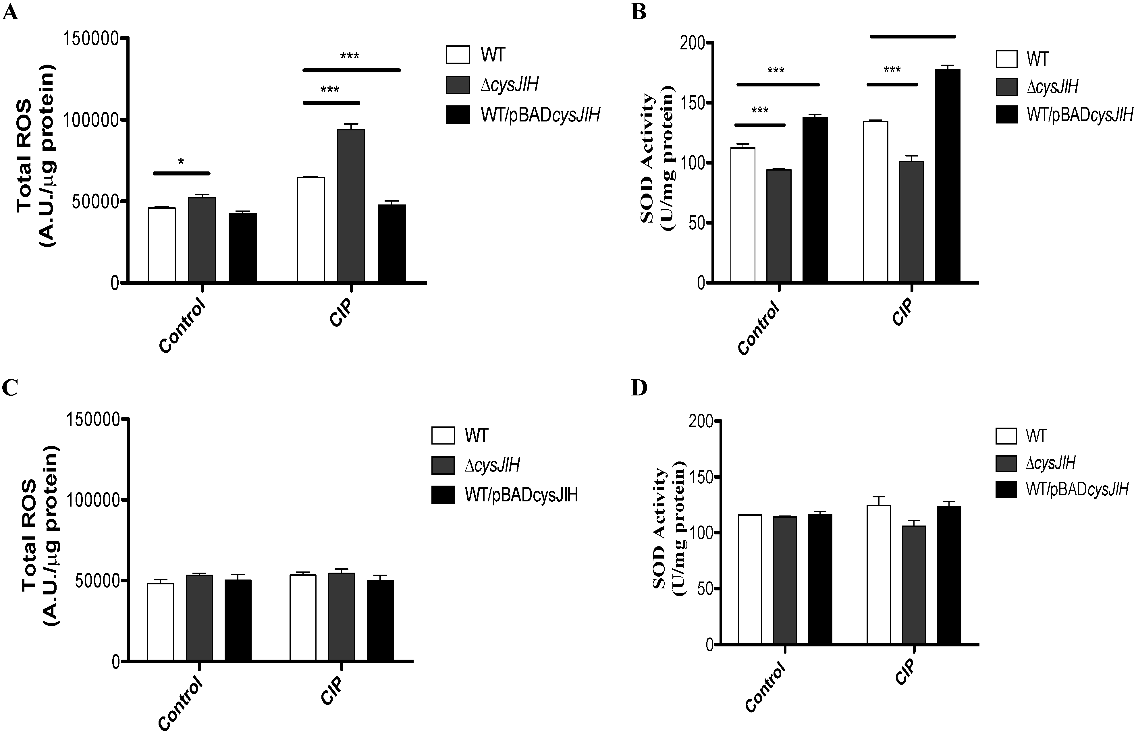
3.1. Deletion of S. Typhimurium cysJIH Results in Increased Intracellular Levels of ROS and Decreased SOD Activity in Presence of Ciprofloxacin When Bacteria Were Cultured with Sulfate as the Sole Sulfur Source

To evaluate the role of cysJIH in the resistance to ciprofloxacin (CIP), we determined the minimal inhibitory concentration (MIC) of S. Typhimurium WT and ΔcysJIH in sulfate and cysteine minimal media. As shown in Table 1, S. Typhimurium ΔcysJIH strain was more sensitive to CIP compared with the WT strain in sulfate medium. This phenotype was reverted in the S. Typhimurium ΔcysJIH complemented with wild-type cysJIH. Furthermore, the S. Typhimurium WT/pBADcysJIH, a cysJIH-overexpressing strain, exhibited even more resistance to CIP than the WT. In cysteine medium, no changes were observed for any strain. Previously, we reported that cysJIH contributes to diminish ROS levels induced by the exposure to antimicrobial agents such as menadione and ceftriaxone by increasing the SOD activity, the levels of reduced thiols, and H2S. This effect was only observed when bacteria were cultured with sulfate as the sole source of sulfur [12], strongly suggesting that cysJIH also contribute to CIP resistance by diminishing ROS. To determine the role of cysJIH in the CIP resistance, we measured oxidative stress markers in S. Typhimurium WT, S. Typhimurium ΔcysJIH, and S. Typhimurium WT/pBADcysJIH in the presence of this antibiotic. As shown in Figure 1, exposure to CIP increased total ROS (Figure 1A) and decreased SOD activity (Figure 1B) in a ΔcysJIH background only when bacteria were cultured with sulfate as the sole sulfur source. In contrast, when bacteria were cultured with cysteine as the sole sulfur source, exposure to CIP had no effect on these same parameters (Figure 1C,D). This result supports the contribution of ROS in the CIP antimicrobial activity (compare Figure 1 and Table 1). Accordingly, we found similar results with ceftriaxone and menadione [12]. Kohanski et al. [15] proposed that some bactericidal antibiotic increases the intracellular levels of ROS. In this sense, several bacterial species could be able to use H2S as a cellular protector to increase resistance to ROS triggered by bactericidal antibiotics [9]. Our results confirm that in a ΔcysJIH strain, ROS response after exposure to CIP is diminished probably due to lower SOD activity in media with sulfate as the sole sulfur source. This effect, as suggested by Shatalin et al. [9], is probably due to a deficient H2S production and hence a less protector effect. The role of H2S in protection to ROS producing agents has been previously suggested. For instance, S. Typhimurium ΔcysK mutant, which could accumulate H2S, is 3-fold more resistant to ciprofloxacin than the WT strain [16,17]. Moreover, upon increased cysteine concentrations, H2S can act as a reducing agent that fuels the Fenton reaction [18]; consequently a transient depletion of free cysteine to produce H2S could allow bacteria to resist the oxidative stress. Altogether, the results presented associates cysJIH with ROS and ciprofloxacin resistance, as described in our previous work with ceftriaxone [12].
Table 1. Minimal Inhibitory Concentration (MIC) determination of strains used in this study
Strain *MIC CIP (μM) sulfateMIC CIP (μM) cysteine
Salmonella Typhimurium ATCC 14028s3.643.64
Salmonella Typhimurium ATCC 14028s ΔcysJIH1.823.64
Salmonella Typhimurium ATCC 14028s ΔcysJIH/pBADcysJIH3.643.64
Salmonella Typhimurium ATCC 14028s WT/pBADcysJIH5.463.64
* All bacteria were treated with ciprofloxacin (CIP) in both M9-sulfate and M9-cysteine media; all determinations were performed 6 times.
Figure 1. Total reactive oxygen species (ROS) and superoxide dismutase (SOD) activity in S. Typhimurium WT and mutant derivative. S. Typhimurium strains were grown in LB medium to OD600 0.45 and subsequently cultured in M9 + sulfate (A,B) or M9 + cysteine (C,D), treated or not with CIP. (A,C) total ROS; (B,D) SOD activity. For all graphics, white bars: S. Typhimurium WT; grey bars: S. Typhimurium ΔcysJIH; black bars: S. Typhimurium WT/pBADcysJIH. Experiments were repeated six times and asterisks represent statistically significant differences as compared with S. Typhimurium WT in each treatment (* p < 0.05; ** p < 0.01; *** p < 0.001).
Antibiotics 04 00321 g001

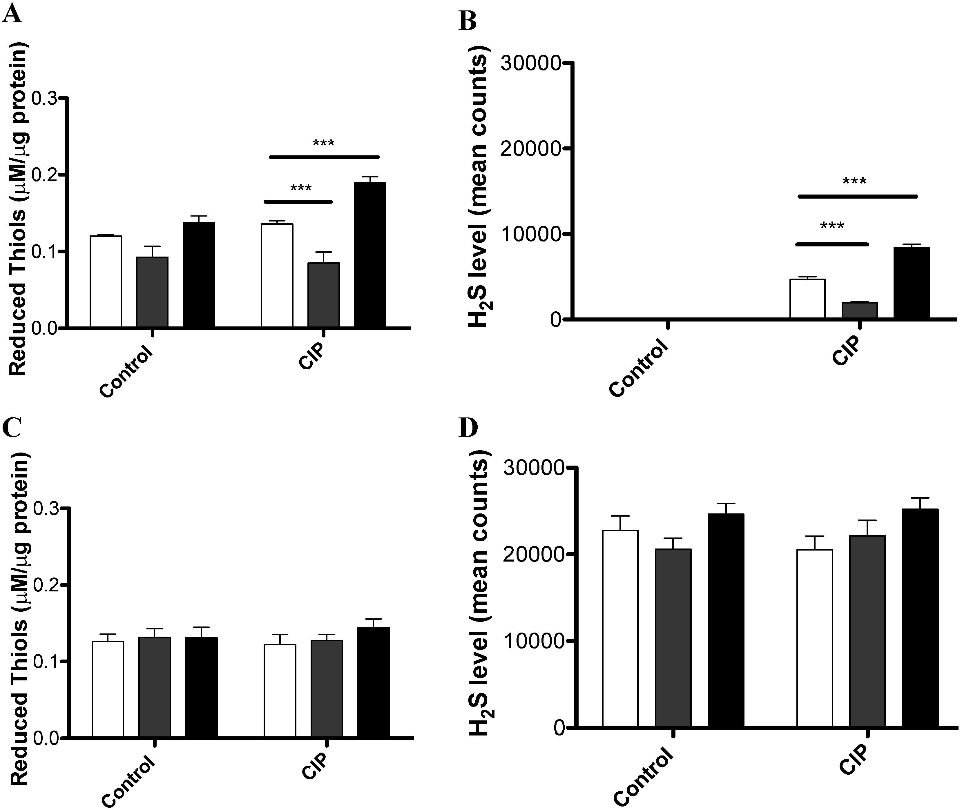
3.2. CIP Induces the Accumulation of Reduced Thiols and H2S in a cysJIH-Dependent Manner with Sulfate as the Sole Sulfur Source

S. Typhimurium ΔcysJIH mutant accumulated more ROS and presented decreased levels of SOD activity as compared with the S. Typhimurium WT, after exposure to CIP. Conceivably, this effect might be explained by a differential H2S accumulation in these strains as reported in our previous work [12]. To test this hypothesis, we evaluated the total reduced thiols and H2S levels induced by CIP in S. Typhimurium WT, S. Typhimurium ΔcysJIH, or S. Typhimurium WT/pBADcysJIH. As shown in Figure 2, S. Typhimurium ΔcysJIH accumulated less reduced thiols (Figure 2A) and less H2S (Figure 2B, data were normalized to the control in which no treatment was amended) compared with S. Typhimurium WT when bacteria were cultured with sulfate in the presence of CIP. Conversely, S. Typhimurium WT/pBADcysJIH, a strain that overexpresses cysJIH, exhibited a higher accumulation of reduced thiols and H2S (Figure 2A,B). In the case of bacteria grown in cysteine, we found that reduced thiols and H2S were accumulated under all of tested culture conditions, where the addition of CIP exerted no effect (Figure 2C,D). Thus, reduced thiols and H2S accumulation perfectly correlate with decreased ROS and increased SOD activity, as shown in our previous work [12].
Altogether, our results show that the exposure to the antimicrobial agent CIP induces H2S accumulation in a cysJIH-dependent manner when bacteria were grown with sulfate as the sole sulfur source. This provides evidence that argues in favor of a mechanism(s) of antibiotic-induced oxidative stress resistance that involves genes that participate in H2S production. Several experiments are required for elucidate the importance of this operon-mechanism relative to CIP resistant enzymes (DNA gyrase and topoisomerase IV) [19] or genes controlling efflux/accumulation of quinolones [20,21], all of which are prevalent and account for clinical failures to CIP and correlate with MIC break points. Such experiments would seemingly define the importance of cysJIH mutations to enhance or decrease CIP susceptibility. A proteomic study could be a good approach to further understand this mechanism at the molecular level, in order to control resistant strains and to develop new therapeutic strategies [21].
Figure 2. Reduced Thiols and H2S production in S. Typhimurium WT and mutant derivative. S. Typhimurium WT and ΔcysJIH strains were grown in LB medium to OD600 0.45 and subsequently cultured in M9 + sulfate (AC) or M9 + cysteine (C,D), treated or not with CIP for 20 min. (A,C) total thiols; (B,D) H2S levels. For all graphics: White bars: S. Typhimurium WT; grey bars: S. Typhimurium ΔcysJIH; black bars: S. Typhimurium WT/pBADcysJIH. Data were normalized to the control in which no treatment was used. Experiments were repeated six times and asterisks represent statistically significant differences as compared with S. Typhimurium WT in each treatment (* p < 0.05; ** p < 0.01; *** p < 0.001).
Antibiotics 04 00321 g002

4. Conclusions

  • cysJIH operon are involved in CIP-resistance by increasing both the levels of H2S and reduced thiols
  • The protective effect of cysJIH operon was observed only when bacteria were cultured in sulfate as the sole sulfur source.

Acknowledgements

This work was supported by grant from FONDECYT #1130074 to FG; FONDECYT #11121262 to PIR; FONDECYT #11110216 and grant from the Research Office of Universidad Andres Bello (DI-340-13/R) to ILC; FONDECYT #11121506 to JAF; and by grants from FONDECYT #1110569, a grant from the Research Office of Universidad Andres Bello (DI-275-13/R), and by a grant from Fondo de Fomento al Desarrollo Científico y Tecnológico (FONDEF) CA13I10077 to DPS. RA received Doctoral fellowship by CONICYT.

Author Contributions

RA and FG conceived the project. RA and FG conducted the results analyses. RA and JF performed the experiments. PIR, JAF, DPS, ILC and FG wrote the paper.

Conflicts of Interest

The authors declare no conflict of interest

References

  1. Sekowska, A.; Kung, H.F.; Danchin, A. Sulfur metabolism in Escherichia coli and related bacteria: Facts and fiction. J. Mol. Microbiol. Biotechnol. 2000, 2, 145–177. [Google Scholar] [PubMed]
  2. Jones, R.T.; Thai, L.P.; Silver, R.P. Genetic and molecular characterization of an Escherichia coli plasmid coding for hydrogen sulfide production and drug resistance. Antimicrob. Agents Chemother. 1978, 14, 765–770. [Google Scholar] [CrossRef]
  3. Wang, R. Physiological implications of hydrogen sulfide: A whiff exploration that blossomed. Physiol. Rev. 2012, 92, 791–896. [Google Scholar] [CrossRef] [PubMed]
  4. Yoshida, A.; Yoshimura, M.; Ohara, N.; Yoshimura, S.; Nagashima, S.; Takehara, T.; Nakayama, K. Hydrogen sulfide production from cysteine and homocysteine by periodontal and oral bacteria. J. Periodontol. 2009, 80, 1845–1851. [Google Scholar] [CrossRef] [PubMed]
  5. Gadalla, M.M.; Snyder, S.H. Hydrogen sulfide as a gasotransmitter. J. Neurochem. 2010, 113, 14–26. [Google Scholar] [CrossRef] [PubMed]
  6. Kimura, H. Hydrogen sulfide: From brain to gut. Antioxid. Redox Signaling 2010, 12, 1111–1123. [Google Scholar] [CrossRef] [PubMed]
  7. Wang, R. Hydrogen sulfide: The third gasotransmitter in biology and medicine. Antioxid. Redox Signaling 2010, 12, 1061–1064. [Google Scholar] [CrossRef] [PubMed]
  8. Lund, M.E.; Matsen, J.M.; Blazevic, D.J. Biochemical and antibiotic susceptibility studies of H2S-negative Citrobacter. Appl. Microbiol. 1974, 28, 22–25. [Google Scholar] [PubMed]
  9. Shatalin, K.; Shatalina, E.; Mironov, A.; Nudler, E. H2S: A universal defense against antibiotics in bacteria. Science 2011, 18, 986–990. [Google Scholar] [CrossRef] [PubMed]
  10. Piras, C.; Soggiu, A.; Bonizzi, L.; Gaviraghi, A.; Deriu, F.; de Martino, L.; Iovane, G.; Amoresano, A.; Roncada, P. Comparative proteomics to evaluate multi drug resistance in Escherichia coli. Mol. Biosyst. 2012, 8, 1060–1067. [Google Scholar] [CrossRef] [PubMed]
  11. Haverkamp, T.; Schwenn, J.D. Structure and function of a cysBJIH gene cluster in the purple sulphur bacterium Thiocapsa roseopersicina. Microbiology 1999, 145, 115–125. [Google Scholar] [CrossRef] [PubMed]
  12. Álvarez, R.; Neumann, G.; Frávega, J.; Díaz, F.; Tejías, C.; Collao, B; Fuentes, J.A.; Paredes-Sabja, D.; Calderón, I.L.; Gil, F. CysB-dependent upregulation of the Salmonella Typhimurium cysJIH operon in response to antimicrobial compounds that induce oxidative stress. Biochem. Biophys. Res. Commun. 2015, 458, 46–51. [Google Scholar] [CrossRef] [PubMed]
  13. Datsenko, K.A.; Wanner, B.L. One-step inactivation of chromosomal genes in Escherichia coli K-12 using PCR products. Proc. Natl. Acad. Sci. USA 2000, 97, 6640–6645. [Google Scholar] [CrossRef] [PubMed]
  14. Guerrero, P.; Collao, B.; Álvarez, R.; Salinas, H.; Morales, E.H.; Calderón, I.L.; Saavedra, C.P.; Gil, F. Salmonella enterica serovar Typhimurium BaeSR two-component system positively regulates sodA in response to ciprofloxacin. Microbiology 2013, 159, 2049–2057. [Google Scholar] [CrossRef] [PubMed]
  15. Kohanski, M.A.; Dwyer, D.J.; Hayete, B.; Lawrence, C.A.; Collins, J.J. A common mechanism of cellular death induced by bactericidal antibiotics. Cell 2007, 130, 797–810. [Google Scholar] [CrossRef] [PubMed]
  16. Turnbull, A.L.; Surette, M.G. L-Cysteine is required for induced antibiotic resistance in actively swarming Salmonella enterica serovar Typhimurium. Microbiology 2008, 154, 3410–3419. [Google Scholar] [CrossRef] [PubMed]
  17. Turnbull, A.L.; Surette, M.G. Cysteine biosynthesis, oxidative stress and antibiotic resistance in Salmonella Typhimurium. Res. Microbiol. 2010, 161, 643–650. [Google Scholar] [CrossRef] [PubMed]
  18. Park, S.; Imlay, J.A. High levels of intracellular cysteine promote oxidative DNA damage by driving the Fenton reaction. J. Bacteriol. 2003, 185, 1942–1950. [Google Scholar] [CrossRef] [PubMed]
  19. Bansal, S.; Tandon, V. Contribution of mutations in DNA gyrase and topoisomerase IV genes to ciprofloxacin resistance in Escherichia coli clinical isolates. Int. J. Antimicrob. Agents 2011, 37, 253–255. [Google Scholar] [CrossRef] [PubMed]
  20. Sharma, V.; Dahiya, S.; Jangra, P.; Das, B.K.; Kumar, R.; Sood, S.; Kapil, A. Study of the role of efflux pump in ciprofloxacin resistance in Salmonella enterica serotype Typhi. Indian J. Med. Microbiol. 2013, 31, 374–378. [Google Scholar] [PubMed]
  21. Piras, C.; Soggiu, A.; Greco, V.; Martino, P.A.; del Chierico, F.; Putignani, L.; Urbani, A.; Nally, J.E.; Bonizzi, L.; Roncada, P. Mechanisms of antibiotic resistance to enrofloxacin in uropathogenic Escherichia coli in dog. J. Proteomics 2015. [CrossRef]

