Article

Investigating the Potential of the Er:YAG Laser for the Removal of Cemented Dust from Limestone and Painted Plaster

by
Lucia Noor Melita
1,*,
Katarzyna Węgłowska
2,
Diego Tamburini
1,† and
Capucine Korenberg
1
1
Department of Scientific Research, The British Museum, Great Russell Street, London WC1B 3DG, UK
2
Department of Conservation, The British Museum, Great Russell Street, London WC1B 3DG, UK
*
Author to whom correspondence should be addressed.
Present Address: Department of Conservation and Scientific Research, Freer Gallery of Art and Arthur M. Sackler Gallery, National Museum of Asian Art, Smithsonian Institution, 1050 Independence Ave SW, Washington, DC 20560, USA.
Coatings 2020, 10(11), 1099; https://doi.org/10.3390/coatings10111099
Submission received: 29 September 2020 / Revised: 6 November 2020 / Accepted: 12 November 2020 / Published: 17 November 2020
(This article belongs to the Special Issue Looking for a Sustainable Cleaning of Cultural Heritage: Agenda 2030)

Abstract

:
A successful application of Er:YAG laser for the cleaning of a restored Assyrian relief sculpture from the British Museum collection is presented. Displayed in the gallery, the sculpture has darkened over time due to the natural deposition of dirt, in particular on restored parts. Since traditional cleaning methods have demonstrated to be unsuccessful, a scientific investigation was performed to identify the composition of the soiling and the materials used for the restoration. The analysis suggested the presence of gypsum, calcium oxalate, carbonates and alumino-silicates on the encrustation. The molded plaster, composed of lime and gypsum and pigmented aggregates, was likely prepared at the end of the 19th century to mimic the stone color. It was repainted with what was identified as a modern oil-based overpaint, applied to cover darkening during a second conservation treatment in the 20th century. Laser trials were first performed on small areas of the objects and on mock-ups to determine the critical fluence thresholds of the surface, investigated through visual examination and analyses using Fourier transform infrared spectroscopy (FTIR) and Pyrolysis-gas chromatography-mass spectrometry (Py–GC–MS). The right parameters and conditions to be used during the cleaning process were, therefore, determined. The chemical selectivity of the cleaning process allowed us to complete the treatment safely while preserving the restoration.

1. Introduction

Dust deposition on museum objects is a challenging issue that requires attention and resources in terms of collection management. Dust not only affects the appearance of objects but, if not regularly removed, can accumulate and lead to the formation of a more cohesive and stubborn layer. Embedded dust particulates can then translate into physical damage and chemical alterations of materials in the long-term, as well as promote pests’ proliferation. To mitigate the risks and support preventive conservation, several studies have focused on different aspects of dust; from identifying the composition of soiling and their effects on substrates [1,2,3,4], to investigating the factors that promote dust adhesion and “cementation”, such as RH fluctuation, salts formation and microbial growth [5,6,7], to understanding the mechanisms of pollutant deposition through air movement and aerosol in indoor heritage [8,9,10].
Once a cemented layer of dust is established on a museum object, it cannot be removed using regular housekeeping practices such as vacuuming or dusting, and a different conservation approach is required. Mechanical and solvent-cleaning methods are often used. However, they may require large amounts of solvents or more than one treatment, which could potentially lead to less precise cleaning and, consequently, damage to the object.
Laser treatment has increasingly become a popular conservation method for the cleaning of stone sculptures and painted surfaces, as it is non-contact, selective and allows greater control, particularly when dealing with delicate substrates [1]. The Nd:YAG laser, emitting at 1064 nm and its harmonics, has demonstrated to be the most employed system for the cleaning of a different range of materials [11,12]. It has been extensively applied for the removal of black crusts of various thickness and biological growth from stone, marble and plaster [13,14,15,16,17,18], as part of the laser energy is selectively absorbed by dark materials and reflected by the underneath substrate. This makes the laser cleaning process self-limiting and ideal for such treatment. Greater progress has been made in the development of laser technology, introducing to the field of conservation a range of different Nd:YAG lasers that not only vary in terms of wavelength but also of pulse duration, between tens of nanoseconds (long Q-switching) to tens of microseconds (short free-running) [19,20]. Research has focused on evaluating the different laser cleaning mechanisms obtained depending on the laser system to overcome effects such as discoloration of pigments on painted surfaces [21,22,23,24,25] and yellowing of stone [11,13,26,27,28,29]. This has further extended the application of the Nd:YAG laser to a wider range of cleaning purposes, including the removal of overpaints on easel paintings [30,31].
However, different types of laser have been introduced to conservation. Among them, the Er:YAG laser is a promising tool for the cleaning of museum objects, although very few instances are reported in the literature [32]. Its cleaning mechanism differs from other laser systems. The Er:YAG laser emits radiation at 2940 nm, which is highly absorbed by materials containing hydroxyl (OH) groups, such as water, solvents and organic substances. When OH groups are present on the surface, the laser energy is absorbed, confining the radiation to the superficial layers of a small volume, thus reducing the depth of ablation. The absorption of the laser wavelength induces direct heating of the irradiated material, which can cause, in most cases, the photo-thermal disaggregation of the surface, without chemical bond dissociation. If the material to remove does not include OH groups, water and solvents such as isopropanol and ethanol can be used to moisten the surface, acting as absorbers and reducing the penetration of the radiation, thus creating explosive localized vaporization, able to eject contaminants and fragments of coatings [32,33]. The Er:YAG laser is effective for the removal of unwanted coating and varnishes [34,35,36,37,38] and for the loosening of contaminants from delicate or unvarnished surfaces [39], often as a two-step cleaning process using solvents. Used to remove soiling, organic coatings and biological growth from stone, marble and plaster [40,41,42,43,44,45], the conditions reported are case-dependent since characteristics such as the chemical composition and the thickness of the encrustation impact the cleaning process and the final outcome. For these reasons, the identification of materials composition is crucial for the selection of the appropriate treatment. However, in some instances, thermally induced chemical transformations may also occur, leading to unwanted alterations of the substrate, such as the color change of pigments containing OH groups [46] as well as the degradation of organic binders on painted surfaces [32]. It is, therefore, important to understand how substrates interact with the laser radiation and determine the critical fluence threshold (CFT) of materials on a case-by-case basis in order to obtain a safe cleaning process.
In 2018 a conservation campaign on a series of objects from the British Museum collection was carried out prior to their display in the exhibition “I am Ashurbanipal, king of the world, king of Assyria” (8 November 2018–24 February 2019). Among the objects, a head of a winged bull was chosen to feature in the exhibition as one of the few surviving sculptures from the palace of Ashurbanipal’s father, King Esarhaddon (680–669 BC), left unfinished at his death. This sculpture had been extensively restored in the past with plaster around the beard, nose, shoulders and hair. These restored parts had probably originally been painted to match the color of the limestone but were covered by a disfiguring dark layer of cemented dust obscuring the details of the sculpture. The object required conservation treatment, which would include structural stabilization, as well as cleaning, in order to improve its appearance (Figure 1). Preliminary tests by the conservator showed that the dark layer had low solubility in water, acetone, industrial methylated spirit (IMS) or enzymes, and well-established solvent cleaning methods proved ineffective. As a Fourier-transform infrared spectroscopy (FTIR) analysis performed on the surface of the dark layer highlighted the presence of strong absorbers in the OH-region centered ~3400 cm−1, it was decided to investigate the use of the Er:YAG laser for the cleaning of the sculpture, in particular the restored parts and fragile paint layer, motivated by the affinity of the laser wavelength with the dark layer to remove.
In this paper, the scientific analysis and conservation treatment of the head sculpture are presented. A multianalytic approach using optical microscopy (OM), FTIR, Raman and scanning electron microscopy with energy-dispersive X-ray spectroscopy (SEM-EDX) was chosen to evaluate and identify the composition of the restored parts and the dark soiling to remove. The effects of the laser irradiation on the substrate and cleaning process were investigated using OM, FTIR and pyrolysis-gas chromatography-mass spectrometry (Py–GC–MS) [47]. The conditions are provided as an example of the applicability of the Er:YAG laser for the cleaning of painted plaster and stone in museum collections.

2. Materials and Methods

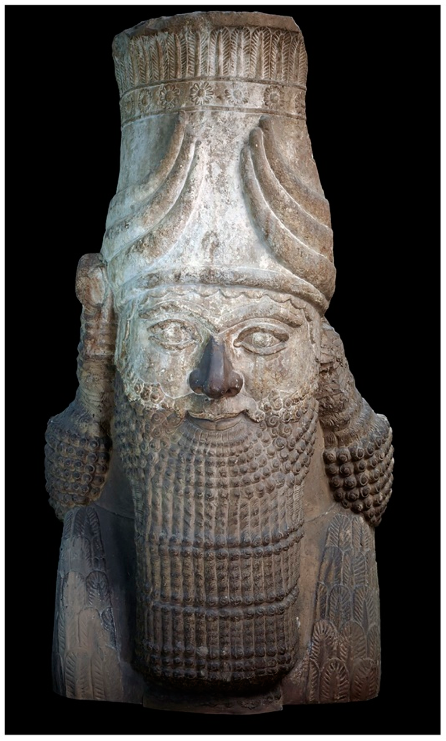
2.1. The Assyrian Limestone Relief Sculpture

The object (Registration Number 1849,1222.1) under investigation is part of a human-headed winged bull (lamassu), a protective figure often used to adorn the entrances of ancient Assyrian palaces. It was excavated by Sir Austen Henry Layard during his archaeological excavations in the Assyrian mounds surrounding Nimrud. Discovered towards the end of 1846 in the South West Palace (entrance c) and resting near the remains of the body [48], the fragment was found “nearly entire”, consisting mostly of the upper part of the face and crown, decorated with bull’s horn carved in a very rough gray limestone [49]. The head, currently on display in the gallery, was brought to the British Museum after its arrival in Britain in August 1849 on board of H.M.S. Meeanee [50].
Little is known about the conservation history of the object, but records indicate that it has been on display for possibly more than 150 years. Some archival information can be found in the official guidebooks of the British Museum. In particular, a complete description of the statue, including the molded beard, nose, shoulders and hair, is included in the 1852 edition, dating a first restoration to the second half of the 19th century shortly after its registration in the Museum Collection [51,52]. In addition to illustrations, the statue appeared in some early photographs: it is shown on a plinth in the background of a view of the Assyrian gallery, taken by Roger Fenton (about 1857–1859) [53], and again in a series of photographs commissioned to Frederick York in 1875 [54]. In the latter, a much darker nose, beard and shoulders are visible, probably as a result of London’s pollution and dirt deposition. It is likely that a second conservation treatment was then performed during the first half of the 20th century to clean and retouch the restored parts, as it appears more homogenous in color in a photograph dated around 1936 [49,55]. When compared with the contemporary appearance of the statue, a missing row of feathers can be observed on the left side of the plain rob. It appears that the statue underwent a localized restoration around the bottom areas to repair part of the missing feathers decoration, potentially damaged when it was moved. Along with other Museum’s objects, the sculpture was evacuated to a safe location to salvage the collection during the Second World War. The Assyrian galleries were then installed in the 1960s for redisplay purposes, and the statue was relocated to its current position, as it was decided that the head, originally part of the lamassu sculpture, should be displayed at appropriate height.

State of Conservation

Through visual examination, it was observed that the restored parts had been more affected by dust deposition than the original limestone. This had led to differential darkening, and cleaning of the restored parts was needed to improve the overall appearance of the sculpture.
A detailed examination allowed acquiring significant information about the internal structure of the restored parts. The lower part of the object, originally missing, had been restored using different materials, such as limestone, clay and plaster. It became clear that the original part of the head had been placed on a limestone block to provide stability and support. Terracotta components, as well as other clay-based ceramic elements, such as bricks, had been mainly used in order to give the restored part a framework for the outermost layer, which was then cast in plaster. The size of the restored parts and the joints, which are still visible on the surface, indicated that the cast had been made using a multi-piece mold. The examination also revealed several cracks indicating that the structure of the restored parts became unstable and required consolidation. Additionally, as mentioned above, it was observed that lower parts were probably damaged and repaired using different materials. The repair, which consisted of the restoration of the bottom part of the feathers, gave the surface an inconsistent and unsightly appearance.

2.2. Analytical Techniques

The materials were investigated using a multianalytic approach. The characterization of the restored parts was performed on samples that were removed from existing damaged areas before and during cleaning tests. The analysis of the limestone was performed in situ, using a portable digital microscope (Dino-Lite, Hemel Hempstead, UK). Evaluation of the laser effects on lead white mock-ups was done using Raman spectroscopy and the portable digital microscope.

2.2.1. Optical Microscopy (OM)

Examination of the surface and cross-sections of the samples was performed using a VHX-5000 digital microscope (Keyence UK Ltd., Milton Keynes, UK), with a magnification between 20× and 1500×.

2.2.2. Scanning Electron Microscopy with Energy Dispersive X-ray Spectroscopy (SEM-EDX)

An Oxford Instruments INCA x-act EDX system (Silicon Drift Detector) attached to a Hitachi S3700 N variable pressure (VP)-SEM (Hitachi High-Technologies Corporation, Tokyo, Japan) was used for elemental analysis of the surface dirt and cross-sections of the restored parts, operating in low vacuum mode (30 or 40 Pa) at 20 kV and at a working distance of 10 mm. The EDX was optimized using a Co (Cobalt) standard. Data were analyzed with the Oxford Instruments Aztec Energy analysis software (3.0 SP1 version) for qualitative analysis.

2.2.3. Raman Spectroscopy

Raman spectroscopy was carried out to characterize the restored parts and mock-ups using a Jobin Yvon LabRAM Infinity spectrometer (Groupe HORIBA, Kyoto, Japan), equipped with a green (532 nm) and red laser (785 nm) with a maximum power of 2.4 mW at the sample, a liquid nitrogen cooled CCD detector and an Olympus microscope system. The instrument was calibrated using a silicon standard, with a wavenumber deviation of 3 cm−1. Spectra were collected for 5–10 s (at least five repetitions) on a spectral range of 100–1600 cm−1. Fine gratings of 1800 g/mm and 900 g/mm were used, and the power of the laser was adjusted depending on the sample (0.1–50%). Samples were placed onto a microscope slide and analyzed without any further treatment.

2.2.4. Fourier-Transform Infrared Spectroscopy (FTIR)

FTIR spectroscopy was performed on a Nicolet 6700 spectrometer (Thermo Fisher Scientific, Waltham, MA, USA) attached to a Continuum IR microscope equipped with MCT/A detectors. Samples taken from the surface of the restored parts before and after cleaning were analyzed in transmission mode, flattened in a diamond microcompression cell. The field of view was controlled by the sliding aperture, which, when fully open, gives a maximum area of analysis of 100 × 100 μm2. The spectra were acquired over a range of 4000–650 cm−1 using 32 scans at a resolution of 4 cm−1 and automatic gain.

2.2.5. Pyrolysis-Gas Chromatography-Mass Spectrometry (Py–GC–MS)

Py–GC–MS analysis was carried out on samples from the overpaint layer applied during the second restoration campaign (first half of the 20th century). The two samples were taken before and after the laser cleaning to evaluate possible compositional changes in the surface layer.
Analytical pyrolysis was performed using 1,1,1,3,3,3–hexamethyldisilazane (HMDS, chemical purity 99.9%, Sigma Aldrich Inc., St. Louis, MI, USA) as a derivatizing (silylating) agent for the in situ thermally-assisted derivatization of pyrolysis products. The instrumentation consisted of a micro-furnace Multi-Shot Pyrolyzer EGA/Py-3030D (Frontier Lab, Fukushima, Japan) coupled to a gas chromatograph 6890 (Agilent Technologies, Palo Alto, CA, USA) equipped with an HP-5MS fused silica capillary column (30 m × 0.25 mm i.d., 0.25 µm-thickness, Agilent Technologies, Palo Alto, CA, USA). The GC was coupled with an Agilent 5973 Mass Selective Detector operating in electron impact mode (EI) at 70 eV. The pyrolysis temperature was 550 °C, and the interface temperature was 300 °C. Similar amounts (ca. 50–100 μg) of sample and HMDS (2 µL) were inserted into stainless steel cups. The GC injector was used with a split ratio of 1:10 at 300 °C. Chromatographic conditions were as follows: initial temperature 36 °C, 10 min isothermal, 10 °C·min−1 up to 280 °C, 2 min isothermal; 20 °C·min−1 up to 310 °C, 20 min isothermal. Helium (purity 99.995%) was used as carrier gas with a constant flow of 1.0 mL·min−1.

2.3. Laser Cleaning Treatment

A Fidelis XS Erbium yttrium aluminum laser (Er:YAG) (Fotona, Ljubljana, Slovenia) emitting at 2940 nm was used for the cleaning tests and treatment of the object. The laser was set with a pulse duration of 100 µs delivered through an articulated arm. The laser spot size, as measured on thermal paper, was between 2.2 and 4.0 mm in diameter. Fluence was determined during the cleaning tests, and frequency was kept at 2 Hz during tests and at 10 and 20 Hz during cleaning. A Dino-Lite microscope was used to monitor the laser cleaning during the tests.

3. Results

3.1. Characterization of the Restored Parts and Soiling Composition

The analysis of the materials used in the restoration of the beard, nose, shoulders and hair was performed on detached fragments. The main results are summarized in Table 1.
A cross-sectional examination is shown in Figure 2a. The layers identified were the following:
  • A bulk of a late 19th-century restoration, a colored plaster, composed of a mixture of fine and coarse pigmented aggregates to imitate the color and texture of the original stone;
  • A thin dark line, as shown at higher magnification in Figure 2b, possibly an early finishing material or organic coating, typical of historical plasterworks in the 19th century. It has probably darkened over time due to degradation and dirt deposition [16,56];
  • A white overpaint, acting as a ground layer;
  • An orange layer, not homogenous in thickness throughout the cross-sections;
  • An additional thin white layer, found in some areas, possibly applied to lighten the surface of the restored parts and mimic the hue and texture of the original stone and intricate beard (Figure 2c);
  • A thin dark deposition, i.e., cemented dirt.
It is possible that the paint layers were added to the object during a later restoration campaign to reduce the contrast between the darkened areas of the restored parts (ii) and the original stone to refresh the color of the restoration [57]. The aim of laser cleaning was to remove layer (vi) only and uncover the paint layer.
The FTIR analysis of the bulk of the restored parts (Figure 3a) revealed the presence of calcium carbonate (CaCO3) as the main component and reaction product of the carbonation process of lime plaster Ca(OH)2. The spectrum is characterized by the CO32− ν3 asymmetric stretching mode at ~1410 cm−1, the ν1 symmetric stretch at ~1087 cm−1 and the O–C–O ν2 out-of-plane bending mode at 874 cm−1. The ν4 in-plane bending mode at 713 cm−1 and 744 cm−1 are assigned to calcite and vaterite, respectively, CaCO3 polymorphs [58]. The broad vibration band in the region 900–1200 cm−1 suggested the presence of Si–O groups, characteristic of clay minerals. Bands at 3549, 1621, 669 and 601 cm−1 were attributed to gypsum. A broad absorption band in the OH stretching region was also observed due to the presence of structural hydroxyl groups [59]. EDX mapping highlighted Ca as the main element, as well as Si, Al, Fe in the inclusions. Elements such as S, K, Cl, Mg and Mn were also detected.
The Raman spectra of the white and orange paint layers are presented in Figure 3b,c. The analysis of the white layer revealed the presence of lead white pigment, i.e., lead carbonate (Pb(OH)2∙2PbCO3). This was confirmed by the characteristic shift at 1053 (s) cm−1 attributed to lead carbonate [60]. EDX analysis highlighted Pb as the main element, as well as Al and Ca. The paint also contains calcium carbonate as a filler, as suggested by the band at 1088 (vs) cm−1 [60]. In the orange layer, Pb and Cr were detected as main components. The Raman spectrum (Figure 3c) showed bands at 140 (m), 320 (w), 339 (m), 350 (vw), 377 (m), 823 (vs), 835 (s), 845 (s) cm−1, attributed to chrome orange, i.e., basic lead chromate (PbCrO4∙PbO) [60,61,62]. Chrome orange is a synthetic pigment that was widely used by artists at the end of the 19th century and the beginning of the 20th century [63,64]. EDX analysis on both thin dark layers highlighted the presence of Al and Ca, as main elements, as well as S, Si, Fe, Cu, K and Cl probably due to environmental pollution.
The FTIR spectrum on the surface prior to laser cleaning (Figure 4a) is characterized by a broad absorbance band centered around 3400 cm−1, suggesting that the dark layer is composed of materials that are rich in OH groups, main absorbers of the Er:YAG wavelength (2940 nm), highlighting the potential of this particular laser for the removal of the unwanted layer [32,45]. The bands at ~3550, 3406 (OH stretching vibration), and additionally 1113 and 668 cm−1 (S–O stretching and bending, respectively) indicated the presence of gypsum, whereas the bands at 1640, 1325 (C=O stretching vibration) and 783 cm−1 (O–C–O bending vibration) suggested calcium oxalate [65]. The broadband centered at ~1445 cm−1, the sharp band at 876 cm−1 and peak at 713 cm−1 were attributed to calcite. Bands in the Si–O stretching region (900–1200 cm−1), in addition to 796 cm−1, suggested the presence of alumino-silicates, as confirmed by the EDX analysis. Peaks at 2924 and 2853 cm−1 were attributed to the CH2 stretching (asymmetric and symmetric, respectively) of aliphatic chains, highlighting the presence of organic compounds. The results suggested that the dark layer is mainly composed of gypsum, calcium oxalate, and inorganic components, consistent with observations on patinas and encrustations found on stone and marble in churches and outdoor environments due to weathering, biological activity, pollution and/or alteration of previous organic-based treatments [66,67]. The object, like other sculptures at the British Museum, was inevitably exposed to high levels of pollution in the 19th and 20th centuries, which have led to the formation of the dark cohesive layers [68].

3.2. Laser Treatment

Laser tests were first performed on commercial pure lead white samples prepared in the laboratory to determine the critical fluence threshold (CFT) of the pigment and evaluate if discoloration may occur. CTF is defined as the lowest fluence (energy per surface unit) at which changes in the material are observed. The samples were irradiated in dry conditions with a single laser shot at increasing fluences, and surface changes, such as color, texture or gloss variations, were observed under a Dino-Lite microscope. During the experiments, a CFT of 1.4–1.6 J/cm2 was found when a color change (yellowing) was detected. Lead white, identified by the shift at 1052 (s) cm−1 before irradiation (Figure 5a, in blue), is converted to lead oxide (PbO), likely massicot and litharge, as evidenced by the broad bands centered at 84, 111, 144 and 284 cm−1 in the Raman spectrum (Figure 5a, in red) [69]. Evidence of yellowing was also observed after several consecutive pulses at 1.2 J/cm2. However, it was found to be reversible. Temporary yellowing effects using the Nd:YAG laser (1064 nm) have been documented on medieval pigments [22,70], and it was reported that the duration of the color change could last from several hours to some days, depending on the laser energy applied. This has been described as a conversion mechanism of lead white into lead monoxide and consequent reconversion [23]. Alteration of pure pigment (without binders) has also been reported when irradiated by the four wavelengths (1064, 532, 355, 266 nm). The pigment turned grey and then black, resulting in a mixture of metallic lead (Pb), lead oxide (PbO) and lead dioxide (PbO2) forms. However, it regained its original color within one week after exposure to air and CO2 [71]. Although the Er:YAG laser mechanism differs from the Nd:YAG laser, it is possible to assume that the energy transferred to the sample has locally sufficiently increased the temperature to induce the conversion. By increasing the fluence, a blackening of lead white was also observed, with some degree of reconversion. The Raman spectrum of blackened areas (Figure 5a, in black) is characterized by bands at 84, 139 and 284 cm−1, which closely resemble bands attributed to massicot. This has been described as a product of laser-induced thermal degradation of plattnerite (PbO2), deriving from the degradation of lead white [72]. Irradiation at fluences above 2.2 J/cm2 caused permanent darkening of the pigment, which also remained after artificial aging. Blackening could be a result of the reduction to metallic lead [70,73], difficult to detect by Raman due to its poor scattering properties [69]. Permanent laser-induced degradation of lead white is shown in Figure 5b,c, where globules of metallic lead were observed after irradiation.
To remove the soiling on the restored plaster parts (Figure 6a), irradiation tests with the Er:YAG laser were conducted with a fluence in the range of 1.1–1.8 J/cm2 in dry and wet conditions with a 10–20 Hz pulse rate. Using a fluence of ~1.8 J/cm2 and above in dry conditions caused cracking and abrasion of the paint layer, visible under high magnification. When used in wet conditions, a microdistillation mechanism was observed, due to the penetration of the solvent underneath the very thin dark layer and into the paint layers, causing the disaggregation of the surface and complete removal of the paint. This left the bulk of the restored part exposed. Cleaning of the restored part was achieved with fluences below 1.8 J/cm2 in dry conditions. The irradiated area turned gray (Figure 6b), most likely due to the enhancement of light scattering phenomena caused by an increase in the surface roughness. The laser treatment was then followed by gentle swabbing using water to remove the soiling and obtain the desired cleaning result (Figure 6c). This was now possible as the overall surface area exposed is higher, improving the cleaning ability of the solvent. The surface was left to fully dry after swabbing, before continuing with the laser. Two-three laser passes were found to be sufficient to remove the dark layer, monitoring the cleaning with the portable microscope. A similar result was reported on the removal of a soot film embedded in the paint of an unvarnished 19th-century oil painting, where the contaminant was turned into fine dust, which was then wiped off with a moistened cotton swab [39]. Although the laser parameters used for the cleaning of the restored parts overlapped with the CFT found for the mock-ups, we did not observe discoloration of the underneath paint layer during cleaning, suggesting that the laser interacted with the dark layer only. Using a fluence very close or even higher than the CFT is not uncommon with the Er:YAG laser [35]. This is because the contaminant is typically not ablated by the Er:YAG laser irradiation, but its top surface is disrupted due to thermal effects. A strong absorber at 2940 nm, such as the dirt covering the sculpture (Figure 4a), acts as a thermal barrier for the material underneath as it absorbs most of the laser energy [33]. The key to using a fluence close to the CFT is to proceed one laser pass at a time. As soon as the top layer is disrupted, the laser stops being used, and the contaminant layer is removed using solvent through swabbing.
A similar approach was used for the limestone; however, on a much smaller scale (Figure 7a), as the original stone did not require as much cleaning as the restoration. In that case, cleaning was performed very locally at a fluence in the range of 1.6–1.8 J/cm2 in dry conditions with a 10 Hz pulse rate (Figure 7b), followed by swabbing the irradiated area with a 50:50 mixture of water and acetone (Figure 7c). Cleaning of the stone allowed highlighting the natural pigmentation of the limestone, as shown in Figure 7d,e.

3.3. Investigation of the Effects of Laser Irradiation

After cleaning the restored parts, a decrease of the broad absorption band in the OH region, as well as of the bands attributed to oxalates, sulfates and alumino-silicate components, was detected by FTIR, as shown in Figure 8a. This is consistent with the removal of the dark layer, highlighting the efficacy of the Er:YAG laser to target OH-rich materials. The IR spectrum is characterized by the CO32− bands at 1409 cm−13 asymmetric stretching), 1045 cm−11 symmetric stretching), 839 cm−12 out-of-plane bending), the split in the ν4 in-plane bending vibration at 690 and 681 cm−1 and the OH stretching vibration near 3540 cm−1, corresponding to lead carbonate of the painted surface [74]. Additionally, the presence of an organic binder was detected, indicated by the C=O stretching vibration bands of carboxylic acids and esters centered at ~1737 cm−1, the C–H stretching bands at 2955 (CH3), 2928 and 2854 cm−1 (CH2, asymmetric and symmetric, respectively) and the C–O stretching bands of esters at 1170 and 1099 cm−1, suggesting the presence of an oil-based paint [75]. A small shoulder at ~1710 cm−1 was also observed, which was attributed to C=O stretching vibrations due to the formation of free fatty acids as a result of hydrolysis of triglycerides and the formation of dicarboxylic acids due to oxidation [76]. The broad shoulder centered at ~1550 cm−1 may indicate metal carboxylates as a result of metal soaps formation due to the interaction of lead salts with the oil binder [77,78].
Py–GC–MS was performed to confirm the identification of the surface layer and highlight any major changes in its composition after laser irradiation. Due to the inhomogeneity of the paint layers, as shown in Figure 2b,c, the sample after cleaning included both the white and the orange layer.
The total ion chromatogram (TIC) of the sample before cleaning showed the predominant presence of aliphatic mono– and dicarboxylic acids (Figure 4b). Among them, hexadecanoic (P–palmitic) and octadecanoic (S–stearic) acids showed the highest relative abundance, followed by nonanedioic (A–azelaic) acid. This was in agreement with the general composition of a drying oil obtained by analytical pyrolysis [79]. In particular, azelaic acid is the final product deriving from the auto-oxidation of the unsaturated side chains of a drying oil [80,81,82], thus showing a relatively high oxidation state of the oil. Py–GC–MS is not the right technique to speculate about the origin of the oil unless specific molecular markers are detected, e.g., 12–hydroxyl–9–cis–octadecenoic (ricinoleic) acid for castor oil [83]. No specific molecular marker was detected in our case. Moreover, parameters traditionally used in GC–MS analysis, such as A/P ratio and P/S ratio, cannot be considered in a quantitative way when pyrolysis is applied [84]. For all these reasons, no further information about the type of oil was obtained. Nevertheless, in addition to carboxylic acids, other minor compounds were detected, including glycerol and some phthalates (isobutyl, diethyl and dibutyl phthalates). These are known additives usually found in modern oil-based paint compositions to modify specific properties [85]. Phthalates are used as plasticizers, and glycerol is a common precursor of modern oil-based paint compositions [86,87]. Although the data did not allow us to further classify the type of binder, the results were generally consistent with a lead white modern oil-based paint with good drying properties, which is in agreement with both the supposed time of application (first half of the 19th century) and a rapid restoration use.
The TIC of the sample after laser cleaning (Figure 8b) showed the presence of all the compounds identified in the sample before cleaning. However, a clear relative reduction of the dicarboxylic acids was observed. These are richer in OH groups, and the result was therefore interpreted as further proof of the efficacy of the laser treatment. Additionally, as dicarboxylic acids are not part of the cross-linked network of the polymer and are partially responsible for the yellowing of oil binders, their partial removal by laser contributed to the better appearance of the surface after laser cleaning.

3.4. Conservation Treatment

The examination revealed a number of significant cracks, which indicated that the structure of the restored part became unstable with time. A network of cracks, their location (mostly at the bottom part of the cast), and pattern provided relevant information about the loss of mechanical strength and cohesion between different layers and materials used in the restoration. This had to be addressed in order to impart strength to the whole structure. The comprehensive consolidation was completed with different solutions of acrylic resin—Paraloid B72—in acetone and IMS, ranging from 5% to 40%. Given different sizes of the cracks and rather complicated internal structure, the resin was introduced using different application methods. This was done in order to reach deeper layers of the structure. The remaining cracks and losses in the cast were filled with microballoons in 20% Paraloid B72 and finished flush with the surface. Finally, the fills were toned down with acrylic paints.
The later repair to the bottom part of the plaster was approached in a slightly different way. The overpaint, shown in Figure 9a, was uncovered after the laser cleaning. It was presumably acrylic paint, in accordance with the conservation materials used in the 1960s for retouches. It was removed using acetone from the plaster fill, which appeared to be structurally sound, and then toned down with acrylic paint.
The fill, which most likely was made as a result of damage to the lower part of the restoration, was completed only in the front and both sides of the sculpture, as shown in Figure 9b. The back remained untreated, with the outer layer of the cast missing (Figure 9c), probably left unfinished as it would not have been visible when displayed in the new location. This area allowed us to investigate the internal structure as well as evaluate the penetration of the consolidation treatment in real time. The edge of the remaining layer was consolidated where necessary, capped with the microballoons paste and finally toned down to color match the limestone (Figure 10).

4. Discussion

It was observed that the painted plaster was more affected by dust deposition than the original limestone, suggesting that the material composition of the substrate had a more pronounced tendency to incorporate dust, embedded in the paint layer by a matrix composed of OH-rich molecules and justifying the use of the Er:YAG laser. Cleaning of the limestone was mostly achieved through mechanical cleaning or using the laser just on difficult areas, while cleaning of the restoration was not possible with traditional methods.
A high relative abundance of dicarboxylic acids was observed on the restored part before laser cleaning, highlighting a high state of oxidation of the paint binder, corresponding to the degraded “skin” of the paint layer. It also indicates a certain degree of hydrolysis of triglycerides, which is influenced by moisture and fluctuations of RH. An increase in water sensitivity in modern oil-based paints, depending on their formulation, has been usually observed in 20th and 21st-century paintings, which are often unvarnished (uncoated), therefore, more vulnerable to aging and to environmental factors [88,89,90]. However, paints containing metal ions such as lead and zinc, although high in diacids content, have been found to be non-water sensitive and to promote metal soaps formation. This has suggested that other factors may contribute to the stability of the paint [91]. It is, therefore, possible to hypothesize that the cementation of dust on the painted plaster has occurred not only as a result of deposition, interaction of hygroscopic particulate with moisture and consequent formation of salts, as observed in indoor environments [6], but also due to an increase in the polarity of the painted surface due to aging. This could have consequently attracted more dust particulates on the restored parts of the statue than on the limestone.
Although a full characterization of the dust was not in the scope of this paper, the presence of organic debris (e.g., fibers, hair, pollen), as well as microorganisms cannot be fully excluded. Recently, a study on dust analysis conducted at the British Museum has demonstrated that several organic components, such as feathers, cotton fibers, pollen and mites, can also be found deposited on objects displayed in galleries [2].
As evidenced by the characterization of the surface after laser cleaning, laser irradiation produced a thermally induced disruption of the organic components and materials containing OH groups in the dark layer, responsible for the cementation of the dust [33]. It is speculated that the weakening of these compounds broke down the cohesiveness of the inorganic components, increasing the roughness and surface area of the layer. After just a few laser passes, the cemented dust was completely removed by swabbing, as a greater surface area increases the exposure to solvents.

5. Conclusions

This study aimed to investigate the applicability of the Er:YAG laser and to understand the cleaning mechanism for the removal of cemented dust in museum collections, in particular painted plaster. Although this laser does not have self-limiting properties when it comes to the removal of dark layers and encrustation, similar to the Nd:YAG laser, it was able to perform selective cleaning of unwanted OH-rich materials in a quick and efficient way, where traditional methods failed, thus reducing treatment time as well as solvent use. In this case, the preliminary FTIR analysis of the dark layer was essential to determine the most appropriate laser wavelength, showing a strong affinity with the Er:YAG laser.
The analysis of samples taken from the soiled plaster enabled the substrate composition to be identified as a lime plaster painted mostly with lead white in a modern oil-based medium, and the dark layer as mainly composed of gypsum, oxalates and inorganic components, present in environmental particulate and pollutants. Additionally, a stratigraphy of the samples suggested that the restored parts had been subjected to past interventions in order to improve their appearance.
The laser treatment was shown to interact primarily with materials rich in OH groups, including degradation products, which was beneficial for this specific case. The material to remove was found to absorb strongly at the laser emission wavelength, enabling the treatment to be performed in dry conditions (without requiring a pre-wetting agent) as well as confining the interaction to the near-surface. On the contrary, the use of a wetting agent (wet conditions) was demonstrated to promote explosive vaporization and ejection of the dark layer as well as the painted surface, probably due to the penetration of the solvent underneath the fragile paint layer. The use of wetting agents is therefore not indicated for the removal of very thin layers, in particular, if OH groups are already found in the composition, or for the cleaning of porous materials, as the solvent would penetrate into the structure, potentially damaging the underneath layers while irradiating the area with the Er:YAG laser.
The analyses demonstrated that, after laser cleaning, removal of soiling compounds was successfully achieved. A notable reduction of the dicarboxylic acids, corresponding to the degraded “skin” of the paint layer, was also observed. The removal of this oxidation layer, responsible for yellowing, improved the overall aspect of the molded plaster.
In addition, no damage was detected visually or using an optical microscope during the cleaning of the paint, such as darkening or yellowing of lead white, which was observed when directly irradiating the surface of mock-ups in laboratory conditions. This suggested that the dark layer covering the sculpture acted as a thermal barrier, absorbing most of the laser energy. The laser treatment was confined to the surface, and heat did not significantly dissipate to the underneath layers.
This study has demonstrated that cleaning with the Er:YAG laser is a promising technique for the removal of cemented dust from unvarnished painted surfaces of museum artifacts.

Author Contributions

Conceptualization, L.N.M. and K.W.; methodology, L.N.M. and K.W.; validation, L.N.M.; formal analysis, L.N.M. and D.T.; investigation, L.N.M. and D.T.; writing—original draft preparation, L.N.M.; writing—review and editing, L.N.M., K.W., D.T., and C.K.; visualization, L.N.M.; supervision, L.N.M. All authors have read and agreed to the published version of the manuscript.

Funding

This research was funded by the Andrew W. Mellon Foundation (G-40900619).

Acknowledgments

The authors would like to thank longstanding supporters Ed and Anne Teppo and the American Friends of the British Museum for the donation of the Er:YAG laser to the British Museum; St John Simpson, Gareth Brereton (Department of Middle East, The British Museum) and Laura Carnelos (Department of Collection Projects and Resources, The British Museum) for their input during the research of historical information; Tracey Sweek (Department of Collection Care, The British Museum) for their input during conservation treatment, the Andrew W. Mellon Foundation which supported Lucia N. Melita and Diego Tamburini’s postdoctoral fellowships.

Conflicts of Interest

The authors declare no conflict of interest.

References

  1. Marczak, J.; Koss, A.; Targowski, P.; Góra, M.; Strzelec, M.; Sarzyński, A.; Skrzeczanowski, W.; Ostrowski, R.; Rycyk, A. Characterization of Laser Cleaning of Artworks. Sensors (Basel) 2008, 8, 6507–6548. [Google Scholar] [CrossRef] [Green Version]
  2. Portoni, F.; Perucchetti, L.; Mak, S.; Cartwright, C. What Lurks under the Microscope? Dust Detective Work. Available online: https://blog.britishmuseum.org/what-lurks-under-the-microscope-dust-detective-work/ (accessed on 4 September 2020).
  3. Fox, S.-J.; Babington, C.; Macalister, F.; Bower, T.; Martin de Fonjaudran, C. Monitoring and Mitigating Particulate Matter Deposition on Decorative Surfaces: Current and Future Approaches in the Palace of Westminster. Stud. Conserv. 2018, 63, 81–86. [Google Scholar] [CrossRef]
  4. Macher, J.M. Review of methods to collect settled dust and isolate culturable microorganisms. Indoor Air 2001, 11, 99–110. [Google Scholar] [CrossRef]
  5. Tarnowski, A.L.; McNamara, C.J.; Bearce, K.A.; Mitchell, R. Sticky microbes and dust on objects in historic houses. In Proceedings of the 32nd Annual Meeting of the American Institute of Conservation (AIC), Portland, OR, USA, 8–13 June 2004; pp. 10–28. [Google Scholar]
  6. Lithgow, K.; Lloyd, H.; Brimblecombe, P.; Yoon, Y.; Thickett, D. Managing dust in historic houses—A visitor/conservator interface. In Proceedings of the ICOM-CC, 14th Triennial Meeting Preprints, The Hague, The Netherlands, 12–16 September 2005; pp. 662–669. [Google Scholar]
  7. Ferreira, M.; Grau-Bove, J.; Mazzei, L.; Blades, N.; Curteis, T.; Altamirano, H.; Faull, J. The Influence of Water Activity and Air Movement in Preventing Mould in Historic Materials. Stud. Conserv. 2018, 63, 348–350. [Google Scholar] [CrossRef]
  8. Shah, B.; Hunter, S.; Adams, S.; Bancroft, A.A.C.R.; Blyth, V.A.C.R. When the Dust Settles: Dust Monitoring in Exhibitions at the Victoria and Albert Museum. Int. Preserv. News 2011, 59, 24–29. [Google Scholar]
  9. Grau-Bové, J.; Mazzei, L.; Thickett, D.; Strlič, M. New Perspectives on the Study of Particulate Matter Deposition within Historic Interiors. Stud. Conserv. 2019, 64, 193–202. [Google Scholar] [CrossRef]
  10. Grau-Bové, J.; Mazzei, L.; Strlic, M.; Cassar, M. Fluid simulations in heritage science. Herit. Sci. 2019, 7, 16. [Google Scholar] [CrossRef]
  11. Siano, S.; Agresti, J.; Cacciari, I.; Ciofini, D.; Mascalchi, M.; Osticioli, I.; Mencaglia, A.A. Laser cleaning in conservation of stone, metal, and painted artifacts: State of the art and new insights on the use of the Nd:YAG lasers. Appl. Phys. A 2012, 106, 419–446. [Google Scholar] [CrossRef]
  12. Siano, S.; Salimbeni, R. Advances in laser cleaning of artwork and objects of historical interest: The optimized pulse duration approach. Acc. Chem. Res. 2010, 43, 739–750. [Google Scholar] [CrossRef]
  13. Klein, S.; Fekrsanati, F.; Hildenhagen, J.; Dickmann, K.; Uphoff, H.; Marakis, Y.; Zafiropulos, V. Discoloration of marble during laser cleaning by Nd:YAG laser wavelengths. Appl. Surf. Sci. 2001, 171, 242–251. [Google Scholar] [CrossRef]
  14. Tanguy, E.; Huet, N.; Vinçotte, A. Lasers Cleaning of Patrimonial Plasters; Dickmann, K., Fotakis, C., Asmus, J.F., Eds.; Springer: Berlin/Heidelberg, Germany, 2005; pp. 125–132. [Google Scholar] [CrossRef] [Green Version]
  15. Bartoli, L.; Pouli, P.; Fotakis, C.; Siano, S.; Salimbeni, R. Characterization of Stone Cleaning by Nd:YAG Lasers with Different Pulse Duration. Laser Chem. 2006, 2006. [Google Scholar] [CrossRef] [Green Version]
  16. Pelosi, C.; Fodaro, D.; Sforzini, L.; Rubino, A.; Falqui, A. Study of the laser cleaning on plaster sculptures. The effect of laser irradiation on the surfaces. Opt. Spectrosc. 2013, 114, 121–132. [Google Scholar] [CrossRef]
  17. Osticioli, I.; Mascalchi, M.; Pinna, D.; Siano, S. Removal of Verrucaria nigrescens from Carrara marble artefacts using Nd:YAG lasers: Comparison among different pulse durations and wavelengths. Appl. Phys. A 2015, 118, 1517–1526. [Google Scholar] [CrossRef]
  18. Barreiro, P.; González, P.; Pozo-Antonio, J.S. IR irradiation to remove a sub-aerial biofilm from granitic stones using two different laser systems: An Nd: YAG (1064 nm) and an Er:YAG (2940 nm). Sci. Total Environ. 2019, 688, 632–641. [Google Scholar] [CrossRef]
  19. Zanini, A.; Trafeli, V.; Bartoli, L. The laser as a tool for the cleaning of Cultural Heritage. IOP Conf. Ser. Mater. Sci. Eng. 2018, 364, 012078. [Google Scholar] [CrossRef]
  20. Siano, S. Multiple Temporal Regime Laser for Art Conservation. Available online: https://spie.org/news/4913-multiple-temporal-regime-laser-for-art-conservation#B3 (accessed on 2 November 2020).
  21. Sansonetti, A.; Realini, M. Nd:YAG laser effects on inorganic pigments. J. Cult. Herit. 2000, 1, S189–S198. [Google Scholar] [CrossRef]
  22. Pouli, P.; Emmony, D.C. The effect of Nd:YAG laser radiation on medieval pigments. J. Cult. Herit. 2000, 1, S181–S188. [Google Scholar] [CrossRef]
  23. Zafiropulos, V.; Stratoudaki, T.; Manousaki, A.; Melesanaki, K.; Orial, G. Discoloration of Pigments Induced by Laser Irradiation. Surf. Eng. 2001, 17, 249–253. [Google Scholar] [CrossRef]
  24. Ciofini, D.; Cacciari, I.; Siano, S. Multi-pulse laser irradiation of cadmium yellow paint films: The influence of binding medium and particle aggregates. Measurement 2018, 118, 311–319. [Google Scholar] [CrossRef]
  25. Oujja, M.; Castillejo, M.; Pouli, P.; Fotakis, C.; Domingo, C. Spectral analysis of the effects of laser wavelength and pulse duration on tempera paints. In Lasers in the Conservation of Artworks: Proceedings of the International Conference LACONA VIII, Sibiu, Romania, 21–25 September 2009; Radvan, R., Asmus, J., Castillejo, M., Pouli, P., Nevin, A., Eds.; CRC Press: London, UK, 2010; pp. 15–22. [Google Scholar]
  26. Berthonneau, J.; Parent, P.; Grauby, O.; Ferry, D.; Laffon, C.; Colombini, A.; Courtois, B.; Bromblet, P. Yellowing of laser-cleaned artworks: Formation of residual hydrocarbon compounds after Nd:YAG laser cleaning of gypsum plates covered by lamp black. J. Cult. Herit. 2019, 39, 57–65. [Google Scholar] [CrossRef]
  27. Licciardi, R.; Mascalchi, M.; Siano, S.; Megna, B. La tecnologia laser nella pulitura dei manufatti lapidei, casi studio a confronto. In Proceedings of the Aplar 5, applicazioni laser nel restauro, Musei Vaticani, Vatican City State, 18–20 September 2014; Brunetto, A., Ed.; Nardini Editore: Florence, Italy, 2017; pp. 15–30. [Google Scholar]
  28. Godet, M.; Vergès-Belmin, V.; Andraud, C.; Saheb, M.; Monnier, J.; Bourgon, J.; Binet, L.; Leroy, E. Laser yellowing effect: Study of the nanophases created by laser irradiation of synthetic black crusts using transmission electron microscopy (TEM) and electron paramagnetic resonance (EPR) spectroscopy. In Proceedings of Lacona XI—Lasers in the Conservation of Artworks, Krakow, Poland, 20–23 September 2016; Targowski, P., Walczak, M., Pouli, P., Eds.; Nicolaus Copernicus University Press: Toruń, Poland, 2017; pp. 63–76. [Google Scholar]
  29. Godet, M.; Vergès-Belmin, V.; Gauquelin, N.; Saheb, M.; Monnier, J.; Leroy, E.; Bourgon, J.; Verbeeck, J.; Andraud, C. Nanoscale investigation by TEM and STEM-EELS of the laser induced yellowing. Micron 2018, 115, 25–31. [Google Scholar] [CrossRef] [PubMed]
  30. Siano, S.; Osticioli, I.; Pavia, A.; Ciofini, D. Overpaint removal from easel paintings using an LQS Nd:YAG laser: The first validation study. Stud. Conserv. 2015, 60, S49–S57. [Google Scholar] [CrossRef]
  31. Striber, J.; Jovanović, V.; Jovanović, M. Easel paintings on canvas and panel: Application of Nd:YAG laser at 355 nm, 1064 nm and UV, IR and visible light for the development of new methodologies in conservation. In Proceedings of Lacona XI—Lasers in the Conservation of Artworks, Krakow, Poland, 20–23 September 2016; Targowski, P., Walczak, M., Pouli, P., Eds.; Nicolaus Copernicus University Press: Toruń, Poland, 2017; pp. 279–292. [Google Scholar]
  32. Pereira-Pardo, L.; Korenberg, C. The use of erbium lasers for the conservation of cultural heritage. A review. J. Cult. Herit. 2018, 31, 236–247. [Google Scholar] [CrossRef]
  33. Teppo, E. Introduction: Er:YAG lasers in the conservation of artworks. J. Inst. Conserv. 2020, 43, 2–11. [Google Scholar] [CrossRef]
  34. Bracco, P.; Lanterna, G.; Matteini, M.; Nakahara, K.; Sartiani, O. L’esperienza dell’Opificio nella sperimentazione del laser ad erbio per la pulitura dei dipinti. OPD Restauro 2001, 13, 192–202. [Google Scholar]
  35. Brunetto, A.; Bono, G.; Frezzato, F. Er:YAG laser cleaning of ‘San Marziale in Gloria’ by Jacopo Tintoretto in the Church of San Marziale, Venice. J. Inst. Conserv. 2020, 43, 44–58. [Google Scholar] [CrossRef]
  36. Chillè, C.; Sala, F.; Wu, Q.; Theodorakopoulos, C. A study on the heat distribution and oxidative modification of aged dammar films upon Er:YAG laser irradiation. J. Inst. Conserv. 2020, 43, 59–78. [Google Scholar] [CrossRef]
  37. Striová, J.; Castellucci, E.; Sansonetti, A.; Camaiti, M.; Matteini, M.; Decruz, A.; Andreotti, A.; Colombini, M.P. Free-running Er: YAG laser cleaning of mural painting specimens treated with linseed oil, ‘Beverone’and Paraloid B72. In Lasers in the Conservation of Artworks: Proceedings of the International Conference LACONA VIII, Sibiu, Romania, 21–25 September 2009; Radvan, R., Asmus, J., Castillejo, M., Pouli, P., Nevin, A., Eds.; CRC Press: London, UK, 2010; pp. 85–91. [Google Scholar]
  38. Andreotti, A.; Brown, W.P.; Camaiti, M.; Colombini, M.P.; Decruz, A. Diagnosis of materials and effectiveness of Er:YAG laser cleaning as complementary treatment in a panel painting attributed to Lluís Borrassà (fifteenth century). Appl. Phys. A 2016, 122, 572. [Google Scholar] [CrossRef]
  39. De Cruz, A.; Wolbarsht, M.L.; Hauger, S.A. Laser removal of contaminants from painted surfaces. J. Cult. Herit. 2000, 1, S173–S180. [Google Scholar] [CrossRef]
  40. De Cruz, A.; Wolbarsht, M.L.; Palmer, R.A.; Pierce, S.E.; Adamkiewicz, E. Er:YAG Laser Applications on Marble and Limestone Sculptures with Polychrome and Patina Surfaces; Springer: Berlin/Heidelberg, Germany, 2005; pp. 113–124. [Google Scholar]
  41. De Cruz, A.; Wolbarsht, M.L.; Andreotti, A.; Colombini, M.P.; Pinna, D.; Culberson, C.F. Investigation of the Er:YAG Laser at 2.94 μm to Remove Lichens Growing on Stone. Stud. Conserv. 2009, 54, 268–277. [Google Scholar] [CrossRef]
  42. De Cruz, A.; Andreotti, A. La pulitura di un’urna romana con il laser Er:YAG a 2940 nm. In Proceedings of Aplar 5, Applicazioni Laser Nel Restauro, Musei Vaticani, Vatican City State, 18–20 September 2014; Brunetto, A., Ed.; Nardini Editore: Florence, Italy, 2017; pp. 101–110. [Google Scholar]
  43. Pereira-Pardo, L.; Camurcuoglu, D.; Orsini, M.; Vasiliou, S.; Weglowska, K.; Kiely, T.; Korenberg, C. The use of an Er:YAG Laser in the removal of biological growth from polychrome archaeological terracotta figurines from Cyprus. In Proceedings of the Recent Advances in Glass and Ceramics Conservation—Interim Meeting of the ICOM-CC Working Group, The British Museum, London, UK, 5–7 September 2019; pp. 77–86. [Google Scholar]
  44. Andreotti, A.; Colombini, M.P.; De Cruz, A. Er:YAG laser cleaning of a marble Roman urn. J. Inst. Conserv. 2020, 43, 12–24. [Google Scholar] [CrossRef]
  45. Pereira-Pardo, L.; Melita, L.N.; Korenberg, C. Tackling conservation challenges using erbium lasers: Case studies at the British Museum. J. Inst. Conserv. 2020, 43, 25–43. [Google Scholar] [CrossRef]
  46. Camaiti, M.; Matteini, M.; Sansonetti, A.; Striova, J.; Castellucci, E.M.; Andreotti, A.; Colombini, M.P.; DeCruz, A.; Palmer, R. The interaction of laser radiation at 2.94 μm with azurite and malachite pigments. In Lasers in the Conservation of Artworks, Proceedings of the International Conference Lacona VII, Madrid, Spain, 17–21 September 2007; Castillejo, M., Moreno, P., Oujja, M., Radvan, R., Ruiz, J., Eds.; CRC Press: London, UK, 2008; pp. 253–258. [Google Scholar]
  47. Andreotti, A.; Colombini, M.P.; Nevin, A.; Melessanaki, K.; Pouli, P.; Fotakis, C. Multi analytical Study of Laser Pulse Duration Effects in the IR Laser Cleaning of Wall Paintings from the Monumental Cemetery of Pisa. Laser Chem. 2006, 2006, 11. [Google Scholar] [CrossRef] [Green Version]
  48. Layard, A.H. Nineveh and Its Remains: With an Account of a Visit to the Chaldaean Christians of Kurdistan, and the Yezidis, Or Devil-worshippers, and an Enquiry Into the Manners and Arts of the Ancient Assyrians, 1st ed.; John Murray: London, UK, 1849; Volume 2, p. 26. [Google Scholar]
  49. Gadd, C.J. The Assyrian Sculptures. By C. J. Gadd, Etc.; British Museum. Order of the Trustees: London, UK, 1934; p. 70. [Google Scholar]
  50. Gadd, C.J. The Stones of Assyria; the Surviving Remains of Assyrian Sculpture, Their Recovery, and Their Original Positions; Chatto and Windus: London, UK, 1936; p. 246. [Google Scholar]
  51. Bonomi, J. Nineveh and Its Palaces: The Discoveries of Botta and Layard, Applied to the Elucidation of Holy Writ/By Joseph Bonomi, 2nd ed.; Office of the Illustrated London library: London, UK, 1852; p. 295. [Google Scholar]
  52. Renouf, P.L.P. Assyrian Antiquities: Guide to the Nimroud Central Saloon; British Museum: London, UK, 1886; pp. 6–7. [Google Scholar]
  53. Fenton, R. The Assyrian Gallery, British Museum. Available online: http://www.getty.edu/art/collection/objects/146261/roger-fenton-the-assyrian-gallery-british-museum-english-about-1857-1859/?dz=0.3582,0.2490,5.41 (accessed on 19 February 2020).
  54. Photograph No 32. In Frederick York Album (1875); The British Museum Archive: London, UK.
  55. Barnett, R.D.; Falkner, M. The Sculptures of Aššur-Nasir-Apli II, 883-859 B.C.: Tiglath-pileser III, 745–727 B.C. and Esarhaddon, 681–669 B.C. from the Central and South-West Palaces at Nimrud; Trustees of the British Museum: London, UK, 1962; p. 165. [Google Scholar]
  56. Salavessa, E.; Jalali, S.; Sousa, L.M.O.; Fernandes, L.; Duarte, A.M. Historical plasterwork techniques inspire new formulations. Constr. Build. Mater. 2013, 48, 858–867. [Google Scholar] [CrossRef] [Green Version]
  57. Doherty, E.; Rivers, S. The removal of lead-and oil-based overpaint from a plaster cast of Hermes Fastening his Sandal. In Gels in the Conservation of Art; Angelova, L.V., Ormsby, B., Townsend, J., Wolbers, R., Eds.; Archetype Publications: London, UK, 2017; pp. 122–125. [Google Scholar]
  58. Cizer, Ö.; Rodriguez-Navarro, C.; Ruiz-Agudo, E.; Elsen, J.; Van Gemert, D.; Van Balen, K. Phase and morphology evolution of calcium carbonate precipitated by carbonation of hydrated lime. J. Mater. Sci. 2012, 47, 6151–6165. [Google Scholar] [CrossRef] [Green Version]
  59. Madejová, J. FTIR techniques in clay mineral studies. Vib. Spectrosc. 2003, 31, 1–10. [Google Scholar] [CrossRef]
  60. Burgio, L.; Clark, R.J.H. Library of FT-Raman spectra of pigments, minerals, pigment media and varnishes, and supplement to existing library of Raman spectra of pigments with visible excitation. Spectrochim. Acta Part A Mol. Biomol. Spectrosc. 2001, 57, 1491–1521. [Google Scholar] [CrossRef]
  61. Correia, A.M.; Clark, R.J.H.; Ribeiro, M.I.M.; Duarte, M.L.T.S. Pigment study by Raman microscopy of 23 paintings by the Portuguese artist Henrique Pousão (1859–1884). J. Raman Spectrosc. 2007, 38, 1390–1405. [Google Scholar] [CrossRef]
  62. Monico, L.; Janssens, K.; Hendriks, E.; Brunetti, B.G.; Miliani, C. Raman study of different crystalline forms of PbCrO4 and PbCr1−xSxO4 solid solutions for the noninvasive identification of chrome yellows in paintings: A focus on works by Vincent van Gogh. J. Raman Spectrosc. 2014, 45, 1034–1045. [Google Scholar] [CrossRef]
  63. Monico, L.; Van der Snickt, G.; Janssens, K.; De Nolf, W.; Miliani, C.; Verbeeck, J.; Tian, H.; Tan, H.; Dik, J.; Radepont, M.; et al. Degradation Process of Lead Chromate in Paintings by Vincent van Gogh Studied by Means of Synchrotron X-ray Spectromicroscopy and Related Methods. 1. Artificially Aged Model Samples. Anal. Chem. 2011, 83, 1214–1223. [Google Scholar] [CrossRef]
  64. Monico, L.; Janssens, K.; Cotte, M.; Sorace, L.; Vanmeert, F.; Brunetti, B.G.; Miliani, C. Chromium speciation methods and infrared spectroscopy for studying the chemical reactivity of lead chromate-based pigments in oil medium. Microchem. J. 2016, 124, 272–282. [Google Scholar] [CrossRef] [Green Version]
  65. Toniolo, L.; Zerbi, C.M.; Bugini, R. Black layers on historical architecture. Environ. Sci. Pollut. Res. 2009, 16, 218–226. [Google Scholar] [CrossRef] [PubMed]
  66. Pinna, D.; Galeotti, M.; Rizzo, A. Brownish alterations on the marble statues in the church of Orsanmichele in Florence: What is their origin? Herit. Sci. 2015, 3, 7. [Google Scholar] [CrossRef] [Green Version]
  67. Rampazzi, L.; Andreotti, A.; Bressan, M.; Colombini, M.P.; Corti, C.; Cuzman, O.; d’Alessandro, N.; Liberatore, L.; Palombi, L.; Raimondi, V.; et al. An interdisciplinary approach to a knowledge-based restoration: The dark alteration on Matera Cathedral (Italy). Appl. Surf. Sci. 2018, 458, 529–539. [Google Scholar] [CrossRef]
  68. Oddy, A. The Conservation of Marble Sculptures in the British Museum Before 1975. Stud. Conserv. 2002, 47, 145–154. [Google Scholar] [CrossRef]
  69. Burgio, L.; Clark, R.J.H.; Firth, S. Raman spectroscopy as a means for the identification of plattnerite (PbO), of lead pigments and of their degradation products. Analyst 2001, 126, 222–227. [Google Scholar] [CrossRef]
  70. Pouli, P.; Emmony, D.C.; Madden, C.E.; Sutherland, I. Analysis of the laser-induced reduction mechanisms of medieval pigments. Appl. Surf. Sci. 2001, 173, 252–261. [Google Scholar] [CrossRef]
  71. Chappé, M.; Hildenhagen, J.; Dickmann, K.; Bredol, M. Laser irradiation of medieval pigments at IR, VIS and UV wavelengths. J. Cult. Herit. 2003, 4, 264–270. [Google Scholar] [CrossRef]
  72. Smith, G.D.; Burgio, L.; Firth, S.; Clark, R.J.H. Laser-induced degradation of lead pigments with reference to Botticelli’s Trionfo d’Amore. Anal. Chim. Acta 2001, 440, 185–188. [Google Scholar] [CrossRef]
  73. Cooper, M.I.; Fowles, P.S.; Tang, C.C. Analysis of the laser-induced discoloration of lead white pigment. Appl. Surf. Sci. 2002, 201, 75–84. [Google Scholar] [CrossRef]
  74. Nodari, L.; Ricciardi, P. Non-invasive identification of paint binders in illuminated manuscripts by ER-FTIR spectroscopy: A systematic study of the influence of different pigments on the binders’ characteristic spectral features. Herit. Sci. 2019, 7, 7. [Google Scholar] [CrossRef]
  75. de Viguerie, L.; Payard, P.A.; Portero, E.; Walter, P.; Cotte, M. The drying of linseed oil investigated by Fourier transform infrared spectroscopy: Historical recipes and influence of lead compounds. Prog. Org. Coat. 2016, 93, 46–60. [Google Scholar] [CrossRef] [Green Version]
  76. Banti, D.; La Nasa, J.; Tenorio, A.L.; Modugno, F.; Jan van den Berg, K.; Lee, J.; Ormsby, B.; Burnstock, A.; Bonaduce, I. A molecular study of modern oil paintings: Investigating the role of dicarboxylic acids in the water sensitivity of modern oil paints. RSC Adv. 2018, 8, 6001–6012. [Google Scholar] [CrossRef] [Green Version]
  77. Mazzeo, R.; Prati, S.; Quaranta, M.; Joseph, E.; Kendix, E.; Galeotti, M. Attenuated total reflection micro FTIR characterisation of pigment–binder interaction in reconstructed paint films. Anal. Bioanal. Chem. 2008, 392, 65–76. [Google Scholar] [CrossRef]
  78. Cotte, M.; Checroun, E.; De Nolf, W.; Taniguchi, Y.; De Viguerie, L.; Burghammer, M.; Walter, P.; Rivard, C.; Salomé, M.; Janssens, K.; et al. Lead soaps in paintings: Friends or foes? Stud. Conserv. 2017, 62, 2–23. [Google Scholar] [CrossRef] [Green Version]
  79. Bonaduce, I.; Andreotti, A. Py-GC/MS of Organic Paint Binders. In Organic Mass Spectrometry in Art and Archaeology; Colombini, M.P., Modugno, F., Eds.; Wiley: Chichester, UK, 2009; pp. 303–326. [Google Scholar]
  80. Colombini, M.P.; Modugno, F.; Fuoco, R.; Tognazzi, A. A GC-MS study on the deterioration of lipidic paint binders. Microchem. J. 2002, 73, 175–185. [Google Scholar] [CrossRef]
  81. Plater, M.J.; De Silva, B.; Gelbrich, T.; Hursthouse, M.B.; Higgitt, C.L.; Saunders, D.R. The characterisation of lead fatty acid soaps in ‘protrusions’ in aged traditional oil paint. Polyhedron 2003, 22, 3171–3179. [Google Scholar] [CrossRef]
  82. Tamburini, D.; Sardi, D.; Spepi, A.; Duce, C.; Tinè, M.R.; Colombini, M.P.; Bonaduce, I. An investigation into the curing of urushi and tung oil films by thermoanalytical and mass spectrometric techniques. Polym. Degrad. Stab. 2016, 134, 251–264. [Google Scholar] [CrossRef] [Green Version]
  83. Wei, S.; Pintus, V.; Schreiner, M. A comparison study of alkyd resin used in art works by Py-GC/MS and GC/MS: The influence of aging. J. Anal. Appl. Pyrolysis 2013, 104, 441–447. [Google Scholar] [CrossRef]
  84. Colombini, M.P.; Andreotti, A.; Bonaduce, I.; Modugno, F.; Ribechini, E. Analytical Strategies for Characterizing Organic Paint Media Using Gas Chromatography/Mass Spectrometry. Acc. Chem. Res. 2010, 43, 715–727. [Google Scholar] [CrossRef]
  85. Scalarone, D.; Chiantore, O. Py-GC/MS of Natural and Synthetic Resins. In Organic Mass Spectrometry in Art and Archaeology; Colombini, M.P., Modugno, F., Eds.; Wiley: Chichester, UK, 2009; pp. 327–361. [Google Scholar]
  86. Muizebelt, W.J.; Donkerbroek, J.J.; Nielen, M.W.F.; Hussem, J.B.; Biemond, M.E.F.; Klaasen, R.P.; Zabel, K.H. Oxidative crosslinking of alkyd resins studied with mass spectrometry and NMR using model compounds. J. Coat. Technol. 1998, 70, 83–93. [Google Scholar] [CrossRef]
  87. Izzo, F.C.; van den Berg, K.J.; van Keulen, H.; Ferriani, B.; Zendri, E. Modern Oil Paints—Formulations, Organic Additives and Degradation: Some Case Studies. In Issues in Contemporary Oil Paint; van den Berg, K.J., Burnstock, A., de Keijzer, M., Krueger, J., Learner, T., Tagle, d.A., Heydenreich, G., Eds.; Springer International Publishing: Cham, Switzerland, 2014; pp. 75–104. [Google Scholar]
  88. La Nasa, J.; Lee, J.; Degano, I.; Burnstock, A.; van den Berg, K.J.; Ormsby, B.; Bonaduce, I. The role of the polymeric network in the water sensitivity of modern oil paints. Sci. Rep. 2019, 9, 3467. [Google Scholar] [CrossRef] [PubMed]
  89. Modugno, F.; Di Gianvincenzo, F.; Degano, I.; van der Werf, I.D.; Bonaduce, I.; van den Berg, K.J. On the influence of relative humidity on the oxidation and hydrolysis of fresh and aged oil paints. Sci. Rep. 2019, 9, 5533. [Google Scholar] [CrossRef] [PubMed]
  90. Burnstock, A.; Van den Berg, K.J. Twentieth Century Oil Paint. The Interface Between Science and Conservation and the Challenges for Modern Oil Paint Research. In Issues in Contemporary Oil Paint; Springer International Publishing: Cham, Switzerland, 2014; pp. 1–19. [Google Scholar]
  91. Lee, J.; Bonaduce, I.; Modugno, F.; La Nasa, J.; Ormsby, B.; van den Berg, K.J. Scientific investigation into the water sensitivity of twentieth century oil paints. Microchem. J. 2018, 138, 282–295. [Google Scholar] [CrossRef]
Figure 1. Human-headed winged bull (Registration number 1849,1222.1) before cleaning. The molded plaster (beard, nose, shoulders and hair) appears darker than the original limestone. ©Trustees of the British Museum.
Coatings 10 01099 g001
Figure 2. Digital microphotographs of a restored part in cross-section, highlighting the stratigraphy (a) shows the bulk of the 19th-century restoration (i) and possibly an early finishing material or organic coating that has darkened due to deposition of dirt (ii), then repainted with coats of white (iii, v) and orange (iv) paint; a thin dark deposition layer (vi) is observed throughout the paint layer. The sequence of the stratigraphy is not homogenous throughout the restored part. (b) and (c) show close-ups of an area of the restored part with evidence of past dirt deposition (ii), then repainted (iii and iv), and of an area where an additional layer of white was applied to lighten the surface color (v). ©Trustees of the British Museum.
Coatings 10 01099 g002
Figure 3. (a) FTIR spectrum of the bulk of the restored parts; Raman spectra (λ = 785 nm) of the white (b) and orange paint layers (c).
Coatings 10 01099 g003
Figure 4. Digital microphotograph and characterization of the soiling layer on the restored parts before cleaning with Er:YAG laser. (a) FTIR spectrum and (b) Py–GC–MS chromatogram: trimethylsilyl (TMS) derivatives of 1, lactic acid; 2, hexanoic acid; 3, glycolic acid; 4, heptanoic acid; 5 glycerol (2TMS); 6, benzoic acid; 7, octanoic acid; 8, glycerol (3TMS); 9, nonanoic acid; 10, decanoic acid; 11, triethylene glycol; 12, diethyl phthalate; 13, dodecanoic acid; 14, octanedioic acid; 15, triethylene glycol; 16, nonanedioic acid; 17, tetradecanoic acid; 18, decanedioic acid; 19, hexadecanoic acid; 20 octadecanoic acid. ©Trustees of the British Museum.
Coatings 10 01099 g004
Figure 5. (a) Raman spectra (λ = 532 nm) of lead white samples before irradiation (in blue), after irradiation at 1.4–1.6 J/cm2 when yellowing occurred (in red) and when darkening was observed (in black); SEM images before (b) and after (c) laser irradiation at fluence above 2.2 J/cm2. ©Trustees of the British Museum.
Coatings 10 01099 g005
Figure 6. Digital microphotographs of the restored parts before (a), after laser irradiation (b) and after swabbing the residues with water (c); close-up of the nose before (d) and after cleaning was completed (e). ©Trustees of the British Museum.
Coatings 10 01099 g006
Figure 7. Digital microphotographs of the stone (eyebrow) before (a) after laser irradiation (b) and after swabbing with a 50:50 mixture of water and acetone (c). Close-up of the eye before (d) and after cleaning was completed (e), highlighting the natural pigmentation of the limestone. ©Trustees of the British Museum.
Coatings 10 01099 g007
Figure 8. Digital microphotograph and characterization of the surface layer after cleaning with Er:YAG laser and swabbing the residues with water. (a) FTIR spectrum and (b) Py–GC–MS chromatogram: refer to Figure 4 for labels.
Coatings 10 01099 g008
Figure 9. Detail of the late repair on the lower part of the sculpture. (a) Laser cleaning revealed that alternated dark and light brushstrokes of paint were used to repaint the damaged part, giving an inconsistent appearance in comparison with the original restoration (b). The repair was not completed in the back, allowing to check the condition of the internal structure of the restored parts (c). ©Trustees of the British Museum.
Coatings 10 01099 g009
Figure 10. Human-headed winged bull (Registration Number 1849,1222.1) after cleaning, retouching and consolidation. ©Trustees of the British Museum.
Coatings 10 01099 g010
Table 1. Summary of the results.
LayerMaterialsSEM/EDXRamanFTIRPy–GC–MSNotes
iBulkCa, Al, Si, Fe
(S, K, Cl, Mg, Mn)
Calcite and vaterite
Gypsum
Lime plaster + gypsum with inclusions of clay and oxide minerals to give color and texture similar to the original stone.
iiDark layerAl, Ca, S, Si, Fe, Cu, KEvidence of previous accumulation of dirt and dust, then covered with paint.
iii, vWhite paint layerPb, Ca, Al
(Ba, S, Ti, Na, Cu, K, Si)
Lead carbonate
Calcium carbonate
Lead carbonate
Oil
Possible metal soaps
Mono- and dicarboxylic acids (oil)
Traces of phthalates
Lead-based paint (Pb(OH)2∙2PbCO3) in modern oil medium. Evidence of oxidation and hydrolysis of triglycerides due to aging. Possible barium sulfate as an extender
ivOrange paint layerPb, Ca, Cr, Ba
(Al, Fe, K, K, Fe, Cu)
Basic lead chromate
Lead carbonate
Mono- and dicarboxylic acids (oil); Traces of phthalatesLead-based paint (PbCrO4∙PbO and Pb(OH)2∙2PbCO3) in modern oil medium
viSurface dark layerAl, Ca, K, S, Cl, Si, P, Ti, Fe, Cu, Ba, Cl (Pb)Gypsum
Calcium oxalate
Calcite
Alumino-silicates
Organic compounds
Mono-, di-, oxy- and hydroxyl-carboxylic acids (highly oxidized oil)
Traces of phthalates
Heterogeneous cemented dust, mainly composed of gypsum and oxalates, as a result of environmental pollution deposition and degradation of the aged organic binder. Pb attributed to the paint layer
Publisher’s Note: MDPI stays neutral with regard to jurisdictional claims in published maps and institutional affiliations.

