Article

Cytotoxicity and Mineralization Potential of Four Calcium Silicate-Based Cements on Human Gingiva-Derived Stem Cells

1
College of Medicine, The Catholic University of Korea, Seoul 06591, Korea
2
Department of Periodontics, College of Medicine, The Catholic University of Korea, Seoul 06591, Korea
3
Department of Conservative Dentistry, Seoul St. Mary’s Dental Hospital, College of Medicine, The Catholic University of Korea, Seoul 06591, Korea
*
Author to whom correspondence should be addressed.
These authors contributed equally to this study.
Coatings 2020, 10(3), 279; https://doi.org/10.3390/coatings10030279
Submission received: 10 February 2020 / Revised: 12 March 2020 / Accepted: 16 March 2020 / Published: 18 March 2020
(This article belongs to the Special Issue Recent Advances in Coatings of Implant and Dental Biomaterials)

Abstract

:
The aim of this study was to evaluate the cytotoxicity and mineralization potential of four calcium silicate-based cements on human gingiva-derived stem cells (GDSCs). The materials evaluated in the present study were ProRoot MTA (Dentsply Tulsa Dental Specialties), Biodentine (Septodont), Endocem Zr (Maruchi), and RetroMTA (BioMTA). Experimental disks of 6 mm in diameter and 3 mm in height were produced and placed in a 100% humidified atmosphere for 48 h to set. We evaluated the cytotoxic effects of the cements using methyl-thiazoldiphenyl-tetrazolium (MTT) and live/dead staining assays. We used a scratch wound healing assay to evaluate cell migratory ability. Mineralization potential was determined with an Alizarin red S (ARS) staining assay. In the MTT assay, no significant differences were found among the ProRoot MTA, Biodentine, and control groups during the test period (p > 0.05). The Endocem Zr and RetroMTA groups showed relatively lower cell viability than the control group at day 7 (p < 0.05). In the wound healing assay, no significant differences were found among the ProRoot MTA, Biodentine, Endocem Zr, and control groups during the test period (p > 0.05). The RetroMTA group had slower cell migration compared to the control group at days 3 and 4 (p < 0.05). In the ARS assay, the ProRoot MTA, Biodentine, and RetroMTA groups exhibited a significant increase in the formation of mineralized nodules compared to the Endocem Zr and control groups on day 21 (p < 0.05). In conclusion, the four calcium silicate-based cements evaluated in the present study exhibited good biological properties on GDSCs. ProRoot MTA, Biodentine, and RetroMTA showed higher mineralization potential than the Endocem Zr and control groups.

1. Introduction

External root resorption (ERR) happens when the periodontal ligament of the cementum is either destructed or removed [1]. Damage to the cementum uncovers the root surface to osteoclasts that can resorb dentin. With additional stimulation provoked by sulcular bacteria in the neighboring area, root resorption constantly progresses [2]. ERR of a permanent tooth is generally unfavorable because it may cause irreversible damage and ultimately loss of the tooth. However, in its early stages, ERR can be stabilized by repairing the cementum with calcium silicate-based cement [3].
Calcium silicate-based cements are hydraulic materials consisting of tricalcium silicate, dicalcium silicate, and tricalcium aluminate [4,5]. The first tricalcium silicate-based cement was mineral trioxide aggregate (MTA), which is a derivative of Portland cement. The physical, chemical, and biological properties of MTA have been studied for decades, and it produces favorable results when applied to direct pulp capping, regenerative endodontic procedure, apical retrograde filling, and repair of ERR or perforations [6]. Tricalcium silicate enhances proliferation and differentiation of dental pulp cells [7,8,9]. However, ProRoot MTA (Dentsply Tulsa Dental Specialties, Tulsa, OK, USA) contains heavy metal components such as bismuth oxide [5]. It also has a long setting time and handling difficulty, and can discolor the tooth and gingiva [10,11]. Novel calcium silicate-based cements have been produced overcome these shortcomings.
Biodentine (Septodont, Saint-Maur-des-Fossés, France) is composed mostly of tricalcium silicate, zirconium oxide, and calcium carbonate powder, which are mixed with a supplied solution that includes calcium chloride [12,13]. The reduced setting time compared to MTA is achieved by diminishing the particle size and adding calcium chloride to expedite the reactions [13,14,15]. The substitution of bismuth oxide with zirconium oxide may also play a role in reduced setting time, because this component has been reported to expedite the primary hydration reaction [13]. Previous studies of this material’s effects on dental pulp stem cells demonstrated its biocompatible ability, odontoblast differentiation ability, and mineralization potential [12,16]. Endocem Zr (Maruchi, Wonju, Korea) and RetroMTA (BioMTA, Seoul, Korea) were developed to cause less tooth discoloration compared to ProRoot MTA [17]. These materials have a reduced setting time compared to ProRoot MTA and are easy to handle [18,19]. Bismuth oxide is replaced by zirconium oxide as a substitute radiopacifier [17]. RetroMTA is composed of fine hydrophilic particles that do not originate from Portland cement [19].
To the best of our knowledge, no study has evaluated the biocompatibility and calcium nodule formation ability of various calcium silicate-based cements on human gingiva-derived stem cells (GDSCs). Therefore, the aim of the present study was to evaluate the cytotoxic effects of four calcium silicate-based cements on GDSC compared to that of intermediate restorative material (IRM; Caulk Dentsply, Midford, DE, USA). IRM is a commonly used temporary filling material that is highly toxic to human stem cells [18,20]; therefore, we used IRM as a negative control. We also evaluated the mineralization potential of the four calcium silicate-based cements on GDSCs.

2. Materials and Methods

2.1. Human Gingiva-Derived Stem Cells

GDSCs were collected using a previously reported method [21]. Gingival tissue was collected from a 70-year-old female undergoing a second implant surgery. The institutional review board of Seoul St. Mary’s Hospital, College of Medicine, The Catholic University of Korea approved this study (KC19SESI0259), and written informed consent was obtained from the participant. All experiments were performed according to relevant guidelines and regulations specified in the Declaration of Helsinki.
Gingival tissue was de-epithelialized, minced into 1–2 mm2 fragments, and digested in an alpha-modified minimal essential medium (α-MEM; Gibco, Grand Island, NY, USA) containing dispase (1 mg/mL; Sigma-Aldrich, St. Louis, MO, USA) and collagenase IV (2 mg/mL; Sigma-Aldrich). Cells were incubated in a humidified incubator at 37 °C. Every 2–3 days nonadherent cells were rinsed with phosphate-buffered saline (PBS; Welgene, Daegu, South Korea) and placed in fresh medium.

2.2. Experimental Disks of Four Calcium Silicate-Based Cements

The calcium silicate-based cements tested in the present study were ProRoot MTA (Dentsply Tulsa Dental Specialties), Biodentine (Septodont), Endocem Zr (Maruchi), and RetroMTA (BioMTA). Their compositions are presented in Table 1. All cements were mixed according to the manufacturer’s guidelines. We produced disks of each cement 6 mm in diameter and 3 mm in height using sterile rubber molds under aseptic conditions. All disks were placed in a 100% humidity incubator at 37 °C for 48 h, then sterilized using ultraviolet light at room temperature for 4 h.

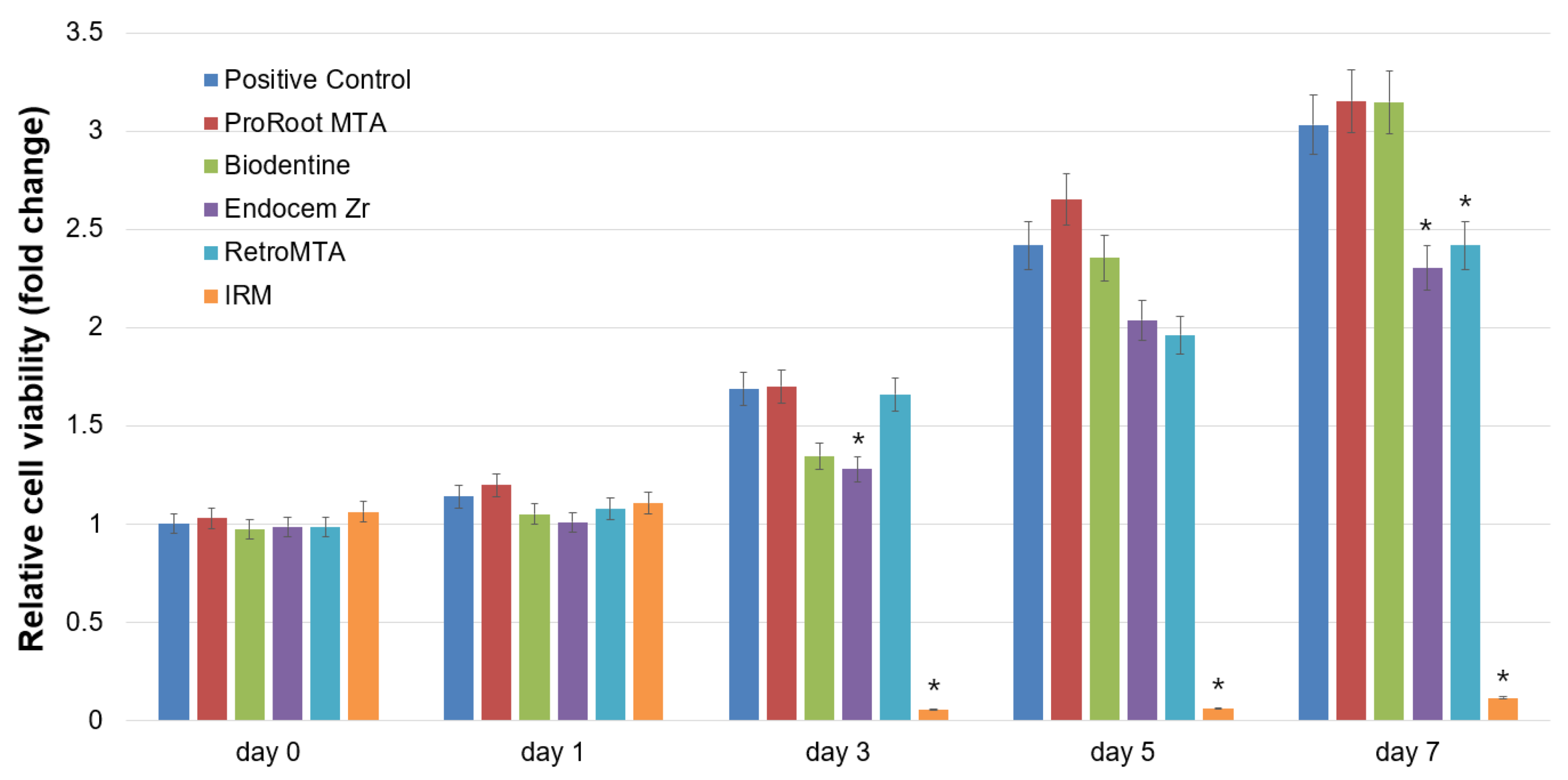
2.3. Cell Viability Assay

We evaluated the cytotoxic effects of the four calcium silicate-based cements using a methyl-thiazoldiphenyl-tetrazolium (MTT) assay (MTT Cell Growth Assay Kit; Chemicon, Rosemont, IL, USA) [25,26]. The proliferation rate of the GDSCs was analyzed after 0, 1, 2, 3, and 5 days of culture growth. GDSCs were seeded at a density of 1.0 × 10⁴ cells/well on 24-well cell culture plates (SPL Life Sciences, Pocheon, Korea) with a growth medium. After 24 h of culture for cell attachment, we obtained the optical density value for day 0. An individual disk was stored in an insert with a 0.4 μm pore size (SPLInsert; SPL Life Sciences) and the insert was stored over the GDSCs. For maintaining the medium level up to the disk, each well was supplemented with an extra 1 mL of growth medium. GDSCs cultured without experimental disks were used as positive controls, and IRM was used as a negative control. MTT solution at a concentration of 500 μg/mL was added to each well for 4 h. Thereafter, each well was washed with PBS and dimethyl sulfoxide was added to dissolve the synthesized formazan. The optical density at 570 nm was determined using an absorbance microplate reader (Power Wave XS; BioTek Instruments, Winooski, VT, USA) with the absorbance at 630 nm as the reference. Each group was evaluated in quadruplicate.

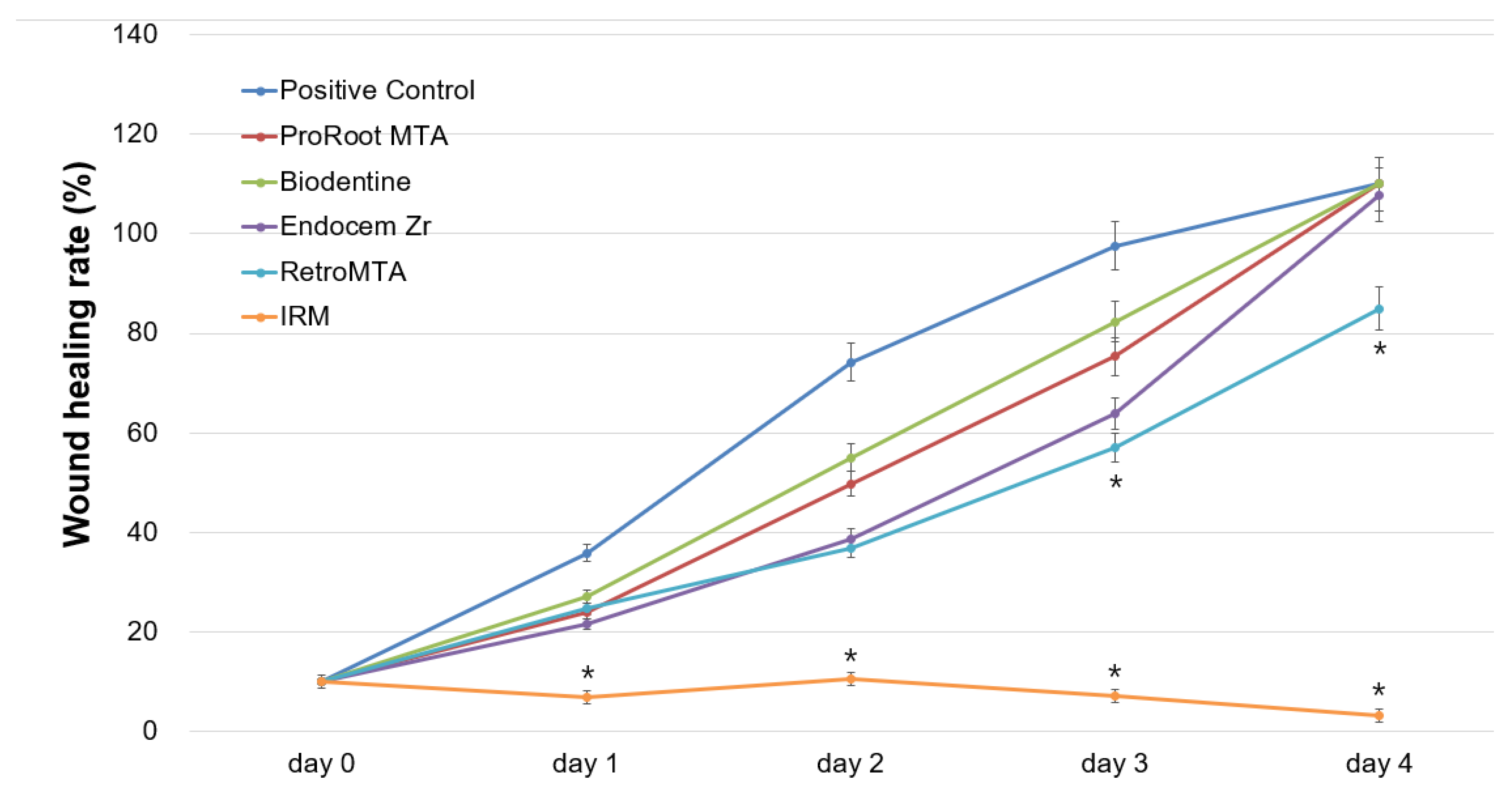
2.4. Cell Migration Assay

We evaluated cell migratory ability using a scratch wound healing assay. GDSCs were seeded at a density of 3.5 × 10⁴ cells/well on 24-well cell culture plates (SPL Life Sciences, Pocheon, Korea) with a growth medium. After 24 h of culture, a scratch wound was created in the middle of the confluent cell layer using a 1,000 μL pipette tip. After scratching, cell debris was rinsed off with PBS. After 24 h of culture, each individual disk was stored in an insert with a 0.4 μm pore size (SPLInsert; SPL Life Sciences) and the insert was stored over the GDSCs. For maintaining the medium level up to the disk, each well was supplemented with an extra 1 mL of growth medium. GDSCs with various calcium silicate-based cement disks were incubated for 4 days, with changing the medium every 2 days. Images of wound healing were observed at 0, 1, 2, 3, and 4 days using a phase-contrast microscope (Olympus, Tokyo, Japan). ImageJ 1.46r (National Institutes of Health, Bethesda, MD, USA) was used to determine the wound healing area. We calculated the area of cell migration into the wound using the initial wound area as the reference. Each group was evaluated in quadruplicate.

2.5. Live/Dead Staining Assay

GDSCs were seeded at a density of 1.0 × 10⁴ cells/well on 24-well plates (SPL Life Sciences) with a growth medium. After 24 h of culture, each individual disk was stored in an insert with a 0.4 μm pore size (SPLInsert; SPL Life Sciences) and the insert was stored over the GDSCs. GDSCs with various calcium silicate cement disks were incubated for 5 days, with changing the growth medium every 2 days. Cells were double-stained with a LIVE/DEAD™ Cell Imaging Kit (488/570; Molecular Probes, Life Technologies, CA, USA) on days 3 and 5, and the stained cells were evaluated under an inverted microscope (Axiovert 200; Carl Zeiss Microscopy, Jena, Germany). Qualitative analyses of cell viability were performed with digital image processing software (ZEN 2012, AxioVision; Carl Zeiss Microscopy).

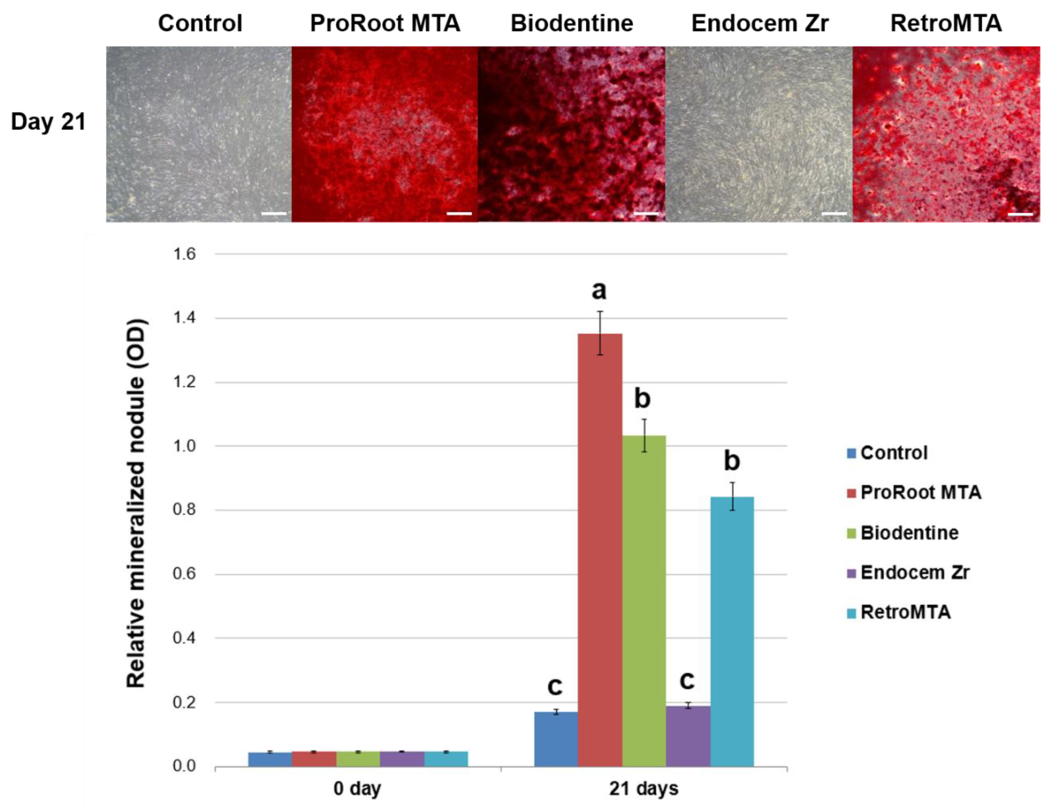
2.6. Alizarin Red S (ARS) Staining Assay

To evaluate the formation of calcified nodules in GDSCs, we used an ARS assay [25,26]. The powder of each experimental calcium silicate-based cement was mixed with an osteogenic medium at a concentration of 5 mg/mL, and the mixture was placed in a 100% humidity incubator at 37 °C for 7 days. The osteogenic medium consisted of complete α-MEM, 50 μg/mL ascorbic acid (Sigma-Aldrich), 0.1 μM dexamethasone (Sigma-Aldrich), and 10 mM beta-glycerophosphate (Sigma-Aldrich). The supernatant fluid was refined through 0.20 μm filters (Minisart; Sartorius Stedim Biotech, Goetingen, Germany). GDSCs were seeded at a density of 2.0 × 10⁴ cells/well on 24-well plates (SPL Life Sciences) and cultured for 21 days in calcium silicate-based cement eluate, with changing the eluate every 3 days. Cells were fixed in 4% paraformaldehyde solution and stained with 2% ARS solution (ScienCell, Carlsbad, CA, USA) for 20 min. The stain was treated with 10% cetylpyridinium chloride (Sigma-Aldrich) for 15 min, and the optical density at 560 nm was evaluated using an absorbance microplate reader (Power Wave XS). Each group was evaluated in quadruplicate.

2.7. pH Measurement

The powder of each experimental calcium silicate-based cement was mixed with deionized water and osteogenic medium at a concentration of 5 mg/mL, and the mixture was placed in a 100% humidity incubator at 37 °C for 7 days. The pH of each liquid was evaluated using a digital pH meter which is adjusted prior calibration (Satorious Docu-pH Meter; Satorious AG, Goettingen, Germany). Three measurements were made for each cement solution. As a control, deionized water and osteogenic medium without experimental powder was also measured.

2.8. Statistical Analyses

The SPSS software program (ver. 24.0; IBM Corp., Armonk, NY, USA) was used for statistical analyses. The Shapiro-Wilk test of normality was used to confirm the data distribution. The data normality was confirmed; thus, repeated measures analyses of variance were performed for general comparisons of MTT and wound healing assays. Independent t tests were performed for pairwise comparisons of experimental groups at each time point. One-way analyses of variance and Tukey post hoc tests were used for the ARS assay. p < 0.05 was considered statistically significant.

3. Results

In the cell viability assay, no significant differences were shown among the ProRoot MTA, Biodentine, and positive control groups during the test period (p > 0.05). The Endocem Zr and RetroMTA groups differed significantly from the control group on days 5 and 7 (p < 0.05). Out of all groups, the IRM group showed the lowest viable cell level after 24 h (p < 0.05) (Figure 1).
In the cell migration assay, no significant differences were found among the ProRoot MTA, Biodentine, Endocem Zr, and control groups during the test period (p > 0.05). The RetroMTA group had lower cell migratory ability than the control group on days 3 and 4 (p < 0.05). Cell migration was not shown in the IRM group, and significant differences were observed between the IRM and positive control groups at days 1–4 (p < 0.05; Figure 2). Representative images of the cell migration in all groups are shown in Figure 3.
In the live/dead staining assay, GDSCs in contact with IRM extract showed low viable cell density, whereas GDSCs in contact with the other experimental cements showed favorable cell growth relative to the control group (Figure 4).
In the ARS assay, GDSCs exposed to ProRoot MTA, Biodentine, and RetroMTA eluates resulted in a meaningful increase in the formation of calcium (Ca) compared to the Endocem Zr and control groups on day 21 (p < 0.05; Figure 5).
In the pH measurement, the pH of all experimental calcium silicate-based cements in deionized water was higher (pH > 10.0) than the pH of deionized water without experimental powder (Table 2).

4. Discussion

In some clinical conditions such as the repair of root resorption, a fast initial setting time is required to prevent the dissolution of materials into blood and oral fluids. Less tooth discoloration is also an important factor esthetically. Some alternative calcium silicate-based cements, such as Biodentine, Endocem Zr, and RetroMTA, were introduced for these reasons to replace ProRoot MTA. Furthermore, biocompatible and bioactive calcium silicate-based cements can promote rapid healing of adjacent periodontal tissue. Therefore, in the present study we evaluated the cytotoxicity and mineralization potential of four calcium silicate-based cements on GDSCs.
In this study, we analyzed the biocompatibility of ProRoot MTA, Biodentine, Endocem Zr, RetroMTA, and IRM using MTT and wound healing assays. The ProRoot MTA, Biodentine, and control groups showed higher cell viability and migratory ability compared to the IRM group. The Endocem Zr and RetroMTA groups showed lower cell viability compared to the control group (Figure 1), and the RetroMTA group also had a slower cell migration rate than the control group (Figure 2).
Biodentine is a novel bioceramic calcium silicate-based cement that possesses biocompatible and noncytotoxic properties [22,27]. In one study, the highest migration rate of dental pulp stem cells was found in the Biodentine group, and scanning electron microscopy revealed superior cell adhesion on disks of Biodentine [22]. In another study, cell viability was highest in the Biodentine group followed by the ProRoot MTA group, although viability on the glass ionomer cement (Ketac Molar Aplicap; 3M ESPE, Seefeld, Germany) was significantly lower [27]. Other published studies showed that Biodentine eluates resulted in low-to-moderate negative consequence on cell viability and on cell migratory ability [28]. One possible explanation is that high density level of Biodentine in the growth medium seriously lower stem cell proliferation [29].
Endocem Zr is a pozzolan-based, white calcium silicate cement developed to improve shortcomings such as a long setting time and tooth discoloration [30]. If bismuth oxide, which is used as a radiopacifier in ProRoot MTA, interacts with the collagen fibrils in dentin, it can lead to tooth discoloration. Bismuth oxide is substituted by zirconium oxide in the Endocem Zr formulation [31]. Lee et al. reported that Endocem Zr showed a similar inflammatory response to dental pulp tissue as did ProRoot MTA; however, its formation of a calcific barrier was inferior to that by ProRoot MTA [30].
RetroMTA is another fast-setting calcium silicate cement thanks to its zirconium component, which shortens the setting time by increasing the hydration rate of Portland cement [32]. In a study by Chung et al., RetroMTA showed similar biocompatibility and angiogenic effects on human dental pulp cells as ProRoot MTA; therefore, it is an effective pulp capping material [33]. In comparison, Endocem Zr showed irregular cytotoxic effects and derived less vascular endothelial growth factor and angiogenin expression [33]. In a previous study, both ProRoot MTA and RetroMTA resulted in significantly higher cell viability compared to the positive control, whereas ProRoot MTA had a higher radiopacity than RetroMTA [23]. Another study found that set RetroMTA showed better biological responses compared to a set calcium-enriched mixture (BioniqueDent, Tehran, Iran) and Angelus MTA (Angelus MTA, Londrina, Paraná, Brazil) in a mouse L929 fibroblast cell line [19].
In this study, we evaluated the calcium nodule formation ability associated with ProRoot MTA, Biodentine, Endocem Zr, and RetroMTA using an ARS assay. We found that ProRoot MTA resulted in more mineralization potential than Biodentine and RetroMTA, which is in accordance with a previous study in which ProRoot MTA showed better osteogenic potential than Biodentine based on real-time polymerase chain reaction expression analysis, alkaline phosphatase activity, and calcium nodule formation data [34]. In another study, Biodentine showed significantly decreased alkaline phosphatase activity compared to ProRoot MTA [35]. Its differences in composition and the rate of dissolution in culture medium may be one reason for the lower mineralization activity of Biodentine [34]. Differences in types of cells, culture condition, and time of culturing may have affected the results. In both studies, alveolar bone marrow stem cells rather than dental pulp stem cells were used [34,35].
Meanwhile, a different study showed that Biodentine has a comparable efficacy to ProRoot MTA in the clinical setting and may be considered as an interesting substitute for ProRoot MTA in pulp capping procedures [36]. In that study, well-arranged odontoblast layers and odontoblast-like cells formed tubular dentin under the osteodentin [36]. Furthermore, Wongwatanasanti et al. reported that only Biodentine showed a positive ARS compared to ProRoot MTA and RetroMTA groups [37]. They concluded that Biodentine, ProRoot MTA, and RetroMTA all induce stem cell apical papilla (SCAP) proliferation; however, only Biodentine induces significant SCAP differentiation [37]. Another study also showed that SCAP mineralization was greater in the Biodentine group than the ProRoot MTA group [38]. These studies used SCAP for the ARS assay [37,38], unlike our study, which used GDSCs. The differences in osteogenic gene expression can be explained by differences in cell origin and developmental status at the time of incubation. Therefore, further investigation is required to clarify the different results.
A previous study reported that Endocem MTA and Endocem Zr are related with remarkably less Ca ion release compared to ProRoot MTA [31]. When the three cements were immersed in PBS for 2 weeks, these cements created Ca- and phosphorous (P)-incorporating apatite-like materials. ProRoot MTA showed precipitates which has a higher Ca/P ratio compared to Endocem Zr [31]. Unlike ProRoot MTA, in which calcium and silicon are the predominant compositions, Endocem Zr is largely composed of zirconia with a small quantity of calcium and silicon. In the present study, Endocem Zr showed the lowest calcium nodule formation ability among the experimental calcium silicate-based cements, in accordance with a previous study [30]. Ca ions contribute to the formation and mineralization of hard tissue. Therefore, extended Ca release from calcium silicate-based cements can influence the osteogenic potential of bone marrow stem cells and osteoblast progenitors [39,40]. In the present study, all tested calcium silicate-based cements were associated with an alkaline pH (Table 2), consistent with the findings of previous studies [23,41,42]. The high alkalinity of the materials contributes to their osteogenic potential as a suitable condition for matrix formation and antimicrobial ability is created.
Unfortunately, the reason why various calcium silicate-based cements elicit different biological responses was not thoroughly investigated in this study. Further study on the association between chemical components of the calcium silicate-based cements and biological responses of cells is necessary. Furthermore, proper characterization of each calcium silicate-based cement is required.

5. Conclusions

In summary, the four calcium silicate-based cements evaluated in this study using GDSCs had good biological properties. The ProRoot MTA, Biodentine, and RetroMTA groups showed higher mineralization potential compared to the Endocem Zr and control groups. Therefore, Biodentine and RetroMTA can be used as alternatives to ProRoot MTA to treat ERR in terms of esthetics. Further in vivo research is needed.

Author Contributions

D.L. and J.-B.P. contributed equally to this work. D.L., J.-B.P., and S.-Y.K. participated in the conceptualization and design of the study. D.L., D.S., and H.-M.K. performed all the experimental procedures and contributed to data acquisition. D.L., J.-B.P., D.S., H.-M.K. and S.-Y.K. contributed substantially to data interpretation and analysis. D.L., J.-B.P., and S.-Y.K. were involved in drafting the manuscript and revising it critically for important intellectual content. All authors have read and agreed to the published version of the manuscript.

Funding

This research was funded by a National Research Foundation of Korea (NRF) grant funded by the Korean government (Ministry of Science, ICT and Future Planning) (no. 2017R1C1B5017098 and 2019R1F1A1058955).

Conflicts of Interest

The authors declare no conflicts of interest.

References

  1. Fuss, Z.; Tsesis, I.; Lin, S. Root resorption—Diagnosis, classification and treatment choices based on stimulation factors. Dent. Traumatol. 2003, 19, 175–182. [Google Scholar] [CrossRef]
  2. Heithersay, G.S. Management of tooth resorption. Aust. Dent. J. 2007, 52, S105–S121. [Google Scholar] [CrossRef] [Green Version]
  3. Karypidou, A.; Chatzinikolaou, I.D.; Kouros, P.; Koulaouzidou, E.; Economides, N. Management of bilateral invasive cervical resorption lesions in maxillary incisors using a novel calcium silicate-based cement: A case report. Quintessence Int. 2016, 47, 637–642. [Google Scholar] [CrossRef]
  4. Duarte, M.A.H.; Marciano, M.A.; Vivan, R.R.; Tanomaru Filho, M.; Tanomaru, J.M.G.; Camilleri, J. Tricalcium silicate-based cements: Properties and modifications. Braz. Oral Res. 2018, 32, e70. [Google Scholar] [CrossRef] [Green Version]
  5. Camilleri, J. Characterization and hydration kinetics of tricalcium silicate cement for use as a dental biomaterial. Dent. Mater. 2011, 27, 836–844. [Google Scholar] [CrossRef]
  6. Torabinejad, M.; Parirokh, M.; Dummer, P.M.H. Mineral trioxide aggregate and other bioactive endodontic cements: An updated overview—Part II: Other clinical applications and complications. Int. Endod. J. 2018, 51, 284–317. [Google Scholar] [CrossRef]
  7. Peng, W.; Liu, W.; Zhai, W.; Jiang, L.; Li, L.; Chang, J.; Zhu, Y. Effect of tricalcium silicate on the proliferation and odontogenic differentiation of human dental pulp cells. J. Endod. 2011, 37, 1240–1246. [Google Scholar] [CrossRef]
  8. Du, R.; Wu, T.; Liu, W.; Li, L.; Jiang, L.; Peng, W.; Chang, J.; Zhu, Y. Role of the extracellular signal-regulated kinase 1/2 pathway in driving tricalcium silicate-induced proliferation and biomineralization of human dental pulp cells in vitro. J. Endod. 2013, 39, 1023–1029. [Google Scholar] [CrossRef]
  9. Rathinam, E.; Rajasekharan, S.; Chitturi, R.T.; Martens, L.; De Coster, P. Gene expression profiling and molecular signaling of dental pulp cells in response to tricalcium silicate cements: A systematic review. J. Endod. 2015, 41, 1805–1817. [Google Scholar] [CrossRef]
  10. Marciano, M.A.; Camilleri, J.; Costa, R.M.; Matsumoto, M.A.; Guimaraes, B.M.; Duarte, M.A.H. Zinc oxide inhibits dental discoloration caused by white mineral trioxide aggregate Angelus. J. Endod. 2017, 43, 1001–1007. [Google Scholar] [CrossRef] [Green Version]
  11. Dawood, A.E.; Parashos, P.; Wong, R.H.K.; Reynolds, E.C.; Manton, D.J. Calcium silicate-based cements: Composition, properties, and clinical applications. J. Investig. Clin. Dent. 2017, 8. [Google Scholar] [CrossRef] [PubMed]
  12. Loison-Robert, L.S.; Tassin, M.; Bonte, E.; Berbar, T.; Isaac, J.; Berdal, A.; Simon, S.; Fournier, B.P.J. In vitro effects of two silicate-based materials, Biodentine and BioRoot RCS, on dental pulp stem cells in models of reactionary and reparative dentinogenesis. PLoS ONE 2018, 13, e0190014. [Google Scholar] [CrossRef] [PubMed] [Green Version]
  13. Li, Q.; Hurt, A.P.; Coleman, N.J. The application of (29)Si NMR spectroscopy to the analysis of calcium silicate-based cement using Biodentine™ as an example. J. Funct. Biomater. 2019, 10, 25. [Google Scholar] [CrossRef] [Green Version]
  14. Setbon, H.M.; Devaux, J.; Iserentant, A.; Leloup, G.; Leprince, J.G. Influence of composition on setting kinetics of new injectable and/or fast setting tricalcium silicate cements. Dent. Mater. 2014, 30, 1291–1303. [Google Scholar] [CrossRef] [PubMed]
  15. Camilleri, J.; Laurent, P.; About, I. Hydration of Biodentine, Theracal LC, and a prototype tricalcium silicate-based dentin replacement material after pulp capping in entire tooth cultures. J. Endod. 2014, 40, 1846–1854. [Google Scholar] [CrossRef] [PubMed]
  16. Zanini, M.; Sautier, J.M.; Berdal, A.; Simon, S. Biodentine induces immortalized murine pulp cell differentiation into odontoblast-like cells and stimulates biomineralization. J. Endod. 2012, 38, 1220–1226. [Google Scholar] [CrossRef] [PubMed]
  17. Kang, S.H.; Shin, Y.S.; Lee, H.S.; Kim, S.O.; Shin, Y.; Jung, I.Y.; Song, J.S. Color changes of teeth after treatment with various mineral trioxide aggregate-based materials: An ex vivo study. J. Endod. 2015, 41, 737–741. [Google Scholar] [CrossRef]
  18. Choi, Y.; Park, S.J.; Lee, S.H.; Hwang, Y.C.; Yu, M.K.; Min, K.S. Biological effects and washout resistance of a newly developed fast-setting pozzolan cement. J. Endod. 2013, 39, 467–472. [Google Scholar] [CrossRef]
  19. Pornamazeh, T.; Yadegari, Z.; Ghasemi, A.; Sheykh-Al-Eslamian, S.M.; Shojaeian, S. In vitro cytotoxicity and setting time assessment of calcium-enriched mixture cement, Retro mineral trioxide aggregate and mineral trioxide aggregate. Iran. Endod. J. 2017, 12, 488–492. [Google Scholar] [CrossRef]
  20. Collado-Gonzalez, M.; Garcia-Bernal, D.; Onate-Sanchez, R.E.; Ortolani-Seltenerich, P.S.; Alvarez-Muro, T.; Lozano, A.; Forner, L.; Llena, C.; Moraleda, J.M.; Rodriguez-Lozano, F.J. Cytotoxicity and bioactivity of various pulpotomy materials on stem cells from human exfoliated primary teeth. Int. Endod. J. 2017, 50, e19–e30. [Google Scholar] [CrossRef]
  21. Jin, S.H.; Lee, J.E.; Yun, J.H.; Kim, I.; Ko, Y.; Park, J.B. Isolation and characterization of human mesenchymal stem cells from gingival connective tissue. J. Periodontal Res. 2015, 50, 461–467. [Google Scholar] [CrossRef] [PubMed]
  22. Tomás-Catalá, C.J.; Collado-González, M.; García-Bernal, D.; Oñate-Sánchez, R.E.; Forner, L.; Llena, C.; Lozano, A.; Moraleda, J.M.; Rodríguez-Lozano, F.J. Biocompatibility of new pulp-capping materials NeoMTA Plus, MTA Repair HP, and Biodentine on human dental pulp stem cells. J. Endod. 2018, 44, 126–132. [Google Scholar] [CrossRef] [PubMed] [Green Version]
  23. Souza, L.C.D.; Yadlapati, M.; Dorn, S.O.; Silva, R.; Letra, A. Analysis of radiopacity, pH and cytotoxicity of a new bioceramic material. J. Appl. Oral Sci. 2015, 23, 383–389. [Google Scholar] [CrossRef] [PubMed] [Green Version]
  24. Youssef, A.R.; Emara, R.; Taher, M.M.; Al-Allaf, F.A.; Almalki, M.; Almasri, M.A.; Siddiqui, S.S. Effects of mineral trioxide aggregate, calcium hydroxide, Biodentine and Emdogain on osteogenesis, odontogenesis, angiogenesis and cell viability of dental pulp stem cells. BMC Oral Health 2019, 19, 133. [Google Scholar] [CrossRef]
  25. Kim, H.S.; Zheng, M.; Kim, D.K.; Lee, W.P.; Yu, S.J.; Kim, B.O. Effects of 1,25-dihydroxyvitamin D3 on the differentiation of MC3T3-E1 osteoblast-like cells. J. Periodontal Implant Sci. 2018, 48, 34–46. [Google Scholar] [CrossRef] [Green Version]
  26. Hwang, J.H.; Oh, S.; Kim, S. Improvement of the osteogenic potential of ErhBMP-2-/EGCG-coated biphasic calcium phosphate bone substitute: In vitro and in vivo activity. J. Periodontal Implant Sci. 2019, 49, 114–126. [Google Scholar] [CrossRef]
  27. Widbiller, M.; Lindner, S.R.; Buchalla, W.; Eidt, A.; Hiller, K.A.; Schmalz, G.; Galler, K.M. Three-dimensional culture of dental pulp stem cells in direct contact to tricalcium silicate cements. Clin. Oral Investig. 2016, 20, 237–246. [Google Scholar] [CrossRef]
  28. Sequeira, D.B.; Seabra, C.M.; Palma, P.J.; Cardoso, A.L.; Peça, J.; Santos, J.M. Effects of a new bioceramic material on human apical papilla cells. J. Funct. Biomater. 2018, 9, 74. [Google Scholar] [CrossRef] [Green Version]
  29. Luo, Z.; Li, D.; Kohli, M.R.; Yu, Q.; Kim, S.; He, W.-X. Effect of Biodentine™ on the proliferation, migration and adhesion of human dental pulp stem cells. J. Dent. 2014, 42, 490–497. [Google Scholar] [CrossRef]
  30. Lee, M.; Kang, C.M.; Song, J.S.; Shin, Y.; Kim, S.; Kim, S.O.; Choi, H.J. Biological efficacy of two mineral trioxide aggregate (MTA)-based materials in a canine model of pulpotomy. Dent. Mater. J. 2017, 36, 41–47. [Google Scholar] [CrossRef] [Green Version]
  31. Han, L.; Kodama, S.; Okiji, T. Evaluation of calcium-releasing and apatite-forming abilities of fast-setting calcium silicate-based endodontic materials. Int. Endod. J. 2015, 48, 124–130. [Google Scholar] [CrossRef] [PubMed]
  32. Li, Q.; Deacon, A.D.; Coleman, N.J. The impact of zirconium oxide nanoparticles on the hydration chemistry and biocompatibility of white Portland cement. Dent. Mater. J. 2013, 32, 808–815. [Google Scholar] [CrossRef] [PubMed] [Green Version]
  33. Chung, C.J.; Kim, E.; Song, M.; Park, J.W.; Shin, S.J. Effects of two fast-setting calcium-silicate cements on cell viability and angiogenic factor release in human pulp-derived cells. Odontology 2016, 104, 143–151. [Google Scholar] [CrossRef]
  34. Margunato, S.; Tasli, P.N.; Aydin, S.; Karapinar Kazandag, M.; Sahin, F. In vitro evaluation of ProRoot MTA, Biodentine, and MM-MTA on human alveolar bone marrow stem cells in terms of biocompatibility and mineralization. J. Endod. 2015, 41, 1646–1652. [Google Scholar] [CrossRef] [PubMed]
  35. Sultana, N.; Singh, M.; Nawal, R.R.; Chaudhry, S.; Yadav, S.; Mohanty, S.; Talwar, S. Evaluation of biocompatibility and osteogenic potential of tricalcium silicate-based cements using human bone marrow-derived mesenchymal stem cells. J. Endod. 2018, 44, 446–451. [Google Scholar] [CrossRef]
  36. Nowicka, A.; Lipski, M.; Parafiniuk, M.; Sporniak-Tutak, K.; Lichota, D.; Kosierkiewicz, A.; Kaczmarek, W.; Buczkowska-Radlinska, J. Response of human dental pulp capped with biodentine and mineral trioxide aggregate. J. Endod. 2013, 39, 743–747. [Google Scholar] [CrossRef] [PubMed]
  37. Wongwatanasanti, N.; Jantarat, J.; Sritanaudomchai, H.; Hargreaves, K.M. Effect of bioceramic materials on proliferation and odontoblast differentiation of human stem cells from the apical papilla. J. Endod. 2018, 44, 1270–1275. [Google Scholar] [CrossRef]
  38. Wattanapakkavong, K.; Srisuwan, T. Release of transforming growth factor beta 1 from human tooth dentin after application of either ProRoot MTA or Biodentine as a coronal barrier. J. Endod. 2019, 45, 701–705. [Google Scholar] [CrossRef]
  39. Khan, S.; Kaleem, M.; Fareed, M.A.; Habib, A.; Iqbal, K.; Aslam, A.; Ud Din, S. Chemical and morphological characteristics of mineral trioxide aggregate and Portland cements. Dent. Mater. J. 2016, 35, 112–117. [Google Scholar] [CrossRef] [Green Version]
  40. Formosa, L.M.; Mallia, B.; Bull, T.; Camilleri, J. The microstructure and surface morphology of radiopaque tricalcium silicate cement exposed to different curing conditions. Dent. Mater. 2012, 28, 584–595. [Google Scholar] [CrossRef]
  41. Kim, M.; Yang, W.; Kim, H.; Ko, H. Comparison of the biological properties of ProRoot MTA, OrthoMTA, and Endocem MTA cements. J. Endod. 2014, 40, 1649–1653. [Google Scholar] [CrossRef] [PubMed]
  42. Parirokh, M.; Torabinejad, M. Mineral trioxide aggregate: A comprehensive literature review—Part I: Chemical, physical, and antibacterial properties. J. Endod. 2010, 36, 16–27. [Google Scholar] [CrossRef] [PubMed]
Figure 1. Relative cell viability based on methyl-thiazoldiphenyl-tetrazolium (MTT) assay. Asterisks represent statistically significant differences between the positive control and experimental groups.
Coatings 10 00279 g001
Figure 2. Wound healing rate of all tested calcium silicate-based cements. Asterisks represent statistically significant differences between the positive control and experimental groups.
Coatings 10 00279 g002
Figure 3. Representative images of cell migration based on wound healing assay (scale bar = 250 μm).
Coatings 10 00279 g003
Figure 4. Results of live/dead staining assay of all tested calcium silicate-based cements (scale bar = 200 μm).
Coatings 10 00279 g004
Figure 5. Relative rate of mineralized nodule formation based on Alizarin red staining assay. Different superscript letters indicate statistically significant differences (scale bar = 500 μm).
Coatings 10 00279 g005
Table 1. The manufacturer and chemical composition of each experimental calcium silicate-based cement used in this study [17,22,23,24].
MaterialManufacturerCompositionBatch Number
ProRoot MTADentsply Tulsa Dental Specialties, Tulsa, OK, USAPortland cement (tricalcium silicate, dicalcium silicate, and tricalcium aluminate) 75%
Calcium sulfate dihydrate (gypsum) 5%
Bismuth oxide 20%
0000186484
BiodentineSeptodont, Saint-Maur-des-Fossés, FranceTricalcium silicate, dicalcium silicate, calcium carbonate, calcium oxide, and zirconium oxide in its powder form
Water, calcium chloride, and soluble polymer as an aqueous liquid
B24553
Endocem ZrMaruchi, Wonju, KoreaCalcium oxide 27%–37%
Silicon dioxide 7%–11%
Aluminum oxide 3%–5%
Magnesium oxide 1.7%–2.5%
Ferrous oxide 1.3%–2.3%
Zirconium dioxide 43%–46%
ZF7812231228
RetroMTABioMTA, Seoul, KoreaCalcium carbonate 60%–80%
Silicon dioxide 5%–15%
Aluminum oxide 5%–10%
Calcium zirconia complex 20%–30%
RM1810D14
Table 2. The pH of each experimental calcium silicate-based cement.
MaterialpH (7 days)
Osteogenic MediaddH2O
Mean Mean
Control7.567.597.507.557.027.067.087.05
ProRoot MTA8.588.638.678.6311.3311.3411.2911.32
Biodentine9.659.719.679.6811.3111.3011.3411.32
Endocem Zr8.618.698.748.6810.7710.7910.8210.79
RetroMTA8.558.578.588.5711.3211.3511.3211.33

