1. Introduction
Microleakage is a result of imperfect bonding that allows the passage of bacteria and fluids between the restorative material and the tooth [
1]. Microleakage can cause negative effects, such as higher sensitivity of the restored tooth, and secondary caries [
2]. Whilst microleakage in clinical conditions is unavoidable, microleakage can be reduced by selecting proper combinations of adhesive materials, application techniques and curing strategies [
3]. Research on microleakage provides useful information on the characteristics of adhesive materials [
4]. Previously, the relationship between bond strength and microleakage of dentistry adhesives has been an object of several studies [
5,
6,
7,
8,
9]. The failure of adhesive materials has been identified as the main cause of secondary caries and dental fracturing [
10]. A wide variety of bonding materials currently available makes it difficult for dentists to choose the best alternative in terms of adhesive materials [
11]. While in vitro testing of dental material may not accurately replicate the clinical situation in vivo, in vitro research for assessing performance and properties of dental materials is an important aspect of dentistry that enables researchers to perform experiments under controlled conditions [
12]. It is common in restorative dentistry to research dental materials using extracted animal or human teeth. For example, image-processing methods were applied on the images of extracted teeth for detection of dental filling size and its gap [
13]. This study, while still correlating with those researching the effects of microleakage is very novel in its purpose not only to analyze the adhesive or bonding strength in vitro, as was the original goal of other researchers and/or manufacturers of such materials, but also to study the microleakage of the adhesive materials, which is one of the less-trialed factors, directly concerning long-term reliability, often leading to multiple dental diseases [
14]. It is important to note that other dye-leakage studies have mostly evaluated the leakage level by degrees only [
15,
16,
17,
18], while Phanombualert et al. [
19] also offered measurements in micrometers. This study combines both. In practical terms, the real value presented here is to allow less experienced dentists to predict/estimate a quality of repair, as reaching the ideal working field conditions when restoring extensive cavities is one of the more skill based tasks. The results of this evaluation study offer a suggestion on choosing a specific material for a specific dampness level within a cavity. In this study, authors follow the guidelines for reporting pre-clinical in vitro studies on dental materials [
12].
The aim of the work was the analysis bonding quality of dental adhesive bonds 3M ESPE “Single bond”, Dentsply “Prime and Bond Active”, Coltene “One Coat 7 Universal”, Kuraray “Clearfil Universal Bond Quick” by studying the microleakage of leading adhesive materials in vitro.
2. Materials and Methods
We collected 136 healthy (caries free) molar teeth extracted due to prosthetic and orthodontic indications from Lithuania (121) and Poland (15) All teeth were immediately submerged into a physiological liquid after extraction. Next, all teeth were cleaned from any calculus, dental plaque and remains of soft body tissues. All teeth were randomly split into 4 groups of 34 teeth (nM = 34). Each group was then subdivided into 3 subgroups representing ideal, damp or too-dry conditions the cavity.
All class V cavities due to Black’s classification were prepared using the same approach on lingual or vestibular tooth surface among the line of cement enamel joint (CEJ), targeting the cavity reach towards both surfaces of enamel and dentin. Upper cavity boundary was always in the enamel zone and lower cavity boundary was always in the cement (dentin) zone. All cavities were prepared using the same type of green diamond rose burrs (using a fast speed tips) and external shape was formulated with a cylinder tool as was determined to be most effective restoration quality-wise by [
20]. Each sample was prepared with the use of cooling water spray. The drill bit was changed to identical new bits after drilling each of the subgroups (after 10 teeth). Each cavity was made to exactly these dimensions: 3 mm × 3 mm × 1.5 mm (depth) ± 0.3 mm. The accuracy was verified and adjusted using a periodontal probe, following recommendations from [
21]. Each cavity was carefully washed and dried.
To explore the adhesive qualities in varying conditions (ideal, damp or too dry dentin) 4 leading brands of universal dental adhesive bonds were selected for our evaluation: 3M ESPE “Single bond”, Dentsply “Prime and Bond Active”, Coltene “One Coat 7 Universal”, and Kuraray “Clearfil Universal Bond Quick”. The chemical properties of each material are summarized in
Table 1.
All cavities were prepared according to these three conditions of each subgroup:
A is ideal conditions as specified by each manufacturer,
B are over-dried conditions,
C are too-damp conditions.
Each adhesive was added (in the amount and application method specified) as per original instruction. A curing lamp of the same strength (GC/Mectron Silverlight, average intensity of 1350 mW/cm2) was used to activate all materials. All teeth were filled using the same Dentsply “Ceram X” composite, strictly following manufacturer’s instructions. The same lamp was used for curing process. Restoration was undertaken carefully, polishing with Dentsply “Ceram X gloss” polishes of different roughness (blue and white). The fill quality was then carefully evaluated using a 3.5× zoom lens (Olympus Corporation, Shinjuku, Tokyo, Japan) with white light-emitting diode (LED) light.
Group 1A was filled using 3M ESPE “Single bond” in ideal working conditions, following manufacturer’s instructions. Group 1B was filled using the same adhesive, only this time over drying a surface of the drilled cavity. Alternatively, group 1C was filled when the cavity surface was too wet. The same process was repeated for the remaining groups only using different adhesive: 2A/B/C were filled using Dentsply “Prime and Bond Active” adhesive, 3A/B/C using Coltene “One Coat 7 Universal” adhesive, 4A/B/C using Kuraray “Clearfil Universal Bond Quick” adhesive.
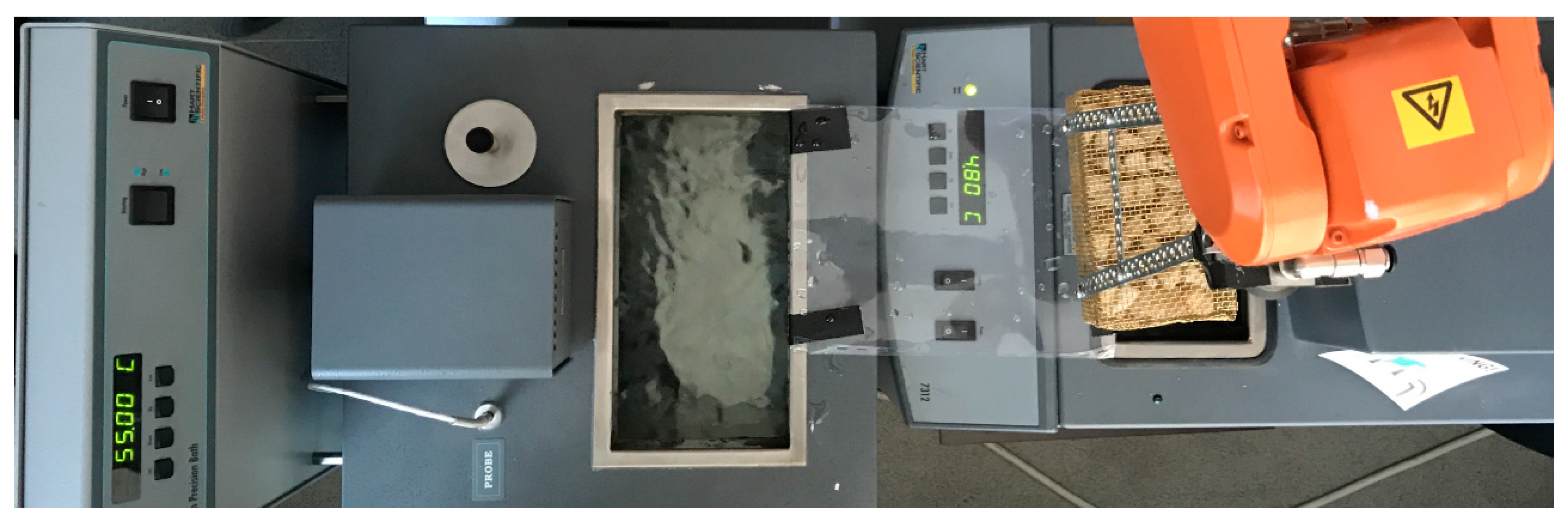
Each sample was exposed to a temperature cycling test to resemble oral cavity environment. An ABB high precision industrial robot (type: IRB120 M2004) (ABB Ltd., Zurich, Switzerland) was programmed to achieve 3000 cycles of submerging the teeth into cold and hot water baths for exactly 1 min following the recommendation of Cevik et al. [
22] and Noda et al. [
23]. The ABB robot was programmed to precisely handle the specimen basket to cyclically submerge and retract into a different temperature environments. Robot was programmed to repeat a constant action of smooth arm swing from one thermal bath to another, following an exact coordinate path with necessary slowdowns and interpolations. The graceful change period to shift a basket with teeth was set to 16 s from bath-to-bath allowing it to avoid spillage of water, not shifting the teeth themselves, and also no measurable cool down. A total length of a full cycle was 136 s. The submersion process was for a total, uninterrupted period of 4 days, 17 h and 13 min, in a clean room, with a person providing 24/7 on-site supervision. Two “Hart Scientific” Temperature Calibration baths were used for a constant and precise upkeep of water temperature. We have used a model 6022 as a hot water container (55 ± 0.3 °C) and model 7312 as a cold water container (5 ± 0.3 °C). Both baths were filled with distilled water and raised to the same top height. Teeth were placed in a custom made brass basket. The whole rig is displayed in
Figure 1.
After the thermocycling the vertices of each tooth were sealed using a dental wax. Each tooth was then fully covered with lacquer. All teeth were then submerged into 1% methylene blue solution for 24 h in room temperature (around 21 °C). All removed teeth were carefully washed in distilled water after the 24 h.
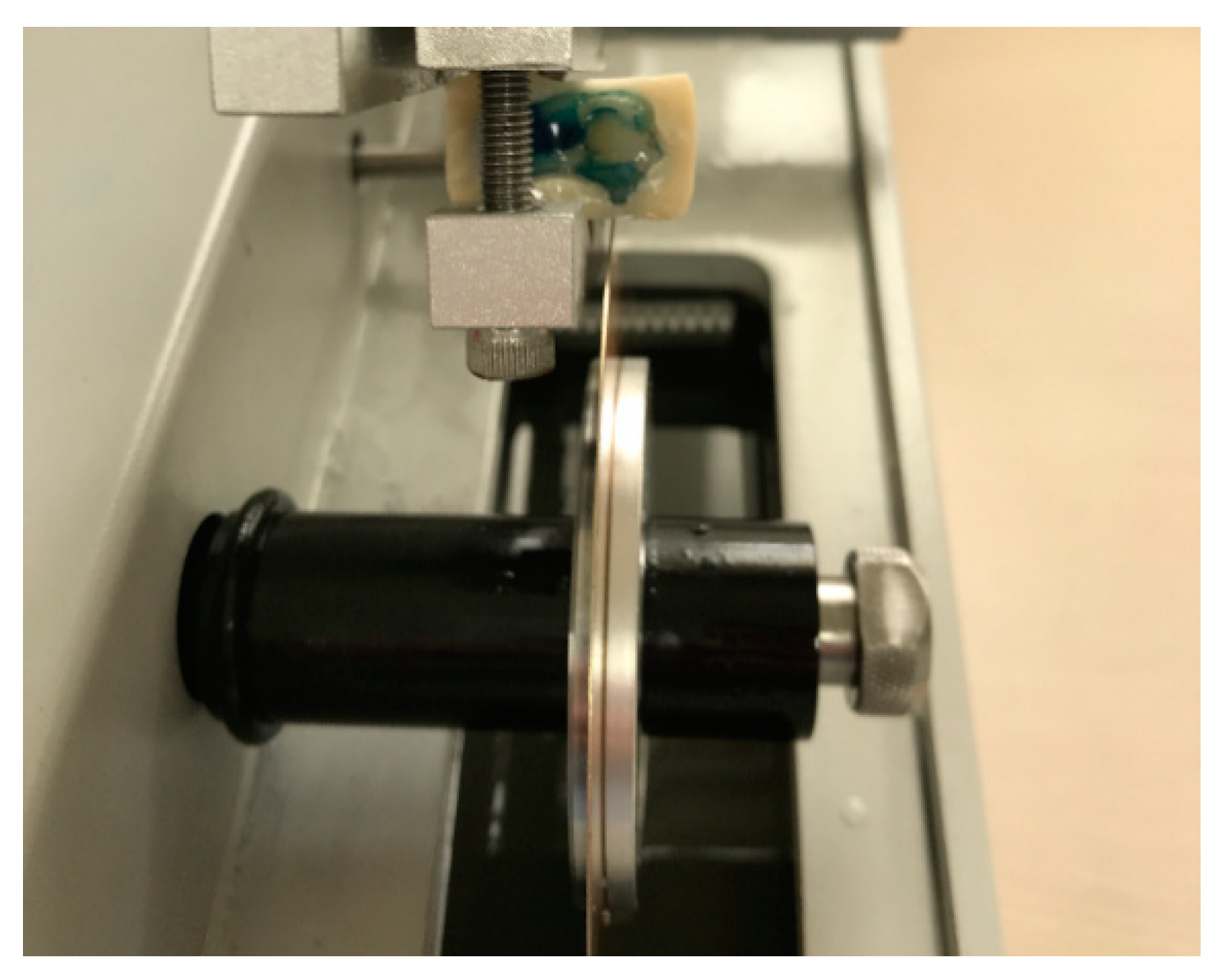
All teeth were mounted one by one, suspended (temporary molded into) in acrylic auto-polymer iTEMP “Self Curing Acrylic Resin”, each block was then assigned a unique ID number and transversely cut through a center of restoration (see
Figure 2 for illustration) using a slow speed diamond saw rig Buehler “IsoMet Low Speed Saw” (Model: 11-1280-250) (Bühler Holding AG, Uzwil, Switzerland) with a ±0.0001 in [±5 µm] precision via a manual micrometer. Series 15LC blade disk was used (No. 11-4254). (Bühler Holding AG, Uzwil, Switzerland). Each cut tooth was analyzed and photographed using Olympus BX43 microscope (Olympus Corporation, Shinjuku, Tokyo, Japan) with white LED lamp (U-LHLEDC), with a 4× zoom lens setting, using a camera sensor Olympus DP72 (Olympus Corporation, Shinjuku, Tokyo, Japan) (12.8 megapixel cooled digital color camera, capturing each color of RGB at 12 bits, at ISO 1600 setting, image color calibrated to AdobeRGB color space.
A hermeticity level of each filling was evaluated using a different degree of leakage of the dye: 0—no leakage; 1—dye has seeped through half of the cavity wall (lengthwise); 2—dye has seeped through the full length of the cavity wall; 3—dye has seeped through the full length of the cavity wall and has reached the bottom of the cavity (the axial wall). All leakage (seep length) measurements were taken using Olympus stream software. Some sample resulting images are given in
Figure 2.
3. Results
The resulting dental image dataset consists of 2004 images; 416 photos were made under the microscope (standard light), as well as grayscale and inverted color modes. Additionally we supplemented the dataset with 252 regular photography images of cut and uncut teeth (also available in color, grayscale and inverted modes). Both halves of 121 teeth datasets were analyzed and the depth of leakage measured in micrometers. The results of analysis are also presented in
Figure 3 as distribution of microleakage vs. degrees of leakage. Generally, larger microleakage translates to a larger degree of leakage. However note that outliers in
Figure 3 shows microleakage through both walls (a high total value in micrometers) although the leak in these cases did not reach the bottom of the cavity (the reason for a different degree than a measurement of leakage depth would indicate).
The experimental analysis has shown that the most effective material when applied to ideally prepared cavity surface was Dentsply “Prime and Bond Active” with least amount of micro leakage (210 microns/degree of 0.6, see
Figure 4a). The second best material was 3M ESPE “Single Bond” (1250 microns/degree of 1.5, see
Figure 4b), third—Coltene “One Coat 7 Universal” (1980 microns/degree of 2.2, see
Figure 4c), fourth—Kuraray “Clearfil Universal Bond Quick” (3250 microns/degree of 2.8, see
Figure 4d).
The most effective material applied to too dry surface was Dentsply “Prime and Bond Active” with least amount of micro leakage (120 microns/degree of 0.4, see
Figure 5a). The second best material was 3M ESPE “Single Bond” (1270 microns/degree of 1.3, see
Figure 5b), third—Coltene “One Coat 7 Universal” (1490 microns/degree of 1.91, see
Figure 5c), fourth—Kuraray “Clearfil Universal Bond Quick” (5050 microns/degree of 2.9, see
Figure 5d).
The most effective material applied on too damp surfaces was also Dentsply “Prime and Bond Active” (670 microns/degree of 0.6, see
Figure 6a). The second best was Coltene “One Coat 7 Universal” (890 microns/degree of 1.2, see
Figure 6b), third—3M ESPE “Single Bond” (2460 microns/degree of 2.1, see
Figure 6c), fourth—Kuraray “Clearfil Universal Bond Quick” (5690 microns/degree of 3, see
Figure 6d).
The characteristics of teeth in the dataset itself are represented as a bubble plot in
Figure 7, which shows the data from the perspective of surface cavity conditions.
The overall measurements have shown that the most effective adhesive overall was Dentsply “Prime and Bond Active”, objectively measured as 2.45 times more effective than the second best material Coltene “One Coat 7 Universal”. Kuraray “Clearfil Universal Bond Quick” was the least effective material in all conditions. The most forgiving non-ideal condition was the over-dried surface.
The analysis of the results used statistical methods.
For statistical analysis of data, we used the Wilcoxon signed test, Mann–Whitney U-test, Kruskal–Wallis test and Nemenyi test. Statistical analysis was performed using MATLAB (ver. 8.6.0.267246 (R2015b), Mathworks, Natick, MA, USA, 2018) software. The results are presented in
Figure 8. The Wilcoxon signed rank test shows significant difference between Dentsply “Prime and Bond Active” and 3M (
z = −4.011), Kuraray “Clearfil Universal Bond Quick” and 3M ESPE “Single bond” (
z = 4.638), Coltene “One Coat 7 Universal” and Dentsply “Prime and Bond Active” (
z = 3.655), Kuraray “Clearfil Universal Bond Quick” and Dentsply “Prime and Bond Active” (
z = 4.782), and Kuraray “Clearfil Universal Bond Quick” and Coltene “One Coat 7 Universal” (
z = 4.453) adhesive bonds (all
p < 0.001). The results were confirmed by the Mann–Whitney U-test, also showing significant difference between Dentsply “Prime and Bond Active” and 3M ESPE “Single bond” (
z = −5.221), Kuraray “Clearfil Universal Bond Quick” and 3M (
z = 5.552), Coltene “One Coat 7 Universal” and Dentsply “Prime and Bond Active” (
z = 4.556), Kuraray “Clearfil Universal Bond Quick” and Dentsply “Prime and Bond Active” (
z = 6.573), and Kuraray “Clearfil Universal Bond Quick” and Coltene “One Coat 7 Universal” (
z = 5.833) adhesive bonds (all
p < 0.001). The Kruskal–Wallis test shows that there was significant effect of bond material on microleakage results at the
p < 0.05 level [F(3,116) = 73.946,
p = 6 × 10
−16]. The Nemenyi post-hoc test was applied to determine whether differences between dental adhesive bonds were statistically significant, with a significance level of
p = 0.05. The Nemenyi test shows significant differences between Dentsply, Kuraray and other bond materials, with Dentsply having the highest rank and Kuraray having the lowest one, while the difference between Coltene and 3M ESPE bonds were statistically insignificant (critical distance = 0.32).
4. Discussion
The principle adopted in restorative dentistry is that restorations should be undertaken with the best possible marginal quality to avoid postoperative sensitivity, marginal discoloration, and secondary caries [
1,
4,
24,
25]. Different laboratory methods claim to predict the clinical performance of restorative materials, for example, tests of bond strength and microleakage and gap analysis. Microleakage studies are sometimes used to assess the bonding quality and questions remain on the validity of such comparisons [
3,
7].
Microleakage studies might differentiate the quality of various materials and also bonding systems used under different conditions so as to simulate clinical situations affecting the quality of the enamel-composite interface in in vitro studies. The significance of the results obtained in the study presented by the authors also lies in the simulation of the oral cavity conditions through the use of the variable temperature cycles in the methodology [
22,
23]. In our experiment, the application of the adhesives in a single layer was used, as indicated by the manufacturer; the possibility of its use in two layers is reported in order to enhance the bond strength. Fujiwara et al. in their work [
26] recommended the application of universal adhesives in two layers improved the adhesive quality when compared to the application in a single layer.
Solvents are one of the most important components of universal bonding systems. The bonding systems evaluated in the present study were no different from each other in relation to their solvents—all contain water and ethanol or isopropanol. This is consistent with the results of other studies which have reported a higher bonding ability in all-in-one adhesives containing a higher amount of ethanol [
27].
The overall quality of the adhesive strength varied according to the level of cavity surface dampness. Over-dried surfaces exhibited a feature of bonding more strongly than those of the too-damp surfaces, probably due to dilution of the material. We have noticed that over-drying leads to demineralization of dentin, which probably depends on poorer levels of monomer penetration. The resulting possible microleakage still exhibits some degree of probability on all types of the materials analyzed, especially in the class V restorations. An important aspect of our experiment is to allow the analysis of the “imperfect” conditions, which most often occur in clinical situations concerning dental cavities class V located in the gingival area. The validity of the results obtained in this way and their usefulness for other researchers is also determined by a very large group of results received on the basis of a huge collection of microscopic images [
12].
With regard to the fact that the variabe humidity conditions of the tissues resulted in a quality of marginal restorative adhesion, its worth considering the introduction and wider promotion of multi-mode universal adhesives in clinical bonds’ application [
28]. Multi-mode universal adhesives give clinicians the choice of using the etch-and-rinse technique, selective enamel etch technique or self-etch technique to bond to tooth substrates and thus offer the possibility to modify the form of application depending on the location of the cavity, the outline of the enamel, the depth of the cavity and humidity [
2,
4].
5. Conclusions
Quality of adhesive varies on the level of cavity surface dampness. Over-dried surfaces exhibit bonding more strongly than over-damp surfaces due to dilution of material.
Due to frequency in maintaining the optimal humidity conditions for the moisture content of the cavity surface, the most expected universal adhesive system should have a strong adhesion/minimal microleakage even in sub-optimal moisture conditions.
With the introduction of the next new generation of bonding systems on the dental materials‘ market, there is a need to test the microleakage by methods that can be applied to the results of other researchers.
The method for assessing dye penetration in a marginal microleak can be an effective tool for analysis, provided that thermocycling simulation of variable conditions in the oral cavity is used.

 ,
, 


