Letter

Laser Irradiation of Super-Nonwettable Carbon Soot Coatings–Physicochemical Implications

by
Karekin D. Esmeryan
1,*,
Yulian I. Fedchenko
2,
Georgi P. Yankov
2 and
Krassimir A. Temelkov
2
1
Acoustoelectronics Laboratory, Georgi Nadjakov Institute of Solid State Physics, Bulgarian Academy of Sciences, 72, Tzarigradsko Chaussee Blvd., 1784 Sofia, Bulgaria
2
Metal Vapor Lasers Laboratory, Georgi Nadjakov Institute of Solid State Physics, Bulgarian Academy of Sciences, 72, Tzarigradsko Chaussee Blvd., 1784 Sofia, Bulgaria
*
Author to whom correspondence should be addressed.
Coatings 2021, 11(1), 58; https://doi.org/10.3390/coatings11010058
Submission received: 7 December 2020 / Revised: 3 January 2021 / Accepted: 5 January 2021 / Published: 6 January 2021
(This article belongs to the Special Issue Laser Nano Fabrication and Nano Deposition)

Abstract

:
Accounting the increasing commercial need of rational strategies for passive icing and anti-microbial protection, the development of simple, time-efficient and scalable laboratory micropatterning techniques is highly desirable. Whilst the laser irradiation is an acknowledged technology for rapidly tuning the properties of any carbon allotropes, including soot aerosols, very barely is known about the impact of the laser beam on the physicochemical profile of the soot if it appears as a functional coating. In this pioneering research, the prolonged laser treatment of a super-nonwettable soot coating actuates morpho-chemical transformations in the material, depending on the laser power and irradiation time, without interfering its anti-wetting capability and optical transmittance. Our observations could be used as a foundation for facilitating the launch of soot coatings with customized anti-icing and anti-microbial performance.

Despite being considered as one of the major anthropogenic contributors to the global warming [1], the carbon soot nanoparticles steadily become popular as a useful tool for the fabrication of liquid-repellent interfaces with potential applicability in cryobiology, medicine, shipbuilding, oil industry, civil and environmental engineering [2,3,4,5,6]. Finding suitable approaches for regulating the physicochemical profile of the as-prepared soot-based nanostructures is critical for their subsequent flexibility and functionality in a variety of practical settings (e.g., for passive icing and anti-microbial protection of civil and industrial facilities).
Currently, the laser irradiation of different carbon allotropes such as diamond, graphite and carbon nanotubes is recognized as a powerful and facile technology for appropriately modulating the relevant surface properties [7,8,9]. It is demonstrated that the structure of graphite and diamond composite films can be varied by adjusting the laser power fluence, wavelength and pulse duration, affecting the degree of graphitization and hindering the laser energy conduction from the surface to the interior of the material [7,8]. Furthermore, the laser pulses can stimulate the formation of new carbon phases such as amorphous carbon and nanocages, next to the original carbon nanotube matrix [9].
Interestingly, the scientific literature published in the last two decades provides also undisputed evidences for the opportunity of triggering laser-induced changes in the structure, morphology, chemical composition and optical properties of soot aerosols [10,11,12,13,14,15,16,17], allowing accurate control of the size and concentration of the incipient soot particles [11], synthesis of fluorescent carbon quantum dots [16], creation of smart optically-reconfigurable surfaces [15] and even subtle manipulation of the sooting propensity of laminar diffusion flames [12]. However, and regardless of the tremendous progress of laser diagnostics in combustion, the impact of laser irradiation on the physicochemical properties of the soot is still far from well-understood at a molecular level [17], but it is of substantial importance for improving the nanoparticle production techniques [11]. Moreover, to the best of our knowledge, there is a profound lack of literature data concerning the laser-soot interactions when the latter occur in a pristine (chemically non-modified) surface coating rather than in an aerosol.
Within the present short communication, we describe the one-of-a-kind research related to laser irradiation of super-nonwettable carbon soot coatings. We demonstrate experimentally that the continuous laser radiation triggers morphological and chemical alterations in the soot, but surprisingly without amending its optical transmittance and wettability, thus, raising many meaningful questions of fundamental significance and opening a space for future systematic studies.
The fabrication of carbon soot coatings maintaining weak contact with a wide range of polar and non-polar liquids [2,3,4] was performed via controlled combustion of rapeseed oil at atmospheric pressure [18]. Shortly, soot with quasisquare particle/aggregate morphology, surface roughness of ~110 nm, thickness of ~25 μm and inherent robustness under watery liquids was generated by setting the rate of the air flow reaching a steel conical chimney, used as a combustion chamber, to 0.0035 m3/min by means of a flow meter/controller (Fisher Scientific 11998014, Sindelfingen, Germany) [18]. Inside the chimney was placed an inflamed paper wick soaked in rapeseed oil, whose vaporization in an oxygen deficient environment triggered desublimation of the nascent polyaromatic hydrocarbons into soot, uniformly deposited for ~20 s on a 25 × 25 mm glass slide.
Surface characterization of the as-prepared soot was executed through scanning electron microscopy (SEM), energy dispersive spectroscopy (EDS) and wetting analysis. Top-view SEM images, revealing the soot morphology prior to and after the laser irradiation, were obtained using a JEOL JSM-5510 scanning electron microscope (JEOL, Akishima, Tokyo, Japan) at magnifications up to 10 k. The chemical composition of the unaffected and laser-affected surface areas was studied with an energy dispersive spectrometer Bruker at 126 keV and an EDAX detector of 1.3 mm2 active area. Static contact angle and contact angle hysteresis measurements of 10 μL water droplets were carried out inside and outside the irradiated soot regions using OCA 15EC analytical instrument (DataPhysics, Filderstadt, Germany).
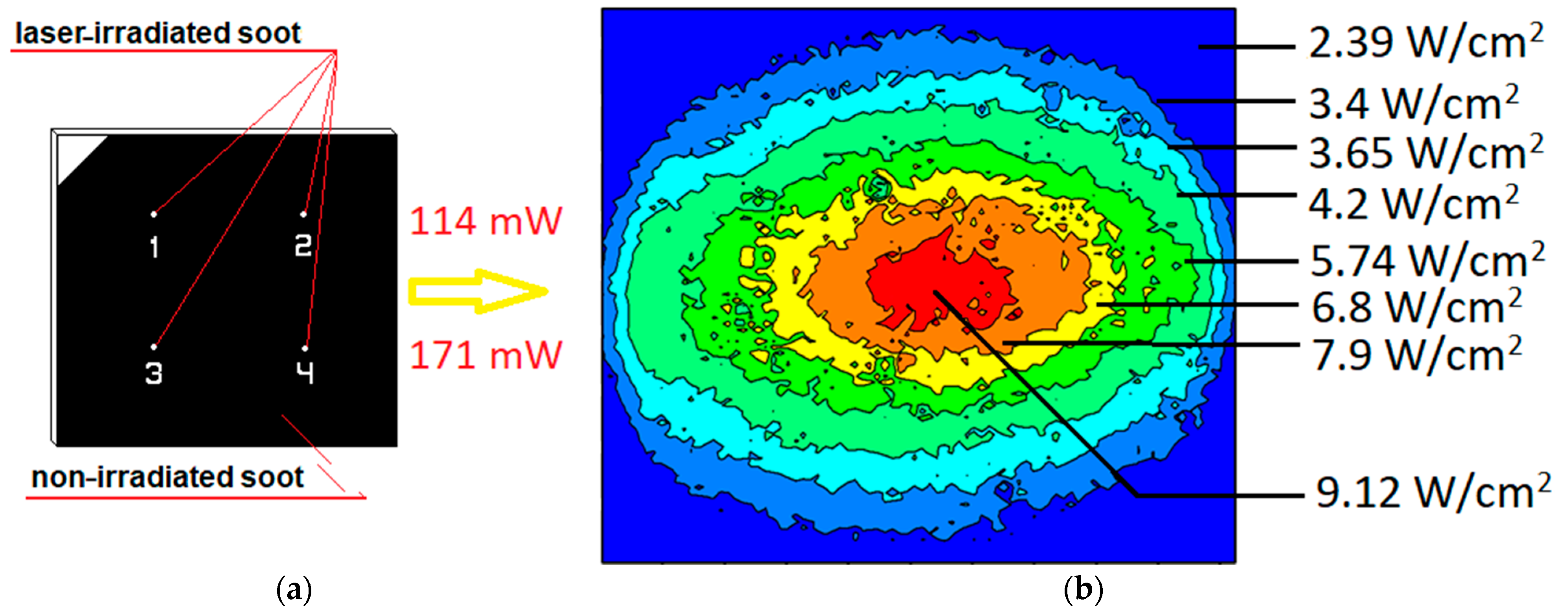
The optical experiments were implemented via a semiconductor laser model LSR-PS-II (Lasever Inc., Glendora, CA, USA), shown in Figure 1, equipped with an optical camera T-560C1/3” having spectral response range of 190–1100 nm, a scanning system supporting CCIR interface and a laser source operating at wavelength λ = 405 nm. The laser beam was focused by a lens with a focal length of 150 mm, chosen to obtain an optimal ratio between the lens’focal length towards the sample and the optimal cross-section of the spot. The lens-to-substrate distance was set to 165 mm in order to ensure the highest possible photon density for modification of the soot at maximum width of the spot, facilitating the subsequent wetting state analysis. The soot coated glass substrate was irradiated in four distinct surface locations with a laser power of p = 114 mW and p = 171 mW, and irradiation time tir = 350–450 s, while during the assays, the laser spot was characterized with asymmetric Gaussian distribution and dimensions x:y = 0.89:0.64 mm, depicted in Figure 2. Although the Gaussian beam profile would not allow uniform heating of the surface, it is appropriate to warrant high homogeneity in the center of the spot.
Finally, the light transmission coefficient of the soot coating in the irradiated and non-irradiated domains was measured via LAMBDA 1050 UV/Vis/NIR spectrophotometer (Perkin Elmer, Waltham, MA, USA) furnished with double monochromator generating a light beam in the range of 320–2500 nm at minimized noise levels within 0.01%–0.1% [18].
Figure 3 and Figure 4 demonstrate the morphological and structural features of the soot coating before and after the laser processing. The surface areas in Figure 3b–d and Figure 4b–d correspond to a spot formed by a Gaussian beam with intensity of 6.8 and 9.12 W/cm2, accordingly. Notably, the original fractal-like structure is preserved, but extensive morphological transformations in the material occur, similarly to the results reported for soot aerosols [10,17]. For instance, the pristine coating is composed of numerous quasisquare-shaped carbonaceous aggregates with dimensions between ~200–2000 nm (see Figure 3a–c and Figure 4a–c) [18], while in the laser irradiated areas, the number of these agglomerates (qualitatively defined by “naked eye”) reduces due to their confluence into much bigger clusters with size up to 6000 nm (see Figure 4b). Moreover, the process of aggregates fusion seems to be controlled by the laser treatment conditions and the most pronounced morphological modifications are induced at p = 171 mW and tir = 350 s, in contrast to the negligible changes observed at p = 114 mW and tir = 350 s (juxtapose Figure 3b and Figure 4b).
Furthermore, the laser beam oxidizes the soot nanostructures and the oxygen content increases by 7 at.%, according to the data in Table 1, but unexpectedly, the morpho-chemical rearrangements do not cause even slight variations in the optical transmittance and non-wettability of the sample.
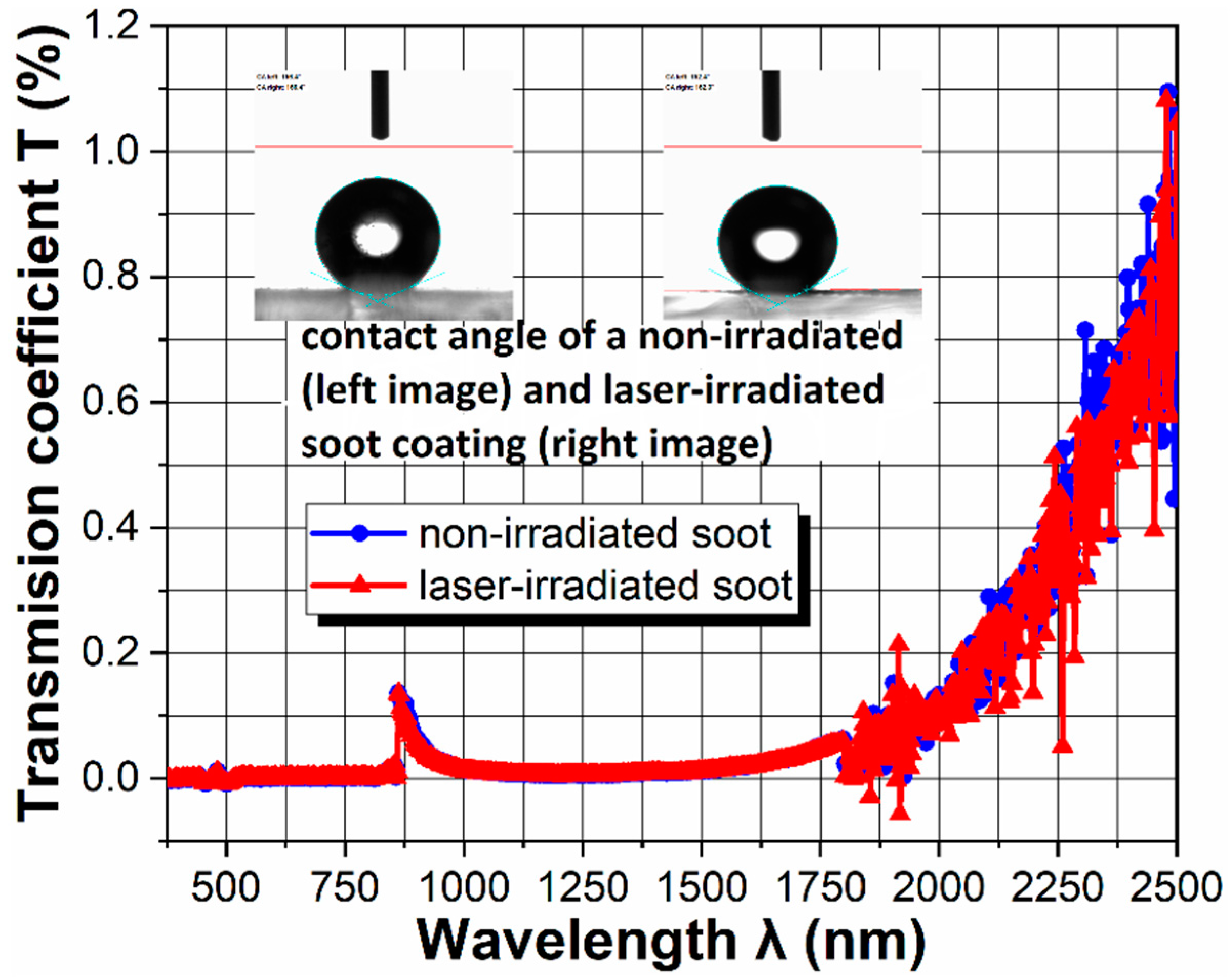
As seen in Figure 5, the graphite-like nature of the soot [4] leads to nearly full and similar light absorption in both the unaffected and affected surface areas, whereas the static contact angle and contact angle hysteresis values of 152–156° and 1.2–2.2°, respectively, confirm the preserved extreme water repellency of the soot (see the insets in Figure 5). Therefore, it can be concluded that the as-performed laser processing does not change the thickness and hybridization (sp2/sp3 ratio) of the coating, since these are the main two parameters governing the light transmission through the soot [18].
Two primary physical mechanisms are considered responsible for the observed physicochemical alterations in the soot, namely thermal annealing and heat-induced oxidation [17]. Upon laser irradiation of the coating, the increased temperatures (~450 °C, defined by performing laser-triggered paper ignition experiments) overcome the energy barrier of the solid-state interatomic forces (in the case of soot, covalent carbon-carbon and carbon-oxygen bonds) and boost the soot particle reorientation and realignment [10]. In turn, energetically more favorable morphological configuration is formed, where the numerous soot aggregates merge into larger clusters composed of smaller individual particles (see Figure 3 and Figure 4), differently from the effects described for soot aerosols [17]. On the other hand, the increased surface oxidation (see Table 1) is attributed likely to the abundance of point defects [4], acting as active sites that accelerate the oxidative processes in the ambient environment due to the laser heating, in agreement with the outcome announced for carbon nanotube films, for example [19]. However, the unaltered light transmission coefficient of our soot coating implies that there is no loss of solid matter, since if the laser inflicts irreversible particle vaporization, the overall film thickness and the optical extinction will decrease [14,17] i.e., the coating will be able to transmit higher % of the incident light beam [18].
Herein, the irradiation of a liquid-repellent soot coating with a continuous laser beam was successfully accomplished for the first time. The laser energy melted the multiple quasisquare-shaped carbon aggregates and merged them into sizeable “soot clumps” with dimensions of a few microns, whereas the protracted heating promoted additional oxidation of the freshly nascent soot agglomerates. Unlike previous studies, it seems there was no loss of material from the nanoparticles, which paves the route for facile single-step modeling of soot-based non-wettable materials with a given morphology, porosity and surface chemistry; features that greatly influence the icephobicity and anti-bioadhesiveness of the interface [4,20]. For that purpose, further experiments encompassing the use of different types of lasers, laser wavelengths, optical lenses and/or soot nanostructures (e.g., those with quasispherical or mixed initial morphology [18]) are mandatory to gain novel insights into the laser-soot interaction mechanisms. These important tasks cannot be solved within the present communication, which focuses on a particular aspect of the problem (i.e., laser irradiation of soot in the form of a thin carbon film) and discloses novel findings that will potentially have significant impact (e.g., for designing optimized passive icephobic and anti-microbial surfaces). Lastly, our coatings must be subjected to transmission electron microscopy, Raman spectroscopy and selected area electron diffraction analyses for elucidating the probability of soot crystallization and graphitization upon laser irradiation, which regrettably is a forthcoming work due to the “COVID-19 pandemic” and the almost complete blocking of the scientific infrastructure (worldwide).

Author Contributions

K.D.E. provided the soot sample, planned and supervised the experiments, processed and interpreted the data, contrived the scientific concept of this short communication and wrote its first and final versions. Y.I.F., along with K.A.T., conceived the research idea for laser irradiation of soot coatings. Y.I.F. organized also the surface characterization analysis, drew some of the figures, found a few of the relevant references and assisted during the data acquisition. G.P.Y. designed the experimental setup, performed the laser irradiation of the soot and calculated the intensity of the Gaussian beam profile. K.A.T. constructively reviewed the first draft of the manuscript. All authors approved the scientific content of this communication prior to submission. All authors have read and agreed to the published version of the manuscript.

Funding

This research received no external funding.

Data Availability Statement

All the required data is available in the short communication.

Acknowledgments

The contact angle measurement system OCA 15EC was purchased thanks to the financial support of Bulgarian National Science Fund under grant №KP-06-H37/7/06.12.2019. The authors wish to gratefully acknowledge Stoyan Russev (Department of Solid State Physics and Microelectronics at Sofia University), Daniela Karashanova (Head of Electron Microscopy Laboratory at IOMT-BAS) and Ilko Miloushev (Optics and Spectroscopy Laboratory at ISSP-BAS) for helping with the EDS, SEM and optical analyses.

Conflicts of Interest

The authors declare no conflict of interest.

References

  1. Bond, T.C.; Doherty, S.J.; Fahey, D.W.; Forster, P.M.; Berntsen, T.; DeAngelo, B.J.; Flanner, M.G.; Ghan, S.; Kärcher, B.; Koch, D.; et al. Bounding the role of black carbon in the climate system: A scientific assessment. J. Geophys. Res. Atmos. 2013, 118, 5380–5552. [Google Scholar] [CrossRef]
  2. Esmeryan, K.D.; Lazarov, Y.; Stamenov, G.S.; Chaushev, T.A. When condensed matter physics meets biology: Does superhydrophobicity benefiting the cryopreservation of human spermatozoa? Cryobiology 2020, 92, 263–266. [Google Scholar] [CrossRef] [PubMed]
  3. Esmeryan, K.D.; Stamenov, G.S.; Chaushev, T.A. An innovative approach for in-situ detection of postejaculatory semen coagulation and liquefaction using superhydrophobic soot coated quartz crystal microbalances. Sens. Actuators A Phys. 2019, 297, 111532. [Google Scholar] [CrossRef]
  4. Esmeryan, K.D.; Castano, C.E.; Chaushev, T.A.; Mohammadi, R.; Vladkova, T.G. Silver-doped superhydrophobic carbon soot coatings with enhanced wear resistance and anti-microbial performance. Colloids Surf. A Physicochem. Eng. Asp. 2019, 582, 123880. [Google Scholar] [CrossRef]
  5. Zulfiqar, U.; Hussain, S.Z.; Subhani, T.; Hussain, I.; Rehman, H. Mechanically robust superhydrophobic coating from sawdust particles and carbon soot for oil/water separation. Colloids Surf. A Physicochem. Eng. Asp. 2019, 539, 391–398. [Google Scholar] [CrossRef]
  6. Xu, Y.; Zhang, G.; Li, L.; Xu, C.; Lv, X.; Zhang, H.; Yao, W. Icephobic behaviors of superhydrophobic amorphous carbon nano-films synthesized from a flame process. J. Colloid Interface Sci. 2019, 552, 613–621. [Google Scholar] [CrossRef] [PubMed]
  7. Komlenok, M.S.; Arutyunyan, N.R.; Freitag, C.; Zavedeev, E.V.; Barinov, A.D.; Shupegin, M.L.; Pimenov, S.M. Effect of tungsten doping on laser ablation and graphitization of diamond-like nanocomposite films. Opt. Laser Technol. 2021, 135, 106683. [Google Scholar] [CrossRef]
  8. Li, W.; Gao, L.; Ma, Z.; Wang, F. Ablation behavior of graphite/SiO2 composite irradiated by high-intensity continuous laser. J. Eur. Ceram. Soc. 2017, 37, 1331–1338. [Google Scholar] [CrossRef]
  9. Santiago, E.V.; Lopez, S.H.; Camacho Lopez, M.A.; Contreras, D.R.; Farias-Mancilla, R.; Flores-Gallardo, S.G.; Hernandez-Escobar, C.A.; Zaragoza-Contreras, E.A. Optical properties of carbon nanostructures produced by laser irradiation on chemically modified multi-walled carbon nanotubes. Opt. Laser Technol. 2016, 84, 53–58. [Google Scholar] [CrossRef]
  10. Vander Wal, R.L.; Choi, M.Y. Pulsed laser heating of soot: Morphological changes. Carbon 1999, 37, 231–239. [Google Scholar] [CrossRef]
  11. Stipe, C.B.; Choi, J.H.; Lucas, D.; Koshland, C.P.; Sawyer, R.F. Nanoparticle production by UV irradiation of combustion generated soot particles. J. Nanopart. Res. 2004, 6, 467–477. [Google Scholar] [CrossRef] [Green Version]
  12. Lee, C.B.; Shin, H.D. The effects of radiation shield and laser heating on the soot formation and oxidation of diffusion flame. JSME Int. J. 2005, 48, 279–285. [Google Scholar] [CrossRef] [Green Version]
  13. Michelsen, H.A.; Tivanski, A.V.; Gilles, M.K.; van Poppel, L.H.; Dansson, M.A.; Buseck, P.R. Particle formation from pulsed laser irradiation of soot aggregates studied with a scanning mobility particle sizer, a transmission electron microscope and a scanning transmission X-ray microscope. Appl. Opt. 2007, 46, 959–977. [Google Scholar] [CrossRef] [PubMed]
  14. Thomson, K.A.; Geigle, K.P.; Köhler, M.; Smallwood, G.J.; Snelling, D.R. Optical properties of pulsed laser heated soot. Appl. Phys. B 2011, 104, 307–319. [Google Scholar] [CrossRef]
  15. Sanchez-Arevalo, F.M.; Garnica-Palafox, I.M.; Jagdale, P.; Hernandez-Cordero, J.; Rodil, S.E.; Okonkwo, A.O.; Robles-Hernandez, F.C.; Tagliaferro, A. Photomechanical response of composites based on PDMS and carbon soot nanoparticles under IR laser irradiation. Opt. Mater. Express 2015, 5, 1792–1805. [Google Scholar] [CrossRef]
  16. Donate-Buendia, C.; Torres-Mendieta, R.; Pyatenko, A.; Falomir, E.; Fernandez-Alonso, M.; Minguez-Vega, G. Fabrication by laser irradiation in a continuous flow jet of carbon quantum dots for fluorescence imaging. ACS Omega 2018, 3, 2735–2742. [Google Scholar] [CrossRef] [PubMed]
  17. Migliorini, F.; De Iuliis, S.; Donde, R.; Commodo, M.; Minutolo, P.; D’Anna, A. Nanosecond laser irradiation of soot particles: Insights on structure and optical properties. Exp. Therm. Fluid Sci. 2020, 114, 110064. [Google Scholar] [CrossRef]
  18. Esmeryan, K.D.; Castano, C.E.; Fedchenko, Y.I.; Mohammadi, R.; Miloushev, I.K.; Temelkov, K.A. Adjustable optical transmittance of superhydrophobic carbon soot coatings by in-situ single-step control of their physicochemical profile. Colloids Surf. A Physicochem. Eng. Asp. 2019, 567, 325–333. [Google Scholar] [CrossRef]
  19. Mialichi, J.R.; Brasil, M.J.S.P.; Iikawa, F.; Verissimo, C.; Moshkalev, S.A. Laser irradiation of carbon nanotube films: Effects and heat dissipation probed by Raman spectroscopy. J. Appl. Phys. 2013, 114, 024904. [Google Scholar] [CrossRef]
  20. Esmeryan, K.D. From extremely water-repellent coatings to passive icing protection–principles, limitations and innovative application aspects. Coatings 2020, 10, 66. [Google Scholar] [CrossRef] [Green Version]
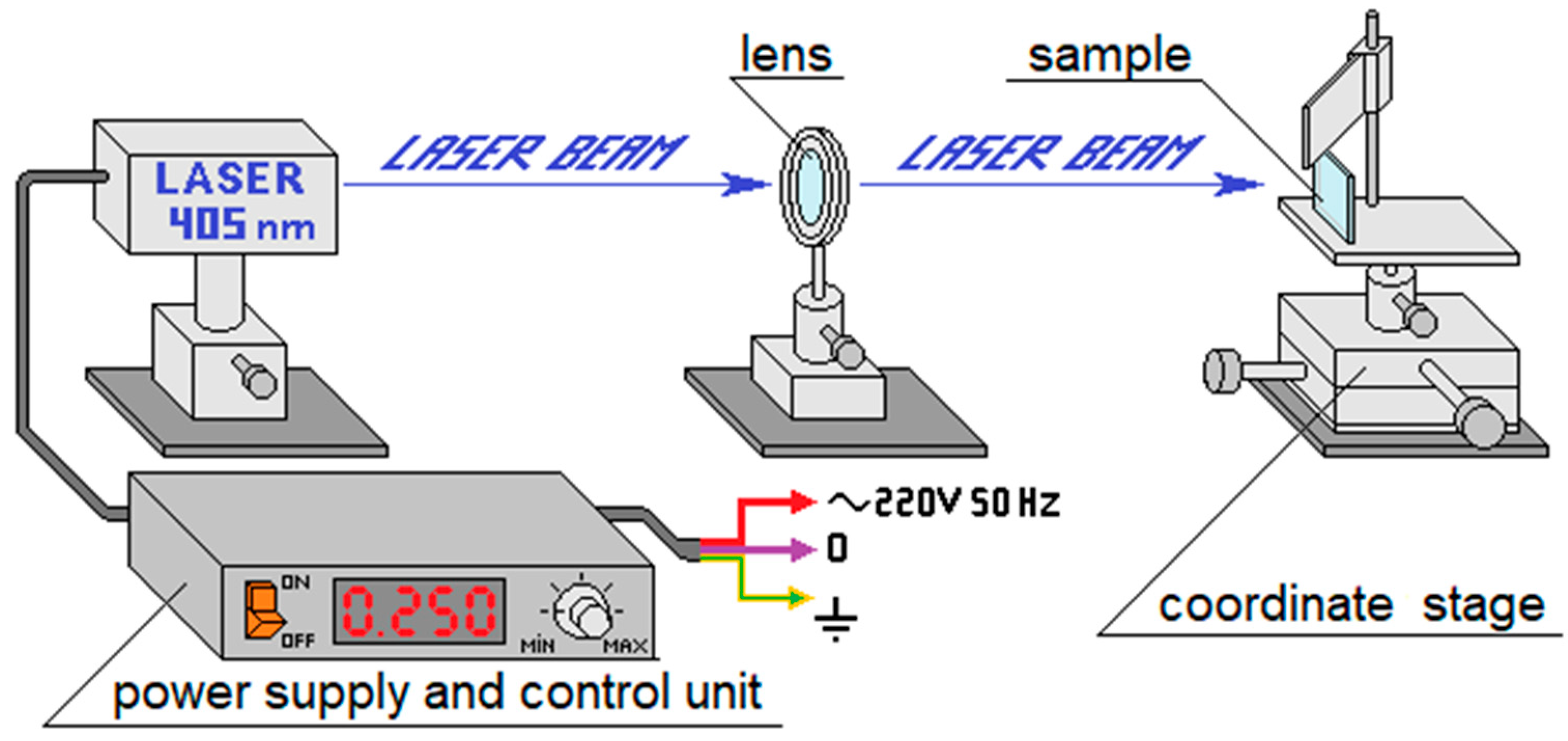
Figure 1. Scheme of the experimental setup.
Coatings 11 00058 g001
Figure 2. Arrangement of the irradiated soot regions (a) and representation of the Gaussian distribution of the laser spot (b).
Coatings 11 00058 g002
Figure 3. Morphology and structure of the soot coating (a,c) in the unaffected areas and (b,d) after laser irradiation with p = 114 mW for 350 and 450 s, respectively.
Coatings 11 00058 g003
Figure 4. Morphology and structure of the soot coating (a,c) in the unaffected areas and (b,d) after laser irradiation with p = 171 mW for 350 and 450 s, respectively.
Coatings 11 00058 g004
Figure 5. Light transmission spectra of the non-irradiated and laser-irradiated soot sample. The insets display the water contact angle before and after the laser treatment at p = 171 mW; tir ~350 s.
Coatings 11 00058 g005
Table 1. Chemical composition of each group soot coatings defined by EDS with an experimental uncertainty within ±0.1 at.%. The elements Na, Mg, Si and Ca are associated with the chemical composition of the glass substrate itself.
Soot AreaIrradiation ConditionsChemical Element (at.%)
CONaMgSICa
non-irradiatedn/a7915.60.70.33.80.6
irradiatedp = 171 mW; tir ~350 s7221.50.90.34.60.7
Publisher’s Note: MDPI stays neutral with regard to jurisdictional claims in published maps and institutional affiliations.