
Er:YAG Laser Cleaning of Painted Surfaces: Functional Considerations to Improve Efficacy and Reduce Side Effects

1
Key Laboratory of Cultural Heritage Research and Conservation, Northwest University, Ministry of Education, Xi’an 710127, China
2
School of Cultural Heritage, Northwest University, Xi’an 710127, China
3
Institute of Culture and Heritage, Northwestern Polytechnical University, Xi’an 710072, China
4
CNR-Institute for Geosciences and Earth Resources, 50121 Florence, Italy
*
Authors to whom correspondence should be addressed.
Coatings 2021, 11(11), 1315; https://doi.org/10.3390/coatings11111315
Submission received: 31 August 2021
/
Revised: 22 October 2021
/
Accepted: 26 October 2021
/
Published: 28 October 2021
(This article belongs to the Special Issue Conservation Tools, Protocols and Treatments on Painted Surfaces, Metal Leaves and Finishes in Cultural Heritage)
Abstract
:The restoration of paintings always involves the removal of darkened superficial layers, which are mainly due to dust deposition and aged varnishes. As cleaning is an irreversible and invasive treatment, physical methods (i.e., laser cleaning) instead of chemical ones are frequently suggested to reduce side effects on pictorial layers. Among the most employed laser systems, the free-running Er:YAG laser is considered very suitable for fine arts cleaning. This laser works at 2.94 μm, at which only –OH and –NH bonds in molecules are excited. This character can become a disadvantage when pigments with these functional groups are present. To understand the potential of the Er:YAG laser in such situations or in the presence of degradable pigments, the effectiveness of varnish removal from paintings prepared with egg yolk as the binder and cinnabar and lead white as the pigments were systematically investigated. Different cleaning conditions were used, and a hyperspectral sensor was innovatively used as a rapid, in situ and non-destructive technique to assess the effects of laser ablation, besides microscopic analysis. Though results obtained show all these pigments are sensitive to this laser radiation, satisfactory cleaning can be achieved without damaging the pictorial layer. The best cleaning conditions were 0.5 W of power (50 mJ and 10 Hz for energy and frequency), with 2-propanol as the wetting agent.
1. Introduction
In order to enhance the color saturation as well as to protect the original pictorial layers, a thin and transparent film (typically less than 100 μm) of organic substances is applied as the finishing layer of paintings (mainly easel paintings and also sometimes mural paintings) [1]. In historical and modern times, both natural substances (e.g., dammar, mastic, shellac resins) and synthetic polymers (e.g., P B67, ketone resins) are used as varnish [1,2]. However, owing to their organic nature, varnishes are vulnerable to natural aging triggered by temperature, relative humidity fluctuations and UV irradiation, which results in darkened, rigid and brittle superficial films. In some cases, micro-cracks of varnish are present. Besides, dust, soot and dirt deposition on varnish also contribute negatively to the precise appreciation of the aesthetic features of artworks. As a consequence, when it comes to the restoration of paintings, the removal or cleaning of darkened superficial layers, either due to dust, dirt deposition or aging of varnish, has always been a major focus for restorers.
Since varnish removal is invasive and irreversible, particular attention must be paid, and all methods shall be properly evaluated before application. In tradition, mechanical and chemical methods are usually adopted. Mechanical cleaning is performed by using a scalpel, and the effect of cleaning mainly depends on the skills of the operator. As the boundary between optimum cleaning and over-cleaning is blurring, damage to paint layers is of great possibility. In fact, chemical methods (or wet methods) that involve the use of organic solvents, e.g., acetone, ethanol, toluene, hexane, etc., were preferred by painting restorers. By simply wetting, dissolving and wiping the targeted material with a cotton swap, chemical methods are a rather convenient way for varnish removal. However, adverse effects of chemical cleaning are also conspicuous. For oil paintings, organic solvents can induce a series of physicochemical alterations called “solvent action”, including swelling of paint films, diffusion and evaporation of solvents, and leaching of pigments and binders off paint films [3]. Such alterations can be temporary (e.g., swelling effect, metal soap formation) and/or permanent (e.g., redistribution/extraction of components and the leaching of components increased brittleness, while the decreased thickness of paint films changed optical properties of overall painting) [3,4,5,6]. Last but not least, the volatility and toxicity of organic solvents should not be neglected, which are harmful to operators and the environment.
Thanks to the rapid advancements of science and technology, novel alternative cleaning methods have been developed in recent decades. In general, there are chemical cleaning methods by physical/chemical gels and physical cleaning methods by laser systems [7,8,9,10]; in this study, we focus on the latter. After the invention of the laser device in the 1960s, the first application of lasers to artworks was in 1971 when John Asmus and his team used a 690 nm ruby laser to acquire holographic images of marble statues in Venice [11]. In the experiment, he not only demonstrated the feasibility of holography but also illustrated the possibility of removing encrustation on stones by a ruby laser. His pioneering work with laser started a new field of research in conservation science. Ever since, there have been mainly three types of lasers employed in art conservation, namely the excimer laser, Nd:YAG laser and Er:YAG laser. In comparison with chemical and mechanical methods, laser cleaning shows various advantages that have been summarized as accuracy, selectivity, safety, control and reliability [10].
With variable emission wavelength (UV, NIR), pulse duration and relatively high energy, excimer lasers and Nd:YAG lasers are more suitable for cleaning rigid, inorganic substances such as stones, metals, glasses and wall paintings [11,12,13,14,15,16,17,18,19]. Due to their high energy (>1 eV) and deep penetration capability, excimer laser and Nd:YAG laser can introduce thermal and mechanical damages to adjacent material surrounding the impact zone, via the so-called “physical amplification” and “delayed amplification” [20]. These characteristics impede their application on delicate, sensitive artifacts. Fortunately, the more recent Er:YAG laser works at 2.94 μm, at which only –OH and –NH bonds in water and other molecules can absorb [21]. Thus, the pattern of energy deposition (actual ablation area) is determined by the distribution of molecules containing –OH and –NH bonds, and consequently, the laser beam is quickly absorbed, achieving very superficial ablation depths. In theory, the efficiency of laser ablation is proportional to the amount of –OH and –NH groups present in the materials. These –OH and –NH bonds, when not present in the original material, can be obtained by the addition of hydroxylated liquids (wetting agents), which can also help to limit the radiation and heat penetration [9,21,22]. These features make the Er:YAG laser particularly suitable for materials with high sensitivity and complex structures, e.g., easel paintings.
Although the Er:YAG laser was successfully applied for varnish removal [23,24,25,26,27,28,29], to our best knowledge, there is limited study on the situation when pigments in pictorial layers are also sensitive to this laser. Besides, laser cleaning is thought to be intuitive, and controlled cleaning can be performed by simply changing the fluence or pulse duration of the laser beam based on the experience or skill of the operator. Therefore, finding a more scientific and in situ method to indicate the extent of the cleaning process is imperative and urgent.
In this study, a free-running Er:YAG laser with different working parameters and conditions, e.g., power, energy, frequency, wetting agents, etc., were employed to clean varnishes on painting mock-ups prepared with laser-sensitive pigments (cinnabar, lead white). During the cleaning, besides the traditional optical microscopy, a portable and non-invasive hyperspectral sensor (ASD-FieldSpec FR Pro, NY, USA) that can characterize organic and inorganic materials [30,31,32,33] was innovatively exploited to characterize painting components and to monitor the progress of varnish cleaning by the Er:YAG laser.
2. Materials and Methods
2.1. Painting Mock-Ups
Painting mock-ups were prepared on the canvas (Zecchi, Florence, Italy), which was already treated with gypsum and animal glue. As support, the canvas was divided into 10 × 10 cm2 dimensions. Several pigments (cinnabar, lead white, malachite, azurite, lapis lazuli, yellow ochre and Naples yellow) and binders (egg yolk, linseed oil and animal glue) were chosen for the testing. Investigations are still ongoing, and only the tests with egg yolk as a binder and cinnabar and lead white as pigments are reported here. The egg yolk was selected as a binder for its wide use in easel paintings and its fast drying compared to linseed oil. Regarding the pigments, cinnabar was selected for its known sensitivity to light, while as one of the most used white pigments, lead white [2PbCO3Pb(OH)2)] has intrinsic –OH groups in the molecule, which may increase its sensitivity to the Er:YAG laser irradiation. Therefore, considering the different interactions and possible sensitivity of pigments under laser irradiation, cinnabar and lead white were applied on the canvas with egg yolk as a binding medium to simulate the tempera painting. Table 1 illustrates the mass of materials used in mock-up preparation. All pigments were purchased from Zecchi (Florence, Italy), while eggs were freshly bought in a local supermarket in Florence. In order to prepare the painting mock-ups, egg yolk was carefully extracted from the whole egg and mixed with different pigments, and then the mixture was brushed onto canvas evenly. Typically, the mass of pigment and egg yolk was almost the same (Table 1). During the mixing, some deionized water was added to decrease the viscosity for easy application. The painted samples prepared were placed in lab conditions for 1 month to achieve complete evaporation of the water (drying). After the complete drying of the pictorial layer, solutions of one natural and one synthetic varnish, i.e., 20% mastic (tears of Chios) in turpentine oil and 44% P B67 (≥98%) in acetone (≥99.5%), were brushed several times until no more absorption was observed (saturation). However, due to the low concentration of mastic, the varnish was applied again on the samples 2 months later to achieve a varnish thickness of about 10–15 μm. After 6 months in the lab conditions, the samples varnished with mastic were placed in an oven at 60 °C for 10 days to accelerate the hardening/drying process of the varnished layer. Finally, the surfaces were gently touched by fingers to confirm the complete drying. The choice of these two varnishes is motivated by their extensive use since ancient times (mastic) or by their use in modern restoration interventions.
2.2. Hyperspectral Sensor
The hyperspectral sensor employed in this study was an Analytical Spectral Devices FieldSpec FR Pro 3 (Analytical Spectral Devices, Inc., Boulder, CO, USA), a portable high-resolution spectroradiometer. It is designed to acquire Visible, Near Infrared (VNIR: 350–1000 nm) and Short-Wave Infrared (SWIR: 1000–2500 nm) punctual reflectance spectra in 0.2 s per spectrum. The sampling interval is 1.4 nm and 2 nm for the VNIR and SWIR spectral regions, respectively. The VNIR spectrometer has a standard spectral resolution (full-width at half maximum of a single emission line) of 3 nm at around 700 nm, while in the SWIR region, the spectral resolution is 10–12 nm from 1400 to 2100 nm. Spectra were firstly collected and then processed using ASD’s RS3 and ViewSpec Pro 6.0 software. The instrument is equipped with a contact reflectance probe (fixed geometry of illumination and shot with a spot analysis of about 1.5 cm2) that provides an internal light source, so spectra can be acquired without involving ambient solar illumination as in the applications of remote sensing.
The characterization of pure varnishes by the hyperspectral sensor was carried out firstly on Teflon support, on which the previously prepared Mastic and P B67 solutions were deposed to form thin films (thickness about 50–70 μm). The drying procedure of the varnish films was the same as that used for the painting mock-ups. The spectral collection was performed after the drying of varnish films. Then, the characterization of painting mock-ups without varnishes was conducted in order to serve as references when evaluating the efficacy of laser cleaning. Afterward, painting mock-ups with pictorial layers and varnishes were analyzed by the hyperspectral sensor before and after laser cleaning.
All reflectance spectra were collected with the Contact Probe by direct contact with the targeted surfaces. In order to eliminate the interferences of the ground layer and the canvas support, the background spectra were also collected on unpainted surfaces. The spectra reported were the average spectrum of 10 acquisitions performed on the same analyte. The spectra were then processed as raw by ViewSpec Pro 6.0 software.
2.3. Varnish Removal by an Er:YAG Laser
The 2.94 μm Er:YAG laser (“Light Brush” made by DEKA, El.En. Group, Calenzano, Italy), which emits 250 μs duration “macropulses” consisting of 1–2 μs micro-pulses, was used for varnish removal tests. The radiation goes directly into a 1 m long, 1 mm bore hollow internal mirrored glass guide with a pen-like tip for aiming at specific target sites. The control of the energy of each pulse to the millijoule level allows the determination of ablation thresholds for each material. For the instrument, the highest macropulse frequency is 15 Hz, and the highest energy is 100 mJ.
The laser cleaning on varnished mock-ups was operated at a relatively constant distance of 5 mm between the surface and the laser. The laser beam was delivered through the internal mirrored glass guide, and a 3–5 mm diameter of spot size was produced. Cleaning was performed without using its original flexible protecting window because the laser beam almost stopped in this case. As shown in Figure 1, each sample of 10 × 10 cm2 dimensions was further divided into nine quadrants for evaluating different cleaning conditions. The cleaning environment was basically “dry” (without wetting agent) and “wet” (with deionized water or 2-propanol (>99.5%, Sigma-Aldrich Inc., Darmstadt, Germany) as wetting agent), and the laser irradiation conditions applied were: (1) 50 mJ, 10 Hz, 0.5 W; (2) 100 mJ, 7 Hz, 0.7 W; (3) 100 mJ, 10 Hz, 1 W for energy, frequency and power, respectively. In order to prevent the pollution of the laser beam from the cleaning substance, a square glass coverslip (1.8 × 1.8 cm2) was placed on the cleaning area during operation. In order to clean an area of this size, 5 scans of laser ablation were taken within 150 s. During and after the cleaning, a portable hyperspectral sensor was exploited to evaluate the efficacy.
2.4. Optical Microscopy
A Leica MZ 125 Stereomicroscope(Leica Microsystems, Wetzlar, Germany) equipped with a digital camera (Canon 500D, Canon Inc., Tokyo, Japan) capturing system was used to document the surface morphology of painting mock-ups before and after laser cleaning. In order to find the same area before and after laser ablation on the surfaces of mock-ups, a paper mask with horizontal and vertical axes was used for precise locating. The position (0, 0) and (−5 mm, −5 mm) in the coordinate system for the left corner of the mock-up was selected for observation. The morphological characterization was performed under 10×, 32× and 80× of magnification.
3. Results and Discussion
3.1. Assessment of the Cleaning of Mastic
3.1.1. Cinnabar
Cinnabar (HgS) is considered an extremely light-sensitive pigment and one of the most sensitive pigments to laser irradiation [34]. Although the ablation action of the pulsed Er:YAG laser is based on the excitation of the –OH and –NH bonds due to the resonance between the radiation at 2.94 µm and the vibration stretching modes [21,35], the interaction between the laser radiation and cinnabar cannot be excluded. The results show that once a lower level of energy (50 mJ, 10 Hz) was applied in dry conditions, the superficial varnish layer was cleaned by laser ablation with no damage to the cinnabar. With the increase in power to 1 W (100 mJ, 10 Hz), the varnish layer was almost removed, but some damage to pictorial layers occurred. As shown in Figure 2b, the cinnabar turned to black spots (marked by white cycles) on which varnish was removed. The appearance of black spots instead of a uniform darkening of the irradiated area is due to the non-uniformity of the varnish layer: thicker the varnish film, less the damaging effect. Without applying wetting agents, a superficial thermal effect was induced by laser irradiation.
As the preferential absorption of the laser radiation by the –OH groups of water may prevent the thermal transformation of the pigments [35], with the presence of water, damages to pigment were not observed under the same working conditions (power 1 W, 0.7 W, 0.5 W). However, since mastic is hydrophobic, water cannot wet the varnish layer uniformly. Therefore, the cleaned surface was not uniform (Figure 2d,d′), and damages were concentrated on the areas where water was not present (marked by black circles).
It is evident that the varnish removal efficacy dramatically increased when using 2-propanol as the wetting agent (Figure 2f,f′). The surfaces (Sections 6–8) were well wetted, and the cleaning was more effective and evenly (Figure 2f). However, it is worth noticing that, even with a lower frequency (7 Hz), higher energy (100 mJ) can induce a little discoloration of cinnabar (0.7 W). The microscopic observation of Section 6 (0.7 W) is shown in Figure 2f,f′. It is clear that mastic was over-cleaned. The cleaned painting layer showed a “whitened” appearance together with some black spots.
3.1.2. Lead White
Lead white (2PbCO3Pb(OH)2) is one of the most important white pigments since ancient times, thanks to their hiding power. It is well known, the presence of –OH groups in their crystal structure may increase its sensitivity to the Er:YAG laser irradiation. However, the results of cleaning in dry conditions showed that lead white was not damaged by laser ablation, even when the highest cleaning power (1 W, by 100 mJ and 10 Hz) was used. This is mainly due to the partial removal of the varnish, which can be deduced from the small concavities created on the surface after 150 s of pulse duration (Figure 3b).
By applying solvents as wetting agents, it was suggested that they could work as barriers to laser irradiation during cleaning [36]. This may be true when water was used as the wetting agent, as in the case of mastic varnish on the cinnabar layer. On the contrary, the surfaces (in Sections 3–5) were not cleaned at all with no varnish removal; even the energy applied was at the highest level (Figure 4). Unlike water, 2-propanol facilitated the removal of mastic already at low power (0.5 W, 50 mJ and 10 Hz), but higher power (in Sections 6 and 8) resulted in permanent discoloration—blacken of lead white (Figure 3d). Where the pigment turned black, the varnish layer was probably very thin; therefore, it was totally removed after a few seconds of these irradiation conditions, with a consequent lack of lead white protection.
3.2. Assessment of the Cleaning of P B67
3.2.1. Cinnabar
In the case that P B67 was the varnish layer, the lowest power (0.5 W, 50 mJ, 10 Hz) did not cause any damage to the cinnabar, and no varnish was removed. By increasing the power to 1 W (100 mJ, 10 Hz), discoloration of the pigment appeared (Figure 5b), whilst the layer of P B67 exfoliated from the painting surfaces but not detached. Under this cleaning condition, the blackening of cinnabar was found, and this is mainly due to the polymorphic transformation from red hexagonal cinnabar (α-HgS) to black cubic metacinnabar (α′-HgS) [34].
When water or 2-propanol were used as a wetting agent, the temperature increase in the irradiated surface was reduced due to the presence of –OH groups in the solvents, which provide a strong absorption at 2.94 µm, ensuring the retention of much of the heat produced [22,35]. However, different from mastic, only 2-propanol provided an effective cleaning. No matter the power used, there was no more damage to the pictorial layer caused by laser irradiation. The difference between these two wet cleaning conditions was that water appeared to have no interaction with the varnish resulting in a better protecting effect than 2-propanol, and consequently, less varnish was removed. With the presence of water on the surface, even though the highest laser power (1 W, 100 mJ, 10 Hz) was adopted, P B 67 was not removed at all (no products on the covering glass), as shown in Figure 5d. Since the surface of P B67 was hydrophobic, water could not wet the entire surface evenly. Only area covered by water was well protected, while other areas were as cleaning in dry conditions, which became a layer with micro-bubbles (marked by a white arrow in Figure 5d). Similar morphological changes were also found by Chillè et al. during the cleaning of aged dammar films by using the Er:YAG laser, as they described “By increasing the fluence, the possibility of identifying differences in appearance of the spots was reduced, due to partial melting of the varnish surface and to the increase in micro-pits and bubbles” [27].
While using 2-propanol, owing to its low surface tension, 2-propanol distributed well on P B67 and led to the evenly cleaning. After laser ablation, the varnish surface became sticky, which was easy to clean with traditional cleaning methods (e.g., cotton swap) afterward. This is probably due to the fact that substances with smaller molecular weights (e.g., chain scission) were produced. With the increase in laser power, the cleaning efficacy also improved. In Figure 5f, it is evident that P B67 was removed under this laser condition without damage to the pigment.
3.2.2. Lead White
With lead white and egg yolk as the pictorial layer, P B67 varnish was cleaned using the same methods. As in the cases of mastic samples, there was no discoloration of lead white under laser ablation in dry condition, which was mainly due to the permanence of the varnish more than the stability of lead white to Er:YAG laser radiation. Figure 6a,b shows the microscopic images (32×) of pigment before and after laser cleaning. Though the highest laser power was applied (1 W), almost no varnish was removed from the surface.
When water was deposited on varnish before laser ablation, P B67 was better cleaned even with the lowest laser power. As marked by the circles in Figure 6d, line-shaped concaves formed after cleaning, indicating the partial removal of superficial varnish. The different results obtained in this case, compared with the ablation of P B67 on cinnabar, may be due to a poor homogeneity of the varnish layer, which favors a better interaction (absorption) of water with the surface. However, in this situation, water not only protected the pictorial layer but also contributed to varnish cleaning. When 2-propanol was used as a wetting agent, better cleaning efficacy was achieved. P B67 layer was cleaned better without damaging the lower layer. From Figure 6f, it was clear that P B67 became very rough with a flake-like texture after laser cleaning.
3.3. In Situ Monitoring of Cleaning by a Portable Hyperspectral Sensor
By aiming to evaluate the cleaning effectiveness of laser ablation, the reflectance spectra in full range (350–2500 nm) of the painted surface, the surface of the varnished painting and the varnished surface after laser cleaning were obtained by the rapid and in situ technique, i.e., a portable hyperspectral sensor (ASD Fieldspec FR Pro 3). The reflectance spectra collected in Section 8 of cinnabar painting mock-ups are shown in Figure 7a, and the original spectrum of pure mastic was also shown as the reference. All spectra contain cinnabar presented very typical sigmoid shape, with an inflection point characterized by a maximum peak in the 1st derivative reflectance spectrum at the wavelengths between 580 and 610 nm. Although the results of optical microscopy manifested that the cinnabar was discolored (as in Section 6, Figure 2f), the characteristic spectral features of cinnabar did not change before and after laser cleaning. In order to definitively demonstrate that the blackening of cinnabar was not induced by a chemical reaction, other techniques should be employed to detect possible new compounds formed. However, the result here found suggests a polymorphic transformation of cinnabar, which is in agreement with other research [34].
The most evident variances in the spectra collected from Section 8 varnished by mastic before and after laser cleaning (100 mJ, 10 Hz, 2-propanol as wetting agent) are plotted in Figure 7b (spectral range 1650–2000 nm). It is clear that the absorption peak of the first overtone of C–H stretching of mastic, which locates at 1703 and 1725 nm, became broader after laser cleaning [37]. In the meanwhile, the broad peak at 1923 nm attributed to the combination band of asymmetric stretching and bending of O–H bond of mastic was even broader and also shifted to a longer wavelength [37,38]. Similar modifications of spectral features were also found in the spectra of mock-ups prepared with lead white and mastic varnish (Figure 8). Regardless of the pigment involved, the reflectance spectra after laser cleaning share the same modifications, i.e., decreasing of the intensity and shifting of the characteristic absorption peaks of mastic. All the above changes make the spectra after cleaning resemble the spectrum of the painting surface without varnish.
In the case of cinnabar tempera painting mock-ups varnished by P B67, we also selected Section 8 to study its reflectance spectra to monitor the efficacy of laser cleaning. In Figure 9a, it is evident that almost all the characteristic absorption peaks of P B67 can be found in the spectrum before and after cleaning. Although the cleaning was not completed, some typical features were found to be strongly weakened due to the partial removal of P B67. The absorption peak ascribed to the first overtone of C–H stretching mode is observed at around 1703, 1730 and 1762 nm for P B67, and they became less evident after cleaning. Besides, the peak intensity of 2275 nm 2308 nm corresponding to the combination band of the first overtone of C–H stretching and bending also reduced [38]. Likewise, very similar behaviors were also found in lead white tempera varnished with P B67 (Figure 10). It is noticed, under the same cleaning condition, the reduction in the intensity of the first overtone of CH2 stretching at around 1703 nm seemed less obvious in the spectrum of P B67 compared to mastic. This can be explained by the fact the intensity of the diffuse reflectance spectrum is closely related to the roughness of the detected surface, as the surface became very rough after cleaning.
3.4. Best Condition for Laser Sensitive Pigments
After the comparative study on cleaning results under different conditions, safe parameters should be determined for the removal of mastic and P B67 from cinnabar and lead white tempera painting through the Er:YAG laser. Based on the microscopic analysis conducted on four different combinations under various cleaning conditions (from Figure 11, Figure 12, Figure 13 and Figure 14), it can be seen that cleaning efficacy increased as the power increased, while the cleaning condition (dry or wet) also strongly affected the final results depending on the type of varnish. Dry and wet cleaning conditions are always discussed by researchers when the Er:YAG laser is adopted for cleaning since wetting agents are able to decrease the temperature in the bulk and obtain a gentler interaction with varnish surface [24,25].
In the dry condition, cleaning without any wetting agents helped us determine the safety threshold for laser ablation (max power 0.5 W, by 50 mJ and 10 Hz) on light-sensitive pigments. In the wet condition, two wetting agents showed distinct influences on the cleaning efficacy. As for water, the water could not wet either the surface of mastic or P B67 evenly due to its relatively high surface tension. During laser ablation, water absorbed the major energy of laser irradiation and acted as a protective layer to the pigments in these four cleaning cases. Consequently, cinnabar and lead white were well protected, although the cleaning was less effective compared with the results obtained with the same operative parameters in dry conditions. This cleaning effect was more obvious for the more sensitive pigment—cinnabar. In Figure 11 and Figure 13, it was clear no matter for cleaning mastic or P B67, when water was present, discoloration of cinnabar did not occur even with the highest power.
However, when 2-propanol was applied as a wetting agent, the situation became more complicated. Thanks to its proper physical–chemical properties, it is well distributed over the entire cleaning surfaces in all cases, facilitating the evenly cleaning by laser ablation. The cleaning efficacy was improved, but damages to the pictorial layer were also observed, i.e., blackening of lead white during cleaning of mastic. These results indicate the discoloration of pigments was not only related to the chemical nature of pigments but also affected by the type of varnish presented.
In the case of mastic removal, both cinnabar and lead white were damaged when high power (0.7 W and 1 W) was applied (Figure 11 and Figure 12). Comparing the result of these 8 sections cleaned in different conditions and parameters, the worst cleaning (least varnish removal) were in Section 6 and Section 8, which implied that the two parameters (0.7 W and 1 W) with the presence of 2-propanol was not suitable for cleaning mastic varnish on cinnabar or lead white pigments. When the lowest power (0.5 W) was used, a limited amount of mastic was removed while no damage was induced to the pictorial layer. In this case, repeated cleaning can give us satisfactory results.
In the case of P B67 removal, the best results were achieved with the pre-treatment of 2-proponal before laser irradiation. For both cinnabar and lead white painting mock-ups, in wet condition, the cleaning efficacy of P B67 increased as the pigments were well protected. After applying the laser power of 0.5 W, the varnish layer became sticky and it was easy to remove without damaging the painting layer, as shown in Figure 13 and Figure 14. Once the power of laser irradiation increased, the surface became stickier, and it was very easy to clean with the traditional method. These results implied, the thin layer of 2-propanol acted as a barrier to absorb the laser energy and limited the penetration depth of irradiation superficially simultaneously. The best cleaning effect was obtained by applying 1 W of power; p B67 was evenly and thoroughly removed on the lead white layer (Figure 14). However, possible damages may be created if a higher power was used for repeated cleaning.
4. Conclusions
In this research, the efficacy and side effects of varnish removal by a free-running Er:YAG laser at 2.94 μm were systematically investigated. By focusing on the validation and evaluation of the applicability of the Er:YAG laser for laser-sensitive pigments, cinnabar and lead white tempera mock-ups varnished with mastic and P B67 were cleaned under varied conditions and parameters. In addition to the traditional optical microscopic analysis, a novel rapid, in situ and non-invasive hyperspectral sensor was exploited to monitor the progress of varnish removal.
In general, under the same laser cleaning conditions, cinnabar was more easily damaged compared with lead white, although lead white has intrinsic O–H groups in its structure. The safety threshold of ablation energy for laser-sensitive pigments was determined as 0.5 W of power (100 mJ and 5 Hz) by tests conducted in dry conditions. Once wetting agents were introduced, they had different influences on the cleaning efficacy. Water, regardless of the pigment and varnish used, demonstrated a protective effect for the pictorial layer during cleaning, but it decreased the removal of varnish. In contrast, 2-propanol improved the cleaning efficacy due to its chemical–physical properties (e.g., lower surface tension and better wettability than water). However, 2-propanol induced the discoloration of both cinnabar and lead white in the case of mastic removal when high ablation power was applied (≥0.7 W). Moreover, by providing the full range reflectance spectra, the portable hyperspectral sensor illustrated good potential in evaluating varnish cleaning as an in situ tool. The progress of varnish removal can be scientifically and rapidly controlled during laser cleaning by simply studying the spectral shape and relative peak intensity change in the spectra of varnishes and pigments.
In conclusion, with 2-propanol as a wetting agent, satisfactory cleaning can be achieved without damaging laser-sensitive pigments by adjusting the working conditions of the Er:YAG laser to 50 mJ of energy and 10 Hz of frequency (0.5 W of power).
Author Contributions
Conceptualization, M.C.; methodology, C.W. and M.C.; investigation, C.W.; writing—original draft preparation, C.W. and Y.C.; writing—review and editing, C.W., Y.C., F.T. and M.C.; supervision, M.C.; funding acquisition, C.W. and M.C. All authors have read and agreed to the published version of the manuscript.
Funding
This research was funded by the National Key Research and Development Project (Grant No. 2020YFC1521904), the Social Science Foundation of Shaanxi Province (Grant NO. 2021G011), the Scientific Research Program of Shaanxi Provincial Education Department (Grant NO. 21JK0947) and the start-up funding from Northwest University, China.
Institutional Review Board Statement
Not applicable.
Informed Consent Statement
Not applicable.
Data Availability Statement
Not applicable.
Acknowledgments
The authors thank El. En. Group for supplying the Er:YAG laser source. Sandro Moretti and Teresa Salvatici from Department of Earth Sciences-University of Florence are also acknowledged for their help with the application of the hyperspectral sensor.
Conflicts of Interest
The authors declare no conflict of interest.
References
- De la Rie, E.R. The inpower of varnish on the appearance of paintings. Stud. Conserv. 1987, 32, 1–13. [Google Scholar]
- Reifsnyder, J.M. A note on a traditional technique of varnish application for paintings on panel. Stud. Conserv. 1996, 41, 120–122. [Google Scholar]
- Phenix, A.; Sutherland, K. The cleaning of paintings: Effects of organic solvents on oil paint films. Stud. Conserv. 2001, 46, 47–60. [Google Scholar] [CrossRef]
- Baij, L.; Hermans, J.; Ormsby, B.; Noble, P.; Iedema, P.; Keune, K. A review of solvent action on oil paint. Herit. Sci. 2020, 8, 43. [Google Scholar] [CrossRef]
- Khandekar, N. A survey of the conservation literature relating to the development of aqueous gel cleaning on painted and varnished surfaces. Stud. Conserv. 2000, 45, 10–20. [Google Scholar] [CrossRef]
- Erhardt, D.; Tsang, J.S. The extraction components of oil paint films. Stud. Conserv. 1990, 45, 93–97. [Google Scholar] [CrossRef]
- Carretti, E.; Dei, L.; Weiss, R.G.; Baglioni, P. A new class of gels for the conservation of painted surfaces. J. Cult. Herit. 2008, 9, 386–393. [Google Scholar] [CrossRef]
- Mastrangelo, R.; Chelazzi, D.; Poggi, G.; Fratini, E.; Buemi, L.P.; Petruzzellis, M.L.; Baglioni, P. Twin-chain polymer hydrogels based on poly (vinyl alcohol) as new advanced tool for the cleaning of modern and contemporary art. Proc. Natl. Acad. Sci. USA 2020, 117, 7011–7020. [Google Scholar] [CrossRef] [PubMed] [Green Version]
- Rui, B.; Morais, P.J.; Helena, G.; Young, C. Laser Cleaning of easel paintings: An overview. Laser Chem. 2006, 2006, 90279. [Google Scholar]
- Ganeev, R. Laser Cleaning of Art. In Laser—Surface Interactions, 1st ed.; Springer: Dordrecht, The Netherlands, 2014; pp. 87–103. [Google Scholar]
- Asmus, J.F.; Guattari, G.; Lazzarini, L.; Musumeci, G.; Wuerker, R.F. Holography in the conservation of statuary. Stud. Conserv. 1973, 18, 49–63. [Google Scholar]
- Siano, S.; Agresti, J.; Cacciari, I.; Ciofini, D.; Mascalachi, M.; Osticioli, I.; Mencaglia, A. Laser cleaning in conservation of stone, metal, and painted artifacts: State of the art and new insights on the use of the Nd:YAG lasers. Appl. Phys. 2012, 106, 419–446. [Google Scholar] [CrossRef]
- Siano, S.; Giamello, M.; Bartoli, L.; Mencaglia, A.; Parfenov, V.; Salimbeni, R. Laser cleaning of stone by different laser pulse duration and wavelength. Laser Phys. 2008, 18, 27–36. [Google Scholar] [CrossRef]
- Osticioli, I.; Mascalchi, M.; Pinna, D.; Siano, S. Removal of verrucaria nigrescens from carrara marble artefacts using Nd:YAG lasers: Comparison among different pulse durations and wavelengths. Appl. Phys. A 2015, 118, 1517–1526. [Google Scholar] [CrossRef]
- Koh, Y.S.; Sárady, I. Cleaning of corroded iron artefacts using pulsed TEA CO2- and Nd:YAG-lasers. J. Cult. Herit. 2003, 4, 129–133. [Google Scholar] [CrossRef]
- Sansonetti, A.; Colella, M.; Letardi, P.; Salvadori, B.; Striova, J. Laser cleaning of a nineteenth-century bronze sculpture: In situ multi-analytical evaluation. Stud. Conserv. 2015, 60, 28–33. [Google Scholar] [CrossRef] [Green Version]
- Bilmes, G.M.; Vallejo, J.; Costa Vera, C.; Garcia, M.E. High efficiencies for laser cleaning of glassware irradiated from the back: Application to glassware historical objects. Appl. Phys. A 2018, 124, 1–11. [Google Scholar] [CrossRef]
- Gaetani, C.; Santamaria, U. The laser cleaning of wall paintings. J. Cult. Herit. 2000, 1, S199–S207. [Google Scholar] [CrossRef]
- Andreotti, A.; Colombini, M.P.; Nevin, A.; Melessanaki, K.; Pouli, P.; Fotakis, C. Multianalytical study of laser pulse duration effects in the IR laser cleaning of wall paintings from the monumental cemetery of Pisa. Laser Chem. 2006, 2006, 39046. [Google Scholar] [CrossRef] [Green Version]
- De Cruz, A.; Wolbarsht, M.L.; Hauger, S.A. Laser removal of contaminants from painted surfaces. J. Cult. Herit. 2000, 1, 173–180. [Google Scholar] [CrossRef]
- Teppo, E. Introduction: Er:YAG lasers in the conservation of artworks. J. Inst. Conserv. 2020, 43, 2–11. [Google Scholar] [CrossRef]
- De Cruz, A.; Andreotti, A.; Ceccarini, A.; Colombini, M.P. Laser cleaning of works of art: Evaluation of the thermal stress induced by Er:YAG laser. Appl. Phys. B 2014, 117, 533–541. [Google Scholar] [CrossRef]
- Bracco, P.; Lanterna, G.; Matteini, M.; Nakahara, K.; Colombini, M.P. Er:YAG laser: An innovative tool for controlled cleaning of old paintings: Testing and evaluation. J. Cult. Herit. 2003, 4, 202–208. [Google Scholar] [CrossRef]
- Striova, J.; Salvadori, B.; Fontana, R.; Sansonetti, A.; Barucci, M.; Pampaloni, E.; Marconi, E.; Pezzati, L.; Colombini, M.P. Optical and spectroscopic tools for evaluating Er:YAG laser removal of shellac varnish. Stud. Conserv. 2015, 60, 91–96. [Google Scholar] [CrossRef] [Green Version]
- Chillè, C.; Papadakis, V.M.; Theodorakopoulos, C. An analytical evaluation of Er:YAG laser cleaning tests on a nineteenth century varnished painting. Microchem. J. 2020, 158, 105086. [Google Scholar] [CrossRef]
- Brunetto, A.; Bono, G.; Frezzato, F. Er:YAG laser cleaning of ‘San Marziale in Gloria’ by Jacopo Tintoretto in the Church of San Marziale, Venice. J. Inst. Conserv. 2020, 43, 44–58. [Google Scholar] [CrossRef]
- Chillè, C.; Sala, F.; Wu, Q.; Theodorakopoulos, C. A study on the heat distribution and oxidative modification of aged dammar films upon Er:YAG laser irradiation. J. Inst. Conserv. 2020, 43, 59–78. [Google Scholar] [CrossRef]
- Pereira-Pardo, L.; Melita, L.N.; Korenberg, C. Tackling conservation challenges using erbium lasers: Case studies at the British Museum. J. Inst. Conserv. 2020, 43, 25–43. [Google Scholar] [CrossRef]
- Striova, J.; Fontana, R.; Barbetti, I.; Pezzati, L.; Fedele, A.; Riminesi, C. Multisensorial assessment of laser effects on shellac applied on wall paintings. Sensors 2021, 21, 3354. [Google Scholar] [CrossRef]
- Camaiti, M.; Vettori, S.; Benvenuti, M.; Chiarantini, L.; Costagliola, P.; Di Benedetto, F.; Moretti, S.; Paba, F.; Pecchioni, E. Hyperspectral sensor for gypsum detection on monumental buildings. J. Geophys. Eng. 2011, 8, S126–S131. [Google Scholar] [CrossRef]
- Wang, C.; Salvatici, T.; Camaiti, M.; Chiara, D.; Moretti, S. A new application of hyperspectral radiometry: The characterization of painted surfaces. In Proceedings of the EGU General Assembly 2016, Vienna, Austria, 23–29 April 2016. [Google Scholar]
- Wang, C. Hyperspectral Sensor: A New Approach for Evaluating the Efficacy of Laser Cleaning in the Removal of Varnishes and Overpaintings. Master’s Thesis, University of Bologna, Bologna, Italy, 2015. [Google Scholar]
- Camaiti, M.; Benvenuti, M.; Costagliola, P.; Benedetto, F.D.; Moretti, S. Hyperspectral sensors for the characterization of cultural heritage surfaces. In Sensing the Past; Masini, N., Soldovieri, F., Eds.; Springer International Publishing: Gewerbestrasse, Switzerland, 2017; Volume 16, pp. 289–311. [Google Scholar]
- Pouli, P.; Emmony, D.C.; Madden, C.E.; Sutherland, I. Studies towards a thorough understanding of the laser-induced discoloration mechanisms of medieval pigments. J. Cult. Herit. 2003, 4, 271s–275s. [Google Scholar] [CrossRef]
- Striova, J.; Camaiti, M.; Castellucci, E.M.; Sansonetti, A. Chemical, morphological and chromatic behavior of mural paintings under er:yag laser irradiation. Appl. Phys. A 2011, 104, 649–660. [Google Scholar] [CrossRef]
- Camaiti, M.; Matteini, M.; Sansonetti, A.; Striová, J.; Castelluci, E.; Andreotti, A.; Colombini, M.P.; De Cruze, A.; Palmer, R. The Interaction of Laser Radiation at 2.94 μm with Azurite and Malachite Pigments. Lasers in the Conservation of Artworks. In Proceedings of the International Conference Lacona VII, Madrid, Spain, 17–21 September 2008; pp. 253–258. [Google Scholar]
- Invernizzi, C.; Rovetta, T.; Licchelli, M.; Malagodi, M. Mid and near-infrared reflection spectral database of natural organic materials in the cultural heritage field. Int. J. Anal. Chem. 2018, 2018, 7823248. [Google Scholar] [CrossRef] [PubMed]
- Schwanninger, M.; Rodrigues, J.C.; Fackler, K. A review of band assignments in near infrared spectra of wood and wood components. J. Near Infrared Spectrosc. 2011, 19, 287–308. [Google Scholar] [CrossRef]
Figure 1.
The different cleaning conditions operated on painting mock-ups.
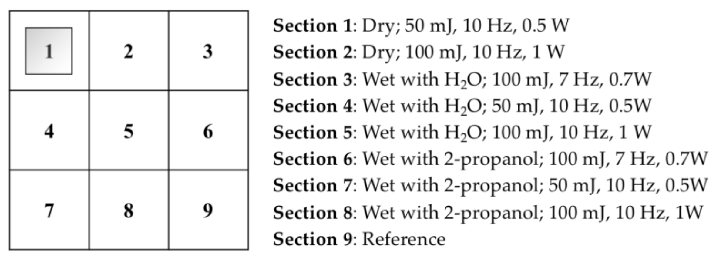
Figure 2.
The microscopic images (10×, 32× and 80×) of mastic removal by Er:YAG laser under varied conditions and parameters, conducted on cinnabar tempera mock-ups. (a,b) is image obtained before and after cleaning in dry condition, while (c,d) was obtained before and after cleaning in wet (H2O) condition. (c′,d′) (80×) was zoomed images of (c,d) at the same location, and (e′,f′) was taken at the same location as (e,f) but with 10× of magnification.
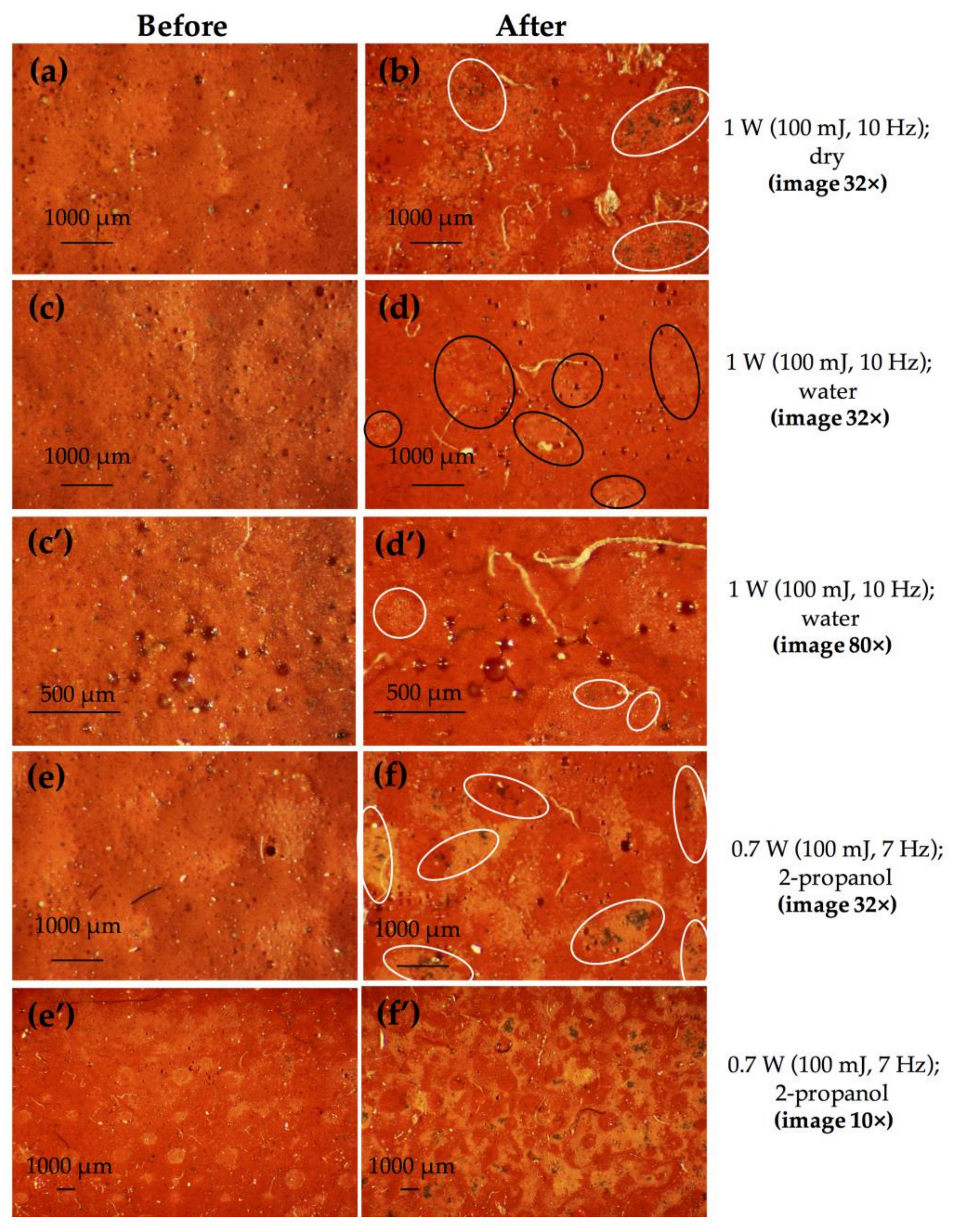
Figure 3.
The microscopic images (32×) of mastic removal by Er:YAG laser under varied conditions and parameters, conducted on lead white tempera mock-ups. (a,b) was obtained before and after cleaning in dry condition with 1 W as laser power, and (c,d) is image before and after cleaning in wet (2-propanol) condition with 0.7 W as laser power.
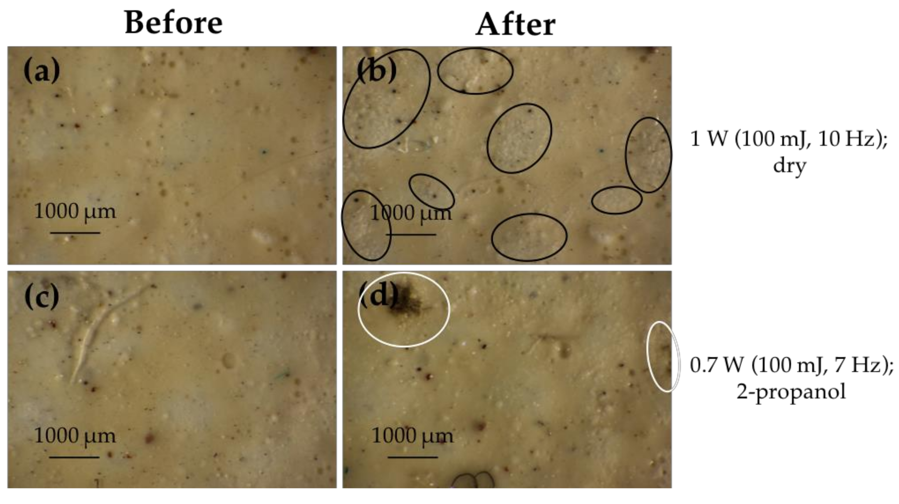
Figure 4.
The microscopic images (32×) of mastic removal by Er:YAG laser under varied conditions and water as wetting agent, conducted on lead white tempera mock-ups. (a,c,e) was taken before cleaning, while (b,d,f) was obtained after cleaning in wet (H2O) conditions with varied laser power (0.7 W, 0.5 W and 1 W respectively).
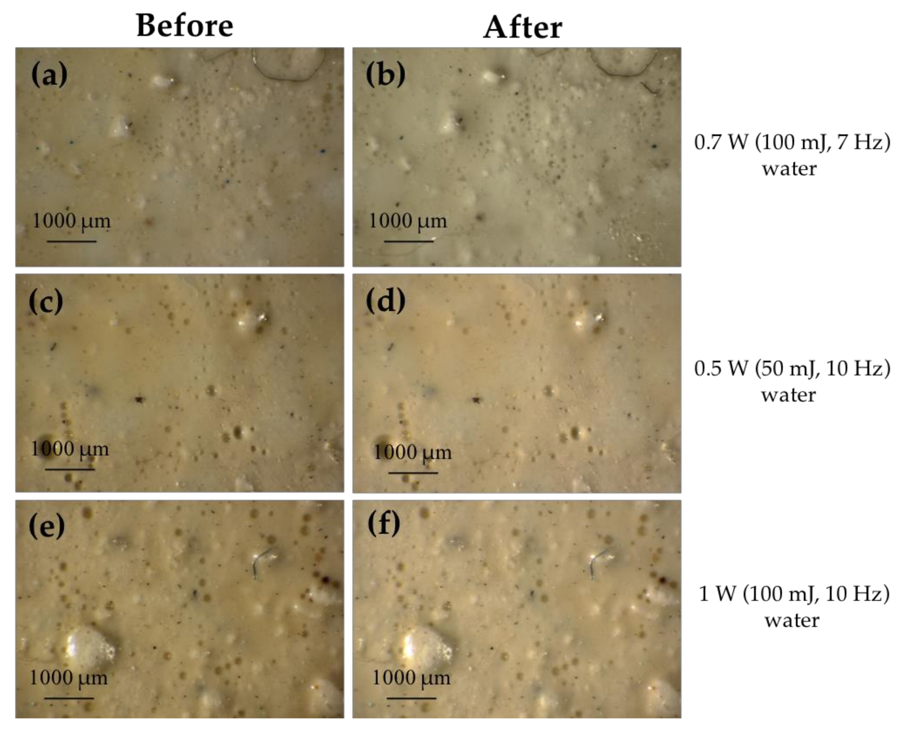
Figure 5.
The microscopic images (32×) of P B67 removal by Er:YAG laser under varied conditions and parameters, conducted on cinnabar tempera mock-ups. (a,c,e) was obtained before cleaning, while (b,d,f) was obtained after cleaning in dry and wet conditions with 1 W as laser power.
Figure 5.
The microscopic images (32×) of P B67 removal by Er:YAG laser under varied conditions and parameters, conducted on cinnabar tempera mock-ups. (a,c,e) was obtained before cleaning, while (b,d,f) was obtained after cleaning in dry and wet conditions with 1 W as laser power.

Figure 6.
The microscopic images (32×) of P B67 removal by Er:YAG laser under varied conditions and parameters, conducted on lead white tempera mock-ups. (a,c,e) was taken before cleaning, while (b,d,f) was obtained after cleaning in dry and wet conditions with different laser power (1 W and 0.5 W).
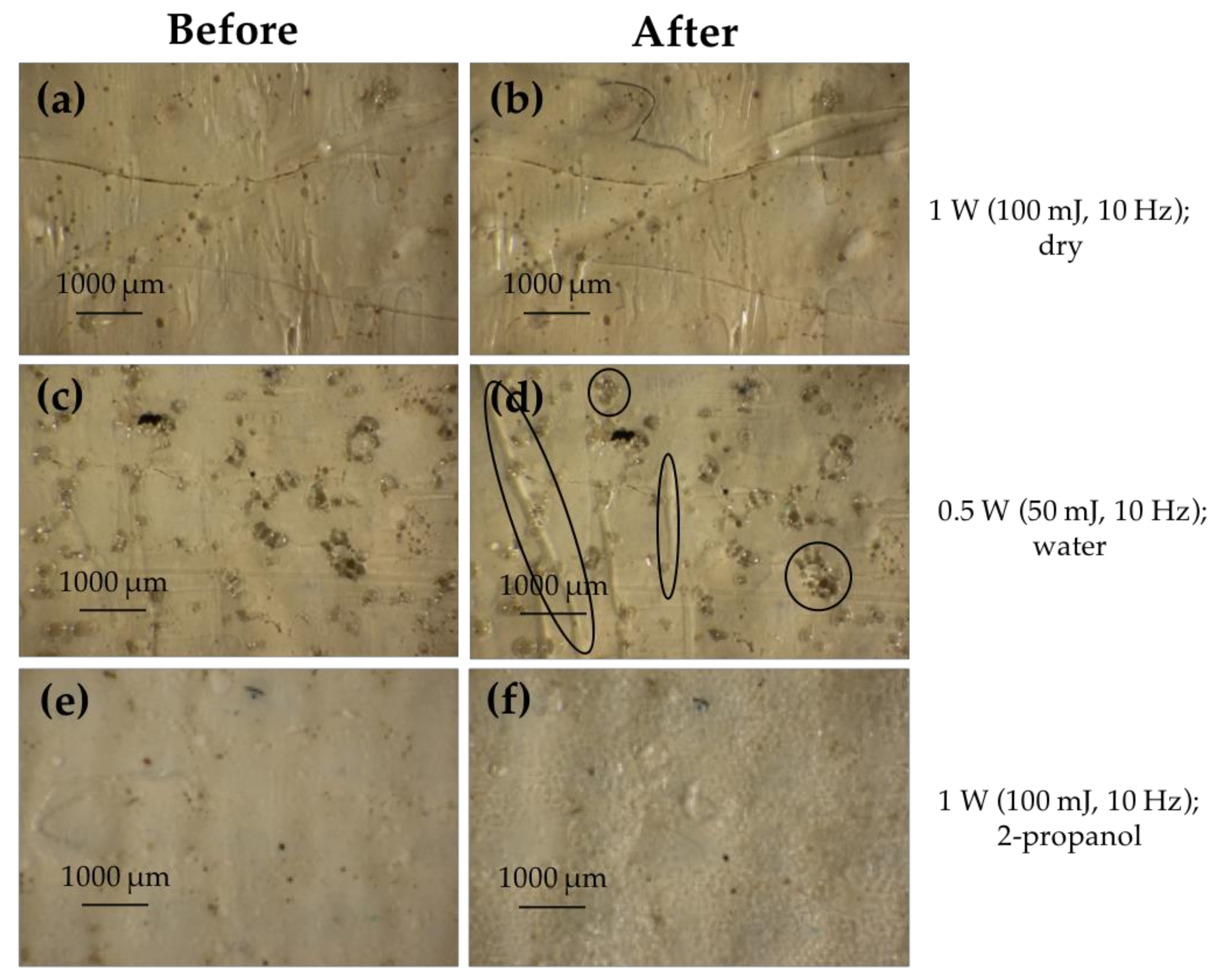
Figure 7.
The full range reflectance spectrum (a) and the selected region reflectance spectrum (b) of pure mastic, cinnabar tempera mock-up (P), cinnabar tempera varnished with mastic (VP) and mock-up after laser cleaning (AL). All spectra reported were collected in Section 8 of painting mock-ups (Figure 1).

Figure 8.
The full range reflectance spectrum (a) and the selected region reflectance spectrum (b) of pure mastic, lead white tempera mock-up (P), lead white tempera varnished with mastic (VP) and mock-up after laser cleaning (AL). All spectra reported were collected in Section 8 of painting mock-ups (Figure 1).
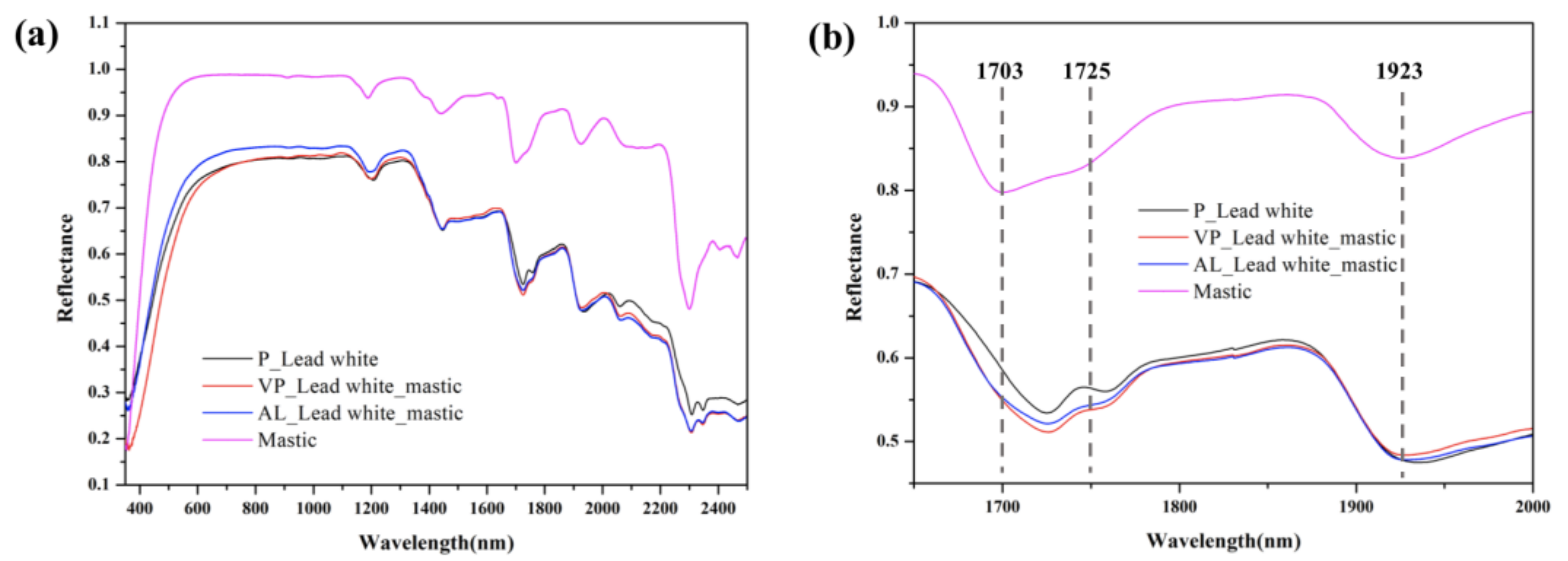
Figure 9.
The full range reflectance spectrum (a) and the selected region reflectance spectrum (b) of pure P B67, cinnabar tempera mock-up (P), cinnabar tempera varnished with P B67 (VP) and mock-up after laser cleaning (AL). All spectra reported were collected in Section 8 of painting mock-ups (Figure 1).
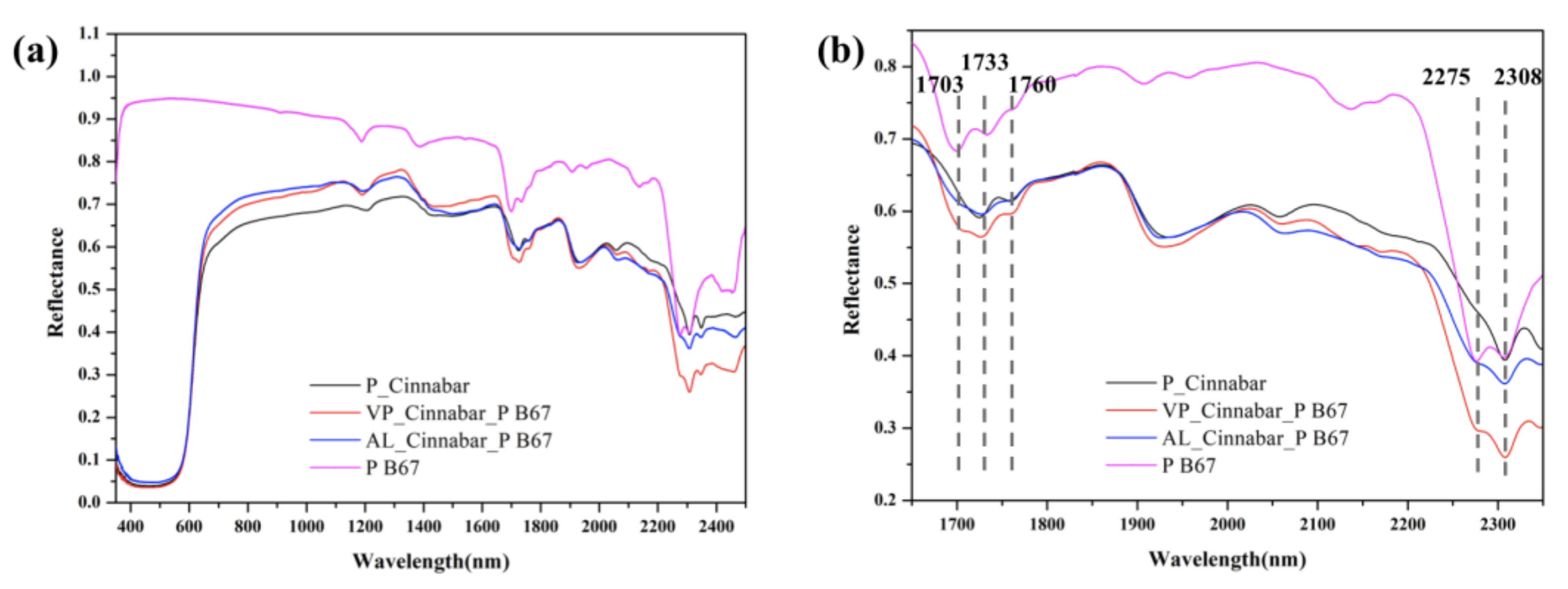
Figure 10.
The full range reflectance spectrum (a) and the selected region reflectance spectrum (b) of pure P B67, lead white tempera mock-up (P), lead white tempera varnished with P B67 (VP) and mock-up after laser cleaning (AL). All spectra reported were collected in Section 8 of painting mock-ups (Figure 1).

Figure 11.
The microscopic images (32×) of the eight sections of mastic varnished cinnabar tempera mock-ups after different laser cleaning conditions and parameters.
Figure 11.
The microscopic images (32×) of the eight sections of mastic varnished cinnabar tempera mock-ups after different laser cleaning conditions and parameters.

Figure 12.
The microscopic images (32×) of the eight sections of mastic varnished lead white tempera mock-ups after different laser cleaning conditions and parameters.
Figure 12.
The microscopic images (32×) of the eight sections of mastic varnished lead white tempera mock-ups after different laser cleaning conditions and parameters.
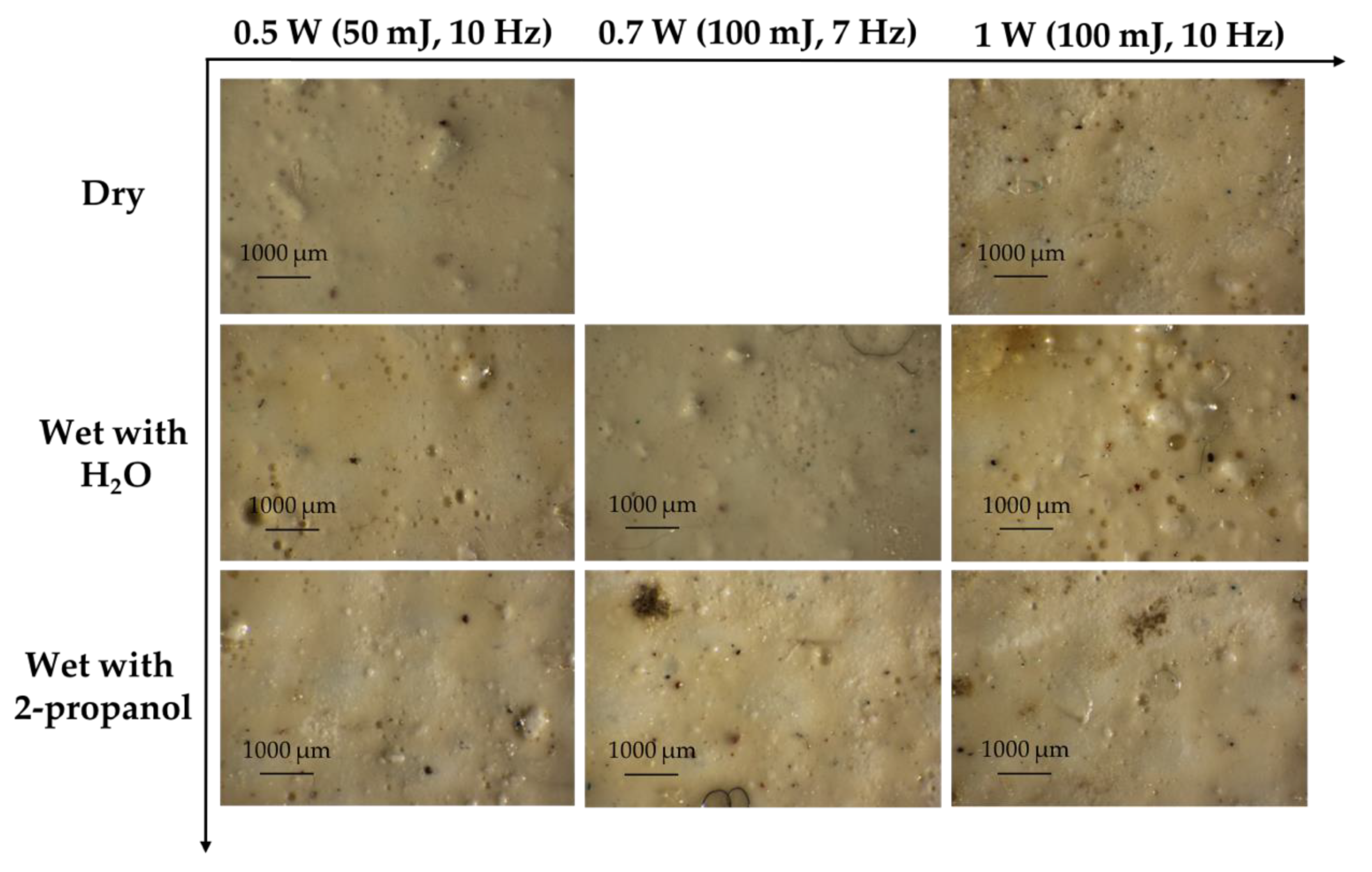
Figure 13.
The microscopic images (32×) of the eight sections of P B67 varnished cinnabar tempera mock-ups after different laser cleaning conditions and parameters.
Figure 13.
The microscopic images (32×) of the eight sections of P B67 varnished cinnabar tempera mock-ups after different laser cleaning conditions and parameters.

Figure 14.
The microscopic images (32×) of the eight sections of P B67 varnished lead white tempera mock-ups after different laser cleaning conditions and parameters.
Figure 14.
The microscopic images (32×) of the eight sections of P B67 varnished lead white tempera mock-ups after different laser cleaning conditions and parameters.


Table 1.
Mass of pigments, egg yolk and water used in mock-up preparation.
| Pigment | Mass of Pigment (g) | Mass of Egg Yolk (g) | Mass of Water (g) |
|---|---|---|---|
| Cinnabar | 3.529 | 3.528 | 0.700 |
| Lead white | 3.589 | 3.960 | 0.800 |
Publisher’s Note: MDPI stays neutral with regard to jurisdictional claims in published maps and institutional affiliations. |
© 2021 by the authors. Licensee MDPI, Basel, Switzerland. This article is an open access article distributed under the terms and conditions of the Creative Commons Attribution (CC BY) license (https://creativecommons.org/licenses/by/4.0/).
Share and Cite
MDPI and ACS Style
Wang, C.; Cao, Y.; Tie, F.; Camaiti, M. Er:YAG Laser Cleaning of Painted Surfaces: Functional Considerations to Improve Efficacy and Reduce Side Effects. Coatings 2021, 11, 1315. https://doi.org/10.3390/coatings11111315
AMA Style
Wang C, Cao Y, Tie F, Camaiti M. Er:YAG Laser Cleaning of Painted Surfaces: Functional Considerations to Improve Efficacy and Reduce Side Effects. Coatings. 2021; 11(11):1315. https://doi.org/10.3390/coatings11111315
Chicago/Turabian StyleWang, Cong, Yijian Cao, Fude Tie, and Mara Camaiti. 2021. "Er:YAG Laser Cleaning of Painted Surfaces: Functional Considerations to Improve Efficacy and Reduce Side Effects" Coatings 11, no. 11: 1315. https://doi.org/10.3390/coatings11111315
Note that from the first issue of 2016, this journal uses article numbers instead of page numbers. See further details here.