Article

Corrosion and Biocompatibility of Pure Zn with a Micro-Arc-Oxidized Layer Coated with Calcium Phosphate

1
Department of Materials Science and Engineering, Taiyuan University of Science and Technology, Taiyuan 030024, China
2
Key Laboratory of Marine Materials and Related Technologies, Zhejiang Key Laboratory of Marine Materials and Protective Technologies, Ningbo Institute of Materials Technology and Engineering, Chinese Academy of Sciences, Ningbo 315201, China
3
Ningbo Powerway Alloy Material Co., Ltd., Ningbo 315145, China
4
Ningbo Medical Center Lihuili Hospital, Ningbo 315040, China
5
Department of Bioengineering, University of California, Riverside, CA 92521, USA
*
Authors to whom correspondence should be addressed.
Coatings 2021, 11(11), 1425; https://doi.org/10.3390/coatings11111425
Submission received: 9 October 2021 / Revised: 1 November 2021 / Accepted: 18 November 2021 / Published: 22 November 2021
(This article belongs to the Special Issue Surface Function Enhancement Film and Coating Technology)

Abstract

:
Recent studies have indicated a great demand to optimize the biocompatibility properties of pure Zn as an implant material. For this purpose, CaZn2(PO4)2·2H2O (CaZnP) was prepared using hydrothermal treatment (HT) combined with micro-arc oxidation (MAO) on pure Zn substrate to generate biodegradable implants. The polarization test and electrochemical impedance spectroscopy indicated that the MAO1−HT coating could modulate the corrosion behavior of MAO1 by filling the crevice between the coating and the substrate. Immersion test evaluation revealed that the osteogenic properties of MAO1−HT coating were better than that of pure Zn substrate, as evidenced by the molar ratio of Ca and P, which increased after soaking in simulated body fluid (SBF) for up to 10 days. In addition, L-929 cells cultured in the 100%, 50%, and 25% extracts of MAO1−HT coated samples exhibited excellent cytocompatibility. Meanwhile, cell adhesion was promoted on the surface with high roughness generated during MAO and HT processes. In summary, the calcified coatings improved biocompatibility and adjusted the degradation rates of pure Zn, broadening the application of Zn alloys.

Graphical Abstract

1. Introduction

The increasing demand for biodegradable implants in aseptic medical surgeries has led to the introduction of completely degradable materials to eliminate the need for the secondary surgical removal of permanent implants when they are no longer necessary [1,2,3,4]. Biodegradable implant materials should meet the following conditions [5,6,7,8,9,10]. Firstly, they should have good biocompatibility with human tissues, with good bioactivity during implementation and degradation processes, i.e., degradation products must not harm the human body and should not affect normal metabolic activities. Secondly, they should have a certain degree of chemical stability and corrode evenly, which will not be affected by biological enzymes.
Mg-based structural alloys are promising candidates due to their low density, natural biodegradability, and good biocompatibility [11,12,13]. However, the high degradation rate of Mg (electronegative potential of −2.37 VNHE) poses severe safety threats, hindering their clinical use. Although researchers have undertaken considerable research in recent years, they have failed to solve the problem of rapid corrosion. In comparison with the high electronegative potential of Mg, studies showed that that of Zn (−0.76 VNHE) does not go rapidly and creates hydrogen gas [14]. Therefore, this key problem might be solved by a zinc-based alloy. Moreover, Zn is an essential trace element required for the human body and plays a vital role in basic biological functions, such as collagen synthesis and osteoblast activity [7,8]. In addition, Zn has antimicrobial and biological properties that considerably reduce the risk of postoperative infection [11] Therefore, the development of degradable Zn for internal fracture fixation, vascular stents, and other implanted medical devices has good prospect. Furthermore, studies have found that Zn ions could react with sulfhydryl groups to inhibit bacterial activities such as glycolysis, transmembrane proton transfer, and acid resistance [15,16,17]. Therefore, scientists have focused on developing degradable Zn alloys as promising medical alloys for internal fracture fixation and as vascular stents and other implanted medical devices. Some literature [8,9,18] has been concerned with the corrosion and biomedical properties of emerging degradable-based alloys such as Zn-Mg, Zn-Ca, and Zn-Sr. However, Zn could apparently inhibit cells, resulting in a decrease in size and cyto-activity after being exposed to Zn. This may be another important factor contributing to low its biocompatibility. However, research on the relatively long degradation time, biocompatibility, and degradation mechanism of biodegradable Zn-based alloys is still in its infancy, and a great deal of basic research is required.
As an effective, rapid, and low-cost method, surface modification technologies, such as the sol-gel method, electrochemical deposition, plasma spraying, micro-arc oxidation (MAO), and hydrothermal treatment (HT), have been established to optimize the corrosion property and biocompatibility of alloys [19,20,21,22,23,24,25,26]. Among these technologies, MAO is a simple, effective, and rapid method for improving the corrosion properties of alloys [27,28]. Usually, MAO coatings with a porous structure consist of oxide and other amorphous phases [29,30], which can improve and increase surface roughness to provide a greater surface area, increasing bone-implant contact [31,32]. In addition, previous research has shown that MAO treatment is beneficial in accelerating the corrosion of an Mg alloy after implantation.
It has been reported that surface engineering of calcium phosphate (CaP) coating can significantly improve the adhesion and biocompatibility of cells because of their inherent bone tissue compatibility [33,34,35,36,37]. Preparing CaP bioceramics through the HT process effectively improves biological activity [38,39,40,41]. During the HT process, the elements Ca and P from the aqueous solution could reprecipitate on the MAO coating surface to form CaP. Nevertheless, limited literature is available on the corrosion and biocompatibility response of Zn alloys used in the HT process. Su et al. [14] first reported zinc phosphate (ZnP) coatings on pure Zn using a chemical conversion method. ZnP coating significantly increased the viability, adhesion, and biocompatibility of cells. Yang et al. [18] prepared Zn-hydroxyapatite composites and improved biocompatibility and osteogenesis. Therefore, none of the reports on MAO-HT of Zn coating materials were targeted on the biocompatibility of Zn in the physiological environment and its further biomedical applications.
In this work, a novel composite CaZnP coating was produced on the surface of pure Zn using MAO and HT treatment to develop good biocompatibility and moderate degradation of the Zn compound and its microstructure. In addition, the chemical composition, corrosion behavior, and biocompatibility of Zn with and without CaZnP coating were compared as biodegradable implants for medical treatment.

2. Materials and Methods

2.1. Material Preparation

Pure Zn sample (99.99%, obtained form Powerway Alloy Material Co., Ningbo, China) with the size of 0.785 cm2 × 4 mm was prepared by wire-electrode cutting. The sample was first soaked with de-oiling agent, ultrasonically cleaned to remove the oil. Afterwards, the Zn samples were ground using 400 to 2000 grit of SiC sandpaper in an orderly manner, washed with deionized water and decontaminated with anhydrous ethanol. The MAO process was carried out in electrolyte composed of Na2SiO3 (15 g/L) and NaOH (3 g/L) under the voltage, applied duty cycle, pulse frequency, and oxidation time were 450 V, 10%, 2000 Hz, and 20 min, respectively. During the MAO treatment, Zn samples were used as anodes, and a stainless-steel water tanks were used as cathodes in the electrolyte bath. A stirring and an external circulating water cooling system was installed to prevent the temperature from being too high. The sample used for HT was named MAO1.
Then, the MAO1 coating was placed in a 50 mL autoclave which was placed in a drying oven (Saidelisi, 101-0AB, SAIDELISIS, Tianjin, China) for the HT using temperature of 110 °C and a holding time of 16 h. The CaP precursor is composed of Ca(NO3)2 0.168 mol/L and NaH2PO4 0.1 mol/L 50 mL with pH value adjusted to 3. After HT, the samples were washed using distilled water two times and dried at room temperature. The treated sample was called MAO1−HT.

2.2. Microstructure and Composition Characterization

Scanning electron microscopy (SEM, Sirion 200, FEI, Portland, OR, USA) and energy dispersive X-ray spectroscopy (EDS) were used to examine the surface, morphologies, and the concentration of the elements in the MAO1 and MAO1−HT coatings, respectively. The composition of both coatings were detected by X-ray diffraction (XRD, D8 ADVANCE DAVINCI, BRUKER, Karlsruhe, Germany) using Cu Kα with a scanning range of 10–80° for 23 min and Fourier-transform infrared spectrometer (NICOLET 6700, Thermo, Waltham, MA, USA), in the range of 4000−400 cm−1 with a resolution of 2 cm−1 for the determination of functional groups. X-ray photoelectron spectrometer (XPS, Axis Ultra DLD, Kratos, Manchester, UK) was used to characterize the coating surface chemical states. ICP-OES (SPECTRO ARCOS Ⅱ, SPECTRO, Cleves, Germany) was used to measure the concentrations of Zn, Ca, and P in the extract culture solution. The standard samples with different concentrations of Ca, P, and Zn were configured. The extracted culture solution of pure Zn and MAO1−HT coatings were measured three times.

2.3. Electrochemical Tests

Electrochemical corrosion tests were conducted using an electrochemical workstation (AUTOLAB-PGSTAT302, Metrohm, Switzerland), with simulated body fluid (SBF) as the electrolyte (NaCl 8.035 g/L, NaHCO3 0.355 g/L, KCl 0.225 g/L, K2HPO4·3H2O 0.231 g/L, MgCl2·6H2O 0.311 g/L, CaCl2 0.292 g/L, Na2SO4 0.072 g/L, TRIS 6.118 g/L, and HCl 1 mol/L), at pH = 7.40 and a temperature of 37.5 ± 0.5 °C. The above substrate was the working electrode, the platinum electrode was the counter electrode, and the saturated calomel electrode was the reference electrode. Electrochemical impedance spectroscopy (EIS) was performed within the frequency range of 100 kHz to 10 mHz and the potential perturbation amplitude of ±10 mV. In the experiments, each group had five samples, with a surface area of 0.785 cm2. Initially, the sample was immersed in SBF for 30 min to stabilize the open circuit potential (OCP). Subsequently, the working electrode was scanned over the voltage range of ±1 V (versus OCP) at a scan rate of 0.5 mV/s. Tafel extrapolation method was used to measure the degradation parameters, including the corrosion potential (Ecorr) and the corrosion current density (icorr).

2.4. Immersion Tests

The uncoated and coated samples’ immersion tests were conducted in the SBF ingredients at 37 ± 0.5 °C which were similar to the human body fluid [42,43]. The SBF solution was updated daily to ensure that ions do not affect the formation of CaP. After soaking for 1, 3 and 10 days, the samples were cleaned with distilled water and dried at 37 °C. The pH value of the solution was monitored during the immersion tests. The surface morphologies and phase composition of the coating were analyzed by SEM and XRD to determine the corrosion products.

2.5. In-Vitro Tests

Murine fibroblast (L-929) was adopted to evaluate the cytocompatibility of uncoated and coated samples. The procedure of cell culture can be found in previous publications [14]. Briefly, for the indirect assays, Dulbecco’s Modified Eagle Medium (DMEM, ATCC, Manassas, VA, USA) with 10% serum was used as culture medium for L-929. Extract media were prepared by incubating samples in the cell culture media at a ratio of 1.5 mL/cm2 for 3 days. Subsequently, 2, 1 and 0.5 mL of the culture media were extracted to form solutions with 100%, 50%, and 25% concentration, respectively. The cell viability was measured by Methyl Thiazolyl Tetrazolium (MTT) assay with a plate reader (Tecan spark, Tecan, Bienne, Switzerland) at 550 nm. MTT refers to the standard test for screening the toxicity of biomaterials and is carried out in accordance with GB/T 16886.5−2003 standard. L-929 cells were seeded in the samples placed in 96-well plates at a density of 1 × 10⁵/mL and cultured in different concentrations of extracted culture solution for 1, 2 and 3 days. Culture media without extract media were used as the control. The cell morphologies in the third day were observed under a metallographic microscope (Nikon eclipse Ti 2-U, Nikon, Tokyo, Japan).

2.6. Wettability Measurement

The contact angles of water on pure Zn and calcified coating surfaces were evaluated using a contact angle measuring instrument (DCAT21, Dataphysics, Ningbo, China) equipped with a digital camera and analysis software (DCATS, 32). Droplets of pure water with volumes of 1 μL were used. Three points were measured for each coating surface and the results were expressed as mean values and standard deviations.

3. Results and Discussion

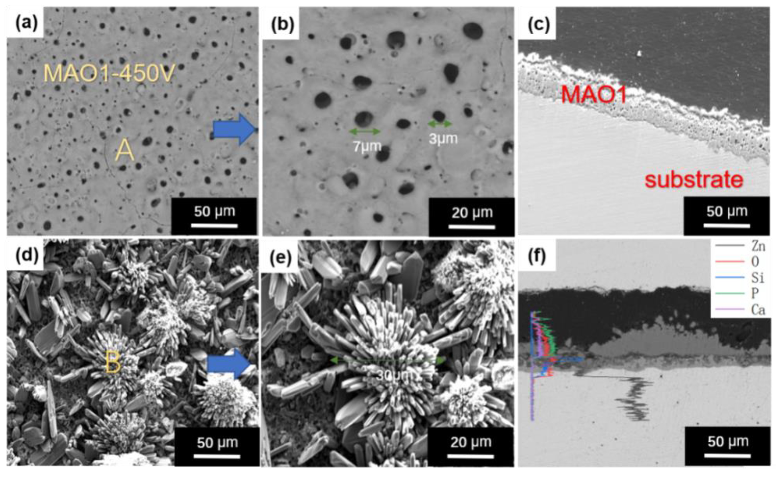
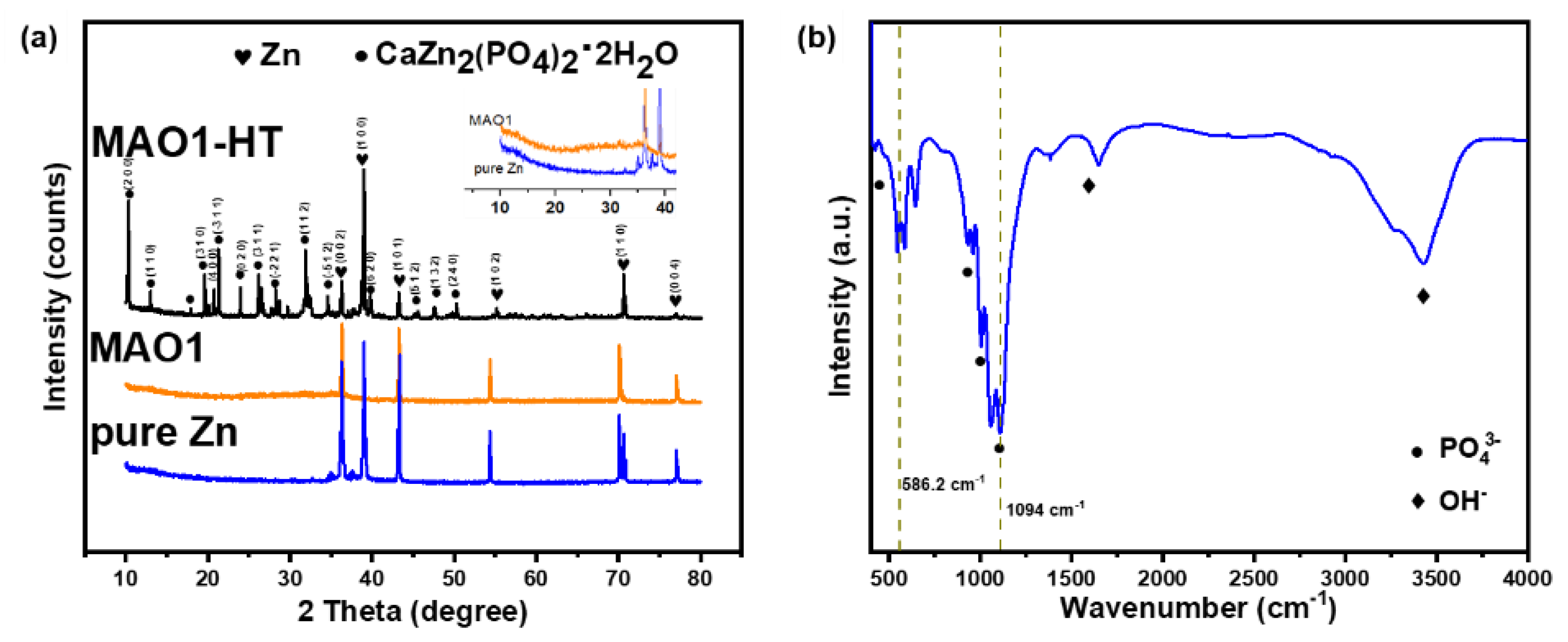
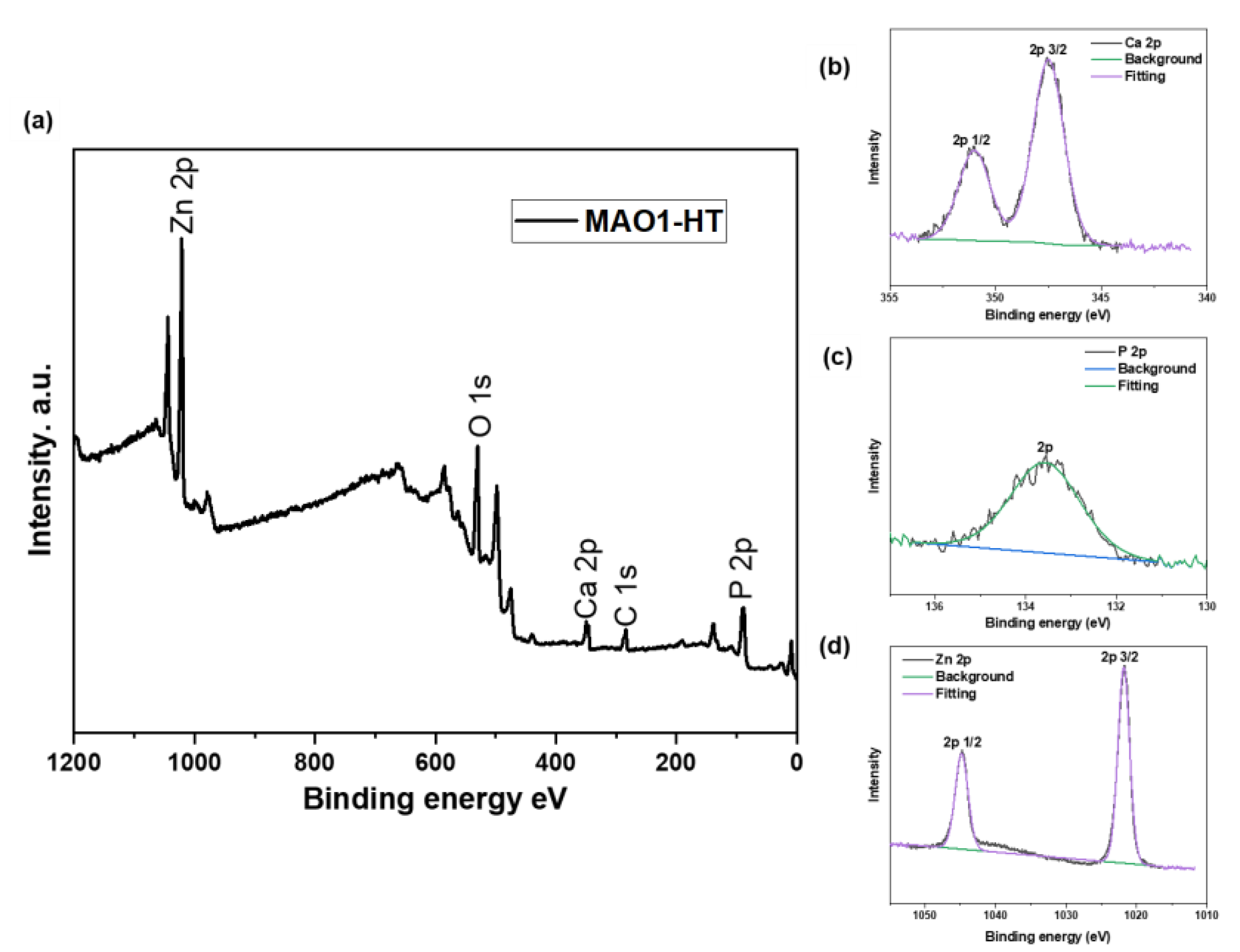
3.1. Microstructure and Composition Identification of MAO Coating before and after HT

Figure 1 presents a comparison of the microstructure and morphologies of MAO1 and MAO1−HT coatings. Figure 1a–c shows that the coating turned smooth with an increased thickness of approximately 40 µm, and tiny pores appeared on the surface after MAO, some of which were connected and the size of these pores was approximately 2–8 µm, and there were some tiny cracks caused by the thermal effect. Figure 1d–f shows the surface and cross-sectional morphologies of MAO1−HT coating.
The microcrystals measured approximately 25–50 µm, appearing as a layered open growth model. Furthermore, the microcrystals with flower-like shapes consisted of rods closely and densely spaced over the surface. Both the MAO1 and MAO1−HT coatings did not exhibit any clear boundary between the two layers. The corresponding elements of areas A and B in Figure 1, as identified by EDS are shown in Table 1. The MAO1 coating was mainly composed of Zn, O and Si from the electrolyte and small amounts of Si were harmless. In fact, a trace amount of Si has been suggested to be essential for the human body [44]. However, MAO1−HT coating mainly consisted of O, Ca, Zn, P, and Si. The Ca/P molar ratio of calcification coatings was approximately 1.1, which is lower than that of the main component of human bone, i.e., hydroxyapatite (1.67).
Figure 2a shows the XRD patterns of the phase components of Zn substrate before and after MAO and MAO-HT processes. The strength of peaks corresponding to Zn was evident, indicating that the oxide film was thin with a porous structure, through which X-rays could pass. However, peaks corresponding to the ZnO were not identified. The curves of pure Zn and MAO1 were inconsistent from 2θ = 20° to 35°, and the MAO1 curve showed an upward trend. A plausible explanation is that ZnO exists in an amorphous state because of rapid solidification by a relatively cool electrolyte. No noticeable change could be observed in the XRD of pure Zn and MAO1. However, after MAO-HT treatment, the Zn peak intensities decreased significantly, and many CaZn2(PO4)2(CaZnP, PDF, No. 35-0495, Parascholzite) peaks appeared, corresponding with the micro-flower morphology in Figure 1d. Mardziah confirmed that Zn ion participates in crystallization during Cap preparation [43]. Considering the number of CaZnP diffraction peaks, the petaling structure was exactly CaZnP.
Figure 2b presents the FT-IR plots of the MAO1−HT coating that match the earlier report [45,46]. The CaZnP molecular identification was associated with a phosphate (PO43−) functional group with four different vibrational modes. At a wavelength of 956 cm−1, the functional group of PO43− corresponded to V1. Other vibration modes of the PO43− functional group were observed at 473 (V2), 1094 (V3), and 568–601 (V4) cm−1. Both cationic and anion substitutions in phosphate can cause Fourier transform spectral changes, such as widening of absorption band and shifting of position. In particular, the relatively higher peak at 600 cm−1 was indicative of CaP [47]. The OH function group was observed at 3500 and 636 cm−1, corresponding to liberation and stretch modes, respectively.
On the other hand, as confirmed by the CaZnP structure in Figure 3, XPS spectrum and high-resolution spectra of P 2p and Ca 2p detected from MAO1−HT coating demonstrated that Ca 2p peaks were assigned to 347.1 (2p 3/2) and 351 eV (2p 1/2), respectively, consistent with the Ca in CaZnP. Meanwhile, the P 2p peak was detected at 133.4 eV, corresponding to the peak of CaZnP [29]. As shown in Figure 3d, the binding energy peaks of 1022.2 and 1045.1 eV correspond to Zn 2p 1/2 and Zn 2p 3/2, respectively. Therefore, the Zn in coating consists of Zn ions, which are incorporated with that in CaZnP.

3.2. Electrochemical Characterization

Figure 4a shows the OCP evaluation of the pure Zn, MAO1, and MAO1−HT coatings as a function of immersion time. The OCP values for the MAO1 and MAO1−HT coatings were slightly lower than the pure Zn. Although pure Zn was the least positive from the beginning, it reached the highest after 100 min. The rapid increase of pure Zn in OCP towards the higher potentials at the initial immersion stage indicated the formation of a certain kind of dense passive oxide layer on the surface. In the sample treated with MAO, the incompactness of the inner layer would lead to local corrosion of the substrate. Both the MAO1 and MAO1−HT coatings exhibited a decrease in their OCPs during the early immersion time. However, the potential stabilization was achieved after 200 min. Furthermore, the fluctuation in the OCP of coated samples was comparatively less than pure Zn.
Figure 4b shows the polarization curves of the uncoated and coated Zn in SBF at 37 ± 0.5 °C. Compared to the pure Zn sample, the corrosion potential of MAO1 and MAO1−HT samples shifted negatively. From a thermodynamics viewpoint, the polarization curves of coated samples exhibited high degradation tendency, with minimal current plateaus, indicating no obvious passivation during the corrosion process. Table 2 lists the calculated electrochemical parameters in this study. A significant increase in icorr for the sample with MAO1 coating was observed in contrast to pure Zn. As a matter of fact, electrochemical corrosion refers to the crevice corrosion between the coating and the substrate. The MAO inner layer provides a channel for the SBF to be transported to the substrate to form a local corrosion cell, leading to the acceleration of corrosion rate, as indicated by the electrochemical test. However, the corrosion rate of the MAO1−HT sample was lower than that of pure Zn, which indicated that the HT process could effectively fill the pores and cracks of the inner layer which generated during MAO treatment.
EIS measurements of pure Zn, MAO1, and MAO1−HT were conducted in SBF to further understand the corrosion resistance of the coatings. Figure 4c presents the coated and uncoated Nyquist diagrams. A qualitative comparison of the plots revealed that the semicircle curves of MAO1 had a smaller radius. In the matrix impedance diagram, two different capacitance loops corresponding to two different constant times were produced by pure Zn. The first one occurred at a high frequency, indicating the charge transfer during Zn corrosion. The second one occurred at the low-frequency region and was represented by the diffusion process through the corrosion layer or surface oxide. On the other hand, the coated samples showed capacitive loops at all frequencies with weak loops in the high frequencies. Different capacitance loops appeared at lower frequency, indicating that the MAO1 layer provided a higher charge transfer rate than others. The high frequency of time constant is related to the outer layer, and the other one at the low frequency is due to the inner layer of the MAO coating. The coated and uncoated samples impedance data were analyzed by the equivalent circuit presented in Figure 4d, two sets of circuits were used for the analysis. For pure Zn and MAO1, the circuit (a) was used whereas the impedance data of MAO1−HT was used (b). The both circuits were based on their double-layer structure, Rs stands for the resistance of electrolyte between the working and reference electrodes, R1 is the resistance of the outer layer parallel to constant phase element Q1, and Rct is the resistance of the inner layer. For MAO1 and MAO1−HT, Q1 represent the capacitance of outer layer (ZnO/CaZnP), Qdl corresponds to the capacitance of the double layer (inner layer and substrate). “n” is the dispersion index in the calculation of constant phase element Q1, which value varies from 0 to 1. The electrochemical results are also represented in Bode plots, in Figure 4e,f, respectively. The high frequency region in Figure 4e represents Rs. Pure Zn and coated samples were all showed resistive behavior in the low frequency region. In contrast, resistive behavior of pure Zn was lower than MAO1−HT. Phase angle vs. log f curves showed (Figure 4f) two time constants for each samples. However, for MAO1 and MAO1−HT, time constants were seen at high frequencies and lower frequencies whereas pure Zn seen at medium frequencies. Normally, the high frequency capacitive loop corresponds to the porous coating. Table 3 lists the EIS fitted values of the equivalent circuit parameters. A comparison of the results revealed that the resistance of the inner layer in both samples was higher than that of the outer layer.

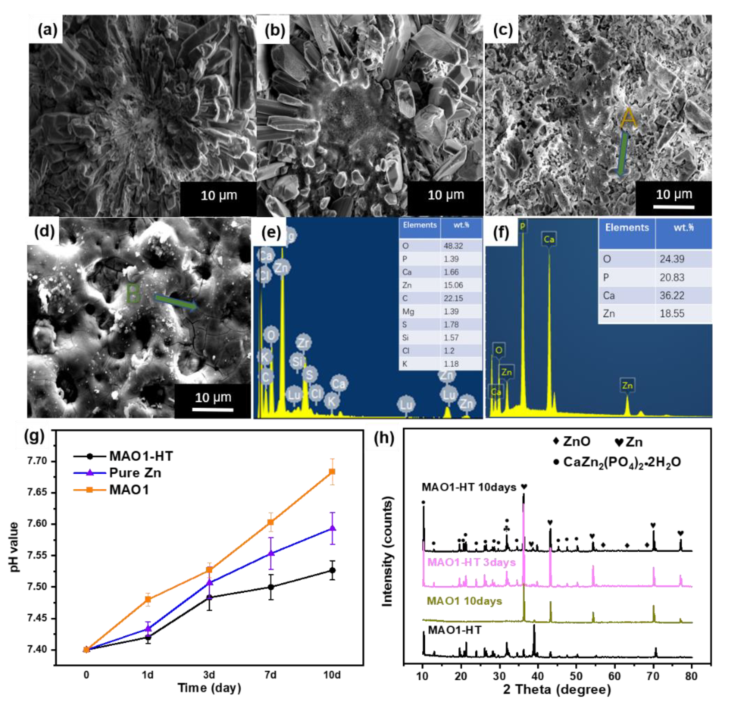
3.3. Immersion Tests

SBF has widely been used to evaluate the in-vitro activity of materials, becoming one of the most advantageous criteria [42,43]. In this case, the CaZnP formation capacity of the coating in SBF was regarded as an indicator of cytocompatibility. Figure 5 shows the surface images of the coated samples soaked in SBF for 1, 3 and 10 days. Figure 5a shows that there was no particle precipitated on surface. The original CaZnP crystals near center may dissolve or fall off; only a few petals were corroded. By increasing the immersion duration to 3 days, more CaZnP disappeared as the central range expands. However, with an increase in soaking time to 10 days, there was apparent corrosion on the surface of the MAO1−HT, and the flower-like center was corroded and dissolved into SBF, where it was replaced by the formed flocculation. Corresponding to Figure 5f,h, the EDS analysis of area A revealed that the molar ratio of Ca/P was approximately 1.36. The comparison of the chemical composition of MAO1−HT before and after the immersion test showed increased amounts of Ca and P, suggesting that the MAO1−HT coating induced an increase in the calcium-to-phosphorus ratio on the surface during immersion in SBF. XRD results revealed that the dominant component of the flocculation on the MAO1−HT surface was CaZnP again, and small amounts of ZnO appeared. Compared with the intensity of MAO1−HT, Zn peaks increased clearly at 36°, 43°, and 70°, indicating a decreased thickness of MAO1−HT and penetration of radiation into the newly deposited CaZnP. Moreover, a few brighter particles precipitated on MAO1, mostly in defect regions, such as small micro-cracks. According to EDS analysis of area B in Figure 5e, most of these precipitated corrosion products were not CaP.
Figure 5g shows the variations of pH value during the 10-day immersion in SBF solution. The pH value of all the samples increased in less than three days, and only the pH value of the MAO1−HT sample increased relatively slowly in the late immersion period, and the final mean value reached 7.51. The corrosion mechanism can be explained by the electrochemical reaction of the cathodic reduction of oxygen and anodic oxidation of Zn after being immersed in nearly neutral physiological SBF [29]. The pH value of MAO1 increased to 7.68 at the end of immersion, demonstrating a higher reactivity in SBF compared with MAO1−HT and pure Zn.

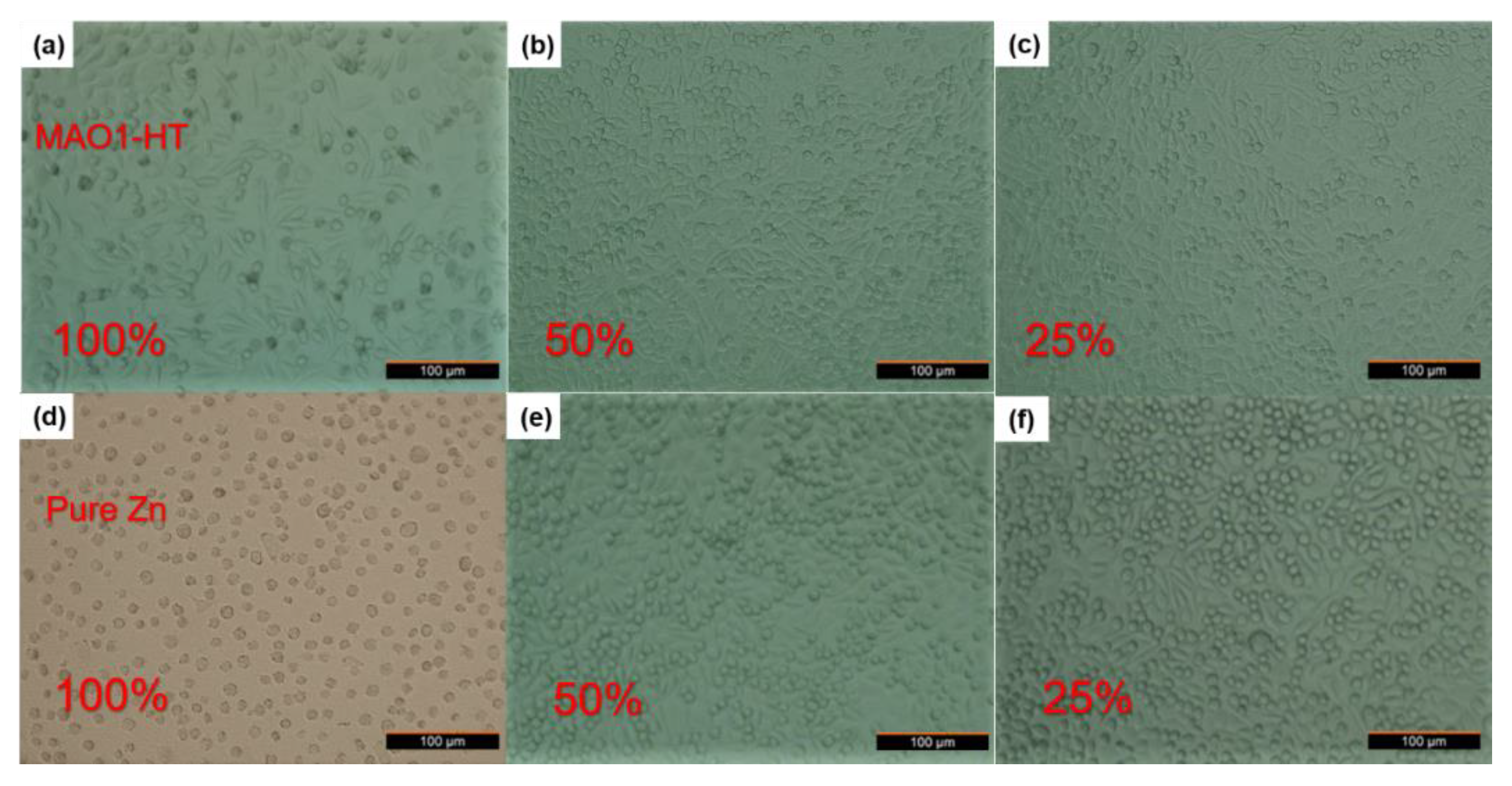
3.4. Cytotoxicity and Cell Morphology
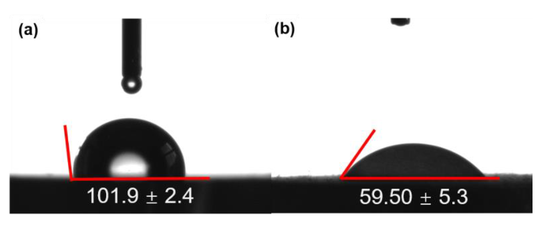
Figure 6 shows the relative growth rate (RGR) of L-929 cells after 1, 2, and 3 days of culture in pure Zn and MAO1−HT extract culture solution to express cell viability. The cell RGR of MAO1−HT at 100%, 50%, and 25% extract concentrations was higher than that with pure Zn. However, the cell survival was relatively good on day 2, which decreased on day 3. This finding could be attributed to the Ca and P contents in the MAO1−HT extract culture solution. The cells in the MAO1−HT groups grew more quickly than those in the pure Zn groups, reaching their peaks earlier. In the 50% and 25% extracts of pure Zn, the RGR of cells in the Zn groups also showed good biocompatibility, indicating that small amounts of Zn ions could promote cell growth, consistent with the fact that the concentration of Zn ions during metabolism would not be too high, in in-vivo experiments. Therefore, the MAO1−HT coating exhibited good biocompatibility with L-929 cells.
Figure 7 shows the cell morphologies of L-929 cells on day 3 of culture in pure Zn and MAO1−HT extract culture solutions. The cell growth in the MAO1−HT group was better than in the pure Zn. For 100% extracts, most cells in the pure Zn extract solution died, indicating apparent cytotoxicity, whereas those in the MAO1−HT extract solution survived and were in good shape. In the culture solutions of 50% and 25% concentrations, the cells in the pure Zn groups showed polycondensation shapes, whereas those in the MAO1−HT groups showed leaf-like shapes. The cell morphology in MAO1−HT groups revealed a better form, i.e., various shapes with good activity due to the influence of Ca and P ions. The formation of CaP crystals on the surface was conducive to cell growth; even when the release of Zn ions, the cells were not affected because of the dissolution of a large amount of CaZnP. Based on the results of ICP-OES (Table 4), the concentration of Zn ions in MAO1−HT 100% extract culture solution was higher than that in pure Zn-soaked solution.

3.5. Wettability

The wettability of the implant is the decisive factor for the growth of osteoblasts. A large number of reports have shown that the surface of biomaterials with moderate hydrophilicity can promote cell growth and have high biocompatibility. Figure 8 shows the contact angle of water on pure Zn and MAO1−HT coatings. The results indicated that the contact angle of the MAO1−HT was 59° compared with 101° in Zn when water was used
Coincidentally, the wettability threshold of ≤60° is considered hydrophilic, and cell attachment is favored [48,49]. Therefore, the electro-negatively charged PO43− ions in the coatings strongly attract the electro-positively charged H ions and the water molecules in the surrounding medium to improve cell adhesion.

4. Conclusions

The corrosion and biocompatibility of pure Zn with MAO layer combined calcium phosphate coating were studied to explore alternative materials for biodegradable implant alloys in the medical field. The following conclusions can be drawn from the current results:
  • A dual-layer structure MAO1−HT coating consisting of ZnO and CaZnP processed under MAO and HT could be an available surface modification technique. Sufficient dense and rich CaZnP formed on the dual-layer MAO1−HT coating, which is a practical approach to adjust the degradation rate and improve biocompatibility.
  • The MAO1 coating could accelerate the corrosion of Zn because of the crevice corrosion between the substrate and the coating. The corrosion performance confirmed by electrochemical measurements could be ranked as: MAO1−HT > pure Zn > MAO1.
  • Cytotoxicity was detected in different extract groups, and each group revealed excellent cytocompatibility by MAO-HT treated Zn, whereas pure Zn in low concentration of extract. In addition, the wettability of MAO1−HT treated Zn with a contact angle of 59° also promoted cell adhesion compared with pure Zn with a contact angle of 101°.

Author Contributions

Conceptualization, L.Y. and Z.S.; writing of the article, Y.S. and Q.Z.; design of experiments, T.L., X.Z., L.W. and J.S.; surface analytical investigations, H.L., Y.S. and W.S.; English editing, Z.S. and Q.Z. All authors have read and agreed to the published version of the manuscript.

Funding

This study was supported by the National Key Research and Development Program (2019YFE0118600), the Ningbo Science Technology Innovation 2025 Major Project (2019B10104) and the Natural Science Foundation of Shanxi Province, China (201901D111272).

Institutional Review Board Statement

Not applicable.

Informed Consent Statement

Not applicable.

Data Availability Statement

The data presented in this study are available on request from the corresponding author.

Conflicts of Interest

The authors declare that they have no known competing financial interests or personal relationships that could have appeared to influence the work reported in this paper.

References

  1. Saini, M.; Singh, Y.; Arora, P.; Arora, V.; Jain, K. Implant biomaterials: A comprehensive review. World J. Clin. Cases 2015, 3, 52–57. [Google Scholar] [CrossRef] [PubMed]
  2. Hornberger, H.; Virtanen, S.; Boccaccini, A.R. Biomedical coatings on magnesium alloys a review. Acta Biomater. 2012, 8, 2442–2455. [Google Scholar] [CrossRef] [PubMed]
  3. Kraus, T.; Fischerauer, S.; Treichler, S.; Martinelli, E.; Eichler, J.; Myrissa, A.; Weinberg, A.M. The influence of biodegradable magnesium implants on the growth plate. Acta Biomater. 2018, 66, 109–117. [Google Scholar] [CrossRef]
  4. Yu, Y.; Lu, H.; Sun, J. Long-term in vivo evolution of high-purity Mg screw degradation—Local and systemic effects of Mg degradation products. Acta Biomater. 2018, 71, 215–224. [Google Scholar] [CrossRef] [PubMed]
  5. Tapiero, H.; Tew, K.D. Trace elements in human physiology and pathology: Zinc and metallothioneins. Biomed. Pharmacother. 2003, 57, 399–411. [Google Scholar] [CrossRef]
  6. Zhu, D.; Su, Y.; Young, M.L.; Ma, J.; Zheng, Y.; Tang, L. Biological responses and mechanisms of human bone marrow mesenchymal stem cells to zn and mg biomaterials. ACS Appl. Mater. Interfaces 2017, 9, 27453–27461. [Google Scholar] [CrossRef]
  7. Lin, J.; Tong, X.; Sun, Q.; Luan, Y.; Zhang, D.; Shi, Z.; Wen, C. Biodegradable ternary Zn-3Ge-0.5X (X=Cu, Mg, and Fe) alloys for orthopedic applications. Acta Biomater. 2020, 115, 432–446. [Google Scholar] [CrossRef]
  8. Li, H.F.; Xie, X.H.; Zheng, Y.F.; Cong, Y.; Zhou, F.Y.; Qiu, K.J.; Qin, L. Development of biodegradable Zn-1X binary alloys with nutrient alloying elements Mg, Ca and Sr. Sci. Rep. 2015, 5, 10719. [Google Scholar] [CrossRef]
  9. Cashman, K.D. Milk minerals (including trace elements) and bone health. Int. Dairy J. 2006, 16, 1389–1398. [Google Scholar] [CrossRef]
  10. Kaur, K.; Gupta, R.; Saraf, S.A.; Saraf, S.K. Zinc: The metal of life. Compr. Rev. Food Sci. F 2014, 13, 358–376. [Google Scholar] [CrossRef]
  11. Jaiswal, S.; McHale, P.; Duffy, B. Preparation and rapid analysis of antibacterial silver, copper and zinc doped sol–gel surfaces. Colloids Surf. B Biointerfaces 2012, 94, 170–176. [Google Scholar] [CrossRef] [PubMed] [Green Version]
  12. Sezer, N.; Evis, Z.; Kayhan, S.M.; Tahmasebifar, A.; Koç, M. Review of magnesium-based biomaterials and their applications. J. Magnes. Alloy. 2018, 6, 23–43. [Google Scholar] [CrossRef]
  13. Wang, X.; Shao, X.; Dai, T.; Xu, F.; Zhou, J.G.; Qu, G.; Liu, Y. In vivo study of the efficacy, biosafety, and degradation of a zinc alloy osteosynthesis system. Acta Biomater. 2019, 92, 351–361. [Google Scholar] [CrossRef] [PubMed]
  14. Su, Y.; Wang, K.; Gao, J.; Yang, Y.; Qin, Y.X.; Zheng, Y.; Zhu, D. Enhanced cytocompatibility and antibacterial property of zinc phosphate coating on biodegradable zinc materials. Acta Biomater. 2019, 98, 174–185. [Google Scholar] [CrossRef] [PubMed]
  15. Kazuyuki, Y.; Yamamoto, O.; Fukuda, M.; Koyota, S.; Koizumi, Y.; Sugiyama, T. In vitro prominent bone regeneration by release zinc ion from Zn-modified implant. Biochem. Biophys. Res. Commun. 2011, 412, 273–278. [Google Scholar] [CrossRef]
  16. Sun, J.; Zhang, X.; Shi, Z.Z.; Gao, X.X.; Liu, X.F.; Wang, J.Q.; Wang, L.N. Adjusting comprehensive properties of biodegradable Zn-Mn alloy through solution heat-treatment. Mater. Today Commun. 2020, 23, 101150. [Google Scholar] [CrossRef]
  17. Mo, X.; Qian, J.; Chen, Y.; Zhang, W.; Xian, P.; Tang, S.; Wan, G. Corrosion and degradation decelerating alendronate embedded zinc phosphate hybrid coating on biodegradable Zn biomaterials. Corros. Sci. 2021, 184, 109398. [Google Scholar] [CrossRef]
  18. Yang, H.; Qu, X.; Lin, W.; Wang, C.; Zhu, D.; Dai, K.; Zheng, Y. In vitro and in vivo studies on zinc-hydroxyapatite composites as novel biodegradable metal matrix composite for orthopedic applications. Acta Biomater. 2018, 71, 200–214. [Google Scholar] [CrossRef]
  19. Bordbar-Khiabani, A.; Ebrahimi, S.; Yarmand, B. In-vitro corrosion and bioactivity behavior of tailored calcium phosphate-containing zinc oxide coating prepared by plasma electrolytic oxidation. Corros. Sci. 2020, 173, 108781. [Google Scholar] [CrossRef]
  20. Su, Y.; Yang, H.; Gao, J.; Qin, Y.X.; Zheng, Y.; Zhu, D. Interfacial zinc phosphate is the key to controlling biocompatibility of metallic zinc implants. Adv. Sci. 2019, 6, 1900112. [Google Scholar] [CrossRef]
  21. Shi, Y.; Qi, M.; Chen, Y.; Shi, P. MAO-DCPD composite coating on Mg alloy for degradable implant applications. Mater. Lett. 2011, 65, 2201–2204. [Google Scholar] [CrossRef]
  22. Sun, J.; Wang, G. Preparation and corrosion resistance of cerium conversion coatings on AZ91D magnesium alloy by a cathodic electrochemical treatment. Surf. Coat. Technol. 2014, 254, 42–48. [Google Scholar] [CrossRef]
  23. Sealy, M.P.; Guo, Y.B. Surface integrity and process mechanics of laser shock peening of novel biodegradable magnesium–calcium (Mg–Ca) alloy. J. Mech. Behav. Biomed. Mater. 2010, 3, 488–496. [Google Scholar] [CrossRef]
  24. Gu, X.N.; Zheng, W.; Cheng, Y.; Zheng, Y.F. A study on alkaline heat treated Mg–Ca alloy for the control of the biocorrosion rate. Acta Biomater. 2009, 5, 2790–2799. [Google Scholar] [CrossRef] [PubMed]
  25. Hussein, R.O.; Northwood, D.O.; Nie, X. The effect of processing parameters and substrate composition on the corrosion resistance of plasma electrolytic oxidation (PEO) coated magnesium alloys. Surf. Coat. Technol. 2013, 237, 357–368. [Google Scholar] [CrossRef]
  26. Rojaee, R.; Fathi, M.; Raeissi, K. Controlling the degradation rate of AZ91 magnesium alloy via sol–gel derived nanostructured hydroxyapatite coating. Mater. Sci. Eng. C 2013, 33, 3817–3825. [Google Scholar] [CrossRef]
  27. Razavi, M.; Fathi, M.; Savabi, O.; Vashaee, D.; Tayebi, L. In vitro study of nanostructured diopside coating on Mg alloy orthopedic implants. Mater. Sci. Eng. C 2014, 41, 168–177. [Google Scholar] [CrossRef]
  28. Hou, Y. Study of ZA43 Alloy Micro-Arc Oxidation; Jiangsu University: Zhenjiang, China, 2005. [Google Scholar]
  29. Zhou, R.; Wei, D.; Cao, J.; Feng, W.; Cheng, S.; Du, Q.; Li, B.; Wang, Y.; Jia, D.; Zhou, Y. Conformal coating containing Ca, P, Si and Na with double-level porous surface structure on titanium formed by a three-step microarc oxidation. RSC Adv. 2015, 5, 28908–28920. [Google Scholar] [CrossRef]
  30. Kaseem, M.; Choe, H.C. Triggering the hydroxyapatite deposition on the surface of PEO-coated Ti–6Al–4V alloy via the dual incorporation of Zn and Mg ions. J. Alloy. Compd. 2020, 819, 153038. [Google Scholar] [CrossRef]
  31. Yuan, W.; Li, B.; Chen, D.; Zhu, D.; Han, Y.; Zheng, Y. Formation mechanism, corrosion behavior, and cytocompatibility of microarc oxidation coating on absorbable high-purity zinc. ACS Biomater. Sci. Eng. 2019, 5, 487–489. [Google Scholar] [CrossRef]
  32. Hu, H.; Zhang, W.; Qiao, Y.; Jiang, X.; Liu, X.; Ding, C. Antibacterial activity and increased bone marrow stem cell functions of Zn-incorporated TiO2 coatings on titanium. Acta Biomater. 2012, 8, 904–915. [Google Scholar] [CrossRef]
  33. Du, Q.; Wei, D.; Wang, S.; Cheng, S.; Wang, Y.; Li, B.; Jia, D.; Zhou, Y. Rapidly formation of the highly bioactive surface with hydroxyapatite crystals on the titania micro arc oxidation coating by microwave hydrothermal treatment. Appl. Surf. Sci. 2019, 487, 708–718. [Google Scholar] [CrossRef]
  34. Onoki, T.; Yamamoto, S.Y.; Onodera, H.; Nakahira, A. New technique for bonding hydroxyapatite ceramics and magnesium alloy by hydrothermal hot-pressing method. Mater. Sci. Eng. C 2011, 31, 499–502. [Google Scholar] [CrossRef]
  35. Sun, J.; Cai, S.; Wei, J.; Ling, R.; Liu, J.; Xu, G. Long-term corrosion resistance and fast mineralization behavior of micro-nano hydroxyapatite coated magnesium alloy in vitro. Ceram. Int. 2020, 46, 824–832. [Google Scholar] [CrossRef]
  36. Yang, Q.; Troczynski, T.; Liu, D.-M. Influence of apatite seeds on the synthesis of calcium phosphate cement. Biomaterials 2020, 23, 2751–2760. [Google Scholar] [CrossRef]
  37. Kroese-Deutman, H.C.; Wolke, J.G.C.; Spauwen, P.H.M.; Jansen, J.A. Closing capacity of cranial bone defects using porous calcium phosphate cement implants in a rabbit animal model. J. Biomed. Mater. Res. Part A 2006, 79, 503–511. [Google Scholar] [CrossRef] [PubMed]
  38. Kizalaite, A.; Grigoraviciute-Puroniene, I.; Asuigui, D.R.C.; Stoll, S.L.; Cho, S.H.; Sekino, T.; Kareiva, A.; Zarkov, A. Dissolution–Precipitation Synthesis and Characterization of Zinc Whitlockite with Variable Metal Content. ACS Biomater. Sci. Eng. 2021, 7, 3586–3593. [Google Scholar] [CrossRef] [PubMed]
  39. Raja, F.N.; Worthington, T.; Isaacs, M.A.; Rana, K.S.; Martin, R.A. The antimicrobial efficacy of zinc doped phosphate-based glass for treating catheter associated urinary tract infections. Mater. Sci. Eng. C 2019, 103, 109868. [Google Scholar] [CrossRef]
  40. Cama, G.; Nkhwa, S.; Gharibi, B.; Lagazzo, A.; Cabella, R.; Carbone, C.; Dubruel, P.; Haugen, H.; di Silvio, L.; Deb, S. The role of new zinc incorporated monetite cements on osteogenic differentiation of human mesenchymal stem cells. Mater. Sci. Eng. C 2017, 78, 485–494. [Google Scholar] [CrossRef] [Green Version]
  41. Boanini, E.; Silingardi, F.; Gazzano, M.; Bigi, A. Synthesis and Hydrolysis of Brushite (DCPD): The Role of Ionic Substitution. Cryst. Growth Des. 2021, 21, 1689–1697. [Google Scholar] [CrossRef]
  42. Kokubo, T.; Takadama, H. How useful is SBF in predicting in vivo bone bioactivity? Biomaterials 2006, 27, 2907–2915. [Google Scholar] [CrossRef] [PubMed]
  43. FAN, X.; Jian, C.; Zou, J.P.; Qian, W.; Zhou, Z.C.; Ruan, J.M. Bone-like apatite formation on HA/316L stainless steel composite surface in simulated body fluid. Trans. Nonferrous Met. Soc. China 2009, 19, 347–352. [Google Scholar] [CrossRef]
  44. Narayanan, T.S.; Park, I.S.; Lee, M.H. Strategies to improve the corrosion resistance of microarc oxidation (MAO) coated magnesium alloys for degradable implants: Prospects and challenges. Prog. Mater. Sci. 2014, 60, 1–71. [Google Scholar] [CrossRef]
  45. Mardziah, C.M.; Ramesh, S.; Wahid, M.A.; Chandran, H.; Sidhu, A.; Krishnasamy, S.; Purbolaksono, J. Effect of zinc ions on the structural characteristics of hydroxyapatite bioceramics. Ceram. Int. 2020, 46, 13945–13952. [Google Scholar] [CrossRef]
  46. Canham, L.T.; Reeves, C.L.; Loni, A.; Houlton, M.R.; Newey, J.P.; Simons, A.J.; Cox, T.I. Calcium phosphate nucleation on porous silicon: Factors influencing kinetics in acellular simulated body fluids. Thin Solid Film. 1997, 297, 304–307. [Google Scholar] [CrossRef]
  47. Bigi, A.; Foresti, E.; Gandolfi, M.; Gazzano, M.; Roveri, N. Isomorphous substitutions in β-tricalcium phosphate: The different effects of zinc and strontium. J. Inorg. Biochem. 1997, 66, 259–265. [Google Scholar] [CrossRef]
  48. Anselme, K. Osteoblast adhesion on biomaterials. Biomaterials 2000, 21, 667–681. [Google Scholar] [CrossRef]
  49. Dulski, M.; Dudek, K.; Grelowski, M.; Kubacki, J.; Hertlein, J.; Wojtyniak, M.; Goryczka, T. Impact of annealing on features of BCP coating on NiTi shape memory alloy: Preparation and physicochemical characterization. Appl. Surf. Sci. 2018, 437, 28–40. [Google Scholar] [CrossRef]
Figure 1. Microstructures of MAO1 and MAO1−HT coatings: (a,b) surface and (c) cross-sectional morphologies of MAO1. (d,e) surface and (f) cross-sectional morphologies of MAO1−HT.
Coatings 11 01425 g001
Figure 2. (a) XRD patterns of pure Zn, MAO1, and MAO1−HT. (b) FT-IR spectra of the MAO1−HT coating.
Coatings 11 01425 g002
Figure 3. (a) XPS analysis detected on the surface of MAO1−HT. High-resolution spectra of (b) Ca 2p, (c) P 2p, and (d) Zn 2p.
Coatings 11 01425 g003
Figure 4. (a) OCP versus time and (b) polarization curves and (c) Nyquist plot of the uncoated and coated Zn. (d) equivalent circuit of MAO1−HT, (e,f) Bode plots.
Coatings 11 01425 g004
Figure 5. The degraded surface morphology and composition after immersion test in SBF for (a) MAO1−HT 1 day, (b) 3 days, (c) 10 days, (d) MAO1 coating immersed for 10 days, and (e) EDS analysis results of area B and (f) A, (g) evolution of pH values with immersion time, and (h) XRD patterns.
Coatings 11 01425 g005
Figure 6. MTT results of L929 cells in pure Zn and MAO1−HT coating leach liquor for (a) 1 day, (b) 2 days, and (c) 3 days.
Coatings 11 01425 g006
Figure 7. Morphologies of cells in different concentration of extract culture solution. (ac) MAO1−HT and (df) pure Zn.
Coatings 11 01425 g007
Figure 8. Wetting angles of water on (a) pure Zn and (b) MAO1−HT coating.
Coatings 11 01425 g008
Table 1. The chemical compositions of the MAO1 and MAO1−HT coatings in Figure 1.
Elementwt.%
MAO1MAO1−HT
O23.1934.84
Zn55.546.8
P/23.99
Ca/34.13
Si21.270.24
Table 2. The results of potentiodynamic polarization test in SBF solution.
Sampleicorr (µA·cm−2)Tafel Slopes (V/dec)Ecorr (V)Rp (kΩ)Corrosion Rate (mm·Year−1)
Pure Zn17.00.11 (bc), 0.13 (ba)−1.171.520.518
MAO130.10.10 (bc), 0.24 (ba)−1.341.020.897
MAO1−HT16.30.08 (bc), 0.23 (ba)−1.311.610.491
Table 3. The results of the impedance elements in the equivalent circuits.
SampleQ1 (10−6·Ω−1·cm−2·s)nR1 (Ω·cm−2)Qdl (10−4·Ω−1·cm−2·s)Rct (Ω·cm−2)
Pure Zn5.1150.8527105.54.7 × 103
MAO17.960.612329.0251.4 × 103
MAO1−HT4.720.593462.688.3 × 103
Table 4. ICP-OES results of elements in pure Zn and MAO1−HT cell culture solution.
ElementConcentration (mg/L)
Pure ZnMAO1−HT
Zn4.9475.342
Ca63.12107.24
P28.9758.96
Publisher’s Note: MDPI stays neutral with regard to jurisdictional claims in published maps and institutional affiliations.

