Article

Characterization of Three Surface Treatments on TiZr—Coating Properies and Corrosion Behavior

by
Manuela Elena Voicu
1,
Andrei Bogdan Stoian
1,
Ioana Demetrescu
1,2 and
Daniela Ionita
1,*
1
Department of General Chemistry, Faculty of Applied Chemistry and Material Science, University Politehnica of Bucharest, 313 Splaiul Independentei, 060042 Bucharest, Romania
2
Academy of Romanian Scientists, 3 Ilfov Street, 050044 Bucharest, Romania
*
Author to whom correspondence should be addressed.
Coatings 2021, 11(6), 615; https://doi.org/10.3390/coatings11060615
Submission received: 15 April 2021 / Revised: 19 May 2021 / Accepted: 19 May 2021 / Published: 21 May 2021
(This article belongs to the Special Issue Innovations in Hybrid Biomaterials and Coatings)

Abstract

:
Titanium implants remain a reliable treatment for patients in need of restorative orthopedic and oral cavity works due to their high flexibility in manufacturing. Multiple strategies for improving Ti implants have been successfully tested and employed including alloying and surface coatings. Polylactic acid (PLA) based nanofibers can be interesting due to their large surface-to-volume ratio and ability to interact with large volumes of other substances. This paper offers an in-depth characterization of a nanofibrous PLA coating obtained by electrospinning on TiZr oxide. Analyses include morphology characterization, investigation of forces present on the surface, and the observation of the evolution of the coatings immersed in two bioelectrolytes.

Graphical Abstract

1. Introduction

Titanium (Ti) implants remain a reliable treatment for patients in need of restorative orthopedic and oral cavity works due to their high flexibility in manufacturing, corrosion and wear resistance and biocompatibility. However, since their introduction in the 1950s, our understanding of material properties and biological processes has evolved, triggering the undergoing of multiple studies and researches aimed at improving Ti implants [1,2,3,4]. Two main strategies for improving Ti implants have been successfully tested and employed during the last two decades, one of them being changing composition adding other elements [5,6] and the other one modifying the surface with physical [7,8] or chemical procedures [9,10].
Alloying of Ti has led to a large variety of materials with improved mechanical properties, such as Ti6Al4V which have been homologated [11,12] and became popular. However, studies have shown that multiple alloying elements released during corrosion can cause unwanted and potentially dangerous side effects. This led to the reorientation of research towards binary alloys. Thus, alloys containing Ti and Zr in different proportions were more intensely studied as alternatives for Ti in applications of restorative works.
A very recent paper [13] with a combined critical and scientometric approach has led to the clear conclusion that Ti50Zr alloys have all the characteristics of a very good biomaterial, exhibiting great biocompatibility, and having significantly improved mechanical properties [14]. Both Ti and Zr being valve metals are naturally protected against corrosion due to a layer of oxides, that can be easily further improved through thermal oxidation or other procedures, such as anodizing in one or more steps with pretreatments and post-treatments or sol–gel coatings which can be used to elaborate various nanostructures that enhance cellular interactions [15,16]. The risk of bacterial biofilm formation increases as well, but besides the antibacterial effect of some of the nanostructures, antibiotic substances or other nanoparticles that inhibit bacterial growth may also be used in the coating [17,18].
Various surface modifications have been studied in order to further improve the qualities of TiZr, including, but not being limited to acid etching, anodization, and surface coatings [19].
It was observed that increasing the roughness of the surface of the implant also improves the biological behavior of cells attached to the materials [20].
Surface coatings usually involve ceramic and biopolymeric compounds. Because of their biodegradability and bio-compatibility, polylactic acid (PLA) nanofibers have attracted recent interest in developing scaffolds in tissue engineering [21].
PLA-based nanofibrous coatings can be interesting due to their large surface areas, allowing them to interact with large volumes of other substances in their environment, and make them similar to the extracellular matrices used in biomedical applications. In addition, the interaction of the cells and the substrate influences their morphology, proliferation, and viability. Their purpose was considered to fill up extracellular space and function as scaffolds, aid in cell binding, allow for tissue formation, playing an important role in the control of cell growth, differentiation, adhesion, migration and proliferation [22].
The wettability characteristics of the newly fabricated implant materials covered with nanofibers were found to be dependent on both chemistry and surface architecture and can improve cell response.
The present manuscript is a first step in the fabrication and in-depth characterization of a PLA nanofibrous deposition obtained via electrospinning on TiZr oxide. The paper is trying as well to achieve chemical-controlled adhesion, knowing that such a process could be reached by introducing hydrophobic and hydrophilic groups on a micro-scale rough surface in a monitored ratio or adjusting the content of specific amphiphilic molecules on the superhydrophobic surfaces [23]. Previously presented in the literature was how Ti50Zr surfaces coated with n-type semiconductor TiO2 electrospun nanowires improved the surface hydrophilicity and corrosion behavior without generating significant inflammatory processes [24]. It is known, as well, that enhanced hydrophilicity promotes better adsorption of certain proteins and osteoblastic differentiation and maturation of human bone mesenchymal stem cells [25], and the aim of the manuscript is to use the new deposited coating to switch adhesion controlling the chemical composition and structural roughness. To the best of our knowledge, our experimental conditions, characterization of the PLA coating and resulting data presented in this manuscript are different than other previously published articles about the produced effect of the PLA layer on contact angle [26], adhesion to metal oxides [27] and corrosion process [28]. Having in mind the present trends of development in using TiZr, a more performant alloy than other Ti systems, the subject is worthy of investigation and dissemination, especially compared to other TiZr treatments.

2. Materials and Methods

2.1. Substrate Preparation

TiZr alloy samples with 50 at. % Zr (20 mm × 20 mm × 2 mm) were polished using SiC paper with increasing grits (P800-P2400 from Buehler, Lake Bluff, IL, USA), cleaned in an ultrasonic bath with acetone, ethanol and distilled water 10 min each, then etched by immersion in an acid mixture of 3HNO3: 1HF: 2H2O for 10 s.

2.2. PLA Solution Preparation and Deposition of Nanofibers

The polymer solutions for electrospinning were prepared by dissolving 0.12 g of mixed molecular weight PLA (Sigma-Aldrich, St. Louis, MO, USA) in 0.8 mL chloroform (CHCl3—Sigma-Aldrich, St. Louis, MO, USA) under magnetic stirring, then 0.53 mL N, N-Dimethylformamide (DMF—Alfa-Aesar, Haverhill, MA, USA) were added and left to stir for 10 h. The mixture was placed before the deposition in an ultrasonic bath for 30 min to ensure homogeneity. The polymer solution was placed in a 10 mL plastic syringe. A pumping system (Legato 180, KD Scientific, Holliston, MA, USA) was used to maintain a constant flow in the needle of the syringe of 0.5 mL/h. A high voltage power supply (PS/EJ30P20, Glassman High Voltage, Inc., High Bridge, NJ, USA) was used to generate an electric field of 20 kV between a collector and a needle. The collector was a copper plate on which an etched TiZr sample was mounted with adhesive tape, covered with aluminum foil cut to allow the surface of the etched TiZr sample to be visible. The distance between the collector and the syringe needle was 10 cm. The PLA nanofibers deposition time was 30 min. The temperature during the deposition was 22 °C and the relative humidity was approximately 32%.

2.3. Characterization of the Obtained Samples

The morphologies of the samples were characterized by scanning electron microscopy (SEM) using a Quanta 650 microscope from Thermo Fisher Scientific, Waltham, MA, USA using an accelerating voltage of 10 kV for the PLA nanofiber samples and 30 kV for the TiZr samples and dwell times of 10 µs. The pressure inside the SEM chamber was approximately 8 × 10⁻⁴ Pa and the working distance was kept at 15 mm. No further preparation of the samples was performed before the acquisition of the images.
Roughness determinations were performed in two ways: for macro roughness, an RT1200 roughness tester (PCE Instruments, Southampton Hampshire, UK) was used; for micro roughness, measurements were performed on 100 μm2 sections using an atomic force microscope (AFM) (A100-SGS—A.P.E. Research, Trieste, Italy) in contact mode. The force set at the cantilever tip was 2 nN, so as to avoid nanofiber damage. The values represent the median from five measurements.
Contact angle measurements were performed to determine the hydrophobicity of the samples using distilled water and a CAM100 optical contact angle equipped with a surface tension meter from KSV Instruments—Espoo, Finland. Static contact angle measurements were performed using ultrapure water. A 5 µL droplet was placed on the surface of the sample and the contact angle value was measured immediately after droplet placement. Each determination was repeated 5 times.
Microhardness tests were performed using a Wilson Tukon 1102 (Berg Engineering, Rolling Meadows, IL, USA) Vickers hardness tester. Three microhardness measurements were performed for each sample, at different points, and the average hardness value was calculated. The applied force was 1 kg, and the dwell time on the surface of the sample was 10 s.
The adhesion of the coatings to the substrate was determined by pull-off tests using an PosiTest AT-M adhesion tester (DeFelsko, Ogdensburg, NY, USA). The samples were fixed on 10 mm diameter aluminum dollies with double adhesive tape. The force necessary to detach the coatings from the TiZr substrates was measured.
The electrochemical determinations were performed with a PGSTAT100N potentiostat (Metrohm Autolab, Barendrecht, The Netherlands) and consisted of monitoring the evolution of the samples immersed in two electrolytes over a period of 7 days. In the three-electrode cells, etched and PLA coated TiZr samples were used as working electrodes, the reference was Ag/AgCl and the counter electrode was a Pt sheet. The electrochemical experiments were performed in two electrolytes: physiological serum (0.9% NaCl) and Kokubo simulated body fluid (SBF) with the composition listed in Table 1. The evolution of the samples was monitored by electrochemical impedance spectroscopy (EIS) performed at opened circuit potential (OCP) in the range 104–10−1 Hz at 10 mV amplitude and corrosion tests (Tafel plots) at ±200 mV vs. OCP.

3. Results and Discussion

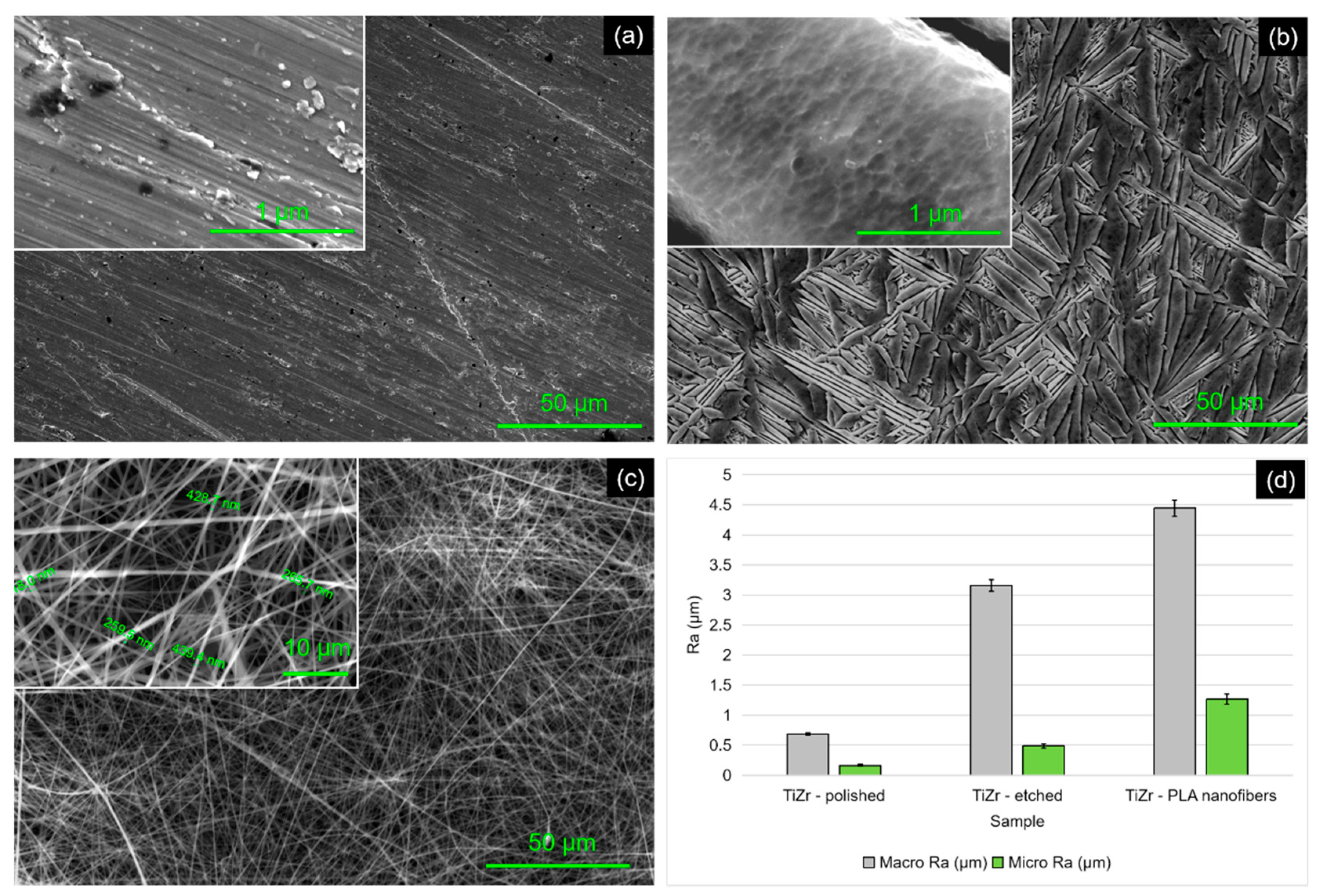
3.1. Sample Morphology

On the polished TiZr sample (Figure 1a) there can be observed grooves left after the polishing process as well as small formations of native oxides. The resulted surface after the etching of the TiZr sample (Figure 1b) shows characteristic needle-like semi-organized structures of the Ti and Zr oxides having lengths of 10–50 μm and widths of 2–5 μm. At the intersections of the structures nanopores with diameters of approximately 200 nm were also formed. Figure 1c shows the TiZr completely coated with PLA nanofibers. The coverage is relatively uniform, however, there are areas with higher fiber densities. The fibers have diameters between about 250 and 450 nm and lengths of hundreds of micrometers. The fibers are not oriented in a certain direction. The straightness of the fibers depends on the distance from the substrate, being straighter at the interface with TiZr and more curved on the surface of the sample.

3.2. Roughness Measurements

The measurements of the average roughness (Figure 1d) show, as expected, that the polished samples have the smoothest surface. The roughness increases after the etching process due to the formation of the oxide structures. The highest roughness value was obtained for the sample covered with PLA nanofibers due to the multi-layers of cross-linking polymer fibers. The higher roughness of the TiZr–PLA samples makes them possible candidates in implant use since it has been proven that certain cell types (e.g., pre-osteoblast MC3T3-E1 cells) have an affinity for rougher surfaces [30]. Although the roughness values were lower when measured with the AFM, the roughness trend remains the same even at the micro-level. The differences in values are considered normal, since the measurement performed by AFM represents only a minor part of the whole sample.

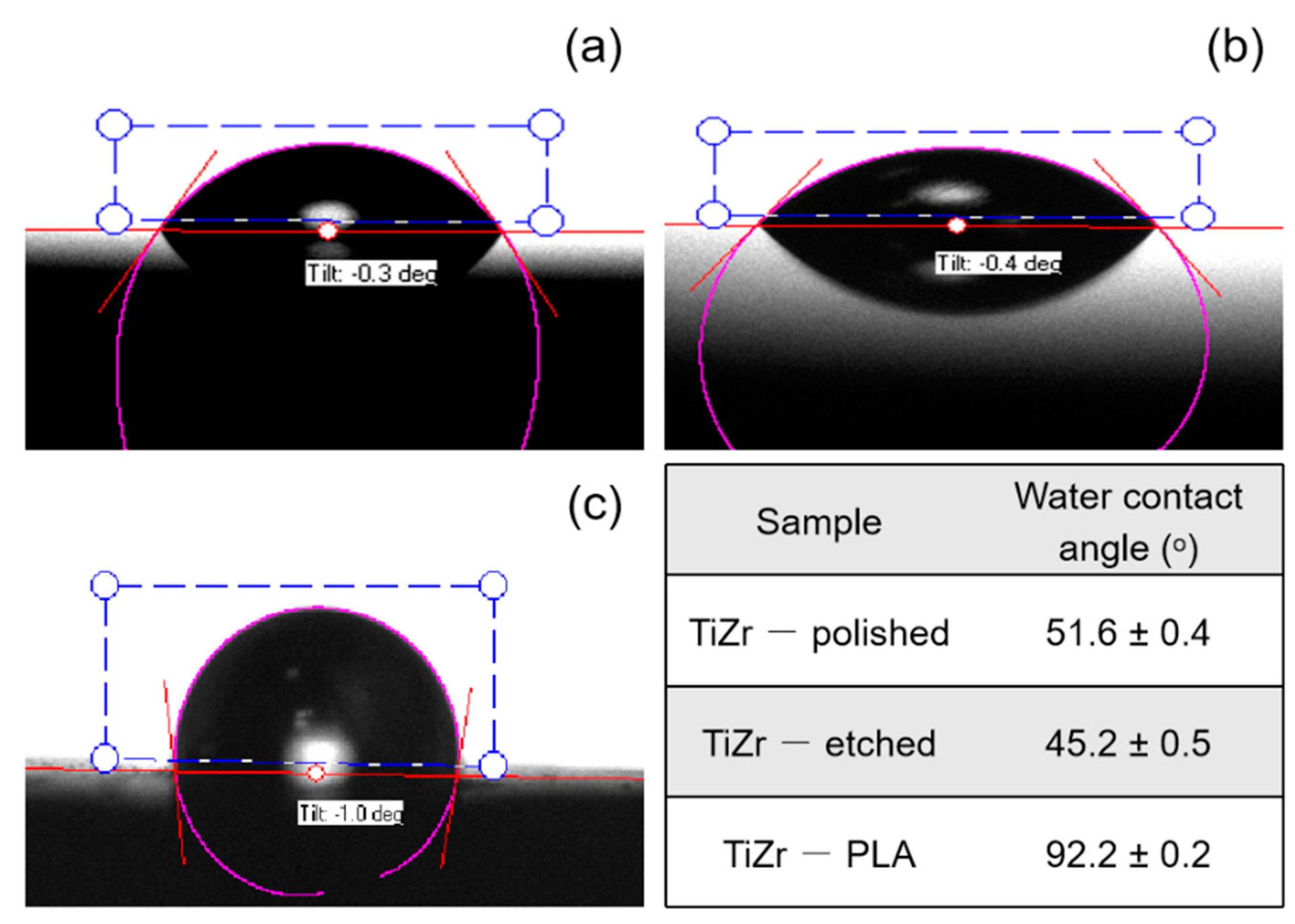
3.3. Contact Angle Measurements

The contact angle measurements show that the polished and etched TiZr samples (Figure 2a,b) have hydrophilic characteristics. The decrease of the contact angle value for the etched TiZr surface can be attributed to an increased oxygen quantity caused by the formation of the oxides, leading to more hydrogen bond interactions between the oxygen-rich Ti and Zr compounds and water [31]. PLA is a relatively hydrophobic polymer with a static water contact angle in the range of 75–85° [32]. Surface micro and structures affect the contact angle values and, in combination with a hydrophobic material, they increase it. Briefly, covering the TiZr surface with PLA nanofibers (Figure 2c) led to an increase of the contact angle value, crossing the limit of hydrophobicity which is an aspect of our aim in this manuscript.

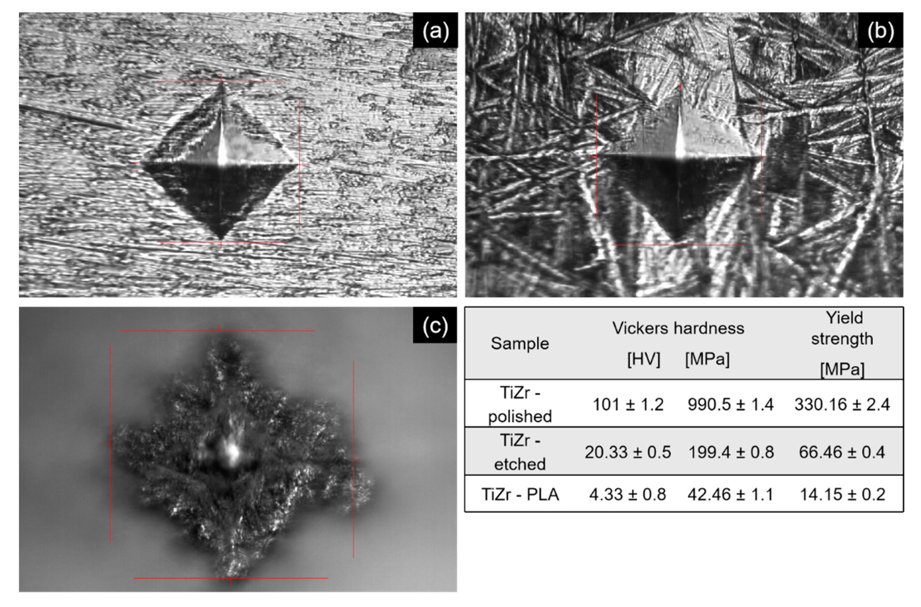
3.4. Micro Hardness Tests

The results obtained after the Vickers micro-hardness tests are presented in Figure 3. In the case of polished and etched TiZr (Figure 3a), the indent mark has sharp edges and no cracks appear at the substrate. On the etched surface, some delamination of the superficial oxide layer could be observed. In the case of the PLA nanofiber coating (Figure 3c), in the center of the indent, the metallic substrate cannot be observed due to the thickness of the coating. The edges of the indent are blurred due to the multiple layers of PLA nanofibers and curved, indicating a partially elastic deformation of the polymer during the experiment. The forces registered during the experiment show, as expected, that the hardest surface is that of the TiZr polished alloy, followed by that of the oxidized sample, the smallest value being that of the polymeric coating.
The Vickers hardness value can be converted to International System (SI) units by multiplying the result with the standard gravity. After this, the yield strength of the material can be estimated using the empirical formula [33]:
σY [MPa] ≈ HV/3

3.5. Adhesion of Coatings

The forces measured during the adhesion experiments are presented in Table 2. The polished TiZr sample had the highest recorded adhesion force. Since there is no coating on this sample, this force basically represents the force of the adhesive tape, since both the dolly and the sample have a smooth surface, a maximum tape–sample interface can be achieved. The forces recorded for the etched TiZr sample decrease in comparison to the polished TiZr. The rough nature of this surface makes the contact with the adhesive tape less than ideal. However, weaker parts of the oxide coating could be detached from the surface of the sample. The PLA coated sample had the weakest adhesion force of all the samples. In this case, as well, the contact between the adhesive tape and the sample was less than ideal, furthermore, some of the PLA nanofibers may have been damaged during the fixation of the dolly. The experiment showed that no oxide was removed from the sample, only layers of PLA nanofibers. So, this value represents the adhesion of the PLA nanofibers to the TiZr oxide layer produced after etching.

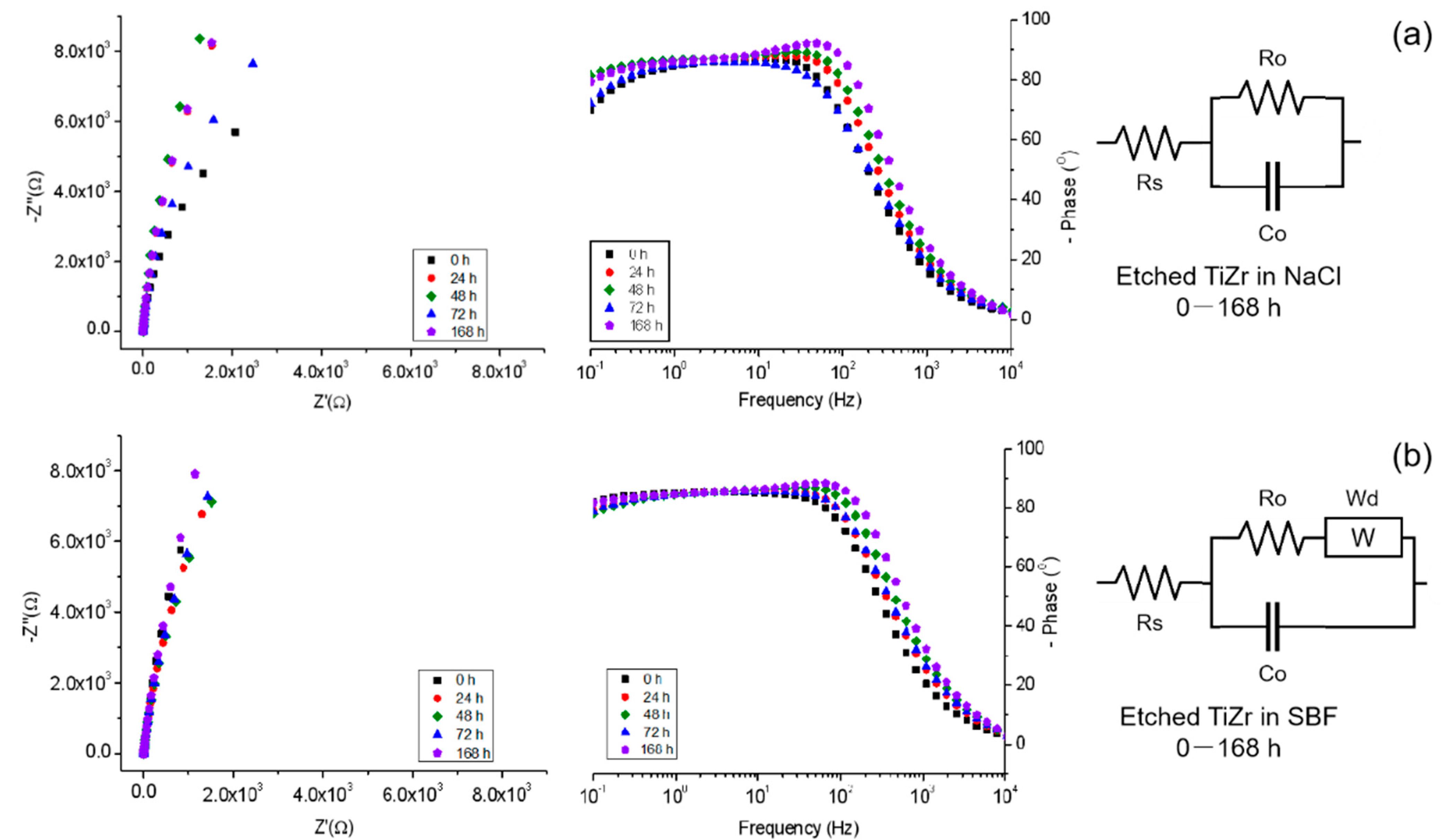
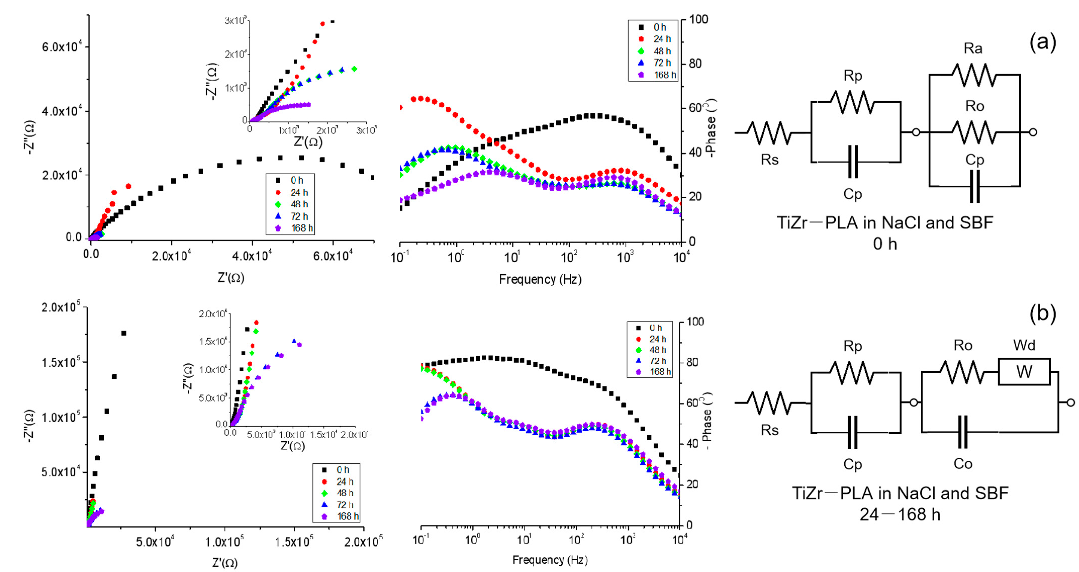
3.6. Electrochemical Impedance Spectroscopy

With EIS one is able to characterize oxide layers on the studied materials and assess the performances of surface layers without accelerating the electrochemical reactions at a sample/solution interface [34]. The evolution of the samples was monitored for a period of 7 days of immersion in two electrolytes (0.9% NaCl and SBF). Figure 4 presents the EIS spectra (Nyquist and Bode Phase) and equivalent circuits used to fit the collected data for the etched TiZr samples. The values obtained after fitting are presented in Table 3.
In 0.9% NaCl (Figure 4a) the etched TiZr sample had a predominantly resistive behavior that increased over time. This can be attributed to the passive nature of the formed oxide that has protective properties [35]. The Bode plot shows that the samples have a resistive behavior at high frequencies that correspond to charge transfer and a capacitive behavior at lower frequencies hinting to diffusion process being dominant at lower frequencies. Similar behavior was observed for the samples immersed in SBF (Figure 4b). However, greater stability was observed in the obtained plots. The fittings were performed with the equivalent circuits where the elements correspond as following: Rs-solution resistance; Ro–TiZr oxide resistance; Co–TiZr oxide capacitance; Wd-diffusion. It was observed that for the samples immersed in 0.9% NaCl there is a high stability of both resistance and capacitance, and although diffusion was hinted in the Bode Phase plot, the fitting did not work with an added diffusion element in the circuit. The samples immersed in SBF had a different behavior, the overall resistance of the oxide dropping fast and the capacitance and diffusion remaining stable. This difference in behavior can be attributed to the greater variety of ionic species present in the SBF solution.
Figure 5 presents the EIS spectra (Nyquist and Bode Phase) and equivalent circuits used to fit the collected data for the TiZr–PLA samples. The values obtained after fitting are presented in Table 4.
The sample immersed in 0.9% NaCl (Figure 5a) showed a predominantly capacitive–diffusive behavior throughout the experiment. It is known that microstructures formed by hydrophobic materials decrease the wettability of the material on which they are deposited. In accordance with other studies [36], it seems likely that in our case as well, pockets of air remained trapped in the polymeric deposition keeping their wetting behavior in the Cassie–Baxter regime when first immersed in the electrolyte, taking some time to be replaced by the liquids. In the Nyquist plot, the high value for the impedance at initial immersion time was attributed to pockets of air trapped between the PLA nanofibers. After the first day of immersion, the impedance value decreased dramatically and the samples acquired a more pronounced capacitive behavior. This behavior, being different than the one observed before for the plain etched TiZr in 0.9% NaCl, can be attributed to phenomena taking place on the PLA nanofibers, some probably form of degradation. The Bode Phase plot shows that this type of sample has a resistive behavior at low frequencies with diffusion happening at medium frequencies and pseudo-capacitive behavior at high frequencies that changes to a more resistive over time. The diffusive behavior starts masking the resistive behavior at immersion time and at high frequencies. The sample immersed in SBF (Figure 5b) had a more resistive-diffusive behavior. At immersion time, the same high-value impedance attributed to air pockets was observed in the Nyquist plot. After the first day, the impedance value decreases and the resistive behavior shifts to diffusive. The Bode Phase plot shows similarities to the sample immersed in 0.9% NaCl at high frequencies, with diffusion being stronger, however. The samples immersed in SBF were more stable through the experiment and a pseudo-capacitive behavior at low frequencies. Two circuits were used for both sample types in order to fit the acquired data. The elements correspond as follows: Rs—solution resistance; Rp—PLA resistance; Cp—PLA capacitance; Ra—air resistance (0 h) Ro—TiZr oxide resistance; Co—TiZr oxide capacitance; Wd—diffusion (24–168 h). It was observed that the resistance and values of the PLA nanofibers remained consistent throughout the experiment, having a decreasing trend. The values of the capacitance increased more rapidly in 0.9% NaCl than in SBF. The resistance of the oxide layer decreased over time. It was observed that, after one day of immersion, the air pockets were filled with electrolyte in both cases and diffusion began.

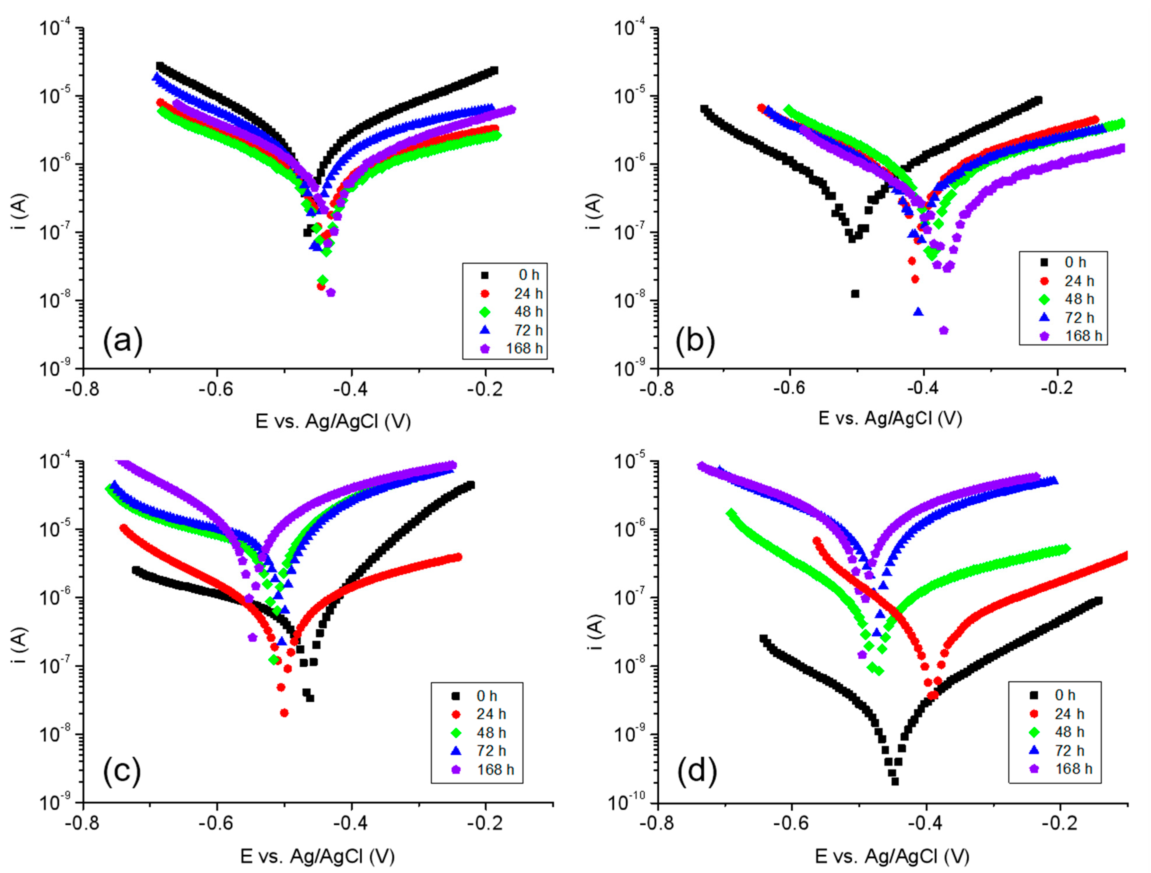
3.7. Corrosion Resistance Tests

Potentiodynamic polarization tests were used to evaluate the corrosion behavior of the samples over time in 0.9% NaCl and SBF. The Tafel plots are presented in Figure 6, and the resulting parameters are summarized in Table 5.
The etched TiZr (Figure 6a) showed great stability when immersed in 0.9% NaCl. The corrosion potential (Ecor) remained stable at around −0.46 ± 0.025 V, the corrosion current (Icor) decreased from 2.42 to 0.286 μA and the corrosion rate (Vcor) decreased from 7.43 to 0.879 μm/year. When immersed in SBF, the etched samples had a slightly different behavior (Figure 6b). The initial Ecor was more electronegative (−0.509 V) than the one registered in 0.9% NaCl, however, both Icor (0.529 μA) and Vcor (1.35 μm/year) had smaller values. Over time, the Ecor for the samples immersed in SBF stabilized at around −400 mV. The Icor and Vcor values were slightly lower in SBF than those observed in 0.9% NaCl. These results show that the TiZr oxides formed on the samples during etching have a passivation effect, protecting the metal from corrosion. The samples immersed in SBF had slower corrosion rates, however, they required more time to stabilize, due to the complexity of the electrolyte. The TiZr samples covered with PLA nanofibers immersed in 0.9% NaCl (Figure 6c) exhibited an accelerated corrosion behavior over time. Ecor increased from −0.462 to −0.546 V, Icor increased from 0.217 to 3.17 μA and Vcor increased from 2.22 to 32.46 μm/year. This gradual change implies a degradation of the PLA nanofibers. This process is dependent on the time of immersion, becoming faster over time, as the inner layers of PLA nanofibers become affected later by the NaCl solution. The immersion in SBF of the TiZr samples covered with PLA nanofibers (Figure 6d) led to completely different results. Initially, the samples were virtually inert. Only after 24 h of immersion, the samples began to show signs of corrosion. In this case, there is a possibility that at earlier stages, the coating is being protected by the carbonate depositions from the SBF solution. Another possibility is that this complex electrolyte, combined with the hydrophobic nature of PLA and the nanostructures present on the surface can create a Cassie–Baxter effect on the surface of the sample, forming air micro pockets, thus hindering corrosion.

4. Conclusions

In this study, we have investigated three surface treatments of TiZr including PLA nanofibrous coatings achieved by electrospinning TiZr oxide.
The morphology of the samples showed that the surfaces of the alloy were covered in a network of randomly deposited PLA fibers. The roughness and contact angles of the coating with electrospun PLA have significantly increased compared to the other two treatments.
Contact angle measurements evidenced the increase of hydrophobicity as a result of mixing the hydrophobic properties of PLA with the microstructure with high roughness given by the nanofibers. The calculated yield strength was, as expected, lower than the standard for PLA due to the fibrous nature of the coating. The adhesion force testing showed poor adhesion between PLA fibers in the coating. The electrochemical experiments showed good resistance to corrosion and good stability over time. A phenomenon of insulation during the first stages of EIS testing was attributed, along with the data from contact angle determination, to the formation of a Cassie–Baxter state on the samples covered with PLA nanofibers.
In conclusion, a deposition of PLA nanofibers on TiZr oxide may be used in a variety of implant procedures, modifying the coating as suited: increasing the roughness for better cell adhesion, filling the gaps between the fibers with various drugs for controlled delivery, or tweaking the wettability as suited.

Author Contributions

Conceptualization, I.D. and D.I.; methodology, I.D.; software, M.E.V. and A.B.S.; validation, I.D. and D.I.; formal analysis, M.E.V. and A.B.S.; investigation, M.E.V. and I.D.; resources, D.I.; data curation, I.D. and D.I.; writing—original draft preparation, M.E.V. and A.B.S.; writing—review and editing, A.B.S., I.D. and D.I.; visualization, I.D. and D.I.; supervision, I.D. All authors have read and agreed to the published version of the manuscript.

Funding

This research received no external funding.

Institutional Review Board Statement

Not applicable.

Informed Consent Statement

Not applicable.

Data Availability Statement

Data is contained within the article.

Acknowledgments

The SEM analyses on Quanta 650 FEG were possible due to European Regional Development Fund through Competitiveness Operational Program 2014–2020, Priority axis 1, Project No. P_36_611, MySMIS code 107066, Innovative Technologies for Materials Quality Assurance in Health, Energy and Environmental—Center for Innovative Manufacturing Solutions of Smart Biomaterials and Biomedical Surfaces—INOVABIOMED.

Conflicts of Interest

The authors declare no conflict of interest.

References

  1. van Hengel, I.A.J.; Gelderman, F.S.A.; Athanasiadis, S.; Minneboo, M.; Weinans, H.; Fluit, A.C.; van der Eerden, B.C.J.; Fratila-Apachitei, L.E.; Apachitei, I.; Zadpoor, A.A. Functionality-packed additively manufactured porous titanium implants. Mater. Today Bio 2020, 7, 100060. [Google Scholar] [CrossRef]
  2. Kaur, M.; Singh, K. Review on titanium and titanium based alloys as biomaterials for orthopaedic applications. Mater. Sci. Eng. C 2019, 102, 844–862. [Google Scholar] [CrossRef]
  3. Brånemark, P.I.; Hansson, B.O.; Adell, R.; Breine, U.; Lindström, J.; Hallén, O.; Ohman, A. Osseointegrated implants in the treatment of the edentulous jaw. Experience from a 10-year period. Scand. J. Plast. Reconstr. Surg. Suppl. 1977, 16, 1–132. [Google Scholar]
  4. Murphy, M.; Walczak, M.S.; Thomas, A.G.; Silikas, N.; Berner, S.; Lindsay, R. Toward optimizing dental implant performance: Surface characterization of Ti and TiZr implant materials. Dent. Mater. 2017, 33, 43–53. [Google Scholar] [CrossRef] [PubMed]
  5. Liu, X.; Chen, S.; Tsoi, J.; Matinlinna, J.P. Binary titanium alloys as dental implant materials—A review. Regen. Biomater. 2017, 4, 315–323. [Google Scholar] [CrossRef] [Green Version]
  6. Todai, M.; Nagase, T.; Hori, T.; Matsugaki, A.; Sekita, A.; Nakano, T. Novel TiNbTaZrMo high-entropy alloys for metallic biomaterials. Scr. Mater. 2017, 129, 65–68. [Google Scholar] [CrossRef] [Green Version]
  7. Zhou, H.; Li, F.; He, B.; Wang, J.; Sun, B. Air plasma sprayed thermal barrier coatings on titanium alloy substrates. Surf. Coat. Technol. 2007, 201, 7360–7367. [Google Scholar] [CrossRef]
  8. Prodana, M.; Nistor, C.E.; Stoian, A.B.; Ionita, D.; Burnei, C. Dual nanofibrous bioactive coatings on TiZr implants. Coatings 2020, 10, 526. [Google Scholar] [CrossRef]
  9. Ionita, D.; Grecu, M.; Ungureanu, C.; Demetrescu, I. Antimicrobial activity of the surface coatings on TiAlZr implant biomaterial. J. Biosci. Bioeng. 2011, 112, 630–634. [Google Scholar] [CrossRef] [PubMed]
  10. Ungureanu, C.; Pirvu, C.; Mindroiu, M.; Demetrescu, I. Antibacterial polymeric coating based on polypyrrole and polyethylene glycol on a new alloy TiAlZr. Prog. Org. Coat. 2012, 75, 349–355. [Google Scholar] [CrossRef]
  11. Standard Specification for Wrought Titanium 6Al 4V ELI Alloy for Surgical Implants; ASTM Designation F136-82; ASTM: Philadelphia, PA, USA, 1994; pp. 19–20.
  12. Standard Specification for Wrought Titanium 6Al 7Nb Alloy for Surgical Implants; ASTM Designation F1295-92; ASTM: Philadelphia, PA, USA, 1994; pp. 687–689.
  13. Nartita, R.; Daniela, I.; Demetrescu, I. A combined scientometric and critical approach in reviewing TiZr implant alloys and coating performances. Coatings 2021, 11, 392. [Google Scholar] [CrossRef]
  14. Cui, W.; Liu, Y. Fatigue behavior of Ti50Zr alloy for dental implant application. J. Alloys Compd. 2019, 793, 212–219. [Google Scholar] [CrossRef]
  15. Grigorescu, S.; Pruna, V.; Titorencu, I.; Jinga, V.V.; Mazare, A.; Schmuki, P.; Demetrescu, I. The two step nanotube formation on TiZr as scaffolds for cell growth. Bioelectrochemistry 2014, 98, 39–45. [Google Scholar] [CrossRef]
  16. Vardaki, M.; Mohajernia, S.; Pantazi, A.; Nica, I.C.; Enachescu, M.; Mazare, A.; Demetrescu, I.; Schmuki, P. Post treatments effect on TiZr nanostructures fabricated via anodizing. J. Mater. Res. Technol. 2019, 8, 5802–5812. [Google Scholar] [CrossRef]
  17. Gulati, K.; Kogawa, M.; Prideaux, M.; Findlay, D.M.; Atkins, G.J.; Losic, D. Drug-releasing nano-engineered titanium implants: Therapeutic efficacy in 3D cell culture model, controlled release and stability. Mater. Sci. Eng. C 2016, 69, 831–840. [Google Scholar] [CrossRef]
  18. Ionita, D.; Bajenaru-Georgescu, D.; Totea, G.; Mazare, A.; Schmuki, P.; Demetrescu, I. Activity of vancomycin release from bioinspired coatings of hydroxyapatite or TiO2 nanotubes. Int. J. Pharm. 2017, 517, 296–302. [Google Scholar] [CrossRef]
  19. Ionita, D.; Pirvu, C.; Stoian, A.B.; Demetrescu, I. The Trends of TiZr Alloy Research as a Viable Alternative for Ti and Ti16 Zr Roxolid Dental Implants. Coatings 2010, 10, 422. [Google Scholar] [CrossRef]
  20. Yun, K.-D.; Yang, Y.; Lim, H.-P.; Oh, G.-J.; Koh, J.-T.; Bae, I.-H.; Kim, J.; Lee, K.-M.; Park, S.-W. Effect of nanotubular-micro-roughened titanium surface on cell response in vitro and osseointegration in vivo. Mater. Sci. Eng. C 2010, 30, 27–33. [Google Scholar] [CrossRef]
  21. Fattahi, F.; Khoddami, A.; Avinc, O. Poly(lactic acid) (PLA) Nanofibers for Bone Tissue Engineering. J. Text. Polym. 2019, 7, 47–64. [Google Scholar]
  22. Saldin, L.T.; Cramer, M.C.; Velankar, S.S.; White, L.J.; Badylak, S.F. Extracellular matrix hydrogels from decellularized tissues: Structure and function. Acta Biomater. 2017, 49, 1–15. [Google Scholar] [CrossRef] [Green Version]
  23. Zhu, H.; Guo, Z.; Liu, W. Adhesion behaviors on superhydrophobic surfaces. Chem. Commun. 2019, 50, 3900–3913. [Google Scholar] [CrossRef] [PubMed]
  24. Manole, C.C.; Dinischiotu, A.; Nica, C.; Demetrescu, I.; Pirvu, C. Influence of electrospun TiO2 nanowires on corrosion resistance and cell response of Ti50Zr alloy. Mater. Corros. 2018, 69, 1609–1619. [Google Scholar] [CrossRef]
  25. Yin, L.; Chang, Y.; You, Y.; Liu, C.; Li, J.; Lai, H.-C. Biological responses of human bone mesenchymal stem cells to Ti and TiZr implant materials. Clin. Implant Dent. Relat. Res. 2019, 21, 550–564. [Google Scholar] [CrossRef] [PubMed]
  26. Vargas-Villagran, H.; Romo-Uribe, A.; Teran-Salgado, E.; Domínguez Díaz, M.; Flores, A. Electrospun polylactic acid non-woven mats incorporating silver nanoparticles. Polym. Bull. 2014, 71, 2437–2452. [Google Scholar] [CrossRef]
  27. Yuan, Q.; Qin, C.; Wu, J.; Xu, A.; Zhang, Z.; Liao, J.; Lin, S.; Ren, X.; Zhang, P. Synthesis and characterization of Cerium-doped hydroxyapatite/polylactic acid composite coatings on metal substrates. Mater. Chem. Phys. 2016, 182, 365–371. [Google Scholar] [CrossRef]
  28. Abdal-hay, A.; Barakat, N.A.M.; Lim, J.K. Hydroxyapatite-doped poly(lactic acid) porous film coating for enhanced bioactivity and corrosion behavior of AZ31 Mg alloy for orthopedic applications. Ceram. Int. 2013, 39, 183–195. [Google Scholar] [CrossRef]
  29. Kokubo, T.; Kushitani, H.; Sakka, S.; Kitsugi, T.; Yamamuro, T. Solutions able to reproduce in vivo surface-structure changes in bioactive glass-ceramic A-W. J. Biomed. Mater. Res. 1990, 24, 721–734. [Google Scholar] [CrossRef]
  30. Ion, R.; Mazare, A.; Dumitriu, C.; Pirvu, C.; Schmuki, P.; Cimpean, A. Nanochannelar topography positively modulates osteoblast differentiation and inhibits osteoclastogenesis. Coatings 2018, 8, 294. [Google Scholar] [CrossRef] [Green Version]
  31. Frank, M.J.; Walter, M.S.; Lyngstadaas, S.P.; Wintermantel, E.; Haugen, H.J. Hydrogen content in titanium and a titanium–zirconium alloy after acid etching. Mater. Sci. Eng. C 2013, 33, 1282–1288. [Google Scholar] [CrossRef] [Green Version]
  32. Baran, E.H.; Erbil, H.Y. Surface Modification of 3D Printed PLA Objects by Fused Deposition Modeling: A Review. Colloids Interfaces 2019, 3, 43. [Google Scholar] [CrossRef] [Green Version]
  33. Zhang, P.; Li, S.X.; Zhang, Z.F. General relationship between strength and hardness. Mater. Sci. Eng. A 2011, 529, 62–73. [Google Scholar] [CrossRef]
  34. Orazem, M.E.; Tribollet, B. Electrochemical Impedance Spectroscopy; John Wiley & Sons, Inc.: Hoboken, NJ, USA, 2008. [Google Scholar] [CrossRef]
  35. Chung, M.K. Study of Anodic Oxide Films on Titanium and Titanium-Zirconium Alloys and Their Potential for Capacitive Energy Storage. Ph.D. Thesis, Case Western Reserve University, Cleveland, OH, USA, 2016. [Google Scholar]
  36. Szewczyk, P.K.; Ura, D.P.; Metwally, S.; Knapczyk-Korczak, J.; Gajek, M.; Marzec, M.M.; Bernasik, A.; Stachewicz, U. Roughness and Fiber Fraction Dominated Wetting of Electrospun Fiber-Based Porous Meshes. Polymers 2018, 11, 34. [Google Scholar] [CrossRef] [Green Version]
Figure 1. SEM micrographs for (a) polished TiZr; (b) etched TiZr; (c) etched TiZr covered with PLA nanofibers; Inset—higher magnifications; (d) average roughness values.
Coatings 11 00615 g001
Figure 2. Contact angle measurements for (a) polished TiZr; (b) etched TiZr; (c) etched TiZr covered with PLA nanofibers. Measured contact angle values.
Coatings 11 00615 g002
Figure 3. Morphology of samples after microhardness tests: (a) polished TiZr; (b) etched TiZr; (c) etched TiZr covered with PLA nanofibers and calculated forces.
Coatings 11 00615 g003
Figure 4. Nyquist, Bode Phase plots and equivalent circuits for etched TiZr in (a) 0.9% NaCl; (b) SBF.
Coatings 11 00615 g004
Figure 5. Nyquist, Bode Phase plots and equivalent circuits for TiZr covered with PLA nanofibers in (a) 0.9% NaCl; (b) SBF. Inset—Nyquist detail at high frequencies.
Coatings 11 00615 g005
Figure 6. Tafel plots over time for (a) etched TiZr in 0.9% NaCl; (b) etched TiZr in SBF; (c) etched TiZr covered with PLA nanofibers in 0.9% NaCl; (d) etched TiZr covered with PLA nanofibers in SBF.
Coatings 11 00615 g006
Table 1. Composition of Kokubo SBF [29].
ComponentConcentration [g/L]
NaCl7.996
NaHCO30.35
KCl0.224
K2HPO4·3H2O0.228
MgCl2·6H2O0.305
CaCl20.278
Na2SO40.071
(CH2OH)3CNH26.057
Table 2. Adhesion forces for the obtained samples.
SampleAdhesion Force [MPa]
TiZr—polished32.2 ± 0.6
TiZr—etched21.4 ± 0.3
TiZr—PLA4.3 ± 0.4
Table 3. Equivalent circuit values for etched TiZr in 0.9% NaCl and SBF.
SampleTime
[h]
Rs
[Ω]
Ro
[kΩ]
Co
[μF]
Wd
[μMho]
TiZr—etched in 0.9% NaCl01.2546.8150-
241.3855.5148-
481.2658.3143-
721.6367.4128-
1681.5861116-
TiZr—etched in SBF01.456313182.7
241.1249514480.7
482.0514.412670.3
721.75.116266
1683.41.4211662.1
Table 4. Equivalent circuit values for etched TiZr covered with PLA nanofibers in 0.9% NaCl and SBF.
SampleTime
[h]
Rs
[Ω]
Rp
[kΩ]
Cp
[μF]
Ro
[kΩ]
Co
[μF]
Ra
[kΩ]
Wd
[μMho]
TiZr—etched in 0.9% NaCl025048.82.184800.492.62-
2412222.553.24821.72-91.2
4872.72.161442043.73-77.4
7271.41.971792074.81-72.1
16862.50.931951765.7-57.3
TiZr—etched in SBF011152.21.521.921.273.02-
245148.34.10.614.1-74.4
4851.342.84.750.594.75-68
7250.120.75.030.515.03-62.5
1683819.75.340.465.34-57.1
Table 5. Electrochemical parameters of potentiodynamic polarization curves of the different samples.
SampleTime
[h]
Ecor
[V]
Icor
[μA]
Vcor
[μm/year]
TiZr—etched in 0.9% NaCl0−0.4632.427.43
24−0.4440.5741.76
48−0.4390.1820.558
72−0.4540.5811.78
168−0.4880.2860.879
TiZr—etched in SBF0−0.5090.5291.35
24−0.4160.3170.988
48−0.3880.1640.419
72−0.4090.3860.812
168−0.3690.3300.239
TiZr—PLA in 0.9% NaCl0−0.4620.2172.22
24−0.4990.2722.79
48−0.5141.8018.46
72−0.5012.3023.56
168−0.5463.1732.46
TiZr—PLA in SBF0−0.4500.721 × 10−30.011
24−0.3940.0120.188
48−0.4720.0280.436
72−0.4680.2573.95
168−0.4920.3054.68
Publisher’s Note: MDPI stays neutral with regard to jurisdictional claims in published maps and institutional affiliations.

