Article

Radiation Effect in Ti-Cr Multilayer-Coated Silicon Carbide under Silicon Ion Irradiation up to 3 dpa

1
Research & Development Group, Hitachi, Ltd., 7-1-1, Omika-cho, Hitachi 319-1292, Japan
2
Open Innovation Institute, Kyoto University, Gokasho, Uji 611-0011, Japan
3
Institute of Advanced Energy, Kyoto University, Gokasho, Uji 611-0011, Japan
4
Hitachi-GE Nuclear Energy, Ltd., 3-1-1, Saiwai-cho, Hitachi 317-0073, Japan
*
Author to whom correspondence should be addressed.
Present Position: Sino-French Institute for Nuclear Energy and Technology, Sun Yat-sen University, Zhuhai 519082, China.
Coatings 2022, 12(6), 832; https://doi.org/10.3390/coatings12060832
Submission received: 29 April 2022 / Revised: 3 June 2022 / Accepted: 9 June 2022 / Published: 14 June 2022
(This article belongs to the Special Issue Advanced Composites and Coatings for Nuclear Applications)

Abstract

:
Replacement of conventional Zircaloy fuel cladding with silicon carbide (SiC) fuel cladding is expected to significantly decrease the amount of hydrogen generated from fuel claddings by the reaction with steam during severe accidents. One of their critical issues addressed regarding practical application has been hydrothermal corrosion. Thus, the corrosion resistant coating technology using a Ti-Cr multilayer was developed to suppress silica dissolution from SiC fuel cladding into reactor coolant under normal operation. The effect of radiation on adhesion of the coating to SiC substrate and its microstructure characteristics were investigated following Si ion irradiation at 573 K up to 3 dpa for SiC. Measurement of swelling in pure Ti, pure Cr and SiC revealed that the maximum inner stress attributed to the swelling difference was generated between the coating and SiC substrate by irradiation of 1 dpa. No delamination and cracking were observed in cross-sectional specimens of the coated SiC irradiated up to 3 dpa. According to analyses using transmission electron microscopy, large void formation and cascade mixing due to irradiation were not observed in the coating. The swelling in the coating at 573 K was presumed to be caused by another mechanism during radiation such as point defects rather than void formation.

1. Introduction

Considering the lessons learned in the aftermath of the Fukushima Daiichi Nuclear Power Plant accident [1], various technologies for light water reactors (LWRs) with an emphasis on safety have been developed. Among candidate alternative materials for fuel cladding and core structures of LWRs, silicon carbide-based (SiC-based) materials—particularly SiC-fiber-reinforced SiC-matrix ceramic composites (SiC/SiC composites)— are thought to provide outstanding passive safety features in beyond-design-basis severe accident scenarios [2,3,4]. Since SiC has a lower hydrogen generation rate and lower heat of reaction than Zr, the amount of hydrogen generated from fuel claddings by reaction with steam during severe accidents is expected to significantly decrease when SiC fuel cladding is used instead of conventional Zircaloy fuel cladding [3,4]. Therefore, the expectation for increased accident tolerance by applying SiC fuel cladding has led to the development of manufacturing and integration technologies [5,6,7,8,9,10,11,12,13,14,15,16], research on material behavior under normal operation and accident conditions [5,6,7,17,18,19,20,21,22,23,24,25,26,27,28,29,30,31], and evaluation of safety and economic benefits [32].
Fabrication of SiC/SiC composite tubing by a chemical vapor infiltration (CVI) process is the most advanced development and various mechanical properties for applying this composite as fuel cladding have been investigated [8,9]. A liquid phase sintering (LPS) process has also been developed for nuclear applications [10]. The LPS process can form matrices that are as highly crystalline as those of a CVI process and the LPS process yields SiC/SiC composite with fewer porosities and higher thermal conductivity than the CVI process [11]. Since end-plug joining is an important technology for hermeticity of a fuel rod, joining technology has been developed [12,13,14] and the end-plug joint portion with corrosion resistant coatings were experimentally produced [15,16].
The advantage for heating resistance of SiC fuel cladding emphasizes accident tolerance. SiC/SiC composite tubing fabricated by a CVI process retained its room temperature strength after being heated to 2173 K in an inert gas environment [17]. Silicon carbide formed by chemical vapor deposition (CVD SiC) is often used to overcoat SiC/SiC composite for protection from corrosive environments. CVD SiC showed a lower steam oxidation rate than Zircaloy and stainless steel [18]. Although the steam oxidation rate of the specimens is affected by pressure and flow rate of steam [19], the corrosion can be limited to the surface for the temperature range of 1973–2073 K in which formation of SiO2 bubbles occurred on the surface by softening and melting of SiO2 [20,22]. CVD-SiC-overcoated SiC/SiC composite tubes maintained a coolable shape after quench tests at 2273 K [21,22].
However, there are many critical issues related to fabrication methods and test methods of the fabricated materials, and understanding of behaviors under normal operation and accident conditions that must be addressed regarding the practical application of SiC fuel cladding. The hydrothermal corrosion is one of the immediate critical issues under normal operation [5,6,7]. In hydrothermal LWR coolant environments, the silicon in SiC undergoes oxidation and produces silica that readily dissolves in water where its concentration in the coolant builds up to the point of saturation, and beyond which the silica can deposit in the cold regions of the coolant loop [23]. Since the dissolved oxygen activity in water can greatly increase SiC recession, the amount of dissolved silica produced from SiC is likely to be larger in the coolant of a boiling water reactor (BWR) under the normal water chemistry (NWC) condition with a higher dissolved oxygen activity than in the coolant of a pressurized water reactor (PWR) [23,24,25,26,27]. Neutron irradiation increases hydrothermal corrosion rate [28]. The radiation effects on hydrothermal corrosion of SiC are thought to be due to the change of surface potential attributed from irradiation-induced defects [29], irradiation damage, and increase of oxidants generated by radiolysis [28]. Pre-damage on the SiC surface by irradiating with Si ions causes a pronounced increase of corrosion rate depending on the amount of dissolved oxygen in water [30]. Radiolysis under a BWR-NWC environment generate some oxidants, such as oxygen and hydrogen peroxide, which promote the oxidation of SiC. Silica concentration in the coolant of BWRs should be kept low because silica carried over to the main steam system adheres to materials of the turbine system and affects the turbine efficiency [33]. Hence, it is necessary to suppress silica dissolution from SiC fuel cladding in BWRs.
Impurities including sintering additives and the crystalline orientation of SiC affect corrosion behavior in high temperature water [25,26,34,35,36]. CVD SiC is expected to have a lower recession rate than SiC formed in other ways because of its higher purity. As affairs now stand, it is difficult to suppress the silica dissolution from CVD SiC completely [26,31].
Corrosion-resistant coatings processed using industrial technologies, such as physical vapor deposition (PVD), CVD, electrolytic deposition, and vacuum plasma spraying (VPS) have been researched [37,38,39,40,41,42,43,44,45,46,47,48,49,50,51]. Metals (such as Cr [37,38,39,40,41,42,43,44,45,46,47,48,49], Zr [37,47] and Ti [47,48,49,50]), metal-doped SiC (such as Ti-doped and Zr-doped SiC [51]) and metal nitrides (such as CrN [37,38,39,40,41,42,43], ZrN [39,40], and TiN [37,39,40,41,42,43]) have been coated onto SiC as a surface layer. Preliminary work included the deposition of Cr, Zr, CrN, TiN, and ZrN coatings on SiC by PVD and VPS [36,37,38,39]. Hydrothermal corrosion testing for 1.4 × 10⁶ s (400 h) without irradiation showed that Cr, CrN, and TiN coatings on SiC by PVD remained in simulated BWR-NWC environment [39,40]. Cr, CrN and TiN coatings on SiC indicated large mass loss at test durations between 2.2 × 10⁶ s (600 h) and 4.0 × 10⁶ s (1100 h) in simulated BWR-NWC environment, while they indicated small mass change for 4.0 × 10⁶ s (1100 h) in simulated BWR- hydrogen water chemistry (HWC) environment and for 1.8 × 10⁶ s (500 h) in simulated PWR environment [41]. After neutron irradiation to a total fluence of 4.8 × 1024 n/m2 (>0.1 MeV), corresponding to about 0.5 dpa in SiC, in an inert environment at 578 K to 613 K, 18-μm-thick Cr coating and 3-μm-thick CrN coating on CVD SiC cracked substantially only 3-μm-thick TiN coating on CVD SiC showed no crack [42,43]. After neutron irradiation to a total fluence of 8.3 × 1024 n/m2 (>0.1 MeV), corresponding to about 1.0 dpa in SiC, in PWR environment at 566 K to 574 K, Cr coating was protective but TiN coating was severely damaged [42,43]. Cracking in coating by irradiation is thought to derive from irradiation-induced differential swelling between coating and SiC substrate. Cr coating technologies by PVD have been improved to develop more compressive residual stress in the coating for mitigating the differential swelling [44,45]. Cr coating remains as the best candidate and evaluation of its steam oxidation behavior has begun [46].
The authors have developed metal coatings on SiC which have corrosion resistance in the BWR-NWC environment. The metal coatings are thought to have the advantage of a lower Young’s modulus in thicker coatings on SiC substrate than the coatings of nitrides and carbides. The thicker coatings have margin for consuming coating material by forming a protective oxide film which provides corrosion resistance. The authors have previously tested Cr coating, Ti coating with a Cr bonding layer, and Zr coating with a Cr bonding layer. During hydrothermal corrosion testing at 561 K in high-purity water with a dissolved oxygen concentration of 8.0 mg/L for 3.6 × 10⁶ s (1000 h), both the Cr coating and the Ti coating were found to protect a CVD SiC substrate, whereas the Zr coating significantly debonded [47]. Additionally, the Cr coating exhibited obvious weight loss while the Ti coating had a small weight change during the tests [47,48,49]. Hence, the Ti coating with Cr bonding layer (hereinafter called Ti-Cr multilayer coating) demonstrated superior corrosion resistance in oxygenated high temperature water.
However, the radiation effect of the coating was not elucidated. Irradiation provides an increase of oxidizing agent by radiolysis in a water environment, and simultaneously leads to damage and strain of materials. The difference in irradiation-induced swelling between the coating and SiC substrate is expected to cause some stress in the coating. Ion irradiation is an effective method to evaluate damage and swelling induced by irradiation.
The objective of this study was to examine the radiation effect of the Ti-Cr multilayer coating on SiC substrates exposed to ion irradiation by evaluating irradiation-induced swelling and by observing and analyzing microstructures in the coating.

2. Experimental Procedure

2.1. Coating Designs

The Ti-Cr multilayer coating for this study was designed to consider corrosion resistance in high temperature water and delamination resistance from SiC substrate. Schematic illustrations of the coating structure obtained after processes of PVD and heat treatment are shown in Figure 1a,b, respectively. The coating has the Ti top layer and the Cr bonding layer between the Ti layer and SiC substrate. The PVD process can be more easily controlled to form a dense multi-layer coating in comparison to other processes. Heat treatment after the coating can strengthen the interface between the coating and a substrate. The Ti top layer forms a highly corrosion-resistant TiO2 film at the top surface in high temperature water [47,48,49]. The Cr bonding layer relieves thermal stress caused by the thermal expansion difference between Ti and SiC, and additionally forms diffusion reaction layers at the interface with SiC by heat treatment [49]. In the authors’ previous study, the heat treatment at 1223 K for 1.8 × 10³ s was the optimized condition to obtain a good adhesive property and corrosion resistance for the coating with a thickness of 10 to 20 μm in oxygenated pure water at 561 K [49]. Elemental Si and C from a SiC layer, Ti from a Ti layer, and Cr from a Cr layer diffused to other layers and formed stable carbide, silicide, and an intermetallic compound in the high temperature phase as shown in Figure 1b.

2.2. Specimens

2.2.1. Ti-Cr Multilayer Coated SiC Specimens

To investigate the radiation effect in the Ti-Cr multilayer coating under ion irradiation, the coated specimens were prepared by PVD using the same conditions as the authors previously reported for the Ti-Cr multilayer coatings [47,48,49]. Base specimens with the substrate surface were prepared by overcoating LPS-SiC/SiC composite plates (40.0 × 9.8 × 1.0 mm3), with high-purity CVD SiC (Na < 0.01, Co < 0.01, K < 0.05, Cu < 0.05, Zn < 0.05, Mn <0.01, Fe < 0.05, Cr < 0.1 in wppm, measured using a glow discharge mass spectrometer). The CVD SiC overcoating was custom-processed to a thickness of 100–150 μm by Ferrotec Material Technologies Co. using a thermal CVD technique. The coating was deposited on the base specimens in an argon gas atmosphere with a bias voltage of 150 V using an unbalanced magnetron sputtering system (Kobe Steel, Ltd.: Kobe, Japan, UBMS202). The coating was layered up to a thickness of 10–20 μm by deposition from a sputtering target of pure Ti (Al < 1, Cr < 1, Fe < 1, Ni < 1 in wppm) after deposition from a sputtering target of pure Cr (Fe 0.02, Al 0.004, Cu 0.002 in wt.%) for the Cr bonding layer on the CVD SiC substrate surface. Following the PVD, the specimens were heat-treated at 1223 K for 1.8 × 10³ s in a vacuum. The heat treatment forms diffused Cr-SiC interface layers consisting of chromium silicides [47,48,49,50]. One of the coated SiC specimens was sliced into 2-mm thick pieces perpendicular to the longitudinal direction using a diamond blade, and their cross-sectional surfaces were mechanically polished to a lap finish with 0.5 μm diamond paste.

2.2.2. Ti and Cr Bulk Specimens

To investigate swelling behavior in the Ti-Cr multilayer coating under ion irradiation, pure Ti and pure Cr bulk specimens were prepared for simulating major portion of the coating. The pure Ti and pure Cr bulk specimens with dimension of 2.0 × 10.0 × 1.2 mm3, the same as the Ti-Cr multilayer-coated SiC specimen, were cut out from a sputtering target of pure Ti (Al < 1, Cr < 1, Fe < 1, Ni < 1 in wppm) and a sputtering target of pure Cr (Fe 0.02, Al 0.004, Cu 0.002 in wt.%), respectively, by electrical discharge machining and slicing with a diamond blade. One of the surfaces (10.0 × 1.2 mm2) was mechanically polished to a lap finish with 0.5 μm diamond paste.

2.3. Ion Irradiation and Analyses

2.3.1. Ion Irradiation

The polished surfaces of the specimens were irradiated with 5.1-MeV Si2+ ions at 573 K in the Dual-Beam Facility for Energy Science and Technology (DuET) at Kyoto University. The polished surfaces were partially protected with a mask during the irradiation to allow determination of swelling by surface profilometry as described in Section 2.3.2. The irradiation temperature in DuET was set based on known surface temperatures of fuel cladding in BWRs. The typical flux was 3.12 × 10¹⁶ ions/m2s during the ion irradiation. The irradiation fluences were set to 0.95 × 10¹⁹, 9.51 × 10¹⁹, and 2.85 × 10²⁰ ions/m2, corresponding to the average number of displacements per atom over the damage range of approximately 0.1, 1.0, and 3.0 dpa for SiC. The irradiation fluences also corresponded to the average number of displacements per atom over the damage range of 0.187, 1.87, and 5.60 dpa for Ti and 0.182, 1.82, and 5.45 dpa for Cr. The depth profiles of the number of displacements and the concentrations of the implanted Si in Ti, Cr, and SiC for the irradiation fluence of 2.85 × 10²⁰ ions/m2, calculated using SRIM-2013 software [52], are shown in Figure 2. The effective displacement energies employed were 30 eV for Ti [53], 40 eV for Cr [53], 35 eV for Si [54], and 20 eV for C [54]. The densities employed were 4.518 g/cm3 for Ti and 7.2 g/cm3 for Cr from default values in SRIM-2013, and 3.21 g/cm3 for SiC [55]. The values of the damage range, d, obtained from the depth profiles of the number of displacements per atom were 2490 nm for Ti, 1720 nm for Cr, and 2400 nm for SiC.

2.3.2. Swelling Evaluation

The swellings produced by ions were evaluated by surface profilometry as described in ASTM E521-16 [53]. The evaluation was carried out on surfaces of the pure Ti and the pure Cr bulk specimens and cross-sectional surfaces of the CVD SiC layer in Ti-Cr multilayer-coated SiC specimens. The step height, Δh, at the boundary between the irradiated and the protected (unirradiated) regions was measured with an atomic force microscope (AFM) (KEYENCE: Osaka, Japan, VN-8000). The swelling, S, produced by ion irradiation is defined as Equation (1);
S = Δ h d
where Δh is the step height and d is the damage range.

2.3.3. Microstructural Analyses of Ti-Cr Multilayer Coating

The scanning electron microscopy (SEM) observation and energy dispersive X-ray spectrometry (EDS) mapping analyses were done on a cross-sectional slice for a Ti-coated specimen to confirm defects such as delamination or cracking and to determine layer structures and phases after ion irradiation. The SEM image was observed at 10 kV and the EDS mappings were analyzed at 30 kV using a field emission-scanning electron microscope (FE-SEM) (Zeiss, Ultra55) with the EDAX AMETEK Inc. TEAMTR EDS system (TEAMTR is a trade name of EDAX AMETEK Inc.: Mahwah, NJ, USA).
Transmission electron microscopy (TEM) observation and EDS mapping analyses were done on a cross-sectional thin foil for a Ti-coated specimen to identify phases and to confirm defect formation after ion irradiation. The cross-sectional thin foils were prepared in a commercial focused-ion-beam (FIB) unit (Hitachi, Ltd.: Tokyo, Japan, FB2200) at 5–40 kV and were finished by Ar gas milling at 5–30 mA using the Technoorg Linda Co. Ltd.: Budapest, Hungary, Gentle Mill. The TEM image was observed at 200 kV and the EDS mappings were analyzed at 30 kV using a field emission-transmission electron microscope (FE-TEM) (JEOL: Akishima, Japan, JEM-2200FS) with the JEOL JED-2300T EDS system. Phases were identified from selected area diffraction (SAD) using ReciPro ver4.828 [56].

3. Results

3.1. Step Height and Swelling

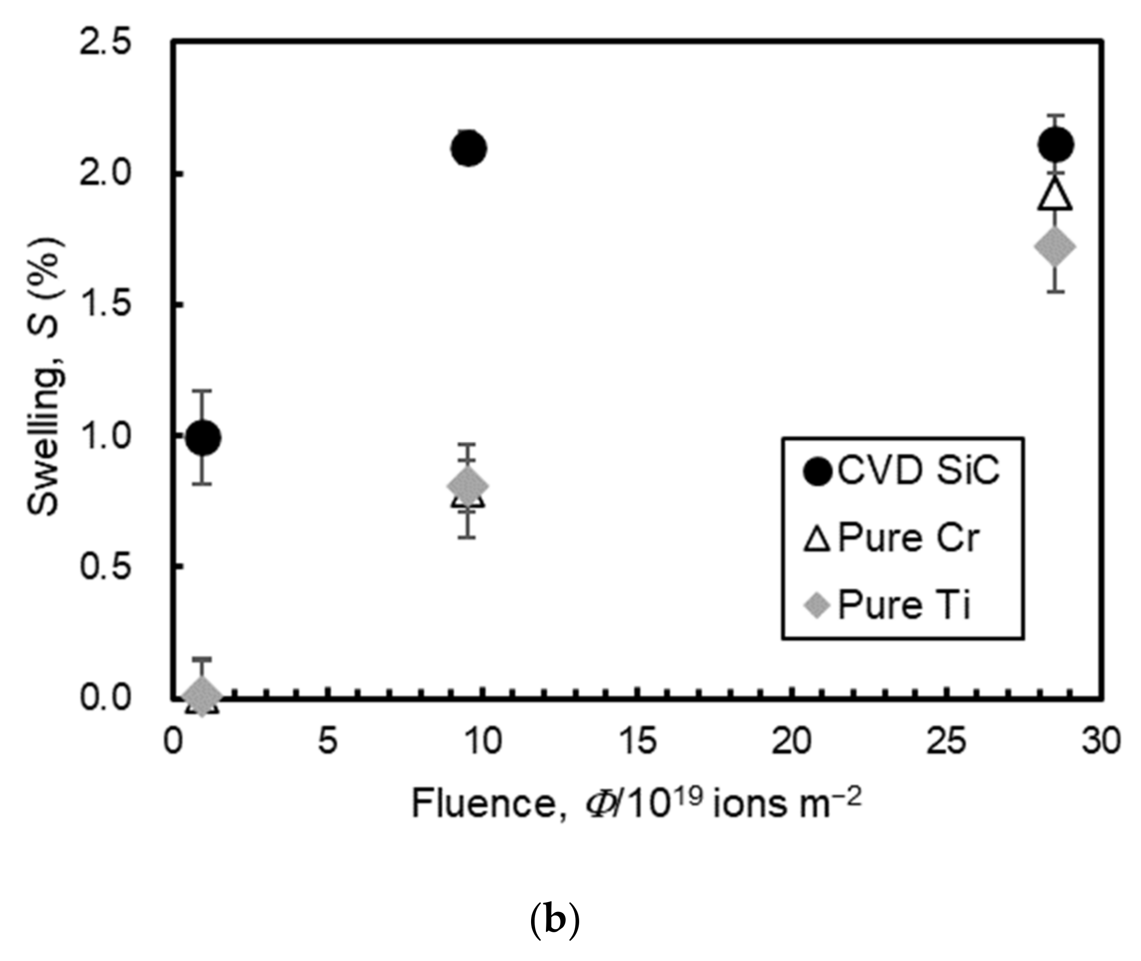
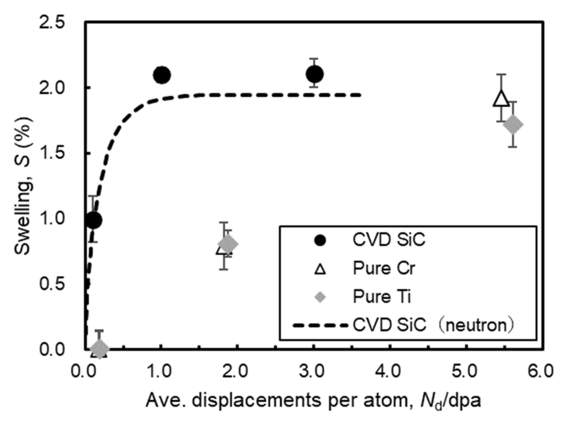
AFM images at the boundary between exposed (irradiated) and protected (unirradiated) regions of the surfaces were obtained. Figure 3 shows AFM images of the irradiated pure Ti bulk specimens. The left color bar in each image indicates height from the lowest point. The red portion indicates a high position and the blue portion indicates a low position. The height of the irradiated region increased with respect to the adjacent unirradiated regions. The step heights between irradiated and unirradiated regions of pure Ti, pure Cr and CVD SiC surfaces are shown in Figure 4a. The step heights of pure Ti and pure Cr were low and off-scale at a fluence of 0.95 × 10¹⁹ ions/m2 and they rose as the irradiation fluence increased to 2.85 × 10²⁰ ions/m2. On the other hand, the step height of CVD SiC rose sharply and had saturated by reaching the fluence of 9.51 × 10¹⁹ ions/m2. The swellings calculated by Equation (1) are plotted in Figure 4b. Pure Ti and pure Cr had almost the same swelling under the irradiation conditions in this study. The amount of their swelling became close to that of the CVD SiC at the fluence of 2.85 × 10²⁰ ions/m2.

3.2. Microstructural Analyses of Ti-Cr Multilayer Coating

3.2.1. SEM-EDS Analysis

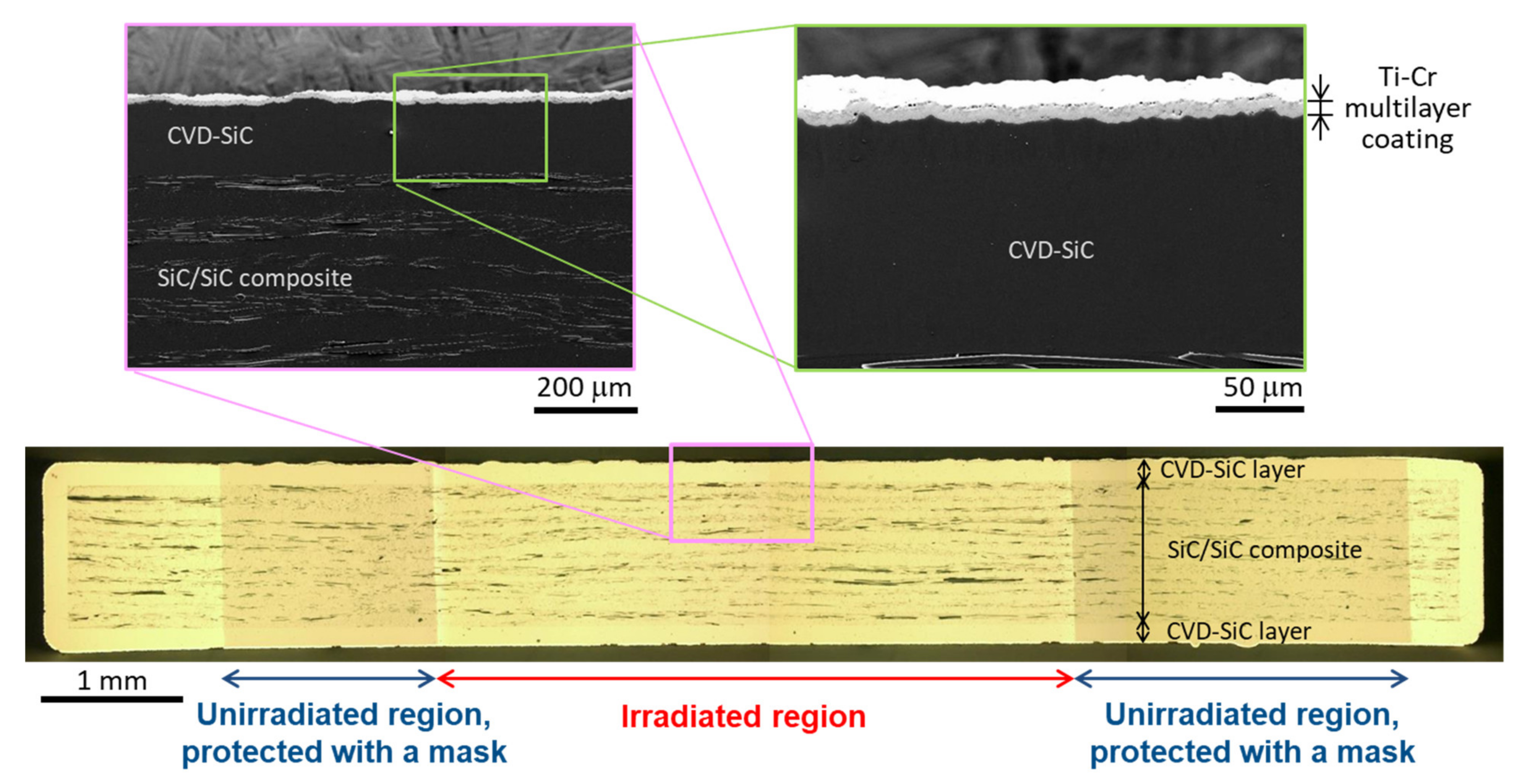
A cross section of the Ti-Cr multilayer-coated SiC after ion irradiation at a fluence of 2.85 × 10²⁰ ions/m2 is shown in Figure 5. CVD SiC with thickness of a about 150 μm was overcoated around SiC/SiC composite and Ti-Cr multilayer coating with a thickness of about 10 μm was on the CVD SiC. The irradiated region on the surface was observed to be brighter than the unirradiated (protected) region in the optical microscopy image of Figure 5.
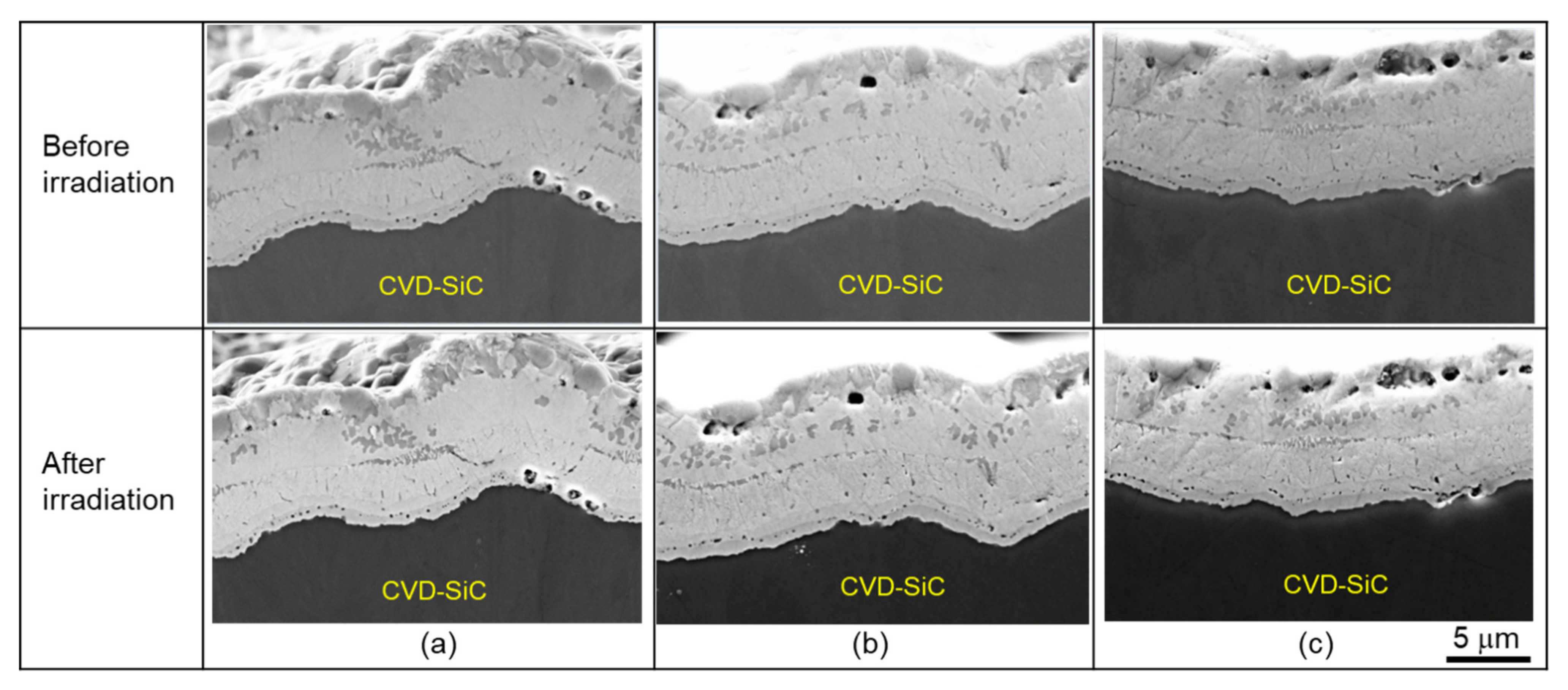
Cross-sectional secondary electron (SE) images of Ti-Cr multilayer-coated SiC before and after ion irradiation at fluences of 0.95 × 10¹⁹, 9.51 × 10¹⁹, and 2.85 × 10²⁰ ions/m2 are shown in Figure 6. The observed coatings on SiC were located near the center of the irradiated region in the specimens. For all three fluences, no differences were observed between the SE images before and after the irradiation.
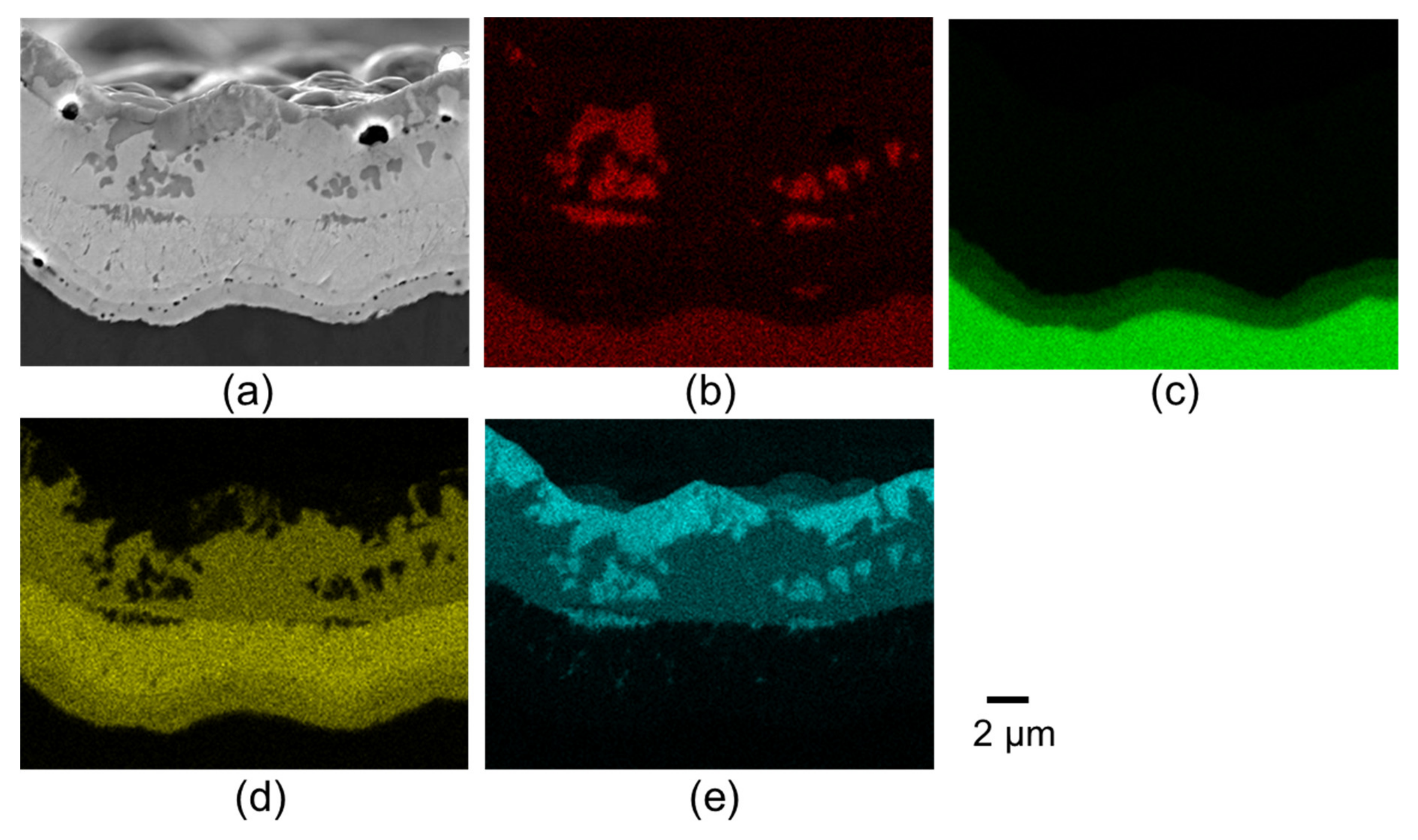
A cross-sectional SE image and EDS mappings of Ti-Cr multilayer-coated SiC after ion irradiation at a fluence of 2.85 × 10²⁰ ions/m2 are shown in Figure 7. The EDS mappings were obtained from the separated peak signals of the Ti L line, Cr L line, Si Kα line, and C K line. The EDS mappings indicated that the coating mainly consisted of the Ti layer, Ti-Cr mixed layer, Cr layer, and diffused Cr-SiC interface layer. The Ti layer consisted of Ti and Ti-and-C areas. The mixed Ti and Cr layer consisted of Ti-and-Cr and Ti-and-C areas. The Cr layer consisted of Cr and Ti-and-Cr areas. The diffused Cr-SiC interface layer consisted of Cr-and-Si (Cr-rich) and Cr-Si-and-C (Si-rich) areas. The multilayer structure and chemical components in each layer were the same as those of unirradiated coating analyzed in the authors’ previous study [49].

3.2.2. TEM-EDS Analysis

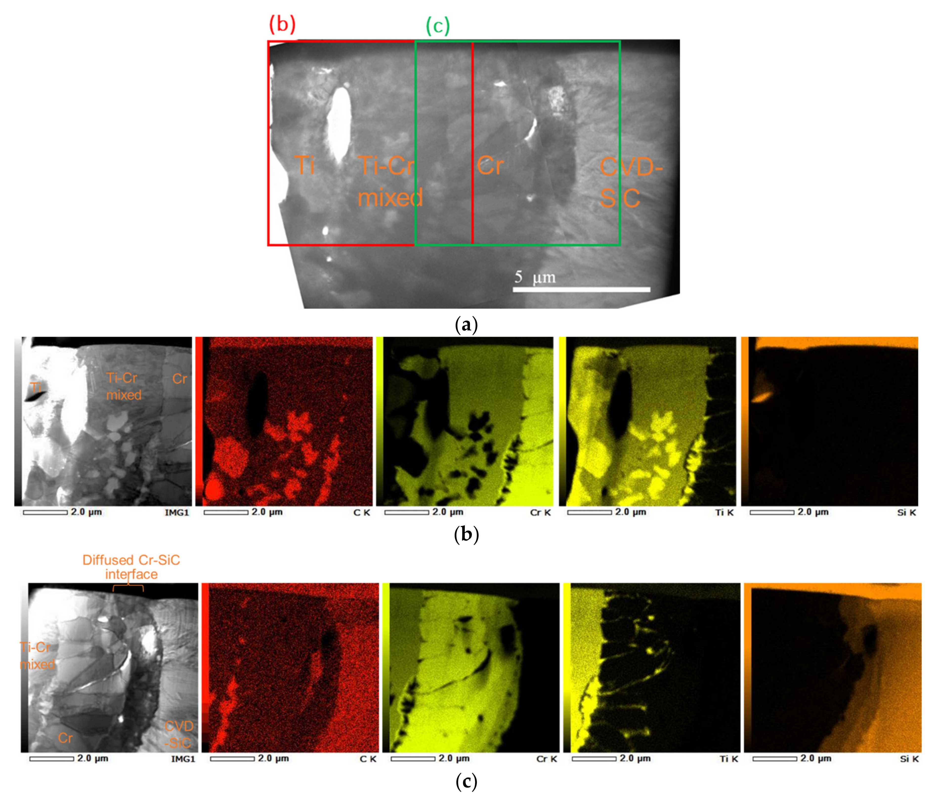
Cross-sectional bright field images and EDS mappings of a Ti-Cr multilayer-coated SiC foil sampled from the unirradiated region are shown in Figure 8. The area described in Figure 8b included Ti, Ti-Cr mixed, and Cr layers, and the other area described in Figure 8c included Ti-Cr mixed, Cr, diffused Cr-SiC interface, and CVD SiC layers. The Ti-and-C areas were distributed in the Ti and Ti-Cr mixed layers and on boundaries in the Cr layer.
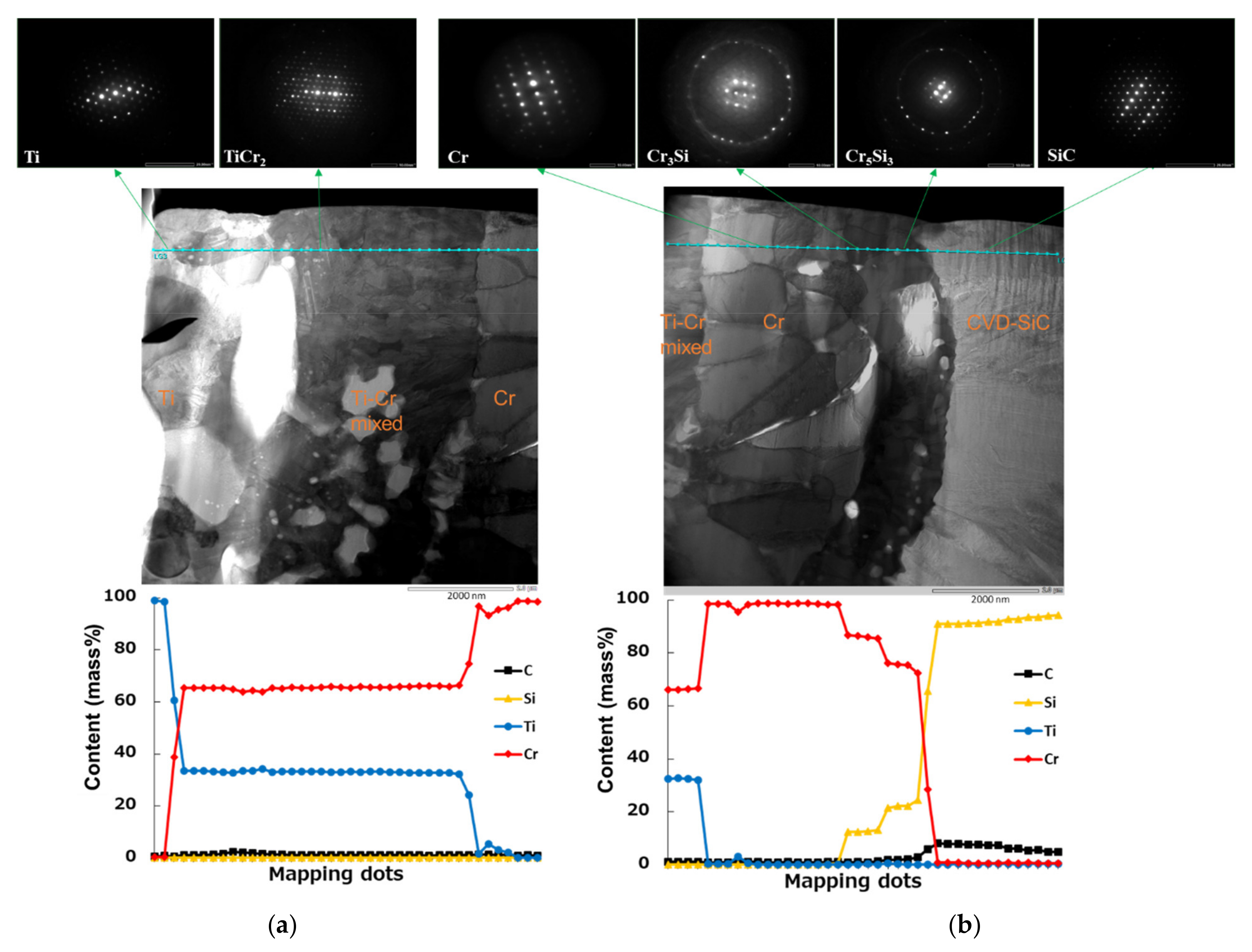
EDS line profiles of chemical composition and constituent phases along the analyzed lines (bule-green) in unirradiated Ti-Cr multilayer coating are shown in Figure 9. The constituent phases were identified by analyzing SADs and chemical compositions. Information for phase identification by SAD analysis using ReciPro ver4.828 [56] is shown in Table 1 from crystal structural data bases [57,58,59,60,61,62,63,64,65]. The Ti area in the Ti layer was α-Ti phase. The Cr area in the Cr layer is α-Cr phase. The SiC area in the CVD SiC layer was β-SiC phase. The Ti-and-Cr areas which were distributed in the Ti and Ti-Cr mixed layers had chemical compositions of Ti 33.4 mass% and Cr 65.5 mass% (Ti 35.2 at.% and Cr 64.6 at.%). The analyzed result of SAD revealed that the Ti-and-Cr area was TiCr2 phase. The Cr-and-Si (Cr-rich) area in the diffused Cr-SiC interface layer had chemical composition of Cr 87.1 mass% and Si 12.8 mass% (Cr 78.6 at.% and Si 21.3 at.%). The Cr-Si-and-C (Si-rich) area in the diffused Cr-SiC interface layer had chemical composition of Cr 76.6 mass% and Si 23.1 mass% (Cr 64.1 at.% and Si 35.7 at.%). From examining the Cr-Si-C ternary phase diagram at 1273 K [66], the chemical compositions suggested that the Cr-and-Si (Cr rich) area was Cr3Si and the Cr-Si-and-C (Si rich) area was Cr5Si3 or Cr5Si3Cx (x = 0.05~0.14 at 1673 K [67]).
Table 2 summarizes the identification of phases in the Ti-Cr multilayer coating heat-treated at 1223 K for 1.8 × 10³ s and also includes results from X-ray diffraction (XRD), SEM-EDS and electron backscatter diffraction (EBSD) identifications in the authors’ previous studies [47,48,49]. TEM-EDS analysis in this study revealed that the diffused Cr-SiC interface consisted of the Cr-and-Si phase (Cr3Si) layer and Cr-Si-and-C phase (Cr5Si3 or Cr5Si3Cx) layer.
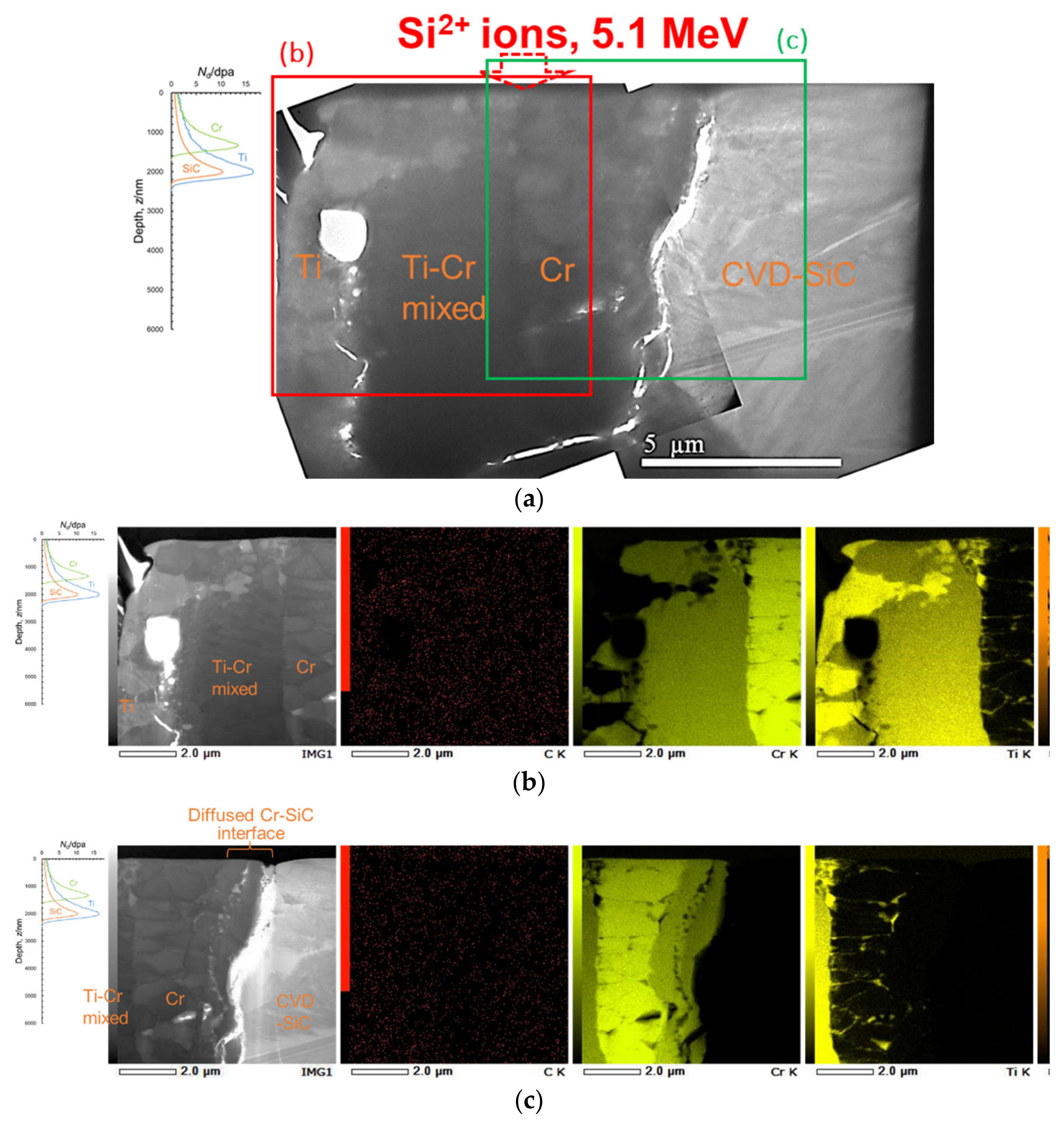
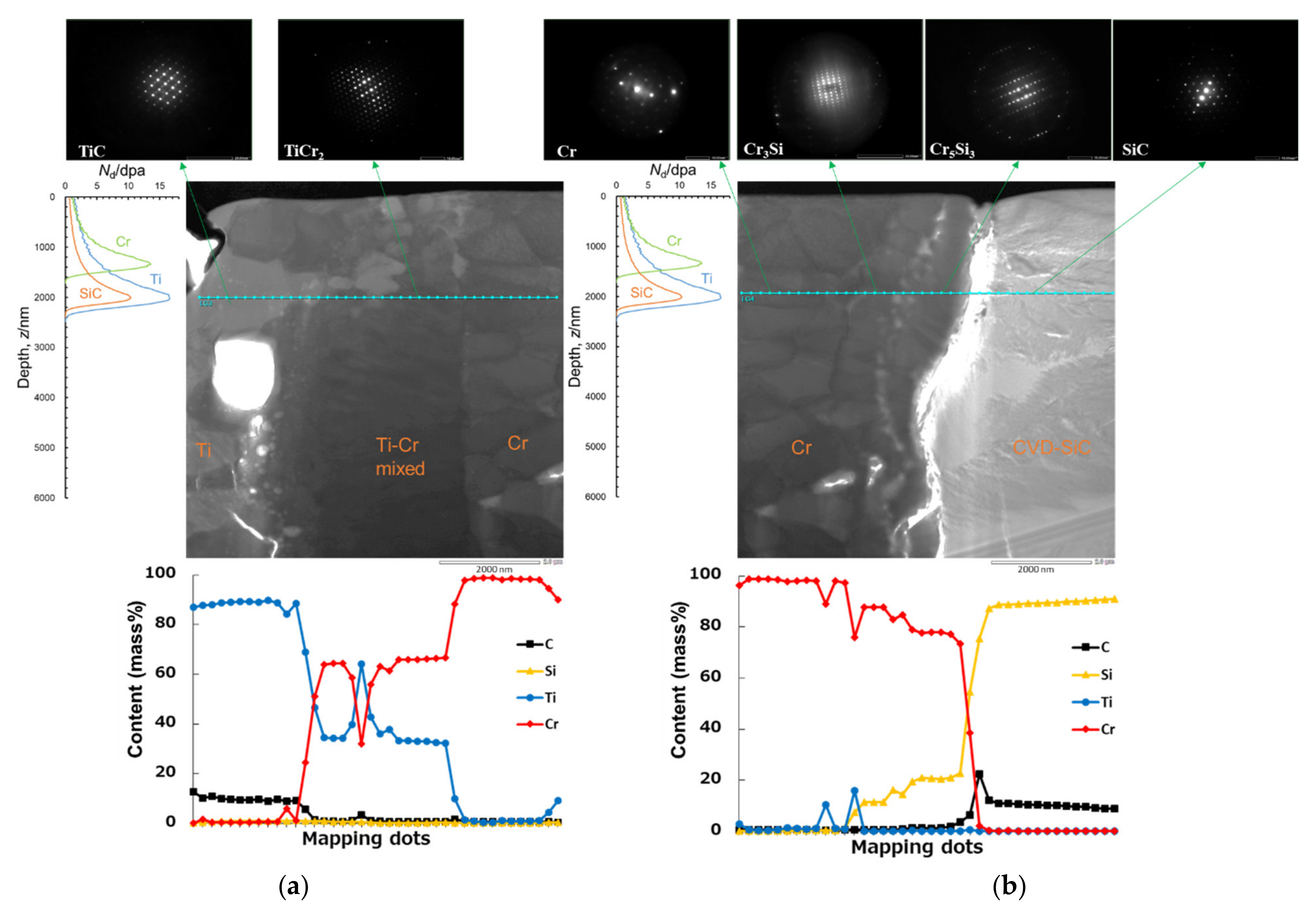
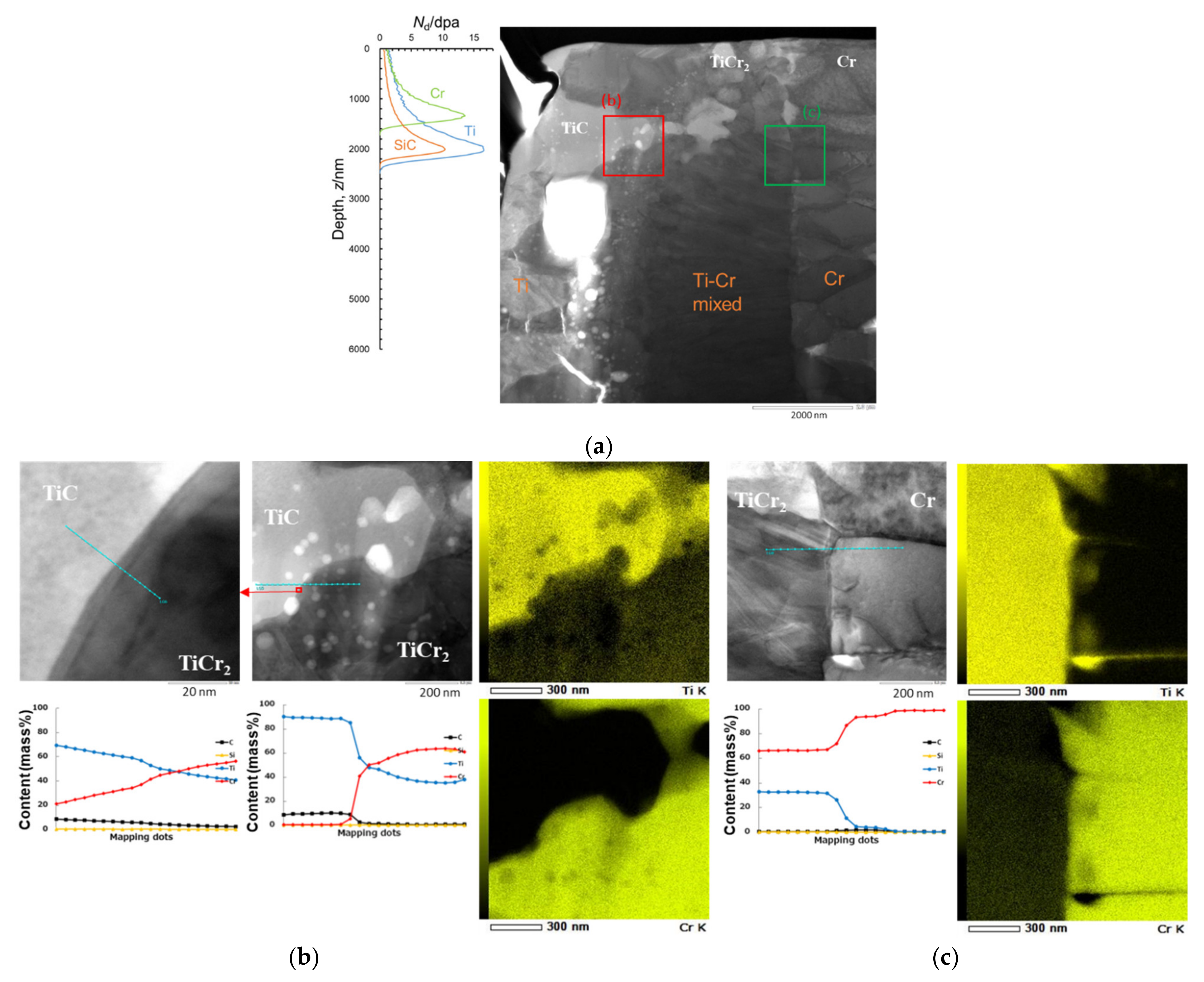
Cross-sectional bright field images and EDS mappings of Ti-Cr multilayer-coated SiC after Si ion irradiation at a fluence of 2.85 × 10²⁰ ions/m2 are shown in Figure 10. The depth profiles of the number of displacements per atom for Ti, Cr, and SiC calculated by SRIM indicated the damage range in the coating and it is shown for reference. The depth of peak number of displacements per atom was approximately 2000 nm for Ti and SiC and 1350 nm for Cr. The EDS line profiles of chemical composition and analyzed results of SAD revealed that TiC, TiCr2, Cr, Cr3Si, Cr5Si3 (or Cr5Si3Cx), and SiC phases were aligned along the analyzed line (bule-green) near the depth at the peak number of displacements per atom as shown in Figure 11. The separation between Cr5Si3 (or Cr5Si3Cx) and SiC phases occurred during the Ar gas milling process after the FIB process.
Bright field images and EDS analyzed the results focusing on the phase interfaces in Figure 11 are shown in Figure 12 and Figure 13. The EDS mappings and line profile at the TiC/TiCr2 phase interface near the peak positions of the number of displacements per atom, shown in Figure 12b, indicate that evidence of cascade mixing was not observed but voids were observed. The voids also were observed at the boundary between Ti and Ti-Cr mixed layers without the radiation effect as shown in the bright field image of Figure 12a. This observation suggests that the voids were not formed due to ion irradiation, but due to other mechanism such as Kirkendall effect. The EDS mappings and line profiles at TiCr2/Cr, Cr/Cr3Si, and Cr3Si/Cr5Si3 (or Cr5Si3Cx) phase interfaces near the depth for peak number of displacements per atom shown in Figure 12c and Figure 13 indicate that evidence of cascade mixing and large void formation was not observed. Therefore, the ion irradiation up to a fluence of 2.85 × 10²⁰ ions/m2 at 573 K did not produce cascade mixing and large void formation in this TEM observation.

4. Discussion

4.1. Swelling

Reports about swelling of Ti and Cr at LWR normal operation temperatures are limited, while swelling of SiC has been researched a lot [22,55,68,69]. The swelling of SiC for the temperature regime from approximately 473 K to 1073 K is referred to as the point-defect swelling regime [55]. The microstructural features are generically classified as black spots [55]. The swelling of SiC seems to approach saturation at a relatively low dose with a highly temperature dependence [55]. Transient swelling behavior of CVD SiC for neutron irradiation can be expressed by the following equation [68,69].
d S d γ = k S γ 1 / 3 e x p γ γ S C
here dS/dγ donates swelling rate, kS is the rate constant for swelling, γ is the fast fluence in dpa, and γsc is the characteristic fluence in dpa for swelling saturation obtained by a negative feedback mechanism.
k S   = 0.10612 1.5904 × 10 4 T + 6.0631 × 10 8 T 2
γ S C = 0.51801 2.7651 × 10 3 T + 9.4807 × 10 6 T 2 1.3095 × 10 8 T 3 + 6.7221 × 10 12 T 4
here T is temperature in Kelvin. The swelling, S, at temperature, T is found by integration (Equation (5)) of the swelling rate (Equation (2)).
S = 0 γ k S γ 1 / 3 e x p γ γ S C d γ
Swellings by ion irradiation in this study are plotted in Figure 14 along the abscissa as the average number of displacements per atom for each material. Swelling of SiC at 573 K by neutrons as predicted by Equation (5) is also described in Figure 14. The swelling of SiC by neutrons saturated at less than 1 dpa and showed relatively good agreement to the swelling by Si ions. Although the number of displacements per atom in the surface irradiated by ions had a depth distribution as shown in Figure 2c, the swelling at 3 dpa was almost the same over the damage region because swelling saturation occurred at less than 1 dpa.
Microstructures and swelling of pure Ti, pure Cr, and Cr coating irradiated at temperatures from 423 K to 723 K are summarized in Table 3 [43,70,71,72,73,74,75]. The microstructural features of pure Ti irradiated with neutrons and ions are generically classified as black spots or dislocation loops at these temperatures. Both black spots and dislocation loops were observed in pure Ti irradiated with Ti ions at 573 K [73]. Few results have been reported about swelling of pure Ti. Voids of 1.5–4 nm in diameter were observed in the microstructure of pure Cr irradiated with neutrons and ions at temperatures between 593 K and 723 K [43,74,75]. The swellings were estimated from size and density of voids. The swellings of pure Cr with ion irradiation at 5–50 dpa at temperatures between 673 K and 723 K are smaller than those estimated from the step height in this study. The difference might be caused by the measuring method and the irradiation temperature. At lower temperature, point defects are predominant rather than void formation. Thus, the mechanism change is presumed to increase swelling.

4.2. Stress in Coating

Provided that both swellings of a single layer coating and substrate are homogeneous, when the coating thickness is much smaller than the substrate thickness, stress, σC, in Ti or Cr single layer coating on SiC is simply estimated by Equation (6) [43].
σ C = E C 1 v C Δ L S Δ L C + T R T T α S α C d T + σ r e s
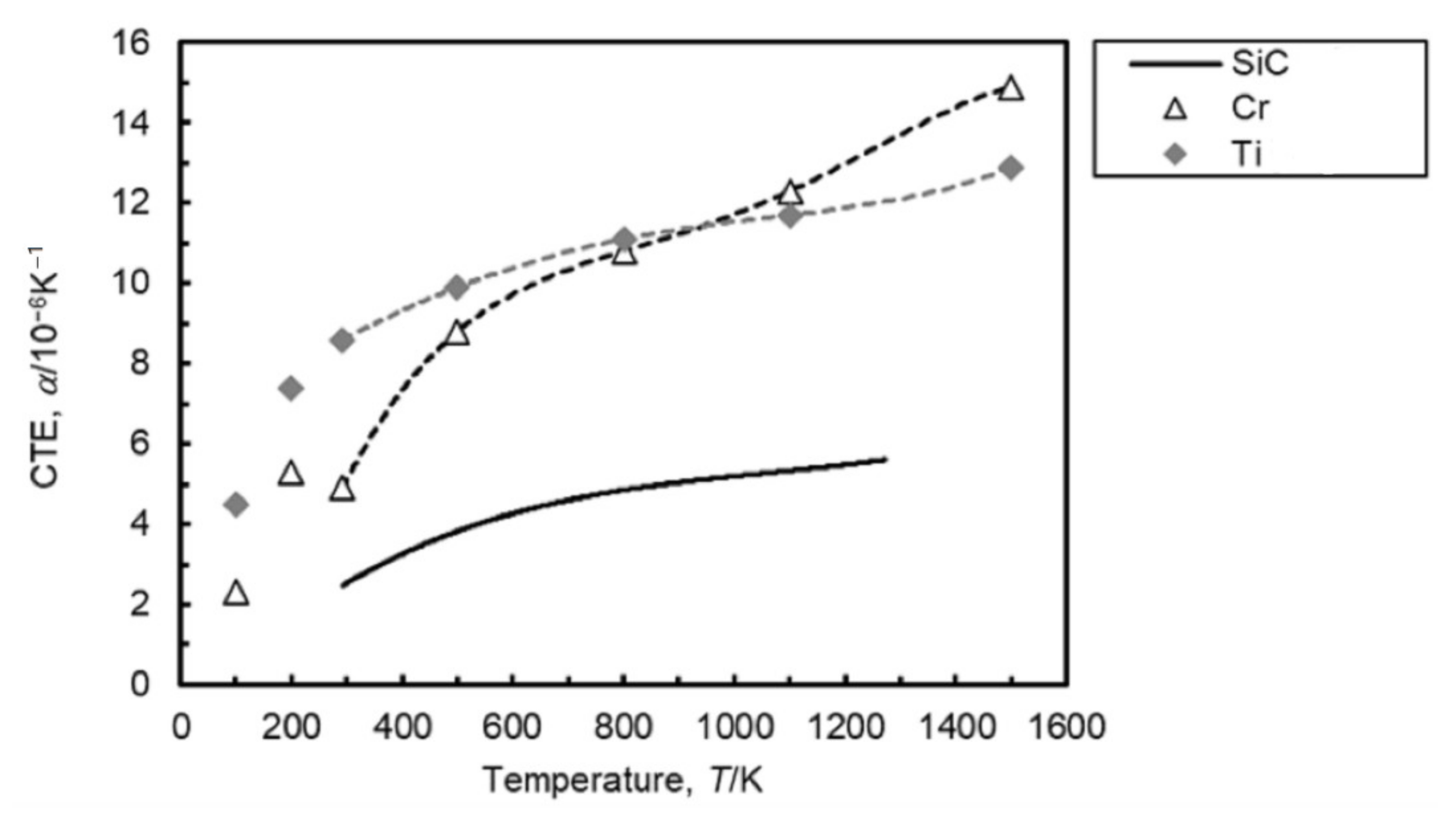
Here EC is Young’s modulus, vC is Poisson’s ratio of coating materials, ΔLS and ΔLC are fractional length change caused by irradiation swelling for substrate and coating, respectively, αS and αC are coefficients of thermal expansion (CTE) functions of substrate and coating, respectively, integrated from room temperature to the irradiation temperature, and σres is the residual stress in the coating at room temperature. The EC and vC are listed in Table 4. The ΔLS and ΔLC correspond to swelling, S defined as Equation (1). The CTEs and their regression curves of SiC, Cr, and Ti are shown in Figure 15. The σres was not considered in this discussion because it is thought to be smaller than stress caused by differential swelling. Residual stress in monolithic Cr coating on SiC is evaluated to be 0.22 GPa [43].
The estimated stresses are presented in Figure 16. Both pure Cr and Ti single layer coatings showed the maximum tensile stress at a fluence of 9.51 × 10¹⁹ ions/m2, corresponding to 1 dpa for SiC. Stress caused by the difference in thermal expansion from 293 K to 573 K was −0.4 GPa for Cr and −0.3 GPa for Ti coating on SiC. Thus, most of the stress in the coating was derived from differential swelling. Although swellings of pure Cr and pure Ti were almost the same as shown in Figure 4b, the Cr coating had higher stress than the Ti coating because of the difference in their Young’s modulus. It should be noted that the pure Ti and pure Cr irradiated with ions did not have uniform swelling that depended on fluence; however, SiC had uniform swelling in most of the irradiated region. It was considered that a larger strain was applied at the low fluence region.
The cross-sectional surface of the Ti-Cr multilayer-coated SiC irradiated up to the fluence of 2.85 × 10²⁰ ions/m2 experienced the maximum tensile stress condition during the ion irradiation. Nevertheless, delamination and cracking in the coating did not occur although the stress was generated over only the damage region from 1720 to 2490 nm in depth. In monolithic Cr coating on SiC irradiated with neutrons to a total fluence of 4.8 × 1024 n/m2 (>0.1 MeV), corresponding to about 0.5 dpa, cracks normal to interface with SiC were observed to propagate through the entire coating [43]. It was thought that tensile stress in the coating caused the cracks normal to the interface. The Ti and Ti-Cr mixed layers outside of the Cr layer in the Ti-Cr multilayer coating possibly suppressed crack generation by constraining the Cr layer. Since the diffused Cr-SiC interface formed by the thermal treatment is known to improve adhesion [49], it was possible to suppress crack generation along the interface.

5. Conclusions

The corrosion-resistant coating technology using Ti-Cr multilayer was developed to suppress silica dissolution from SiC fuel cladding into reactor coolant under normal operation conditions. The effect of radiation on adhesion of the coating to SiC substrate and its microstructure characteristics was investigated at 573 K for the Si ion irradiation up to a fluence of 2.85 × 10²⁰ ions/m2 corresponding to 3 dpa for SiC. The conclusions are listed as follows:
  • Measurement of swelling in pure Ti, pure Cr, and SiC revealed that the maximum tensile strain equivariant of the swelling difference might be generated in the coating on the SiC substrate by irradiation corresponding to 1 dpa for SiC;
  • No delamination and cracking were observed in cross-sectional specimens of the coated SiC irradiated up to 3 dpa, although it had experienced the maximum tensile strain condition;
  • According to analyses using TEM-EDS, large void formation and cascade mixing due to irradiation were not observed in the coating. The swelling in the coating irradiated at 573 K was presumed to be caused by other mechanisms such as point defects rather than void formation.

Author Contributions

Conceptualization, R.I. and T.H.; methodology, T.H.; validation, R.I., Y.H. and T.H.; formal analysis, Y.H. and H.B.; investigation, R.I., Y.H. and H.B.; resources, R.I.; data curation, R.I. and T.H.; writing—original draft preparation, R.I.; writing—review and editing, T.H.; visualization, R.I.; supervision, T.K.; project administration, T.H.; funding acquisition, T.K. All authors have read and agreed to the published version of the manuscript.

Funding

This research received no external funding.

Institutional Review Board Statement

Not applicable.

Informed Consent Statement

Not applicable.

Data Availability Statement

The data presented in this study are openly available in The Materials Project at https://doi.org/10.1063/1.4812323 [64] (accessed on 28 April 2022), in AFLOW at https://doi.org/10.48550/arXiv.2012.05961 [65] (accessed on 28 April 2022) and in Tables of Physical and Chemical Constants at www.kayelaby.npl.co.uk [76,77] (accessed on 28 April 2022).

Acknowledgments

The authors are grateful to Yutaka Watanabe, Akira Hasegawa, Eiji Akiyama, Hiroshi Abe, and Sosuke Kondo of Tohoku University for discussions about microstructural formation of the Ti-Cr multilayer coating. The authors would like to thank Tsuneyuki Hashimoto and Yasuhisa Aono of Hitachi, Ltd. for their advice in the course of this work. The contributions of partner-researchers of the Japan Atomic Energy Agency for their views on this topic are also appreciated.

Conflicts of Interest

The authors declare no conflict of interest.

References

  1. The Fukushima Daiichi Accident; International Atomic Energy Agency: Vienna, Austria, 2015.
  2. Zinkle, S.J.; Terrani, K.A.; Gehin, J.C.; Ott, L.J.; Snead, L.L. Accident tolerant fuels for LWRs: A perspective. J. Nucl. Mater. 2014, 448, 374–379. [Google Scholar] [CrossRef]
  3. Ishibashi, R.; Ikegawa, T.; Noshita, K.; Kitou, K.; Kamoshida, M. Hydrogen explosion prevention system using SiC fuel cladding for large scale BWRs with inherently safe technologies. Mech. Eng. J. 2016, 3, 15-00215. [Google Scholar] [CrossRef] [Green Version]
  4. Ikegawa, T.; Kondo, T.; Sakamoto, K.; Yamashita, S. Performance Evaluation of Accident Tolerant Fuel Claddings during Severe Accidents of BWRs. In Proceedings of the TopFuel 2018, Prague, Czech Republic, 30 September–4 October 2018; European Nuclear Society: Brussels, Belgium, 2018. Paper No. A0131. [Google Scholar]
  5. Katoh, Y.; Terrani, K.A.; Snead, L.L. Systematic Technology Evaluation Program for SiC/SiC Composite-Based Accident-Tolerant LWR Fuel Cladding and Core Structures; ORNL/TM-2014/210; Oak Ridge National Laboratory: Oak Ridge, TN, USA, 2014. [Google Scholar]
  6. Terrani, K.A. Accident tolerant fuel cladding development: Promise, status, and challenge. J. Nucl. Mater. 2018, 501, 13–30. [Google Scholar] [CrossRef]
  7. State-of-the-Art Report on Light Water Reactor Accident-Tolerant Fuels; NEA No. 7317; OECD: Paris, France, 2018.
  8. Rohmer, E.; Martin, E.; Lorrette, C. Mechanical properties of SiC/SiC braided tubes for fuel cladding. J. Nucl. Mater. 2014, 453, 16–21. [Google Scholar] [CrossRef]
  9. Shapovalov, K.; Jacobsen, G.M.; Alva, L.; Truesdale, N.; Deck, C.P.; Huang, X. Strength of SiCf-SiCm composite tube under uniaxial and multiaxial loading. Trans. Am. Nucl. Soc. 2018, 120, 371–374. [Google Scholar] [CrossRef]
  10. Hinoki, T.; Kano, F.; Kondo, S.; Kawaharada, Y.; Tsuchiya, Y.; Lee, M.; Sakai, H. Development of liquid phase sintering silicon cabide composites for light water reactor. Coatings 2022, 12, 623. [Google Scholar] [CrossRef]
  11. Shimoda, K.; Hinoki, T.; Kishimoto, H.; Kohyama, A. Enhanced high-temperature performances of SiC/SiC composites by high densification and crystalline structure. Compos. Sci. Technol. 2011, 71, 326–332. [Google Scholar] [CrossRef]
  12. Katoh, Y.; Snead, L.L.; Cheng, T.; Shih, C.; Lewis, W.D.; Koyanagi, T.; Hinoki, T.; Henager, C.H., Jr.; Ferraris, M. Radiation-tolerant joining technologies for silicon carbide ceramics and composites. J. Nucl. Mater. 2014, 448, 497–511. [Google Scholar] [CrossRef]
  13. Ishibashi, R.; Takamori, Y.; Zhang, X.; Kondo, T.; Miyazaki, K. Development of Joining Technology Using Local Heating for SiC Fuel Cladding. In Proceedings of the Top Fuel 2016, Boise, ID, USA, 11–15 September 2016; American Nuclear Society: La Grange Park, IL, USA, 2016. Paper No. 17636. pp. 805–813. [Google Scholar]
  14. Khalifa, H.E.; Koyanagi, T.; Jacobsen, G.; Deck, C.P. Radiation stable, hybrid, chemical vaper infiltration/preceramic polymer joining of silicon carbide components. J. Nucl. Mater. 2017, 487, 91–95. [Google Scholar] [CrossRef]
  15. Deck, C.P.; Gonderman, S.; Jacobsen, G.M.; Sheeder, J.; Oswald, S.; Haefelfinger, R.; Shapovalov, K.S.; Khalifa, H.E.; Gazza, J.; Lyons, J.; et al. Overview of general atomics SiGAtm SiC-SiC composite development for accident toletrant fuel. Trans. Am. Nucl. Soc. 2019, 120, 371–374. [Google Scholar]
  16. Ishibashi, R.; Kida, M.; Shibata, M.; Kondo, T.; Yamashita, S.; Kawanishi, T.; Fukahori, T. Joining Technology with Corrosion Resistant Coating for Silicon-carbide Fuel Cladding. In Proceedings of the Top Fuel 2019, Seattle, WA, USA, 22–27 September 2019; American Nuclear Society: La Grange Park, IL, USA, 2019. Paper No. 29441. pp. 753–759. [Google Scholar]
  17. Shapovalov, K.; Jacobsen, G.M.; Shih, C.; Deck, C.P. C-ring testing of nuclear grade silicon carbide composites at temperatures up to 1900 °C. J. Nucl. Mater. 2019, 522, 184–191. [Google Scholar] [CrossRef]
  18. Pint, B.; Terrani, K.; Brady, M.; Cheng, T.; Keiser, J. High temperature oxidation of fuel cladding candidate materials in steam-hydrogen environements. J. Nucl. Mater. 2013, 440, 420–427. [Google Scholar] [CrossRef]
  19. Terrani, K.A.; Bruce, P.; Parish, C.; Silva, C.; Snead, L.; Katoh, Y. Silicon carbide oxidation in steam up to 2 MPa. J. Am. Ceram. Soc. 2014, 97, 2331–2352. [Google Scholar] [CrossRef]
  20. Pham, H.; Nagae, Y.; Kurata, M.; Bottomley, D.; Furumoto, K. Oxidation kinetics of silicon carbide in steam at temperature range of 1400 to 1800 °C studied by laser heating. J. Nucl. Mater. 2020, 529, 151939. [Google Scholar] [CrossRef]
  21. Avincola, V.; Grosse, M.; Stegmaier, U.; Steinbrueck, M.; Seifert, H. Oxidation at high temperatures in steam atmosphere and quench of silicon carbide composites for nuclear application. Nucl. Eng. Des. 2015, 195, 468–478. [Google Scholar] [CrossRef]
  22. Pham, H.V.; Kurata, M.; Steinbrueck, M. Steam Oxidation of Silicon Carbide at High Temperatures for Application as Accident Tolerant Fuel Cladding, an Overview. Thermo 2021, 1, 151–167. [Google Scholar] [CrossRef]
  23. Terrani, K.A.; Yang, Y.; Kim, Y.J.; Rebak, R.; Meyer, H.M.; Gerczak, T.J. Hydrothermal corrosion of SiC in LWR coolant environments in the absence of irradiation. J. Nucl. Mater. 2015, 465, 488–498. [Google Scholar] [CrossRef] [Green Version]
  24. Pantano, M.A.; McKrell, T.J.; Guenoun, P.B.; Kazimi, M.S.; Carpenter, D.M.; Kohse, G.E. Silicon Carbide Behavior Under Prototypic LWR Chemistry/Neutron Flux and Accident Conditions, Accident Tolerant Fuel Concepts for Light Water Reactors; IAEA-TECDOC-1797; International Atomic Energy Agency: Vienna, Austria, 2016; pp. 334–350. [Google Scholar]
  25. Parish, C.M.; Terrani, K.A.; Kim, Y.-J.; Koyanagi, T.; Katoh, Y. Microstructure and hydrothermal corrosion behavior of NITE-SiC with various sintering additives in LWR coolant environments. J. Eur. Ceram. Soc. 2017, 37, 1261–1279. [Google Scholar] [CrossRef] [Green Version]
  26. Doyle, P.J.; Raiman, S.S.; Rebak, R.; Terrani, K.A. Characterization of the hydrothermal corrosion behavior of ceramics for accident tolerant fuel cladding. In Proceedings of the 18th International Conference on Environmental Degradation of Materials—Water Reactors, Portland, OR, USA, 13–17 August 2017; The Minerals, Metals & Materials Society: Pittsburgh, PA, USA, 2018; pp. 269–280. [Google Scholar]
  27. Doyle, P.J.; Zinkle, S.; Raiman, S.S. Hydrothermal corrosion behavior of CVD SiC in high temperature water. J. Nucl. Mater. 2020, 539, 152241. [Google Scholar] [CrossRef]
  28. Doyle, P.; Sun, K.; Snead, L.; Katoh, Y.; Bartels, D.; Zinkle, S.; Raiman, S. The effects of neutron and ionizing irradiation on the aqueous corrosion of SiC. J. Nucl. Mater. 2020, 536, 152190. [Google Scholar] [CrossRef]
  29. Kondo, S.; Mouri, S.; Hyodo, Y.; Hinoki, T.; Kano, F. Role of irradiation-induced defects on SiC dissolution in hot water. Corros. Sci. 2016, 112, 402–407. [Google Scholar] [CrossRef]
  30. Kondo, S.; Lee, M.; Hinoki, T.; Hyodo, Y.; Kano, F. Effect of irradiation damage on hydrothermal corrosion of SiC. J. Nucl. Mater. 2015, 464, 36–42. [Google Scholar] [CrossRef]
  31. Shin, J.H.; Kim, D.; Lee, H.J.; Lee, H.-G.; Park, J.Y.; Kim, W.-J. Factors affecting the hydrothermal corrosion behavior of chemically vaper deposited silicon carbides. J. Nucl. Mater. 2019, 518, 350–356. [Google Scholar] [CrossRef]
  32. Csontos, A.; Capps, N. Accident-Tolerant Fuel Valuation: Safety and Economic Benefits (Revision 1); EPRI Technical Report 3002015091; Electric Power Research Institute: Palo Alto, CA, USA, 2019. [Google Scholar]
  33. AESJ-SC-S007:2019; The Standards of the Atomic Energy Society of Japan, Water Chemistry Guidelines for Boiling Water Reactors: 2019. Atomic Energy Society of Japan: Tokyo, Japan, 2019. (In Japanese)
  34. Hirayama, H.; Kawakubo, T.; Goto, A.; Kaneko, T. Corrosion behavior of silicon carbide in 290 °C water. J. Am. Ceram. Soc. 1989, 72, 2049–2053. [Google Scholar] [CrossRef]
  35. Kim, W.-J.; Hwang, H.S.; Park, J.Y. Corrosion behavior of reaction-bonded silicon carbide ceramics in high-temperature water. J. Mater. Sci. Lett. 2002, 21, 733–735. [Google Scholar] [CrossRef]
  36. Kim, W.-J.; Hwang, H.S.; Park, J.Y.; Ryu, W.-S. Corrosion behaviors of sintered and chemically vapor deposited silicon carbide ceramics in water at 360 °C. J. Mater. Sci. Lett. 2003, 22, 581–584. [Google Scholar] [CrossRef]
  37. Ang, C.K.; Terrani, K.A.; Burns, J.; Katoh, Y. Examination of Hybrid Metal Coatings for Mitigation of Fission Product Release and Corrosion Protection of LWR SiC/SiC; ORNL/TM-2016/332; Oak Ridge National Laboratory: Oak Ridge, TN, USA, 2016. [Google Scholar]
  38. Ang, C.; Katoh, Y.; Kemary, C.; Kiggans, J.O.; Terrani, K.A. Chromium-based mitigation coatings on SiC materials for fuel cladding. Trans. Am. Nucl. Soc. 2016, 114, 1095–1097. [Google Scholar]
  39. Raiman, S.S.; Doyle, P.; Ang, C.; Katoh, Y.; Terrani, K.A. Hydrothermal corrosion of coatings on silicon carbide in boiling water reactor conditions. Corrosion 2019, 75, 217–223. [Google Scholar] [CrossRef]
  40. Raiman, S.S.; Ang, C.; Doyle, P.; Terrani, K.A. Hydrothermal Corrosion of SiC Materials for Accident Tolerant Fuel Cladding with and Without Mitigation Coatings. In Proceedings of the 18th International Conference on Environmental Degradation of Materials in Nuclear Power Systems—Water Reactors, Portland, OR, USA, 13–17 August 2017; The Minerals, Metals & Materials Society: Pittsburgh, PA, USA, 2018; pp. 259–267. [Google Scholar]
  41. Doyle, P.J.; Raiman, S.S.; Ang, C.; Katoh, Y.; Zinkle, S. Evaluation of the Corrosion Kinetics of SiC with and without Mitigation Coatings in LWR Chemistries. In Proceedings of the 19th International Conference on Environmental Degradation of Materials in Nuclear Power Systems—Water Reactors, Boston, MA, USA, 18–22 August 2019; The American Nuclear Society: La Grange Park, IL, USA, 2019; pp. 417–427. [Google Scholar]
  42. Raiman, S.S.; Doyle, P.J.; Ang, C.; Koyanagi, T.; Carpenter, D.; Terrani, K.A.; Katoh, Y. Irradiation-induced Cracking of Dual-purpose Coatings on SiC. In Proceedings of the 18th International Conference on Environmental Degradation of Materials in Nuclear Power Systems—Water Reactors, Boston, MA, USA, 18–22 August 2019; The American Nuclear Society: La Grange Park, IL, USA, 2019; pp. 428–433. [Google Scholar]
  43. Doyle, P.J.; Doyle, P.J.; Koyanagi, T.; Ang, C.; Snead, L.; Mouche, P.; Katoh, Y.; Raiman, S.S. Evaluation of the effects of neutron irradiation on first-generation corrosion mitigation coatings on SiC for accident-tolerant fuel cladding. J. Nucl. Mater. 2020, 536, 152203. [Google Scholar] [CrossRef]
  44. Quillin, K.; Yeom, H.; Dabney, T.; McFariand, M.; Sridharan, K. Experimental evaluation of direct current magnetron sputtered and high-power impulse magnetron sputtered Cr coatings on SiC for light water reactor applications. Thin Solid Films 2020, 716, 138431. [Google Scholar] [CrossRef]
  45. Mouche, P.A.; Evans, A.; Zhong, W.; Koyanagi, T.; Katoh, Y. Effects of sample bias on adhesion of magnetron sputtered Cr coatings on SiC. J. Nucl. Mater. 2021, 556, 153251. [Google Scholar] [CrossRef]
  46. Kain, K.A.; Stack, P.I.M.; Mouche, P.A.; Pillai, R.R.; Pint, B.A. Steam oxidation of chromium corrosion barrier coatings for sic-based accident tolerant fuel cladding, J. Nucl. Mater. 2021, 543, 152561. [Google Scholar] [CrossRef]
  47. Ishibashi, R.; Tanabe, S.; Kondo, T.; Yamashita, S.; Nagase, F. Improving the Corrosion Resistance of Silicon Carbide for Fuel in BWR Environments by Using a Metal Coating. In Proceedings of the WRFPM 2017, Jeju Island, Korea, 10–14 September 2017; Korean Nuclear Society: Daejeon, Korea, 2017. Paper No. A-177_F-177. [Google Scholar]
  48. Ishibashi, R.; Tanabe, S.; Kondo, T.; Yamashita, S.; Fukahori, T. Improvement of Corrosion Resistant Coating for Silicon-carbide Fuel Cladding in Oxygenated High Temperature Water. In Proceedings of the TopFuel 2018, Prague, Czech Republic, 30 September–4 October 2018; European Nuclear Society: Brussels, Belgium, 2018. Paper No. A0072. [Google Scholar]
  49. Ishibashi, R.; Ishida, K.; Kondo, T.; Watanabe, Y. Corrosion-resistant metallic coating on silicon carbide for use in high-temperature water. J. Nucl. Mater. 2021, 557, 153214. [Google Scholar] [CrossRef]
  50. Ishibashi, R.; Shibata, M.; Sasaki, M.; Kondo, T. Hydrothermal Corrosion Evaluation of Titanium-coated Silicon Carbide Tube with End-plug Joints in Oxygenated High Temperature Water. In Proceedings of the Top Fuel 2021, Santander, Spain, 24–28 October 2021; European Nuclear Society: Brussels, Belgium, 2021. No. 82. [Google Scholar]
  51. Yueh, K. SiC Composite for Fuel Structure Applications; DOE-EPRI-0000539; Electric Power Research Institute: Palo Alto, CA, USA, 2017. [Google Scholar]
  52. Ziegler, J.F.; Ziegler, M.D.; Biersak, J.P. SRIM—The stopping and range of ions in matter. Nucl. Instr. Meth. 2010, B 268, 1818–1823. [Google Scholar] [CrossRef] [Green Version]
  53. ASTM E521-16; Standard Practice for Investigating the Effects of Neutron Radiation Damage Using Charged-Particle Irradiation. ASTM International: West Conshohocken, PA, USA, 2016.
  54. Devanathan, R.; Weber, W.J.; Gao, F. Atomic scale simulation of defect production in irradiated 3C-SiC. J. Appl. Phys. 2001, 90, 2303–2309. [Google Scholar] [CrossRef]
  55. Snead, L.L.; Nozawa, T.; Katoh, Y.; Byun, T.S.; Kondo, S.; Petti, D.A. Handbook of SiC properties for fuel performance modeling. J. Nucl. Mater. 2007, 371, 329–377. [Google Scholar] [CrossRef]
  56. Seto, Y.; Ohtsuka, M. ReciPro: Free and open-source multipurpose crystallographic software integrating a crystal model database and viewer, diffraction and microscopy simulators, and diffraction data analysis tools. J. Appl. Cryst. 2022, 55, 397–410. [Google Scholar] [CrossRef]
  57. The Materials Project mp-72. Available online: https://materialsproject.org/materials/mp-72/ (accessed on 4 April 2022).
  58. The Materials Project mp-631. Available online: https://materialsproject.org/materials/mp-631/ (accessed on 4 April 2022).
  59. The Materials Project mp-1589. Available online: https://materialsproject.org/materials/mp-1589/ (accessed on 4 April 2022).
  60. The Materials Project mp-90. Available online: https://materialsproject.org/materials/mp-90/ (accessed on 4 April 2022).
  61. The Materials Project mp-729. Available online: https://materialsproject.org/materials/mp-729/ (accessed on 4 April 2022).
  62. AFLOW Prototype: A5B3_oC16_65_aeh_bj. Available online: https://aflow.org/material/?id=aflow:1016082d8a829e2f (accessed on 1 April 2022).
  63. AFLOW Prototype: AB_cF8_225_b_a. Available online: https://aflow.org/material/?iid=aflow:0e5c7dd4d4402dff (accessed on 18 May 2022).
  64. Jain, A.; Omg, S.P.; Hautier, G.; Chen, W.; Richards, W.D.; Dacek, S.; Cholia, S.; Gunter, D.; Skinner, D.; Ceder, G.; et al. The Materials Project: A materials genome approach to accelerating materials innovation. APL Mater. 2013, 1, 011002. [Google Scholar] [CrossRef] [Green Version]
  65. Hicks, D.; Mehl, M.J.; Esters, M.; Oses, C.; Levy, O.; Hart, G.L.W.; Toher, C.; Curtarolo, S. The AFLOW Library of Crystallographic Prototypes: Part 3. Comp. Mater. Sci. 2021, 199, 110450. [Google Scholar] [CrossRef]
  66. Du, Y.; Schuster, J.C.; Perring, L. Experimental investigation and thermodynamic description of the constitution of the ternary system Cr-Si-C. J. Am. Ceram. Soc. 2000, 83, 2067–2073. [Google Scholar] [CrossRef]
  67. Pellegrini, P.W.; Giessen, B.C.; Feldman, J.M. A survey of the Cr-rich area of the Cr-Si-C phase diagram. J. Electrochem. Soc. 1972, 119, 535–537. [Google Scholar] [CrossRef]
  68. Katoh, Y.; Snead, L.L.; Parish, C.M.; Hinoki, T. Observation and possible mechanism of irradiation induced creep in ceramics. J. Nucl. Mater. 2013, 434, 141–151. [Google Scholar] [CrossRef]
  69. Katoh, Y.; Ozawa, K.; Shih, C.; Nozawa, T.; Shinavski, R.J.; Hasegawa, A.; Snead, L.L. Continuous SiC fiber, CVI SiC matrix composites for nuclear applications: Properties and irradiation effects. J. Nucl. Mater. 2014, 448, 448–476. [Google Scholar] [CrossRef]
  70. Higashiguchi, Y.; Kayano, H.; Yajima, S. Relation between microstructure and recovery behavior in fast neutron irradiated titanium. J. Nucl. Sci. Technol. 1976, 13, 454–458. [Google Scholar] [CrossRef]
  71. Brumhall, J.L.; Kulcinski, G.L.; Kissinger, H.E.; Mastel, B. Microstructural analysis of neutron irradiated titanium and rhenium. Radiat. Eff. 1971, 9, 273–278. [Google Scholar] [CrossRef]
  72. Jostsons, A.; Black, R.C.; Kelly, P.M. Characterization of dislocation loops in neutron-irradiated titanium. Philos. Mag. A 1980, 41, 903–916. [Google Scholar] [CrossRef]
  73. Jouanny, E.; Doriot, S.; Malaplate, J.; Dehmas, M.; Allais, L.; Le Thuaut, M.; Millot, T. Evolution of defects in titanium grade 2 under Ti2+ ion irradiation. J. Microsc. 2017, 265, 275–286. [Google Scholar] [CrossRef] [Green Version]
  74. Kuprin, A.S.; Belous, V.A.; Voyevodin, V.N.; Vasilenko, R.L.; Ovcharenko, V.D.; Tolstolutskaya, G.D.; Kopanets, I.E.; Kolodiy, I.V. Irradiation resistance of vacuum arc chromium coatings for zirconium alloy fuel claddings. J. Nucl. Mater. 2018, 510, 163–167. [Google Scholar] [CrossRef]
  75. Ryabikovskaya, E.; French, A.; Gabriel, A.; Kim, H.; Wang, T.; Shirvan, K.; Garner, F.A.; Shao, L. Irradiation-induced swelling of pure chromium with 5 MeV Fe ions in the temperature range 450–650 °C. J. Nucl. Mater. 2021, 543, 152585. [Google Scholar] [CrossRef]
  76. 2.3.5 Thermal Expansion, Kaye and Laby Online. In Tables of Physical and Chemical Constants, 16th ed.; Version 1.0 (2005); The National Physical Laboratory: Middlesex, UK, 1995; Available online: www.kayelaby.npl.co.uk (accessed on 28 April 2022).
  77. 2.2.2 Elasticities and Strengths, Kaye and Laby Online. In Tables of Physical and Chemical Constants, 16th ed.; Version 1.0 (2005); The National Physical Laboratory: Middlesex, UK, 1995; Available online: www.kayelaby.npl.co.uk (accessed on 28 April 2022).
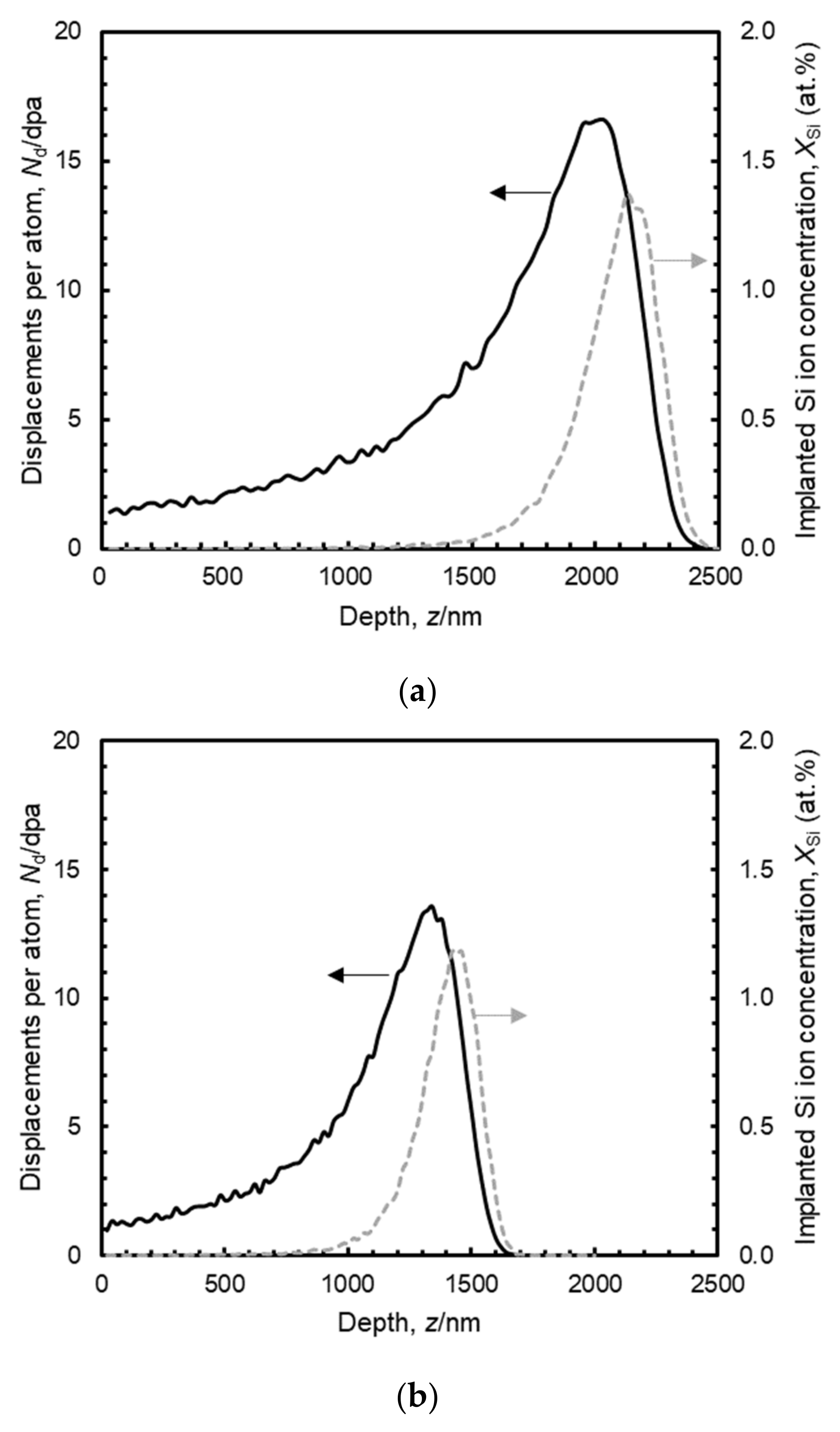
Figure 1. Cross-sectional illustrations of (a) the PVD Ti-Cr multilayer coating and (b) its transition after heat treatment at 1223 K for 1.8 × 10³ s.
Coatings 12 00832 g001
Figure 2. Number of displacements per atom and implanted Si ion concentration in (a) pure Ti, (b) pure Cr and (c) SiC after irradiation with 5.1 MeV Si2+ ions to a fluence of 2.85 × 10²⁰ ions/m2 calculated by SRIM-2013.
Coatings 12 00832 g002aCoatings 12 00832 g002b
Figure 3. Example AFM images including a boundary between unirradiated (protected) and irradiated (exposed) regions of pure Ti bulk surfaces irradiated with 5.1-MeV Si2+ ions at 573 K, at fluences of (a) 9.51 × 10¹⁹ and (b) 2.85 × 10²⁰ ions/m2.
Coatings 12 00832 g003
Figure 4. Changes in (a) height step and (b) swelling of pure Ti, pure Cr and CVD SiC after irradiation with 5.1 MeV Si2+ ions up to a fluence of 2.85 × 10²⁰ ions/m2.
Coatings 12 00832 g004aCoatings 12 00832 g004b
Figure 5. Cross section of Ti-Cr multilayer-coated SiC after irradiation with 5.1-MeV Si2+ ion at 573 K at a fluence of 2.85 × 10²⁰ ions/m2.
Coatings 12 00832 g005
Figure 6. SE images of cross-sectional Ti-Cr multilayer coating surface irradiated with Si2+ ion at 5.1 MeV and 573 K to fluences of (a) 0.95 × 10¹⁹, (b) 9.51 × 10¹⁹, and (c) 2.85 × 10²⁰ ions /m2.
Coatings 12 00832 g006
Figure 7. Cross-sectional SE image and EDS mappings for Ti-Cr multilayer coating irradiated at 573 K, 2.85 × 10²⁰ ions /m2 with Si2+ ion at 5.1 MeV. (a) Cross-sectional SE image. (be) EDS mappings for the same specimen as in (a) obtained from the separated peak signals of the: (b) C K line, (c) Si Kα line, (d) Cr L line, and (e) Ti L line.
Coatings 12 00832 g007
Figure 8. Cross-sectional bright field images and EDS mappings of unirradiated Ti-Cr multilayer coating. (a) Bright field image overview. (b,c) Analyzed areas described in (a). Far left, two bright field images, the other eight images are the EDS mappings. In (b) Ti, Ti-Cr mixed, and Cr layers were identified and in (c) Ti-Cr mixed, Cr, diffused Cr-SiC interface, and CVD SiC layers were identified.
Coatings 12 00832 g008
Figure 9. EDS line profile of chemical composition and constituent phases along the analyzed lines (bule-green) in unirradiated Ti-Cr multilayer coating. The analyzed lines includes (a) Ti, Ti-Cr mixed, and Cr layers and (b) Ti-Cr mixed, Cr, diffused Cr-SiC interface, and CVD SiC layers.
Coatings 12 00832 g009
Figure 10. Cross-sectional bright field images and EDS mappings of Ti-Cr multilayer coating after being irradiated with Si2+ ions up to a fluence of 2.85 × 10²⁰ ions /m2. (a) Bright field image overview. (b,c) Analyzed areas described in (a). Far left, two bright field images, the other eight images are the EDS mappings. In (b) Ti, Ti-Cr mixed, and Cr layers were identified and in (c) Ti-Cr mixed, Cr, diffused Cr-SiC interface and CVD SiC layers were identified.
Coatings 12 00832 g010
Figure 11. EDS line profiles of chemical composition and constituent phases along the analyzed line (bule-green) at the peak positions for the number of displacements per atom and constituent phases in Ti-Cr multilayer coating after being irradiated with Si2+ ions up to a fluence of 2.85 × 10²⁰ ions /m2. The analyzed lines included (a) Ti, Ti-Cr mixed and Cr layers and (b) Ti-Cr mixed, Cr, diffused Cr-SiC interface, and CVD SiC layers.
Coatings 12 00832 g011
Figure 12. EDS line profiles and mappings of chemical composition across phase interfaces in Ti-Cr multilayer coating after being irradiated with Si2+ ions up to a fluence of 2.85 × 10²⁰ ions /m2. (a) Overview of the analyzed areas. (b,c) Analyzed areas described in (a). In (b) the TiC/ TiCr2 phase interface was included and in (c) the TiCr2/Cr phase interface was included.
Coatings 12 00832 g012
Figure 13. EDS line profiles and mappings of chemical composition across Cr/Cr3Si and Cr3Si/Cr5Si3 phase interfaces in Ti-Cr multilayer coating after being irradiated with Si2+ ions up to a fluence of 2.85 × 10²⁰ ions /m2.
Coatings 12 00832 g013
Figure 14. Changes in swelling of pure Ti, pure Cr and CVD SiC. The abscissa is the average number of displacements per atom for each material.
Coatings 12 00832 g014
Figure 15. Coefficient of thermal expansion (CTE) of SiC, Cr, and Ti. The dashed lines are regression curves used for calculation of thermal stress. SiC [69], Cr [76], Ti [76].
Coatings 12 00832 g015
Figure 16. Simply estimated stress in pure Ti or Cr coating on CVD SiC after irradiation with 5.1 MeV Si2+ ions up to a fluence of 2.85 × 10²⁰ ions/m2 at 573 K.
Coatings 12 00832 g016
Table 1. Information for phase identification by SAD analysis using ReciPro ver4.828.
PhaseCrystal SystemSpace GroupLattice ConstantDatabase
α-TiHexagonal P 6 / m m m a = b = 0.4577 nm, c = 0.2829 nm, α = β = 90°, γ = 120°The Materials Project *1 mp-72 [57]
TiCCubic F m 3 - m a = b = c = 0.4336 nm, α = β = γ = 90°The Materials Project *1 mp-631 [58]
TiCr2Hexagonal P 6 3 / m m c a = b = 0.4877 nm, c = 0.7868 nm,
α = β = 90°, γ = 120°
The Materials Project *1 mp-1589 [59]
CrCubic I m 3 - m a = b = c = 0.2874 nm, α = β = γ = 90°The Materials Project *1 mp-90 [60]
Cr3SiCubic P m 3 - n a = b = c = 0.4519 nm, α = β = γ = 90°The Materials Project *1 mp-729 [61]
Cr5Si3Orthorhombic C m m m a = 0.5543 nm, b = 0.8206 nm, c = 0.4179 nm,
α = β = γ = 90°
AFLOW *2 Prototype A5B3_oC16_65_aeh_bj [62]
SiCCubic F m 3 - m a = b = c = 0.4058 nm, α = β = γ = 90°AFLOW *2 Prototype: AB_cF8_225_b_a [63]
*1 The Materials Project [64]. *2 AFLOW (Automatic-FLOW for Materials Discovery) [65].
Table 2. Identification of phases in Ti-Cr multilayer coating heat-treated at 1223 K for 1.8 × 10³ s.
LayerChemical Components by SEM-EDS [49]Lattice Structure by EBSD [49]Phase Identified by XRD [47,48,49]Phase Identified by TEM-EDS and SAD Analysis (This Study)
TiTiα-Tiα-Tiα-Ti
TiBCCβ-Ti-
Ti and Cr(Fairly low IQ *1)TiCr2TiCr2
Ti and CFCCTiCTiC
Ti-Cr mixedTi and CrBCCβ-Ti-
Ti and Cr(Fairly low IQ *1)TiCr2TiCr2
Ti and CFCCTiC-
CrCrBCCCrCr
Ti, Cr and O
[grain boundary]
BCC--
Diffused Cr-SiC interfaceCr and Si (Cr-rich)Cr3Si-Cr3Si
Cr, Si and C (Si-rich)(Fairly low IQ *1)-Cr5Si3 or Cr5Si3Cx
SiCSi and CFCC-β-SiC
*1 IQ: Image quality defined in EDAX OIM system.
Table 3. Microstructure and swelling of pure Ti, pure Cr and Cr coating irradiated at temperatures from 423 to 723 K.
MaterialIrradiationTemperatureExposureMicrostructureSwellingRef.
Pure Ti
(bulk)
Neutrons423 K1.3 × 1024 n/m2 (E > 1 MeV)Black spots-[70]
Neutrons723 K3 × 1025 n/m2 (E > 0.1 MeV)Dislocation loops-[71]
Neutrons673 K3.9 × 1023 n/m2 (E > 1 MeV)Dislocation loops-[72]
6 MeV
Ti2+ ions
573 K0.6, 3 dpaBlack spots
Dislocation loops
-[73]
703 K0.6, 3 dpaDislocation loops-
Cr coating1.4 MeV Ar ions673 K5, 15, 25 dpaVoid (1.5–4 nm)0.16% for 5 dpa *1
0.66% for 25 dpa *1
[74]
Neutrons593–613 K4.8 × 1024 n/m2 (E > 0.1 MeV)
~0.5 dpa
Void (2 nm)0.2% *1
(~0.07% length change)
[43]
Pure Cr
(bulk)
5 MeV
Fe ions
723 K50 dpa (peak)Void (4 nm)0.5 % *1[75]
*1 Estimated from void size and density.
Table 4. Properties of SiC, Cr, and Ti at room temperature.
MaterialCTE *1, α
[10−6 K−1]
Young’s Modulus, E
[GPa]
Poisson’s Ratio, νRef.
SiC2.24600.21[55]
Cr4.9279.10.210[76,77]
Ti8.6115.70.321[76,77]
*1 CTE: coefficient of thermal expansion.
Publisher’s Note: MDPI stays neutral with regard to jurisdictional claims in published maps and institutional affiliations.