Communication

In Situ Fabrication of Polydeoxyribonucleotide-Impregnated Hydroxyapatite onto a Magnesium Surface

1
Department of Materials Science and Engineering, Seoul National University, Seoul 08826, Republic of Korea
2
Department of Carbon and Nano Materials Engineering, Jeonju University, Jeonju 55069, Republic of Korea
*
Author to whom correspondence should be addressed.
Coatings 2023, 13(1), 72; https://doi.org/10.3390/coatings13010072
Submission received: 27 November 2022 / Revised: 29 December 2022 / Accepted: 29 December 2022 / Published: 31 December 2022
(This article belongs to the Section Bioactive Coatings and Biointerfaces)

Abstract

:
In this study, in situ polydeoxyribonucleotide-impregnated hydroxyapatite (PDRN/HA) was coated on a magnesium (Mg) substrate to form a biocompatible HA layer by chemical conversion for effective PDRN delivery. The HA layer showed needle-like morphology, and the PDRN impregnation did not affect the coating structure. The loading amount of PDRN via the proposed in situ method was 2.5 times higher than that by the conventional dipping method. An in vitro cell proliferation test demonstrated that the PDRN loading was more effective through this one-step method than through the dipping method. The results indicate that in situ PDRN/HA coating can enhance the potential of Mg-based implants.

1. Introduction

Magnesium (Mg)-based materials are one of the most promising implantable metallic materials for hard tissue owing to their biocompatibility, mechanical properties, and biodegradability [1,2,3,4]. However, the biodegradation rate of bare Mg is higher than the bone regeneration rate, which limits the applications of Mg-based implants [5,6,7]. To mitigate the rapid degradation of Mg-based implants, various types of coating and alloying have been proposed [8,9,10].
Among them, the addition of a hydroxyapatite (HA) layer onto a Mg surface through chemical conversion is a very simple method for improving the bioactivity and corrosion resistance of Mg-based implants [11,12,13]. In our recent study [14], we found that the chemical conversion of HA could be applied not only to bulk Mg but also to micropatterned Mg on a silicon wafer. The resulting HA pattern was transferred onto a biodegradable polymer matrix, and the resulting HA-pattern-embedded polymer showed improved biocompatibility and osteogenic properties. Furthermore, the hydrophilic nature of the HA pattern improved the drug-loading efficiency of the polymer matrix. However, the amount of drug loaded is limited because the drug was loaded only on the outer surface of the HA layer. The application of the HA conversion method can be more promising if the drug-loading efficiency could be increased. One of the simplest ways to increase the loading amount of a drug is to load it during the HA conversion process. This in situ method also eliminates the additional drug-loading step. The HA conversion process is, however, performed at 90 °C; hence, normal drugs would be thermally damaged [15]. Therefore, it is essential to search for a good drug candidate that can be applied to the in situ HA conversion process.
Polydeoxyribonucleotide (PDRN) is a type of DNA derived from the sperm of salmon. Through various studies, it is known that PDRN is effective in increasing cell growth and activity, which is known as angiogenesis effect through the expression of vascular endothelial growth factor (VEGF) [16,17,18]. Moreover, it is known for exerting increasing cell growth and activity on various soft tissues. Furthermore, it has been confirmed that PDRN is effective for bone regeneration by acting as an osteoblast growth stimulator [19,20]. This effect was also proven in animal experiments using rats and mice [18,21]. In addition, the synthesis of PDRN comprises a high-temperature procedure [22]. PDRN is thus considered a good candidate for drugs that can be used for an in situ drug-loading process. In addition, since PDRN maintains its activity even at 121 °C [23], it is free from reduction in drug effect by known magnesium and magnesium derivatives [24]. Therefore, in this study, we developed a facile method to fabricate a PDRN-impregnated HA layer onto a Mg surface. Through this, PDRN, which acts as a drug, was expected to exist even inside the HA coating layer to exert a continuous osteoconductive effect. This method was compared with the method of simply dipping the drug after forming the HA coating layer. Finally, the loading efficiency and the effect of the released PDRN on the cell proliferation behavior were investigated.

2. Materials and Methods

2.1. PDRN-Impregnated HA Coating

First, a 10 × 10 × 1-mm3 Mg plate (Yirium, Jiangxi, China) was polished using a 400–2000 grit SiC abrasive paper (Deerfos, Pusan, Republic of Korea) to diminish impurities and smoothen the surface. Then, a solution was prepared by mixing 0.05-M ethylenediaminetetraacetic acid calcium disodium salt hydrate (Sigma-Aldrich, Burlington, MA, USA) and 0.05-M KH2PO4 (Sigma-Aldrich, Burlington, MA, USA). PDRN (Genoss, Suwon, Republic of Korea) at various concentrations (0, 200, 500, and 1000 μg/mL) was added to the solution. PDRN-impregnated HA (PDRN/HA) coating was performed by immersing the Mg plate into the solution at 90 °C, and its pH was adjusted to 8.9 using sodium hydroxide (NaOH) for 2 h. After the coating process, the specimens were rinsed with distilled water briefly and dried in air.

2.2. Characterization of PDRN-Impregnated HA Layer on Mg

The surface morphology of bare, HA-coated, and PDRN/HA-coated Mg was observed using a field-emission scanning electron microscope (FE-SEM, Sigma, Carl Zeiss, Jena, Germany). Cross-section images were obtained using the focused ion beam (FIB, Auriga, Carl Zeiss, Jena, Germany) technique. The chemical composition of each layer was determined using an energy dispersive spectroscope (EDS, Auriga, Carl Zeiss, Jena, Germany), which was connected to the FIB instrument. The crystalline phase of the surface layer was investigated through X-ray diffraction (XRD, D8-advance, Bruker Miller Co., Middlesex County, MA, USA).

2.3. Drug Release Test

The PDRN-loaded samples were immersed in Dulbecco’s phosphate-buffered saline solution (DPBS, Welgene, Gyeongsan, Republic of Korea), and the released PDRN was monitored for up to 1 week. The absorbance of PDRN was measured using a UV/visible spectroscope (V-770, Jasco, Tokyo, Japan) at predetermined times. The amount of PDRN released from the sample was compared to that prepared using the dipping method (Dip). HA-coated Mg was dipped in 1000 μg/mL PDRN solution overnight, rinsed with distilled water, and then dried in the air.

2.4. Effect of PDRN on In Vitro Cellular Proliferation

The effects of the PDRN released on cellular behavior were evaluated using in vitro cell proliferation tests. The Dip and PDRN/HA samples were immersed into the cell culture medium comprising an α-minimum essential medium (Welgene, Gyeongsan, Republic of Korea), 10% fetal bovine serum (Life Technologies Inc., Gaithersburg, MD, USA), and 1% antibiotics (100 U/mL penicillin and 100 μm/mL streptomycin, GIBCO, Grand Island, NY, USA) for 1 day to release the PDRN into the medium. After releasing the drug, the samples were removed, and the preosteoblast cell line (MC3T3-E1, ATCC, Manassas, VA, USA) with a density of 2 × 10⁴ cells/mL was incubated in each medium for 1 and 3 days at 37 °C and 5% CO2 conditions. Cell proliferation was evaluated using a methoxyphenyl tetrazolium salt (MTS) assay kit (CellTiter 96 Aqueous One Solution, Promega, Madison, WI, USA).

2.5. Statistical Analysis

Statistical analysis of the drug release test and in vitro proliferation test was conducted using the Statistical Package for the Social Sciences software (SPSS 23, SPSS Inc., Chicago, IL, USA). One-way analysis of variance with Tukey post hoc comparison was performed, and a p-value below 0.05 was considered statistically significant.

3. Results and Discussion

The surface morphologies of bare-, HA-coated, and PDRN/HA-coated Mg are shown in Figure 1a–c, respectively. Both HA- and PDRN/HA-coated surfaces showed clusters with a needle-like morphology, whereas the bare-Mg surface had only mechanical grooves caused by the polishing process. This needle-like structure is a typical morphology of chemically converted HA from a Mg surface [15,25]. Figure 1d,e show the FIB-milled cross-section images of HA- and PDRN/HA-coated Mg, respectively. Both coating layers had a thickness of 3–4-μm and needle-like structures. Moreover, the surface morphology of the HA coating layer was not affected by PDRN impregnation. The EDS mapping image of the PDRN/HA-coated Mg showed that the coating layer (orange) included both Ca (yellow) and P (red), whereas the substrate only showed Mg (blue), as shown in Figure 1f. As carbon and oxygen, the main components of PDRN, are elements that also exist in HA, they could not be confirmed via EDS. Through the EDS image, it was confirmed that Mg was successfully converted into a calcium phosphate–based material; however, the crystal structure must be checked to determine the type of material. The crystal structure of the coating layer was further investigated via XRD. As shown in Figure 2, the peak of the Mg substrate appeared clearly in all groups (JCPDS No. 35-0821). Weak HA peaks were detected in the HA- and PDRN/HA-coated groups at 26°, 28°, 32°, 47°, 49°, and 53° groups (JCPDS No. 09-0432) [25,26]. Moreover, PDRN does not have crystallinity. The difference between HA- and PDRN/HA-coated groups could not be confirmed through XRD, which indicated that there were no side effects, such as the occurrence of other crystalline phases due to the addition of PDRN. These results indicated that the HA layer with PDRN incorporation on the Mg substrate was successfully created.
Figure 3 depicts the in vitro drug release behavior. Figure 3a shows the release profile of PDRN from PDRN/HA-coated Mg at different PDRN concentrations (200, 500, and 1000 μg/mL). The amount of released impregnated PDRN was proportional to the original PDRN concentration of the coating solution. Therefore, it is expected that the required amount of drug can be controlled simply by adjusting the concentration of the drug in the coating solution. The drug-loading efficiency of this new in situ method was compared with that of the conventional dip-coating method, as shown in Figure 3b. For comparison, Dip and PDRN/HA specimens were prepared using solutions with the same PDRN concentration (1000 μg/mL). The release of PDRN was performed for 1 day, and then, the cumulative PDRN amounts were compared because most of PDRN was released within 24 h. The total amount of PDRN released from the Dip group was 15 μg while that from the PDRN/HA group was 40 μg. The higher PDRN-loading efficiency of the in situ method can mainly be attributed to the difference in the drug-loading space. For the Dip group, most of PDRN is deposited on the surface layer and a part of PDRN is diffused in the coating layer. In contrast, for the in situ PDRN/HA group, PDRN can be deposited inside the coating layer during the coating process. Therefore, it was confirmed that this in situ method can not only reduce the number of steps but also enhance drug-loading efficiency.
Finally, the PDRN activity after the coating process was examined using an in vitro cell proliferation test. The cell test was not conducted on the specimen surface to minimize the effect of the HA coating layer. In addition, the Mg substrate reacts with the cell-counting reagent and affects the results. Thus, the PDRN was released to the cell culture media and then added to the cells. Figure 4 shows the proliferation of MC3T3-E1 cells after 1 and 3 days of culturing with PDRN released from the Dip and PDRN/HA groups. The HA-coated Mg without PDRN (HA) was also immersed in the culture media for 1 and 3 days, and then, this media was used as a control group. The difference in proliferation was observed on Day 3. The PDRN/HA group showed almost two times higher level of proliferation compared to the Dip group. These results are consistent with PDRN release data and existing literature. According to Shin et al., the effect of PDRN appears when the amount of release is greater than 20 ug; however, according to Figure 3, Dip is lower than this and PDRN/HA is higher than this, showing a difference in cell proliferation [27]. This result corresponds to the PDRN release data. As PDRN is known to affect the proliferation of cells, more PDRN would result in higher proliferation. This effect is induced because PDRN is cleaved by active cell membrane enzymes and acts as a source of deoxynucleotides and deoxynucleosides, which aid in the growth or proliferation of cells [21,28,29]. Furthermore, it can be confirmed that the PDRN activity was not affected by the heat during the coating process. PDRN is stable at the temperature of this in situ coating process because it has already undergone a high-temperature process during its fabrication process.
In summary, the in situ PDRN/HA coating using the chemical conversion process showed enhanced PDRN-loading efficiency compared to when using the conventional dip-coating method.

4. Conclusions

A new strategy to coat a biodegradable Mg surface with a PDRN-impregnated HA layer using a one-step coating process was presented. The surface showed a needle-like HA crystalline structure, and the PDRN impregnation did not affect the coating structure. The loading amount of PDRN was easily controlled by adjusting the PDRN concentration of the coating solution. The in situ loading of PDRN was significantly more effective than the dip-coating method. An in vitro cell test revealed that PDRN was not damaged by the high temperatures during the coating process and indicated that the proposed in situ method would be useful in the field of hard tissue engineering.

Author Contributions

Conceptualization, C.-M.H.; methodology, J.-Y.K. and I.-G.K.; formal analysis, J.-Y.K.; investigation, J.-Y.K. and I.-G.K.; data curation, J.-Y.K.; writing—original draft preparation, J.-Y.K.; writing—review and editing, C.-M.H.; supervision, C.-M.H.; funding acquisition, C.-M.H. All authors have read and agreed to the published version of the manuscript.

Funding

This research was supported by the National Research Foundation of Korea (NRF) grant funded by the Korea Government (MSIT) (NRF-2021R1F1A1049167 and NRF-2022R1I1A3054310).

Institutional Review Board Statement

Not applicable.

Informed Consent Statement

Not applicable.

Data Availability Statement

Not applicable.

Conflicts of Interest

The authors declare no conflict of interest.

References

  1. Staiger, M.P.; Pietak, A.M.; Huadmai, J.; Dias, G. Magnesium and its alloys as orthopedic biomaterials: A review. Biomaterials 2006, 27, 1728–1734. [Google Scholar] [CrossRef]
  2. Kumar, K.; Gill, R.S.; Batra, U. Challenges and opportunities for biodegradable magnesium alloy implants. Mater. Technol. 2017, 33, 153–172. [Google Scholar] [CrossRef]
  3. Zhao, D.; Witte, F.; Lu, F.; Wang, J.; Li, J.; Qin, L. Current status on clinical applications of magnesium-based orthopaedic implants: A review from clinical translational perspective. Biomaterials 2017, 112, 287–302. [Google Scholar] [CrossRef] [PubMed]
  4. Chakraborty Banerjee, P.; Al-Saadi, S.; Choudhary, L.; Harandi, S.E.; Singh, R. Magnesium implants: Prospects and challenges. Materials 2019, 12, 136. [Google Scholar] [CrossRef] [PubMed] [Green Version]
  5. Song, G. Control of biodegradation of biocompatable magnesium alloys. Corr. Sci. 2007, 49, 1696–1701. [Google Scholar] [CrossRef]
  6. Waizy, H.; Seitz, J.M.; Reifenrath, J.; Weizbauer, A.; Bach, F.W.; Meyer-Lindenberg, A.; Denkena, B.; Windhagen, H. Biodegradable magnesium implants for orthopedic applications. J. Mater. Sci. 2012, 48, 39–50. [Google Scholar] [CrossRef]
  7. Wang, J.L.; Xu, J.K.; Hopkins, C.; Chow, D.H.; Qin, L. Biodegradable magnesium-based implants in orthopedics—A general review and perspectives. Adv. Sci. 2020, 7, 1902443. [Google Scholar] [CrossRef] [PubMed] [Green Version]
  8. Gu, X.N.; Zhou, W.R.; Zheng, Y.F.; Cheng, Y.; Wei, S.C.; Zhong, S.P.; Xi, T.F.; Chen, L.J. Corrosion fatigue behaviors of two biomedical Mg alloys—AZ91D and WE43—In simulated body fluid. Acta Biomater. 2010, 6, 4605–4613. [Google Scholar] [CrossRef] [PubMed]
  9. Cheon, K.H.; Park, C.; Kang, M.H.; Kang, I.G.; Lee, M.K.; Lee, H.; Kim, H.E.; Jung, H.D.; Jang, T.S. Construction of tantalum/poly(ether imide) coatings on magnesium implants with both corrosion protection and osseointegration properties. Bioact. Mater. 2021, 6, 1189–1200. [Google Scholar] [CrossRef]
  10. Kirkland, N.T.; Birbilis, N.; Staiger, M.P. Assessing the corrosion of biodegradable magnesium implants: A critical review of current methodologies and their limitations. Acta Biomater. 2012, 8, 925–936. [Google Scholar] [CrossRef]
  11. Byun, S.H.; Lim, H.K.; Kim, S.M.; Lee, S.M.; Kim, H.E.; Lee, J.H. The bioresorption and guided bone regeneration of absorbable hydroxyapatite-coated magnesium mesh. J. Craniofac. Surg. 2017, 28, 518–523. [Google Scholar] [CrossRef] [PubMed]
  12. Kang, M.H.; Lee, H.; Jang, T.S.; Seong, Y.J.; Kim, H.E.; Koh, Y.H.; Song, J.H.; Jung, H.D. Biomimetic porous Mg with tunable mechanical properties and biodegradation rates for bone regeneration. Acta Biomater. 2019, 84, 453–467. [Google Scholar] [CrossRef] [PubMed]
  13. Guo, Y.; Su, Y.; Gu, R.; Zhang, Z.; Li, G.; Lian, J.; Ren, L. Enhanced corrosion resistance and biocompatibility of biodegradable magnesium alloy modified by calcium phosphate/collagen coating. Surf. Coat. Technol. 2020, 401, 126318. [Google Scholar] [CrossRef]
  14. Kang, I.G.; Kim, J.; Kim, J.; Park, S.; Kim, H.E.; Han, C.M. PLLA Membrane with embedded hydroxyapatite patterns for improved bioactivity and efficient delivery of growth factor. Macromol. Biosci. 2020, 20, e2000136. [Google Scholar] [CrossRef] [PubMed]
  15. Kim, S.M.; Kang, M.H.; Kim, H.E.; Lim, H.K.; Byun, S.H.; Lee, J.H.; Lee, S.M. Innovative micro-textured hydroxyapatite and poly(l-lactic)-acid polymer composite film as a flexible, corrosion resistant, biocompatible, and bioactive coating for Mg implants. Mater. Sci. Eng. C Mater. Biol. Appl. 2017, 81, 97–103. [Google Scholar] [CrossRef] [PubMed]
  16. Kim, S.K.; Huh, C.K.; Lee, J.H.; Kim, K.W.; Kim, M.Y. Histologic study of bone-forming capacity on polydeoxyribonucleotide combined with demineralized dentin matrix. Maxillofac. Plast. Reconstr. Surg. 2016, 38, 7. [Google Scholar] [CrossRef] [Green Version]
  17. Koo, Y.; Yun, Y. Effects of polydeoxyribonucleotides (PDRN) on wound healing: Electric cell-substrate impedance sensing (ECIS). Mater. Sci. Eng. C 2016, 69, 554–560. [Google Scholar] [CrossRef]
  18. Buffoli, B.; Favero, G.; Borsani, E.; Boninsegna, R.; Sancassani, G.; Labanca, M.; Rezzani, R.; Nocini, P.F.; Albanese, M.; Rodella, L.F. Sodium-DNA for bone tissue regeneration: An experimental study in rat calvaria. BioMed. Res. Int. 2017, 2017, 7320953. [Google Scholar] [CrossRef] [Green Version]
  19. Veronesi, F.; Dallari, D.; Sabbioni, G.; Carubbi, C.; Martini, L.; Fini, M. Polydeoxyribonucleotides (PDRNs) from skin to musculoskeletal tissue regeneration via adenosine A2A receptor involvement. J. Cell Physiol. 2017, 232, 2299–2307. [Google Scholar] [CrossRef]
  20. Lim, H.K.; Kwon, Y.J.; Hong, S.J.; Choi, H.G.; Chung, S.M.; Yang, B.E.; Lee, J.H.; Byun, S.H. Bone regeneration in ceramic scaffolds with variable concentrations of PDRN and rhBMP-2. Sci. Rep. 2021, 11, 11470. [Google Scholar] [CrossRef]
  21. Guizzardi, S.; Martini, D.; Bacchelli, B.; Valdatta, L.; Thione, A.; Scamoni, S.; Uggeri, J.; Ruggeri, A. Effects of heat deproteinate bone and polynucleotides on bone regeneration: An experimental study on rat. Micron 2007, 38, 722–728. [Google Scholar] [CrossRef] [PubMed]
  22. Squadrito, F.; Bitto, A.; Irrera, N.; Pizzino, G.; Pallio, G.; Minutoli, L.; Altavilla, D. Pharmacological activity and clinical use of PDRN. Front. Pharmacol. 2017, 8, 224. [Google Scholar] [CrossRef] [PubMed] [Green Version]
  23. Raposio, E.; Guida, C.; Coradeghini, R.; Scanarotti, C.; Parodi, A.; Baldelli, I.; Fiocca, R.; Santi, P.L. In vitro polydeoxyribonucleotide effects on human pre-adipocytes. Cell Prolif. 2008, 41, 739–754. [Google Scholar] [CrossRef] [PubMed]
  24. Jelić, D.; Papović, S.; Vraneš, M.; Gadžurić, S.; Berto, S.; Alladio, E.; Gajic, D.; Janković, B. Thermo-analytical and compatibility study with mechanistic explanation of degradation kinetics of ambroxol hydrochloride tablets under non-isothermal conditions. Pharmaceutics 2021, 13, 1910. [Google Scholar] [CrossRef]
  25. Tomozawa, M.; Hiromoto, S. Growth mechanism of hydroxyapatite-coatings formed on pure magnesium and corrosion behavior of the coated magnesium. Appl. Surf. Sci. 2011, 257, 8253–8257. [Google Scholar] [CrossRef]
  26. Guillen-Romero, L.D.; Oropeza-Guzman, M.T.; Lopez-Maldonado, E.A.; Iglesias, A.L.; Paz-Gonzalez, J.A.; Ng, T. Serena-Gomez, E.; Villarreal-Gomez, L.J. Synthetic hydroxyapatite and its use in bioactive coatings. J. Appl. Biomater. Funct. Mater. 2019, 17, 2280800018817463. [Google Scholar]
  27. Shin, D.Y.; Park, J.U.; Choi, M.H.; Kim, S.; Kim, H.E.; Jeong, S.H. Polydeoxyribonucleotide-delivering therapeutic hydrogel for diabetic wound healing. Sci. Rep. 2020, 10, 16811. [Google Scholar] [CrossRef]
  28. Guizzardi, S.; Galli, C.; Govoni, P.; Boratto, R.; Cattarini, G.; Martini, D.; Belletti, S.; Scandroglio, R. Polydeoxyribonucleotide (PDRN) promotes human osteoblast proliferation: A new proposal for bone tissue repair. Life Sci. 2003, 73, 1973–1983. [Google Scholar] [CrossRef]
  29. Nakamura, E.I.; Uezono, Y.; Narusawa, K.I.; Shibuya, I.; Oishi, Y.; Tanaka, M.; Yanagihara, N.; Nakamura, T.; Izumi, F. ATP activates DNA synthesis by acting on P2X receptors in human osteoblast-like MG-63 cells. Am. J. Physiol. Cell Physiol. 2000, 279, C510–C519. [Google Scholar] [CrossRef]
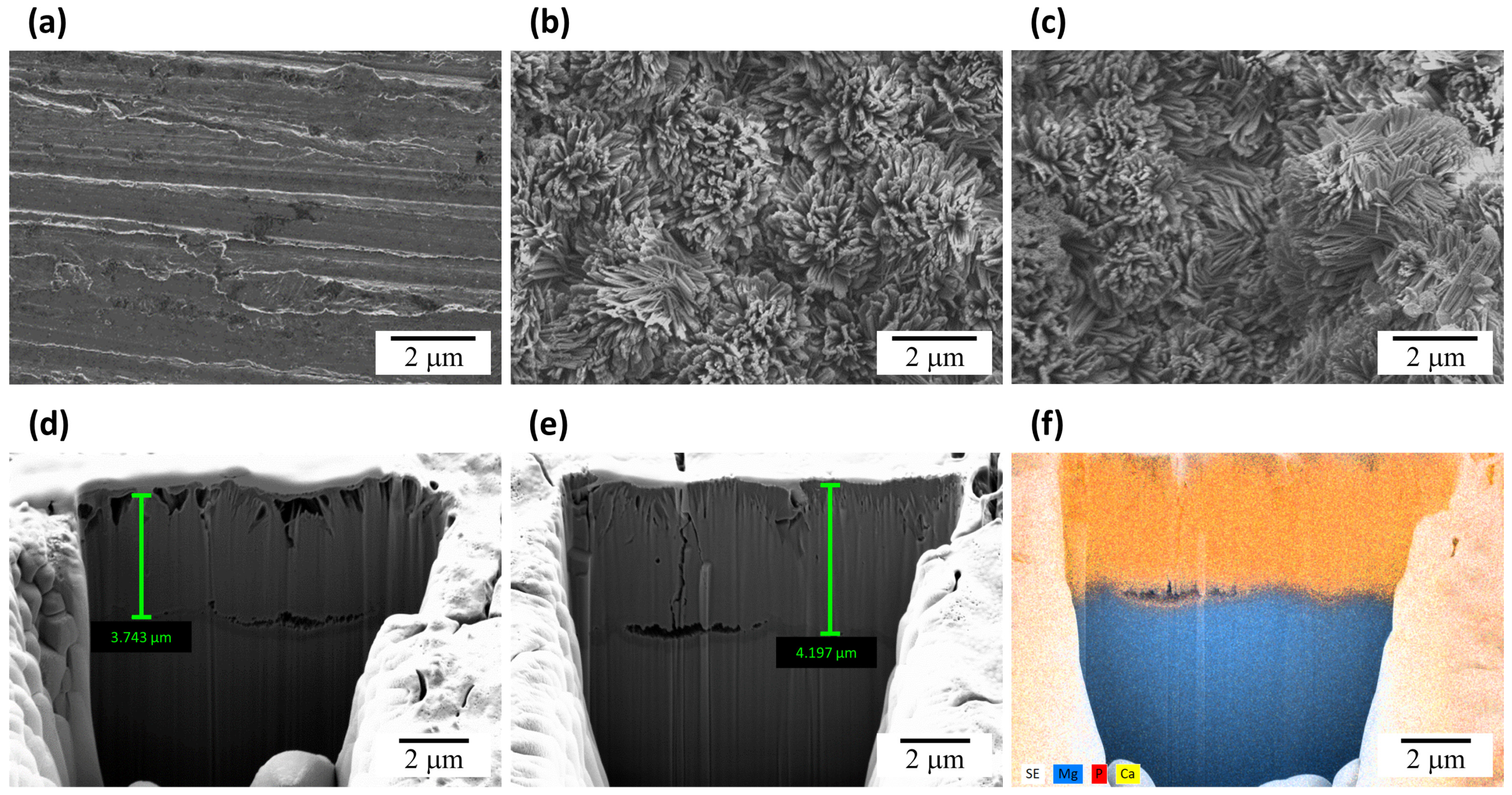
Figure 1. Morphology of (a) bare-, (b) HA-coated, and (c) PDRN/HA-coated Mg surfaces and FIB-dissected cross-section of (d) HA-coated and (e) PDRN/HA-coated Mg; (f) EDS mapping image of the cross-section of PDRN/HA-coated Mg (Blue: Mg, red: P, and yellow: Ca).
Coatings 13 00072 g001
Figure 2. XRD spectra of (a) bare-, (b) HA-coated, and (c) PDRN/HA-coated Mg sample groups.
Coatings 13 00072 g002
Figure 3. (a) PDRN release profile of PDRN/HA-coated Mg at various loading concentrations (200, 500, and 1000 μg/mL). (b) Total amount of PDRN released from Dip and PDRN/HA groups (*: p < 0.05).
Coatings 13 00072 g003
Figure 4. Cell viability of HA, Dip, and PDRN/HA groups measured using the MTS method (*: p < 0.05).
Coatings 13 00072 g004
Disclaimer/Publisher’s Note: The statements, opinions and data contained in all publications are solely those of the individual author(s) and contributor(s) and not of MDPI and/or the editor(s). MDPI and/or the editor(s) disclaim responsibility for any injury to people or property resulting from any ideas, methods, instructions or products referred to in the content.