Article

Strong Interface Interaction of ZnO Nanosheets and MnSx Nanoparticles Triggered by Light over Wide Ranges of Wavelength to Enhance Their Removal of VOCs

1
School of Environmental and Material Engineering, Center of Advanced Functional Materials, Yantai University, Yantai 264005, China
2
National Laboratory of Industrial Control Technology, Institute of Cyber-Systems and Control, Zhejiang University, Hangzhou 310027, China
*
Author to whom correspondence should be addressed.
Coatings 2023, 13(10), 1727; https://doi.org/10.3390/coatings13101727
Submission received: 27 August 2023 / Revised: 29 September 2023 / Accepted: 1 October 2023 / Published: 3 October 2023

Abstract

:
The characteristics of the surface and interface of nanocomposites are important for exerting multi-functional properties and widening interdisciplinary applications. These properties are mainly depending on the electronic structures of materials. Some key factors, such as the surface, interface, grain boundaries, and defects take vital roles in the contribution of desired properties. Due to the excellent sensitivity of the QCM (quartz crystal microbalance) device, the surface and interface features of the nanocomposite were studied with the aid of the gas-response of the sensors (Sensor’s Gas-Sensitivity) in this work. To make full use of the visible light and part of NIR, a ZnO/MnSx nanocomposite was constructed using hydrothermal synthesis for narrowing the bandgap width of wide bandgap materials. The results indicated that the absorbance of the resulting nanocomposite was extended to part of the NIR range due to the introduction of impurity level or defect level, although ZnO and MnS belonged to wide bandgap semiconductor materials. To explore the physical mechanism of light activities, the photoconductive responses to weak visible light (650 nm, etc.) and NIR (near-infrared) (808 nm, 980 nm, and 1064 nm, etc.) were studied based on interdigital electrodes of Au on flexible PET (polyethylene terephthalate) film substrate with the casting method. The results showed that the on/off ratio of ZnO/MnSx nanocomposite to weak visible light and part of NIR light were changed by about one to five orders of magnitude, with changes varying with the amount of MnSx nanoparticle loading due to defect-assisted photoconductive behavior. It illustrated that the ZnO/MnSx nanocomposite easily produced photo-induced free charges, effectively avoiding the recombination of electrons/holes because of the formation of strong built-in electrical fields. To examine the surface and interface properties of nanocomposites, chemical prototype sensor arrays were constructed based on ZnO, ZnO/MnSx nanocomposite, and QCM arrays. The adsorption response behaviors of the sensor arrays to some typical volatile compounds were examined under a similar micro-environment. The results exhibited that in comparison to ZnO nanosheets, the ZnO nanosheets/MnSx nanocomposite increased adsorption properties to some typical organic volatile compounds significantly. It would have good potential applications in photo-catalysts, self-cleaning films, multi-functional coatings, and organic pollutants treatment (VOCs) of environmental fields for sustainable development. It provided some reference value to explore the physical mechanism of materials physics and photophysics for photo-active functional nanocomposites.

1. Introduction

Recently, the demands for energy and from the environment have promoted the great progress of advanced functional materials and their nanocomposites, especially metal oxides, metal sulfides, carbon nanotubes, graphene, carbon dots, low-dimensional noble metals, functional polymers, organic/inorganic hybrids systems, etc. Among so many metal oxides, ZnO is one kind of multi-functional material due to its low-cost, simple synthesis, outstanding optical properties, multi-functionalities, and good biocompatibility. ZnO-based nanocomposites have good potential applications in solar cells [1], photoelectrical devices [2], energy storage [3,4], water splitting [5], supercapacitors [6], gas sensors [7,8,9,10,11], biosensors [12,13], photodetectors [14,15,16,17,18,19,20,21,22,23], photocatalysts [24,25,26,27,28,29,30,31,32,33,34,35,36,37], removal of toxic gases [38], and so on. Because ZnO is only active in the UV region, this led to low efficiency in the energy and environmental fields. To utilize visible light more effectively, many reports are focused on ZnO-based nanocomposites including morphology tailoring, bandgap engineering, multi-element doping or co-doping, surface modification, heterostructures, nanocomposites, etc. Although some excellent progress in ZnO-based nanocomposites was obtained, emphasizing enhanced properties and widening the applications of materials is still one of the popular fields of study. Some typical examples are as follows: Kegel and co-workers [39] examined the effect of surface and defect chemistry on the photocatalytic properties of defect-rich ZnO nanorod arrays. Cox and co-workers [40] carried out defect manipulation to control ZnO micro/nanowire metal contacts. Lai and co-workers [41] studied the multiphonon process in Mn-doped ZnO nanowires. Labégorre and co-workers [42] reported on phonon scattering and electron doping from 2D structural defects in In/ZnO. Liu and co-workers [43] tailored the catalysis property of defective mesocrystal ZnO-supported gold catalysts via vacancy defects. Huang and co-workers [44] discussed the role of the interface between Ag and ZnO in the electric conductivity of Ag nanoparticle-embedded ZnO. Wolf and co-workers [45] modified the structural, electronic, and surface properties of nanostructured ZnO with F-doping. Liang and co-workers [46] reviewed the recent advances in the fabrication of graphene/ZnO heterojunctions for optoelectronic device applications. Pham and co-workers [47] used Ag nanoparticles on ZnO nanoplates as a hybrid SERS-active substrate for trace detection of methylene blue. ZnO is used as an N-type semiconductor in most cases. However, as a P-typical material, it still presents great a challenge, because of some difficulties of the p-doping. Chi and co-workers [48] utilized ZnO as an effective hole transport layer for organic solar cells. Wu and co-workers [49] studied bismuth oxysulfide-modified ZnO nanorod arrays as an efficient electron transport layer for polymer solar cells. Du and co-workers [50] performed the interface engineering of palladium and zinc oxide nanorods with strong metal/support interactions for enhanced hydrogen production. Weng and co-workers [51] reviewed the research progress of stimuli-responsive ZnO-based nanomaterials in biomedical applications. Banerjee and co-workers [52] shared insights on the impact of photophysical processes and defect state evolution on the emission properties of surface-modified ZnO nanoplates for applications in photocatalysis and hybrid LEDs. Li and co-workers [53] studied ZnO nanowire arrays with self-assembled vertically oriented CdS nanosheets as superior photoanodes for photoelectrochemical water splitting. Indubala and co-workers [54] increased carrier concentration in the ZnO/CuI heterojunction diode with L-alanine capping of ZnO nanorods. Khan and co-workers [55] investigated the photoelectrochemical performance of polycrystalline Bi-doped ZnO thin films. Deva Kumar and co-workers [56] reported on gold-reduced graphene oxide and BiVO4/ZnO mixed oxide composite with leveraged charge carrier transport under solar radiation. Zhang and co-workers [57] examined the effect of graphene film thickness on the photoluminescence properties of ZnO/graphene composite films. Wang and co-workers [58] investigated the optical-electrical characteristics of ZnS/ZnO films. Khoshsang and co-workers [59] carried out the biosynthesis of ZnO and CuO nanoparticles using sunflower petal extract. Raha and co-workers [60] reviewed the ZnO nanostructured materials and their potential applications, focusing on the progress, challenges, and perspectives. Mota and co-workers [61] synthesized the polyvinyl carbazole/reduced graphite oxide/ZnO nanocomposites. Camarda and co-workers [62] investigated the luminescence mechanisms of defective ZnO nanomaterials. Suzuki and co-workers [63] tailored the photoluminescence property through defect engineering of carbon dots in ZnO macroporous films. Sawant and co-workers [64] synthesized ZnO@C core–shell nanomaterials as photocatalysts. Bitenc and co-workers [65] studied the effects of ZnO load, stability and morphology on the kinetics of the photocatalytic degradation. Farooq and co-workers [66] enhanced the photocatalytic property of reduced zinc oxide (ZnO) under solar light irradiation. Lu and co-workers [67] examined ZnO nanowire array electrodes, which exhibited high photocurrent densities. Banik and co-workers [68] investigated the role of silica nanospheres with their light scattering and energy barrier properties in enhancing the photovoltaic performance of ZnO-based solar cells. Gupta and co-workers [69] prepared the visible-light-driven N and Cu co-doped ZnO, and so on. From the above, it is shown that defect and interface engineering are the key approaches to improve the properties of ZnO-based nanocomposites in the design of advanced functional materials or composites.
Currently, pollution in China is still considered a serious issue, especially organic pollutants solutions and organic solvents, which are one kind of the main pollution sources (such as coatings and adhesives from house decoration, organic wastewater from chemical factories and pharmaceutical factories, etc.). Therefore, solving the current pollution issue of China is not easy over a short period, and will need hard work for a long time, which requires a variety of new ideas and multi-functional materials or nanocomposites. Some advanced materials or different products with various forms holding self-adsorption or self-cleaning characteristics utilizing photocatalysis effects are also good selections for sustainable solving of environmental issues. As a good photocatalyst for the treatment of organic pollutants, some key chemical and physical issues of photocatalysts are needed, such as widened absorbance in the visible light range, charge generation, separation, transfer of nanocomposites easily under irradiation of visible light, delaying the time of recombination of photo-induced charge generation, etc. The chemical and physical properties of materials are mainly depending on the electronic structures of materials, i.e., electron-rich or hole-rich characteristics. The electron-rich materials generally have a good reductive ability, and the hole-rich materials hold good oxidation activity. The characteristics of being electron-rich or hole-rich can be controlled by the microstructures of materials, including different components, phases, surfaces, interfaces, grain boundaries, defects, their integration, and so on. Among so many factors, the surface, interface, grain boundaries, and defects take vital roles in the contribution of desired properties for material design, and they also are the emphases of this study for overcoming key issues. Through enhancing the utilization of the visible light range and paving some effective conductive channels for charge transfer and separation, it is possible to enhance the efficiency of photocatalysts for treatment of organic pollutants.
In review of the progress of ZnO-based nanocomposites, some non-metal element doping or co-doping, such as C-, N-, S-, P-, or F-doping, are used to improve functionality. Additionally, modification with other different materials, for example, carbon nanotubes, carbon dots, graphene and its oxide, functional polymers, noble metals, metal oxides, metal sulfides, or construction of P/N junction, heterostructures, core–shell structured nanocomposites, etc., have been used extensively to improve the properties of ZnO. The interface and defect engineering of ZnO are still hot-spot fields aiming to enhance its properties and widen its applications. Among metal sulfides, modifying ZnO or TiO2 with MnS is possible, though PbS and CdS are more popularly used in references. There are a few references utilizing MnS nanomaterials.
MnS is a p-type and abundant semiconductor material, which has potential applications in sensitized solar cells [70,71], lithium-ion batteries [72,73,74], supercapacitors [75,76,77,78], photodetectors [79], magnetic resonance imaging and fluorescent imaging for biomedical fields [80,81,82], photocatalytic applications [83,84,85,86], etc. Manganese sulfide (MnS) embraces a wide range of electronic and magneto-optical properties on account of the unpaired electrons in high-spin Mn2+. These unpaired electrons in Mn2+ can act as electron-donors, and Mn2+ is an abundant and low-cost material resources. The study of the structural, optical, and electronic properties of MnS nanostructures is still focused on the emphasis to control the properties and applications for desired materials [87,88]. Therefore, the combination of ZnO (n-type semiconductor) with MnS (p-type semiconductor) is possibly effective to improve the optical and physical properties for the applications of treatment of organic pollutants due to the formation of the P/N junction. In recent decades, there have still been some references on the investigations of MnS, including its synthesis, defect engineering, and properties. For example, Heiba and co-workers [87] examined the effect of sulfur deficiency on the structural, optical, and electronic properties of MnS nanostructures. Caillet and co-workers [88] explored the structural property variations in the MnO/MnS system, and so on. Some good results were obtained. Generally, oxygen or sulfur vacancy strongly affected the properties of nanocomposites containing oxygen and sulfur elements.
For many years, the authors have been very interested in studies on low-dimensional polymer functional materials or organic–inorganic functional nanocomposites and their properties [89,90,91,92,93,94]. In several previous reports, the authors’ groups have published some references on ZnO- or TiO2-based nanocomposites and their properties [92,93,94,95,96,97]. On the basis of previous studies, the ZnO nanosheets/MnSx heterostructure was developed using the hydrothermal method for improving the utilization of the light spectrum range and properties of charge transfer on nanoscale in this work, and some excellent results were obtained based on QCM device arrays and Au gap electrode structure. Other similar materials systems, such as TiO2 nanotube/PbS and ZnO/ZnS nanocomposites, also obtained some similar results.

2. Materials and Methods

2.1. Materials

Zinc nitrate (AR), and manganese sulfate (AR) were purchased from Sinopharm Chemical Reagent Co., Ltd, Shanghai, China. Hexamethylenetetramine (AR), sodium sulfide (AR), rhodamine B (AR), and methylene blue (AR), were purchased from Tianjin Bodi Chemical Co., Ltd., Tianjin, China. Toluene (AR), acetaldehyde (AR), chloroform (AR), tetrahydrofuran (AR), N,N-dimethyl formamide (AR), etc., were commercially available.

2.2. Synthesis of ZnO Nanosheets

ZnO nanosheets were synthesized using a hydrothermal approach according to the previous report [92,93,94].

2.3. Preparation of ZnO/MnSx Nanocomposites

First, 0.5 g of as-prepared ZnO nanosheets was added into 60 mL deionized water. About 0.1, 0.2, 0.3, and 0.4 g manganese sulfate, and an appropriate amount of sodium sulfide was added. Then the above-mentioned mixture was transferred into a Teflon-lined stainless-steel autoclave. The hydrothermal reaction was held at 130–160 °C for 10–15 h. The resulting product was washed with deionized water repeatedly 5–6 times, filtrated, and dried at room temperature, which were named as the 1#, 2#, 3#, 4#-ZnO/MnSx nanocomposites.

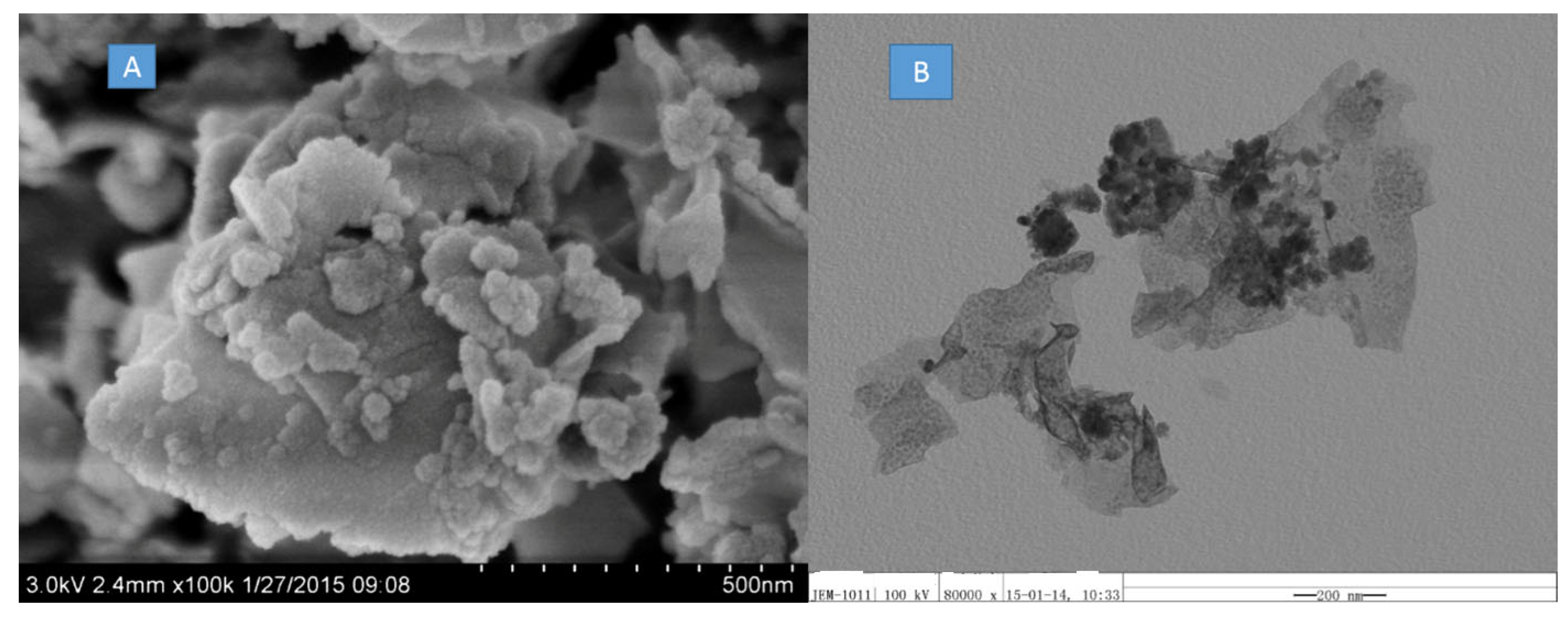
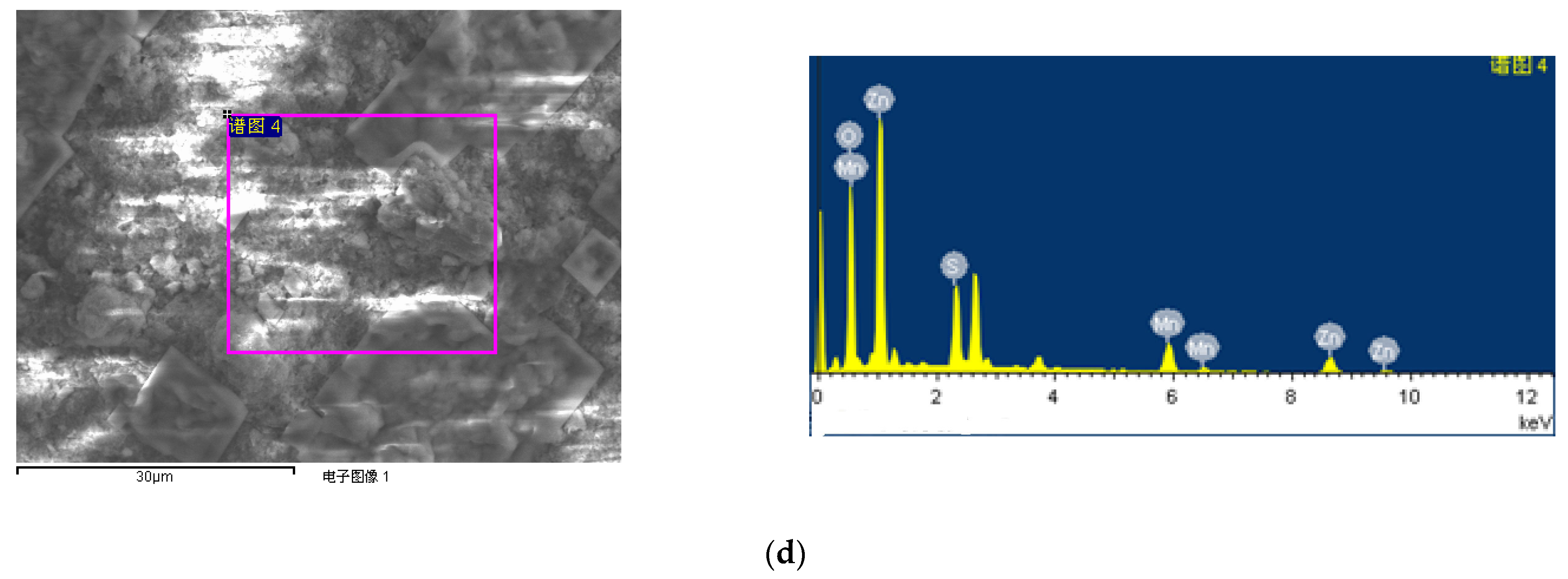
2.4. Morphology Observation with SEM

The scanning electron microscopy (SEM) observation was carried out with a Hitachi S-4800 (Hitachi, Tokyo, Japan). The obtained sample was washed with deionized water, deposited on an aluminum foil substrate, dried at room temperature, and then sputtered with a thin layer of Pt on its surface for SEM observation.

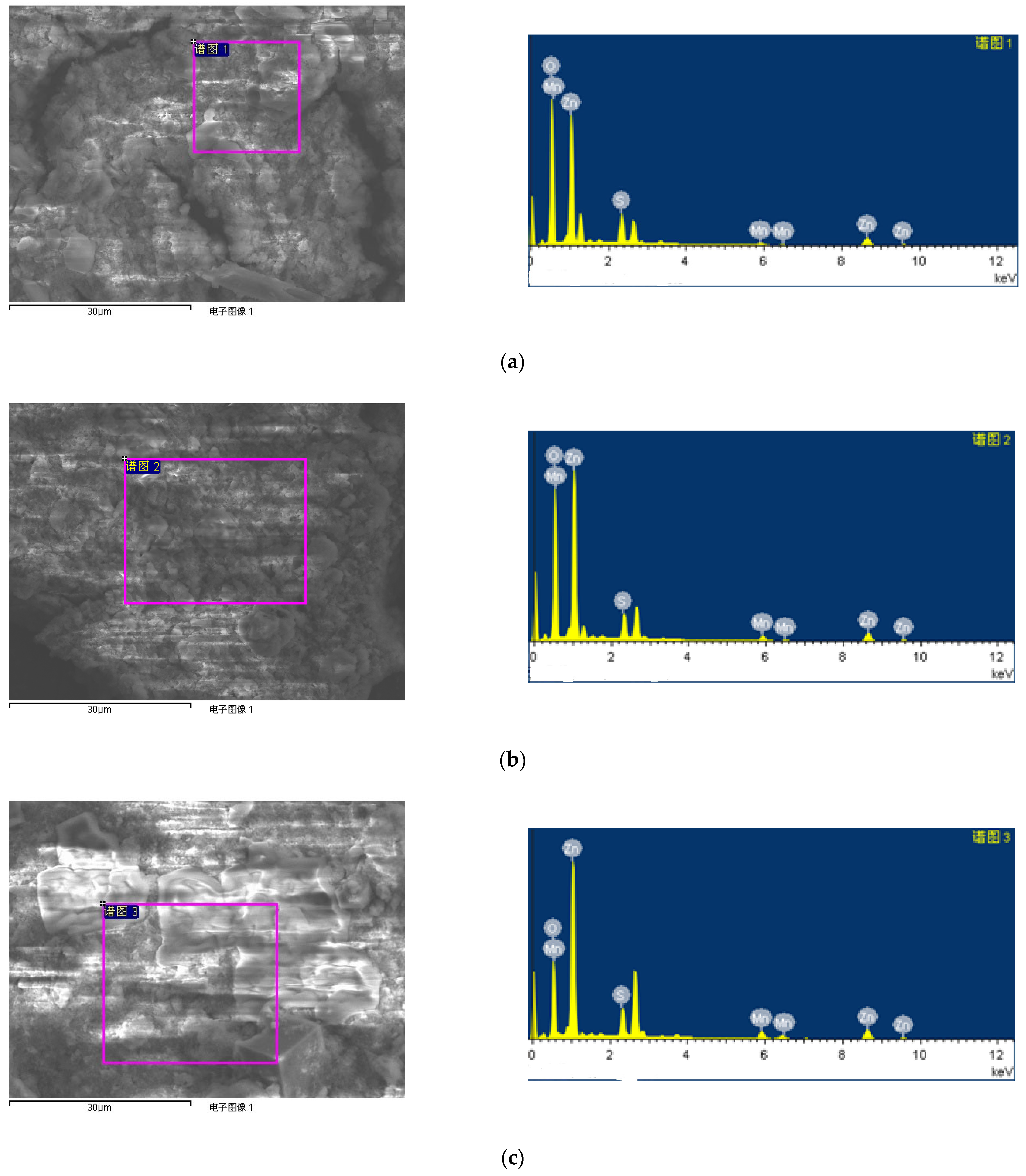
2.5. Energy Dispersive Spectroscopy (EDS) Examination

The energy dispersive spectroscopy (EDS) examination was carried out using a Hitachi S-4800 (HITACHI, Tokyo, Japan). The 1#, 2#, 3#, and 4#-ZnO/MnSx heterojunctions were washed with deionized water, deposited on Al foils, and dried at room temperature. The fabrication process of the sample is similar to that of SEM observation. The only difference is as follows. The sample for EDS examination did not need to have a layer of Pt deposited on its surface. The EDS data were obtained.

2.6. Morphology Observation with TEM

The TEM observation was carried out with a JEM-1011 (Nippon Electronics Co., LTD, Tokyo, Japan). The samples were dispersed in absolute alcohol, deposited on copper mesh coated with carbon film, and dried at room temperature for 30 min.

2.7. XRD Characterization

The powders’ X-ray diffraction (XRD) patterns were determined using the XRD-7000 SHIMADZU diffraction device (Shimadzu, Kyoto, Japan), a rotating anode X-ray generator was working at 40 kV, 300 mA, with Cu Ka monochromatic radiation. The sample was dispersed in an aqueous solution, then cast on a glass substrate and dried for 48–96 h at room temperature for determination.

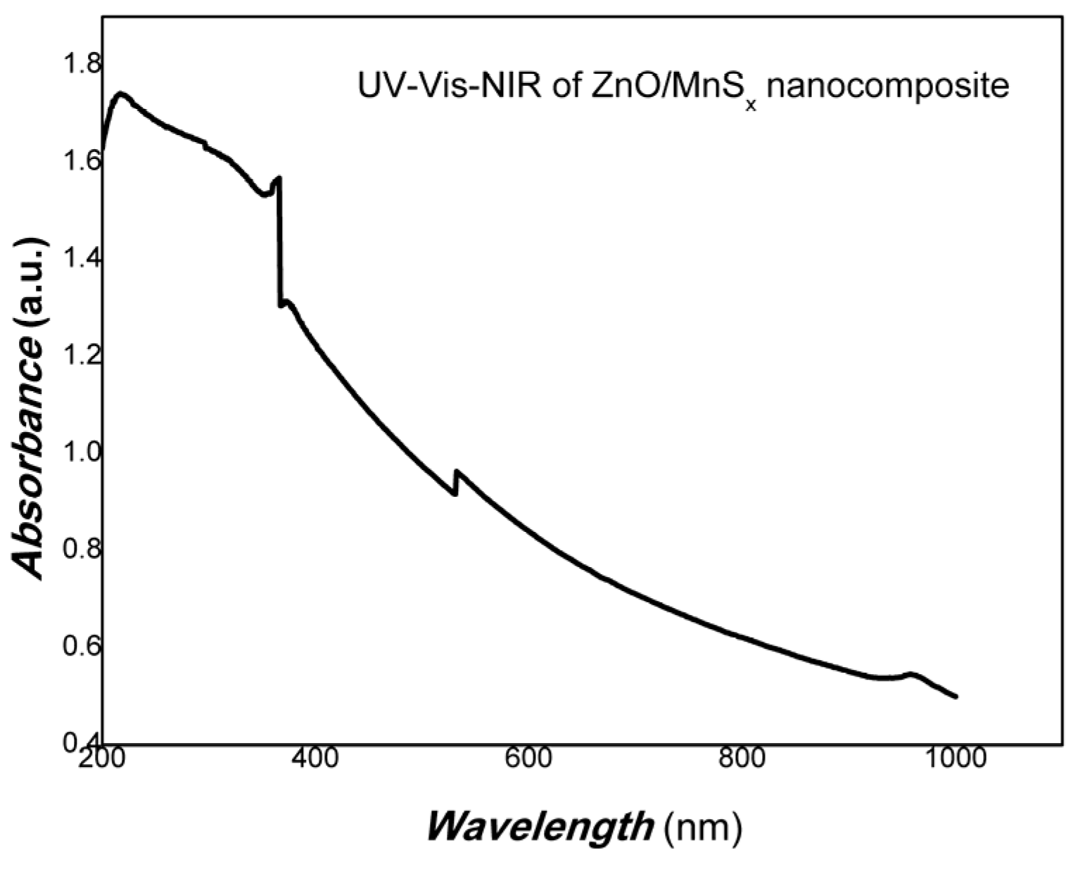
2.8. Measurement of UV-Vis-NIR Spectrum

The UV-Vis-NIR was determined by a TU-1810 spectrophotometer (Shanhai Yuan Analysis Instrument Co., LTD, Shanhai, China) and the samples were taken using a suspension.

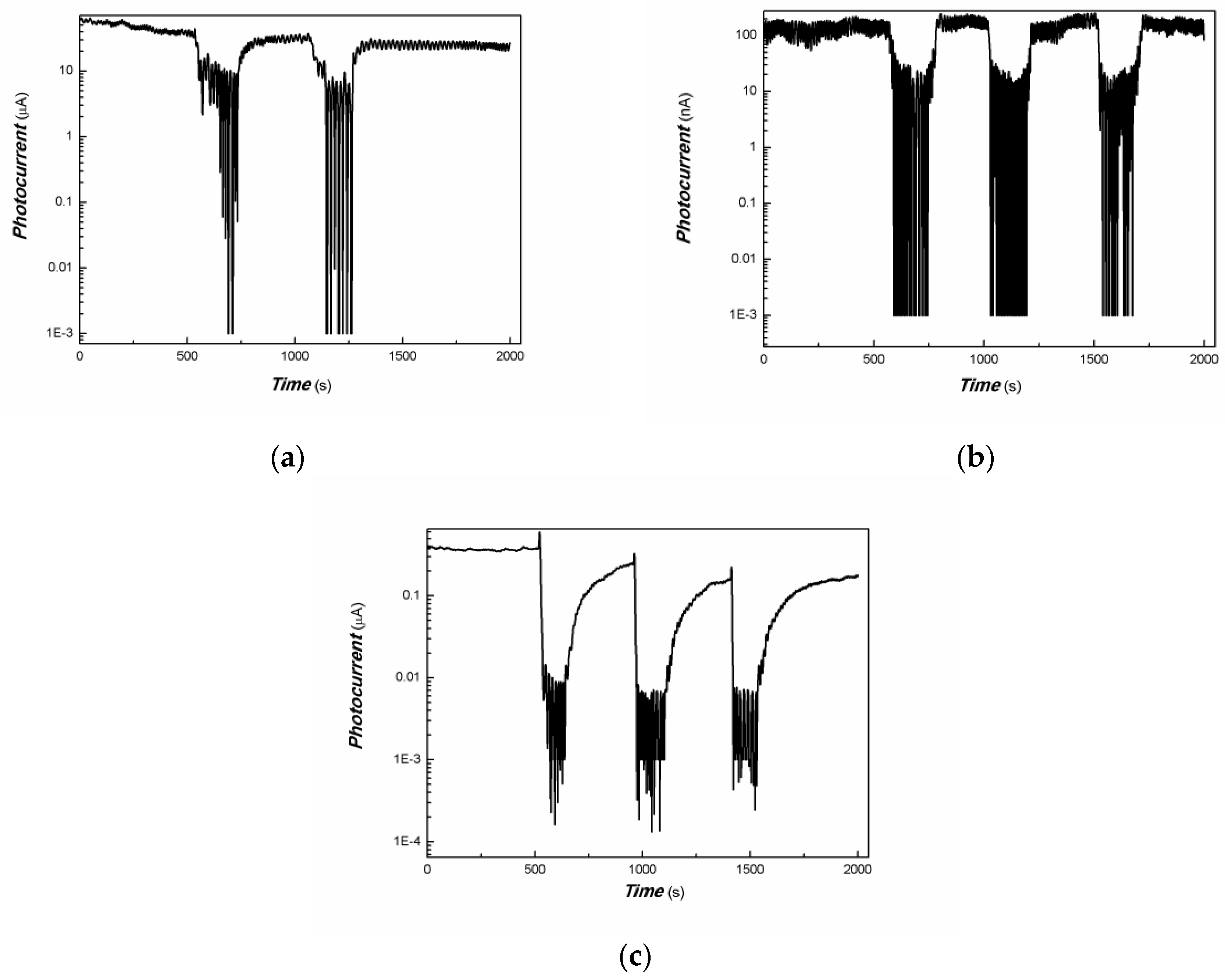
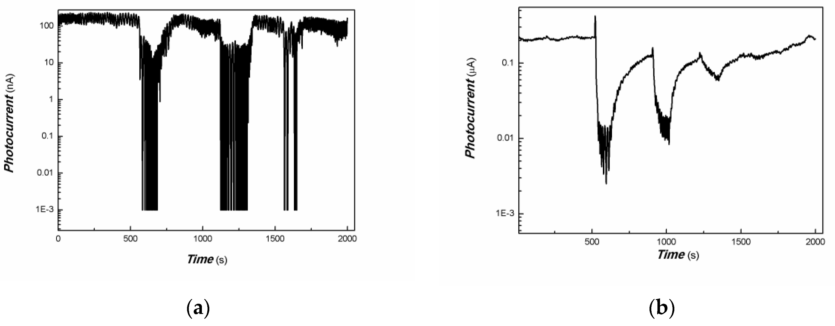
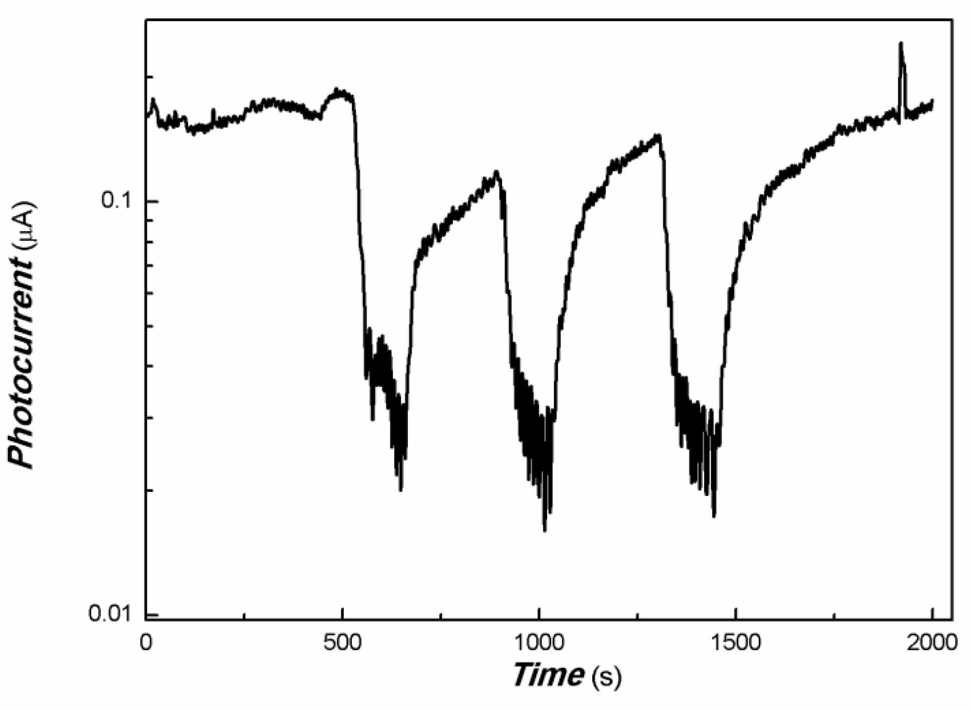
2.9. Photocurrent Response of Nanocomposite

The nanocomposite suspension was cast on the interdigital electrodes of Au on a flexible PET film substrate, after drying, the photoconductivity responses to weak visible light (20–25 W) (650 nm etc.) and NIR light (808, 980, and 1064 nm, etc.) were determined with an LK2000A Electrochemical Work Station from LANLIKE Chemistry and Electron High Technology Co., Ltd. (LANLIKE Chemistry and Electron High Technology Co., LTD, Tianjin, China). A 1 V DC bias was applied [89,90,91].

2.10. Construction of Prototype Sensors and Arrays

ZnO nanosheets and ZnO nanosheets/MnSx nanocomposite were firstly dispersed in deionized water (about 0.1–0.2% wt.), which was used as the sensing layer of the QCM sensors. This is also shown in our previous reports [92,93,96,98]. The QCM sensors were prepared as follows: the AT-cut 6000 MHz crystal was rinsed repeatedly into ethanol or deionized water and dried in air at room temperature. Two microliters of ZnO nanosheets and ZnO nanosheets/MnSx nanocomposite dispersion aqueous solutions were dispensed onto the surfaces of the electrodes using a micropipette, separately. After the devices were dried at room temperature, ZnO nanosheets and ZnO nanosheets/MnSx nanocomposite-treated QCM sensors were formed.

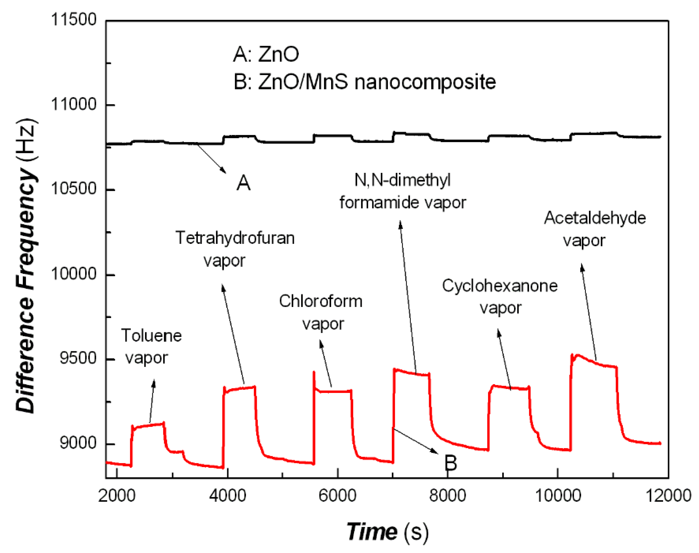
2.11. Characterization of Surface and Interface Features of Nanocomposite with the Help of QCM Arrays

The experimental details refer to our previous reports [92,93,96,98].

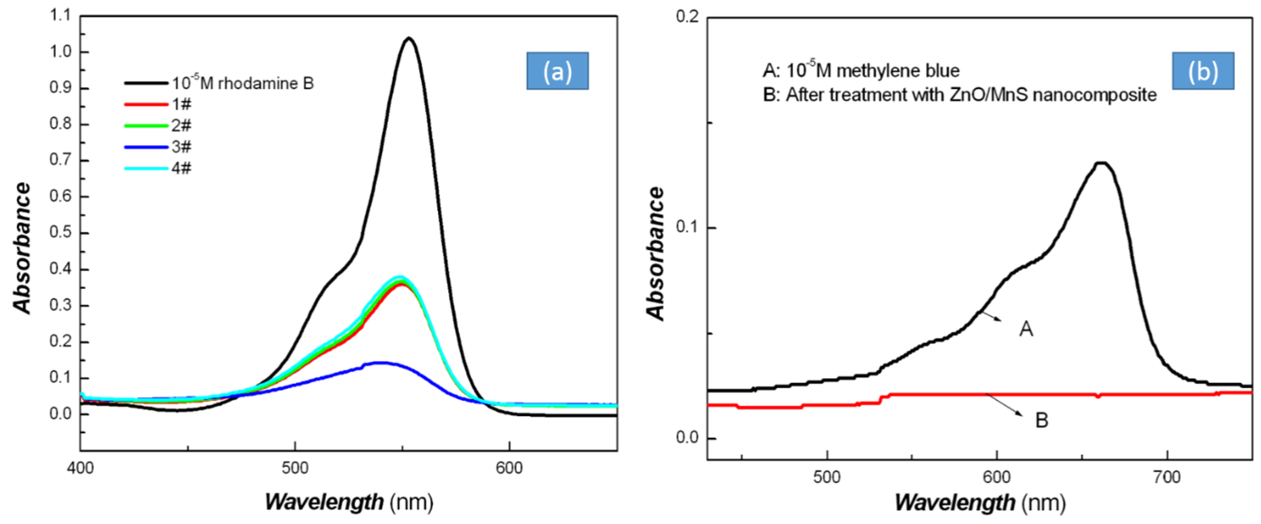
2.12. Comparison of the Nanocomposites to the Removal of Organic Pollutants

Comparison of the nanocomposites for the removal of organic pollutants: In 50 mL transparent vials (made from PET plastic), about 0.05 g as-prepared nanocomposite was added either into 40 mL of 10−5 M rhodamine B solution or methylene blue (rhodamine B and methylene blue were selected as simulating organic pollutants). The references were 10−5 M rhodamine B or methylene blue solution. The above-mentioned of transparent vials were placed for 2–3 days under exposure to natural sunlight indoors and at room temperature.

3. Results and Discussion

ZnO nanosheets utilized were the same as that of our previous reports [92,93,94] in the experiments. The ZnO nanosheets/MnSx nanocomposite was analyzed with SEM and TEM examinations. The representative SEM and TEM images of ZnO nanosheets/MnSx nanocomposite are shown in Figure 1.
Figure 1 shows that the majority of morphologies of materials are two-dimensional nanosheets; this is characteristic of ZnO nanomaterials, which is in line with that of the previous report [92,93,94], and the size of ZnO nanosheets is not uniform. Two-dimensional nanosheets are in favor of contact with other materials, which is helpful for interface charge transfer for multi-component nanocomposites. A small amount of material is presented in the nanoparticle form morphology, which is indicative of MnSx. The distribution of MnSx nanoparticles on the ZnO nanosheets is also not uniform, which can be observed from the TEM images of Figure 1B. The intimate contact between ZnO nanosheets and MnSx nanoparticles contributed to the interface charge transfer.
The XRD of ZnO nanosheets/MnSx nanocomposite was also determined. The results are shown in Figure 2.
As shown in Figure 2, there are some diffraction peaks of 2θ in 34.13 (002), 36.15 (101), 47.30 (102), 56.16 (110), 62.75 (103), 67.55 (112), 72.38 (004), 76.93 (202), etc., in the ZnO/MnSx nanocomposite. These diffraction peaks belong to the hexagonal structure of ZnO (PDF#36-1451). Also present are some weak diffraction peaks of 2θ in 31.60 (200), 54.13 (311), and 66.29 (400). These peaks belong to the diffraction of face-centered MnS cubic crystals (PDF#40-1288). Due to the exchange of oxygen and sulfur atoms in the synthesis of ZnO/MnSx nanocomposite, it is possible to produce some MnOx and MnSx or ZnS products. As shown in Figure 2, the diffraction peaks of 54.15 (230), 66.15 (331), and 76.67 (422) belong to the MnS2 (PDF#25-0549). The diffraction peaks of 47.33 (311), 64.53 (203), and 68.90 (610) belong to the MnO2 (PDF#39-0375). The diffraction peaks of 28.37 (002), 39.42 (102), and 72.43 (203) are ZnS (PDF#36-1450). The diffraction peaks of 28.91 (103), 32.20 (105), 48.57 (1011), 56.33 (118), and 58.51 (205) belong to the ZnS (PDF#39-1363). Because the content of MnSx in the nanocomposite is relatively low, the diffraction peaks of MnSx look a little weak. The main diffraction peaks are still ZnO nanosheets.
The UV-Vis-NIR of ZnO nanosheets/MnSx nanocomposite was examined. The results are shown in Figure 3.
As shown in Figure 3, it is interesting that the absorbance edge of ZnO nanosheets/MnSx nanocomposite was extended to part of the NIR (near-infrared) region. Therefore, it would be utilized more efficiently over the whole range of visible light and part of the NIR range for environmental or biomedical fields, etc., since the bandgap widths of ZnO and MnS are about 3.32 and 3 eV, respectively. The bandgap width of ZnS is about 3.52 eV, MnS2 is about 0.5 eV, and ZnS2 is about 2.7 eV. The absorbance in the 400–1000 nm range is a possible result of the introduction of impurity levels or defect levels. Therefore, the resulting nanocomposite is possibly containing a certain amount of MnSx. The oxygen or sulfur vacancy took an important role to enhance the absorbance in the longer wavelength range.
To explore the physical mechanism of ZnO nanosheets/MnSx nanocomposite with the activity of visible light, the photoconductive response to visible light with low-power was studied based on interdigital electrodes of Au on flexible PET (polyethylene terephthalate) film substrate with the casting method. The representative results are shown in Figure 4.
Figure 4 shows that the ratio of on/off of ZnO nanosheets/MnS to weak visible light was changed greatly, by about two orders of magnitude. Since a large number of defects in the interface between ZnO nanosheets and MnSx nanoparticles were presented, the electrons generated by light inducement were trapped by these defects. The holes were accumulated at these interfaces, which resulted in the decrease in the thick film current of the nanocomposite. It is expected that these holes generated by light inducement had a strong oxidation ability, which led to the degradation of some organic pollutants. This illustrated that the ZnO nanosheets/MnSx nanocomposite showed good activity to weak visible light, which can easily produce a photo-induced charge, avoiding the recombination of charges produced by visible light.
The repeatability of the photoresponse of ZnO nanosheets/MnSx to weak visible light was also examined. The repeatability was also very good, which is shown in Figure 5. Otherwise, the effect of the amount of MnSx on the photoresponse of ZnO nanosheets/MnSx to weak visible light was studied. The representative results are shown in Figure 6.
Figure 6 shows that the effect of the amount of MnSx on the photoresponse of ZnO nanosheets/MnSx nanocomposites to weak visible light was great at the initial stage, then changed to smooth and steady. The ratio of on/off of ZnO nanosheets/MnSx nanocomposites was changed by about one to two orders of magnitude, varying with the amount of MnSx. This illustrated that more electrons generated by light excitation were captured by the defects from the increasing content of MnSx. Further increasing the loading of MnSx, holes that accumulated at the interfaces became to smooth due to the aggregation of MnSx nanoparticles. The contact interface between ZnO nanosheets and MnSx nanoparticles would take an important role in the production of holes generated by light. Figure 6 also shows that the 2#-ZnO nanosheets/MnSx nanocomposites exhibited more sensitivity than other nanocomposites samples to weak visible light excitation.
As shown in Figure 3, the absorbance edge of ZnO nanosheets/MnSx nanocomposite extended to part of the NIR (near-infrared) region. Therefore, 808 nm, 980 nm, and 650 nm were selected the typical light resources to examine the photoconductive responses. The representative results are shown in Figure 7, Figure 8 and Figure 9.
Figure 7 shows that the on/off ratio of 2#-, 3#-, and 4#-ZnO/MnSx nanocomposites to 200 mW 808 nm change by about two, five, and two orders of magnitude, respectively. Among them, sample 3# is more sensitive than other samples to the 200 mW 808 nm light resource. On the other hand, it could be illustrated that the defect at 1.53 eV or so of the 3#-ZnO/MnSx nanocomposite was more than that of other samples.
Figure 8 shows that the on/off ratio of 2#-, 3#-, and 4#-ZnO/MnSx nanocomposites to 200 mW 980 nm change by about four, five, and four orders of magnitude, respectively. Among them, sample 3# is more sensitive than other samples to the 200 mW 980 nm light resource as well. Similarly, the defect at 1.26 eV or so of the 3#-ZnO/MnSx nanocomposite was more than that of other samples.
Figure 9 shows that the on/off ratio of ZnO/MnSx nanocomposites to 100 mW 650 nm is changed by about five orders of magnitude. Samples 3# and 4# showed a similar response to the 100 mW 650 nm light resource.
To examine the carriers’ photo-generated extracting ability with low-power light resource excitation, photocurrent responses were examined with a lower power of typical light resources and the representative nanocomposites. The results are shown in Figure 10 and Figure 11.
As shown in Figure 10 and Figure 11, it is found that the resulting nanocomposites still had good photocurrent responses to 50 mW 650 and 980 nm of light. However, the nanocomposite had little photocurrent response to 5 mW 650 and 980 nm of light. This also showed that the 3# nanocomposite was still more sensitive than that of sample 4# to 980 nm NIR. Meanwhile, as shown in Figure 10 and Figure 11, it is also found that the photocurrent of ZnO nanosheets/MnSx nanocomposites to weak 650 nm and 980 nm light resources were increased at the initial stage, and then decreased to smooth and steady. This illustrated that a large amount of photo-generated electrons were extracted effectively at the initial stage, then more electrons generated by the light trigger were captured by the defects. Holes accumulated at the interfaces changed to smooth, and the direction of built-in electrical fields produced is opposite to that of bias fields applied, which led to the decrease in the film’s current. This also indicated that more electron traps were present in the resulting nanocomposites. These traps in the resulting nanocomposites took on a vital role in exerting the physical and chemical properties of the films.
The cyclic stability was also examined; we selected a typical sample and representative light resource. The results are shown in Figure 12.
As shown in Figure 12, the resulting nanocomposite showed a good cyclic stability to the 200 mW 808 nm light resource as a whole.
Due to exhibiting good photocurrent responses to the 808 nm and 980 nm light resources, the photocurrent responses to 1064 nm were also examined. We selected a typical sample; since 1064 nm is another important light resource in the bio-imaging field, it was used. The results are shown in Figure 13.
Figure 13 shows that the on/off ratio of the resulting nanocomposite to a 20 mW 1064 nm light is changed by about one order of magnitude. It is illustrated that the resulting nanocomposite was photoconductive sensitive to a weak 1064 nm light resource, and therefore has a potential application in biomedical fields.
Due to the excellent sensitivity of the QCM sensor (QCM device could weigh the weight of a single molecule), the QCM arrays were constructed to compare the adsorption properties of ZnO nanosheets and ZnO nanosheets/MnSx nanocomposite under similar micro-environments for functional material design and devices or photocatalyst applications. Among organic pollutants, organic pollutant solutions and organic solvents are one kind of the main pollution sources (such as coatings and adhesives from house decoration, organic wastewater from chemical factories and pharmaceutical factories, etc.). Therefore, some representative VOCs (volatile organic compounds) were selected for determination, such as toluene vapor, acetaldehyde vapor, cyclohexanone vapor, N, N-dimethyl formamide vapor, chloroform vapor, and tetrahydrofuran vapor. The comparative results of ZnO nanosheets and ZnO nanosheets/MnSx nanocomposite under the same test examination conditions based on QCM arrays devices are shown in Figure 14.
Figure 14 shows the response behavior comparisons of ZnO nanosheets and ZnO nanosheets/MnSx nanocomposite to 30 mL saturated toluene vapor, acetaldehyde vapor, cyclohexanone vapor, N,N-dimethyl formamide vapor, chloroform vapor, and tetrahydrofuran vapor diluted in a 1000 mL chamber with N2 at similar test conditions. As shown in Figure 14, it also can be found that compared with ZnO nanosheets (as a reference), the ZnO nanosheets/MnSx nanocomposite exhibited enhanced adsorption properties to some representative VOCs dramatically under the same micro-environment experimental conditions based on QCM array examinations. The presence of interfaces between ZnO nanosheets and MnSx nanoparticles or defects of nanocomposites contributes to this excellent adsorption property. A large number of defects at the interface has high chemical activity, and is in favor of adsorption to VOCs. On the other hand, MnSx nanoparticles attached to the ZnO nanosheets also avoid the aggregation of ZnO nanosheets, and improved the adsorption property. This illustrated that the ZnO nanosheets/MnSx nanocomposite has an excellent ability to remove VOCs.
To explore the interaction between VOCs and the surface of ZnO nanosheets/MnSx nanocomposite, the selectivity to different organic vapors of ZnO nanosheets/MnSx nanocomposite based on QCM arrays was also examined. The results are shown in Figure 15.
Figure 15 shows that the selectivity to different VOCs of ZnO nanosheets/MnSx nanocomposite based on QCM arrays was also clear, and the bigger the polarity of vapor, the larger the adsorption response to VOCs. Due to the presence of a large number of hydroxyl groups on the surface of ZnO nanosheets, the ZnO nanosheets/MnSx nanocomposite tended to absorb the VOCs with higher polarity due to presence of some strong chemical interaction and physical interaction forces.
Based above obtained good results, the treatment of organic pollutants in solution was also investigated. Thus, photocatalytic efficiency was examined by selecting two typical organic pollutant solutions under natural sunlight indoors; some good results were also obtained. For methylene blue as an organic pollutant, the decolorization is complete for 1#-, 2#-, 3#-, and 4#- ZnO nanosheets/MnSx nanocomposite; regarding rhodamine B, the effects of degradation are also very clear, although some fluctuations were present. This is possibly the results of the nonuniformity of MnSx in ZnO nanosheets (which is shown in TEM images of Figure 1B). The representative results are shown in Figure 16.
Figure 16 shows that the ZnO/MnSx nanocomposite exhibited better degradation effects on methylene blue than on rhodamine under natural sunlight indoors. On the other hand, the dosage of the resulting nanocomposite is very small (about 0.05 g) in the experiments compared to most of references reported. It is interesting and useful to remove the organic pollutants using such a small dosage. This also illustrated that the resulting nanocomposite had great selectivity to different organic pollutants due to different adsorption and degradation mechanisms. It is well known that the distribution of the sunlight spectrum reaching the surface of earth is about as follows: the light below 400 nm in wavelength is about less than 5%; the 400–700 nm range is about 45%–46%; the NIR region is about 48%–49%. Since the experiments were carried out using exposure to natural sunlight indoors, there is a little UV light in this environment. The results are also meaningful for utilizing low-power light resources, such as the visible light range (400–700 nm) and part of the NIR range (808, 980, and 1064 nm).
Combining the results of Figure 4, Figure 5, Figure 6, Figure 7, Figure 8, Figure 9, Figure 10, Figure 11, Figure 12 and Figure 13, it is shown that under the irradiation of weak visible light and the NIR, a large number of holes were collected at the interface between ZnO nanosheets and MnSx nanoparticles. The reaction between holes and H2O molecules adsorbed on the surface of ZnO would take place and result in the production of free radicals. These fresh free radicals are in favor of degrading some organic pollutants due to their strong oxidation ability. Otherwise, ZnO and MnS belong to wide bandgap semiconductors, the contact of ZnO nanosheets and MnSx nanoparticles formed a P/N junction. The presence of a P/N junction led to the formation of strong built-in electrical fields. Strong built-in electrical fields would sustainably produce holes generated by light inducement and promote the separation of carriers by light inducement. Additionally, the energy level position of ZnO and MnS is shown in Figure 17.
As shown in Figure 17, the ZnO nanosheets/MnS contact belongs to the II-type heterojunction classification, which is a result of preventing the recombination of electron/hole pairs. The electrons generated by light were transferred to the CB (conductive band) of MnS from the CB of ZnO, and the holes were transferred to the VB (valence band) of ZnO from the VB of MnS. Since ZnO and MnS belong to the group of wide bandgap semiconductor materials, the absorbance of light should occur with a wavelength less than 400 nm. The good activities of the visible light and part of the NIR range of the resulting nanocomposite were contributed to the presence of impurities or defects. Among the impurity levels or defect levels, the distribution of deep energy levels and shallow energy levels is different for the nanocomposite with the adjusted composition, which led to the great changes in photoconductive sensitivity under different wavelength excitation light resources. The nonstoichiometric factor and the exchange of oxygen and sulfur atoms in the preparation of nanocomposite also led to some oxygen or sulfur vacancy, which enhanced the absorbance in the NIR. Combined with the results of XRD, UV-Vis-NIR, and photocurrent extracted from the visible range to 1064 nm light resources, it can be found that the resulting nanocomposite is possibly ZnO/MnSx. The impurity level or defect level in the nanocomposite plays an important role in the process of photodynamics over a wide range of excitation light resources. This study provides some reference values to explore the physical mechanisms of materials physics and photophysics for photoactive functional nanocomposite further.
The above results would also support our viewpoints on the visible light and NIR light activities of ZnO nanosheets/MnSx nanocomposite. Therefore, it would be used for developing low-cost nanocomposites with visible light and NIR light activities, and applied for photocatalysts, nano-reactors, self-cleaning multi-functional films or nano-coatings, treatment of organic pollutants, interdisciplinary fields, etc.
To explore the relationship between the composition of ZnO/MnSx nanocomposite and properties, the EDS data of 1–4# ZnO/MnSx nanocomposites were determined. The atomic percent and weight percent (%) of Zn, O, Mn, and S elements in 1–4# ZnO/MnSx heterojunctions were examined. The typical results are shown in Table 1 and Figure 18.
As shown in Table 1, it is found that the content of Mn increased from the 1#- to 4#-ZnO/MnSx nanocomposites. On the whole, the content of S also showed an increasing trend. The S element of sample 2# exhibited a slightly unusual content, which is the result of loss of S due to the generation of vacancy. The molar ratios of S/Mn in 1–4# ZnO/MnSx are about 5.58, 2.25, 1.74, and 1.30, respectively. The molar ratios of O/Zn in 1–4# ZnO/MnS are about 3.94, 3.24, 1.91, and 2.74. Although the detecting error of EDS is a little high compared with other analytical methods, the comparative results still can be discussed using the changing trend. It is shown that 1–4# ZnO/MnSx are oxygen-enriched and sulfur-enriched nanocomposites. The oxygen-rich feature is possibly the result of absorbed O2 or H2O molecules. On the other hand, it is also possible for the O and S elements to exchange, leading to small amounts of ZnS, MnS2, or Mn oxide. Combined with the results of XRD, some diffraction peaks of MnO2, MnS2, and ZnS could also be distinguished in detailed analysis.

4. Conclusions

In summary, the ZnO nanosheets/MnSx nanocomposite was prepared using hydrothermal synthesis. The photoconductive responses of nanocomposites to the visible light range and NIR range with low power light excitation were examined based on interdigital electrodes of Au on a flexible PET film substrate. The results showed that the ZnO nanosheets/MnSx nanocomposite produced a photo-induced charge very easily to weak visible light and NIR light and possessed good visible light and NIR activities. Strong charge transfer at the interface between ZnO nanosheets and MnSx nanoparticles took place via the light trigger. At the same time, this nanocomposite also showed greatly enhanced adsorption properties to some representative organic volatile compounds based on QCM device array examinations. Since ZnO and MnS both belong to the group of wide bandgap semiconductor materials, the absorbance of light should occur on wavelengths less than 400 nm. The presence of impurities or defects contributed to the good activities in the visible light and NIR ranges of the resulting nanocomposite. The nonstoichiometric factor and the exchange of oxygen and sulfur atoms in the preparation of the nanocomposite also led to some oxygen or sulfur vacancy, which enhanced the absorbance in the NIR. Combined with the results of XRD, UV-Vis-NIR, and photocurrent responses from the visible range to the NIR light resources, it can be concluded that the resulting nanocomposite was possibly ZnO/MnSx. The impurity level or defect level in the nanocomposite plays an important role in the process of photodynamics over a wide range of excitation light resources. This study provides some reference values to explore the physical mechanism of materials physics and photophysics for a photoactive functional nanocomposite. This would have good potential applications in photocatalysts, nano-reactors, self-cleaning multi-functional films, coatings, and treatment of organic pollutants in the gas phase or liquid phase, used in the environmental fields for sustainable development.

Author Contributions

X.M. conceived and designed the experiments; X.Z. (XRD), M.G. (UV-Vis-NIR), and X.M. performed the experiments; Y.W. carried out the fabrication of Au electrodes on a PET film substrate, G.L. checked the English of the paper; all authors analyzed the data; X.M., wrote the paper; all authors discussed the results on the paper. All authors have read and agreed to the published version of the manuscript.

Funding

This project had ever been supported by the Natural Science Foundation of Shandong Province (project No. ZR2013EMM008).

Institutional Review Board Statement

Not applicable.

Informed Consent Statement

Not applicable.

Data Availability Statement

The data presented in this study are available on request from the corresponding author.

Acknowledgments

Thanks to You Wang and Guang Li of Zhejiang University for the fabrication of Au electrodes on PET film substrate and for checking the English of our papers for many years. TEM was performed by Chunsheng Wang, School of Chemistry and Chemical Engineering, Shandong University. SEM and EDS were conducted by Wenhai Wang, Yantai Institute of Coastal Zone Research, Chinese Academy of Sciences. Bin Guo, a Master Student of Yantai University, carried out part of the experiments. Kaihuan Zhang of Zhejiang University, assisted the QCM array experiments. Jianxun Qiu of Yantai University assisted the XRD experiments for many years.

Conflicts of Interest

We declare that we have no conflict of interest.

References

  1. Ali, U.; Qureshi, A.A.; Javed, S.; Rehman, G.; Akram, M.A. Graphene oxide incorporation in Ag-doped ZnO nanocomposite as efficient electron extraction material for planar perovskite solar cells. Results Opt. 2023, 12, 100486. [Google Scholar] [CrossRef]
  2. Zhao, X.; Li, Q.; Xu, L.; Zhang, Z.; Kang, Z.; Liao, Q.; Zhang, Y. Interface Engineering in 1D ZnO-Based Heterostructures for Photoelectrical Devices. Adv. Funct. Mater. 2021, 32, 112106887. [Google Scholar] [CrossRef]
  3. Tian, F.; Han, W.; Hu, J.; Wang, H.; Li, H.; Geng, F.; Wei, T.; Li, D. Oxygen vacancy-enriched bilayer flower-like structure of ZnO&NiO@C-ZnO nanorod arrays on carbon cloth with improved electrochemical performance. J. Energy Storage Part B 2023, 72, 108316. [Google Scholar]
  4. Kumar, R.D.; Kumar, A.J.; Balachandran, S.; Kusmartsev, F.V.; Trabelsi, A.B.G.; Alkallas, F.H.; Nagarani, S.; Sethuraman, V.; Lee, B. High-performance chrysanthemum flower-like structure of Ni-doped ZnO nanoflowers for pseudo-supercapacitors. J. Energy Storage Part B 2023, 72, 108441. [Google Scholar] [CrossRef]
  5. Zhang, H.; Liu, F.; Mou, Z.; Liu, X.; Sun, J.; Lei, W. A facile one-step synthesis of ZnO quantum dots modified poly (triazine imide) nanosheets for enhanced hydrogen evolution under visible light. Chem. Commun. 2016, 52, 13020–13023. [Google Scholar] [CrossRef] [PubMed]
  6. Li, G.; Liu, P.; Liu, R.; Liu, M.; Tao, K.; Zhu, S.; Wu, M.; Yi, F.; Han, L. MOF-derived hierarchical double-shelled NiO/ZnO hollow spheres for high-performance supercapacitors. Dalton Trans. 2016, 45, 13311–13316. [Google Scholar] [CrossRef] [PubMed]
  7. Chao, J.; Yu, H.; Zhang, K.; Zhou, Y.; Meng, D.; Sun, Y. Integration of ZnO and Au/ZnO Nanostructures into Gas Sensor Devices for Sensitive Ethanolamine Detection. ACS Appl. Nano Mater. 2023, 6, 5994–6001. [Google Scholar] [CrossRef]
  8. Hyun, S.K.; Nam, B.; Ko, T.K.; Lee, C.; Choi, S.B.; Lee, W.I. Optimal Composition of ZnO/WO3 Composite Nanoparticle Gas Sensors. Phys. Status Solidi A 2020, 217, 1900874. [Google Scholar] [CrossRef]
  9. Ramakrishnan, V.; Nair, K.G.; Dhakshinamoorthy, J.; Ravi, K.R.; Pullithadathil, B.I. Porous, n–p type ultra-long, ZnO@Bi2O3 heterojunction nanorods—Based NO2 gas sensor: New insights towards charge transport characteristics. Phys. Chem. Chem. Phys. 2020, 22, 7524–7536. [Google Scholar] [CrossRef]
  10. Cao, P.; Cai, Y.; Pawar, D.; Han, S.; Xu, W.; Fang, M.; Liu, X.; Zeng, Y.; Liu, W.; Lu, Y.; et al. Au@ZnO/rGO nanocomposite-based ultra-low detection limit highly sensitive and selective NO2 gas sensor. J. Mater. Chem. C 2022, 10, 4295–4305. [Google Scholar] [CrossRef]
  11. Vishnuraj, R.; Aleem, M.; Nair, K.G.; Pullithadathil, B. 1D aligned, n–p and n–n type ZnO heterojunction nanofibers for NO2 sensors: Exploration of conduction mechanism using in situ impedance spectroscopy. Mater. Adv. 2023, 4, 3010–3025. [Google Scholar] [CrossRef]
  12. Beitollahi, H.; Tajik, S.; Nejad, F.G.; Safaei, M. Recent advances in ZnO nanostructure-based electrochemical sensors and biosensors. J. Mater. Chem. B 2020, 8, 5826–5844. [Google Scholar] [CrossRef] [PubMed]
  13. Krishna, M.S.; Singh, S.; Batool, M.; Fahmy, H.M.; Seku, K.; Shalan, A.E.; Lanceros-Mendez, S.; Zafar, M.N. A review on 2D-ZnO nanostructure-based biosensors: From materials to devices. Mater. Adv. 2023, 4, 320–354. [Google Scholar] [CrossRef]
  14. Tang, K.; Jiang, M.; Yang, B.; Xu, T.; Liu, Z.; Wan, P.; Kan, C.; Shi, D. Enhancing UV photodetection performance of an individual ZnO microwire p–n homojunction via interfacial engineering. Nanoscale 2023, 15, 2292–2304. [Google Scholar] [CrossRef] [PubMed]
  15. Nandi, S.; Kumar, S.; Misra, A. Zinc oxide heterostructures: Advances in devices from self-powered photodetectors to self-charging supercapacitors. Mater. Adv. 2021, 2, 6768–6799. [Google Scholar] [CrossRef]
  16. Wang, H.; Wang, H.; Li, L.; Gu, Y.; Kim, B.; Huang, J. Self-Powered Broadband Photodetectors Material Based on Co3O4–ZnO Heterojunction with Bottlebrush Nanostructure. ACS Appl. Electron. Mater. 2023, 5, 3224–3231. [Google Scholar] [CrossRef]
  17. Kim, H.; Baek, S.; Lee, S. Surface Plasmon-Enhanced High-Performance ZnO/Ni/ZnO Ultraviolet Photodetectors. Phys. Status Solidi Rapid Res. Lett. 2020, 14, 1900685. [Google Scholar] [CrossRef]
  18. Ning, Y.; Zhang, Z.; Teng, F.; Fang, X. Novel Transparent and Self-Powered UV Photodetector Based on Crossed ZnO Nanofiber Array Homojunction. Small 2018, 14, 1703754. [Google Scholar] [CrossRef]
  19. Zhou, X.; Yan, C.; Deng, R.; Hu, Y.; Deng, Z.; Cui, Q.; Lou, Z.; Hou, Y.; Teng, F. High-Performance Polymer Photodetectors using ZnO Nanocrystal Trap States. Phys. Status Solidi Rapid Res. Lett. 2021, 15, 2100003. [Google Scholar] [CrossRef]
  20. Yashaswini, M.; Naik, K.G. Fabrication of ZnO nanorods and ZnO/CdS core-shell nanorods for the ultraviolet photo-sensing application. Inorg. Chem. Commun. 2023, 154, 110975. [Google Scholar] [CrossRef]
  21. Zhou, J.; Qiao, Q.; Tan, Y.; Wu, C.; Hu, J.; Qiu, X.; Wu, S.; Zheng, J.; Wang, R.; Zhang, C.; et al. The improvement of polymer photodetector based on 1D-ZnO nanorod arrays/0D-ZnO quantum dots composite film. Opt. Mater. 2023, 142, 114086. [Google Scholar] [CrossRef]
  22. Peng, Y.; Jiang, D.; Zhao, M.; Duan, Y.; Wei, H.; Li, H.; Liang, Q.; Wang, S. High-performance UV–visible photodetectors based on ZnO/perovskite heterostructures. J. Alloys Compd. 2023, 965, 171372. [Google Scholar] [CrossRef]
  23. Boruah, B.D. Zinc oxide ultraviolet photodetectors: Rapid progress from conventional to self-powered photodetectors. Nanoscale Adv. 2019, 1, 2059–2085. [Google Scholar] [CrossRef] [PubMed]
  24. Liu, R.; Fu, X.; Guo, Y.; Zhang, J.; Tian, W. A study on Ag or Ce doped and co-doped ZnO for the photocatalytic degradation of RhB dye. Vacuum 2023, 215, 112337. [Google Scholar] [CrossRef]
  25. Vo, N.T.T.; You, S.; Pham, M.; Pham, V.V. A green synthesis approach of p-n CuO/ZnO junctions for multifunctional photocatalysis towards the degradation of methyl orange, phenol, and nitric oxide. Environ. Technol. Innov. 2023, 32, 103285. [Google Scholar] [CrossRef]
  26. Shi, Q.; Luo, Z.; Jiang, L.; Li, X.; Bai, C.; Yu, Q. Fabrication and photocatalytic properties of Co-doped ZnO nanomaterials. Mater. Lett. 2023, 350, 134952. [Google Scholar] [CrossRef]
  27. Al-Zaqri, N. Highly operative near full spectrum Cu doped NiO/ZnO photocatalyst: Photodegradation of phenol and organic dyes. Opt. Mater. 2023, 143, 114139. [Google Scholar] [CrossRef]
  28. Alawamleh, H.K.; Amin, A.H.; Ali, A.M.; Alreda, B.A.; Lagum, A.A.; Pecho, R.D.C.; Taqi, N.; Salman, H.M.; Nassar, M.F. Solar light driven enhanced photocatalytic treatment of azo dye contaminated water based on Co-doped ZnO/g-C3N4 nanocomposite. Chemosphere 2023, 335, 139104. [Google Scholar] [CrossRef]
  29. Bi, K.; Qin, X.; Cheng, S.; Liu, S. ZnO/g-C3N4 S scheme photocatalytic material with visible light response and enhanced photocatalytic performance. Diam. Relat. Mater. 2023, 137, 110143. [Google Scholar] [CrossRef]
  30. Yan, M.; An, B.; Li, X.; Zai, Z.; Wu, S.; Ma, J.; Zhang, L. Effect of different electronegative oxygen atoms of cellulose nanofibrils on the formation and photocatalytic property of ZnO/cellulose composite. Appl. Surf. Sci. 2023, 637, 157974. [Google Scholar] [CrossRef]
  31. Xie, Z.; Xie, L.; Qi, F.; Liu, H.; Meng, L.; Wang, J.; Xie, Y.; Chen, J.; Lu, C. Efficient photocatalytic hydrogen production by space separation of photo-generated charges from S-scheme ZnIn2S4/ZnO heterojunction. J. Colloid Interface Sci. Part A 2023, 650, 784–797. [Google Scholar] [CrossRef] [PubMed]
  32. Liang, Y.; Wang, Y. Optimizing crystal characterization of WO3–ZnO composites for boosting photoactive performance via manipulating crystal formation conditions. CrystEngComm 2021, 23, 3498–3509. [Google Scholar] [CrossRef]
  33. Wang, Q.; Chen, K.; Wang, S.; Jiang, D.; Ma, C.; Zhu, L.; Xu, X. An in situ-fabricated p-Co3O4@n-ZnO surface heterojunction photocatalyst for solar-to-fuel conversion of CO2. Mater. Chem. Front. 2023, 7, 523–534. [Google Scholar] [CrossRef]
  34. Feng, C.; Chen, Z.; Jing, J.; Hou, J. The photocatalytic phenol degradation mechanism of Ag-modified ZnO nanorods. J. Mater. Chem. C 2020, 8, 3000–3009. [Google Scholar] [CrossRef]
  35. Jayswal, S.; Moirangthem, R.S. Construction of a solar spectrum active SnS/ZnO p–n heterojunction as a highly efficient photocatalyst: The effect of the sensitization process on its performance. New J. Chem. 2018, 42, 13689–13701. [Google Scholar] [CrossRef]
  36. Sher, M.; Javed, M.; Shahid, S.; Iqbal, S.; Qamar, M.A.; Bahadur, A.; Qayyum, M.A. The controlled synthesis of g-C3N4/Cd-doped ZnO nanocomposites as potential photocatalysts for the disinfection and degradation of organic pollutants under visible light irradiation. RSC Adv. 2021, 11, 2025–2039. [Google Scholar] [CrossRef] [PubMed]
  37. Huang, M.; Lin, J.; Li, R.; Su, Y.; Zhao, X.; Liu, Y.; Chen, W.; Lian, X.; Chen, X.; Pan, X. Hierarchical ZnO Nanosheet-Reduced Graphene Oxide Composites for Photocatalytic Ethylene Oxidation. ACS Appl. Nano Mater. 2022, 5, 1828–1835. [Google Scholar] [CrossRef]
  38. Lonkar, S.L.P.; Pillai, V.; Abdala, A.; Mittal, V. In situ formed graphene/ZnO nanostructured composites for low-temperature hydrogen sulfide removal from natural gas. RSC Adv. 2016, 6, 81142–81150. [Google Scholar] [CrossRef]
  39. Kegel, J.; Zubialevich, V.Z.; Schmidt, M.; Povey, I.M.; Pemble, M.E. Effect of Surface and Defect Chemistry on the Photocatalytic Properties of Intentionally Defect-Rich ZnO Nanorod Arrays. ACS Appl. Mater. Interfaces 2018, 10, 17994–18004. [Google Scholar] [CrossRef]
  40. Cox, J.W.; Foster, G.M.; Jarjour, A.; Wenckstern, H.; Grundmann, M.; Brillson, L.J. Defect Manipulation To Control ZnO Micro-/Nanowire-Metal Contacts. Nano Lett. 2018, 18, 6974–6980. [Google Scholar] [CrossRef]
  41. Lai, J.; Farooq, M.U.; Sun, Y.; Tan, P.; Zhang, J. Multiphonon Process in Mn-Doped ZnO Nanowires. Nano Lett. 2022, 22, 5385–5391. [Google Scholar] [CrossRef] [PubMed]
  42. Labégorre, J.; Lebedev, O.I.; Bourgès, C.; Rečnik, A.; Košir, M.; Bernik, S.; Maignan, A.; Mercier, T.L.; Pautrot-d’Alençon, L.; Guilmeau, E. Phonon Scattering and Electron Doping by 2D Structural Defects in In/ZnO. ACS Appl. Mater. Interfaces 2018, 10, 6415–6423. [Google Scholar] [CrossRef] [PubMed]
  43. Liu, M.; Chen, Y.; Lin, T.; Mou, C. Defective Mesocrystal ZnO-Supported Gold Catalysts: Facilitating CO Oxidation via Vacancy Defects in ZnO. ACS Catal. 2018, 8, 6862–6869. [Google Scholar] [CrossRef]
  44. Huang, P.; Qin, F.; Lee, J. Role of the Interface between Ag and ZnO in the Electric Conductivity of Ag Nanoparticle-Embedded ZnO. ACS Appl. Mater. Interfaces 2020, 12, 4715–4721. [Google Scholar] [CrossRef] [PubMed]
  45. Wolf, E.H.; Millet, M.; Seitz, F.; Redeker, F.A.; Riedel, W.; Scholz, G.; Hetaba, W.; Teschner, D.; Wrabetz, S.; Girgsdies, F.; et al. F-doping of nanostructured ZnO: A way to modify structural, electronic, and surface properties. Phys. Chem. Chem. Phys. 2020, 22, 11273–11285. [Google Scholar] [CrossRef] [PubMed]
  46. Liang, F.; Gao, Y.; Xie, C.; Tong, X.; Li, Z.; Luo, L. Recent advances in the fabrication of graphene–ZnO heterojunctions for optoelectronic device applications. J. Mater. Chem. C 2018, 6, 3815–3833. [Google Scholar] [CrossRef]
  47. Pham, T.T.H.; Vu, X.H.; Dien, N.D.; Trang, T.T.; Chi, T.T.K.; Phuong, P.H.; Nghia, N.T. Ag nanoparticles on ZnO nanoplates as a hybrid SERS-active substrate for trace detection of methylene blue. RSC Adv. 2022, 12, 7850–7863. [Google Scholar] [CrossRef] [PubMed]
  48. Chi, C.; Shih, C.; Sauter, E.; Das, S.K.; Liang, Y.; Lien, H.; Chang, S.; Zharnikov, M.; Tai, Y. ZnO as an effective hole transport layer for water resistant organic solar cells. J. Mater. Chem. A 2018, 6, 6542–6550. [Google Scholar] [CrossRef]
  49. Wu, Z.; Yu, H.; Shi, S.; Li, Y. Bismuth oxysulfide modified ZnO nanorod arrays as an efficient electron transport layer for inverted polymer solar cells. J. Mater. Chem. A 2019, 7, 14776–14789. [Google Scholar] [CrossRef]
  50. Du, L.; Qian, K.; Zhu, X.; Yan, X.; Kobayashi, H.; Liu, Z.; Lou, Y.; Li, R. Interface engineering of palladium and zinc oxide nanorods with strong metal–support interactions for enhanced hydrogen production from base-free formaldehyde solution. J. Mater. Chem. A 2019, 7, 8855–8864. [Google Scholar] [CrossRef]
  51. Weng, Z.; Xu, Y.; Gao, J.; Wang, X. Research progress of stimuli-responsive ZnO-based nanomaterials in biomedical applications. Biomater. Sci. 2023, 11, 76–95. [Google Scholar] [CrossRef] [PubMed]
  52. Banerjee, D.; Banerjee, P.; Kar, A.K. Insights into the impact of photophysical processes and defect state evolution on the emission properties of surface-modified ZnO nanoplates for application in photocatalysis and hybrid LEDs. Phys. Chem. Chem. Phys. 2022, 24, 2424–2440. [Google Scholar] [CrossRef] [PubMed]
  53. Li, C.; Lin, H.; Jing, M.; Zhang, L.; Yuan, W.; Li, C. ZnO nanowire arrays with in situ sequentially self-assembled vertically oriented CdS nanosheets as superior photoanodes for photoelectrochemical water splitting. Sustain. Energy Fuels 2022, 6, 3240–3248. [Google Scholar] [CrossRef]
  54. Indubala, E.; Dhanasekar, M.; Sudha, V.; Malar, E.J.P.; Divya, P.; Sherine, J.; Rajagopal, R.; Bhat, S.V.; Harinipriya, S. L-Alanine capping of ZnO nanorods: Increased carrier concentration in ZnO/CuI heterojunction diode. RSC Adv. 2018, 8, 5350–5361. [Google Scholar] [CrossRef] [PubMed]
  55. Khan, H.R.; Akram, R.; Aamir, M.; Malik, M.A.; Tahir, A.A.; Choudhary, M.A.; Akhtar, J. Investigations of photoelectrochemical performance of polycrystalline Bi-doped ZnO thin films. J. Phys. Chem. Solids 2023, 181, 111529. [Google Scholar] [CrossRef]
  56. Kumar, E.T.D.; Easwaramoorthi, S.; Rao, J.R. Gold-reduced graphene oxide intimated BiVO4–ZnO mixed oxide composite with leveraged charge carrier transport under solar radiation. Opt. Mater. 2023, 142, 114054. [Google Scholar] [CrossRef]
  57. Zhang, H.; Chen, C.; Zhao, W.; Li, Z.; Kou, L.; Zhang, Z.; Stiens, J. Effect of graphene film thickness on photoluminescence properties of ZnO/graphene composite films. Ceram. Int. 2023, 49, 30864–30874. [Google Scholar] [CrossRef]
  58. Wang, C. Fabrication and optical-electrical characteristics of ZnS/ZnO films by pulsed laser deposition. Thin Solid Film. 2023, 780, 139971. [Google Scholar] [CrossRef]
  59. Khoshsang, H.; Abbasi, K.; Ghaffarinejad, A. Biosynthesis of ZnO and CuO nanoparticles using sunflower petal extract. Inorg. Chem. Commun. 2023, 155, 111083. [Google Scholar] [CrossRef]
  60. Raha, S.; Ahmaruzzaman, M. ZnO nanostructured materials and their potential applications: Progress, challenges, and perspectives. Nanoscale Adv. 2022, 4, 1868–1925. [Google Scholar] [CrossRef]
  61. Mota, I.C.; Marques, M.F.V. Synthesis of Polyvinylcarbazole/Reduced Graphite Oxide-ZnO Nanocomposites. Macromol. Symp. 2019, 383, 1700081. [Google Scholar] [CrossRef]
  62. Camarda, P.; Messina, F.; Vaccaro, L.; Agnello, S.; Buscarino, G.; Schneider, R.; Popescu, R.; Gerthsen, D.; Lorenzi, R.; Gelardi, F.M.; et al. Luminescence mechanisms of defective ZnO nanoparticles. Phys. Chem. Chem. Phys. 2016, 18, 16237–16244. [Google Scholar] [CrossRef] [PubMed]
  63. Suzuki, K.; Takahashi, M.; Malfattiac, L.; Innocenzi, P. Carbon dots in ZnO macroporous films with controlled photoluminescence through defects engineering. RSC Adv. 2016, 6, 55393–55400. [Google Scholar] [CrossRef]
  64. Sawant, S.Y.; Cho, M.H. Facile and single-step route towards ZnO@C core-shell nanoparticles as an oxygen vacancy induced visible light active photocatalyst using the thermal decomposition of Zn(an)2(NO3)2. RSC Adv. 2016, 6, 70644–70652. [Google Scholar] [CrossRef]
  65. Bitenc, M.; Horvat, B.; Likozar, B.; Dra, G.; Orel, Z.C. The impact of ZnO load, stability and morphology on the kinetics of the photocatalytic degradation of caffeine and resazurin. Appl. Catal. B Environ. 2013, 136–137, 202–209. [Google Scholar] [CrossRef]
  66. Farooq, M.H.; Aslam, I.; Anam, H.S.; Tanveer, M.; Ali, Z.; Ghani, U.; Boddula, R. Improved photocatalytic performance of reduced zinc oxide (ZnO) novel morphology of astray like microstructure under solar light irradiation. Mater. Sci. Energy Technol. 2019, 2, 181–186. [Google Scholar] [CrossRef]
  67. Lu, Y.; Liu, C.; Hu, C.; Kuo, J.; Boddula, R. Fabrication and characterization of ZnO nanowires array electrodes with high photocurrent densities: Effects of the seed layer calcination time. Mater. Chem. Phys. 2017, 189, 56–63. [Google Scholar] [CrossRef]
  68. Banik, A.; Ansari, M.S.; Sahu, T.K.; Qureshi, M. Understanding the role of silica nanospheres with their light scattering and energy barrier properties in enhancing the photovoltaic performance of ZnO based solar cells. Phys. Chem. Chem. Phys. 2016, 18, 27818–27828. [Google Scholar] [CrossRef]
  69. Gupta, R.; Eswar, N.K.; Modaka, J.M.; Madras, G. Visible light driven efficient N and Cu co-doped ZnO for photoinactivation of Escherichia coli. RSC Adv. 2016, 6, 85675–85687. [Google Scholar] [CrossRef]
  70. Luo, J.; Wang, Y.; Sun, J.; Yang, Z.; Zhang, Q. MnS passivation layer for highly efficient ZnO–based quantum dot-sensitized solar cells. Environ. Res. 2023, 231, 2116218. [Google Scholar] [CrossRef]
  71. An, S.; Gao, Q.; Zhang, X.; Li, X.; Duan, L.; Lü, W. Introducing of MnS passivation layer on TiO2 mesoporous film for improving performance of quantum dot sensitized solar cells. Appl. Catal. B Environ. 2021, 283, 119638. [Google Scholar] [CrossRef]
  72. Liu, Y.; Li, L.; Zhu, J.; Meng, T.; Ma, L.; Zhang, H.; Xu, M.; Jiang, J.; Li, C. One-Dimensional Integrated MnS@Carbon Nanoreactors Hybrid: An Alternative Anode for Full-Cell Li-Ion and Na-Ion Batteries. ACS Appl. Mater. Interfaces 2018, 10, 27911–27919. [Google Scholar] [CrossRef] [PubMed]
  73. Jin, P.; Zhang, X.; Wang, J. Mesoporous γ-MnS nanospheres as anode materials for Li-ion batteries. Mater. Lett. 2017, 188, 13–16. [Google Scholar] [CrossRef]
  74. Ha, D.; Ly, T.; Caron, J.M.; Zhang, H.; Fritz, K.E.; Robinson, R.D. A General Method for High-Performance Li-Ion Battery Electrodes from Colloidal Nanoparticles without the Introduction of Binders or Conductive-Carbon Additives: The Cases of MnS, Cu2−xS, and Ge. ACS Appl. Mater. Interfaces 2015, 7, 25053–25060. [Google Scholar] [CrossRef] [PubMed]
  75. Naveenkumar, P.; Maniyazagan, M.; Yesuraj, J.; Yang, H.; Kang, N.; Kim, K.; Kalaignan, G.P.; Kang, W.S.; Kim, S. Electrodeposited MnS@Ni(OH)2 core-shell hybrids as an efficient electrode materials for symmetric supercapacitor applications. Optik 2021, 243, 167457. [Google Scholar] [CrossRef]
  76. Zhang, C.; Lin, Z.; Huang, C.; Zheng, B.; Li, Y.; Wang, J.; Deng, M.; Tang, S.; Du, Y. Surface Sulfur Vacancies Induced the Direct Growth of Mesoporous MnS Nanosheets on Three-Dimensional Reduced Graphene Oxide with Ultrahigh Capacity as Electrode Materials for Supercapacitors. ACS Appl. Energy Mater. 2019, 2, 6599–6607. [Google Scholar] [CrossRef]
  77. Quan, H.; Cheng, B.; Chen, D.; Su, X.; Xiao, Y.; Lei, S. One-pot synthesis of a-MnS/nitrogen-doped reduced graphene oxide hybrid for high-performance asymmetric supercapacitors. Electrochim. Acta 2016, 210, 557–566. [Google Scholar] [CrossRef]
  78. Pujari, R.B.; Lokhande, A.C.; Yadav, A.A.; Kim, J.H.; Lokhande, C.D. Synthesis of MnS microfibers for high-performance flexible supercapacitors. Mater. Des. 2016, 108, 510–517. [Google Scholar] [CrossRef]
  79. Kadhm, A.J.; Ismail, R.A.; Atwan, A.F. Preparation of nanostructured cerium-doped MnS/Si heterojunction photodetector by chemical spray pyrolysis: Influence of doping concentration. J. Alloys Compd. 2021, 889, 161662. [Google Scholar] [CrossRef]
  80. Li, M.; Zhao, Q.; Yi, X.; Zhong, X.; Song, G.; Chai, Z.; Liu, Z.; Yang, K. Au@MnS@ZnS Core/Shell/Shell Nanoparticles for Magnetic Resonance Imaging and Enhanced Cancer Radiation Therapy. ACS Appl. Mater. Interfaces 2016, 8, 9557–9564. [Google Scholar] [CrossRef]
  81. Meng, J.; Zhao, Y.; Li, Z.; Wang, L.; Tian, Y. Phase transfer preparation of ultrasmall MnS nanocrystals with a high-performance MRI contrast agent. RSC Adv. 2016, 6, 6878–6887. [Google Scholar] [CrossRef]
  82. Zhao, Y.; Meng, J.; Sheng, X.; Tian, Y. Synthesis of Ultrathin MnS Shell on ZnS: Mn Nanorods by One-Step Coating and Doping for MRI and Fluorescent Imaging. Adv. Opt. Mater. 2016, 4, 1115–1123. [Google Scholar] [CrossRef]
  83. Saleem, A.; Alharbi, F.F.; Ashiq, M.N.; Manzoor, S.; Shah, S.I.A.; Khan, K.Z.; Aman, S.; Ahmad, N.; Alzahrani, H.A.; Messali, M. Facile Hydrothermal Synthesis of Visible-Light-Driven MnS/rGO Nanocomposite for Photocatalytic Degradation of Methylene Blue Dye. Phys. Status Solidi A 2023, 220, 2200734. [Google Scholar] [CrossRef]
  84. Wang, Y.; He, Y.; Chi, Y.; Yin, P.; Wei, L.; Liu, W.; Wang, X.; Zhang, H.; Song, H. Construction of S-scheme p-n heterojunction between protonated g-C3N4 and α-MnS nanosphere for photocatalytic H2O2 production and in situ degradation of oxytetracycline. Inorg. Chem. Commun. 2023, 155, 111003. [Google Scholar] [CrossRef]
  85. Farid, H.M.T.; Altuijri, R.; Maati, L.A.E.; Jawhari, A.H. One pot development of MnS/g-C3N4 nanocomposite via hydrothermal synthesis for photodegradation of methylene blue. Chem. Phys. Lett. 2021, 779, 138877. [Google Scholar]
  86. Yin, J.; Wu, Z.; Fang, M.; Xu, Y.; Zhu, W.; Li, C. In situ synthesis of C3N4/Bi2S3 composites with enhanced photocatalytic degradation performance under visible light irradiation. J. Chin. Chem. Soc. 2018, 65, 1044–1052. [Google Scholar] [CrossRef]
  87. Heiba, Z.K.; Mohamed, M.B.; Badawi, A.; Farag, N.M. Effect of sulfur deficiency on the structural, optical, and electronic properties of MnS nanostructures. Electrochim. Acta 2021, 412, 140138. [Google Scholar] [CrossRef]
  88. Caillet, D.A.; Harrison, D.P. Structural property variations in the MnO/MnS system. J. Ind. Eng. Chem. 2023, 120, 1–26. [Google Scholar] [CrossRef]
  89. Ma, X.; Li, C.; Zhang, X.; Gao, M.; Li, G. Broadband Spectrum Light-Driven PANI/Au/Beta-Cyclodextrin Nanocomposite and Its Light-Triggered Interfacial Carrier Transfer. Coatings 2022, 12, 1401. [Google Scholar] [CrossRef]
  90. Ma, X.; Li, C.; Gao, M.; Zhang, X.; Wang, Y.; Li, G. Interface Optimization of Metal Quantum Dots/Polymer Nanocomposites and Their Properties: Studies of Multi-Functional Organic/Inorganic Hybrid. Materials 2023, 16, 150. [Google Scholar] [CrossRef]
  91. Ma, X.; Gao, M.; Zhang, X.; Wang, Y.; Li, G. Polymer-Derived Carbon Nanofiber and Its Photocurrent-Switching Responses of Carbon Nanofiber/Cu Nanocomposite in Wide Ranges of Excited Light Wavelength. Polymers 2023, 15, 3528. [Google Scholar] [CrossRef] [PubMed]
  92. Ma, X.; Zhang, B.; Cong, Q.; He, X.; Gao, M.; Li, G. Organic/inorganic nanocomposites of ZnO/CuO/chitosan with improved properties. Mater. Chem. Phys. 2016, 178, 88–97. [Google Scholar] [CrossRef]
  93. Cong, Q.; He, X.; Gao, M.; Ma, X.; Li, G. ZnO/CuS heterostructured nanocomposite and its organic functionalization. Mater. Res. Innov. 2014, 18, 740–746. [Google Scholar] [CrossRef]
  94. Cong, Q.; Geng, H.; He, X.; Gao, M.; Ma, X.; Li, G. Surface modification of ZnO nanosheets with Au/polyaniline and their properties. Mater. Res. Innov. 2014, 18, 30–36. [Google Scholar]
  95. Ma, X.; Wang, M.; Li, G.; Chen, H.; Bai, R. Preparation of Polyaniline-TiO2 Composite Film with in-situ Polymerization Approach and Its Gas-sensitivity at Room Temperature. Mater. Chem. Phys. 2006, 98, 241–247. [Google Scholar] [CrossRef]
  96. Ma, X.; Zhu, T.; Xu, H.; Li, G.; Zheng, J.; Liu, A.; Zhang, J.; Du, H. Rapid response behavior of nanofiber-structured TiO2 sensor to selected stimulant chemical warfare agents operating at room temperature. Anal. Bioanal. Chem. 2008, 390, 1133–1137. [Google Scholar] [CrossRef]
  97. Ma, X.; Wang, Y.; Gao, M.; Xu, H.; Li, G. A novel strategy to prepare ZnO/PbS heterostructured functional nanocomposite utilizing the surface adsorption property of ZnO nanosheets. Catal. Today 2010, 158, 459–463. [Google Scholar] [CrossRef]
  98. Ma, X.; Zhang, B.; Cong, Q.; He, X.; Gao, M.; Li, G. Highly-Enhanced Performance of TiO2 Nanotube Attached CdS Quantum Dots. Curr. Nanosci. 2016, 12, 500–507. [Google Scholar] [CrossRef]
Figure 1. The representative SEM and TEM images of ZnO/MnSx nanocomposite ((A) SEM; (B) TEM).
Coatings 13 01727 g001
Figure 2. XRD pattern of ZnO/MnSx nanocomposite.
Coatings 13 01727 g002
Figure 3. UV-Vis-NIR of ZnO/MnSx nanocomposite.
Coatings 13 01727 g003
Figure 4. The photocurrent response of 2#-ZnO/MnSx nanocomposite to weak visible light.
Coatings 13 01727 g004
Figure 5. The photocurrent response repeatability of 2#-ZnO/MnSx nanocomposite to weak visible light.
Coatings 13 01727 g005
Figure 6. The photocurrent response of ZnO/MnSx nanocomposite to weak visible light.
Coatings 13 01727 g006
Figure 7. The comparative photocurrent responses of (a) 2#-, (b) 3#-, and (c) 4#-ZnO/MnSx nanocomposites to a 200 mW 808 nm light resource.
Coatings 13 01727 g007
Figure 8. The comparative photocurrent responses of (a) 2#-, (b) 3#-, and (c) 4#-ZnO/MnSx nanocomposites to a 200 mW 980 nm light resource.
Coatings 13 01727 g008
Figure 9. The comparative photocurrent responses of (a) 3#- and (b) 4#-ZnO/MnSx nanocomposites to a 100 mW 650 nm light resource.
Coatings 13 01727 g009
Figure 10. The comparative photocurrent responses of (a) 3#- and (b) 4#-ZnO/MnSx nanocomposites to 200, 100, 50, and 5 mW 980 nm light resources.
Coatings 13 01727 g010
Figure 11. The photocurrent responses of 4#-ZnO/MnSx nanocomposites to 100, 50, and 5 mW 650 nm light resources.
Coatings 13 01727 g011
Figure 12. (ac)The cyclic photocurrent responses of 4#-ZnO/MnSx nanocomposites to a 200 mW 808 nm light resource.
Coatings 13 01727 g012
Figure 13. The photocurrent responses of 4#-ZnO/MnSx nanocomposites to a 20 mW 1064 nm light resource.
Coatings 13 01727 g013
Figure 14. Frequency responses of ZnO/MnSx nanocomposite-based QCM arrays to VOCs ((a) toluene vapor; (b) acetaldehyde vapor; (c) cyclohexanone vapor; (d) N, N-dimethyl formamide vapor; (e) chloroform vapor; (f) tetrahydrofuran vapor).
Coatings 13 01727 g014
Figure 15. Selectivity of ZnO/MnSx nanocomposite-based QCM arrays to different VOCs.
Coatings 13 01727 g015
Figure 16. Degradation of ZnO/MnSx nanocomposite to organic pollutants ((a) rhodamine B; (b) methylene blue).
Coatings 13 01727 g016
Figure 17. Scheme of the energy position of ZnO and MnS.
Coatings 13 01727 g017
Figure 18. The EDS data and the local area selected from the 1#-, 2#-, 3#-, and 4#- ZnO/MnSx nanocomposites. (ad)—Samples 1#–4# respectively. (left)—the local area selected of ZnO/MnSx nanocomposite, (right)—the distribution diagram of elements in ZnO/MnSx nanocomposite.
Coatings 13 01727 g018aCoatings 13 01727 g018b
Table 1. The composition of ZnO/MnSx nanocomposites by EDS data.
Sample No.Zn ElementO ElementThe Molar Ratio of O/ZnMn ElementS ElementThe Molar Ratio of S/MnTotal
Atomic
Percent
(%)
1# ZnO/MnSx nanocomposite18.9474.553.940.995.525.58100
2# ZnO/MnSx nanocomposite22.0671.573.241.964.412.25100
3# ZnO/MnSx nanocomposite30.3958.171.914.187.271.74100
4# ZnO/MnSx nanocomposite22.3761.332.747.079.211.30100
Weight
percent
(%)
1# ZnO/MnSx nanocomposite46.5144.80-2.046.64-100
2# ZnO/MnSx nanocomposite50.8540.37-3.804.99-100
3# ZnO/MnSx nanocomposite58.7827.53-6.796.89-100
4# ZnO/MnSx nanocomposite46.7431.37-12.469.44-100
Disclaimer/Publisher’s Note: The statements, opinions and data contained in all publications are solely those of the individual author(s) and contributor(s) and not of MDPI and/or the editor(s). MDPI and/or the editor(s) disclaim responsibility for any injury to people or property resulting from any ideas, methods, instructions or products referred to in the content.

