Article

Novel Photocatalyst Ag/ZnO/BC Nanofilms Degradation of Low Concentration Ammonia Nitrogen Wastewater

1
School of Food Engineering, Harbin University of Commerce, Harbin 150028, China
2
School of Light Industry, Harbin University of Commerce, Harbin 150028, China
*
Authors to whom correspondence should be addressed.
Coatings 2023, 13(12), 2043; https://doi.org/10.3390/coatings13122043
Submission received: 25 October 2023 / Revised: 2 December 2023 / Accepted: 4 December 2023 / Published: 5 December 2023

Abstract

:
In this study, an innovative Ag/ZnO/BC nanofilms composite material was synthesized by loading zinc oxide and silver on biochar nanofilms using a combination of hydrothermal and calcination methods using zinc oxide, silver and biochar as raw materials. Subsequent characterization analysis confirmed the successful synthesis of Ag/ZnO/BC nanofilms photocatalysts, and the Ag/ZnO nanocomposite particles were effectively loaded on the biochar nanofilms (BC). The composite exhibited robust photocatalytic removal under visible light irradiation under simulated wastewater conditions with an ammonia nitrogen concentration of 50 mg/L. The photocatalytic removal of ammonia and nitrogen pollutants in the composite was achieved by the use of Ag/ZnO nanoparticles. Specifically, the degradation of ammonia nitrogen pollutant reached a peak efficiency of 83.28%. Notably, the photocatalyst maintained over 80% degradation efficiency after four cycles, highlighting its sustained photocatalytic activity and stability. In conclusion, this study elucidated a feasible method to fabricate metal oxide–biochar thin-film composites with excellent adsorption and photocatalytic properties, thus providing a promising pathway for the remediation of organic wastewater, especially wastewater containing ammonia and nitrogen pollutants.

1. Introduction

Contemporary industrial practices often result in the discharge of wastewater containing low concentrations of ammonia nitrogen, posing significant risks to ecological systems as well as animal and human health
Established methodologies for treating this form of wastewater encompass chemical, physical, and biological strategies. However, these traditional methods frequently fall short in adequately addressing the escalating challenges associated with ammonia nitrogen wastewater remediation. Quartaroli [1] demonstrated that Membrane Bioreactor (MBR) technology exhibits excellent denitrification performance under conditions of high salinity (40 g/L); however, its application is limited due to issues such as membrane fouling, leading to elevated operational and maintenance costs. Similarly, Hao [2] established that sodium hypochlorite outperformed calcium hypochlorite and chlorine dioxide in the chlorine oxidation of ammonia nitrogen wastewater under identical conditions. Nonetheless, the method is constrained by its high cost and the risk of generating secondary pollutants. Given these limitations, the pursuit of innovative, environmentally benign techniques for ammonia nitrogen wastewater treatment has emerged as a focal point of research.
In recent years, photocatalysis has garnered substantial attention as an efficient, eco-friendly, and cost-effective technology for treating wastewater containing low concentrations of ammonia nitrogen. Existing research illustrates that hydroxyl radicals (·OH) and superoxide anions (·O2) generated by semiconductor materials during the photocatalytic process can effectively degrade ammonia nitrogen pollutants present in wastewater. For instance, Zhou et al. [3] reported that their B-SiO2@TiO2 composite catalyst achieved a removal rate of 60.7% for ammonia nitrogen pollutants in a 200 mL water sample with an initial concentration of 50 mg/L when irradiated under simulated sunlight for a duration of 510 min. Despite these promising results, conventional photocatalytic materials present several limitations, including their sensitivity exclusively to ultraviolet light and the rapid recombination of photogenerated electron-hole pairs, which can compromise their overall efficacy in the degradation of ammonia nitrogen pollutants.
It has been established that the deposition of precious metals onto photocatalytic materials can significantly broaden their photoresponsive spectral range and enhance overall photocatalytic efficiency. As elucidated by Vincenzo Vaiano [4], the surface of ZnO photocatalysts was successfully modified by depositing silver (Ag) nanoparticles via a light deposition technique. Subsequent analyses revealed that the Ag/ZnO composite could achieve complete degradation of caffeine, with Total Organic Carbon (TOC) removal rates reaching approximately 70% after four hours of exposure to visible light when the Ag weight percentage was increased to 1%. Concurrently, biochar substrates, characterized by their high specific surface area, excellent electrical conductivity, and robust adsorptive capacity, can further augment the performance of conventional photocatalytic materials by mitigating photogenerated electron-hole recombination. The adsorption-enrichment effect of biochar nanomembranes on pollutants in water improves the photocatalytic degradation efficiency of the photocatalyst, and the cross-linking effect between biochar nanomembranes and the photocatalyst can inhibit the aggregation of the catalyst, thus reducing the single particle size, shortening the time of photogenerated carrier migration, and improving the photocatalytic efficiency. For example, the ZnO/biochar nanofilm composites synthesized by Yu [5] by the ball milling method achieved remarkable results. Under visible light irradiation, the rhodamine B removal of these composites reached 95.19% (at an initial concentration of 160 mg/g), which exceeded the performance of standalone ZnO materials. Therefore, the development of environmentally sustainable and easy-to-synthesize metal oxide–biochar nanofilm composites is of great research importance, especially for advanced treatment of low concentration ammonia nitrogen wastewater.
In this study, a novel Ag/ZnO/BC nanofilms composite photocatalyst was synthesized by a two-step method. First, the Ag/ZnO composite was prepared by a one-step hydrothermal method, and then it was composited with biochar nanofilms extracted from corn stover by calcination. The study had three main objectives: (1) to synthesize and characterize the Ag/ZnO/BC nanofilms composite, (2) to evaluate the efficacy of the composite for the treatment of low concentration of ammonia-nitrogen wastewater, and (3) to elucidate the underlying mechanism by which the Ag/ZnO/BC nanofilms composite facilitates the removal of ammonia-nitrogen pollutants.

2. Experiment

2.1. Reagents

The chemicals employed in the experimental procedures included Zinc Nitrate Hexahydrate (Zn(NO3)2·6H2O, analytical reagent grade, sourced from Tianjin Beilian Fine Chemical Development Co., Ltd., Tianjin, China), Urea (CH4N2O, analytical reagent grade, procured from Tianjin BASF Chemical Co., Ltd., Tianjin, China), Propanetriol (C3H8O3, obtained from Tianjin Fuyu Fine Chemical Co., Ltd., Tianjin, China), Ammonium Chloride (NH4Cl, 60% purity, provided by Tianjin Yongda Chemical Reagent Co., Ltd., Tianjin, China), Sodium Hydroxide (NaOH, acquired from Tianjin Continental., Ltd., Tianjin, China), Potassium Sodium Tartrate (C4H4O6KNa·4H2O, analytical reagent grade, sourced from Tianjin Guangfu Technology Development Co., Ltd., Tianjin, China), Potassium Iodide (KI, analytical reagent grade, obtained from Tianjin Beilian Fine Chemicals Development Co., Ltd., Tianjin, China), Mercury Iodide (HgI2, 99% purity, supplied by Tianjin Beilian Fine Chemicals Development Co., Ltd., Tianjin, China), and Silver Nitrate (AgNO3, analytical reagent grade, procured from Nanjing Chemical Reagent Co., Nanjing, China). Deionized water was utilized throughout the experimental process as the solvent medium.

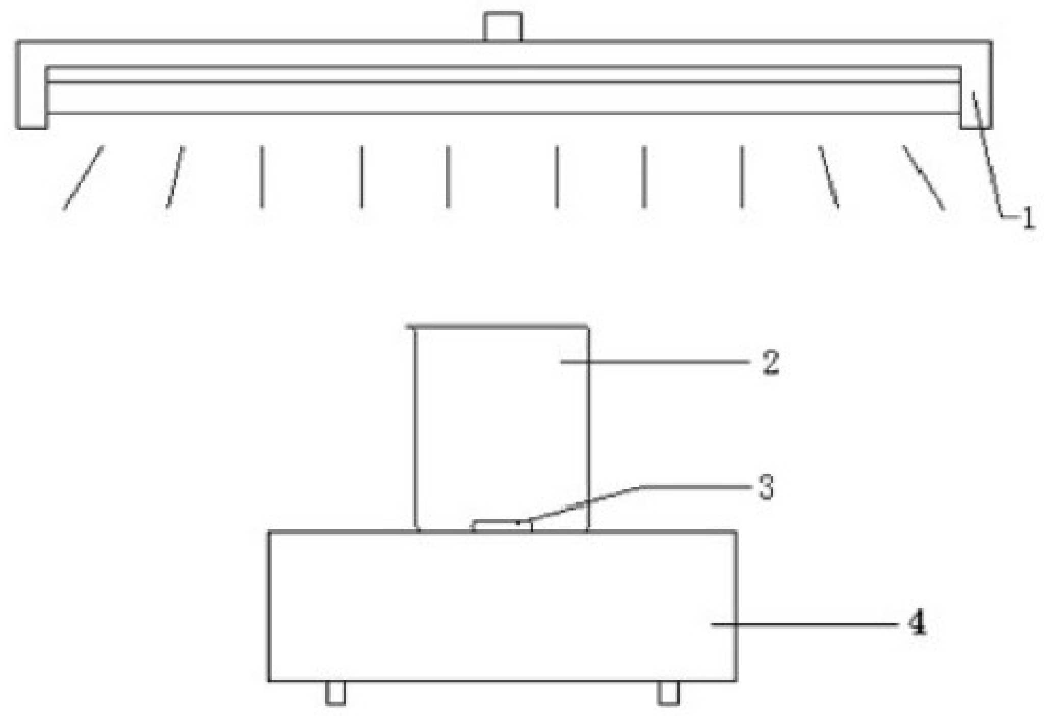
2.2. Experimental Setup

The photocatalytic reaction device in this study is shown in Figure 1. During the experiment, in order to reduce the effect of reagent evaporation, a layer of condensing jacket can be placed on the beaker and condensed water can be passed into it.

2.3. Preparation of ZnO and Ag/ZnO materials

In the current investigation, ZnO materials were synthesized employing hydrothermal methodology. A precise mass of 2.38 g of zinc nitrate hexahydrate and 1.2 g of urea were initially dispensed into a beaker to which 10 mL of deionized water and an equal volume of propanetriol were added. This mixture was stirred rigorously until complete dissolution was achieved, thereby yielding the target solution. Subsequently, the prepared solution was transferred into a pristine 100 mL autoclave and subjected to hydrothermal conditions at a temperature of 130 °C for a duration of 7 h. Following this, the resultant material was allowed to cool to ambient conditions, washed thrice with deionized water, and similarly with anhydrous ethanol. It was then oven-dried at 80 °C for a period of 4 h and ultimately calcined in a muffle furnace at 550 °C for 2.5 h to yield the final ZnO material.
For the fabrication of the Ag/ZnO composite, a one-step hydrothermal synthesis was employed. Precisely 0.119 g of silver nitrate, 2.38 g of zinc nitrate hexahydrate, and 1.2 g of urea were measured into a beaker, and the ensuing procedure mirrored that which was implemented for the production of ZnO material. Lastly, Ag/ZnO-5% composites were engineered, featuring a mass ratio between silver nitrate and zinc nitrate hexahydrate of 1:5.

2.4. Preparation of Ag/ZnO/BC Nanofilm composites

Synthesis of corn stover biochar: Corn stover was first harvested from the experimental field of the School of Food Engineering, Harbin University of Commerce, cut into 2–3 cm pieces, and then rigorously washed with deionized water. Subsequently, the stover samples were dried in an electrically heated blast dryer set at 85 °C. The dried stover was then pulverized using a high-speed multifunctional pulverizer and the fine powder was sieved through a 100-mesh sieve. The quantitative corn stover powder was carefully placed into a pure ceramic crucible, and then the crucible was placed in a high temperature tubular furnace. During heating, the temperature was controlled from ambient to 550 °C at a rate of 5 °C per minute while maintaining a nitrogen atmosphere in order to complete the calcination within 3 h. Upon completion of the reaction, the samples were cooled to room temperature and then stored to obtain the final corn stover biochar nanofilms (BC).
Weigh 0.15 g of the prepared corn straw biochar in a beaker and add 10 mL of methanol solution and sonicate for 30 min to obtain liquid A. At the same time, weigh 0.2 g of Ag/ZnO composite and add 10 mL of methanol, stir for 30 min to obtain liquid B. Solution A and solution B were then mixed and stirred thoroughly. The mixed solution was then heated in a water bath to promote evaporation. After the evaporation process, the residual material was transferred to a high-temperature tube furnace and calcined at 250 °C for 3 h under nitrogen. The final product was an Ag/ZnO/BC nanofilms-3:4 composite characterized by a 3:4 mass ratio of Ag/ZnO composite to biochar film (BC). The ratio of Ag, ZnO and biocarbon composite films is 1.7:1.3:4.

2.5. Sample Characterisation

In the physical characterization phase, the crystallographic structure and morphology of the sample were assessed using a Bruker D8 ADVANCE X-ray diffractometer of German origin (Bruker, Bremen, Germany). According to the XRD pattern information, it can be determined whether the sample is amorphous or crystal: an amorphous sample has a large packet peak and no fine spectrum peak structure, whereas crystals have rich spectral line characteristics. By comparing with the standard spectrum, it can be known which phase composes the measured sample. If the composition elements or groups of crystalline substances are different, or if their structures are different, their diffraction spectra will show differences in the number of diffraction peaks, angle position, relative intensity and diffraction peak shape. A wide-angle diffraction scan was conducted over a 2θ range of 3° to 140°. The mean particle size of the sample was quantitatively determined through application of the Scherrer equation (Equation (1)):
D = K γ B cos θ
where D represents particle size in nanometers nm; K is a constant valued at 0.89; γ denotes the incident wavelength, set at 0.154 nm; B is the diffraction peak half-height width; and θ is the diffraction angle.
Microstructural and morphological attributes of the samples were analyzed using a Hitachi S-4800 Scanning Electron Microscope (SEM) (Hitachi, Japan) and a JEOL JEM-2100F Transmission Electron Microscope (TEM) (Hitachi, Japan). Prior to analysis, samples were thoroughly desiccated and sputter-coated with gold; the SEM was operated at an Extra High Tension (EHT) of 15 kV. Elemental composition was investigated using X-ray Photoelectron Spectroscopy (XPS) (Brucker, Bremen, Germany), with Alka rays radiation at 1486.6 eV and a vacuum pressure maintained below 5 × 10⁻¹⁰ mbar. Optical properties were characterized using a UV/Vis spectrophotometer (Shanghai Yuan analytical Instrument Co., Shanghai, China). employing BaSO4 as a blank substrate and with a spectral scan range of 220–850 nm. Photoluminescence characteristics were ascertained using a F-7000 fluorescence spectrometer (Hitachi, Japan) at an excitation wavelength of 325 nm. Structural information was garnered using a Bruker VERTEX 80 Infrared-Raman spectrometer (Brucker, Bremen, Germany), operating within a spectral range of 7500–50 cm−1 and at a resolution of 0.07 cm−1.

2.6. Photocatalytic Activity Test

In this section, the photocatalytic efficacy of the synthesized ZnO material was evaluated through the degradation kinetics of a simulated wastewater containing 50 mg/L of ammonia nitrogen. This simulated wastewater was formulated using a dry and analytically pure NH4Cl reagent. An aliquot of 100 mL of the wastewater, containing an ammonia nitrogen concentration of 50 mg/L, was dispensed into a 100 mL quartz beaker, into which 200 mg of the catalyst was accurately weighed and added. Prior to photoactivation, the wastewater–catalyst mixture was magnetically stirred for 30 min in dark conditions to establish an adsorption–desorption equilibrium between the catalyst and the ammonia nitrogen pollutants; the baseline absorbance of this system was subsequently recorded. Photocatalytic degradation was initiated by illuminating the mixture with a 250 W mercury lamp positioned 10 cm away from the beaker wall. At predetermined time intervals, aliquots of the reaction mixture were extracted and filtered through a 0.22 μm microporous membrane. The resultant absorbance values were quantified via UV-Visible spectrophotometry, employing the Nahre reagent spectrophotometric method as stipulated by HJ 535-2009 standards [6]. Concurrently, the concentration of ammonia nitrogen in the simulated wastewater was determined.
To investigate the active species that play a role in the degradation reactions of ZnO, Ag/ZnO and Ag/ZnO/BC nanofilms materials by degraded photocatalysis, 1 mM disodium EDTA (Na2EDTA), p-benzoquinone (C6H4O2), and isopropanol [(CH3)2CHOH] were added to the reaction solution as trapping agents for vacancies (h+), superoxide radicals (·O2−), and hydroxyl radicals (·OH), respectively, and other experiments. The conditions and methods were the same as above.
The degradation rate η for ammonia nitrogen simulated wastewater can be calculated from (Equation (2)):
η = [(C0 − Ct)/C0] × 100%
where C0 represents the initial concentration of ammonia nitrogen in the simulated wastewater, while Ct signifies the concentration of ammonia nitrogen in the simulated wastewater following exposure to light.

3. Results and Discussion

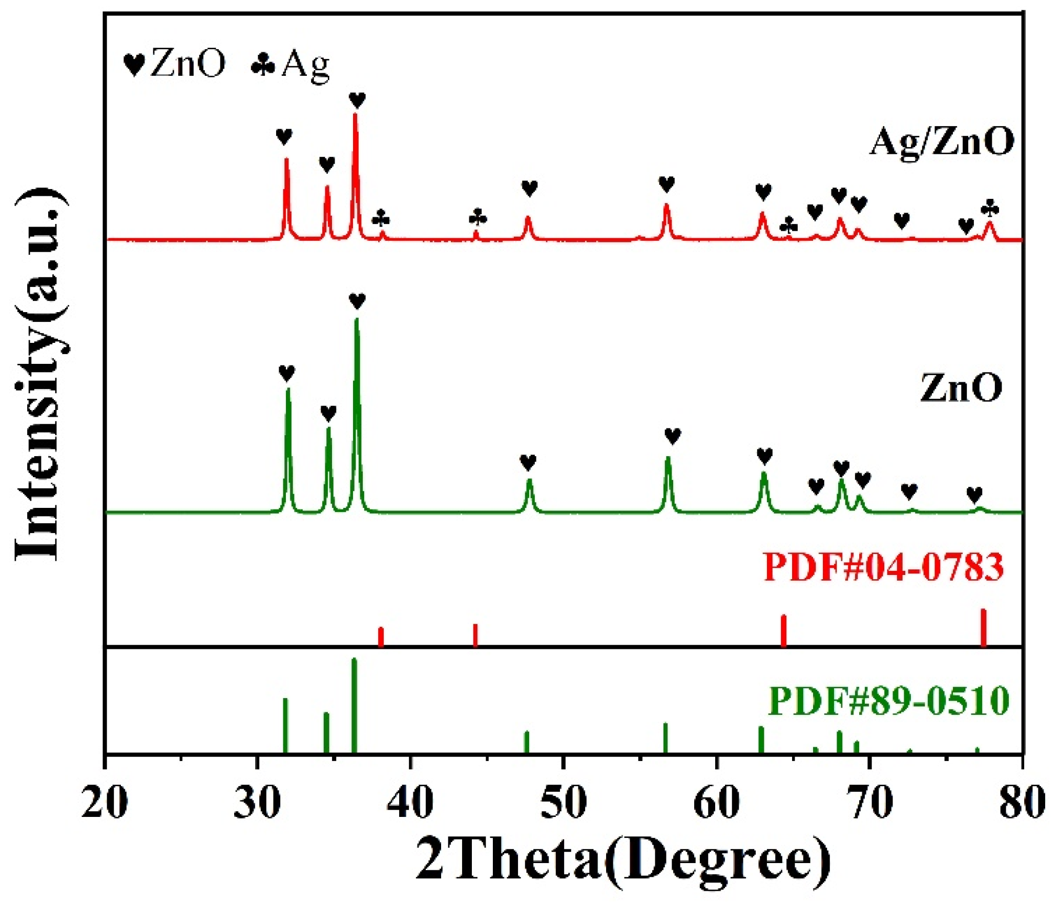
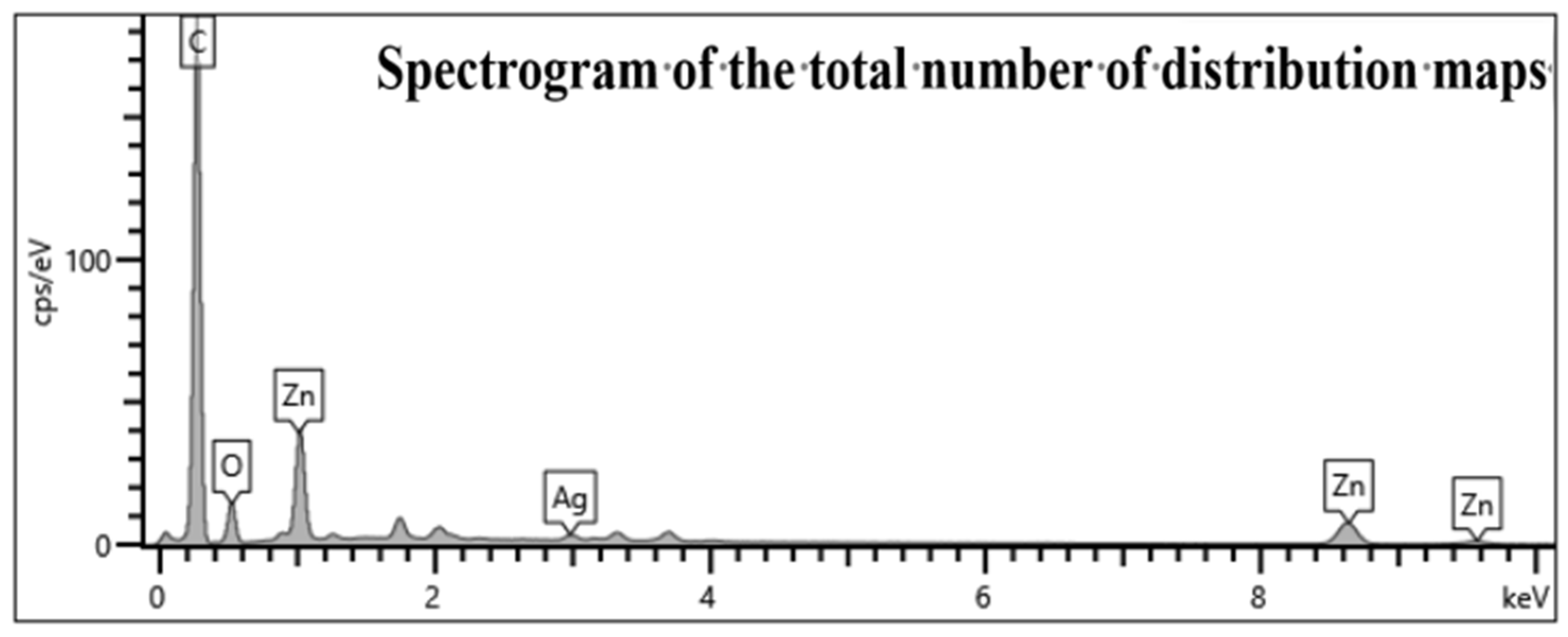
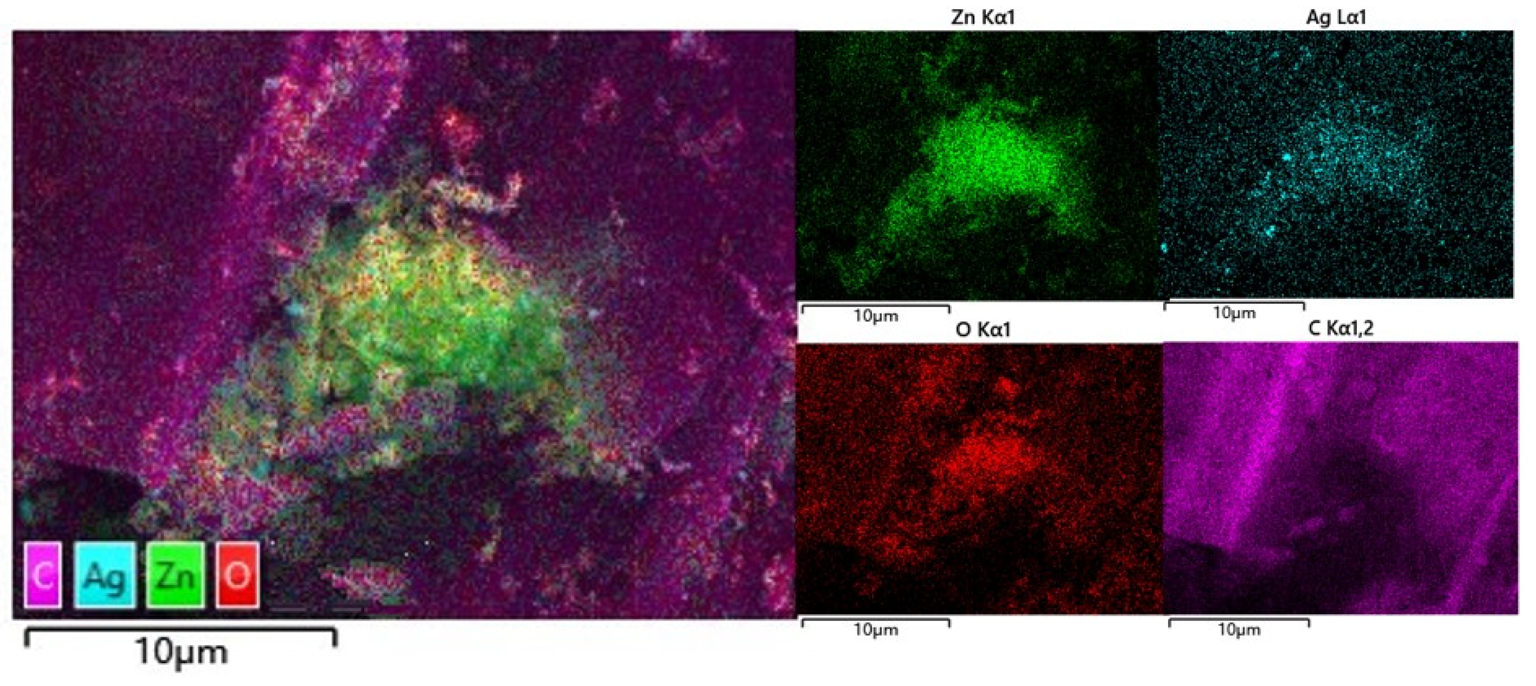
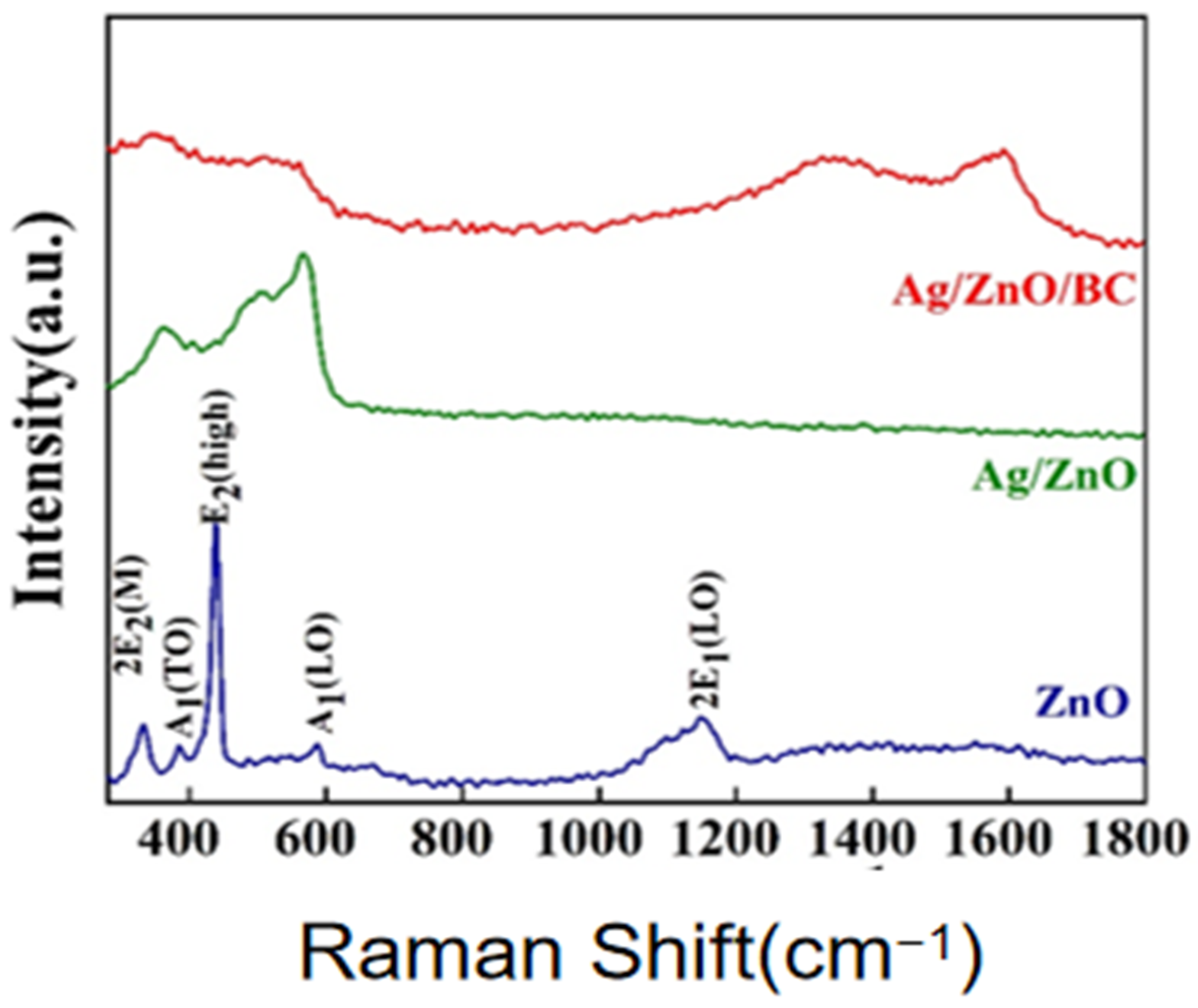
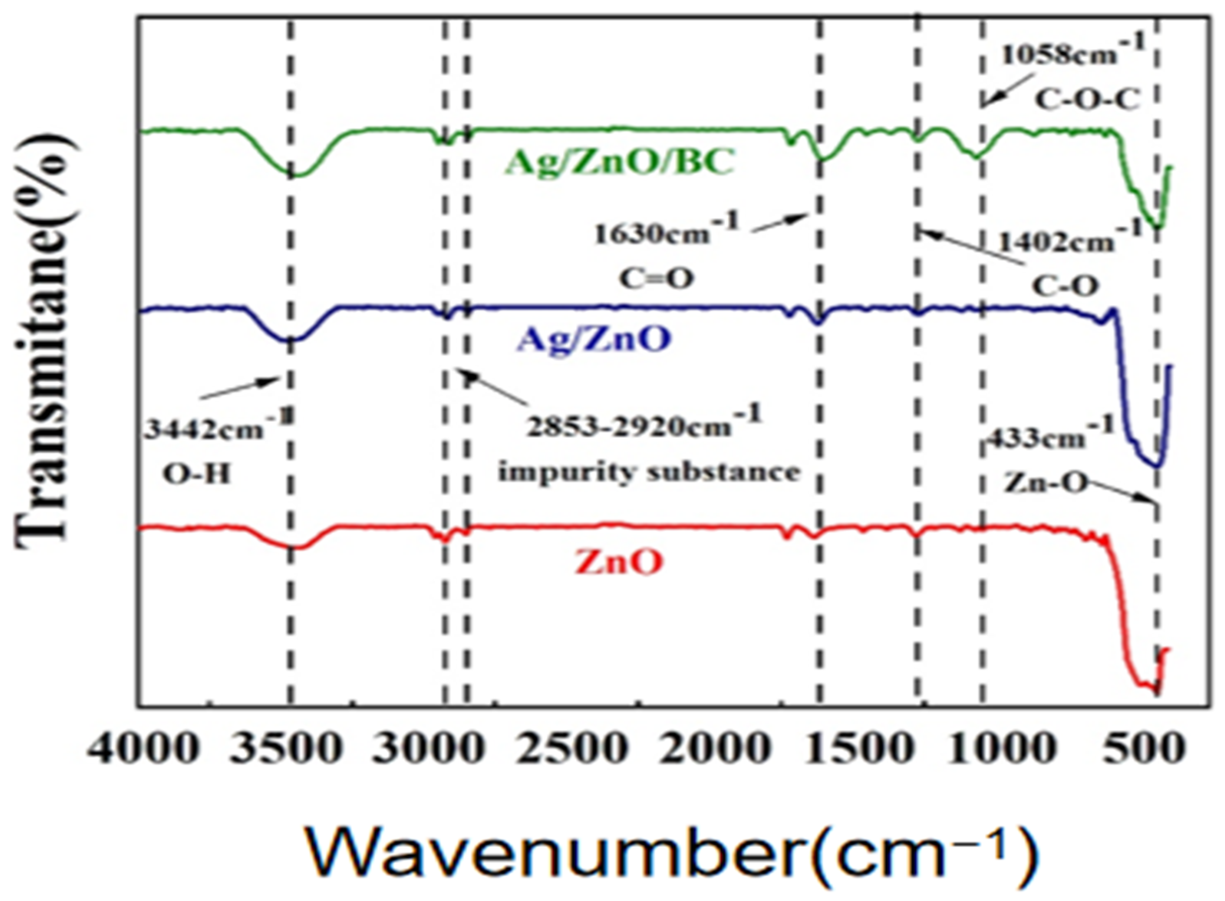
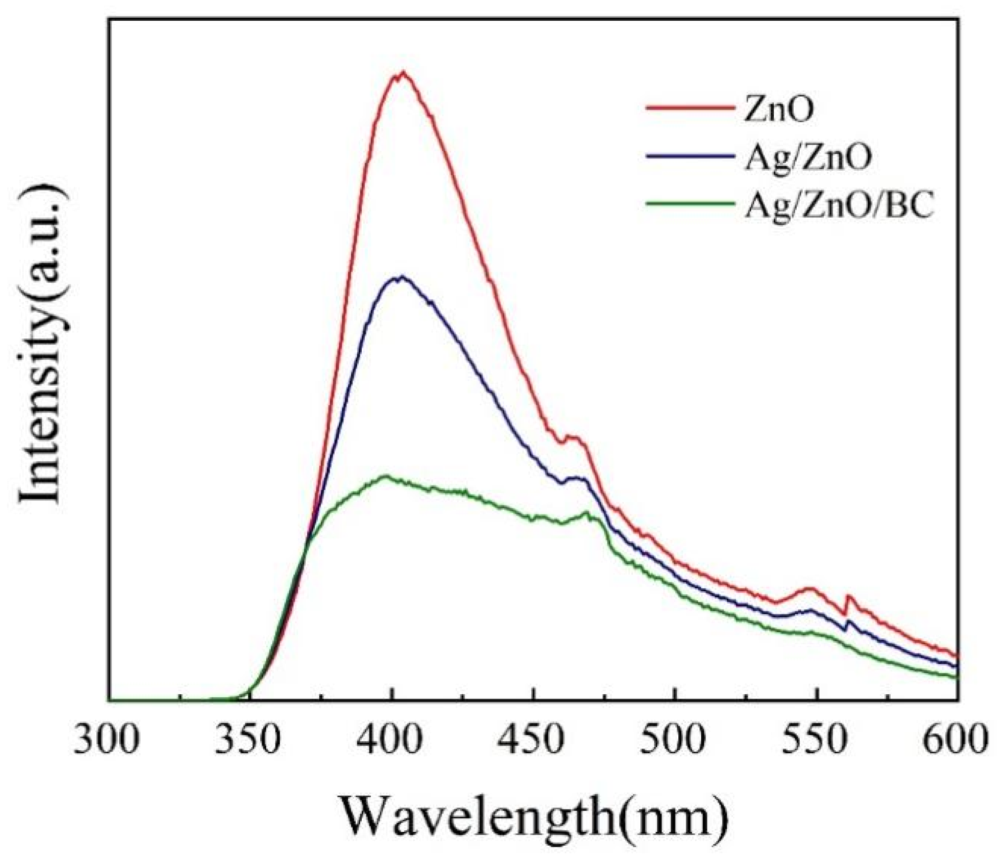
3.1. Sample Characterisation Analysis

The crystallographic characteristics of the Ag/ZnO/BC nanofilm composites were scrutinized using X-ray diffraction (XRD), the results of which are depicted in Figure 2. The observed diffraction peaks from pristine, monolithic ZnO were in alignment with those of the hexagonal, fibrous crystalline structure of ZnO, as referenced by PDF number 89-0510 [7]. Notably, no extraneous characteristic peaks were detected in the spectra. The modified Ag/ZnO presented well-defined, intense diffraction peaks at 31.8°, 34.4°, 36.2°, 47.6°, 56.6°, 62.9°, 66.3°, 68.0°, 69.1°, 72.6°, and 77.0°, corroborating a high degree of congruence with the XRD patterns of the original ZnO.
The diffraction pattern for the Ag/ZnO/BC nanofilms composite largely mimics that of the Ag/ZnO composite, suggesting that the biochar-induced peaks are inconspicuous and overshadowed by dominant, well-resolved ZnO peaks. No spurious peaks were observed. However, the Ag/ZnO/BC nanofilms composite exhibited a notable alteration in the ratio among the three crystalline planes at (100), (002), and (101). Furthermore, the intensity of these peaks was markedly diminished compared to both the Ag/ZnO composite and the original ZnO material. This suggests a substantive interaction between the corn stover biochar and the ZnO crystalline planes, thereby inducing modifications to the crystallographic architecture of the Ag/ZnO composite. Overall, the empirical data affirm the successful synthesis of Ag/ZnO/BC nanofilm composites, characterized by both robust crystallinity and high purity.
The microscopic morphology of the synthesized Ag/ZnO/BC nanofilm composites was elucidated through Scanning Electron Microscopy (SEM), as illustrated in Figure 3. Figure 3a,b reveal that the ZnO component manifests a unique, snowball-like structure. Figure 3c further highlights that the integrated Ag/ZnO configuration is more uniformly dispersed, featuring cleanly defined lamellae. Examination of Figure 3d indicates that the biochar employed exhibits an amorphous, lumpy structural morphology. Figure 3e,f present SEM images showcasing the Ag/ZnO/BC nanofilm composites, revealing a persistently irregular, porous lamellar architecture with an approximate dimension of 80 nm. These observations are largely congruent with grain size data derived from X-ray Diffraction (XRD) analyses. The nanoparticles are observed to be irregularly dispersed across the biochar surface and within its pores. This porous architecture is conducive to inhibiting the recombination of electron-hole pairs on the composite’s surface. Moreover, the reduced lamellar structure indicates an augmentation in specific surface area, which is suggestive of enhanced electron transport efficiency. This increase is likely to contribute to improved light responsiveness and pollutant interaction, thereby accelerating the photocatalytic reaction rate [8].
The Energy-Dispersive Spectroscopy (EDS) pattern, depicted in Figure 4, corroborates the observations derived from the Scanning Electron Microscopy (SEM) analysis presented in Figure 2. As evidenced by Figure 4, the elemental composition of the Ag/ZnO/BC nanofilms composite is restricted to four elements—Zinc (Zn), Oxygen (O), Ag, and Carbon (C)—each manifesting robust and uniformly distributed spectral peaks. Concurrently, owing to the gold sputter coating process as well as the presence of environmental C and bench-induced Copper (Cu), additional diffraction peaks with diminished intensity are discernible in the EDS energy spectrum [9]. These observations are in alignment with the preliminary X-Ray Diffraction (XRD) findings, thereby substantiating the successful synthesis of the Ag/ZnO/BC nanofilms composite.
The crystalline phase structure of the material was analyzed by Raman spectroscopy and the results are shown in Figure 5. The experimentally prepared ZnO spectra have distinct characteristic peaks near 331 cm−1, 382 cm−1, 438 cm−1, 586 cm−1 and 1150 cm−1 corresponding to 2E2 (M), A1 (TO), E2H modes, A1 (LO), 2 E1 (LO) of the ZnO material, respectively. In addition, the scattering of the A1 (LO) branch in the Raman spectrum of the Ag/ZnO material broadens and shifts the Raman peak of the A1 (LO) symmetric vibrational mode to a lower energy, shifting it to 568 cm−1, and a significant enhancement of the intensity of this Raman feature peak appears [10]. With the addition of biochar, the Raman spectra of the Ag/ZnO/BC nanofilm composites show a decrease in the intensity of the ZnO stretching vibrational modes at 331 cm−1 and 438 cm−1 compared to the ZnO and Ag/ZnO materials, and two typical peaks are detected at 1358 cm−1 and 1587 cm−1, mainly due to the D and G bands of the biochar material, where the D peak at 1358 cm−1 is the signal of disordered carbon or the edge of graphitic carbon, while the G-peak at 1587 cm−1 is usually due to the in-plane vibrational mode of sp2 heterogeneous carbon (graphitic carbon), and the defect density is usually determined with ID/IG [11]. Therefore, the presence of characteristic bands of ZnO and biochar in the Ag/ZnO/BC nanofilms composite is evident from the Raman spectra, indicating that ZnO is well bound to biochar.
From the full spectrum of XPS shown in Figure 6a, it is obvious that Ag/ZnO/BC nanomembrane composites prepared under the best preparation conditions are composed of Zn, O, C and Ag elements. These findings are consistent with data obtained from the eds analysis. The high-resolution XPS spectrum of the Zn 2p state, shown in Figure 6b, has an obvious peak at 1022.76 eV and 1045.76 eV, corresponding to the Zn 2p3/2 and Zn 2p1/2 states, respectively [4]. Furthermore, the binding energy is poor.
The high-resolution XPS spectra for the O 1s state in Figure 6c exhibits peaks at 531.51 eV and 532.71 eV, attributed to Zn-OH and C-O-C/C-O-Zn structures, respectively [12]. Figure 6d displays the high-resolution XPS energy spectrum for the C 1s state, revealing spectral features at 284.83 eV and 288.73 eV. These peaks are attributed to the introduction of CO into ZnO and Ag/ZnO due to prolonged atmospheric exposure, resulting in orbital electron forbidden energy vibrations of carbon at these specific energies. Furthermore, the Ag/ZnO/BC nanofilm composite exhibits additional peaks at 284.86 eV, 285.59 eV, 286.71 eV, and 288.85 eV, which correspond to C-C/C=C, C-O, C=O, and O-C=O bonds, respectively [13].
Lastly, the XPS energy spectrum for the Ag 3d state, illustrated in Figure 6e, indicates the presence of orbital electron forbidden band energy vibrational peaks at 367.68 eV (Ag 3d5/2) and 373.68 eV (Ag 3d3/2). The energy gap of 6 eV between these peaks substantiates that Ag exists as monomeric entities in a 0-valent state within both the Ag/ZnO material and the Ag/ZnO/BC nanofilm composite [14].
In conclusion, the XPS pattern analysis shows that there is residual oxygen-containing functionality on the carbon surface in the Ag/ZnO/BC nanofilm composite, and that C can replace Zn on the Ag/ZnO material surface or H in -OH, forming C-O-Zn bonds in the ZnO lattice, particularly at the interface between carbon and ZnO [15]. All of the above analyses indicate that ZnO was successfully compounded with biochar nanofilms.
The chemical bonding and functional group composition of the Ag/ZnO/BC nanofilm composites were analyzed by FTIR. As shown in Figure 7, Ag/ZnO/BC nanofilm composites show stretching vibration peaks at 433 cm−1 (stretching vibration peak of Zn-O bond), 1058 cm−1 (stretching vibration of C-O-C), 1402 cm−1 (stretching vibration of C-O bond/C=O bond), 1630 cm−1 (stretching vibration of C=O bond), 2853–2920 cm−1, 3442 cm−1, while also showing a stretching vibration peak at 1058 cm−1 (impurities from potassium bromide used in the compression process) [16]. It is worth noting that the peak intensity of Ag/ZnO/BC nanofilm composites at 1402 cm−1 and 1630 cm−1 is significantly higher than that of ZnO and Ag/ZnO materials, indicating that maize straw biochar is rich in functional groups such as hydroxyl, carboxyl and carbonyl groups, and the peak intensity of the stretching vibration peak at 3442 cm−1 is further increased, indicating that the adsorbed oxygen content on the surface of the material has increased, which is more beneficial to improve the photocatalytic activity of ZnO materials. The photocatalytic activity of the ZnO material was improved by the increase in oxygen adsorption on the surface of the material [17].
The optical properties were analyzed by UV-Vis DRS, as shown in Figure 8. As shown in Figure 8a, the optical absorption band edge of the Ag/ZnO/BC nanofilms composite shifts to approximately 460 nm, which is the visible range; in addition, the Ag/ZnO/BC nanofilm composite has a red-shifted response range to the light region, with a stronger degree of light response to the visible region, mainly attributed to the chemical interaction between the biochar and the ZnO particles [18]. Owing to the porous architecture of the biochar, a light-trapping phenomenon is induced, enabling the dispersion and internal scattering of incident light, thereby widening the absorption spectrum. Concurrently, the anchoring of ZnO particles onto the biochar surface leads to an increased number of surface defects, which not only roughens the surface, but also enhances its light-absorptive capacity [19]. Figure 8b shows the curves of Ag/ZnO/BC nanofilm materials (ahv)2 and (hv). According to the Tauc plot formula, the bandgaps of ZnO, Ag/ZnO and Ag/ZnO/BC nanofilm composites are 3.21 eV, 3.17 eV and 3.07 eV, respectively. The reduction in the band gap of the Ag/ZnO/BC nanofilm composites is mainly due to the formation of new chemical bonds between the biochar nanofilms and the ZnO particles, which further reduces the reactive band gap of the material. The introduction of biochar reduced the band width of the Ag/ZnO material to a certain extent and improved its utilization of visible light and electron-hole utilization, resulting in the synthesis of Ag/ZnO/BC nanofilm composites with superior photocatalytic performance [20].
As delineated in Figure 9, the emission peaks for all three investigated materials manifest predominantly at wavelengths near 400 nm and 470 nm. The pronounced UV emission peak proximal to 400 nm can be ascribed to correlated emission phenomena arising from free exciton collisions. Conversely, the emission peak in the visible spectrum near 470 nm is attributable to correlated emissions induced by lattice defects in ZnO, as reported in previous studies [21]. Notably, the UV emission peak for the pristine ZnO material is considerably more intense, resulting in suboptimal photogenerated electron-hole pair separation due to elevated photoluminescence (PL) fluorescence intensity. In contrast, the composite Ag/ZnO material exhibits attenuated emission peaks in both the UV and visible ranges. This diminished fluorescence intensity suggests that the incorporation of Ag functions as an electron acceptor, thereby mitigating electron-hole pair recombination and consequently augmenting photocatalytic activity [22]. Upon the further addition of biochar to form the Ag/ZnO/BC nanofilms composite, a significant reduction in both emission peak and fluorescence intensities is observed. This is principally attributed to the superior electrical conductivity of biochar, facilitating the expedited separation of photogenerated electron-hole pairs and further inhibiting their recombination. These attributes collectively serve to enhance the material’s photocatalytic activity [23]. The observed PL behavior aligns closely with our preceding experimental outcomes obtained via UV-Vis DRS.
To elucidate the variations in surface properties of the synthesized Ag/ZnO/BC nanofilm composites, nitrogen adsorption–desorption curves were obtained as shown in Figure 10, and the pore properties are shown in Table 1. In Figure 10, the adsorption of all three materials was low in the low pressure region and the amount of adsorbed gas gradually increased with the increase in component partial pressure. The specific surface areas of ZnO, Ag/ZnO and Ag/ZnO/BC nanofilms are shown in Table 1 as 41.38 m2·g−1, 27.37 m2·g−1 and 62.14 m2·g−1, respectively. The reduction in specific surface area of Ag/ZnO composites compared to ZnO is probably due to the addition of Ag, which caused some of the mesopores of ZnO to be filled or covered, thus reducing its specific surface area and pore volume [24,25]. Therefore, the Ag/ZnO/BC nanofilm composites were prepared by adding biochar to increase the specific surface area and provide more reactive sites, which can facilitate the mass transfer process in photocatalytic degradation and allow more electron holes to participate in the reaction [26,27], further improving the photocatalytic efficiency.

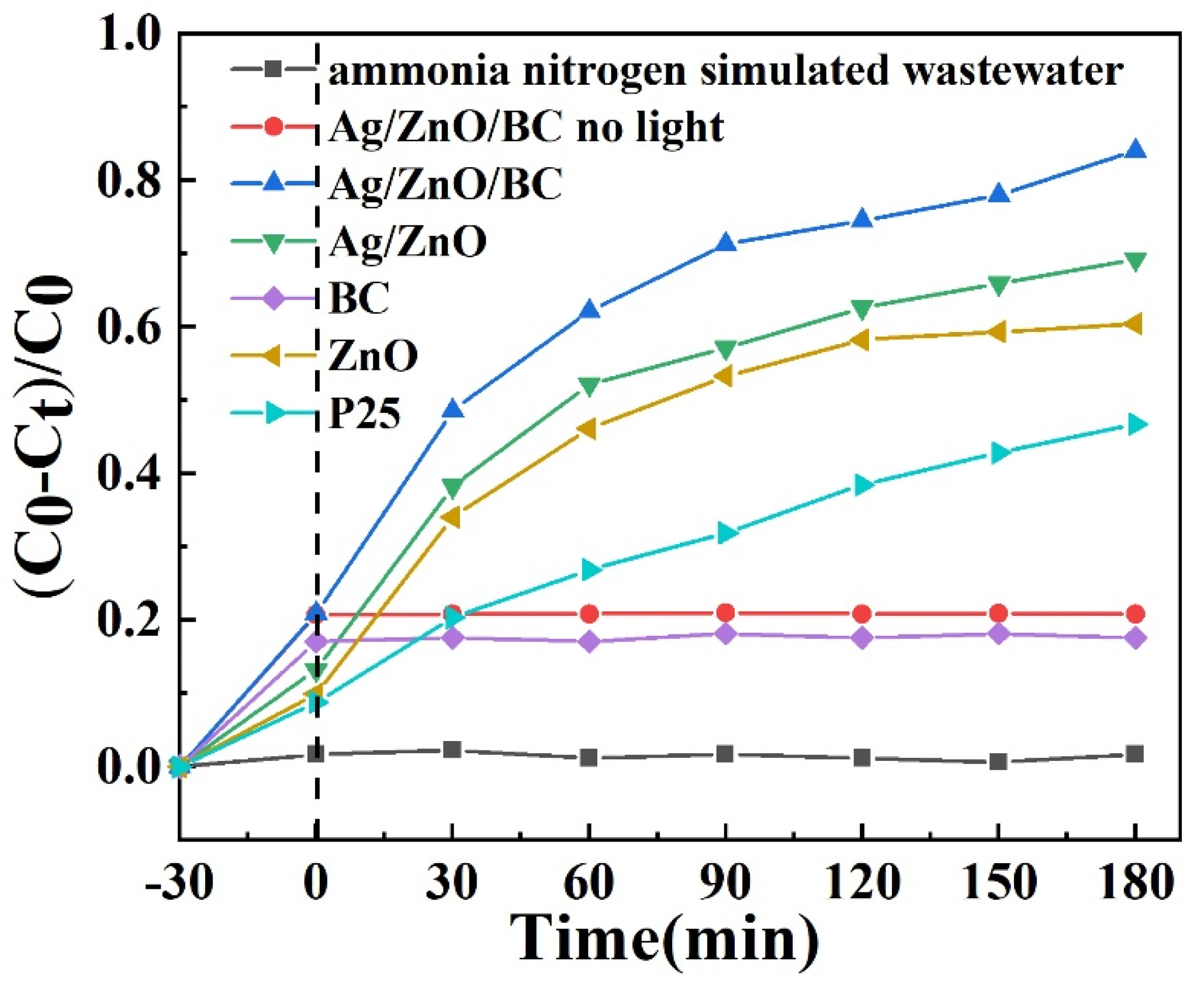
3.2. Photocatalytic Performance Tests

As illustrated in Figure 11, optimal photocatalytic performance for the degradation of ammonia nitrogen pollutants in simulated wastewater (with a concentration of 50 mg/L) was observed when the mass ratio of the AgNO3 reagent to the synthesized ZnO material was maintained at 5%. The mass of zero valence Ag is 0.0756 g. Specifically, the Ag/ZnO-5% composite underwent a 30 min dark reaction followed by 180 min of exposure to a 250 W mercury lamp, achieving unparalleled photocatalytic efficiency. Additionally, the Ag/ZnO/BC nanofilm composite fabricated with a mass ratio of Ag/ZnO to BC of 3:4 exhibited superior photocatalytic performance. In this case, the degradation rate of ammonia nitrogen in the simulated wastewater reached 71.11% after a 30 min dark reaction and 180 min of illumination under a 250 W mercury lamp. Strikingly, the degradation rate further escalated to 84.03% when subjected to a similar set of conditions.
As shown in Figure 12, the photocatalytic activity of the commercial TiO2 (P25), ZnO material, Ag/ZnO composite and Ag/ZnO/BC nanofilm composite were tested under 250 W mercury lamp irradiation to examine their photocatalytic performance in terms of degradation rates of 50 mg/L ammonia nitrogen simulated wastewater, and light experiments with 50 mg/L ammonia nitrogen simulated wastewater alone, and Ag/ZnO/BC nanofilm composites were compared with light-free adsorption experiments. The adsorption of Ag/ZnO/BC nanofilm composites was better than that of commercial TiO2 (P25), ZnO materials and Ag/ZnO composites in the dark reaction adsorption phase, mainly because the introduction of biochar increased the specific surface area of Ag/ZnO/BC nanofilm composites, resulting in better adsorption performance, and accelerated the adsorption of ammonia nitrogen onto its surface, increasing the contact between the active material and pollutant molecules and improving the catalytic activity. Consequently, under light-activated conditions, the degradation rate of ammonia nitrogen pollutants reached an impressive 84.03% within 180 min of reaction time. This degradation rate surpassed those achieved by Ag/ZnO composite at 71.11%, ZnO material at 60.43%, and commercial TiO2 (P25) at 45.37%.

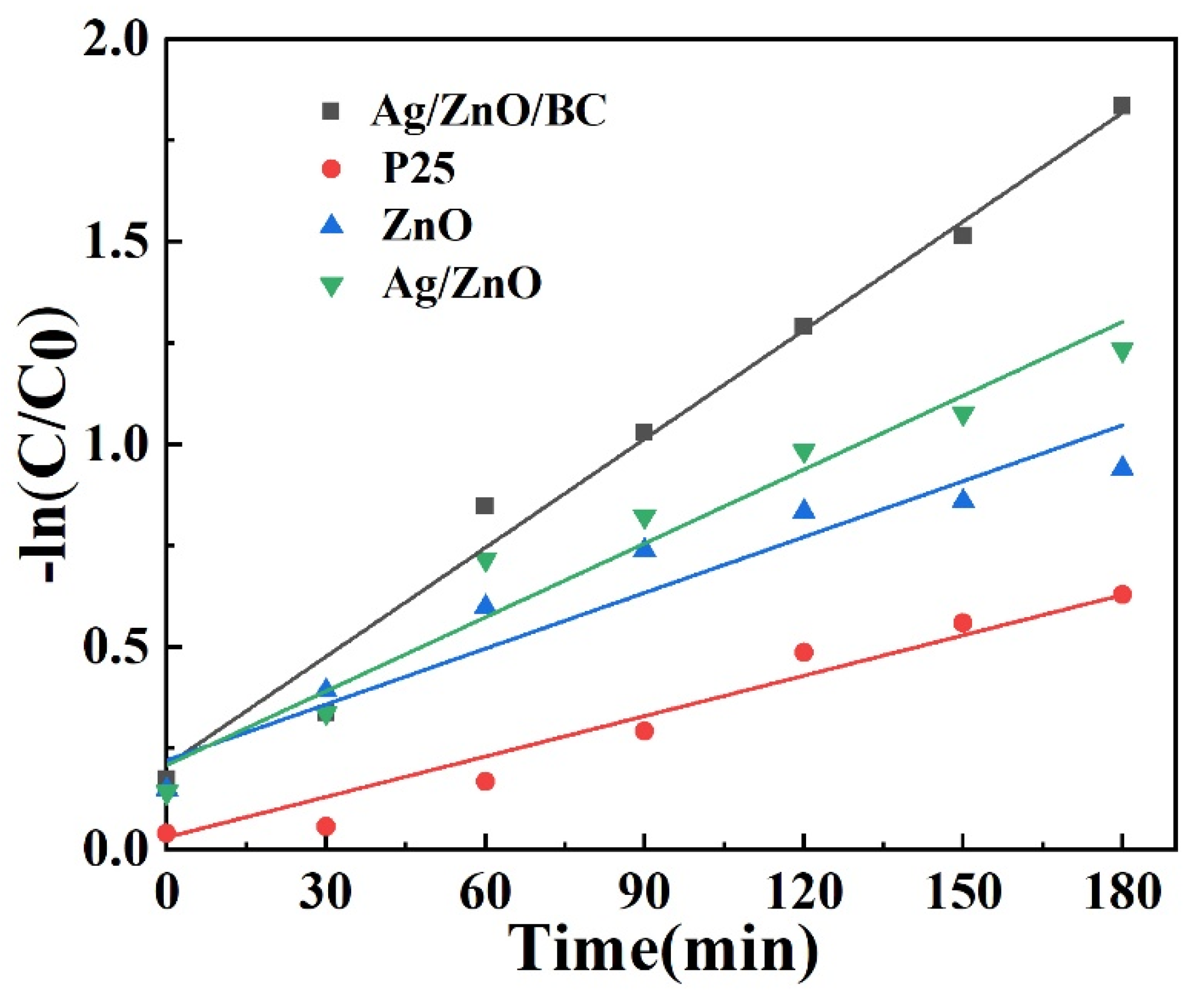
3.3. Kinetic Analysis

As evidenced in Figure 13, linear relationships were found for TiO2 (P25), ZnO material, Ag/ZnO composite and Ag/ZnO/BC nanofilm composite, indicating that the degradation process of all samples for the 50 mg/L ammonia simulated wastewater was highly consistent with the quasi first order kinetic equation. As shown in Table 2, the K values are in descending order: Ag/ZnO/BC nanofilms > Ag/ZnO > ZnO > TiO2 (P25) and the linear correlation coefficients R2 are 0.986, 0.966, 0.945 and 0.930, respectively. The K value of Ag/ZnO/BC nanofilms was 2.1 times higher than that of ZnO, indicating that the introduction of the noble metal Ag and biochar helped to improve the photocatalytic performance of ZnO, which may be attributed to the addition of biochar, which led to more rapid adsorption of ammonia nitrogen pollutants onto the surface of the composite thus accelerating the photocatalytic reaction efficiency [28,29]. Additionally, the enhanced photocatalytic performance can be attributed to the effective charge carrier separation, promoted by the favorable electrical conductivity of both the Ag monomer and biochar substrate [30].

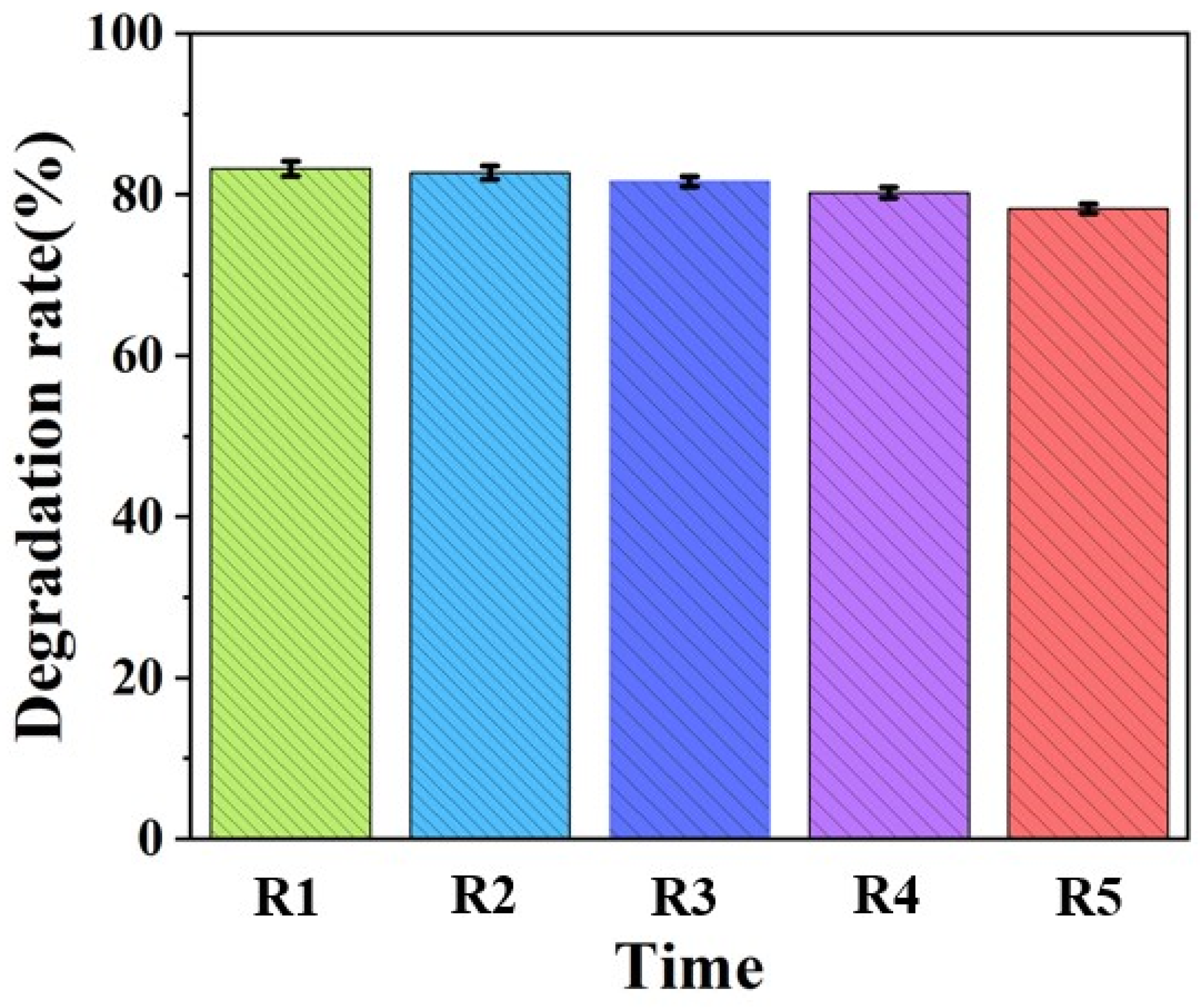
3.4. Photocatalytic Stability Analysis

The long-term stability of the Ag/ZnO/BC nanofilm composite constitutes a critical variable for its real-world applicability and was rigorously assessed across five photocatalytic degradation cycles using simulated wastewater with an ammonia nitrogen concentration of 50 mg/L. The experiments were conducted under the irradiation of a 250 W mercury lamp. Following each degradation cycle, the catalyst powder was isolated via centrifugation, subsequently washed with deionized water, and air-dried prior to its reemployment in the subsequent degradation experiment. As delineated in Figure 14, the composite manifested high stability, The photocatalytic activities of the first three cycles were 82.36%, 81.95% and 80.96%, respectively, retaining 80.6% of its original photocatalytic activity after four consecutive cycles. In these trials, the ammonia nitrogen concentration in the treated solution was reduced to 9.7 mg/L. However, a marginal decline in performance was noted in the fifth cycle, where the composite displayed 78.3% of its initial activity. With the progress of the experiment cycle, a small amount of the void in the catalyst is blocked by the solution, resulting in a reduction of the pollutants absorbed by the catalyst, and due to the competition between the catalysts, the migration of excited electron-hole pairs slows, resulting in a slight degradation of the catalytic performance. These findings substantiate the composite’s robust photocatalytic efficiency under conditions mimicking natural sunlight and underscore its reusability, sustaining elevated catalytic activity even after four consecutive cycles of operation.

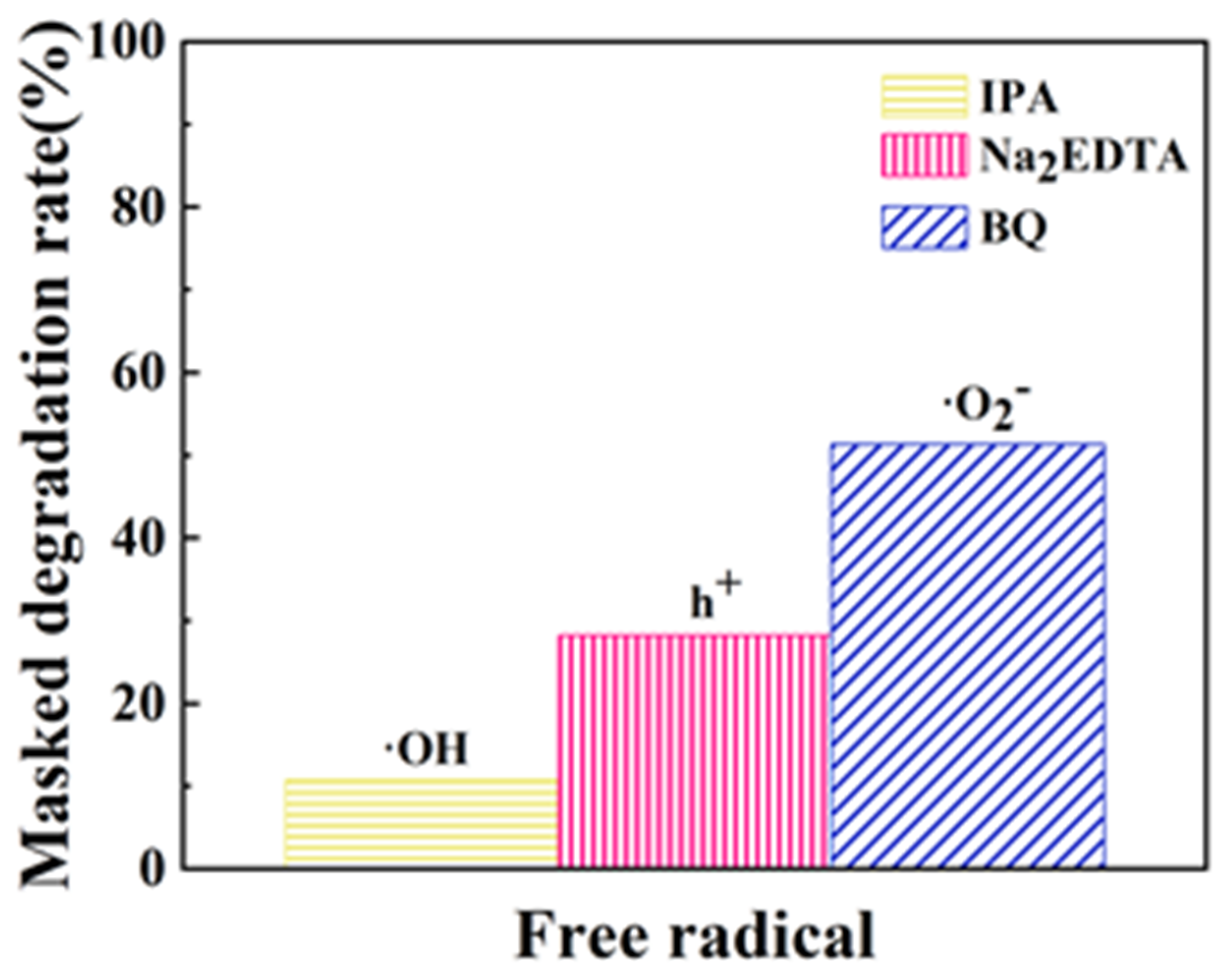
3.5. Free Radical Masking Experiments

The photocatalytic efficacy of the Ag/ZnO/BC nanofilm composites was markedly attenuated upon the introduction of p-benzoquinone (BQ) as a specific ·O2 scavenger in the reaction milieu. Under these conditions, the degradation rate for simulated wastewater containing 50 mg/L of ammonia nitrogen plummeted to a mere 34.63%. As delineated in Figure 15, in a comparative analysis involving various scavenging agents, the degradation rates were recorded as 10.74%, 28.27%, and 51.35% for isopropanol (IPA), disodium ethylenediaminetetraacetate (Na2EDTA), and BQ, respectively. Results from these masking experiments unequivocally indicate that superoxide anions (·O2) serve as the most influential reactive species in the photocatalytic mechanisms of the Ag/ZnO/BC nanofilm composite. This is followed in significance by photogenerated holes (h+), while hydroxyl radicals (·OH) exerted the least impact on the composite’s photocatalytic activity.

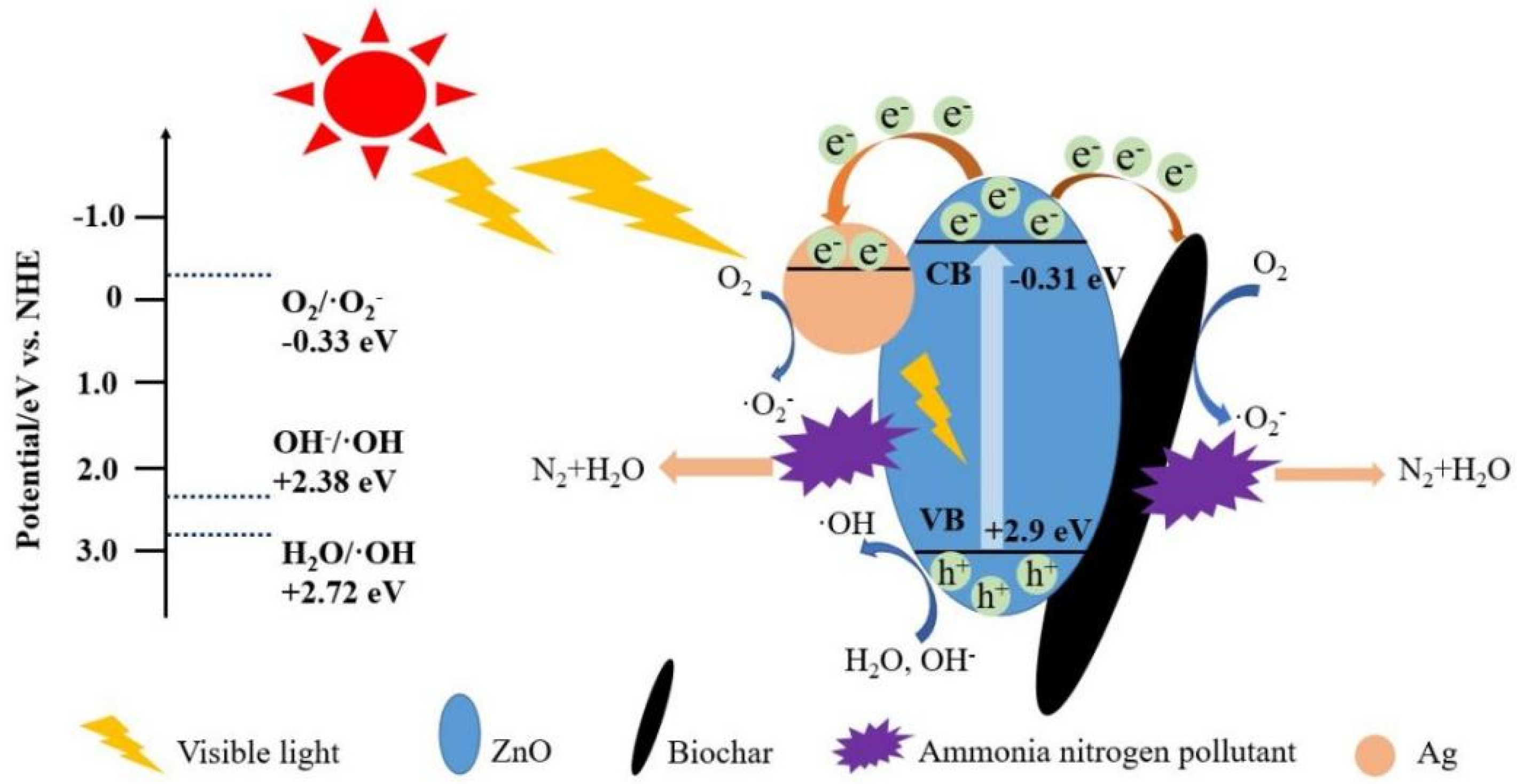
3.6. Analysis of the Photocatalytic Mechanism

The photocatalyst can not only capture energy to release powerful reducing and oxidizing power, but also achieve efficient electron and hole transfer. The underlying mechanism governing the photocatalytic activity of the Ag/ZnO/BC nanofilm composite is depicted in Figure 16. Within this framework, ammonia (NH) molecules adsorbed onto the catalyst’s surface undergo continuous oxidation facilitated by potent oxidizing species, namely superoxide radicals (·O2) and hydroxyl radicals (·OH). This oxidative process effectively cleaves the N-H bond, culminating in the formation of molecular N2 in a zero-valent state, as supported by the previous literature [27]. In addition, within the Ag/ZnO/BC nanofilm composite catalyst, monomeric Ag exists with a lower Fermi energy level compared to that of ZnO [31]. As a result, electrons can readily transfer from ZnO’s conduction band to the surface of the monomeric Ag. The Schottky barrier formed between the two components serves as an effective “electron trap” [9], significantly inhibiting the recombination of electron-hole pairs. This, in turn, amplifies the composite’s photocatalytic efficacy.

4. Conclusions

In summary, this study successfully synthesized Ag/ZnO/BC nanofilm photocatalytic materials through a multi-step approach involving hydrothermal and calcination techniques, resulting in composites with exceptional photocatalytic activity and stability. It can also be seen from Table 3 that the new photocatalytic nanomembranes prepared have a good treatment effect on ammonia nitrogen wastewater.
(1)
Characterization results substantiate that Ag/ZnO nanocomposites are effectively anchored onto the biochar nanofilms. The introduction of biochar increased the specific surface area of Ag/ZnO/BC nanomembrane composite, thereby improving the adsorption performance, accelerating the adsorption of ammonia nitrogen on its surface, increasing the contact between active substances and pollutant molecules, and improving the catalytic activity. Under the condition of photoactivation, the degradation rate of ammonia nitrogen pollutants reached 84.03% within 180 min after the reaction Consequently, the utilization of biochar as a substrate for catalyst loading not only augments photocatalytic performance, but also facilitates catalyst recovery.
(2)
The photocatalytic stability experiment showed that the catalyst had a very high stability and still had a very high catalytic efficiency after four cycles, and the catalytic efficiency could still reach 78.3%.
(3)
Results from these masking experiments unequivocally indicate that superoxide anions (·O2) serve as the most influential reactive species in the photocatalytic mechanisms of the Ag/ZnO/BC nanofilm composite. This is followed in significance by photogenerated holes (h+), while hydroxyl radicals (·OH) exerted the least impact on the composite’s photocatalytic activity.
(4)
This methodology thereby opens up a novel avenue for the synthesis of loaded composite photocatalysts and expands their potential applications, particularly in the removal of ammonia and nitrogenous pollutants from wastewater.

Author Contributions

Methodology, J.L. (Junsheng Li); Software, S.L.; Formal analysis, L.J.; Resources, J.X. and J.L. (Jiahui Li); Data curation, Z.X.; Writing—original draft, C.T., J.Z. and Y.W.; Writing—review & editing, J.Z. and Y.W. All authors have read and agreed to the published version of the manuscript.

Funding

This research was funded by 2023 Heilongjiang Provincial Natural Science Foundation Joint Guidance Project, grant number LH2023E029.

Institutional Review Board Statement

Not applicable.

Informed Consent Statement

Not applicable.

Data Availability Statement

Date and materials will be made available on reasonable request.

Conflicts of Interest

The authors declare no conflict of interest.

References

  1. Quartaroli, L.; Silva, C.M.; Silva, L.C.F.; Lima, H.S.; de Paula, S.O.; Dias, R.S.; da Silva, C.C. Effect of the gradual increase of salt on stability and microbial diversity of granular sludge and ammonia removal. J. Environ. Manag. 2019, 248, 109273. [Google Scholar] [CrossRef] [PubMed]
  2. Hao, L.; Chen, Y.; Cui, X. Study on the treatment of ammonia-nitrogen wastewater by oxidization methods. Adv. Mater. Res. 2015, 1092–1093, 938–942. [Google Scholar] [CrossRef]
  3. Yuan, R.; Qin, Y.; He, C.; Wang, Z.; Bai, L.; Zhao, H.; He, X. Fe-Mn-Cu-Ce/Al2O3 as an efficient catalyst for catalytic ozonation of bio-treated coking wastewater: Characteristics, efficiency, and mechanism. Arab. J. Chem. 2023, 16, 104415. [Google Scholar] [CrossRef]
  4. Vaiano, V.; Jaramillo-Paez, C.A.; Matarangolo, M. UV and visible-light driven photocatalytic removal of caffeine using ZnO modified with different noble metals (Pt, Ag and Au). Mater. Res. Bull. 2019, 112, 251–260. [Google Scholar] [CrossRef]
  5. Yu, F.; Tian, F.; Zou, H. ZnO/biochar nanocomposites via solvent free ball milling for enhanced adsorption and photocatalytic degradation of methylene blue. J. Hazard. Mater. 2021, 415, 125511. [Google Scholar] [CrossRef] [PubMed]
  6. HJ 535-2009; Water Quality-Determination of Ammonia Nitrogen—Nessler’s Reagent Spectrophotometry. Ministry of Environmental Protection: Beijing, China, 2009.
  7. Mou, H.; Song, C.; Zhou, Y.; Zhang, B.; Wang, D. Design and synthesis of porous Ag/ZnO nanosheets assemblies as super photocatalysts for enhanced visible-light degradation of 4-nitrophenol and hydrogen evolution. Appl. Catal. B Environ. 2018, 221, 565–573. [Google Scholar] [CrossRef]
  8. Shi, L.; Liang, L.; Ma, J.; Meng, Y.; Zhong, S.; Wang, F.; Sun, J. Highly efficient visible light-driven Ag/AgBr/ZnO composite photocatalyst for degrading Rhodamine B. Ceram. Int. 2014, 40, 3495–3502. [Google Scholar] [CrossRef]
  9. Jiang, X.; Li, Z.; Lin, Q.; Dong, K.; Zhang, Y.; Sun, Z. Structure, optical properties and photocatalysis performance of Cu2O microspheres prepared by hydrothermal method. J. Mater. Sci. Mater. Electron. 2016, 27, 8856–8861. [Google Scholar] [CrossRef]
  10. Liang, Y.; Jiao, C.; Pan, L.; Zhao, T.; Liang, J.; Xiong, J.; Zhou, Q. Degradation of chlorine dioxide bleaching wastewater and response of bacterial community in the intimately coupled system of visible-light photocatalysis and biodegradation. Environ. Res. 2021, 195, 110840. [Google Scholar] [CrossRef]
  11. Bera, M.; Lee, D.S.; Cho, E.J. Advances in N-centered intermediates by energy transfer photocatalysis. Trends Chem. 2021, 3, 877–891. [Google Scholar] [CrossRef]
  12. Shen, Z.; Zhang, X.; Mi, R.; Liu, M.; Chen, Y.; Chen, C.; Ruan, S. On the high response towards TEA of gas sensors based on Ag-loaded 3D porous ZnO microspheres. Sens. Actuators B Chem. 2018, 270, 492–499. [Google Scholar] [CrossRef]
  13. Meshram, S.P.; Adhyapak, P.V.; Amalnerkar, D.P.; Mulla, I.S. Cu doped ZnO microballs as effective sunlight driven photocatalyst. Ceram. Int. 2016, 42, 7482–7489. [Google Scholar] [CrossRef]
  14. Ozdemir, E.; Orkan Ucar, I.; Akay, R.G.; Aytac, A. The effect of Ag, ZnO, and CuO nanoparticles on the properties of the compatibilized polyethylene/thermoplastic starch blend films. J. Vinyl Addit. Technol. 2021, 27, 543–554. [Google Scholar] [CrossRef]
  15. Kasim, N.; Hassan, Z.; Lim, W.F.; Quah, H.J. A comparison study of ZnO, InZnO, GaZnO and InGaZnO physical properties and optical bandgap. Int. J. Nanotechnol. 2022, 19, 241–251. [Google Scholar] [CrossRef]
  16. Amirouche, B.; Abderrahmane, Y. Effect of ZnO, SiO2 and Al2O3 Doped on Morphological, Optical, Structural and Mechanical Properties of Polylactic Acid. Key Eng. Mater. 2022, 6323, 105–113. [Google Scholar]
  17. Jung, K.-W.; Lee, S.Y.; Lee, Y.J. Hydrothermal synthesis of hierarchically structured birnessite-type MnO2/biochar composites for the adsorptive removal of Cu(II) from aqueous media. Bioresour. Technol. 2018, 260, 204–212. [Google Scholar] [CrossRef]
  18. Soltani, S.; Khanian, N.; Rashid, U.; Choong, T.S.Y. Core-shell ZnO-TiO2 hollow spheres synthesized by in-situ hydrothermal method for ester production application. Renew. Energy 2020, 151, 1076–1081. [Google Scholar] [CrossRef]
  19. Khan, H.; Habib, M.; Khan, A.; Boffito, D.C. A modified sol-gel synthesis to yield a stable Fe3+/ZnO photocatalyst: Degradation of water pollutants and mechanistic insights under UV and visible light. Engineering 2020, 8, 104282. [Google Scholar] [CrossRef]
  20. Ambedkar, A.K.; Singh, M.; Kumar, V.; Kumar, V.; Singh, B.P.; Kumar, A.; Gautam, Y.K. Structural, optical and thermoelectric properties of Al-doped ZnO thin films prepared by spray pyrolysis. Surf. Interfaces 2020, 19, 100504. [Google Scholar] [CrossRef]
  21. Jung, S.H.; Oh, E.; Lee, K.H.; Yang, Y.; Park, C.G.; Park, W.; Jeong, S.H. Sonochemical preparation of shape selective ZnO nanostructures. Cryst. Growth Des. 2008, 8, 265–269. [Google Scholar] [CrossRef]
  22. Mahana, A.; Guliy, O.I.; Momin, S.C.; Lalmuanzeli, R.; Mehta, S.K. Sunlight-driven photocatalytic degradation of methylene blue using ZnO nanowires prepared through ultrasonication-assisted biological process using aqueous extract of Anabaena doliolum. Cryst. Growth Des. 2020, 108, 110205. [Google Scholar] [CrossRef]
  23. Nava, O.J.; Luque, P.A.; Gómez-Gutiérrez, C.M.; Vilchis-Nestor, A.R.; Castro-Beltrán, A.; Mota-González, M.L.; Olivas, A. Influence of Camellia sinensis extract on Zinc Oxide nanoparticle green synthesis. Opt. Mater. 2017, 1134, 121–125. [Google Scholar] [CrossRef]
  24. Zhou, Q.; Yin, H.; Wang, A.; Si, Y. Preparation of hollow B-SiO_2@TiO_2 composites and their photocatalytic performances for degradation of ammonia-nitrogen and green algae in aqueous solution. Chin. J. Chem. Eng. 2019, 27, 2535–2543. [Google Scholar] [CrossRef]
  25. Manzoor, M.F.; EjazAhmed; Ahmad, M. Enhanced photocatalytic activity of hydrogen evolution through Cu incorporated ZnO nano composites. Mater. Sci. Semicond. Process. 2020, 120, 105278. [Google Scholar] [CrossRef]
  26. Gharaghani, M.A.; Malakootian, M. Photocatalytic deg-radation of the antibiotic ciprofloxacin by ZnO nanoparti-cles immobilized on a glass plate. Desalnation Water Treat. 2017, 89, 304–314. [Google Scholar] [CrossRef]
  27. Gong, H.; Hu, J.Q.; Wang, J.H.; Ong, C.H.; Zhu, F.R. Nano-crystalline Cu-doped ZnO thin film gas sensor for CO. Sens. Actuators B Chem. 2006, 115, 247–251. [Google Scholar] [CrossRef]
  28. Emamifar, A.; Kadivar, M.; Shahedi, M.; Soleimanian-Zad, S. Evaluation of nanocomposite packaging containing Ag and ZnO on shelf life of fresh orange juice. Innov. Food Sci. Emerg. Technol. 2010, 11, 742–748. [Google Scholar] [CrossRef]
  29. Shafei, A.E.; Abou-Okeil, A. ZnO/carboxymethyl chitosan bionano-composite to impart antibacterial and UV protection for cotton fabric. Carbohydr. Polym. 2011, 83, 920–925. [Google Scholar] [CrossRef]
  30. Wu, C.L. Facile one-step synthesis of N-doped ZnO micropolyhedrons for efficient photocatalytic degradation of formaldehyde under visible-light irradiation. Appl. Surf. Sci. 2014, 319, 237–243. [Google Scholar] [CrossRef]
  31. Jalili-Jahani, N.; Rabbani, F.; Fatehi, A. Rapid one-pot synthesis of Ag-decorated ZnO nanoflowers for photocatalytic degradation of tetracycline and product analysis by LC/APCI-MS and direct probe ESI-MS. Adv. Powder Technol. 2021, 32, 3075–3089. [Google Scholar] [CrossRef]
Figure 1. 1—UVlamp; 2—Quartz glass; 3—The rotor; 4—Magnetic stirrer.
Coatings 13 02043 g001
Figure 2. XRD spectrum of ZnO, Ag/ZnO and Ag/ZnO/BC nanofilm composites.
Coatings 13 02043 g002
Figure 3. (a) shows the SEM of ZnO; Figure (b,c) shows the SEM of Ag/ZnO; Figure (d) shows the SEM of BC; Figure (e,f) shows the SEM of Ag/ZnO/BC nanofilms composite.
Coatings 13 02043 g003
Figure 4. EDS energy spectrum and elemental distribution of Ag/ZnO/BC nanofilm composites.
Coatings 13 02043 g004aCoatings 13 02043 g004b
Figure 5. Raman spectra of ZnO, Ag/ZnO and Ag/ZnO/BC nanofilms.
Coatings 13 02043 g005
Figure 6. (a) XPS full spectra of ZnO, Ag/ZnO and Ag/ZnO/BC nanofilm composites; (b) high resolution XPS spectra of Zn 2p; (c) O 1s; (d) C1s; (e) Ag 3d.
Coatings 13 02043 g006
Figure 7. Infrared spectra of ZnO, Ag/ZnO and Ag/ZnO composites.
Coatings 13 02043 g007
Figure 8. (a) UV-Vis DRS spectra of BC, ZnO, Ag/ZnO and Ag/ZnO/BC nanofilm composites; (b) optical band gap maps of ZnO, Ag/ZnO and Ag/ZnO/BC nanofilm composites.
Coatings 13 02043 g008
Figure 9. PL spectra of ZnO, Ag/ZnO and Ag/ZnO/BC nanofilms.
Coatings 13 02043 g009
Figure 10. Nitrogen adsorption–desorption isotherms for ZnO, Ag/ZnO and Ag/ZnO/BC nanofilms.
Coatings 13 02043 g010
Figure 11. Degradation of 50 mg/L ammonia nitrogen simulated wastewater by Ag/ZnO and Ag/ZnO/BC nanofilms at different compound ratios.
Coatings 13 02043 g011
Figure 12. Photocatalytic activity test of Ag/ZnO/BC nanofilms.
Coatings 13 02043 g012
Figure 13. Results of linear fitting of kinetics for the degradation of 50 mg/L ammonia nitrogen simulated wastewater for each sample.
Coatings 13 02043 g013
Figure 14. Effect of number of catalyst uses on photocatalytic performance.
Coatings 13 02043 g014
Figure 15. Free radical degradation masking rate.
Coatings 13 02043 g015
Figure 16. Photocatalytic mechanism diagram.
Coatings 13 02043 g016
Table 1. Pore properties of ZnO, Ag/ZnO and Ag/ZnO/BC nanofilms.
Samples
Sample
Specific Surface Area
Specific Surface Area/(m2·g)−1
Kong Rong
Pore Volume (cm3/g)
Average Pore Size
Average Aperture (nm)
ZnO41.380.11310.93
Ag/ZnO27.370.079.75
Ag/ZnO/BC nanofilms62.140.138.37
Table 2. Kinetic parameters for photocatalytic degradation of each sample.
Samplesk (min)−1R2
Ag/ZnO/BC nanofilms0.00950.986
P250.00330.945
ZnO0.00460.930
Ag/ZnO0.00610.966
Table 3. Treatment efficiency of ammonia nitrogen wastewater by different materials.
Material/Method NameProcessing ObjectConcentration of Ammonia Nitrogen in Primary Solution/(mg·L−1)Removal Rate/(mg·g−1)
TiO2
(Hydrothermal process)
Simulated ammonia nitrogen wastewater30.00 mg/L79.00%
ZnO
(Hydrothermal process)
Simulated ammonia nitrogen wastewater50.00 mg/L64.80%
BiOI/BiOBr/1 wt%MoS2Simulated ammonia nitrogen wastewater50.00 mg/L80.52%
ZnO-PMMASimulated ammonia nitrogen wastewater50.00 mg/L66.00%
ZnFe2O4/NGSimulated ammonia nitrogen wastewater100.00 mg/L62.84%
TiO2-CuO/HSCSimulated ammonia nitrogen wastewater100.00 mg/L60.7%
Fe3O4/ZnO—BCSimulated ammonia nitrogen wastewater50.00 mg/L80.50%
Ag/ZnO/BCSimulated ammonia nitrogen wastewate50.00 mg/L84.03%
Disclaimer/Publisher’s Note: The statements, opinions and data contained in all publications are solely those of the individual author(s) and contributor(s) and not of MDPI and/or the editor(s). MDPI and/or the editor(s) disclaim responsibility for any injury to people or property resulting from any ideas, methods, instructions or products referred to in the content.

