Article

Preparation and Characterization of Biomimetic SiO2-TiO2-PDMS Composite Hydrophobic Coating with Self-Cleaning Properties for Wall Protection Applications

1
Hunan Province Key Laboratory of Engineering Rheology, Central South University of Forestry and Technology, Changsha 410004, China
2
School of Civil Engineering, Central South University of Forestry and Technology, Changsha 410004, China
3
School of Materials Science and Engineering, Hunan University of Science and Technology, Xiangtan 411201, China
*
Author to whom correspondence should be addressed.
Coatings 2023, 13(2), 224; https://doi.org/10.3390/coatings13020224
Submission received: 12 December 2022 / Revised: 10 January 2023 / Accepted: 10 January 2023 / Published: 18 January 2023
(This article belongs to the Section Surface Characterization, Deposition and Modification)

Abstract

:
Superhydrophobic surfaces have great potential for self-cleaning, anti-icing, and drag-reducing characteristics because of their water repellent property. This study demonstrates the potential application of coatings to protect architectures from detrimental atmospheric effects via a self-cleaning approach. In this research, a SiO2-TiO2-PDMS composite coating was prepared on the surface of building walls by the sol-gel method. Tetraethyl orthosilicate (TEOS) and titanium isopropoxide (TTIP) were used as inorganic precursors, and polydimethylsiloxane (PDMS) was used as low surface energy substances. The effects of TEOS and PDMS content on microstructure, wettability, and self-cleaning performance of coating wall surfaces were investigated by conducting various tests, including scanning electron microscopy (SEM), X-ray energy spectroscopy (EDS), angle measurement, and Fourier transform infrared spectroscopy (FTIR). The results indicated that hydrolysis and condensation reactions of TEOS, TTIP, and PDMS were performed on the surface of the substrates, leading to a micro- and nano-structure similar to the surface of lotus leaves. When the molar ratio of PDMS to TEOS was 1:5, the static contact angle of the coating reached a maximum of 152.6°. At this point, the coated surface was able to resist the adhesion of particle pollutants and liquid pollutants, which could keep the walls clean and possess a good ability of self-cleaning. In conclusion, SiO2-TiO2-PDMS composite coating is potentially useful in wall protection applications with its hydrophobic and environmentally friendly superhydrophobic properties.

1. Introduction

With the rapid development of industrialization and a social economy, environmental problems have gradually accumulated and emerged. Extreme weather conditions such as acid rain and haze have many adverse effects on the surface of building walls [1]. Building walls are exposed to the air, and the surfaces are washed by rainwater, which makes the walls prone to peeling and cracking. At the same time, organic and inorganic contaminants can easily adhere to the surface of building walls and accumulate to produce black spots, which affect the aesthetics of the walls [2,3]. It was found that the humidity and cleanliness of the walls are closely related to environmental health [4], and it is urgent to improve the hydrophobicity and cleanliness of building wall surfaces [5].
The construction of biomimetic super-impregnated interfacial materials holds significant promise for applications in water repellency, stain resistance, and self-cleaning [6,7]. In the 1970s, Neinhuis and Barthlott observed the surface of lotus leaves with SEM and found that the surface was composed of hierarchical rough structures and hydrophobic wax-like crystals [8]. The most important feature of the superhydrophobic surface of lotus leaf is the combined micron and nanoscale rough structure with low surface energy wax. With the help of this theory, scholars have begun to prepare superhydrophobic coatings on the surface of construction materials using both micro-nano structures and low surface energy.
Scientists have employed various methods to fabricate superhydrophobic surfaces, including chemical etching [9], chemical vapor deposition [10], the solution immersion method [11], the sol-gel method [12], and laser fabrication [13]. Ivan P. Parkin’s team [14,15] constructed superhydrophobic surfaces on aluminum sheets, copper mesh, glass, and other substrates by aerosol-assisted chemical vapor deposition. Chunde Jin’s team [16,17,18,19] used the template method and prepared several types of superhydrophobic bamboo materials, such as low-adhesion superhydrophobic bamboo materials, high-temperature resistant superhydrophobic bamboo materials, acid-resistant superhydrophobic bamboo materials, and etc. However, the above-mentioned preparation processes are complex, and the required equipment is demanding, which limits the large-scale preparation and application of the coatings [20]. Due to the advantages of a simple operation and easy control of reaction conditions [21,22], the sol-gel method has been widely used for the preparation of superhydrophobic coatings on wood and stone surfaces [23,24,25,26]. Zhou H et al. [27,28] modified silica sol with 1H, 1H, 2H, 2H-perfluorodecyltriethoxysilane and prepared aqueous superhydrophobic coatings on the surface of substrates such as glass, fibers, and wood. However, such coatings were expensive and tended to pollute the environment due to the fluorinated groups contained in fluorinated silanes. Minzhen Zhong et al. [29] synthesized a superhydrophobic coating on the surface of glass by using TEOS and methyltriethoxysilane (MTES). However, the coating was easy to fail in an external environment due to its poor mechanical stability. In view of the low surface energy and adhesive effect of PDMS, PDMS mixed with inorganic oxides (SiO2, TiO2, and ZnO) has been used to prepare fluorine-free and environmentally friendly superhydrophobic coatings in some studies [30,31,32]. R. Zarraga et al. [33] prepared a SiO2-PDMS composite sol on stone surfaces. They found that the introduction of PDMS could improve the elasticity of the coating, effectively prevent the coating from cracking, and improve the hydrophobic property of the stone. Nakajima et al. [34] added a small amount of TiO2 nanoparticles to the PDMS-SiO2 hydrophobic sol and found that the addition of nano-TiO2 could improve the durability of the coating under outdoor conditions. Peng S et al. [35] prepared a highly efficient, cost-effective, and wide-applicable functionalized SiO2/TiO2@PDMS superhydrophobic coating. The coating exhibited superior photocatalytic activity and strong UV resistance that could repeatedly degrade organic oil pollutants for as many as 50 times, while still reserved superhydrophobicity even exposure to UV light with high intensity of 80 mW/cm2 for as long as 36 h. Sheng Y et al. [36,37] prepared a photocatalytic and superhydrophobic PDMS/SiO2-TiO2 coating on the surface of a textile. The treated textile has a water contact angle of 157.7° and a photocatalytic degradation ratio for methylene blue of 87.08% after a 4 h illumination. However, few studies have focused on the application of this coating in building walls, particularly for the materials’ hydrophobicity from both a microscopic and macroscopic perspective.
In this study, a SiO2-TiO2-PDMS composite coating was prepared by the sol-gel method, which exhibits superhydrophobic properties and has potential in the application of building walls. There are two innovation points for the present work. Firstly, the SiO2-TiO2-PDMS composite is a novel system. TEOS is used as the precursor material, and PDMS is used as a low surface energy substance. It is not only environmentally friendly, but also reduces the cost of raw materials. Secondly, the changes of the microscopic morphology and chemical structure of the coating during the formation process are investigated. Moreover, the superhydrophobic and self-cleaning mechanisms are systematically revealed by chemical and microscopic analysis methods.

2. Materials and Methods

2.1. Materials

The following materials were used for the preparation of the coatings. Oxalic acid (AR), polydimethylsiloxane (PDMS, Purity ≥ 99%), and tetraethyl orthosilicate (TEOS, Purity ≥ 99%) were purchased from Sinopharm Group Chemical Reagent Co., Ltd. (Shanghai, China). Ethanol absolute (EtOH, Purity ≥ 99.7%) was purchased from Tianjin Fuyu Fine Chemical Co., Ltd. (Tianjin, China), and titanium isopropoxide (TTIP, Purity ≥ 97%) was purchased from Shanghai Maclean Biochemical Technology Co., Ltd. (Shanghai, China). All the chemicals were used without further purifification.
The following materials were purchased from the local market and used to prepare the substrates: cement (P.O 42.5), sand (MX ≈ 2.26), and putty paste.

2.2. Preparation of Substrates

Cement mortar specimens with dimensions of 70 mm × 70 mm × 15 mm were prepared according to the “Test method of cement mortar strength” [38]. The specimens were demoulded after 24 h of forming and maintained under standard conditions for 28 d. After the completion of maintenance, the putty paste was scraped three times on the surface of the specimens.

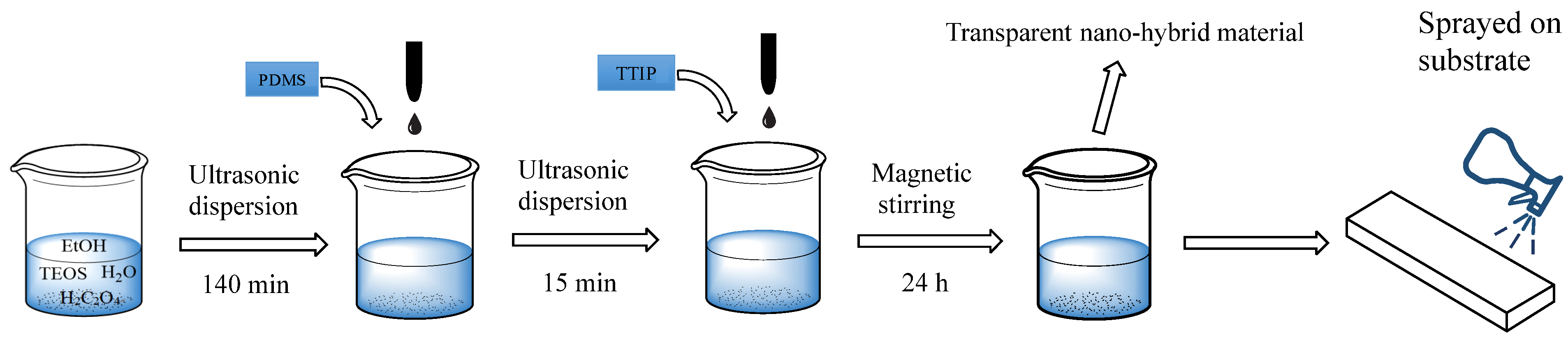
2.3. Synthesis and Deposition of the Surface Coating

As in a typical synthesis process, 0.05 mol of oxalic acid was mixed with 5.6 mol of DI water. Later on, TEOS and EtOH were added to the mixture of H2O/H2C2O4 solution. The mixed solution was ultrasonically dispersed at room temperature for 140 min and prehydrolyzed the TEOS under acidic condition. Then, PDMS was added to the solution and ultrasonically dispersed at room temperature for 15 min. Finally, 0.05 mol of TTIP was added drop-wise and magnetically stirred for 24 h to obtain SiO2-TiO2-PDMS nanocomposite sol. The molar ratio of the solution was TEOS: H2O:EtOH:H2C2O4:PDMS:TTIP = 1:5.6:5.5:0.05:X:0.05 (X = 0, 0.1, 0.2, 0.3, 0.4).
The sol-solution was deposited on the surface of specimens by a spray-coating method with a spray can [39,40]. When spraying, the nozzle was placed approximately 10–15 cm away from the base, and the spraying pressure was 0.05 MPa. A schematic illustration of the process is shown in Figure 1.

2.4. Characterization

The morphologies and chemical compositions of the superhydrophobic substrate surfaces were assessed using a scanning electron microscope (SEM, TESCAN MIRA LMS) equipped with energy-dispersive X-ray spectroscopy (EDS, Xplore). Operating conditions of the SEM were an accelerating voltage of 20 kV and a probe current of 45 nA, while the working distance was set at 10 mm. The functional groups existing in the sol were assessed by Fourier transform infrared (FTIR, Thermo Scientific Nicolet iS10) analysis in attenuated total reflection (ATR) mode. FTIR spectra were recorded in the range of 600–4000 cm−1 with a resolution of 4 cm−1. The water contact angle (CA) of the coated samples was measured by the sessile drop method with 10 µL of deionized water droplet each time. The sliding angle (SA) was measured using the tilted drop method. Both water CA and SA results were presented as the average deviation of 3 measurements taken for each sample [41].

2.5. Water Absorption Test

The samples with dimensions (70 × 70 × 15 mm) were used for the measurement of water absorption. The specimens were periodically taken out of the water, wiped with tissue paper to remove surface water, reweighed, and immediately put back into water. The pre-dried (m0) was determined and used to calculate the degree of water absorption through the following formula [42]:
w i = m i - m 0 m 0 × 100 %
where wi is the water absorption rate of the specimen, m0 is the mass of the dried specimen, and mi is the surface dry mass of the specimen.

2.6. Self-Cleaning Test

The self-cleaning property was evaluated by comparing the color of the substrate surface before and after contamination. To evaluate the self-cleaning property, Fe2O3 powder was scattered on the sample to mimic contaminant, and the sample was then laid on a tray tilted at an angle of 10°. Droplets of DI water were then trickled down from a pipette at 5 cm above the samples. Meanwhile, methyl orange solution was used to simulate liquid contaminant. The appearance of the sample was characterized using a colorimeter (3nh, NR110). We adopted the CIE L*a*b* color system, where L* indicates luminosity, 0 indicates black, and 100 indicates white; a*, which ranges from positive to negative, indicates colors from red to green; b*, which ranges from positive to negative, indicates colors from yellow to blue. The color difference ∆E was calculated as follows [43,44]:
Δ E = ( Δ L ) 2 + ( Δ a ) 2 + ( Δ b ) 2
where ∆L*, ∆a*, and ∆b* are the variations in the L*, a*, and b* values of the samples before and after treatment, respectively. Each sample was measured three times, and we reported the average.

2.7. Mechanical Stability Test

Chemical stability of the coated samples was evaluated by immersing in corrosion bath [45,46]. Acid and alkali solutions were prepared with diluting NaOH and H2SO4 with a range of pH concentrations: 1, 2, 3, 4, 5, 6, 7, 8, 9, 10, 11,12, 13 and 14. The coated samples were immersed in these corrosive solutions for 3 h at room temperature. The mechanical stability of the superhydrophobic coating was evaluated by a linear abrasion test and tape-peeling test. In the sandpaper abrasion test, the coated sample was placed facing down on 320 grit SiC sandpaper, with a weight of 300 g placed on top [47,48]. The sample was then linearly abraded across a 10 cm distance along a ruler to complete one abrasion cycle. The changes in water CAs and SAs of coated samples were subsequently evaluated at specific intervals until the samples completed 100 abrasion cycles. The tape-peeling test was conducted and adapted to Method B described in ASTM D3359-09 [49]. A transparent Scotch tape was used to assess the adhesion strength between substrate and coating. To ensure homogeneous contact within the tape with the surface, a 300 g load was rolled back and forth for 15 s and then peeled off. This was set as one peeling cycle, and the tape was replaced with a new one after every peeling cycle and repeated for 200 cycles. The water CA was measured after every 50 peeling cycles.

3. Results and Discussion

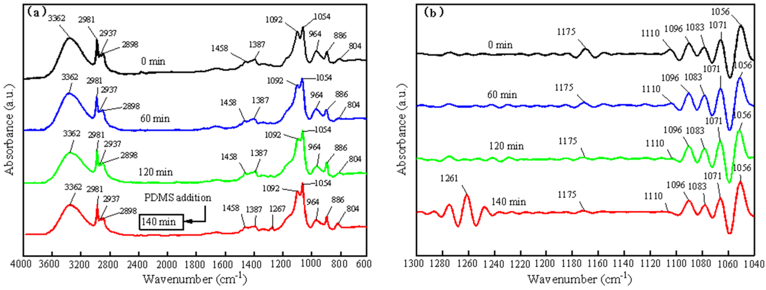
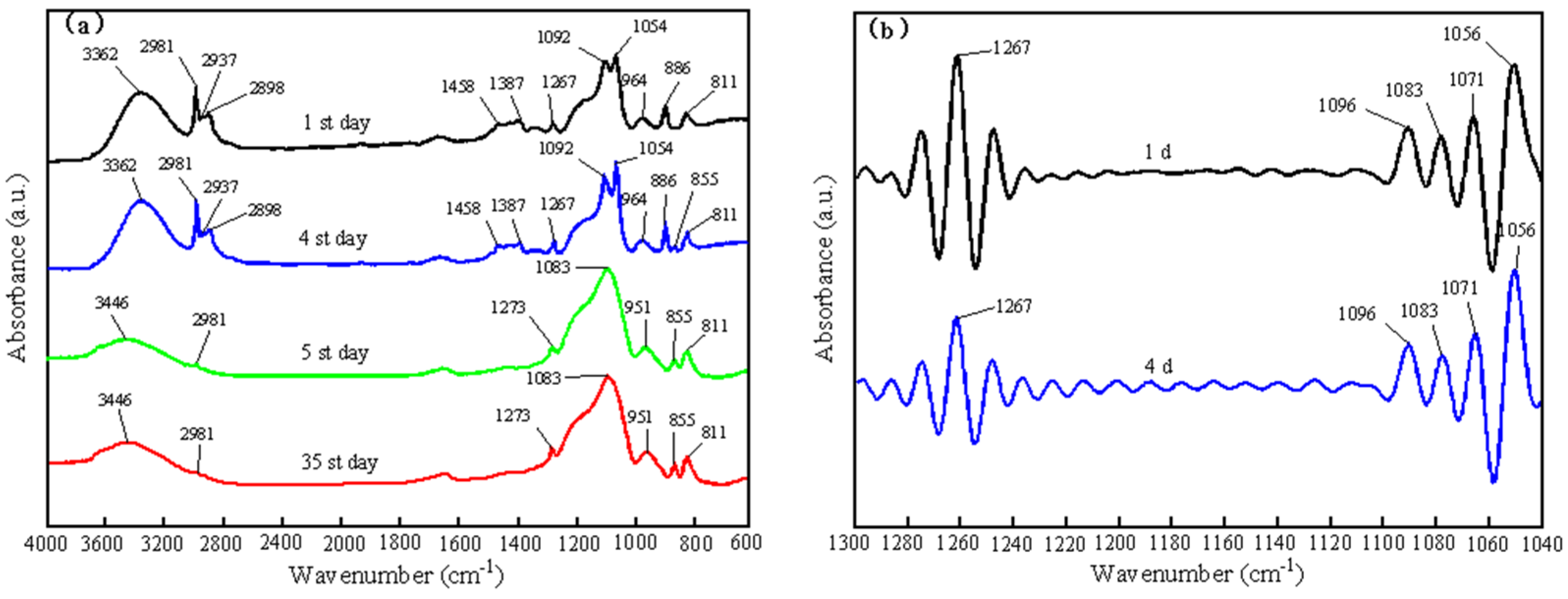
3.1. Characterization of the SiO2-TiO2-PDMS Sol and Coating

The formation mechanism of the coating was evaluated by FTIR spectroscopy. Due to the presence of overlapping peaks of EtOH and TEOS in the wave number region of 1300–1040 cm−1. In this wave number range, Nicolet EZ Omnic 6.0 was used to perform the self-deconvolution process, and the results are shown in Figure 2b, Figure 3b and Figure 4b. The parameters of Fourier deconvolution were set as Bandwidth = 28.0 ± 0.2 and Enhancement = 2.0 [50,51].
Figure 2 shows the FTIR spectra of the raw materials TEOS, PDMS, and EtOH. The characteristic peak at 3362 cm−1 in Figure 2a corresponded to the O-H stretching in ethanol. The characteristic peaks at 2800–3200 cm−1 corresponded to the bending, symmetric stretching, and antisymmetric stretching of the C-H bond in TEOS, PDMS, and EtOH, which is listed in detail in Table 1 [52,53,54].
Figure 3 presents the FTIR spectra of the TEOS hydrolysis process. As shown in Figure 3b, the characteristic peak at 1175 cm−1 was the C-H stretching vibration peak in TEOS [55], which disappeared with the completion of TEOS hydrolysis. The antisymmetric stretching vibrational peak of Si-O-C in TEOS was at 1110 cm−1, and its decrease in 0–140 min proved the occurrence of TEOS hydrolysis. The stretching vibration peak of CH3 in TEOS and EtOH was at 1096 cm−1, and this absorption peak showed the same intensity in all spectra due to the occurrence of TEOS hydrolysis and the production of EtOH. The Si-O-C stretching vibration peak in TEOS and C-O stretching vibration peak in EtOH were shown at 1083 cm−1. After the addition of PDMS at 140 min, a new characteristic peak appeared at 1267 cm−1 due to the bending vibration of Si-(CH3) in PDMS. With the hydrolysis of TEOS, a Si-O-Si bond was gradually formed, and the absorption peak at 804 cm−1 shifted toward 811 cm−1.
Figure 4 shows the FTIR spectra of the gel after 1, 4, 5, and 35 days of curing. As shown in Figure 4, a new characteristic peak was observed at 855 cm−1 on day four, which was associated with the co-polymerization of PDMS and TEOS. On day five, the C-H absorption peaks at 2937, 2898, 1458, 1387, 1092, and 886 cm−1 disappeared due to the volatilization of EtOH and the conversion of sol to gel. At the same time, the absorption peak at 964 cm−1 shifted toward 951 cm−1, which was due to the combination of the TiO2-SiO2 network and the formation of a Si-O-Ti bond. The characteristic peak at 1273 cm−1 confirmed the co-polymerization of PDMS molecules with Ti-OH and Si-OH.
The results of the FTIR analysis indicated that the formation of SiO2-TiO2-PDMS coating can be roughly divided into three stages. The first stage mainly included the hydrolysis and condensation of TEOS. In the second stage, when the hydrolysis and condensation reaction of TEOS reached a certain degree, TEOS and PDMS co-polymerized. At this point, the long chains of PDMS broke apart and oligomers were produced. In the third stage, after the nano-hybrid sol was applied to the surface of the building wall, the co-polymerization reaction between TiO2 and PDMS occurred within the SiO2 network, resulting in the formation of a uniform organic-inorganic hybrid gel five days later. The related chemical [56] reactions are shown in Equations (3)–(8).
Coatings 13 00224 i001
Coatings 13 00224 i002
Coatings 13 00224 i003
Coatings 13 00224 i004
Coatings 13 00224 i005
Coatings 13 00224 i006

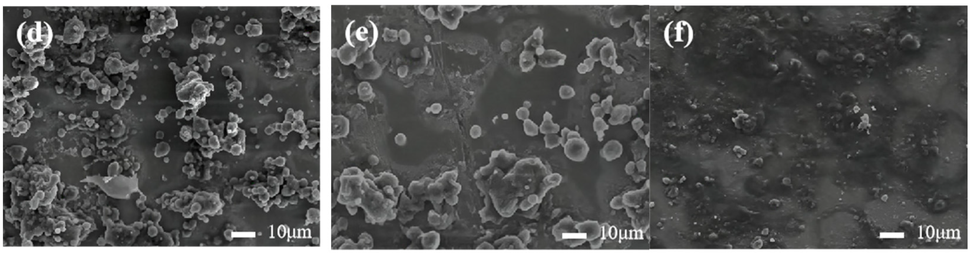
3.2. Microstructure of the Coating

In the present study, the SiO2-TiO2-PDMS coating was fabricated by mixing organic and inorganic solvents, which was actually a chemical reaction process. As shown in Figure 5, the microstructure of the coating with different amounts of PDMS varies greatly.
The morphology of the substrate surface changed significantly before and after spraying, as shown in Figure 5a,b. Before spraying, a large number of irregular and relatively loose blocks with large gaps could be observed on the surface. After covering the coating, the pores on the surface were filled and covered by a surface film with uneven surface bumps. The uneven bumps were tightly attached to the substrate, and the joints were relatively smooth. It indicates that the alkyl functional groups in the silane-based hydrophobic material were successfully grafted onto the surface of SiO2 and TiO2. Thus, the bumps were tightly bonded to the substrate surface. At the same time, the superhydrophobic material could penetrate into the interior of the substrate and fill the pores inside it. Therefore, it could effectively prevent the liquid from entering the interior of the substrate and improve the hydrophobic performance of the specimen. Generally, the “filling” and “covering” effects of micro-nano particles and PDMS changed the morphology of the substrate surface, thus improving the hydrophobic property of the substrate.
Figure 5b–f showed the morphology of the coating surface with different PDMS-to-TEOS molar ratios. As shown in Figure 5b, the surface of the coating without PDMS exhibited a rough structure. However, cracks existed on the surface, and the coating had a tendency to peel off from the substrate. As shown in Figure 5c–f, the cracks on the coating gradually decreased with the increased addition of PDMS. The surface of the coating was basically free of cracks and showed an obvious micro-nano rough structure when the PDMS to TEOS molar ratio was between 1:5 and 3:10. When the molar ratio of PDMS to TEOS was 2:5, the micro-nano rough structure that formed on the surface of the coating was gradually covered by PDMS, and the coating was relatively smooth, resulting in the decrease of hydrophobicity of the coating.
The EDS results of the wall surface before and after spraying are shown in Figure 6. The wall surface mainly contained C, O, and Ca elements before spraying, among which the Ca element reached the highest content, as shown in Figure 6a. After spraying, five elements (C, O, Si, Ca, Ti) were found on the surface. On the other hand, the content of Si element increased significantly. It can be inferred from the above findings that SiO2 and TiO2 nanoparticles have been grafted onto the surface of the coating.

3.3. Water-Repellent Performance

3.3.1. Contact Angle

The effect of the molar ratio of PDMS to TEOS on the contact angle was obtained based on the measurement test of contact angles, and the results are shown in Figure 7. When PDMS was not added, the contact angle of the coating was 98° and the rolling angle was 10.2°. With the addition of PDMS, the roll angle decreased gradually, while the contact angle increased first and then decreased. When the molar ratio of PDMS to TEOS was 1:5, the contact angle of the coating reached a maximum of about 152.6°, and the rolling angle was about 5.2°. Because the static contact angle was larger than 150° and the roll-off angle was less than 10°, the coating reached a superhydrophobic state.

3.3.2. Water Absorption

Figure 8 shows the effect of PDMS and TEOS ratio on water absorption of building walls. As depicted in Figure 8, water absorption of the coated building walls was reduced to varying degrees compared to the uncoated specimens. In the absence of a surface coating, the building wall reached water absorption saturation at 24 h with a water absorption rate of 5.28%. With the increase of PDMS content, the hydrophobic protection effect of the coating gradually increased, and the water absorption rate of the building wall gradually decreased. When the molar ratio of PDMS to TEOS was 1:5 or even higher, the building wall reached water absorption saturation at 7 d, and the water absorption was 3.3%. At this time, the coating showed excellent water-repellent performance.

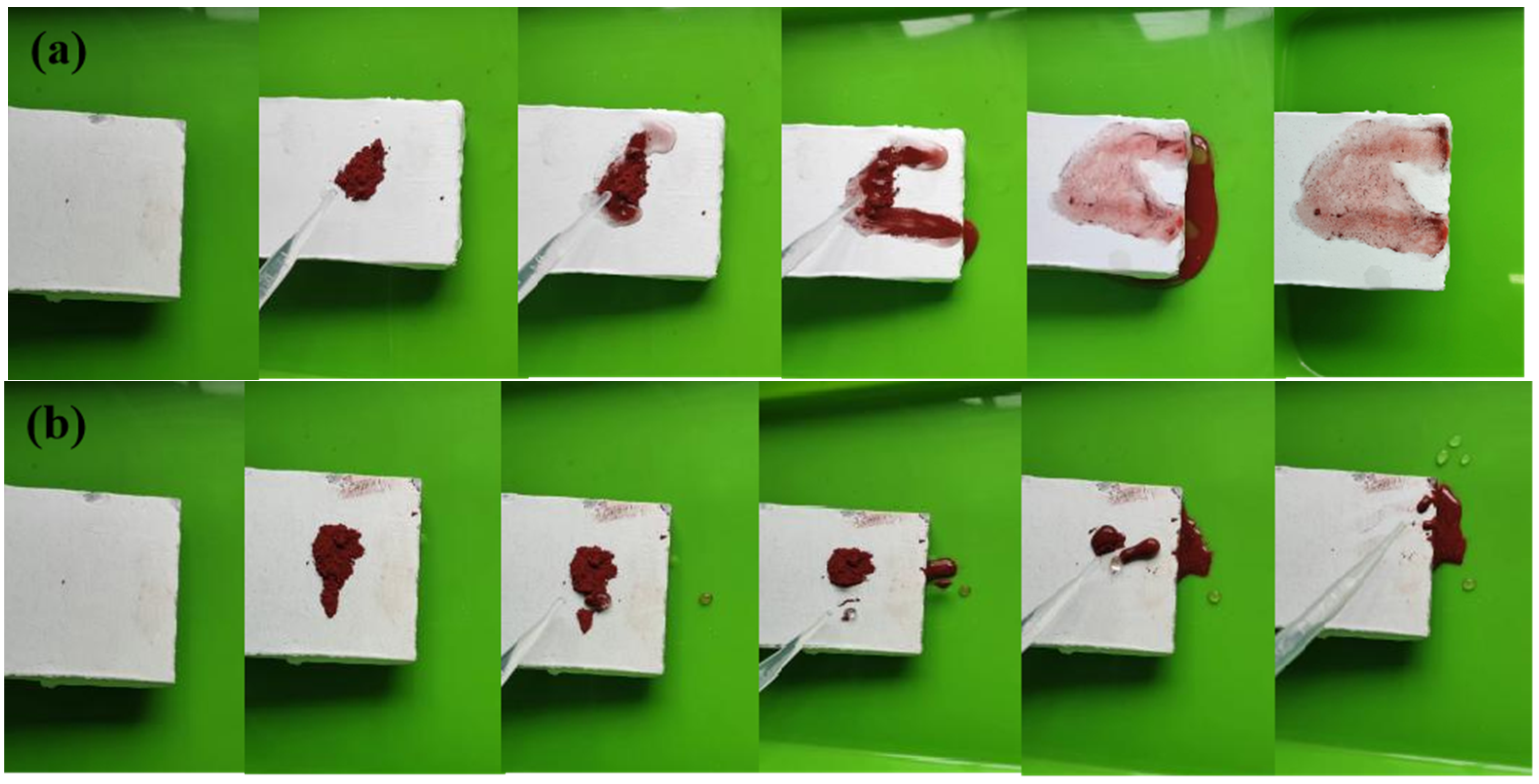
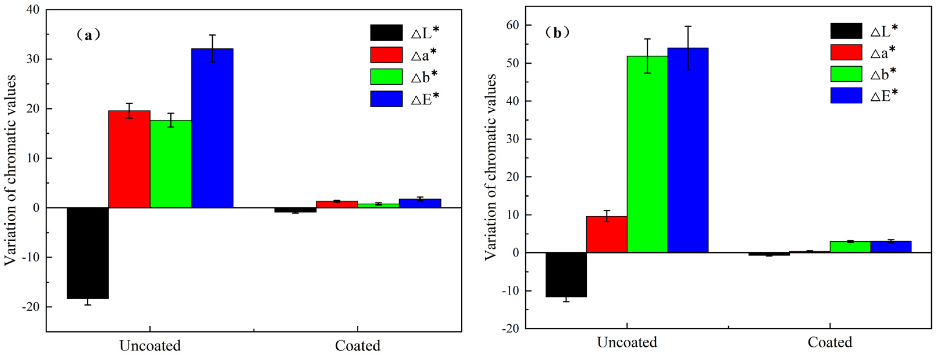
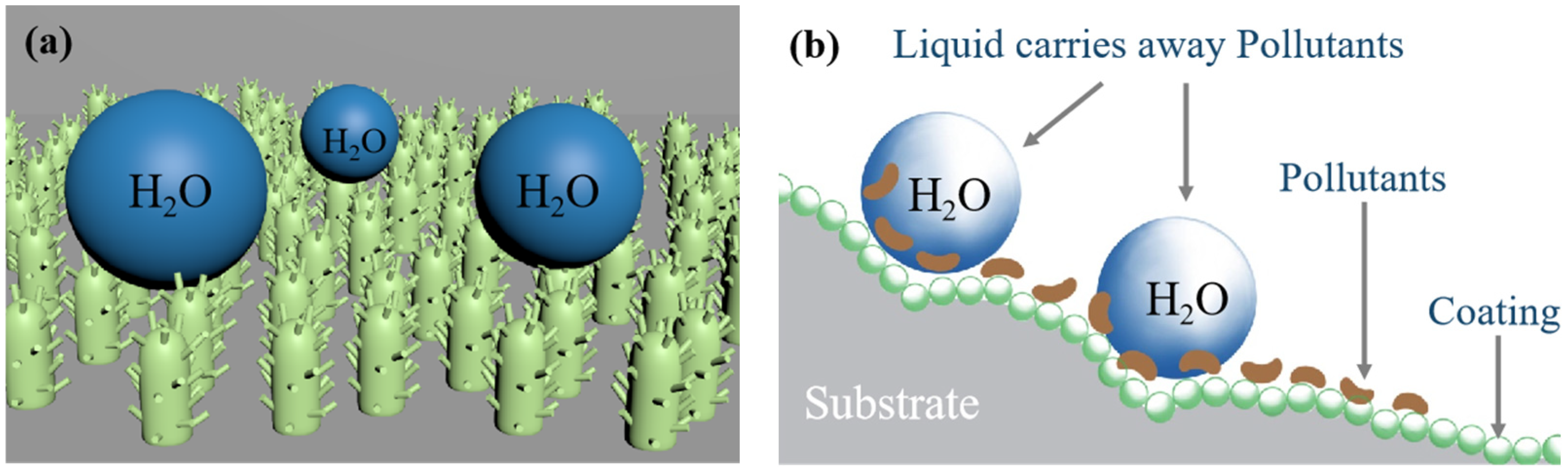
3.4. Self-Cleaning Ability of the Coating

Fe2O3 powder was used to simulate particulate pollutants and methyl orange solution was used to simulate liquid pollutants [57]. Figure 9 shows the results for the surface of coated and uncoated walls contaminated with Fe2O3. It can be found in Figure 9a that the surface of the uncoated wall was easily adhered by Fe2O3 powder, and the contaminants were difficult to be washed away by water. In contrast, Figure 9b shows that the coated walls could be easily cleaned out with deionized water even if particulate contaminants were adhered to the surface.
Figure 10 depicts the optical images of uncoated and coated walls contaminated with methyl orange solution. Figure 10a demonstrates that the uncoated wall’s surface was easily contaminated by the methyl orange solution and was difficult to clean out with water. As expected, as shown in Figure 10b, due to the presence of the superhydrophobic coating, the liquid contaminant dropped onto the coating in a spherical form and quickly rolled off. As a result, it was difficult for the methyl orange solution to adhere to the surfaces of the coated walls.
The variation in chroma values of the coated and uncoated walls contaminated by pollutants is shown in Figure 11. This is due to the fact that a large amount of air is stored between tiny bumps due to the presence of the micron and nano papillary rough structure on the coating surface, thus reducing the liquid-solid surface contact area and expanding the interface between the liquid and the air.
According to the self-cleaning model depicted in Figure 12b, when a certain angle of inclination is applied to the substrate, the rough surface of the substrate will hold the water droplets and carry the contaminants away via gravity. Therefore, the surface of the modified substrate is self-cleaning. The microscopic model of the coated surface and the self-cleaning model are shown in Figure 12a,b.

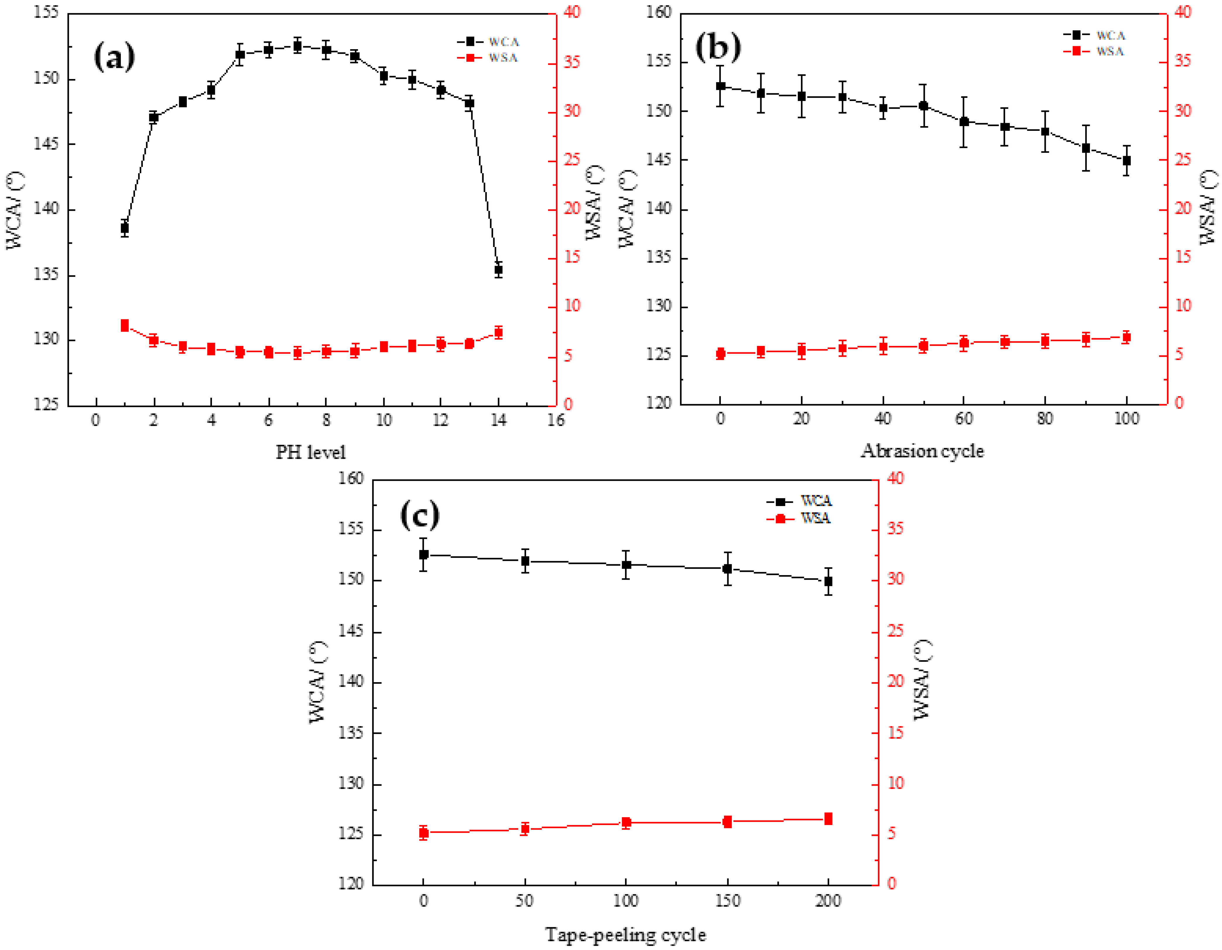
3.5. Mechanical Stability of the Coating

The chemical stability of the superhydrophobic surface is an important parameter to be considered in practical applications of engineering materials. Figure 13a shows the results of water CA and SA on the surface of the coating after being subjected to a corrosive bath with different pH values for 3 h. The results showed that the solution with a PH of 2–13 did not cause excessive fluctuations in CA of the coating, but the coating was sensitive to solutions with pH values of 1 and 14. This is because the wettability of the heterogeneous surface can be illustrated by the Cassie–Baxter equation [58,59]:
c o s θ c = f ( c o s θ 0 + 1 ) 1
where θc is the apparent contact angle, θ0 is the intrinsic contact angle, and f is the solid-liquid contact area fraction. According to Cassie–Baxter’s theory, a rough surface is a composite air-solid interface, and the apparent contact angle increases as the solid-liquid contact area decreases. When the molar ratio of PDMS to TEOS is 1:5, the apparent contact angle of the coating is 152.6° and f is 0.1122, resulting in a solid-air contact area of 88.78%. In Cassie–Baxter’s mode, the presence of the air pocket layer formed within the interface with a rough structure and low surface energy acts as a barrier layer to block the chemical to penetrate across the surface. This gives the coating a greater range of acid and alkali resistance. However, the coating is sensitive to solutions with pH values of 1 and 14, which is because the high PH value seriously destroys the rough structure of the coating surface, resulting in a rapid decline in superhydrophobic properties. It shows that the coating can resist the solution with a pH value of 2–13 and has the ability to resist acid and alkali.
The mechanical stability of a superhydrophobic coating is a significant requirement to realize its practical applications. In this study, the linear abrasion test and tape-peeling test were performed to evaluate the mechanical durability of the SiO2-TiO2-PDMS superhydrophobic surface. Figure 13b shows the changes of CA and SA of the superhydrophobic SiO2-TiO2-PDMS coating over 100 abrasion cycles. The results show that with the increase of the friction distance, there is a slight decrease in water CA and a slight increase in SA results. However, the hydrophobicity of the coating was retained, as the CAs were maintained above 120° and the SAs below 10°. In order to further evaluate the mechanical properties of the prepared superhydrophobic surface, the tape-peeling test was also conducted in this study using a transparent adhesive Scotch tape. As shown in Figure 13c, the CA and SA results showed that the superhydrophobic coating remained above 120° even after 200 cycles. This is because PDMS, as a binder, can effectively improve the interfacial adhesion between nanoparticles and matrix in the coating solution. This ensures the stability of the coating against abrasion.

4. Conclusions

The effect of SiO2-TiO2-PDMS coating on superhydrophobicity and self-cleaning performance of building walls was investigated by various characterization methods, and the main conclusions are as follows:
(1)
A novel hybrid material for building wall protection was prepared by the sol-gel process with TEOS and TTIP as precursors and PDMS as low surface energy substances under the catalytic action of oxalic acid. When the molar ratio of PDMS to TEOS was 1:5, the static contact angle of the coating surface was 152.6°, and the superhydrophobic state was achieved.
(2)
The coating exhibited a hydrophobic protective effect on the building wall. Without coating, the building wall reached water absorption saturation at 24 h with 5.28% water absorption. After coating, the building wall reached water absorption saturation after 4 days, and the water absorption rate was 3.3%, which was 37.5% lower than before.
(3)
The results of FTIR showed that the coating formed a micro-nano secondary structure similar to the surface of lotus leaf after condensation and co-polymerization. Short term immersion measurements revealed that the coating exhibited excellent chemical durability at different pH concentrations.

Author Contributions

X.X.: Investigation, writing–original draft, writing—review & editing. J.L.: Supervision, writing—review & editing. Y.L.: Supervision, review. Z.L.: Investigation. Y.H.: Investigation. Z.Z.: Investigation. J.Y.: Supervision. All authors have read and agreed to the published version of the manuscript.

Funding

This research was funded by the National Natural Science Foundation of China (No. 51904357), the Natural Science Foundation of Hunan Province (No. 2020JJ5985) and the scientific research project of the education department of Hunan Province (No. 20B623).

Institutional Review Board Statement

Not applicable.

Informed Consent Statement

Not applicable.

Data Availability Statement

The data that support the findings of this study are available from the corresponding author, upon reasonable request.

Acknowledgments

We thank Ting Yang and Lu Huang, the working technicians of Science Compass, who performed SEM and FTIR tests, respectively. Thanks to Huanyu Su for his enlightening suggestions for data analysis.

Conflicts of Interest

The authors declare no conflict of interest.

References

  1. Gradeci, K.; Labonnote, N.; Time, B.; Köhler, J. A Probabilistic-Based Methodology for Predicting Mould Growth in Façade Constructions. Build. Environ. 2018, 128, 33–45. [Google Scholar] [CrossRef]
  2. Chen, G.; Wang, H.; Chen, Y.; Liu, X.; Guo, X. Contrastive Study on Indoor Temperature-humidity Threshold Value for Mould Germination Risk on the Hygroscopic Wall. J. Univ. S. China (Sci. Technol.) 2019, 33, 1–7. [Google Scholar] [CrossRef]
  3. Kurbanova, A.; Myrzakhmetova, N.; Akimbayeva, N.; Kishibayev, K.; Nurbekova, M.; Kanagat, Y.; Tursynova, A.; Zhunussova, T.; Seralin, A.; Kudaibergenova, R.; et al. Superhydrophobic SiO2/Trimethylchlorosilane Coating for Self-Cleaning Application of Construction Materials. Coatings 2022, 12, 1422. [Google Scholar] [CrossRef]
  4. Salimifard, P.; Rim, D.; Gomes, C.; Kremer, P.; Freihaut, J.D. Resuspension of Biological Particles from Indoor Surfaces: Effects of Humidity and Air Swirl. Sci. Total Environ. 2017, 583, 241–247. [Google Scholar] [CrossRef]
  5. Dalawai, S.P.; Saad Aly, M.A.; Latthe, S.S.; Xing, R.; Sutar, R.S.; Nagappan, S.; Ha, C.S.; Kumar Sadasivuni, K.; Liu, S. Recent Advances in Durability of Superhydrophobic Self-Cleaning Technology: A Critical Review. Prog. Org. Coat. 2020, 138, 105381. [Google Scholar] [CrossRef]
  6. Bai, Y.; Zhang, H.; Shao, Y.; Zhang, H.; Zhu, J. Recent Progresses of Superhydrophobic Coatings in Different Application Fields: An Overview. Coatings 2021, 11, 116. [Google Scholar] [CrossRef]
  7. Xiang, T.; Lv, Z.; Wei, F.; Liu, J.; Dong, W.; Li, C.; Zhao, Y.; Chen, D. Superhydrophobic Civil Engineering Materials: A Review from Recent Developments. Coatings 2019, 9, 753. [Google Scholar] [CrossRef] [Green Version]
  8. Neinhuis, C.; Barthlott, W. Characterization and Distribution of Water-Repellent, Self-Cleaning Plant Surfaces. Ann. Bot. 1997, 79, 667–677. [Google Scholar] [CrossRef] [Green Version]
  9. Qian, B.; Shen, Z. Fabrication of Superhydrophobic Surfaces by Dislocation-Selective Chemical Etching on Aluminum, Copper, and Zinc Substrates. Langmuir 2005, 21, 9007–9009. [Google Scholar] [CrossRef]
  10. Yu, J.; Qin, L.; Hao, Y.; Kuang, S.; Bai, X.; Chong, Y.M.; Zhang, W.; Wang, E. Vertically Aligned Boron Nitride Nanosheets: Chemical Vapor Synthesis, Ultraviolet Light Emission, and Superhydrophobicity. ACS Nano 2010, 4, 414–422. [Google Scholar] [CrossRef]
  11. Chaudhary, A.; Barshilia, H.C. Nanometric Multiscale Rough CuO/Cu(OH)2 Superhydrophobic Surfaces Prepared by a Facile One-Step Solution-Immersion Process: Transition to Superhydrophilicity with Oxygen Plasma Treatment. J. Phys. Chem. C 2011, 115, 18213–18220. [Google Scholar] [CrossRef]
  12. Rao, A.V.; Latthe, S.S.; Mahadik, S.A.; Kappenstein, C. Mechanically Stable and Corrosion Resistant Superhydrophobic Sol–Gel Coatings on Copper Substrate. Appl. Surf. Sci. 2011, 257, 5772–5776. [Google Scholar] [CrossRef]
  13. Dong, C.; Gu, Y.; Zhong, M.; Li, L.; Sezer, K.; Ma, M.; Liu, W. Fabrication of Superhydrophobic Cu Surfaces with Tunable Regular Micro and Random Nano-Scale Structures by Hybrid Laser Texture and Chemical Etching. J. Mater. Proc. Technol. 2011, 211, 1234–1240. [Google Scholar] [CrossRef]
  14. Guo, X.J.; Xue, C.H.; Sathasivam, S.; Page, K.; He, G.; Guo, J.; Promdet, P.; Heale, F.L.; Carmalt, C.J.; Parkin, I.P. Fabrication of Robust Superhydrophobic Surfaces via Aerosol-Assisted CVD and Thermo-Triggered Healing of Superhydrophobicity by Recovery of Roughness Structures. J. Mater. Chem. A 2019, 7, 17604–17612. [Google Scholar] [CrossRef]
  15. Li, S.; Page, K.; Sathasivam, S.; Heale, F.; He, G.; Lu, Y.; Lai, Y.; Chen, G.; Carmalt, C.J.; Parkin, I.P. Efficiently Texturing Hierarchical Superhydrophobic Fluoride-Free Translucent Films by AACVD with Excellent Durability and Self-Cleaning Ability. J. Mater. Chem. A 2018, 6, 17633–17641. [Google Scholar] [CrossRef] [Green Version]
  16. Wang, F. Study on Bionic Preparation of Micro-Nanostructures and Properties of Plant Leaves onto Bamboo Surfaces through Morph-Genetic Method. Ph.D. Thesis, Zhejiang A&F University, Zhejiang, China, 2018. [Google Scholar]
  17. Wang, F.; Li, S. The super-hydrophobic structure of lotus leaf generated on the bamboo surface based on soft lithography. Sci. Technol. Rev. 2016, 34, 101–104. [Google Scholar] [CrossRef]
  18. Wang, F.; Wu, H.; Li, S.; Zhang, W. The Bionic Rose-like Super-hydrophobic Structure on Bamboo Surface. J. Bamboo Res. 2017, 36, 49–52. [Google Scholar] [CrossRef]
  19. He, X.; Fu, S.; Dai, Y.; Jin, C.; Wang, F. Research on Surface Characteristics of Bamboo with Bionic Super-hydrophobic Chinese Rose/TiO2. World Bamboo Ratt. 2018, 15, 11–15. [Google Scholar] [CrossRef]
  20. Wen, N.; Miao, X.; Yang, X.; Long, M.; Deng, W.; Zhou, Q.; Deng, W. An Alternative Fabrication of Underoil Superhydrophobic or Underwater Superoleophobic Stainless Steel Meshes for Oil-Water Separation: Originating from One-Step Vapor Deposition of Polydimethylsiloxane. Sep. Purif. Technol. 2018, 204, 116–126. [Google Scholar] [CrossRef]
  21. Xie, A.; Cui, J.; Chen, Y.; Lang, J.; Li, C.; Yan, Y.; Dai, J. One-Step Facile Fabrication of Sustainable Cellulose Membrane with Superhydrophobicity via a Sol-Gel Strategy for Efficient Oil/Water Separation. Surf. Coat. Technol. 2019, 361, 19–26. [Google Scholar] [CrossRef]
  22. Arun Kumar, K.V.; John, J.; Sooraj, T.R.; Raj, S.A.; Unnikrishnan, N.V.; Selvaraj, N.B. Surface Plasmon Response of Silver Nanoparticles Doped Silica Synthesised via Sol-Gel Route. Appl. Surf. Sci. 2019, 472, 40–45. [Google Scholar] [CrossRef]
  23. Calia, A.; Lettieri, M.; Masieri, M. Durability Assessment of Nanostructured TiO2 Coatings Applied on Limestones to Enhance Building Surface with Self-Cleaning Ability. Build. Environ. 2016, 110, 1–10. [Google Scholar] [CrossRef]
  24. Pedna, A.; Pinho, L.; Frediani, P.; Mosquera, M.J. Obtaining SiO2–Fluorinated PLA Bionanocomposites with Application as Reversible and Highly-Hydrophobic Coatings of Buildings. Prog. Org. Coat. 2016, 90, 91–100. [Google Scholar] [CrossRef]
  25. Peng, M.; Wang, L.; Guo, L.; Guo, J.; Zheng, L.; Yang, F.; Ma, Z.; Zhao, X. A Durable Nano-SiO2-TiO2/Dodecyltrimethoxysilane Superhydrophobic Coating for Stone Protection. Coatings 2022, 12, 1397. [Google Scholar] [CrossRef]
  26. Adamopoulos, F.G.; Vouvoudi, E.C.; Pavlidou, E.; Achilias, D.S.; Karapanagiotis, I. TEOS-Based Superhydrophobic Coating for the Protection of Stone-Built Cultural Heritage. Coatings 2021, 11, 135. [Google Scholar] [CrossRef]
  27. Zhou, H.; Wang, H.; Niu, H.; Zhao, Y.; Xu, Z.; Lin, T. A Waterborne Coating System for Preparing Robust, Self-Healing, Superamphiphobic Surfaces. Adv. Funct. Mater. 2017, 27, 1604261. [Google Scholar] [CrossRef]
  28. Wei, X.; Chen, F.; Wang, H.; Zhou, H.; Ji, Z.; Lin, T. Efficient Removal of Aerosol Oil-Mists Using Superoleophobic Filters. J. Mater. Chem. A 2018, 6, 871–877. [Google Scholar] [CrossRef]
  29. Zhong, M. Study on the Preparation and Application of Superhydrophobic Silica Coatings. Master’s Thesis, Fuzhou University, Fuzhou, China, 2018. [Google Scholar]
  30. Yang, C.; Wang, F.; Li, W.; Ou, J.; Li, C.; Amirfazli, A. Anti-Icing Properties of Superhydrophobic ZnO/PDMS Composite Coating. Appl. Phys. A Mater. Sci. Process. 2016, 122, 1. [Google Scholar] [CrossRef]
  31. Deng, Z.Y.; Wang, W.; Mao, L.H.; Wang, C.F.; Chen, S. Versatile Superhydrophobic and Photocatalytic Films Generated from TiO2–SiO2@PDMS and Their Applications on Fabrics. J. Mater. Chem. A 2014, 2, 4178–4184. [Google Scholar] [CrossRef]
  32. Song, Y.; Lei, X.; Liu, J.; Guan, X.; Yuan, H. Preparation and Properties of Superhydrophobic Surface with Titanium Dioxide and Attapulgite. J. Chin. Ceram. Soc. 2021, 49, 2242–2250. [Google Scholar] [CrossRef]
  33. Zárraga, R.; Cervantes, J.; Salazar-Hernandez, C.; Wheeler, G. Effect of the Addition of Hydroxyl-Terminated Polydimethylsiloxane to TEOS-Based Stone Consolidants. J. Cult. Herit. 2010, 11, 138–144. [Google Scholar] [CrossRef]
  34. Hu, Y.; Xu, J.; Yuan, C.; Lin, J.; Yin, Z. A single TiO2-coated side-glowing optical fiber for photocatalytic wastewater treatment. Chin. Sci. Bull. 2005, 50, 1978–1984. [Google Scholar] [CrossRef]
  35. Peng, S.; Meng, W.; Guo, J.; Wang, B.; Wang, Z.; Xu, N.; Li, X.; Wang, J.; Xu, J. Photocatalytically Stable Superhydrophobic and Translucent Coatings Generated from PDMS-Grafted-SiO2/TiO2@PDMS with Multiple Applications. Langmuir 2019, 35, 2760–2771. [Google Scholar] [CrossRef] [PubMed]
  36. Sheng, Y.; Xu, L.; Meng, Y.; Shen, Y.; Wang, L.; Pan, H. Preparation of superhydrophobic, photocatalytic and UV-blocking textiles based on SiO2/TiO2 composite aerogels. J. Text. Res. 2019, 40, 90–96. [Google Scholar] [CrossRef]
  37. Liu, X.; Gu, Y.; Mi, T.; Wang, X.; Zhang, X. Dip-Coating Approach to Fabricate Durable PDMS/STA/SiO2 Superhydrophobic Polyester Fabrics. Coatings 2021, 11, 326. [Google Scholar] [CrossRef]
  38. Test Method of Cement Mortar Strength (ISO Method). Available online: https://wenku.baidu.com/view/bf9f196074232f60ddccda38376baf1ffd4fe379.html?_wkts_=1674023709816 (accessed on 3 November 2022).
  39. Aslanidou, D.; Karapanagiotis, I.; Panayiotou, C. Tuning the Wetting Properties of Siloxane-Nanoparticle Coatings to Induce Superhydrophobicity and Superoleophobicity for Stone Protection. Mater. Des. 2016, 108, 736–744. [Google Scholar] [CrossRef]
  40. Chatzigrigoriou, A.; Karapanagiotis, I.; Poulios, I. Superhydrophobic Coatings Based on Siloxane Resin and Calcium Hydroxide Nanoparticles for Marble Protection. Coatings 2020, 10, 334. [Google Scholar] [CrossRef] [Green Version]
  41. Yin, Z.; Chen, X.; Zhou, T.; Xue, M.; Li, M.; Liu, K.; Zhou, D.; Ou, J.; Xie, Y.; Ren, Z.; et al. Mussel-Inspired Fabrication of Superior Superhydrophobic Cellulose-Based Composite Membrane for Efficient Oil Emulsions Separation, Excellent Anti-Microbial Property and Simultaneous Photocatalytic Dye Degradation. Sep. Purif. Technol. 2022, 286, 120504. [Google Scholar] [CrossRef]
  42. Standard Test Method for Determining the Water Absorption of Hardened Concrete Treated with a Water Repellent Coating. Available online: https://www.astm.org/d6489-99r20.html (accessed on 3 November 2022).
  43. La Russa, M.F.; Ruffolo, S.A.; Rovella, N.; Belfiore, C.M.; Palermo, A.M.; Guzzi, M.T.; Crisci, G.M. Multifunctional TiO2 Coatings for Cultural Heritage. Prog. Org. Coat. 2012, 74, 186–191. [Google Scholar] [CrossRef]
  44. García, O.; Malaga, K. Definition of the Procedure to Determine the Suitability and Durability of an Anti-Graffiti Product for Application on Cultural Heritage Porous Materials. J. Cult. Herit. 2012, 13, 77–82. [Google Scholar] [CrossRef]
  45. Elzaabalawy, A.; Meguid, S.A. Development of Novel Superhydrophobic Coatings Using Siloxane-Modified Epoxy Nanocomposites. Chem. Eng. J. 2020, 398, 125403. [Google Scholar] [CrossRef]
  46. Zhang, X.; Si, Y.; Mo, J.; Guo, Z. Robust Micro-Nanoscale Flowerlike ZnO/Epoxy Resin Superhydrophobic Coating with Rapid Healing Ability. Chem. Eng. J. 2017, 313, 1152–1159. [Google Scholar] [CrossRef]
  47. Yin, Z.; Yuan, F.; Li, M.; Xue, M.; Zhou, D.; Chen, Y.; Liu, X.; Luo, Y.; Hong, Z.; Xie, C.; et al. Self-Cleaning, Underwater Writable, Heat-Insulated and Photocatalytic Cellulose Membrane for High-Efficient Oil/Water Separation and Removal of Hazardous Organic Pollutants. Prog. Org. Coat 2021, 157, 106311. [Google Scholar] [CrossRef]
  48. Yin, Z.; Yuan, F.; Xue, M.; Xue, Y.; Xie, Y.; Ou, J.; Luo, Y.; Hong, Z.; Xie, C. A Multifunctional and Environmentally Safe Superhydrophobic Membrane with Superior Oil/Water Separation, Photocatalytic Degradation and Anti-Biofouling Performance. J. Colloid Interface Sci. 2022, 611, 93–104. [Google Scholar] [CrossRef]
  49. Yap, S.W.; Johari, N.; Mazlan, S.A.; Hassan, N.A. Mechanochemical Durability and Self-Cleaning Performance of Zinc Oxide-Epoxy Superhydrophobic Coating Prepared via a Facile One-Step Approach. Ceram. Int. 2021, 47, 15825–15833. [Google Scholar] [CrossRef]
  50. Vazhnova, T.; Lukyanov, D.B. Fourier Self-Deconvolution of the IR Spectra as a Tool for Investigation of Distinct Functional Groups in Porous Materials: Brønsted Acid Sites in Zeolites. Anal. Chem. 2013, 85, 11291–11296. [Google Scholar] [CrossRef]
  51. Kauppinen, J.K.; Moffatt, D.J.; Mantsch, H.H.; Cameron, D.G. Fourier Self-Deconvolution: A Method for Resolving Intrinsically Overlapped Bands. Appl. Spectrosc. 1981, 35, 271–276. [Google Scholar] [CrossRef]
  52. Jia, S.; Liu, M.; Qing, Y.; Wang, S.; Wu, Y. Preparation of Superhydrophobic Surface on Wood Substrate by One-step Sol-gel. China Wood Ind. 2016, 30, 17–20. [Google Scholar] [CrossRef]
  53. Song, X.; Cui, W.; Gao, G. Water Resistance of Catalyzed and Hybridized TEOS Waterproof Agent. Bull. Chin. Ce Ramic Soc. 2016, 35, 358–362. [Google Scholar] [CrossRef]
  54. Kapridaki, C.; Maravelaki-Kalaitzaki, P. TiO2–SiO2–PDMS Nano-Composite Hydrophobic Coating with Self-Cleaning Properties for Marble Protection. Prog. Org. Coat. 2013, 76, 400–410. [Google Scholar] [CrossRef]
  55. Zhou, S. PreParation of PDMS/SiO2 Hybrid Material and Its Application in the Protection of Ancient Ivory. Master’s Thesis, Chengdu University of Technology, Chengdu, China, 2008. [Google Scholar]
  56. Wang, H. Study on the PreParation and Properties of PDMS/SiO2 Hybrid Membrafl. Master’s Thesis, Beijing Jiaotong University, Beijing, China, 2011. [Google Scholar]
  57. Ye, X.; Cai, D.; Hou, J.; Ruan, X. Superhydrophobic and self-cleaning coating for building wall protection. Acta Mater. Compos. Sin. 2018, 35, 3271–3279. [Google Scholar] [CrossRef]
  58. Yin, Z.; Xue, M.; Luo, Y.; Hong, Z.; Xie, C.; Ren, Z.; Wang, H. Excellent Static and Dynamic Anti-Icing Properties of Hierarchical Structured ZnO Superhydrophobic Surface on Cu Substrates. Chem. Phys. Lett. 2020, 755, 137806. [Google Scholar] [CrossRef]
  59. Yin, Z.; Yuan, F.; Zhou, D.; Xue, M.; Luo, Y.; Hong, Z.; Xie, C. Ultra Dynamic Water Repellency and Anti-Icing Performance of Superhydrophobic ZnO Surface on the Printed Circuit Board (PCB). Chem. Phys. Lett. 2021, 771, 138558. [Google Scholar] [CrossRef]
Figure 1. Preparation process of the coating.
Coatings 13 00224 g001
Figure 2. FTIR spectra. (a) FTIR spectra of TEOS, PDMS, and EtOH. (b) Fourier self-deconvoluted infrared spectra of TEOS, PDMS, and EtOH at 1040 cm−1 to 1300 cm−1.
Coatings 13 00224 g002
Figure 3. FTIR spectra. (a) FTIR spectra of TEOS hydrolysis at 0 min, 60 min, 120 min, and 140 min. (b) Fourier self-deconvolution infrared spectra of TEOS hydrolysis at 1040 cm−1 to 1300 cm−1 for 0 min, 60 min, 120 min, and 140 min.
Coatings 13 00224 g003
Figure 4. FTIR spectra. (a) FTIR spectra of sol-gel curing at 1, 4, 5, and 35 days. (b) Fourier self-deconvolution infrared spectra of sols cured at 1040 cm−1 to 1300 cm−1 for 1 and 4 days.
Coatings 13 00224 g004
Figure 5. Microstructure of coating with different molar ratios of PDMS and TEOS. (a) Uncoated wall surface. (b) n (PDMS):n (TEOS) = 0. (c) n (PDMS):n (TEOS) = 1:10. (d) n (PDMS):n (TEOS) = 1:5. (e) n (PDMS):n (TEOS) = 3:10. (f) n (PDMS):n (TEOS) = 2:5.
Coatings 13 00224 g005aCoatings 13 00224 g005b
Figure 6. EDS energy spectrum of wall surface before and after coating. (a) Uncoated. (b) Coated with n (PDMS):n (TEOS) = 1:5.
Coatings 13 00224 g006
Figure 7. Effect of molar ratio of PDMS and TEOS on the contact angle.
Coatings 13 00224 g007
Figure 8. Water absorption of building walls with different PDMS and TEOS ratios. (a) Cumulative water absorption in 14 days. (b) Cumulative water absorption in 12 h.
Coatings 13 00224 g008
Figure 9. Optical images of particulate contaminants on the surface of building wall samples before and after spraying. (a) Particle pollutants contaminate the uncoated wall surface. (b) Particle pollutants contaminate the coating with n (PDMS):n (TEOS) = 1:5.
Coatings 13 00224 g009
Figure 10. Optical images of liquid contaminants on the surface of building wall samples before and after spraying. (a) Liquid pollutants contaminate the uncoated wall surface. (b) Liquid pollutants contaminate the coating with n (PDMS):n (TEOS) = 1:5.
Coatings 13 00224 g010
Figure 11. Variation in chromatic values of the coated and uncoated walls. (a) Building walls contaminated with particulate pollutants. (b) Building walls contaminated with liquid pollutants.
Coatings 13 00224 g011
Figure 12. Models. (a) Microscopic model of coated surfaces. (b) Self-cleaning model of coated surfaces.
Coatings 13 00224 g012
Figure 13. Mechanical Stability of the Coating. (a) Water CA and SA result over corrosive bath immersion in different pH range. (b) Changes in water CA and SA of the coating over 100 abrasion cycles. (c) Changes in water CA and SA of the coating over 200 tape-peeling cycles.
Coatings 13 00224 g013
Table 1. Antisymmetric stretching, symmetric stretching, and bending peaks of C-H bonds in TEOS, PDMS, and EtOH.
Wavenumber (cm−1)Characteristic PeaksReferences
2981, 2933Antisymmetric stretching of C-H bond in EtOH and TEOS[48,49]
2968Antisymmetric stretching of C-H bond in PDMS[51]
2911Symmetric stretching of C-H bonds in PDMS[51]
2898Symmetric stretching of C-H bond in EtOH and TEOS[48,49]
1460Antisymmetric bending of the C-H bond in EtOH[49]
1451Antisymmetric bending of the C-H bond in TEOS[49]
1396Symmetric bending of C-H bond in EtOH and TEOS[48,49]
1267Symmetric bending of C-H bond in PDMS[51]
1175Rocking of C-H bond in TEOS[49]
1089Rocking of C-H bond in EtOH[49]
Disclaimer/Publisher’s Note: The statements, opinions and data contained in all publications are solely those of the individual author(s) and contributor(s) and not of MDPI and/or the editor(s). MDPI and/or the editor(s) disclaim responsibility for any injury to people or property resulting from any ideas, methods, instructions or products referred to in the content.

