Article

Dispersion and Demineralization Inhibition Capacity of Novel Magnesium Oxide Nanoparticles Varnish on Enamel Surfaces against Streptococcus mutans (an In Vitro Study)

by
Sonya Jamal Hamalaw
,
Fadil Abdulla Kareem
and
Arass Jalal Noori
*
Department of Pedodontics, Orthodontics and Preventive Dentistry, College of Dentistry, University of Sulaimani, Sulaymaniyah 46001, Iraq
*
Author to whom correspondence should be addressed.
Coatings 2023, 13(6), 1018; https://doi.org/10.3390/coatings13061018
Submission received: 10 May 2023 / Revised: 25 May 2023 / Accepted: 26 May 2023 / Published: 30 May 2023

Abstract

:
This research analyzed the dispersion and impact of magnesium oxide nanoparticles (MgONPs) varnish on inhibiting enamel demineralization. A novel MgONPs varnish was prepared in absolute ethanol with rosin in 10%, 5%, 2.5%, and 1.25% concentrations. The samples were classified into six groups, including four tested with MgONPs varnish, one commercial 5% NaF varnish, and control groups of non-protected and sound dental enamel groups. Each group included five enamel samples and three broths of 20 mL per sample. The examinations were started by applying different concentrations of varnishes on the enamel surfaces, which were then exposed to Streptococcus mutans (S. mutans) in three sequences of time for 144 h. A scanning electron microscope (SEM) and energy dispersive X-ray spectroscopy (EDX) were used to examine the MgONPs’ dispersion. Inductively coupled plasma optical emission spectroscopy (ICP-OES) was used to quantify the calcium (Ca) released from the enamel. The SEM and EDX evaluations of the enamel samples showed a significantly increased dispersion for the 5% MgONPs varnish, with the highest median. The ICP-OES test showed significant inhibition levels of the Ca release capacity in the 2.5% and 1.25% MgONPs varnishes, similar to the 5% NaF varnish. The MgONPs varnish revealed increasing dispersion of MgONPs, from 1.25% to 5%, and the maximum protection capacity was associated with the 1.25% and 2.5% varnishes, which was similar to the 5% NaF varnish in inhibiting the demineralization effect on enamel.

Graphical Abstract

1. Introduction

Tooth enamel is the hardest and most condensed tissue in extinct and extant vertebrates. It is a complex structure with exceptional quality, intended to last a lifetime and tolerate significant external or biological chemical and temperature changes while insulating the dentition from outside deterioration [1,2]. Nevertheless, structural differences in enamel composition determine an individual’s susceptibility to dental damage [3,4].
The effects of any disruptions are crucial because, unlike bone, once enamel tissue has calcified, it remains acellular and does not rebuild [1,5]. The most common disorder that affects tooth enamel is microbial damage that results in dental caries formation. Dental caries are a major worldwide public health issue [6,7]. According to the Global Burden of Disease Study 2019, nearly 3.5 billion people worldwide suffer from oral disorders, with caries of the permanent teeth being the most frequent problem. Around the world, 2.3 billion people are considered to experience persistent dental caries, and 532 million children have caries in their primary dentition [8].
Dental caries are a complex, dynamic, biofilm-mediated disease that causes periodic demineralization and remineralization of dental hard tissues [9]. Streptococcus mutans (S. mutans) is a bacterium that was isolated from carious lesions by J. Clarke, who believed the oval-shaped cells he had seen were Mutans strains of Streptococci [10]. S. mutans subsequently attracted considerable interest from scientists. By the mid-1960s, clinical and animal-based laboratory research had established S. mutans as a significant causative agent in dental caries because of its virulency [11]; other investigators subsequently confirmed this finding [12,13,14,15,16].
However, the assessment of individual caries risk criteria is a challenge. If determined properly, it could enable a practitioner to deliver a more individualized approach in preventing dental caries [9]. Decreasing the S. mutans interaction in the dental caries process lowers the overall damage to a tooth’s structure. Protecting the tooth surface was an interesting achievement. Indeed, Bibby introduced the professionally applied topical fluoride as a caries preventive measure in 1942 [17]. Topically used fluoride is considered a safe agent if not ingested accidentally in an amount of more than 5 mg/kg [18], but this cannot be guaranteed.
Fortunately, nanomaterial technology innovation has encouraged researchers’ use of different nanomaterials in dental products because of their superior surface-to-volume ratios and unique optical, physical, and chemical characteristics, as well as their higher bioavailability to cells [19,20,21]. Magnesium’s (Mg) biocompatibility and physiologically dissolving [22,23] and antibacterial properties inspired researchers to use an Mg derivative in medical applications [24,25,26,27]. Most animals have approximately 0.4 g/kg of Mg in their entire body [28]. Despite the importance of MgONPs, studies on these nanoparticles’ activities as topical dental applicants are lacking. Reviewing MgONPs’ properties was the inspiration for the current study, and we prepared a novel dental varnish using MgONPs and analyzed their dispersion and protective capacity against the most cariogenic bacteria when applied topically on the surface of enamel.

2. Materials and Methods

2.1. Materials

Our materials consisted of the MgONPs (MgO, 99.9%, 10–30 nm, SkySpring Nanomaterials, Inc., Houston, TX, USA), absolute ethanol (CAS No: 64-17-5, Scharlab, Sentmenat, Spain), fully hydrogenated rosin (Foral AX-E, Code: P75041E1, Eastman Chemical Middelburg BV), TYCSB (Tryptone Yeast Extract Cystine Bacitracin) agar (Ref: M1975-500G, Himedia, Mumbai, India), BHI (Brain Hart Infusion) broth (Ref: 610008, Liofilchem, Roseto d. Abruzzi (TE), Italy), BHI (Brain Hart Infusion) agar (Ref: 610007, Liofilchem, Roseto d. Abruzzi (TE), Italy), MH (Mueller Hinton) agar (Ref: M173-500G, Himedia, Mumbai, India), and 5% sodium fluoride varnish (NPN: 80022817, AMD Medicom Inc. Montreal, QC, Canada).

2.2. Methods

2.2.1. Ethical Approval
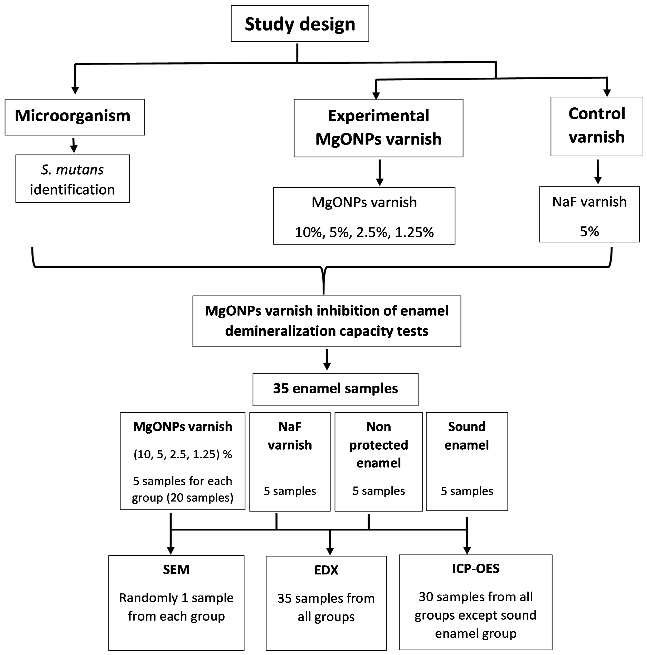
Approval was obtained from the scientific committee at the College of Dentistry/University of Sulaimani (approval no. 23/153).

2.2.2. Study Groups

The MgONPs varnish, NaF varnish, and control (non-protected enamel and sound enamel) groups were included in the study.
The MgONPs varnish was prepared in four concentrations (Table 1), and the commercial 5% NaF [29] and control groups were included in the corresponding groups according to the tests (Figure 1).

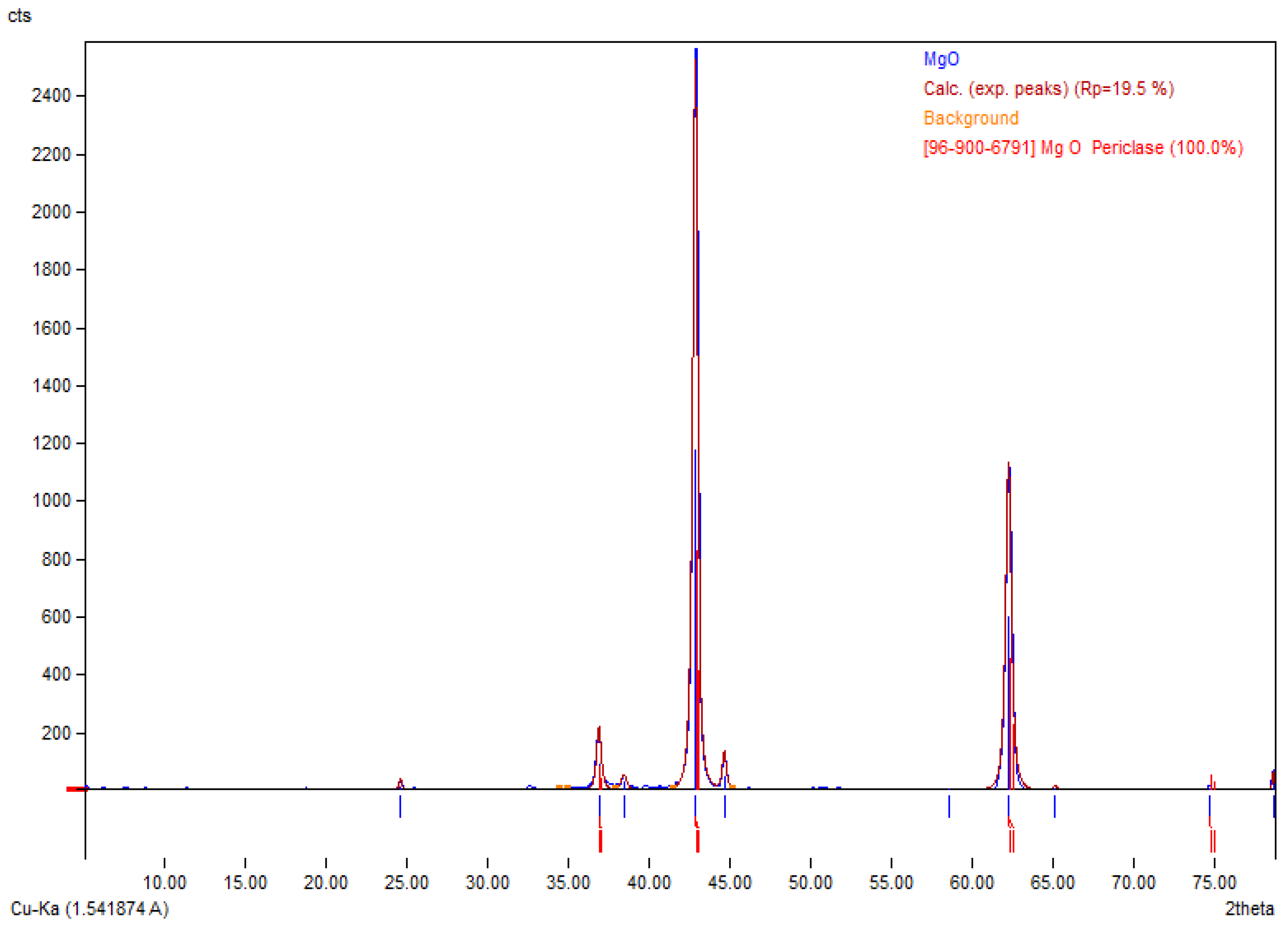
2.2.3. MgONPs Characterization

The MgONPs were characterized by X-ray diffraction (XRD) using a PAN analytical X’Pert PRO at 45 kV with a current of 40 mA (CuKα = 1.5406 Å). The scanning rate was 1 °/min in the 2θ range, from 5° to 75°.

2.2.4. Experimental MgONPs Varnish Preparation

Different concentrations of the MgONPs varnish were prepared with fully hydrogenated rosin in absolute ethanol with a two-fold dilution (Table 1). Absolute ethanol was applied as a dispensing solution with the MgONPs due to use of ethanol as the main solvent in production of commercial and experimental varnish by other researchers [30,31,32]. The prepared varnish with the MgONPs was mixed with an ultrasonic homogenizer for 10 min (Ti horn, 20 kHz at 65% efficiency, Bandelin electronic, Berlin, Germany). The sonication of the mixture decreased the agglomeration of the MgONPs, and we used the statistical acceptance time for sonication that was demonstrated in the Pradhan et al. study [33].

2.2.5. Microorganism

Streptococcus mutans Isolation and Identification

The Streptococcus mutans samples were obtained from four children with severe dental caries by collecting the total dental plaque with a sterile swab after obtaining verbal approval from the children and their parents. The swabs were placed in a 10 mL test tube with 10 mL PBS (phosphate buffer saline) and transported to the laboratory within 30 min. The plaque sample was vortexed for 30 s to release the bacteria into the PBS, and then 25 µL of the PBS solution was transported to the Tryptone Yeast Extract Cystine Bacitracin agar (TYCSB) and incubated for 48 h in 5% CO2 at 37 °C [34]. Consequently, a specific primer was designed according to the most appropriate method (as identified by other researchers [35], who considered that the Ohu et al. method produced the least false results [36]). The primer was designed to target the GtbB-F and GtfB-R on the glucosyl transferase gene of the S. mutans to amplify a 517-bp DNA fragment of the GtfB sequence of S. mutans. The final confirmation of S. mutans was achieved by DNA sequencing. When the DNA sequencing results were verified, glycerol stock cultures were created from single pure colonies of the indicated isolates and stored at −80 °C for long-term preservation.

2.2.6. Tooth Sections Preparation

The first permanent and sound upper premolars, which were indicated for extraction for orthodontic purposes, were obtained from patients in Sulaimani City, with verbal approval taken by the dentists. After collecting the teeth and the simple removal of the debris and blood, the teeth were preserved in deionized water that was changed weekly until the time of use. The teeth were polished by a slow-speed handpiece (geared angle handpiece- Foshan COXO Medical Instrument Co., Ltd., Foshan, China, model-CX235C1) with rubber polishing cups (TPC, INC 851 St. Lawson, CA, USA) and pumice (30-micron, PD, Bern, Switzerland) to remove residue and organic particles on the surfaces of the teeth. Next, the teeth were cleaned in an ultrasonic bath (Ultrasonic cleaner-VGT-900-, Beijing, China) with deionized water for 60 min to remove any attached pumice. All the teeth were examined by stereomicroscope to exclude teeth with demineralization, cracks, and dental caries. Each tooth was dissected mesiodistally by a slow-speed micromotor with a diamond disk and separated from the root. The enamel sections were sterilized by autoclaving for 15 min at 121 °C. An adhesive tape with a width and length of 5 mm was used to standardize the enamel area exposed to the experiment. Subsequently, the enamel and the dentin area around the adhesive tape were coated with acid-resistant nail varnish. Then, the enamel section was attached to a sterile spatula of a fecal specimen container with wax to facilitate the manipulation of the samples during the experiment.

2.2.7. MgONPs Varnish Application

The teeth sections were randomly divided into seven groups, with five samples per group. The first four groups were treated with 10%, 5%, 2.5%, and 1.25% MgONPs varnishes in sequence, and the fifth group was treated with 5% NaF varnish. These six groups, without any protection (non-protected enamel), were exposed to the S. mutans in the same way as the other groups. Meanwhile, the last group consisted of sound tooth sections that were tested without any protection or procedure (the sound enamel group).
The enamel sections were covered with 10 µL of the relevant varnish and left for one minute to dry. Then, the samples were incubated in 20 mL of deionized water for 4 h to provide time for ionic exchange [37]. The samples were then carefully placed in 20 mL of BHI broth with 1% sucrose taken from the 20% sucrose stock solution and with the 0.5 McFarland S. mutans [38]. The samples were incubated for 144 h in 5% CO2 at 37 °C in a static position, with a hole having been made in the container cover to enhance the homogenous atmosphere. Additionally, new fresh broth was prepared for each sample and the samples were transferred to new containers every 48 h. Each broth sample with the enamel sample was vortexed for 10 s before exchanging the container to expel the attached bacteria and demineralized surface of the enamel sample. The three containers with broth belonging to each sample were stored in the refrigerator until they were needed for analysis.

2.2.8. Dispersion of the MgONPs Varnish tests on the Enamel Surfaces

Scanning Electron Microscope (SEM)

All enamel samples were washed thoroughly for 30 s with deionized water to detach the unattached bacteria. Then, each enamel surface was cleaned with a sterile swab immersed in 99% ethanol to expose the enamel. One enamel sample was randomly selected to investigate the nanoparticle dispersion and S. mutans’ adherence to the enamel surface by SEM (SEM, Quanta, Ann Arbor, MI, USA).

Energy Dispersive X-ray Spectroscopy (EDX)

The chemical composition, including the proportions of calcium (Ca), phosphorous (P), magnesium (Mg), oxygen (O), and fluoride (F), of the superficial enamel samples were measured by EDX (Bruker Nano, Berlin, Germany) performed in SEM.

2.2.9. Enamel Demineralization Inhibition Capacity of the MgONPs Varnish Test

Inductively Coupled Plasma Optical Emission Spectroscopy (ICP-OES)

The previously stored 30 samples with 20 mL of broth were subjected to Ca measurement using an ICP-OES machine (WinLab32, PerkinElmer, Waltham, MA, USA) that quantitatively recorded the concentrations of Ca in mg/L [39,40]. The analytic wavelength of Ca was 315.887 nm, the estimated detection limit was 30 µg/L, the plasma gas flow rate was 15 L/min, and the pump injection volume was 1.50 mL/min. This test analyzed the dissolved Ca from the enamel surface in the BHI broth containing S. mutans in three sequences of time: 48 h, 96 h, and 144 h, after coating the surface of the enamel with the corresponding varnish.

2.3. Statistical Analysis

For the quantitative values, normality tests were applied, and the data were non-normally distributed. A Kruskal–Wallis H-test was utilized to compare the medians of the experimental groups. Additionally, a Friedman test supported by a post hoc analysis with Wilcoxon signed-rank tests was conducted with a Bonferroni correction to interpret the data that had consequences.

3. Results

3.1. MgONPs Characterization

The X-ray diffraction test (XRD) result is shown in Figure 2. The MgONPs peak was identical to the referenced peak indexed in Match version 3.15 build 252.

3.2. Microorganism

Streptococcus mutans Isolation and Identification

According to the National Library of Medicine web BLAST and the accession number (OQ656871) from Genbank, the DNA sequences of S. mutans confirmed the successful isolation and identification of S. mutans from the dental plaque samples.

3.3. Dispersion of MgONPs Varnish on the Enamel Surface

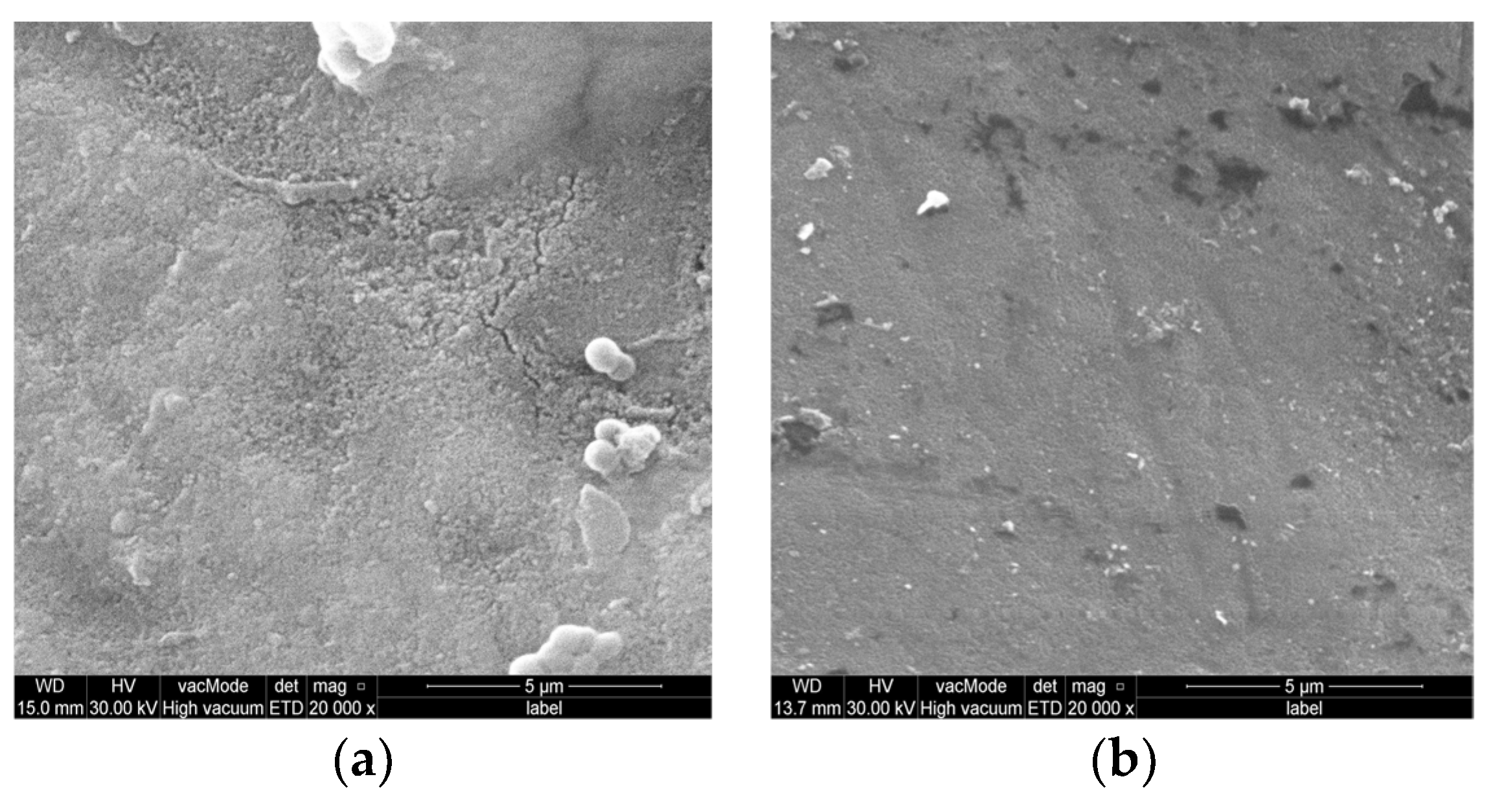
3.3.1. Scanning Electron Microscope (SEM)

The SEM analysis revealed that the surface of the sound enamel had remained nearly the same and was without S. mutans attachment as it had been sterilized (Figure 3A). At the same time, the non-protected enamel surface revealed localized distributed microporosity with S. mutans colonization (Figure 3B-1). The mean size of S. mutans that had attached to the enamel was approximately 79 nm (±std = 8), with active division detected. In addition, an area of demineralization was observed in the non-protected enamel following the repelling of the S. mutans (Figure 3B-2). This was despite the attachment of many other single S. mutans, which was not obvious in the other groups. The enamel surface protected by the NaF varnish exhibited a homogenous (Figure 3C-1) smooth surface, with a strip of S. mutans interrupted by the varnish smeared on the tooth enamel (Figure 3C-2).
The SEM showed that the MgONPs were dispersed over the surface and could not be isolated from the enamel structure in any of the experimental varnishes except for the 10% MgONPs varnish. Consequently, 1.25%, the MgONPs varnish exhibited a homogeneous surface but with S. mutans attachments seen over the enamel, largely with single S. mutans (Figure 4A). The enamel surface appeared more homogenous in the 2.5% and 5% MgONPs, with fewer S. mutans attachments (Figure 4B,C). The SEM identified that the 10% MgONPs varnish had agglomerated on the enamel surface. The last concentration showed coalescence drops with the rosin content, and the underlying enamel was not seen in the magnified field (Figure 4D).

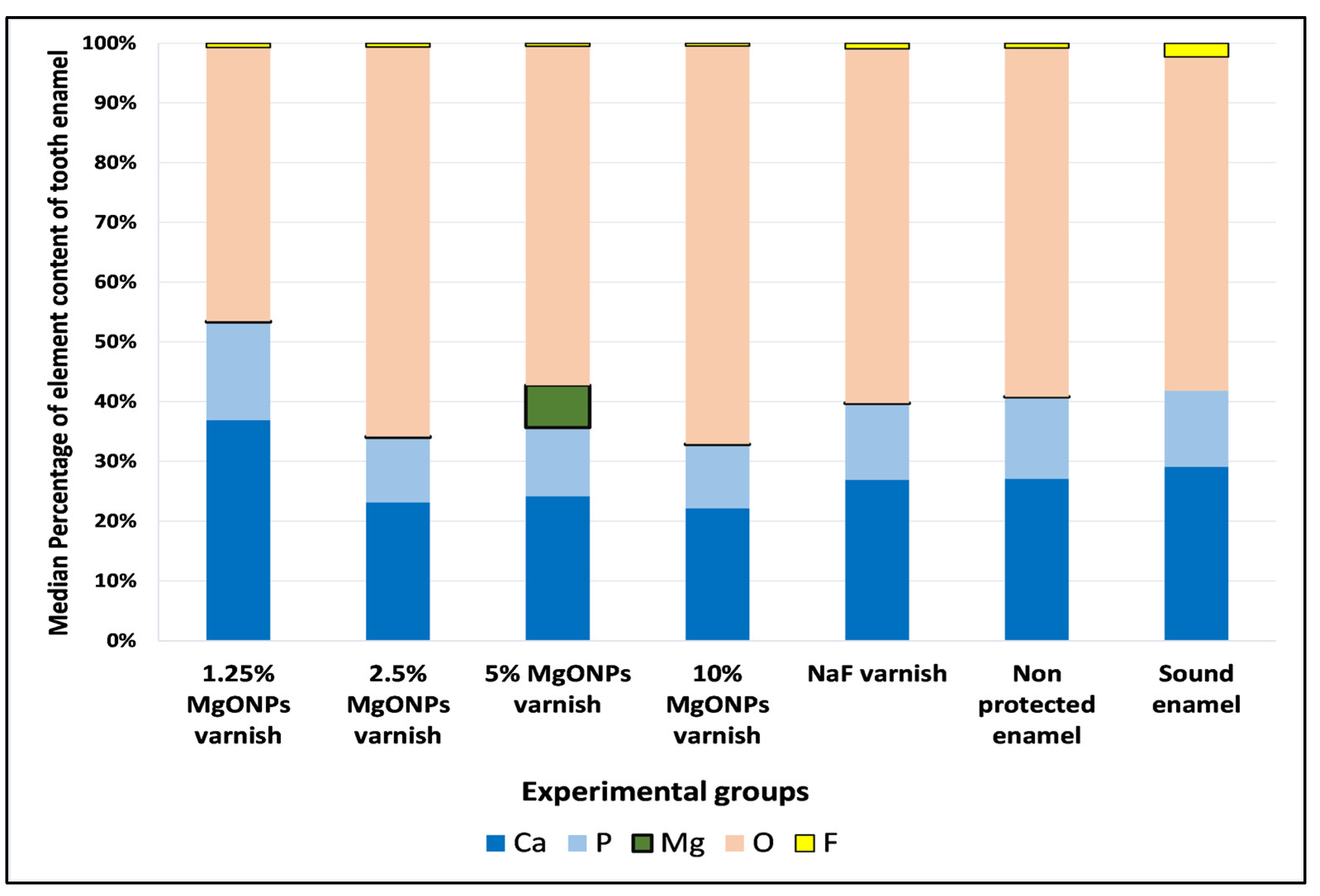
3.3.2. Energy Dispersive X-ray Spectrophotometry (EDX)

The values measured by the EDX machine were non-normally distributed; therefore, a Kruskal–Wallis H-test was applied to compare the medians of the experimental groups. Ca, P, Mg, O, and F were the elements analyzed by EDX on the human enamel surfaces of all groups, with five samples per group (Figure 5). Ca, P and O were revealed to be the main elements in the enamel crystals. In addition, a small amount of F was found in the composition of all groups, but more was found in the sound enamel. The EDX measurements showed the compositional Mg element of the enamel surface in the four experimental MgONPs varnish groups were seen in a higher amount than those groups not treated by the MgONPs. Despite the differences in the medians, only the analysis result for the Mg element in the 5% MgONPs varnish was statistically significant.
Additionally, EDX spectrum photos showed the low compositional distribution of Mg elements in the sound enamel, non-protected enamel, and NaF varnish groups (Figure 6A–C). While the MgONPs varnish groups exhibited homogenous and higher Mg element deposition on the enamel surface, with the more prominent Mg element observed in the 5% MgONPs varnish group, followed by 2.5% MgONPs varnish (Figure 6E,F).

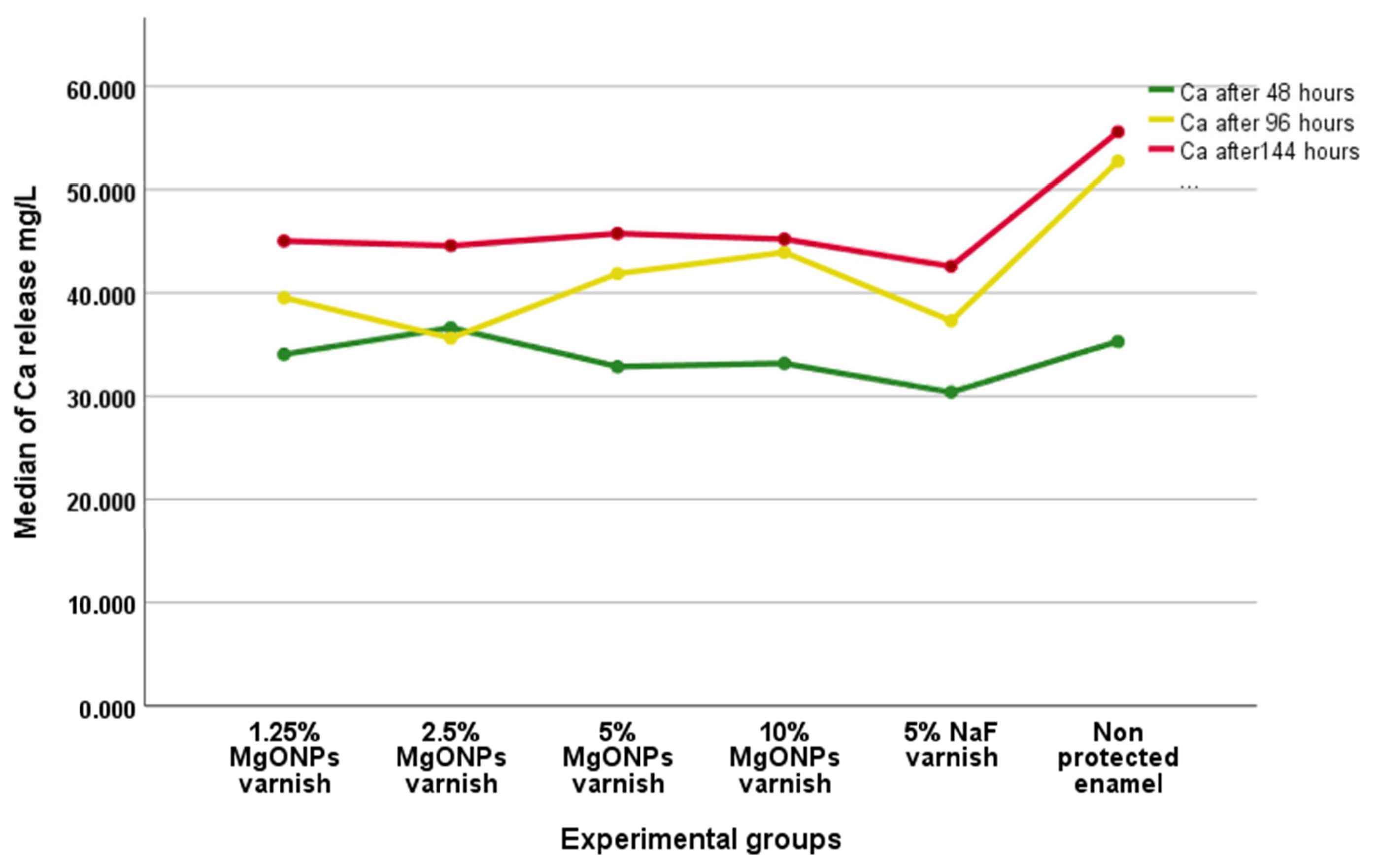
3.4. Enamel Demineralization Inhibition Capacity of the MgONPs Varnish

Inductively Coupled Plasma Optical Emission Spectroscopy (ICP-OES)

A Friedman test was applied to the non-normally distributed data from three respective series in this test. There was a statistically significant difference in the Ca release depending on the time of the evaluation (χ2(2) = 39.200, p = 0.000). A post hoc analysis with Wilcoxon signed-rank tests was conducted with a Bonferroni correction, resulting in a significance level set at p < 0.017. The median (IQR) perceived Ca releases after 48, 96, and 144 h were 33.21 (31.825 to 35.1325), 40.51 (35.975 to 44.8025), and 45.1945 (42.015 to 49.89275), respectively. There was a significant difference in the Ca release between 48 and 96 h (Z = −4.186, p = 0.000) and 96 and 144 h (Z = −3.445, p = 0.001). In addition, a statistically significant increase in Ca release was observed within 48 to 144 h of exposure of the enamel samples to S. mutans (Z = −4.721, p = 0.000). When comparing the Ca release median of each group, it was found that all groups with enamel varnish had inhibited Ca release (Figure 7). At the same time, the 2.5% MgONPs varnish had a Ca release inhibition capacity approximate to that of the NaF varnish, which was discovered when the summation of the three median records of each group was evaluated (Figure 8).

4. Discussion

Microdamage, demineralization, and infection all impair mineralized tissues. Osseous tissues rebuild (turn over) to preserve structural integrity, whereas severely loaded dentition does not remodel (turn over), putting it in danger of eventual collapse [41]. Streptococcus mutans is a well-known cariogenic bacteria that demineralizes and destroys enamel [12,13,14,15,16]. Based on this, S. mutans was isolated and identified successfully from high caries individuals. Indeed, the unavailability of effective coating materials with antibacterial and remineralizing capacity has motivated the investigation of different elements being combined with modern technology, such as nanomaterials. Recently, magnesium oxide nanoparticles (MgONPs) have attracted the interest of many researchers because of their antimicrobial activity [25,42,43], in addition to their biocompatibility [22,23]. The use of fluoride varnish has had a long history in the protection of the enamel layer [17]; however, its antibacterial capacity was not found to be promising [18,44,45]. Unfortunately, fluoride overdose toxicity from dental products remains a local and systemic problem that must be considered [46,47], especially in high caries-risk individuals.
The reason for preparing MgONPs in the different concentrations was to determine the most effective MgONPs varnish concentration suitable for this study’s aims since nanoparticle capacity is affected by the dose and contact time, as determined by a previous investigation [48]. Therefore, rosin was added to the MgONPs to increase the viscosity and form a dental varnish that could be applied to the enamel surface within a few minutes to mimic the clinical use of dental varnish.
The MgONPs varnish dispersion to the enamel was examined by SEM and EDX. In addition, Ca, P, O, and F were evaluated with the Mg dispersion to analyze the variations in the enamel constitution samples [3]. Moreover, the noticeably increased F content of the sound enamel group could be associated with the exposure of the enamel to the daily care routine practiced by the volunteers. In addition, the high O element in the enamel revealed the presence of O with other elements, such as P (Figure 5) [49]. Nonetheless, the analysis was statistically significant only for the 5% MgONPs varnish (p < 0.05). The cause of this difference in the incorporation of the MgONPs to the enamel could be open to speculation: first, it may have been due to the concentration of the MgONPs and the limited time of exposure; second, it may have been because the high concentration of the 10% MgONPs increased the probability of agglomeration on the enamel surface; third, it could have been due to differences in the enamel compositions of the analyzed teeth; and fourth, it may have been because the test’s principle was to protect sound enamel and not remineralize it.
Furthermore, the demineralization inhibition capacities of the MgONPs varnishes tested by ICP-OES differed significantly across all the experimental varnish and control groups (non-protected enamel), with the maximum inhibition of Ca release seen in the 2.5% and 1.25% MgONPs varnishes, which was comparable to the NaF varnish and much lower than the control group.
Research related to novel MgONPs varnish preparations and the topical application of MgONPs on a tooth’s surface was found to be lacking. Meanwhile, other researchers have used Mg in different compositions to illustrate the incorporation of Mg into the surface of enamel. Abdulla et al. applied an additional Mg-containing solution to the enamel of an extracted human tooth, and they concluded that Mg could reprecipitate the enamel and enhance the enamel’s properties [50]. The same finding was produced by Kis et al., who applied an MgCl2 solution for the improvement of the mechanical properties of the enamel [32]. Other investigators have reported that topical protection with toothpaste containing Mg provides higher protection than fluoride varnish and toothpaste in the enamel protection of bovine teeth [51], though they did not illustrate the dispersion of the Mg element to the enamel. However, their results confirmed the impact of Mg on enamel protection.
This research analyzed, for the first time, the topical influence of an MgONPs varnish on the surface of enamel. SEM, EDX, and ICP-OES were used to investigate, in vitro, the dispersion and inhibition of the demineralization capability of the MgONPs after exposing them to S. mutans in various time sequences. As with any other in vitro study, the exact oral environment could not be rebuilt. Therefore, further studies are necessary to investigate the topical application of MgONPs against intra-oral bacteria and to clinically employ MgONPs varnish in preventive dentistry procedures (using both in vitro and in vivo studies). This study’s findings suggest a viable strategy for producing a biocompatible dental varnish with superior qualities to those currently available.

5. Conclusions

This experiment indicated that MgONPs varnish disperses and inhibits enamel demineralization against S. mutans, even at low concentrations. The SEM and EDX tests illustrated the deposition of 5% MgONPs varnish by increasing the Mg element’s binding to the enamel. The variations in the medians of the Ca, P, O, and F across the enamel sample groups were statistically insignificant. In contrast, the inhibition of Ca release capacity, according to the three time sequences of 144 h for all groups, was significant, with greater enamel protection (inhibited Ca release) associated with the 2.5% MgONPs varnish, which was comparable to the enamel protection impact of the NaF varnish, followed by the 1.25% MgONPs varnish. Additional in vivo and in vitro studies are recommended to examine the protective qualities of MgONPs for hard dental tissues.

Author Contributions

Conceptualization, F.A.K. and A.J.N.; methodology, S.J.H.; software, S.J.H.; validation, F.A.K., S.J.H. and A.J.N.; formal analysis, S.J.H.; investigation, S.J.H.; resources, F.A.K. and A.J.N.; data curation, S.J.H.; writing—original draft preparation, S.J.H.; writing—review and editing, S.J.H. and A.J.N.; visualization, S.J.H.; supervision, F.A.K. and A.J.N.; project administration, F.A.K. and A.J.N. All authors have read and agreed to the published version of the manuscript.

Funding

This research received no external funding.

Institutional Review Board Statement

This research was approved by the Ethics Committee of the College of Dentsitry—University of Sulaimani application No. 153/23 issued on 29/3/2023.

Informed Consent Statement

Verbal consent were obtained from orthodontic patients for collecting extracted premolar teeth.

Data Availability Statement

The data that support the findings of this study are available from the corresponding author upon reasonable request.

Acknowledgments

The authors appreciate the cooperation of their colleagues and the staff of the College of Dentistry at the University of Sulaimani, and they extend their deepest gratitude to Soran University and the Kurdistan Institute for Strategic Study and Scientific Research.

Conflicts of Interest

The authors declare no conflict of interest.

References

  1. Lacruz, R.S.; Habelitz, S.; Wright, J.T.; Paine, M.L. Dental enamel formation and implications for oral health and disease. Physiol. Rev. 2017, 97, 939–993. [Google Scholar] [CrossRef] [PubMed]
  2. Thesleff, I. Developmental Biology and Building a Tooth. Quintessence Int. 2003, 34, 613–620. [Google Scholar] [PubMed]
  3. Eimar, H.; Ghadimi, E.; Marelli, B.; Vali, H.; Nazhat, S.N.; Amin, W.M.; Torres, J.; Ciobanu, O.; Albuquerque Junior, R.F.; Tamimi, F. Regulation of Enamel Hardness by Its Crystallographic Dimensions. Acta Biomater. 2012, 8, 3400–3410. [Google Scholar] [CrossRef] [PubMed]
  4. Ghadimi, E.; Eimar, H.; Song, J.; Marelli, B.; Ciobanu, O.; Abdallah, M.-N.; Stähli, C.; Nazhat, S.N.; Vali, H.; Tamimi, F. Regulated Fracture in Tooth Enamel: A Nanotechnological Strategy from Nature. J. Biomech. 2014, 47, 2444–2451. [Google Scholar] [CrossRef] [PubMed]
  5. Gil-Bona, A.; Bidlack, F.B. Tooth Enamel and Its Dynamic Protein Matrix. Int. J. Mol. Sci. 2020, 21, 4458. [Google Scholar] [CrossRef]
  6. Bernabe, E.; Marcenes, W.; Hernandez, C.R.; Bailey, J.; Abreu, L.G.; Alipour, V.; Amini, S.; Arabloo, J.; Arefi, Z.; Arora, A.; et al. Global, Regional, and National Levels and Trends in Burden of Oral Conditions from 1990 to 2017: A Systematic Analysis for the Global Burden of Disease 2017 Study. J. Dent. Res. 2020, 99, 362–373. [Google Scholar] [CrossRef]
  7. Wen, P.Y.F.; Chen, M.X.; Zhong, Y.J.; Dong, Q.Q.; Wong, H.M. Global Burden and Inequality of Dental Caries, 1990 to 2019. J. Dent. Res. 2022, 101, 392–399. [Google Scholar] [CrossRef]
  8. Global Burden of Disease Collaborative Network Global Burden of Disease Study 2019. Available online: https://ghdx.healthdata.org/gbd-2019 (accessed on 20 May 2022).
  9. Pitts, N.B.; Zero, D.T.; Marsh, P.D.; Ekstrand, K.; Weintraub, J.A.; Ramos-Gomez, F.; Tagami, J.; Twetman, S.; Tsakos, G.; Ismail, A. Dental Caries. Nat. Rev. Dis. Prim. 2017, 3, 17030. [Google Scholar] [CrossRef]
  10. Clarke, J.K. On the Bacterial Factor in the Ætiology of Dental Caries. Br. J. Exp. Pathol. 1924, 5, 141–147. [Google Scholar]
  11. Loesche, W.J. Role of Streptococcus Mutans in Human Dental Decay. Microbiol. Rev. 1986, 50, 353–380. [Google Scholar] [CrossRef]
  12. Li, Z.-R.; Sun, J.; Du, Y.; Pan, A.; Zeng, L.; Maboudian, R.; Burne, R.A.; Qian, P.-Y.; Zhang, W. Mutanofactin Promotes Adhesion and Biofilm Formation of Cariogenic Streptococcus Mutans. Nat. Chem. Biol. 2021, 17, 576–584. [Google Scholar] [CrossRef] [PubMed]
  13. Matsumoto-Nakano, M. Role of Streptococcus Mutans Surface Proteins for Biofilm Formation. Jpn. Dent. Sci. Rev. 2018, 54, 22–29. [Google Scholar] [CrossRef] [PubMed]
  14. Tang, X.; Kudo, Y.; Baker, J.L.; LaBonte, S.; Jordan, P.A.; McKinnie, S.M.K.; Guo, J.; Huan, T.; Moore, B.S.; Edlund, A. Cariogenic Streptococcus Mutans Produces Tetramic Acid Strain-Specific Antibiotics That Impair Commensal Colonization. ACS Infect. Dis. 2020, 6, 563–571. [Google Scholar] [CrossRef]
  15. Wang, Y.; Lee, S.M.; Dykes, G.A. Potential Mechanisms for the Effects of Tea Extracts on the Attachment, Biofilm Formation and Cell Size of Streptococcus Mutans. Biofouling 2013, 29, 307–318. [Google Scholar] [CrossRef] [PubMed]
  16. Zhang, Q.; Ma, Q.; Wang, Y.; Wu, H.; Zou, J. Molecular Mechanisms of Inhibiting Glucosyltransferases for Biofilm Formation in Streptococcus Mutans. Int. J. Oral Sci. 2021, 13, 30. [Google Scholar] [CrossRef]
  17. Bawden, J.W. Fluoride Varnish: A Useful New Tool for Public Health Dentistry. J. Public Health Dent. 1998, 58, 266–269. [Google Scholar] [CrossRef]
  18. Kanduti, D.; Sterbenk, P.; Artnik, B. Fluoride: A Review of Use and Effects on Health. Mater. Socio-Med. 2016, 28, 133–137. [Google Scholar] [CrossRef]
  19. Corrie, S.R.; Thurecht, K.J. Nano-Bio Interactions: Guiding the Development of Nanoparticle Therapeutics, Diagnostics, and Imaging Agents. Pharm. Res. 2016, 33, 2311–2313. [Google Scholar] [CrossRef]
  20. Deyhle, H.; Bunk, O.; Buser, S.; Krastl, G.; Zitzmann, N.U.; Ilgenstein, B.; Beckmann, F.; Pfeiffer, F.; Weiger, R.; Müller, B. Bio-Inspired Dental Fillings. Biomim. Bioinspiration 2009, 7401, 74010E. [Google Scholar] [CrossRef]
  21. Jeevanandam, J.; Barhoum, A.; Chan, Y.S.; Dufresne, A.; Danquah, M.K. Review on Nanoparticles and Nanostructured Materials: History, Sources, Toxicity and Regulations. Beilstein J. Nanotechnol. 2018, 9, 1050–1074. [Google Scholar] [CrossRef]
  22. Sezer, N.; Evis, Z.; Kayhan, S.M.; Tahmasebifar, A.; Koç, M. Review of Magnesium-Based Biomaterials and Their Applications. J. Magnes. Alloys 2018, 6, 23–43. [Google Scholar] [CrossRef]
  23. Zhou, H.; Liang, B.; Jiang, H.; Deng, Z.; Yu, K. Magnesium-Based Biomaterials as Emerging Agents for Bone Repair and Regeneration: From Mechanism to Application. J. Magnes. Alloys 2021, 9, 779–804. [Google Scholar] [CrossRef]
  24. Bhushan, J.; Maini, C. Nanoparticles: Apromising Novel Adjunct for Dentistry. Indian J. Dent. Sci. 2019, 11, 167–173. [Google Scholar] [CrossRef]
  25. Yamamoto, O.; Ohira, T.; Alvarez, K.; Fukuda, M. Antibacterial Characteristics of CaCO3-MgO Composites. Mater. Sci. Eng. B Solid-State Mater. Adv. Technol. 2010, 173, 208–212. [Google Scholar] [CrossRef]
  26. Nguyen, N.-Y.T.; Grelling, N.; Wetteland, C.L.; Rosario, R.; Liu, H. Antimicrobial Activities and Mechanisms of Magnesium Oxide Nanoparticles (NMgO) against Pathogenic Bacteria, Yeasts, and Biofilms. Sci. Rep. 2018, 8, 16260. [Google Scholar] [CrossRef] [PubMed]
  27. Arass, J.; Fadil, A. The Effect of Magnesium Oxide Nanoparticles on the Antibacterial and Antibiofilm Properties of Glass-Ionomer Cement. Heliyon 2019, 5, e02568. [Google Scholar] [CrossRef]
  28. Maguire, M.E.; Cowan, J.A. Magnesium Chemistry and Biochemistry. Biometals 2002, 15, 203–210. [Google Scholar] [CrossRef]
  29. Medicom Safety Data Sheet Duraflour Varnish. Revision No. 1, Issued 23 November 2017. Available online: https://medicom.com/ (accessed on 20 May 2023).
  30. Shahmoradi, M.; Hunter, N.; Swain, M. Efficacy of Fluoride Varnishes with Added Calcium Phosphate in the Protection of the Structural and Mechanical Properties of Enamel. BioMed Res. Int. 2017, 2017, 7834905. [Google Scholar] [CrossRef]
  31. Haghgoo, R.; Saderi, H.; Eskandari, M.; Haghshenas, H.; Rezvani, M. Evaluation of the Antimicrobial Effect of Conventional and Nanosilver-Containing Varnishes on Oral Streptococci. J. Dent. 2014, 15, 57–62. [Google Scholar]
  32. Kis, V.K.; Sulyok, A.; Hegedűs, M.; Kovács, I.; Rózsa, N.; Kovács, Z. Magnesium Incorporation into Primary Dental Enamel and Its Effect on Mechanical Properties. Acta Biomater. 2021, 120, 104–115. [Google Scholar] [CrossRef]
  33. Pradhan, S.; Hedberg, J.; Blomberg, E.; Wold, S.; Odnevall Wallinder, I. Effect of Sonication on Particle Dispersion, Administered Dose and Metal Release of Non-Functionalized, Non-Inert Metal Nanoparticles. J. Nanopart. Res. 2016, 18, 285. [Google Scholar] [CrossRef] [PubMed]
  34. Wan, A.; Seow, W.; Walsh, L.; Bird, P. Comparison of Five Selective Media for the Growth and Enumeration of Streptococcus Mutans. Aust. Dent. J. 2002, 47, 21–26. [Google Scholar] [CrossRef] [PubMed]
  35. Villhauer, A.L.; Lynch, D.J.; Drake, D.R. Improved Method for Rapid and Accurate Isolation and Identification of Streptococcus Mutans and Streptococcus Sobrinus from Human Plaque Samples. J. Microbiol. Methods 2017, 139, 205–209. [Google Scholar] [CrossRef] [PubMed]
  36. Oho, T.; Yamashita, Y.; Shimazaki, Y.; Kushiyama, M.; Koga, T. Simple and Rapid Detection of Streptococcus Mutans and Streptococcus Sobrinus in Human Saliva by Polymerase Chain Reaction. Oral Microbiol. Immunol. 2000, 15, 258–262. [Google Scholar] [CrossRef] [PubMed]
  37. Van Loveren, C.; Buijs, J.F.; Buijs, M.J.; ten Cate, J.M. Protection of Bovine Enamel and Dentine by Chlorhexidine and Fluoride Varnishes in a Bacterial Demineralization Model. Caries Res. 1996, 30, 45–51. [Google Scholar] [CrossRef]
  38. Wassel, M.O.; Khattab, M.A. Antibacterial Activity against Streptococcus Mutans and Inhibition of Bacterial Induced Enamel Demineralization of Propolis, Miswak, and Chitosan Nanoparticles Based Dental Varnishes. J. Adv. Res. 2017, 8, 387–392. [Google Scholar] [CrossRef]
  39. Vallapragada, V.V.; Inti, G.; Ramulu, J.S. A Validated Inductively Coupled Plasma-Optical Emission Spectrometry (ICP-OES) Method to Estimate Free Calcium and Phosphorus in In Vitro Phosphate Binding Study of Eliphos Tablets. Am. J. Anal. Chem. 2011, 2, 718–725. [Google Scholar] [CrossRef]
  40. Hou, X.; Amais, R.S.; Jones, B.T.; Donati, G.L. Inductively Coupled Plasma Optical Emission Spectrometry. In Encyclopedia of Analytical Chemistry; John Wiley & Sons, Ltd.: Hoboken, NJ, USA, 2021; pp. 1–29. ISBN 978-0-470-02731-8. [Google Scholar]
  41. Roberts, W.E.; Mangum, J.E.; Schneider, P.M. Pathophysiology of Demineralization, Part II: Enamel White Spots, Cavitated Caries, and Bone Infection. Curr. Osteoporos. Rep. 2022, 20, 106–119. [Google Scholar] [CrossRef]
  42. Vidic, J.; Stankic, S.; Haque, F.; Ciric, D.; Le Goffic, R.; Vidy, A.; Jupille, J.; Delmas, B. Selective Antibacterial Effects of Mixed ZnMgO Nanoparticles. J. Nanoparticle Res. 2013, 15, 1595. [Google Scholar] [CrossRef]
  43. Rodríguez-Hernández, A.-P.; Vega-Jiménez, A.L.; Vázquez-Olmos, A.R.; Ortega-Maldonado, M.; Ximenez-Fyvie, L.-A. Antibacterial Properties In Vitro of Magnesium Oxide Nanoparticles for Dental Applications. Nanomaterials 2023, 13, 502. [Google Scholar] [CrossRef]
  44. Buzalaf, M.A.R.; Pessan, J.P.; Honório, H.M.; ten Cate, J.M. Mechanisms of Action of Fluoride for Caries Control. Monogr Oral Sci. 2011, 22, 97–114. [Google Scholar] [PubMed]
  45. Gupta, A.; Sharda, S.; Nishant; Shafiq, N.; Kumar, A.; Goyal, A. Topical Fluoride-Antibacterial Agent Combined Therapy versus Topical Fluoride Monotherapy in Preventing Dental Caries: A Systematic Review and Meta-Analysis. Eur. Arch. Paediatr. Dent. 2020, 21, 629–646. [Google Scholar] [CrossRef] [PubMed]
  46. Martínez-Mier, E.A. Fluoride: Its Metabolism, Toxicity, and Role in Dental Health. J. Evid.-Based Complement. Altern. Med. 2012, 17, 28–32. [Google Scholar] [CrossRef]
  47. Ullah, R.; Zafar, M.S.; Shahani, N. Potential Fluoride Toxicity from Oral Medicaments: A Review. Iran J. Basic Med. Sci. 2017, 20, 841–848. [Google Scholar] [CrossRef] [PubMed]
  48. Haghshenas, L.; Amini, A.; Bashir Bahati, A.; Rahimi, G. In Vitro Antibacterial Biofilm Effect of Magnesium Oxide Nanoparticles on Streptococcus Mutans. Micro Nano Biomed. 2016, 1, 21–27. [Google Scholar] [CrossRef]
  49. Trayler, R.B.; Kohn, M.J. Tooth Enamel Maturation Reequilibrates Oxygen Isotope Compositions and Supports Simple Sampling Methods. Geochim. Cosmochim. Acta 2017, 198, 32–47. [Google Scholar] [CrossRef]
  50. Abdallah, M.-N.; Eimar, H.; Bassett, D.C.; Schnabel, M.; Ciobanu, O.; Nelea, V.; McKee, M.D.; Cerruti, M.; Tamimi, F. Diagenesis-Inspired Reaction of Magnesium Ions with Surface Enamel Mineral Modifies Properties of Human Teeth. Acta Biomater. 2016, 37, 174–183. [Google Scholar] [CrossRef] [PubMed]
  51. Lale, S.; Solak, H.; Hınçal, E.; Vahdettin, L. In Vitro Comparison of Fluoride, Magnesium, and Calcium Phosphate Materials on Prevention of White Spot Lesions around Orthodontic Brackets. BioMed Res. Int. 2020, 2020, 1989817. [Google Scholar] [CrossRef]
Figure 1. Study design and groups with tests.
Coatings 13 01018 g001
Figure 2. XRD test for the MgONPs powder.
Coatings 13 01018 g002
Figure 3. SEM of enamel groups: (A) sound enamel; (B-1) non-protected enamel with S. mutans strip (black arrows); (B-2) demineralization at the position of S. mutans repelling (yellow arrows); (C-1) NaF varnish on enamel (C-2) same sample with S. mutans strip interrupted with NaF varnish (black arrows).
Coatings 13 01018 g003
Figure 4. SEM of enamel groups with MgONPs varnish: (a) 1.25%; (b) 2.5%; (c) 5%; (d) 10%.
Coatings 13 01018 g004aCoatings 13 01018 g004b
Figure 5. The median of enamel elements (Ca, P, Mg, O, and F) of experimental groups examined by EDX.
Coatings 13 01018 g005
Figure 6. EDX analysis of enamel Mg element of experimental groups: (A) sound enamel; (B) non-protected enamel; (C) NaF varnish; (D) 1.25% MgONPs varnish; (E) 2.5% MgONPs varnish; (F) 5% MgONPs varnish; (G) 10% MgONPs varnish.
Coatings 13 01018 g006
Figure 7. ICP-OES test of the experimental groups analyzing the median Ca release from enamel against S. mutans at 48 h, 96 h, and 144 h.
Coatings 13 01018 g007
Figure 8. ICP-OES test of the experimental groups: the summation of the median of Ca release from enamel against S. mutans after 144 h.
Coatings 13 01018 g008
Table 1. Experimental varnish compositions.
Varnish Component
ConcentrationFluoride (g)Nanoparticle (g)Hydrogenated Rosin (g)Ethanol (mL)
MgONPs varnish10%-101080
5%-51085
2.5%-2.51087.5
1.25%-1.251088.75
NaF varnish (Duraflour) [29]5%1–10-50–7010–30
Disclaimer/Publisher’s Note: The statements, opinions and data contained in all publications are solely those of the individual author(s) and contributor(s) and not of MDPI and/or the editor(s). MDPI and/or the editor(s) disclaim responsibility for any injury to people or property resulting from any ideas, methods, instructions or products referred to in the content.

