Article

Enhancement of Antibacterial Silk Face Covering with the Biosynthesis of Silver Nanoparticles from Garcinia mangostana Linn. Peel and Andrographis paniculata Extract and a Bacterial Cellulose Filter

by
Phannaphat Phromphen
1,
Pithalai Phoophat
1,
Udomlak Sukatta
2,
Prapassorn Rugthaworn
2,
Nattadon Rungruangkitkrai
1,
Pawarin Tuntariyanond
1,
Nawarat Chartvivatpornchai
1,
Preeyanuch Sichola
2,
Jirachaya Boonyarit
2,
Thanyachol Apipatpapha
2 and
Rungsima Chollakup
2,*
1
Department of Textile Science, Faculty of Agro-Industry, Kasetsart University, Bangkok 10900, Thailand
2
Kasetsart Agricultural and Agro-Industrial Product Improvement Institute, Kasetsart University, Bangkok 10900, Thailand
*
Author to whom correspondence should be addressed.
Coatings 2024, 14(4), 379; https://doi.org/10.3390/coatings14040379
Submission received: 25 January 2024 / Revised: 17 March 2024 / Accepted: 19 March 2024 / Published: 24 March 2024
(This article belongs to the Section Bioactive Coatings and Biointerfaces)

Abstract

:
Since the late 2010-s and early 2020s, people around the world have not only encountered the pandemic crisis, but also in some places, they have had to deal with serious levels of air pollution. Personal protective equipment is essential to protect from microorganisms or fine particulate matter. Consequently, this study aimed to develop a silk face covering that could meet the international requirements with the addition of having an antibacterial property. The developed silk face covering consisted of three layers; the outer layer was water-repellent mulberry silk, the inner layer was oil-repellent eri silk, while the middle layer was cotton fabric coated with biosynthesized silver nanoparticles from mangosteen peels mixed with Andrographis paniculata extract. A biodegradable bacterial cellulose filter made of nata de coco waste was also prepared to improve filtration efficiency. It was found that the silver nanoparticles extracted from mangosteen peel and Andrographis paniculata inhibited S. aureus and E. coli by more than 99.9%, even after 20 washing cycles. The performance properties of the silk barrier face covering met Level I of the ASTM F3502-2021 standard, as well as being comfortable for public use.

1. Introduction

The latest standard specification regarding face coverings was released to reduce the number of expelled droplets and aerosols from the wearer’s nose and mouth into the air and to potentially offer a degree of particulate filtration to reduce the amount of inhaled particulate matter by the wearer. Even though the requirement of the new standard only focused on sub-micron particulate filtration efficiency and airflow resistance, most of the fabric masks available recently have underperformed in these requirements [1]. Moreover, cloth masks could not be used as barrier face coverings, nor could they be used for respiratory source control in the healthcare workplace [2]. Recently, there have been studies about novel cloth face masks for public use which could not be a source of protection but they aim to be used to reduce the spread of infection and could be washed, reused, and worn for a longer time [3,4]. Another study showed that a handmade fabric mask could not filter aerosol sizes between 30 and 600 nm, being only effective at capturing large respiratory droplets [5]. Subsequently, multi-layered structures of woven fabric were introduced to improve the filtration efficiency of materials [6,7]. Four layers of silk fabric could protect against particulate penetration ranging from 10 nm to 6 µm [8]. However, the enhanced performance and additional functionality of face covering materials has gained much attention regarding antimicrobial [9,10] and water-repellent properties [11,12]. Recent studies proposed several alternative methods for producing functional face coverings such as zinc-ion embedded onto 100% nylon masks [13], polypropylene masks coated with copper salt and organic antimicrobial agents [14,15], and cotton fabric coated with a mixture of chitosan, eugenol, and oleic acid [16].
The biosynthesis of silver nanoparticles is an alternative method to enhance the antibacterial properties of textile because it inhibits microorganisms at lower concentrations than other heavy metals and shows great bactericidal potential against various Gram-positive and Gram-negative bacteria [17]. Furthermore, it is cost-effective, environmentally friendly, and easier to produce, with it being possible to synthesize a large amount of nanoparticles of any size and shape, while in the synthesis process, the reducing agent can come from a natural source, such as flavonoids, alkaloids, terpenoids, or polyphenols [18,19]. The reducing agent acts on the electrons in the metal ions to transform them into metal nanoparticles. Various types of natural reducing agents have been reported, such as basil [20], banana peel [21], sweet flag root [22], mangosteen peel [4], Moringa leaves, and green chiretta [23]. Some previous research also used the biosynthesis of silver nanoparticles as coating on textile materials for various applications [24,25].
Andrographis paniculata or green chiretta is an annual plant whose entire above-ground parts (leaves, flowers, and stems) can be used as a medicinal plant to treat common cold and fever [26]. The active ingredients include andrographolide, neoandrographolide, isoandrographolide, and 14-deoxyandrographolide [27]. The active ingredients in Andrographis paniculata consist of various types of secondary substances, such as an isoandrographolide that inhibits bacteria and fungi [28] and exerts antiviral activity [29]. In addition, the andrographolide could inhibit bacteria [30] and biofilm formation [31] and had an anti-influenza property [32]. Also, Andrographis paniculata was used with other plants and biosynthesized silver nanoparticles to achieve synergistic antimicrobial activity [33,34]. However, there is limited research on applying the silver nanoparticles derived from the Garcinia mangostana Linn. synergized with Andrographis paniculata extract onto textile materials to achieve antibacterial property.
Bacterial cellulose is an exopolysaccharide biopolymer produced from the fermentation process by some Gram-negative bacteria under specific nutrient conditions, including sources of carbon and nitrogen. A famous dessert derived from bacterial cellulose—called “nata de coco”—is produced from the fermentation of Komagataeibacter xylinus using coconut water and other proteins in the culture medium. Bacterial cellulose can also be used in paper applications as its properties reinforce and improve mechanical properties during paper or pulp production. Thus, it could be a high-quality, sustainable material to replace the use of forest resources. In addition, the network of nanofibers in bacterial cellulose contains many hydrogen bonds that influence water absorption, producing low internal porosity, while being highly durable and biodegradable. Other studies proposed the use of soy protein with bacterial cellulose as an environmentally friendly air filter with high filtration efficiency [35]. Furthermore, the application of bacterial cellulose with silver nanowire was reported to improve the antibacterial, porosity, air permeability and filtration efficiency of particulate matter 2.5 and particulate matter 10 by as much as 99.7% and 99.8%, respectively [36].
While numerous barrier face coverings have been proposed for public use to mitigate the spread of infections or shield individuals from harmful air pollution, only a few studies have focused on developing antibacterial face coverings by incorporating biosynthesized silver nanoparticles onto the face covering [9,37,38]. The novelty of this work is that we attempted to produce a functional silk face covering using cotton coated with a mixture of silver nanoparticle synthesized from the extracted peel of Garcinia mangostana Linn. synergized with Andrographis paniculata, and bacterial cellulose filter sheets as an additional layer, to enhance the antibacterial properties and the sub-micron particulate filtration efficiency, respectively. These two layers were assembled with sustainable materials including mulberry silk and eri silk to meet the barrier covering standards outlined in ASTM F3502-2021 [39]. Initially, the physical properties of bacterial cellulose filter sheets produced using nata de coco, which were integrated into the face covering to enhance sub-micron particulate filtration efficiency, were studied. Subsequently, the synergistic antibacterial effect of silver nanoparticles combined with Andrographis paniculata extract was evaluated and coated onto cotton fabric to enhance antibacterial properties. Moreover, the presence and the durability of the biosynthesized silver nanoparticles were also assessed before and after washing. Lastly, the performance of the assembled silk face covering was evaluated to ensure compliance with international standard requirements for barrier face coverings.

2. Materials and Methods

2.1. Materials

The silk face covering consisted of three layers. The outer layer was 87.14 g/m2 mulberry silk (100 ends per inch and 177 picks per inch). The middle layer was 76.61 g/m2 cotton fabric (100 ends per inch and 91 picks per inch). The inner layer was 73.15 g/m2 eri silk fabric (100 ends per inch and 74 picks per inch). The mulberry silk outer layer was coated with a commercial water-repellent agent, perfluorooctanoic acid-free fluorocarbon (PFOA-free fluorocarbon), to produce a water-repellent layer. This commercial water-repellent agent was composed of C6- Fluorocarbon resin with hyperbranched polymers in a hydrocarbon matrix, cationic Free of perfluorooctanoic acid (PFOA*), perfluorooctane sulfonic acid (PFOS*), and alkylphenol ethoxylate (APEO).
The water repellency of the outer layer was shown at the ISO 4–5 level according to AATCC 22-2001 and the water contact angle was approximately 138.31 ± 3.75. In addition, the eri silk inner layer was coated with another commercial fluorocarbon chemical to produce easy-care properties to facilitate washing off cosmetic powder and lipstick. The oil repellency level (according to AATCC 118-2013) and the soil-release properties (according to AATCC 130-2018) of the fluorocarbon coated eri silk are shown in Table 1.
The peel of Garcinia mangostana Linn. was obtained from a local grocery store. The Andrographis paniculata leaves were obtained from Vejpong pharmacy company limited, Thailand. The nata de coco waste, which was produced from coconut juice and Gluconacetobacter xylinus, was received from Taweephon products Co., Ltd. (Bangkok, Thailand). NaOH and AgNO3 were purchased from Sigma-Aldrich (Bangkok, Thailand). The commercial silver nanoparticles were purchased from Prime Nanotechnology Co., Ltd. (Bangkok, Thailand).
Staphylococcus aureus DMST 8840 was obtained from the Department of Medical Sciences Thailand DMST Culture Collection, and Escherichia coli TISTR 117 was obtained from the Thailand Institute of Scientific and Technological Research Culture Collection, Pathum Thani, Thailand.

2.2. Methods

2.2.1. Preparation of Bacterial Cellulose Filter Sheet from Nata de Coco

The other filter layer in the face covering was designed to be removable, replaceable, and disposable. It was produced from nata de coco waste according to another study [39]. First, the pieces of nata de coco waste were placed in a filtration bag and boiled in 2% NaOH for 1 h (material ratio = 1:10). Then, the nata de coco was rinsed and boiled in 2% NaOH for another 1 h. After that, the nata de coco was rinsed until neutral, followed by mixing with eucalyptus pulp at different ratios (5%, 10%, 50%, and 100%) to produce 50 g/m2 filter paper samples. Then, the mixture was distributed in the disintegrator (Kumagai Riki Kogyo Co., Ltd.; Tokyo, Japan) under controlled conditions. The volumes of the pulp mixtures were adjusted using distilled water, and the mixtures were poured separately into the paper-forming machine (Kumagai Riki Kogyo Co., Ltd.; Tokyo, Japan). Finally, the paper was rolled at 105 °C and kept in the conditioning room for 4–6 h for further study.

2.2.2. Biosynthesis of Silver Nanoparticles

The extraction of Garcinia mangostana Linn. peel and the biosynthesis of silver nanoparticles from Garcinia mangostana Linn. peel were carried out according to another study [4].

2.2.3. Preparation of Andrographis paniculata Crude Extract

The Andrographis paniculata leaves were dried until the moisture content was less than 9 percent. Then, the leaves were crushed through a 20–60 mesh. After that, 100 g of crushed leaves was soaked in 1 L of 95% ethanol for 3 days before the solution was filtered and extracted with 95% ethanol another two times. Then, the solution was evaporated in a rotary evaporator.

2.2.4. Silver Nanoparticle Coating Method

The silver nanoparticles synthesized from the extracted Garcinia mangostana Linn. peel were combined with the Andrographis paniculata crude extract and coated on the cotton fabric using the exhaustion method. In brief, 100 ppm of silver nanoparticles was mixed with 2 g/L of wetting agent at 100 °C. Then, cotton fabric was immersed in the solution for 30 min (L:R = 1:15). The coated fabric was rinsed in running water and dried at room temperature for further study.

2.2.5. Characterization Method

The tensile strength of the bacterial cellulose filter was tested according to TAPPI T 494 om-96, based on Schopper tensile testing (Nidec-Shimpo, Kyoto, Japan). The internal tearing resistance of the bacterial cellulose filter was tested according to TAPPI T 414 om-98, using an Elmendorf tearing tester (Kumagai Riki Kogyo Co., Ltd., Tokyo, Japan). The water absorptiveness (Cobb test) of the bacterial cellulose filter was tested according to TAPPI T 441 om-98, using a Cobb size tester (Kumagai Riki Kogyo Co., Ltd., Tokyo, Japan). The air permeability values of the mulberry silk fabric, eri silk fabric, and bacterial cellulose filter were measured according to ASTM D373 using Airperm Air Permeability Tester (M021A, SDL Atlas, Rock Hill, SC, USA). The initial sub-micron filtration efficiency of the bacterial cellulose filter in an airflow using latex spheres was determined based on the ASTM F 1215-1989 method. The morphologies and pore structures of the mulberry silk fabric, eri silk fabric, bacterial cellulose filter, and the silver nanoparticle-loaded cotton fabric were investigated using a scanning electron microscope (SEM) (Quanta 450 FEI, Brno, Czech Republic). Energy-dispersive X-ray spectroscopy (EDX) was used for the element content analysis of the coated cotton fabric.

2.2.6. Antimicrobial Properties of Silver Nanoparticles and Andrographis paniculata Extract

The antibacterial properties of the Andrographis paniculata extract, commercial silver nanoparticles, and the mixture of biosynthesized silver nanoparticles and Andrographis paniculata extract were evaluated using the microdilution test. First, the sample solution was prepared at 10,000 µg/mL and sterilized in a 0.45 μm syringe filter (Whatman®, Maidstone, UK). Second, S. aureus and E. coli that had been grown on tryptone soy agar at 37 °C for 24 h were diluted using 0.85% NaCl, and the turbidity was McFarland No. 0.5 (108 CFU/mL (Kirby-Bauer). Then, the bacterial solution was diluted using Muller–Hinton broth (MHB) with a 1:200 ratio. Later, 100 μL of MHB was pipetted into 96-well microtiter plates. Then, 100 μL of the sample solution was pipetted into the first well and diluted using the two-fold dilution method. The wells containing the agar medium and the sample solution were used as positive controls, and the wells containing the agar medium and bacteria were used as negative controls. The plates were incubated at room temperature. The minimum inhibitory concentration (MIC) was evaluated by adding 30 μL of 0.02% resazurin solution and noting the final well that did not change the color of the resazurin solution [40]. The minimum bactericidal concentration (MBC) was evaluated by incubating the solution from the final unchanged color wells in Muller–Hinton agar medium [41].

2.2.7. Synergistic Antibacterial Effect of Silver Nanoparticles and Andrographis paniculata Extract

The MIC values for the Andrographis paniculata extract, silver nanoparticles, and the mixture of biosynthesized silver nanoparticles and Andrographis paniculata extract were determined according to another study [42]. The MIC was used to calculate the fractional inhibitory concentration (FIC) index according to Equation (1):
F I C   i n d e x = F I C A n d r o g r a p h i s   p a n i c u l a t a   e x t r a c t + F I C S i l v e r   n a n o p a r t i c l e
where
F I C A n d r o g r a p h i s   p a n i c u l a t a   e x t r a c t = M I C C o m b i n a t i o n   o f   t h e   e x t r a c t   a n d   s i l v e r   n a n o p a r t i c l e M I C A n d r o g r a p h i s   p a n i c u l a t a   e x t r a c t
F I C S i l v e r   n a n o p a r t i c l e = M I C C o m b i n a t i o n   o f   t h e   e x t r a c t   a n d   s i l v e r   n a n o p a r t i c l e M I C s i l v e r   n a n o p a r t i c l e
The FIC index was interpreted as follows: <1 = synergism; 1 = additive; >1 = antagonistic.

2.2.8. Antibacterial Activity Evaluation

The antibacterial activity of the silver nanoparticles synthesized from the extracted Garcinia mangostana Linn. peel and the Andrographis paniculata crude extract coated on the cotton fabric was evaluated before and after washing by using ISO 6330:2012 method 11B gentle setting. In brief, the sample underwent washing with a non-phosphate powder detergent in a top-loading agitator washing machine, set at 30 °C for 8 min, with the water level maintained at full capacity, for 20 and 30 cycles. Then, the antibacterial activity was evaluated by the AATCC 100-2019 standard. Briefly, 1 mL of test organism suspension containing approximately 1 × 10⁶ CFU/mL is added to the cotton filter (4.8 cm. diameter). Following inoculation (0 contact time), a neutralizing solution is applied to half of the samples to prevent bactericidal effect and maintain the proper pH balance. The remaining cotton filters are incubated at 37 ± 2 °C for 18 h. Following incubation, neutralizing solution is added to the samples. To ensure adequate mixing, components are shaken either by hand or with a vortex. To determine the residual bacteria, serial dilutions of the neutralizing solution are prepared and plated. By comparing a reduction in the viable count of bacteria on the treated cotton filter with the untreated cotton filter, antibacterial activity is ascertained. The percentage reduction (%R) of bacteria was calculated according to Equation (2):
%R = [100(C-A)]/C
where A is the amount of bacteria recovered from inoculated silver nanoparticle-coated specimen swatches in the bottle incubated over 18 h, and C is the amount of bacteria recovered from inoculated uncoated specimen swatches in the bottle incubated over desired contact.
The differences in the morphology of bacterial cells on both silver-containing cotton layers and the cotton layers, before and after washing at 20 and 30 cycles were investigated using SEM. The samples were sterilized using UV radiation for 15 min in a laminar flow cabinet. Then, 1 mL of the tested bacteria suspension containing approximately 1 × 10⁶ CFU/mL was placed on each of the samples (4.8 cm in diameter). The samples were then incubated at 37 °C ± 2 °C for 18 h. In the next step, the samples were fixed with 2.5% glutaraldehyde, followed by them being dehydrated in a series of ethanol solutions. Finally, the samples were dried using a critical point dryer (CPD 030 unit, BalTec, Balzers, Liechtenstein).

2.2.9. Performance of the Silk Face Covering

The performance of the developed face covering was tested according to ASTM F3502-2021. Sub-micron particulate filtration efficiency and airflow resistance were assessed using an electrostatic classifier (TSI model 3082, TSI, Inc., Shoreview, MN, USA).

3. Results

3.1. Physical Properties of Bacterial Cellulose Filter

The results of the analysis of the physical properties of the bacterial cellulose filter made from the different percentages of nata de coco are shown in Table 2.
The tensile index of the bacterial cellulose filter increased as the quantity of nata de coco increased because the bacterial cellulose had high crystallinity and high strength [43,44]. However, increasing the quantity of nata de coco did not influence the tearing strength, with the values being similar for sheets containing 10%–100% nata de coco. According to Van den Akker, tearing was a result of two phenomena: fiber breakage and friction from the pulling of bacterial cellulose fibers that are interconnected in undamaged areas [45]. Therefore, this result may have been related to the aggregation of the bacterial cellulose resulting in the index of tear strength being reduced. The accumulation of bacterial cellulose fibers was inversely related to the strength of the sheets [46,47]. It was also found that increasing the amount of nata de coco changed the water absorption properties of the bacterial cellulose sheets as the sheets became more water-resistant. Furthermore, increasing the nata de coco content in the bacterial cellulose reduced air permeability until it was more than 50% of the nata de coco in the bacterial cellulose sheet, making it less permeable to air because the bacterial cellulose consisted of small, fine fibers that were tightly entangled, as can be seen in Figure 1a,b, compared to the loosed network structure of cellulose fiber (Figure 1c). This resulted in sub-micron filtration efficiency; however, this was unable to be measured (not detected) due to the tight structure of the fibers. From these properties, it was found that 100% nata de coco content had high strength and low water absorption. These properties would help to increase the efficiency if they were used as filter paper that could be removed and replaced in the face covering. Therefore, the filter layer with 100% nata de coco content was considered for practical use in further study.

3.2. Antimicrobial Property of Silver Nanoparticle and Andrographis paniculata Extract

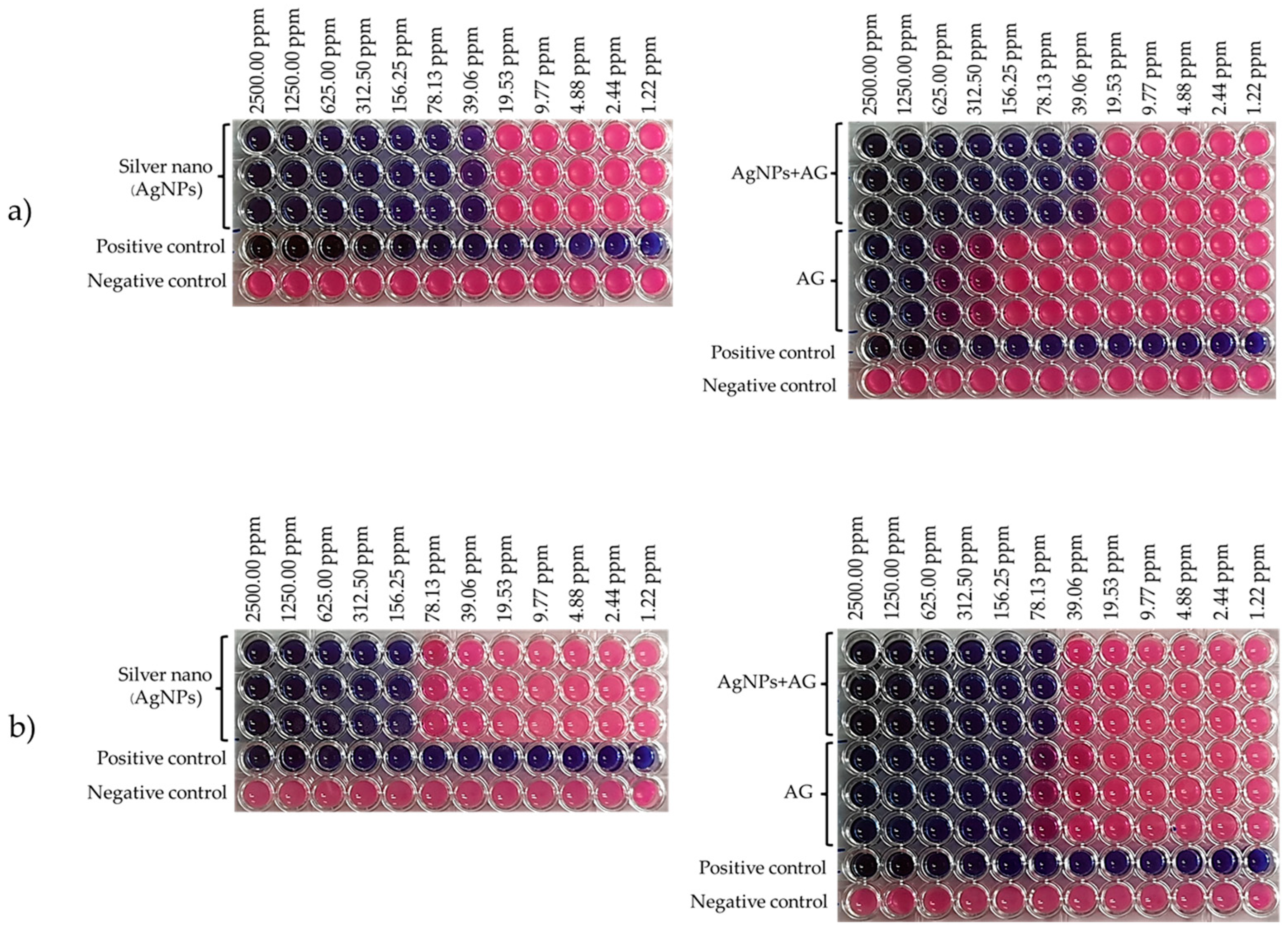
The antibacterial properties of the mixture of the biosynthesized silver nanoparticles from mangoesteen peels and the Andrographis paniculata extract were compared to the biosynthesis silver nanoparticles and the Andrographis paniculata extract using Staphylococcus aureus DMST 8840 and Escherichia coli TISTR 117, as shown in Table 3. Moreover, the images of the antibacterial test are presented in Figure 2.
Table 3 reveals that the mixture of Andrographis paniculata extract and silver nanoparticles derived from mangosteen peels inhibits both Gram-positive (S. aureus DMST 8840) and Gram-negative (E. coli. TISTR 117) bacteria. This is due to nano silver inhibiting a variety of microorganisms by releasing silver ions (Ag+) into their cells [48], where the Ag+ could bind to the proteins of the cell wall and enzymes, affecting the generation of ATP, and causing abnormalities in the cell’s functioning system [49]. Then, the silver nanoparticles, having soft acid properties, could interact with the soft base molecules within the cell, namely, the sulfhydryl group. Subsequently, the enzyme formed a hydrogen bond with the sulfhydryl group, resulting in the inactivation of the enzyme and causing the microbial cell to die. This inhibition mechanism of silver nanoparticles was similar to the inhibition mechanism of Andrographis paniculata containing andrographolide, as the andrographolide passed into bacterial cells. Then, the DNA, RNA, and protein syntheses of the bacteria were disrupted and inhibited. Furthermore, andrographolide affected the quorum sensing system of the bacteria and caused their death [27]. The experiment’s findings indicate that Andrographis paniculata extract can enhance nano silver’s ability to eliminate Gram-positive bacteria. This is because Andrographis paniculata extract is efficient at inhibiting infection, particularly from Gram-positive pathogens. This is similar to the findings of [36], where the methanol extract of Andrographis paniculata demonstrated the largest inhibition zone (26 mm) against S. aureus ATCC 25923, but did not show efficacy against Proteus vulgaris and E. coli ATCC 25922. As a consequence, it can improve the mixed formula’s efficiency and enhance its antibacterial effect. Therefore, the development of cotton fabric coated with silver nanoparticles could be effective in stopping acne-causing bacteria and protecting against the SARS-CoV-19 virus, as other research found that phenolic compound groups, such as betulinic acid, indigo, aloeemodine, luteolin, and quinomethyl triterpenoids, quercitin, or gallates, might inhibit the self-replication mechanism of this virus [50]. In the current study, the Andrographis paniculata extract had a high amount of phenolic compounds and andrographolide, which were reported to inhibit the SARS-CoV-19 virus by inhibiting the activity of the protease enzyme, affecting the survival of the infection [51].

3.3. Synergistic Antibacterial Activity of Garcinia mangostana Linn. and Andrographis paniculata

When the biosynthesized silver nanoparticles from the Garcinia mangostana Linn. extract and the Andrographis paniculata extract were mixed in a 1:1 ratio, it was found that both substances acted synergistically when testing for inhibition against two strains of microorganisms (S. aureus and E. coli), with the FIC values in the range 0.281–0.531, indicating synergistic efficiency in inhibition (Table 4).
The results showed the efficiency of the mixture. This was consistent with another study that reported the synergistic effects of a mixture of clove and cinnamon essential oils in increasing antimicrobial efficiency more than by using only one of the essential oils [52]. The bioactive compounds in the mixtures could enhance antimicrobial activity [53]. In addition, silver nanoparticles with gentamicin and zinc oxide with gentamycin synergized the inhibition of S. aureus [54]. Increasing the effectiveness of the two substances would reduce the amount of use of either substance in excessive amounts.

3.4. Antibacterial Property of Silver Nanoparticles Coated on Cotton Fabric

The ability to inhibit the microbial growth of cotton fabric coated with silver nanoparticles and Andrographis paniculata extract at 100 ppm is shown in Table 5. It was found that the coated fabric inhibited the growth of S. aureus and E. coli at a very high level (%R~100). Furthermore, even if the silver nanoparticle-coated fabric had been washed for 20 consecutive cycles, it still had an excellent inhibition of the growth of both types of microorganisms. With the S. aureus strain, there was a substantial decrease in inhibition after 30 washing cycles. On the other hand, the antimicrobial property of the coated cotton fabric to E. coli was only slightly decreased after 30 washing cycles. These results were similar to another study that suggested that increasing the number of washing cycles decreased the loading capacity of silver and reduced the antibacterial properties of cotton fiber [55].
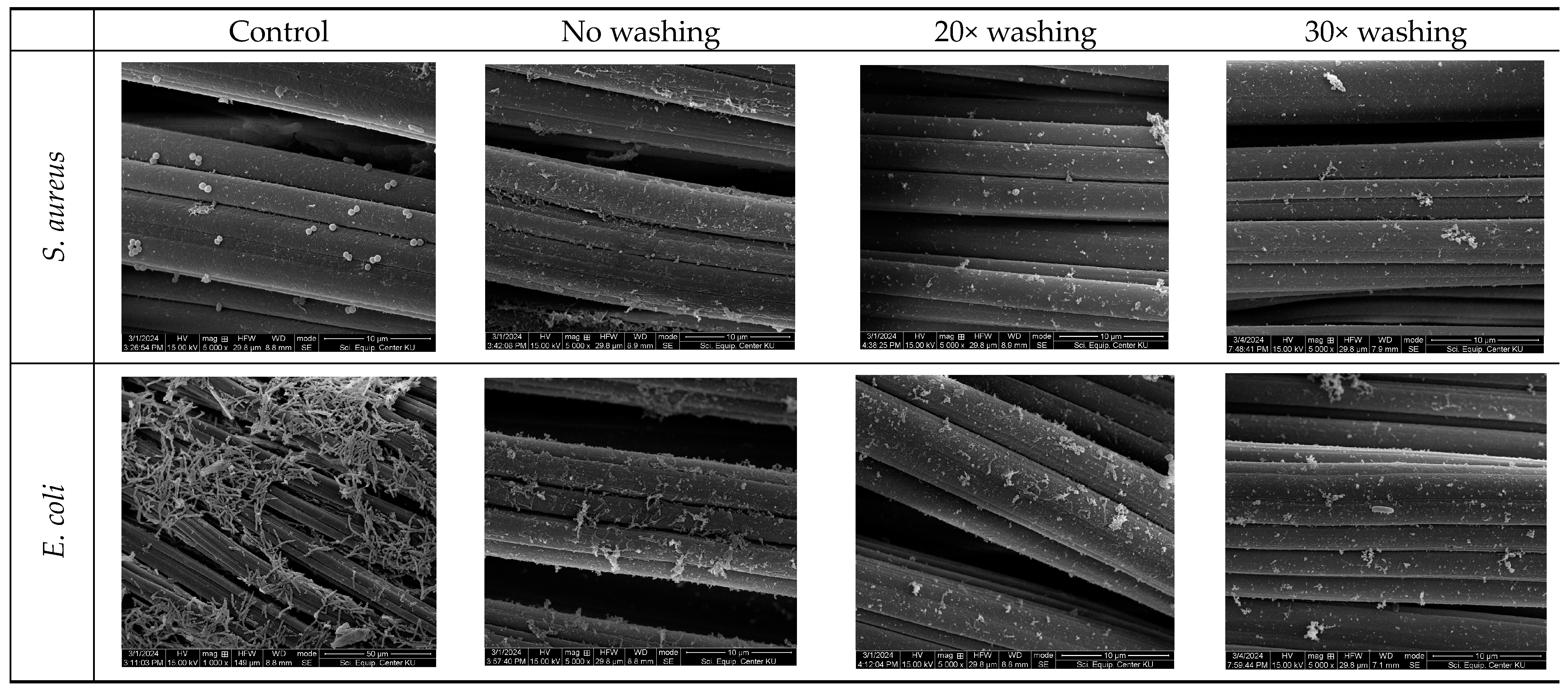
The morphologies of the AgNPs+AG-coated cotton fabric before and after 20 and 30 washing cycles was evaluated using SEM images (Figure 3). Small crystalline particles were spread on the surface of the AgNPs+AG-coated cotton fabric and some areas were coated with a thick layer of AgNPs+AG nanoparticles, indicating that the AgNPs+AG extract could form a good coating on the cotton fabric. However, after washing, while there were still some nanoparticles spread across the surface of the cotton fabric, the AgNPs+AG nanoparticle layer seemed to have lessened. This may have been due to the fact that the AgNPs+AG nanoparticles were washed off during the washing process. One of the requirements of the silk face covering in this study was its antimicrobial property, which was achieved by successfully coating the cotton fabric with a mixture of AgNPs+AG at a concentration of 100 ppm. As depicted in the SEM images (Figure 4), numerous bacteria cells are observed on the surface of the uncoated cotton fabric. In contrast, the surface of the cotton fabric coated with AgNPs+AG exhibits numerous lysed and leaking bacteria cells, indicating effective antimicrobial action. Furthermore, SEM images revealed a decrease in the antibacterial efficacy of the AgNPs+AG-coated cotton fabric with an increase in the number of washing cycles. After 20 washing cycles, damaged bacterial structures were observed on the coated fabric. However, after 30 washing cycles, numerous S. aureus cells in normal shape were detected on the fabric surface, while only a small amount of E. coli cells in normal shape were present (Figure 4). Subsequently, this was used as the middle layer component in the developed silk face covering.
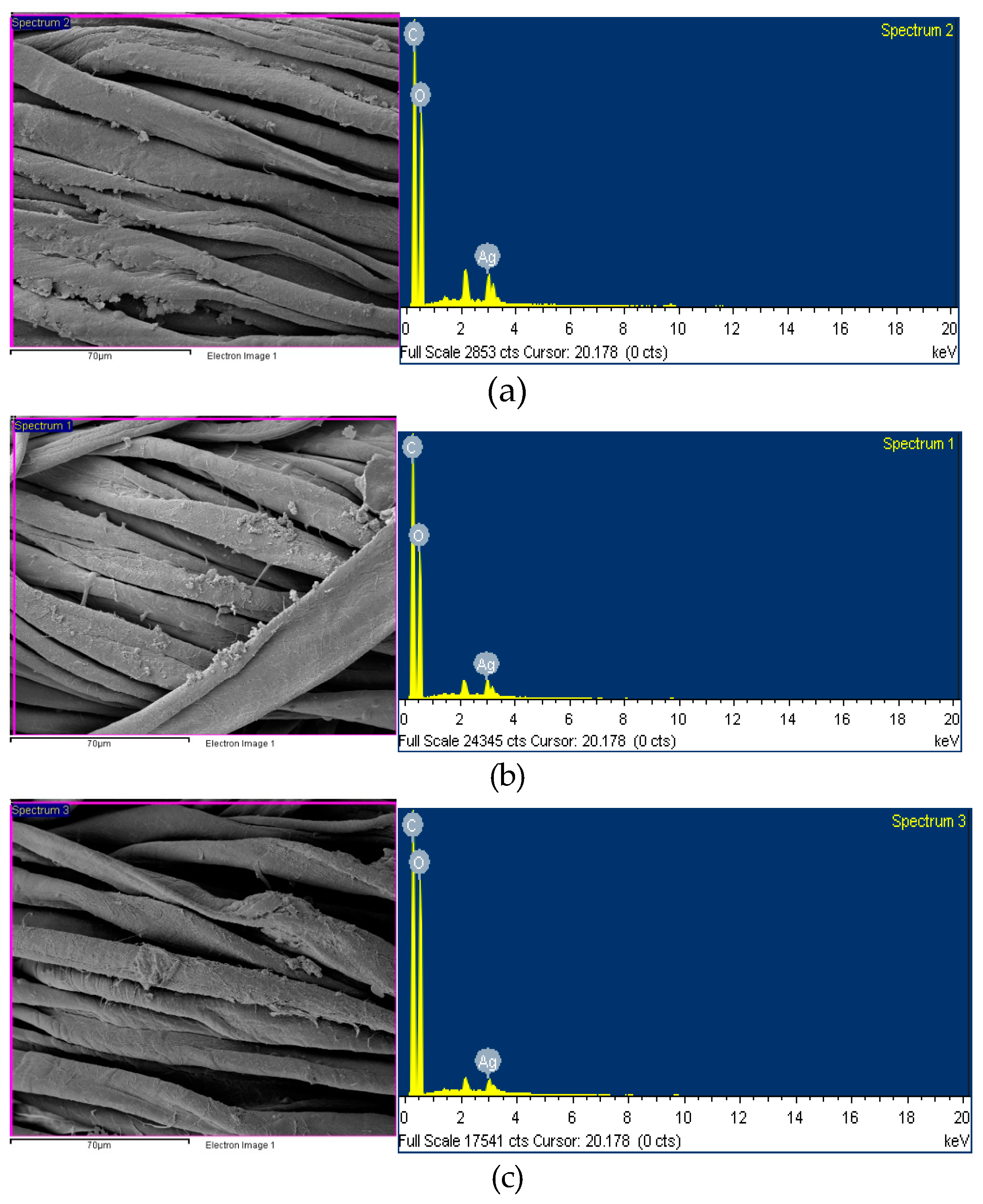
The analysis of the silver nanoparticle content on the surface of cotton fabric based on EDS is shown in Table 6 and Figure 5. The peak at 3 keV confirmed the presence of the silver nanoparticles [26]. The amounts of silver nanoparticles decreased from before washing (3.70%) to after 20 and 30 washing cycles (2.48% and 1.63%, respectively), representing a reduction of 32.97% and 55.95% after washing. This indicates that the washing process could remove the silver nanoparticles from the surface of the cotton fabric [44]. In comparison, a previous study found that the coating of the biosynthesis silver nanoparticles on cotton fabric decreased to around 60% reduction of S. aureus and 45% reduction of E.coli after 15 washing cycles [56], although some previous research showed a slightly decreased antibacterial activity after washing [4,57]. However, approximately one-half of the silver nanoparticles still remained on the fabric after 30 washing cycles.

3.5. Performance of the Silk Face Covering

The silk face covering was assembled using the water-repellent mulberry silk fabric as the outer layer, the silver nanoparticle-coated cotton fabric as the middle layer, the bacterial cellulose sheet as a filter layer, and the oil-repellent eri silk fabric as the inner layer. Then, the assembled face covering was evaluated for its sub-micron particulate filtration efficiency and airflow resistance, as shown in Table 7.
The sub-micron particulate filtration efficiency was 35.5%, and the airflow resistance was 7.7 mm H2O, which met the Level I requirements of the ASTM F3502-2021 standard. Other studies reported that the filtration efficiency and airflow resistance of an N95 mask were approximately 95% and 2.65 mm H2O, respectively [58,59]. While both the filtration efficiency and breathing comfort properties of this developed face covering may not be comparable to an N95 mask, another study suggested that a multi-layer cloth mask might not be effective enough to filter particles or infection [60]. Thus, the additional bacterial cellulose filter layer was introduced. On the other hand, the air impermeability property of the bacterial cellulose might restrict air passing through the material, making breathing more difficult.

4. Conclusions

The silk face covering adhered to the latest standards for commercial non-medical masks outlined in the ASTM F3502-21 Barrier Face Covering Standard, offering a unique blend of comfort, protection, and sustainability. Its three-layer design boasts a water-repellent mulberry silk outer layer, an oil-repellent eri silk inner layer, and a cotton mid-layer coated with silver nanoparticles. To enhance its antimicrobial efficacy, the cotton layer underwent treatment with biosynthesized silver nanoparticles extracted from Garcinia mangostana Linn. peel and Andrographis paniculata extract, effectively inhibiting S. aureus and E. coli by more than 99.99% even after 20 washing cycles. Furthermore, the filtration efficiency of the silk face covering was enhanced by the inclusion of a biodegradable bacterial cellulose filter layer. Overall, the silk barrier face covering met the performance benchmarks outlined in the ASTM F3502-2021 standard, achieving a filtration efficiency of 35.5% and an airflow resistance of 7.7 mm H2O, thereby fulfilling Level I requirements. This innovative mask caters to various needs, providing comfort for sensitive skin, breathability for active individuals, and style for fashion-conscious consumers. Its eco-friendly materials and durable performance position it as a leader in the sustainable mask market, attracting environmentally conscious buyers.
Because of the application of our silk mask innovation in the mask industry, it must be competitive with many different types and brands. By building brand awareness, the silk face covering can stand out from the competition and be seen as a unique and desirable option. Brand awareness allows the silk face covering to be more than just a mask. It becomes a product with a specific identity and perceived value that can potentially transform the mask industry by offering a unique and desirable option for consumers. By addressing specific needs, quantifying benefits, and building brand awareness, this silk face covering has the potential to transform the mask industry, offering a unique and desirable option for consumers.

Author Contributions

Conceptualization, R.C., P.T, and P.P. (Pithalai Phoophat); methodology, N.C., J.B. and T.A.; validation, P.P. (Phannaphat Phromphen) and P.P. (Pithalai Phoophat); formal analysis, N.C. and P.R.; investigation, U.S. and N.R.; resources, U.S. and P.S.; data curation, P.P. (Phannaphat Phromphen); writing—original draft preparation, P.R., T.A. and P.P. (Phannaphat Phromphen); writing—review and editing, P.P. (Phannaphat Phromphen) and R.C.; visualization, R.C.; supervision, P.S. and N.R.; project administration, T.A. and J.B.; funding acquisition, R.C. and P.T. All authors have read and agreed to the published version of the manuscript.

Funding

This research was funded by the Kasetsart University Research and Development Institute (KURDI), grant number (COVID-19)-1.64.

Institutional Review Board Statement

Not applicable.

Informed Consent Statement

Not applicable.

Data Availability Statement

Data are contained within the article.

Acknowledgments

The authors are also grateful to the Department of Medical Sciences, Ministry of Public Health, and the Microbiological Resources Centre, Thailand Institute of Scientific and Technological Research (TISTR), for microorganism support, and to Natural Nich Co., Ltd., for silk fabric production. The authors would like to thank Taveephol Product Co., Ltd., Bangkok, Thailand, for kindly providing the bacterial cellulose residue, and SMS Corporation Co., Ltd., Pathum Thani, Thailand, for supplying the cationic modified starch used in this experiment.

Conflicts of Interest

The authors declare no conflicts of interest.

References

  1. Freeman, C.; Burch, R.; Strawderman, L.; Black, C.; Saucier, D.; Rickert, J.; Wilson, J.; Bealor, S.A.; Ratledge, M.; Fava, S. Preliminary evaluation of filtration efficiency and differential pressure ASTM F3502 testing methods of non-medical masks using a face filtration mount. Int. J. Environ. Res. Public Health 2021, 18, 4124. [Google Scholar] [CrossRef] [PubMed]
  2. Brosseau, L.M.; Stull, J. Criticism of workplace protection barrier face covering article mischaracterizes ASTM standard and its potential utility. New Solut. 2023, 33, 195–197. [Google Scholar] [CrossRef] [PubMed]
  3. Alby, L.; Jayswal, A.; Morris, S.; McAtee, W.; Raghav, V.; Adanur, S. A novel face mask design with improved properties for COVID-19 prevention. Text. Res. J. 2023, 93, 2754–2764. [Google Scholar] [CrossRef]
  4. Phromphen, P.; Sukatta, U.; Rugthaworn, P.; Boonyarit, J.; Phoophat, P.; Rungruangkitkrai, N.; Tuntariyanond, P.; Chartvivatpornchai, N.; Apipatpapha, T.; Chollakup, R. Biosynthesis of silver nanoparticles enhanced antibacterial silk face covering. J. Nat. Fibers 2023, 20, 2212926. [Google Scholar] [CrossRef]
  5. Hao, W.; Parasch, A.; Williams, S.; Li, J.; Ma, H.; Burken, J.; Wang, Y. Filtration performances of non-medical materials as candidates for manufacturing facemasks and respirators. Int. J. Hyg. Environ. 2020, 229, 113582. [Google Scholar] [CrossRef] [PubMed]
  6. Zhao, M.; Liao, L.; Xiao, W.; Yu, X.; Wang, H.; Wang, Q.; Lin, Y.L.; Kilinc-Balci, F.S.; Price, A.; Chu, L.; et al. Household materials selection for homemade cloth face coverings and their filtration efficiency enhancement with triboelectric charging. Nano Lett. 2020, 20, 5544–5552. [Google Scholar] [CrossRef]
  7. Ayodeji, O.J.; Hilliard, T.A.; Ramkumar, S. Particle-size-dependent filtration efficiency, breathability, and flow resistance of face coverings and common household fabrics used for face masks during the covid-19 pandemic. Int. J. Environ. Res. 2022, 16, 11. [Google Scholar] [CrossRef]
  8. Konda, A.; Prakash, A.; Moss, G.A.; Schmoldt, M.; Grant, G.D.; Guha, S. Aerosol filtration efficiency of common fabrics used in respiratory cloth masks. ACS Nano 2020, 14, 6339–6347. [Google Scholar] [CrossRef]
  9. Li, Y.; Leung, P.; Yao, L.; Song, Q.W.; Newton, E. Antimicrobial effect of surgical masks coated with nanoparticles. J. Hosp. Infect. 2006, 62, 58–63. [Google Scholar] [CrossRef] [PubMed]
  10. Kharaghani, D.; Khan, M.Q.; Shahzad, A.; Inoue, Y.; Yamamoto, T.; Rozet, S.; Tamada, Y.; Kim, I.S. Preparation and in-vitro assessment of hierarchal organized antibacterial breath mask based on polyacrylonitrile/silver (PAN/AgNPs) nanofiber. Nanomaterials 2018, 8, 461. [Google Scholar] [CrossRef] [PubMed]
  11. Ray, S.S.; Park, Y.I.; Park, H.; Nam, S.E.; Kim, I.C.; Kwon, Y.N. Surface innovation to enhance anti-droplet and hydrophobic behavior of breathable compressed-polyurethane masks. Environ. Technol. Innov. 2020, 20, 101093. [Google Scholar] [CrossRef]
  12. Han, M.C.; Cai, S.Z.; Wang, J.; He, H.W. Single-side superhydrophobicity in Si3N4-doped and SiO2-treated polypropylene nonwoven webs with antibacterial activity. Polymers 2022, 14, 2952. [Google Scholar] [CrossRef]
  13. McQuerry, M.; Dodson, A. An antimicrobial zinc ion fiber for COVID-19 prevention in nonwoven face coverings for healthcare settings. J. Occup. Environ. Hyg. 2024, 1–8. [Google Scholar] [CrossRef]
  14. Calais, G.B.; Rocha Neto, J.B.M.; Bataglioli, R.A.; Chevalier, P.; Tsukamoto, J.; Arns, C.W.; Mantovani, D.; Beppu, M.M. Bioactive textile coatings for improved viral protection: A study of polypropylene masks coated with copper salt and organic antimicrobial agents. Appl. Surf. Sci. 2023, 638, 158112. [Google Scholar] [CrossRef]
  15. SadrHaghighi, A.; Sarvari, R.; Fakhri, E.; Poortahmasebi, V.; Sedighnia, N.; Torabi, M.; Mohammadzadeh, M.; Azhiri, A.H.; Eskandarinezhad, M.; Moharamzadeh, K.; et al. Copper-Nanoparticle-Coated Melt-Blown Facemask Filter with Antibacterial and SARS-CoV-2 Antiviral Ability. ACS Appl. Nano Mater. 2023, 6, 12849–12861. [Google Scholar] [CrossRef]
  16. Punjabi, K.; Bhatia, E.; Keshari, R.; Jadhav, K.; Singh, S.; Shastri, J.; Banerjee, R. Biopolymer Coating Imparts Sustainable Self-Disinfecting and Antimicrobial Properties to Fabric: Translated to Protective Gears for the Pandemic and Beyond. ACS Biomater. Sci. Eng. 2023, 9, 1116–1131. [Google Scholar] [CrossRef]
  17. Huq, M.A.; Ashrafudoulla, M.; Rahman, M.M.; Balusamy, S.R.; Akter, S. Green synthesis and potential antibacterial applications of bioactive silver nanoparticles: A review. Polymers 2022, 14, 742. [Google Scholar] [CrossRef]
  18. Yaqoob, A.A.; Umar, K.; Ibrahim, M.N.M. Silver nanoparticles: Various methods of synthesis, size affecting factors and their potential applications—A review. Appl. Nanosci. 2020, 10, 1369–1378. [Google Scholar] [CrossRef]
  19. Zhou, Y.; Tang, R.-C. Facile and eco-friendly fabrication of colored and bioactive silk materials using silver nanoparticles synthesized by two flavonoids. Polymers 2018, 10, 404. [Google Scholar] [CrossRef]
  20. Brahmachari, G.; Sarkar, S.; Ghosh, R.; Barman, S.; Mandal, N.C.; Jash, S.K.; Banerjee, B.; Roy, R. Sunlight-induced rapid and efficient biogenic synthesis of silver nanoparticles using aqueous leaf extract of Ocimum sanctum Linn. with enhanced antibacterial activity. Bioorganic Med. Chem. Lett. 2014, 4, 18. [Google Scholar] [CrossRef] [PubMed]
  21. Bankar, A.; Joshi, B.; Kumar, A.R.; Zinjarde, S. Banana peel extract mediated novel route for the synthesis of silver nanoparticles. Colloids Surf. A Physicochem. Eng. 2010, 368, 58–63. [Google Scholar] [CrossRef]
  22. Nakkala, J.R.; Mata, R.; Bhagat, E.; Sadras, S.R. Green synthesis of silver and gold nanoparticles from Gymnema sylvestre leaf extract: Study of antioxidant and anticancer activities. J. Nanopart. Res. 2015, 17, 151. [Google Scholar] [CrossRef]
  23. Labh, A.K.; Rajeshkumar, S.; Lakshmi, T.; Roy, A. Antibacterial efficacy of silver nanoparticles synthesized using herbal formulation against clinical pathogens. Drug Invent. Today 2019, 11, 2362. [Google Scholar]
  24. Kibria, M.G.; Chowdhury, K.P.; Ashik, A.H.; Riyad, M.E.H. Wound healing functionality of mangosteen extracts on viscose fabric. Text. Leather Rev. 2022, 5, 147–164. [Google Scholar] [CrossRef]
  25. Maghimaa, M.; Alharbi, S.A. Green synthesis of silver nanoparticles from Curcuma longa L. and coating on the cotton fabrics for antimicrobial applications and wound healing activity. J. Photochem. Photobiol. B Biol. 2020, 204, 111806. [Google Scholar] [CrossRef] [PubMed]
  26. Senthamarai Kannan, M.; Hari Haran, P.S.; Sundar, K.; Kunjiappan, S.; Balakrishnan, V. Fabrication of anti-bacterial cotton bandage using biologically synthesized nanoparticles for medical applications. Prog. Biomater. 2022, 11, 229–241. [Google Scholar] [CrossRef]
  27. Hossain, S.; Urbi, Z.; Karuniawati, H.; Mohiuddin, R.B.; Moh Qrimida, A.; Allzrag, A.M.M.; Ming, L.C.; Pagano, E.; Capasso, R. Andrographis paniculata (Burm. f.) Wall. ex Nees: An updated review of phytochemistry, antimicrobial pharmacology, and clinical safety and efficacy. Life 2021, 11, 348. [Google Scholar] [CrossRef] [PubMed]
  28. Sule, A.; Ahmed, Q.U.; Latip, J.; Samah, O.A.; Omar, M.N.; Umar, A.; Dogarai, B.B.S. Antifungal activity of Andrographis paniculata extracts and active principles against skin pathogenic fungal strains in vitro. Pharm. Biol. 2012, 50, 850–856. [Google Scholar] [CrossRef] [PubMed]
  29. Sharma, V.; Kaushik, S.; Pandit, P.; Dhull, D.; Yadav, J.P.; Kaushik, S. Green synthesis of silver nanoparticles from medicinal plants and evaluation of their antiviral potential against chikungunya virus. Appl. Microbiol. Biotechnol. 2019, 103, 881–891. [Google Scholar] [CrossRef]
  30. Banerjee, M.; Parai, D.; Chattopadhyay, S.; Mukherjee, S.K. Andrographolide: Antibacterial activity against common bacteria of human health concern and possible mechanism of action. Folia Microbiol. 2017, 62, 237–244. [Google Scholar] [CrossRef]
  31. Banerjee, M.; Moulick, S.; Bhattacharya, K.K.; Parai, D.; Chattopadhyay, S.; Mukherjee, S.K. Attenuation of Pseudomonas aeruginosa quorum sensing, virulence and biofilm formation by extracts of Andrographis paniculata. Microb. Pathog. 2017, 113, 85–93. [Google Scholar] [CrossRef] [PubMed]
  32. Chen, J.-X.; Xue, H.-J.; Ye, W.-C.; Fang, B.-H.; Liu, Y.-H.; Yuan, S.-H.; Yu, P.; Wang, Y.-Q. Activity of andrographolide and its derivatives against influenza virus in vivo and in vitro. Biol. Pharm. Bull. 2009, 32, 1385–1391. [Google Scholar] [CrossRef]
  33. Abass, S.; Zahiruddin, S.; Ali, A.; Irfan, M.; Jan, B.; Haq, Q.M.R.; Husain, S.A.; Ahmad, S. Development of synergy-based combination of methanolic extract of Andrographis paniculata and Berberis aristata against E. coli and S. aureus. Curr. Microbiol. 2022, 79, 223. [Google Scholar] [CrossRef] [PubMed]
  34. Kongcharoensuntorn, W.; Kuawaiyakul, K.; Thongsri, S. Synergistic Effect of Andrographis paniculata with Medicinal Plants to Inhibit the Growth of Opportunistic Bacteria. Burapha Sci. J. 2023, 28, 789–808. [Google Scholar]
  35. Liu, X.; Souzandeh, H.; Zheng, Y.; Xie, Y.; Zhong, W.-H.; Wang, C. Soy protein isolate/bacterial cellulose composite membranes for high efficiency particulate air filtration. Compos. Sci. Technol. 2017, 138, 124–133. [Google Scholar] [CrossRef]
  36. Wu, A.; Hu, X.; Ao, H.; Chen, Z.; Chu, Z.; Jiang, T.; Deng, X.; Wan, Y. Rational design of bacterial cellulose-based air filter with antibacterial activity for highly efficient particulate matters removal. Nano Sel. 2022, 3, 201–211. [Google Scholar] [CrossRef]
  37. Valdez-Salas, B.; Beltran-Partida, E.; Cheng, N.; Salvador-Carlos, J.; Valdez-Salas, E.A.; Curiel-Alvarez, M.; Ibarra-Wiley, R. Promotion of Surgical Masks Antimicrobial Activity by Disinfection and Impregnation with Disinfectant Silver Nanoparticles. Int. J. Nanomed. 2021, 16, 2689–2702. [Google Scholar] [CrossRef] [PubMed]
  38. Ahmed, T.; Ogulata, R.T.; Sezgin Bozok, S. Silver nanoparticles against SARS-CoV-2 and its potential application in medical protective clothing—A review. J. Text. Inst. 2022, 113, 2825–2838. [Google Scholar] [CrossRef]
  39. Pradipasena, P.; Chollakup, R.; Tantratian, S. Formation and characterization of BC and BC-paper pulp films for packaging application. J. Thermoplast. Compos. Mater. 2018, 31, 500–513. [Google Scholar] [CrossRef]
  40. Sarker, S.D.; Nahar, L.; Kumarasamy, Y. Microtitre plate-based antibacterial assay incorporating resazurin as an indicator of cell growth, and its application in the in vitro antibacterial screening of phytochemicals. Methods 2007, 42, 321–324. [Google Scholar] [CrossRef]
  41. Basri, D.F.; Fan, S. The potential of aqueous and acetone extracts of galls of Quercus infectoria as antibacterial agents. Indian J. Pharmacol. 2005, 37, 26. [Google Scholar] [CrossRef]
  42. Davidson, P.M.; Taylor, T.M.; Schmidt, S.E. Chemical preservatives and natural antimicrobial compounds. In Food Microbiology: Fundamentals and Frontiers; Michael, P., Doyle, R.L.B., Eds.; ASM Press: Washington, DC, USA, 2012; pp. 765–801. [Google Scholar]
  43. Nishi, Y.; Uryu, M.; Yamanaka, S.; Watanabe, K.; Kitamura, N.; Iguchi, M.; Mitsuhashi, S. The structure and mechanical properties of sheets prepared from bacterial cellulose: Part 2 Improvement of the mechanical properties of sheets and their applicability to diaphragms of electroacoustic transducers. J. Mater. Sci. 1990, 25, 2997–3001. [Google Scholar] [CrossRef]
  44. Iguchi, M.; Yamanaka, S.; Budhiono, A. Bacterial cellulose—A masterpiece of nature’s arts. J. Mater. Sci. 2000, 35, 261–270. [Google Scholar] [CrossRef]
  45. Tabarsa, T.; Sheykhnazari, S.; Ashori, A.; Mashkour, M.; Khazaeian, A. Preparation and characterization of reinforced papers using nano bacterial cellulose. Int. J. Biol. Macromol. 2017, 101, 334–340. [Google Scholar] [CrossRef] [PubMed]
  46. Yuan, J.; Wang, T.; Huang, X.; Wei, W. Dispersion and beating of bacterial cellulose and their influence on paper properties. BioResources 2016, 11, 9290–9301. [Google Scholar] [CrossRef]
  47. Xiang, Z.; Zhang, J.; Liu, Q.; Chen, Y.; Li, J.; Lu, F. Improved dispersion of bacterial cellulose fibers for the reinforcement of paper made from recycled fibers. Nanomaterials 2019, 9, 58. [Google Scholar] [CrossRef]
  48. Qing, Y.a.; Cheng, L.; Li, R.; Liu, G.; Zhang, Y.; Tang, X.; Wang, J.; Liu, H.; Qin, Y. Potential antibacterial mechanism of silver nanoparticles and the optimization of orthopedic implants by advanced modification technologies. Int. J. Nanomed. 2018, 13, 3311–3327. [Google Scholar] [CrossRef]
  49. Klueh, U.; Wagner, V.; Kelly, S.; Johnson, A.; Bryers, J. Efficacy of silver-coated fabric to prevent bacterial colonization and subsequent device-based biofilm formation. J. Biomed. Mater. Res. 2000, 53, 621–631. [Google Scholar] [CrossRef]
  50. Chojnacka, K.; Witek-Krowiak, A.; Skrzypczak, D.; Mikula, K.; Młynarz, P. Phytochemicals containing biologically active polyphenols as an effective agent against Covid-19-inducing coronavirus. J. Funct. Foods 2020, 73, 104146. [Google Scholar] [CrossRef] [PubMed]
  51. Ervina, M.; Pratama, M.R.F.; Poerwono, H.; Siswodihardjo, S. The coronavirus disease 2019 main protease inhibitor from Andrographis paniculata (Burm. f) Ness. J. Adv. Pharm. Technol. Res. 2020, 11, 157–162. [Google Scholar] [CrossRef]
  52. Goñi, P.; López, P.; Sánchez, C.; Gómez-Lus, R.; Becerril, R.; Nerín, C. Antimicrobial activity in the vapour phase of a combination of cinnamon and clove essential oils. Food Chem. 2009, 116, 982–989. [Google Scholar] [CrossRef]
  53. Khanoonkon, N.; Rugthaworn, P.; Kongsin, K.; Sukyai, P.; Harnkarnsujarit, N.; Sothornvit, R.; Chollakup, R.; Sukatta, U. Enhanced antimicrobial effectiveness of synergistic mixtures of rambutan peel extract and cinnamon essential oil on food spoilage bacteria and bio-based food packaging. J. Food Saf. 2022, 42, e12976. [Google Scholar] [CrossRef]
  54. Punjabi, K.; Mehta, S.; Chavan, R.; Chitalia, V.; Deogharkar, D.; Deshpande, S. Efficiency of biosynthesized silver and zinc nanoparticles against multi-drug resistant pathogens. Front. Microbiol. 2018, 9, 2207. [Google Scholar] [CrossRef]
  55. Zhang, S.; Zhang, T.; He, J.; Dong, X. Effect of AgNP distribution on the cotton fiber on the durability of antibacterial cotton fabrics. Cellulose 2021, 28, 9489–9504. [Google Scholar] [CrossRef]
  56. Fouda, A.; Shaheen, T.I. Silver Nanoparticles: Biosynthesis, Characterization and Application on Cotton Fabrics. Microbiol. Res. J. Int. 2017, 20, 1–14. [Google Scholar] [CrossRef]
  57. Andra, S.; Balu, S.k.; Jeevanandam, J.; Muthalagu, M.; Danquah, M.K. Surface cationization of cellulose to enhance durable antibacterial finish in phytosynthesized silver nanoparticle treated cotton fabric. Cellulose 2021, 28, 5895–5910. [Google Scholar] [CrossRef]
  58. Wang, A.-B.; Zhang, X.; Gao, L.-J.; Zhang, T.; Xu, H.-J.; Bi, Y.-J. A review of filtration performance of protective masks. Int. J. Environ. Res. Public Health 2023, 20, 2346. [Google Scholar] [CrossRef] [PubMed]
  59. Skaria, S.D.; Smaldone, G.C. Respiratory Source Control Using Surgical Masks with Nanofiber Media. Ann. Occup. Hyg. 2014, 58, 771–781. [Google Scholar] [CrossRef] [PubMed]
  60. MacIntyre, C.R.; Seale, H.; Dung, T.C.; Hien, N.T.; Nga, P.T.; Chughtai, A.A.; Rahman, B.; Dwyer, D.E.; Wang, Q. A cluster randomised trial of cloth masks compared with medical masks in healthcare workers. BMJ Open 2015, 5, e006577. [Google Scholar] [CrossRef] [PubMed]
Figure 1. SEM images of bacterial cellulose sheet (a) 500×; (b) 1000×; and cellulose sheet (c) 500×.
Coatings 14 00379 g001
Figure 2. Images of MIC test on bacterial cellulose sheet: (a) S. aureus DMST 8840; (b) E. coli TISTR 117.
Coatings 14 00379 g002
Figure 3. SEM images of silver nanoparticles coated on cotton fabric (1000×): (a) unwashed; (b) after 20 washing cycles; and (c) after 30 washing cycles.
Coatings 14 00379 g003
Figure 4. SEM images of the bacteria grown on the silver-containing and control samples before and after washing at 5000×.
Coatings 14 00379 g004
Figure 5. EDX of (a) silver nanoparticles on cotton fabric; (b) silver nanoparticles on cotton fabric after 20 washing cycles; and (c) silver nanoparticles on cotton fabric after 30 washing cycles.
Coatings 14 00379 g005
Table 1. Oil repellency and stain release rating of eri silk fabric.
Property Fluorocarbon-Treated Eri Silk
Oil repellency
(seconds)
Vegetable oil300
Paraffin oil300
n-Heptane60
Stain release rating 4
Table 2. Physical properties and air permeability of bacterial cellulose filter.
% Nata de Coco by Weight051050100
Basis weight (g/m2)52.27 ± 0.8253.73 ± 2.9955.98 ± 1.5651.84 ± 1.2251.27 ± 3.78
Tensile index (N·m/g)6.09 ± 0.3211.40 ± 0.0017.27 ± 0.0051.30 ± 0.6463.57 ± 3.33
Tear index (mN·m2/g)0ND3.72 ± 0.265.49 ± 0.563.61 ± 0.00
Cobb test (g/m2)56.62 ± 0.9768.97 ± 0.6266.52 ± 3.2629.06 ± 2.0219.57 ± 2.68
Air permeability (cm3/cm2/s)7.90 ± 1.201.95 ± 0.040.44 ± 0.020.00 ± 0.000.00 ± 0.00
Values presented as mean ± standard deviation. ND = not determined.
Table 3. MIC and MBC of the biosynthesized silver nanoparticles (AgNPs), the Andrographis paniculata extract (AG), and the combination of biosynthesized silver nanoparticles and Andrographis paniculata extract (AgNPs+AG) against S. aureus DMST 8840 and E. coli TISTR 117.
SamplesS. aureus DMST 8840E. coli TISTR 117
MICMBCMICMBC
AgNPs39.06 156.25156.25156.25
AG1250.002500.001250.002500.00
AgNPs+AG39.0678.1378.1378.13
Table 4. FIC index of biosynthesized silver nanoparticles (AgNO) and Andrographis paniculata extract.
RatioFICindex
AgNOAndrographis paniculata ExtractS. aureusE. coli
1001.0001.000
550.5160.281
0101.0001.000
Table 5. Viable bacterial counts (CFU/mL) at zero and 18 h contact time intervals of cotton fabric coated with silver nanoparticles before and after washing.
SamplesS. aureusE. coli
Bacteria Count
(CFU/mL)
% RBacteria Count
(CFU/mL)
% R
0 h18 h0 h18 h
Unwashed1.91 × 106<1001006.77 × 105<100100
After 20 washing cycles1.91 × 106<1001006.77 × 105<100100
After 30 washing cycles1.91 × 1063.05 × 1060.006.77 × 1058.53 × 10398.74
Table 6. EDS of silver nanoparticles coated on cotton fabric.
SamplesSilver (% Weight)% Reduction after Washing
Unwashed3.700
After 20 washing cycles2.4832.97
After 30 washing cycles1.6355.95
Table 7. Performance of the silk face covering.
ASTM F3502–2021 (Level l)CriterionResult
Sub-micron particulate filtration efficiency (%)≥20%35.5%
Airflow resistance (mm H2O)≤157.7
Disclaimer/Publisher’s Note: The statements, opinions and data contained in all publications are solely those of the individual author(s) and contributor(s) and not of MDPI and/or the editor(s). MDPI and/or the editor(s) disclaim responsibility for any injury to people or property resulting from any ideas, methods, instructions or products referred to in the content.