Article

Microstructure and Biocompatibility of Graphene Oxide/BCZT Composite Ceramics via Fast Hot-Pressed Sintering

1
College of Material and Metallurgy, Guizhou University, 550025 Guiyang, China
2
Key Laboratory of Advanced Manufacturing Technology of the Ministry of Education, 550025 Guiyang, China
*
Author to whom correspondence should be addressed.
Coatings 2024, 14(6), 689; https://doi.org/10.3390/coatings14060689
Submission received: 10 May 2024 / Revised: 25 May 2024 / Accepted: 26 May 2024 / Published: 1 June 2024
(This article belongs to the Special Issue Advances of Ceramic and Alloy Coatings, 2nd Edition)

Abstract

:
Improving fracture toughness, electrical conductivity, and biocompatibility has consistently presented challenges in the development of artificial bone replacement materials. This paper presents a new strategy for creating high-performance, multifunctional composite ceramic materials by doping graphene oxide (GO), which is known to induce osteoblast differentiation and enhance cell adhesion and proliferation into barium calcium zirconate titanate (BCZT) ceramics that already exhibit good mechanical properties, piezoelectric effects, and low cytotoxicity. Using fast hot-pressed sintering under vacuum conditions, (1 − x)(Ba0.85Ca0.15Zr0.1Ti0.9)O3−xGO (0.2 mol% ≤ x ≤ 0.5 mol%) composite piezoelectric ceramics were successfully synthesized. Experimental results revealed that these composite ceramics exhibited high piezoelectric properties (d33 = 18 pC/N, kp = 62%) and microhardness (173.76 HV0.5), meeting the standards for artificial bone substitutes. Furthermore, the incorporation of graphene oxide significantly reduced the water contact angle and enhanced their wettability. Cell viability tests using Cell Counting Kit-8, alkaline phosphatase staining, and DAPI staining demonstrated that the GO/BCZT composite ceramics were non-cytotoxic and effectively promoted cell proliferation and growth, indicating excellent biocompatibility. Consequently, with their superior mechanical properties, piezoelectric performance, and biocompatibility, GO/BCZT composite ceramics show extensive potential for application in bone defect repair.

1. Introduction

Bone, a nanocomposite material composed of living cells and minerals, plays a critical role in the structural integrity of the human body [1]. Congenital factors such as heredity and infection, along with acquired factors such as trauma, bone disease, and inflammation, can lead to the formation of bone defects [2,3]. Current treatments for bone defects include stem cell therapy, the use of customized bone scaffolds through 3D printing, and bone grafting [4,5,6]. Stem cell therapy encounters ethical and legal challenges, while the use of 3D-printed bone scaffolds is impeded by high costs and technical complexities. Bone grafting is extensively employed to resolve bone defects. This technique includes autologous bone grafts, allogeneic bone grafts, and synthetic bone replacement materials [7]. Autologous bone grafting is considered the gold standard for repairing bone defects; however, its application is limited by donor scarcity and potential complications [8]. Allogeneic bone grafting, known for its low histocompatibility, frequently leads to rejection reactions [9]. In recent years, synthetic bone replacement materials designed to emulate the structure and properties of human bone have been extensively researched and increasingly utilized [10].
Artificial bone replacement materials primarily consist of medical-grade metallic materials [11], polymer materials [12], bioceramics materials [13], and composite/hybrid biomaterials [14]. Metal materials appropriate for load-bearing implants may cause osteoporosis and rejection owing to their stress shielding effects [15]. Medical polymer materials tailored for different bone repair needs can trigger immune responses upon implantation and suffer from issues such as poor biocompatibility, poor mechanical properties, and high cost [16]. Bioceramics, specifically designed for medical use, exhibit inherent piezoelectric properties, allowing them to facilitate the conversion between mechanical energy and electrical energy and conversely. They are highly biocompatible and find extensive applications in the biomedical field [17,18]. Additionally, the piezoelectric effects observed in both piezoelectric ceramic materials and bone both involve the generation of electric charges under mechanical stress. Fukada and Yasuda were the first to observe the piezoelectric effect in bone, demonstrating that human bone tissue has inherent piezoelectric properties [19]. Subsequent research has indicated that materials exhibiting piezoelectric properties can promote osteoblast proliferation and differentiation, thus enhancing bone regeneration [20]. Therefore, biocompatible piezoelectric ceramic materials are frequently utilized in the repair and replacement of bone defects [21].
Common biological piezoelectric ceramic materials primarily consist of piezoelectric single crystals such as lithium niobate [22], piezoelectric polycrystals including barium titanate and lead zirconate titanate [23], and composite piezoelectric materials formed by combining these materials with polymers [24]. In 1981, Park [25] et al. pioneered the use of barium titanate (BaTiO3) as a piezoelectric ceramic material in bone repair, demonstrating its capacity to emit piezoelectric signals that enhance osteoblast proliferation and differentiation. Furthermore, BaTiO3 ceramics meets the requisite mechanical strength for physiological bone loads, making it extensively utilized in bone tissue engineering research [26,27,28]. However, compared with BaTiO3 ceramics, barium calcium zirconate titanate (BCZT) ceramics exhibit superior properties. In 2009, Professor Ren Xiaobing’s research group [29] demonstrated that BCZT ceramics possess enhanced piezoelectric characteristics. Furthermore, Kara K et al. reported that BCZT ceramics show low cytotoxicity to human osteoblasts [30], suggesting that they are more suitable for biomedical applications involving direct contact with biological tissues [31,32]. When used directly as materials for bone defect repair, pure piezoelectric ceramics may display inadequate fracture toughness, brittle unreliability, low electrical conductivity, and limited biocompatibility [33]. The literature has shown that the addition of various additives to reinforced composite ceramics can significantly improve these properties [34]. For example, Chelli Sai et al. enhanced BCZT ceramics by incorporating hydroxyapatite (HA), finding that this addition not only increased the biocompatibility of the ceramics but also facilitated bone cell growth [35]. Therefore, the exploration of novel and diverse additives for the modification of composite ceramics is a current research focus.
In recent years, there has been considerable research interest in materials such as graphene, primarily attributed to their exceptional capacity to stimulate osteoblast differentiation [36]. Cahit et al. used graphene as a nanocarrier for gold (I)–monoene complexes, and the experimental results showed very favorable adsorption, underscoring the potential of graphene for medical applications [37]. These materials enhance various cellular responses, including adhesion, proliferation, and differentiation, thereby improving the biocompatibility of composites [38,39]. Notably, graphene oxide exhibits considerable potential in biomedical applications, attributed to its nanoscale dimensions, expansive specific surface area, exceptional mechanical strength, significant biocompatibility, elevated electrical conductivity, and antimicrobial capabilities [40,41]. Guo et al. investigated the interaction of graphene and its derivatives with stem cells in different materials, demonstrating that graphene oxide accelerates the osteogenic differentiation of mesenchymal stem cells from the bone marrow [42]. Wu et al. produced graphene oxide-modified β-tricalcium phosphate bioceramics, with studies indicating that an optimal amount of graphene enhances piezoelectric properties and stimulates bone formation in vivo [38]. Palmieri and his team used graphene oxide to interact with human pathogens, confirming its effectiveness as an antimicrobial agent [43]. Additionally, Kanwal et al. synthesized graphene oxide/bioactive glass composites using an alkali-catalyzed sol-gel method, which were found to possess good cytocompatibility [44]. Joy et al. incorporated the graphene oxide effective inclusion of anchor deposit nanostructure (GO-Au) into polycaprolactone (PCL) matrix, and the results showed that the polymer nanocomposite has good hydrophilicity, mechanical stability, and antibacterial activity [45]. However, the method for doping graphene oxide into BCZT ceramics remains unexplored.
In this study, the ceramic matrix composed of Ba0.85Ca0.15Zr0.1Ti0.9 (BCZT) composite ceramics and graphene oxide (GO) was introduced as a dopant to augment the material properties. It was mainly to combine the advantages of BCZT ceramics that can independently generate surface potential and have a high piezoelectric coefficient and good biocompatibility with the advantages of GO’s excellent electrical and thermal properties and high specific surface area to achieve complementarity and optimization of performance. Compared with existing bone defect repair materials, it can further promote bone formation and has better biocompatibility and mechanical properties. A fast hot-pressed sintering technique was employed to facilitate the densification and high-temperature synthesis of the composite ceramics. This process was conducted under vacuum conditions, utilizing direct current heating and axial pressure application to achieve optimal sintering outcomes.

2. Materials and Methods

2.1. Materials and Preparation

The graphene oxide raw material was sourced from a dispersion solution provided by Shanghai Aladdin Biotechnology Co., Ltd. (Shanghai, China). Similarly, the raw materials for the BCZT piezoelectric ceramics, including BaCO3, CaCO3, ZrO2, and TiO2 (≥99.0%, AR), were procured from the same company. The (1 − x)(Ba0.85Ca0.15Zr0.1Ti0.9)O3−xGO (0.2 mol% ≤ x ≤ 0.5 mol%) composite piezoelectric ceramics were prepared by the following process.
Initially, BCZT-based ceramics powders were synthesized using the solid-phase synthesis method. High-purity BaCO3, CaCO3, TiO2, and ZrO2 were precisely weighed according to their stoichiometric ratios. These powders were mixed in a zirconia ball mill jar using zirconia balls of 3, 5, and 10 mm diameters, and anhydrous ethanol was used as the milling medium. The mixture was milled for 12 h at a speed of 500 rpm in a planetary ball mill and then calcined at 1200 °C for 2 h. After this, the material was milled again for another 12 h and then sintered at 1400 °C in a tube furnace. The final BCZT-based ceramics powders were obtained following additional grinding and sieving.
Subsequently, the BCZT ceramic powder was combined with a graphene dispersion in a specific ratio and subjected to ball milling. In this process, the materials were placed in a zirconia milling jar and milled at 300 rpm for 2 h using a planetary ball mill. After milling, the mixture was dried, sieved, and reserved for further processing. In the final step, the composite powder was sintered at 1400 °C and a pressure of 50 MPa for 30 min in a vacuum environment, producing ceramic discs of 12 mm in diameter and 1.5 mm in thickness.

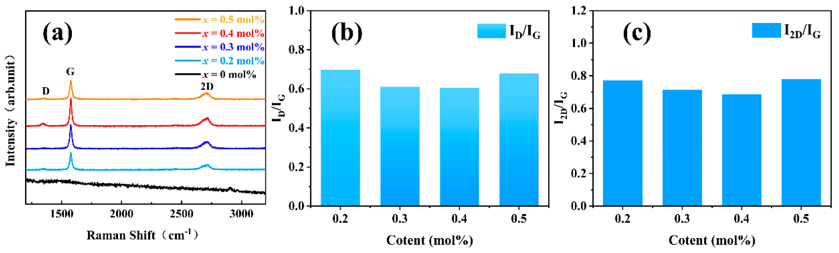
2.2. Characterization of Materials

The phase composition of the xGO/BCZT composite ceramic was analyzed by X-ray diffraction (XRD, Bruker D8 advance, Berlin, Germany) using Cu-Kα radiation over 2θ = 20~80° at a scan rate of 2°/min. A confocal Raman spectrometer (Suzhou Only Light Tan Zhen Technology Co., Ltd., Suzhou, China) with an incident light wavelength of 532 nm was used for further analysis. The microstructure and crystallite size were assessed by scanning electron microscopy (SEM, SU 8600, Hitachi, Tokyo, Japan), and the materials were treated by etching if necessary. The piezoelectric coefficient was determined using a quasi-static d33 measuring instrument (ZJ-3A, Institute of Acoustics, Chinese Academy of Sciences, Beijing, China). Additionally, the planar electromechanical coupling coefficient (kp) was assessed utilizing an impedance analyzer ( E4991A, Agilent Technologies, Inc., Santa Clara, CA, USA) employing resonance and antiresonance techniques. The microhardness of the GO/BCZT composite ceramics was assessed via an HV-1000 Vickers indenter, with a 5N load applied for 10 s.

2.3. Evaluation of Vitro Cell Compatibility

2.3.1. Surface Wettability

Surface wettability significantly influences cell adhesion as demonstrated in prior studies [46]. Typically, the water contact angle serves as a key metric for evaluating the hydrophilic or hydrophobic nature of solid surfaces, where moderately hydrophilic surfaces are deemed advantageous for cellular proliferation [47]. In this paper, the wettability of xGO/BCZT composite ceramic materials was determined through static contact angle measurements using a Krüss DSA100S device. Measurements were conducted at five distinct points on each sample to ensure representativeness, with the mean value of these measurements utilized to characterize the overall surface wettability of the materials.

2.3.2. CCK-8 Assay

Osteoblasts play a crucial role in the repair of bone defects and are one of the main cells in contact with the implant [48]. In this paper, mouse embryonic osteoblast precursor cells (MC3T3-E1) were employed for cytotoxicity testing to assess the biocompatibility of xGO/BCZT composite ceramics. Initially, xGO/BCZT composite ceramics were ultrasonically cleaned with anhydrous ethanol for 1 h and subjected to ultraviolet radiation for 30 min. MC3T3-E1 cells at a concentration of 2 × 10⁵ cells/mL were seeded onto ceramics in 24-well plates and cultured under standard conditions (37 °C, 5% CO2) for 1, 4, and 7 days. CCK-8 was applied to the cultures and incubated for 2 h. Subsequently, the culture medium was then transferred to a 96-well plate in triplicate, and the absorbance (OD value) was measured at 450 nm using an enzyme linked immunosorbent assay reader.

2.3.3. DAPI Staining

In this study, DAPI staining experiments were performed as evidence to further support the CCK8 cell proliferation assay. The apoptotic effects of the control and xGO/BCZT composite ceramics on MC3T3-E1 cells were elucidated through DAPI staining. The materials were first sterilized by ultrasonic cleaning in anhydrous ethanol for 30 min, washing with sterile PBS 5 times, drying for 2 h, and ultraviolet irradiation on both sides for 30 min. The ceramic plates were then transferred to 24-well culture plates and inoculated with MC3T3-E1 cells, while the control wells received no samples. After 24 h of incubation, the cells were subjected to DAPI staining, which involved a PBS wash, fixation of cells for 20 min, permeabilization for 5 min, another PBS wash, and then staining with DAPI solution for 15 min in darkness. Finally, the cell nuclei were visualized using a fluorescence microscope (Nikon Eclipse Ts2R, Shanghai, China).

2.3.4. Alkaline Phosphatase Activity

Alkaline phosphatase (ALP) is an essential enzyme for bone formation and serves as an early indicator of osteoblast differentiation and functional maturity [49]. Consequently, ALP staining is employed to identify the presence of ALP in bone cells, bone tissue, kidneys, and liver, resulting in ALP appearing black and nuclei appearing light blue. ALP activity in the cell lysate was measured using an ALP assay kit, adhering to the provided protocol. The procedure entailed washing the cells twice with PBS, fixing them with 4% paraformaldehyde at room temperature for 10 min, applying BCIP/NBT solution (Beyotime, Shanghai, China) to the fixed cells, and documenting the stained cells with an inverted microscope. By observing and analyzing the staining intensity and distribution, the activity of ALP in cells and its role in cell differentiation and tissue structure can be evaluated.

3. Results and Discussion

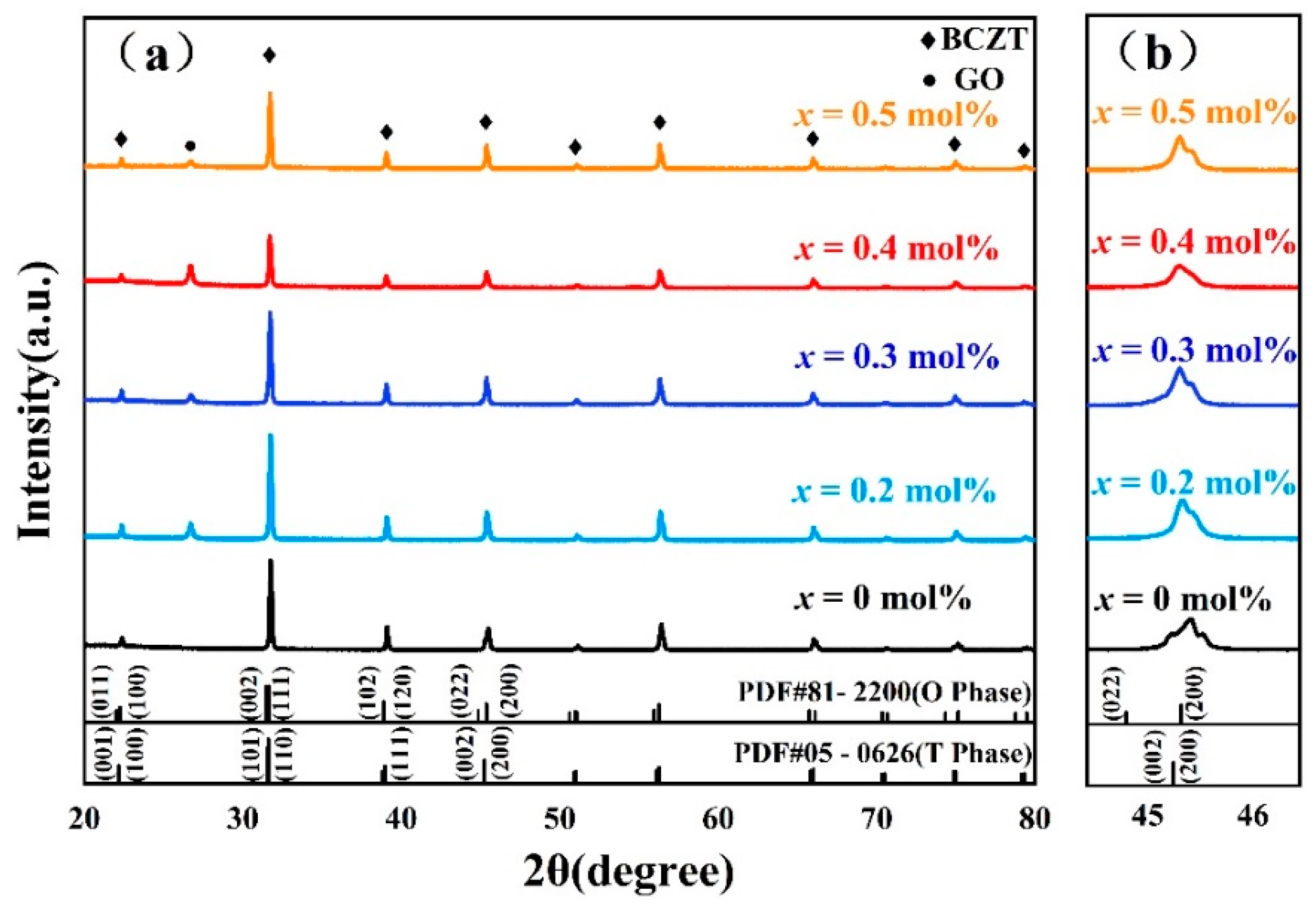
3.1. Crystal Phase Structure

The XRD patterns of xGO/BCZT (0.2 mol% ≤ x ≤ 0.5 mol%) composite ceramics analyzed under room temperature are shown in Figure 1. Additionally, the vertical line diagram corresponds to the card information of the standard diffraction peaks of BaTiO3 ceramics, including both the orthorhombic (PDF#81-2200) and tetragonal (PDF#05-0626) phases. It can be seen from Figure 1 that all composite piezoelectric ceramics had a perovskite structure [50,51]. Furthermore, as graphene oxide concentration increased, the xGO/BCZT composite ceramics exhibited the O phase and a distinctive diffraction peak at 26.7°, correlating with the (002) diffraction peak of graphene [52]. This diffraction peak was related to the reflection generated by the normal graphite structure, indicating a highly ordered crystal structure with a certain layer spacing within the composite ceramics [53].
Figure 2a presents the Raman spectra of xGO/BCZT (0.2 mol% ≤ x ≤ 0.5 mol%) composite ceramics under room temperature and a spectral range of 1000~3500 cm−1. Previous reports have indicated that the characteristic peaks of graphene are G peak, D peak, and 2D peak [54].
The G peak, located at 1581 cm−1, arises from the in-plane vibrations of sp2-hybridized carbon atoms and effectively reflects the number of graphene layers [55]. The D peak, positioned at 1350 cm−1, is associated with the disorder-induced vibrational modes and is closely related to the wavelength of the laser used, serving as a marker for identifying structural defects or edge characteristics in graphene samples [56]. The 2D peak, located around 2700 cm−1, is a second-order Raman peak that maps the stacking of carbon layers [57].
At x = 0 mol%, no characteristic peaks of graphene were observed in the composite ceramics. However, as the content of graphene oxide increased, the characteristic peaks of graphene gradually became evident. Simultaneously, the Raman spectra were instrumental in determining graphene’s disorder degree by analyzing the ID/IG peak intensity ratio [58]. The ID/IG ratio increased with low defect density and decreased with high defect density [59]. Moreover, the I2D/IG ratio, representing the peak intensities between the 2D and G peaks, was closely associated with the graphene layers’ quantity [60,61]. Specifically, for single- or bi-layer graphene, the I2D/IG exceeded 1, showcasing a single, sharp 2D peak. Conversely, with increasing graphene layer number, the I2D/IG ratio decreased from 1 to 0.5. Notably, in the case of xGO/BCZT composite ceramics, the I2D/IG lay between 0.5 and 1, as shown in Figure 2c, confirming the presence of a multilayer graphene structure [60,61]. As evidenced by Figure 2a, this G peak intensity initially rose with an increase in graphene oxide content, reaching a maximum value at x = 0.4 mol%, before subsequently declining. This trend suggested that the quantity of graphene layers within the composite ceramics initially increased and then decreased [55]. The Raman spectroscopy analysis in this study further substantiated the phase composition of the composite ceramics.

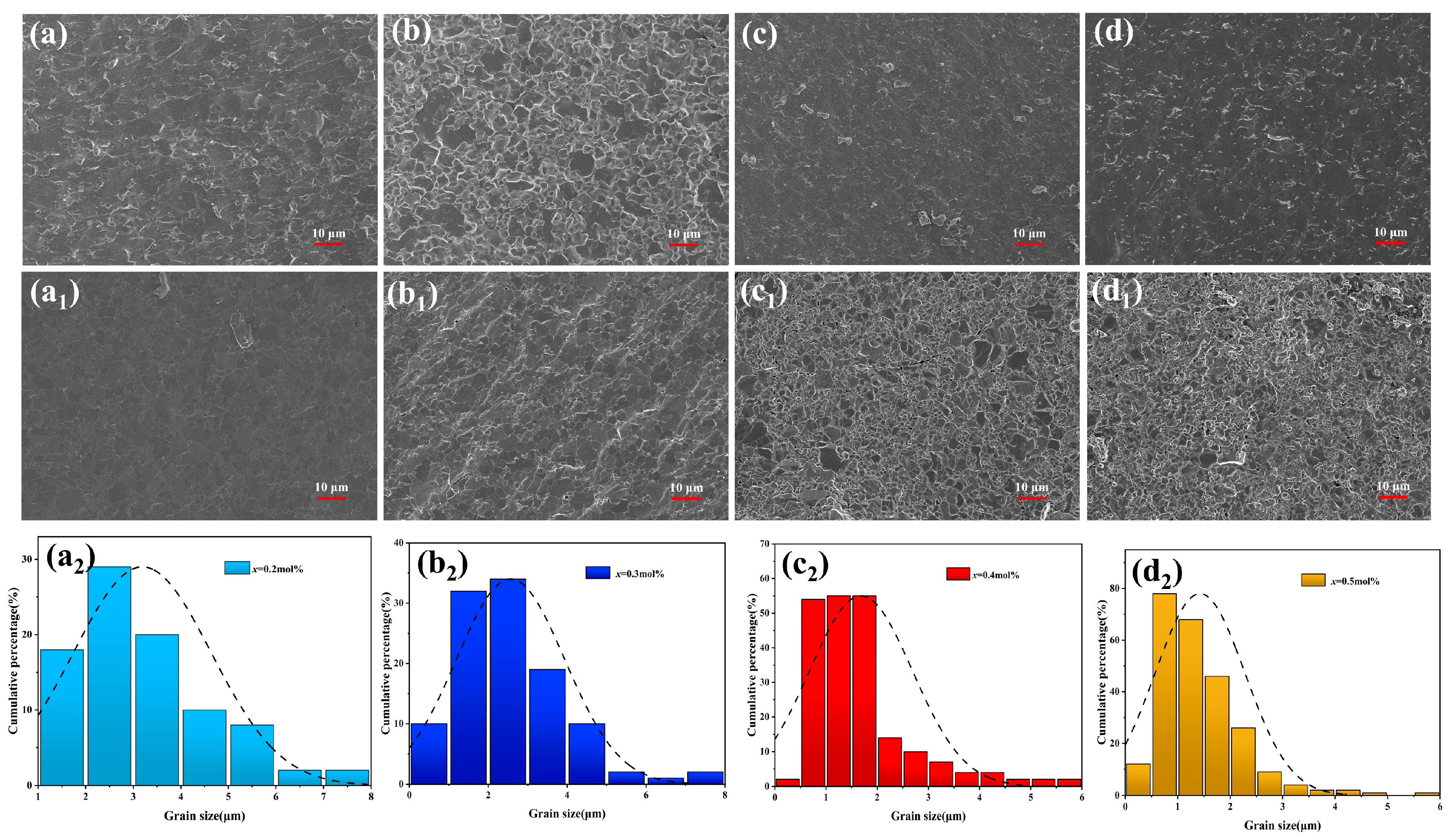
3.2. Microstructure and Fracture Morphology

Figure 3 presents the SEM images of xGO/BCZT (0.2 mol% ≤ x ≤ 0.5 mol%) composite ceramics. The images reveal that the composite ceramics all possessed relatively small grain sizes, similar to those reported in other studies involving graphene-doped composite materials [62]. Moreover, the average grain size (AGS) of the composite ceramics was largely contingent on the quantity of graphene oxide incorporated. It can be seen from the SEM images after thermal etching in Figure 3(a1–d1) that the grain size of the composite ceramics gradually decreased and the densification progressively improved with the increases of graphene oxide content. The AGS showed a decreasing trend with values of 3.19, 2.56, 1.68, and 1.42 μm, as depicted in Figure 3(a2–d2).
The reduction in grain size witnessed within the composite ceramics was attributed to the pinning phenomenon facilitated by graphene oxide. This mechanism impeded the growth of grains and constrained the movement of boundaries, thereby resulting in the enhancement of microstructural refinement and densification [63]. In comparison with BCZT-based ceramics without graphene oxide, the inclusion of graphene oxide yielded a denser structure, potentially enhancing the composite material’s mechanical properties. Energy dispersive spectrometer (EDS) analyses validated the presence of carbon (C), oxygen (O), calcium (Ca), titanium (Ti), and barium (Ba) in the xGO/BCZT composites, as enumerated in Table 1.

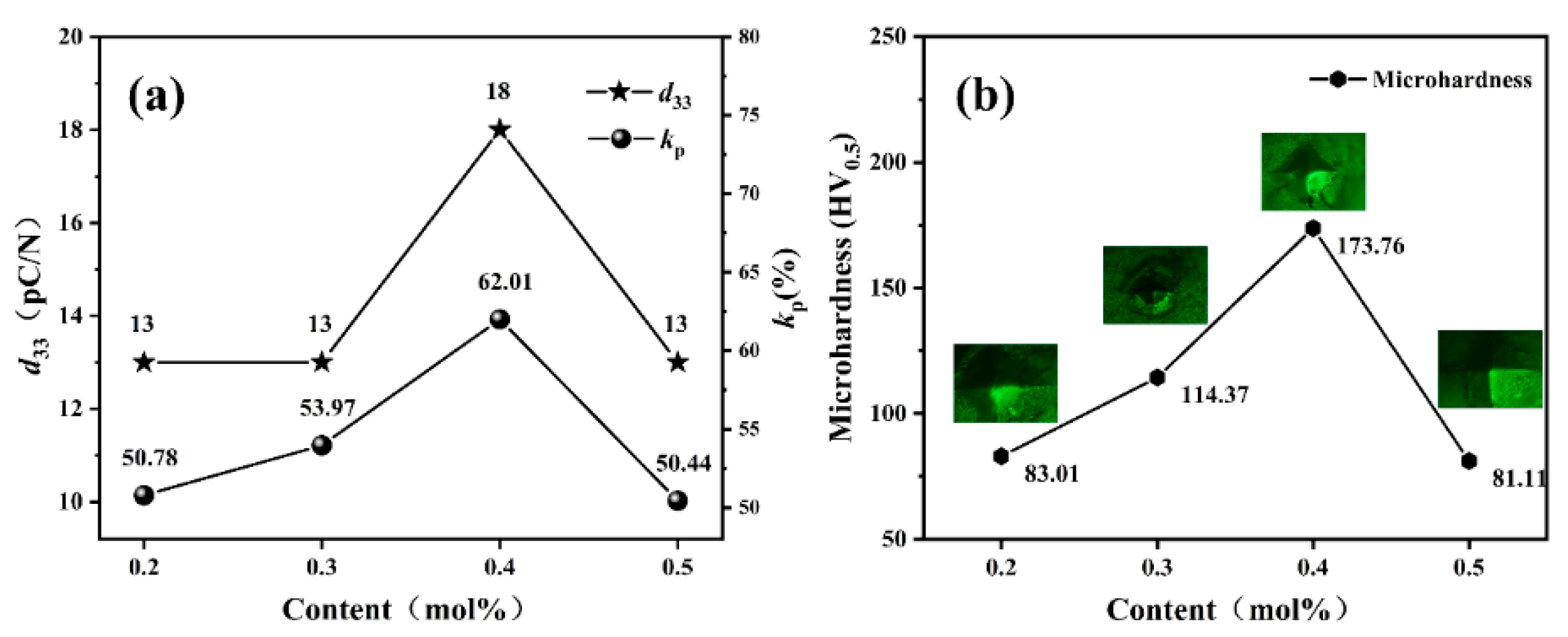
3.3. Piezoelectric Properties

Figure 4a illustrates the test results of the piezoelectric coefficient and the electromechanical coupling coefficient of xGO/BCZT (0.2 mol% ≤ x ≤ 0.5 mol%) ceramics. Notably, the piezoelectric performance of the composite ceramics initially rose and subsequently declined with the increasing content of graphene oxide; optimal piezoelectric properties were achieved at x = 0.4 mol% (d33 = 18 pC/N, kp = 62%). The incorporation of graphene oxide enhanced the electrical conductivity of the composite ceramics, which promoted their piezoelectric response, thus improving the piezoelectric performance [64]. Moreover, owing to its high tensile strength and toughness, graphene oxide may effectively transmit stress to the BCZT ceramic matrix, thereby activating more piezoelectric domains and augmenting the material’s piezoelectric performance. It can be seen from the previous SEM analysis that the grain size of the composite ceramics began to decrease when x = 0.5 mol%. The alteration in piezoelectric properties could be linked to the variation in grain size of composite ceramics. When the grain size was small, significant coupling between grain boundaries and domain walls occurred. This made domain rearrangement and domain wall motion more difficult and consequently reduced the piezoelectric performance of xGO/BCZT ceramics [65,66].

3.4. Mechanical Properties

Figure 4b presents the variations in microhardness for xGO/BCZT (0.2 mol% ≤ x ≤ 0.5 mol%) composite ceramics. The microhardness was obtained by multiple measurements at different points on the sample surface using a 5N test force. Prior to measurement, all samples were coated with silver to achieve as clear a surface as possible. The graph shows that the microhardness of the composite ceramics increased and then decreased with an increase in the content of graphene oxide, reaching a peak at x = 0.4 mol%, where the maximum average microhardness recorded was 173.76 HV0.5. The increase in microhardness was likely due to the refinement of grains caused by the incorporation of graphene oxide, which increased the number and length of grain boundaries and hindered dislocation motion [67]. The subsequent decrease in microhardness beyond this peak was associated with the disruption of van der Waals bonds within the perimeter of the graphene oxide layers, affecting the structural cohesion and thereby reducing hardness [63].

3.5. Water Contact Angle

The hydrophilic–hydrophobic balance was assessed by water contact angle measurements of xGO/BCZT (0.2 mol% ≤ x ≤ 0.5 mol%) composite ceramics, as depicted in Figure 5a. This figure shows that the contact angle of all composite ceramic samples was less than 90°, showing good hydrophilicity. As graphene oxide concentration increased, there was a noticeable decline in contact angles, reflecting enhanced hydrophilic characteristics of the composite ceramics [47]. Notably, at x = 0.4 mol%, the composite ceramics achieved their lowest contact angle of 54.7°, showcasing superior wetting properties. This improved hydrophilicity was attributed to the functional groups of graphene oxide, including hydroxyl and epoxy, which significantly contributed to its water affinity. As a result, the hydrophilic nature and wetting properties of the composites were enhanced by combining graphene oxide with BCZT ceramics [68].

3.6. Cell Cytotoxicity

Cytotoxicity and cell proliferation are crucial metrics for evaluating the biological characteristics of composite ceramics, particularly in applications involving bone defects [69]. Figure 5b presents the experimental results from the CCK-8 cytotoxicity assay for xGO/BCZT (0.2 mol% ≤ x ≤ 0.5 mol%) composite ceramics. The initial observations after one day of culture indicated a marginally higher optical density (OD) value for the 0.4 mol% composite compared with others. Upon cultivation for 4 and 7 days, the composite material containing 0.4 mol% graphene oxide demonstrated a notable increase in cell count. This improvement could be attributed to the enhanced wettability of the composite ceramics due to graphene oxide, which in turn enhanced cell adhesion to the ceramic surface. Additionally, graphene oxide promoted cellular proliferation and differentiation [70]. These findings indicated that graphene oxide doping in BCZT composite ceramics was non-cytotoxic and actually supported cell proliferation, thereby exhibiting excellent biocompatibility.

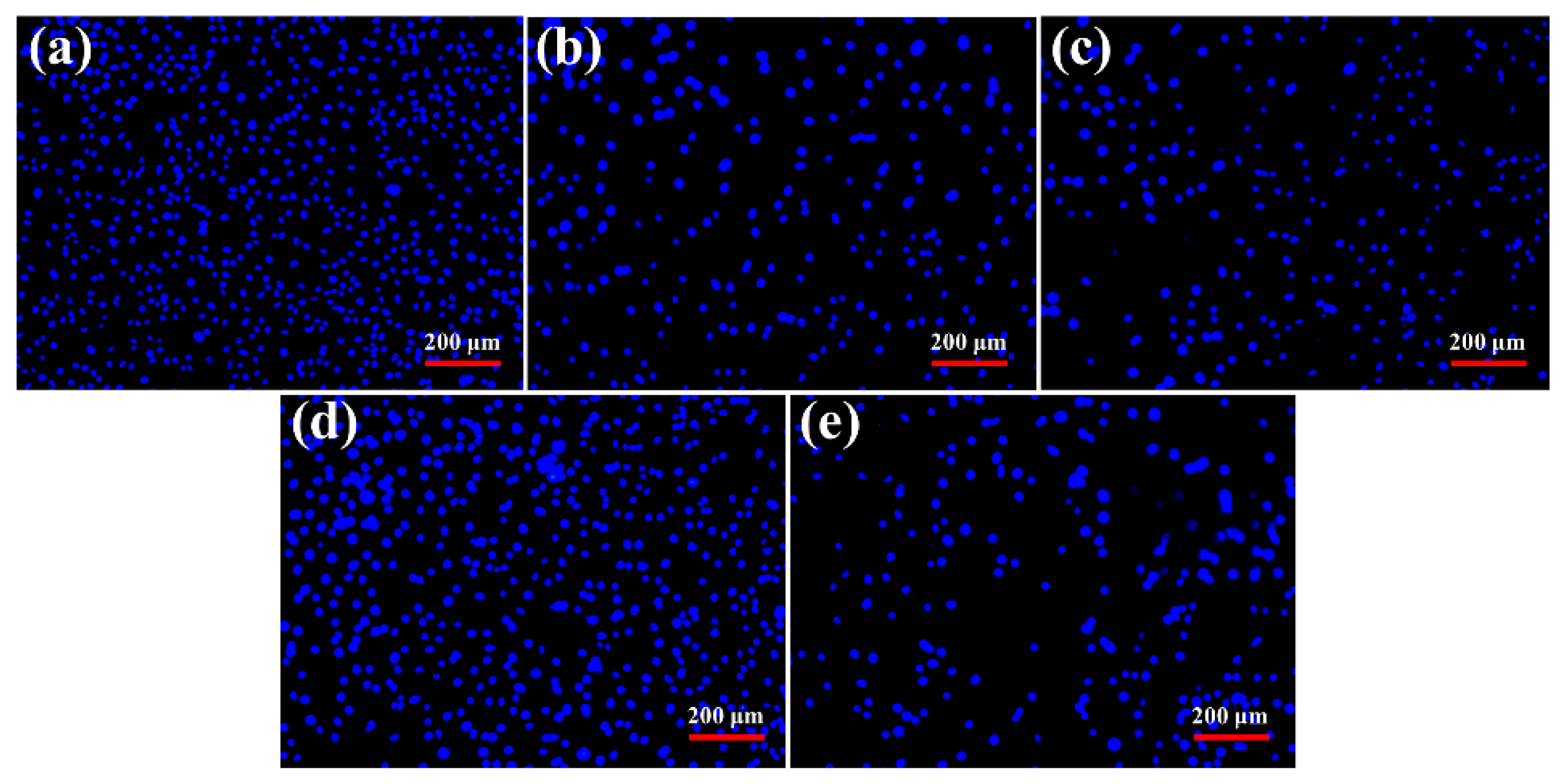
3.7. Apoptosis Evaluation

This paper further substantiated the CCK-8 assay findings through DAPI staining experiments. Fluorescence microscopy facilitated the observation of density and morphological changes in MC3T3-E1 cells incubated for 24 h with xGO/BCZT (0.2 mol% ≤ x ≤ 0.5 mol%) composite ceramics and a control group, as depicted in Figure 6. Cells treated with the composite ceramics showed no changes in chromatin morphology, exhibiting uniform nuclear staining similar to the control group. This demonstrated the biocompatibility of xGO/BCZT composite ceramics across different graphene concentrations, confirming the cytotoxicity study outcomes.

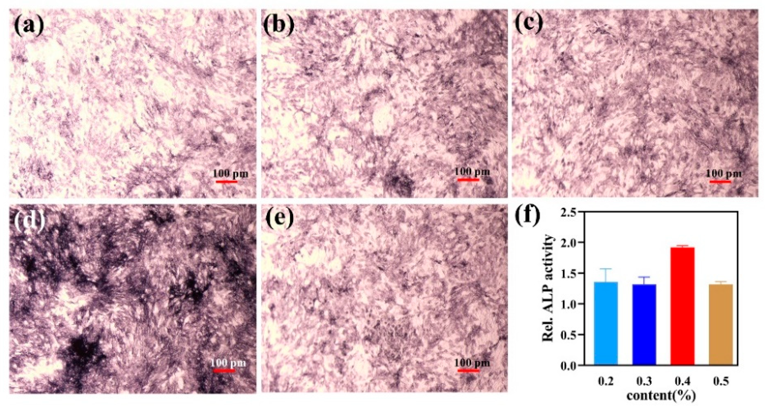
3.8. Alkaline Phosphatase Activity

During the current experiment, MC3T3-E1 cells were seeded onto xGO/BCZT (0.2 mol% ≤ x ≤ 0.5 mol%) ceramics. These cells were then cultured in osteogenic differentiation medium for 7 days, after which the expression and activity of ALP were compared, as illustrated in Figure 7. The results indicated that the most prominent staining for ALP occurred at x = 0.4 mol%, suggesting the highest ALP activity and the most effective promotion of osteoblastic differentiation under this condition. Previous research has shown that the presence of functional groups of graphene oxide can enhance cell adhesion capabilities, which in turn supports cell proliferation and differentiation. The incorporation of GO may influence intracellular signaling pathways, enhancing the expression of genes related to osteogenic differentiation [71]. Furthermore, the xGO/BCZT composite ceramic material might provide a favorable surface for the deposition of minerals such as calcium phosphate, thereby enhancing ALP activity [72]. Therefore, xGO/BCZT composite ceramics have no cytotoxicity and can promote the proliferation and growth of osteoblasts, which is expected to be applied in the treatment of bone defects.

4. Conclusions

In this paper, (1 − x)(Ba0.85Ca0.15Zr0.1Ti0.9)O3−xGO (0.2 mol% ≤ x ≤ 0.5 mol%) composite piezoelectric ceramics were synthesized through fast hot-pressed sintering under a vacuum environment, 1400 °C, and 50 MPa for 30 min. We systematically explored the influence of graphene oxide content on the piezoelectric, mechanical, and biological characteristics. XRD and Raman spectroscopy confirmed the successful synthesis of GO and BCZT ceramics via this method. The analysis revealed a decrease in grain size and an increase in densification of the composite ceramics with rising graphene oxide content, as observed through SEM. Optimal properties, including the piezoelectric constant, electromechanical coupling coefficient, Vickers microhardness, and wettability, were achieved at a graphene oxide concentration of 0.4 mol% (d33 = 18 pC/N, kp = 62%, microhardness =173.76 HV0.5, contact angle = 54.7°). Furthermore, when the content of graphene oxide was 0.4mol%, the CCK-8 assays, DAPI staining, and ALP staining showed that the composite ceramics could enhance the proliferation, growth, and adhesion of cells without significant cytotoxic effects. The results show that the xGO/BCZT composite ceramic has a good biocompatibility and has the potential to be used in the biomedical field.

Author Contributions

B.Z., superior, conceptualization, investigation, writing—original draft, and methodology; Q.L., supervision, writing—review and editing, resources, and project administration; G.T., data curation, formal analysis, investigation, and validation; D.W., data curation, investigation, and formal analysis. All authors have read and agreed to the published version of the manuscript.

Funding

This work is supported by the High-level Innovative Talents Plan of Guizhou Province (Grant No. (2015) 4009) and the National Natural Science Foundation of China (Grant No. 51602066).

Data Availability Statement

Data are contained within the article.

Conflicts of Interest

The authors declare that they have no known competing financial interests or personal relationships that could have appeared to influence the work reported in this paper.

References

  1. Khan, A.F.; Saleem, M.; Afzal, A.; Ali, A.; Khan, A.; Khan, A.R. Bioactive behavior of silicon substituted calcium phosphate based bioceramics for bone regeneration. Mater. Sci. Eng. C-Biomim. Supramol. Syst. 2014, 35, 245–252. [Google Scholar] [CrossRef] [PubMed]
  2. Yılmaz, M.D.; Altuntaş, A. Congenital vomeral bone defect. Am. J. Otolaryngol. 2005, 26, 64–66. [Google Scholar] [CrossRef] [PubMed]
  3. Shen, Y.; Zhang, Y.; Zhou, Z.; Wang, J.; Han, D.; Sun, J.; Chen, G.; Tang, Q.; Sun, W.; Chen, L. Dysfunction of macrophages leads to diabetic bone regeneration deficiency. Front. Immunol. 2022, 13, 990457. [Google Scholar] [CrossRef] [PubMed]
  4. Watanabe, Y.; Harada, N.; Sato, K.; Abe, S.; Yamanaka, K.; Matushita, T. Stem cell therapy: Is there a future for reconstruction of large bone defects? Int. J. Care Inj. 2016, 47, S47–S51. [Google Scholar] [CrossRef] [PubMed]
  5. Lu, M.; Li, Y.; Luo, Y.; Zhang, W.; Zhou, Y.; Tu, C. Uncemented three-dimensional-printed prosthetic reconstruction for massive bone defects of the proximal tibia. World J. Surg. Oncol. 2018, 16, 47. [Google Scholar] [CrossRef] [PubMed]
  6. Liu, Z.; He, X.; Chen, S.; Yu, H. Advances in the use of calcium silicate-based materials in bone tissue engineering. Ceram. Int. 2023, 49, 19355–19363. [Google Scholar] [CrossRef]
  7. Valtanen, R.S.; Yang, Y.P.; Gurtner, G.C.; Maloney, W.J.; Lowenberg, D.W. Synthetic and Bone tissue engineering graft substitutes: What is the future? Int. J. Care Inj. 2021, 52, S72–S77. [Google Scholar] [CrossRef]
  8. Jung, R.E.; Glauser, R.; Schärer, P.; Hämmerle, C.H.; Sailer, H.F.; Weber, F.E. Effect of rhBMP-2 on guided bone regeneration in humans. Clin. Oral Implant. Res. 2003, 14, 556–568. [Google Scholar] [CrossRef] [PubMed]
  9. O’Sullivan, E.D.; Battle, R.K.; Zahra, S.; Keating, J.F.; Marson, L.P.; Turner, D.M. Allosensitization Following Bone Graft. Am. J. Transplant. 2017, 17, 2207–2211. [Google Scholar] [CrossRef]
  10. Wang, W.; Yeung, K.W.K. Bone grafts and biomaterials substitutes for bone defect repair: A review. Bioact. Mater. 2017, 2, 224–247. [Google Scholar] [CrossRef]
  11. Phuoc, H.D.; Hoang, P.N.; Yang, S.; Fraser, D.; Nguyen, V.T. Osseointegrability of 3D-printed porous titanium alloy implant on tibial shaft bone defect in rabbit model. PLoS ONE 2023, 18, e0282457. [Google Scholar] [CrossRef] [PubMed]
  12. Wang, E.; Han, J.; Zhang, X.; Wu, Y.; Deng, X.-L. Efficacy of a mineralized collagen bone-grafting material for peri-implant bone defect reconstruction in mini pigs. Regen. Biomater. 2019, 6, 107–111. [Google Scholar] [CrossRef] [PubMed]
  13. Sivakumar, P.M.; Yetisgin, A.A.; Demir, E.; Sahin, S.B.; Cetinel, S. Polysaccharide-bioceramic composites for bone tissue engineering: A review. Int. J. Biol. Macromol. 2023, 250, 126237. [Google Scholar] [CrossRef] [PubMed]
  14. Koons, G.L.; Diba, M.; Mikos, A.G. Materials design for bone-tissue engineering. Nat. Rev. Mater. 2020, 5, 584–603. [Google Scholar] [CrossRef]
  15. Majee, P.; Dhar, S.; Mitra, P.K.; Lalzawmliana, V.; Nandi, S.K.; Basak, P.; Kundu, B. In vivo bone regeneration analysis of trilayer coated 316L stainless steel implant in rabbit model. J. Mater. Res. 2018, 33, 2106–2117. [Google Scholar] [CrossRef]
  16. Yu, H.; Liu, H.; Shen, Y.; Ao, Q. Synthetic biodegradable polymer materials in the repair of tumor-associated bone defects. Front. Bioeng. Biotechnol. 2023, 11, 1096525. [Google Scholar] [CrossRef] [PubMed]
  17. Li, Z.; Zhu, W.; Bi, S.; Li, R.; Hu, H.; Lin, H.; Tuan, R.S.; Khor, K.A. Incorporating silica-coated graphene in bioceramic nanocomposites to simultaneously enhance mechanical and biological performance. J. Biomed. Mater. Res. A 2020, 108, 1016–1027. [Google Scholar] [CrossRef]
  18. Budharaju, H.; Suresh, S.; Sekar, M.P.; De Vega, B.; Sethuraman, S.; Sundaramurthi, D.; Kalaskar, D.M. Ceramic materials for 3D printing of biomimetic bone scaffolds—Current state-of-the-art & future perspectives. Mater. Des. 2023, 231, 112064. [Google Scholar] [CrossRef]
  19. Fukada, E.; Yasuda, I. On the Piezoelectric Effect of Bone. J. Phys. Soc. Jpn. 1957, 12, 1158–1162. [Google Scholar] [CrossRef]
  20. Tang, H.; Liao, R.; Tian, J. Application and design of piezoelectric materials for bone defect repair. Chin. J. Tissue Eng. Res. 2023, 27, 1117–1125. [Google Scholar] [CrossRef]
  21. Perić Kačarević, Ž.; Rider, P.; Alkildani, S.; Retnasingh, S.; Pejakić, M.; Schnettler, R.; Gosau, M.; Smeets, R.; Jung, O.; Barbeck, M. An introduction to bone tissue engineering. J. Artif. Organs 2019, 43, 69–86. [Google Scholar] [CrossRef] [PubMed]
  22. Bhunia, S.; Karan, S.K.; Chowdhury, R.; Ghosh, I.; Saha, S.; Das, K.; Mondal, A.; Nanda, A.; Khatua, B.B.; Reddy, C.M. Mechanically flexible piezoelectric organic single crystals for electrical energy harvesting. Chem 2024. [Google Scholar] [CrossRef]
  23. Zheng, T.; Zhao, H.; Huang, Y.; Gao, C.; Zhang, X.; Cai, Q.; Yang, X. Piezoelectric calcium/manganese-doped barium titanate nanofibers with improved osteogenic activity. Ceram. Int. 2021, 47, 28778–28789. [Google Scholar] [CrossRef]
  24. Mokhtari, F.; Azimi, B.; Salehi, M.; Hashemikia, S.; Danti, S. Recent advances of polymer-based piezoelectric composites for biomedical applications. J. Mech. Behav. Biomed. Mater. 2021, 122, 104669. [Google Scholar] [CrossRef] [PubMed]
  25. Migliorini, F.; Cuozzo, F.; Torsiello, E.; Spiezia, F.; Oliva, F.; Maffulli, N. Autologous Bone Grafting in Trauma and Orthopaedic Surgery: An Evidence-Based Narrative Review. J. Clin. Med. 2021, 10, 4347. [Google Scholar] [CrossRef]
  26. Gheno, E.; Alves, G.G.; Ghiretti, R.; Mello-Machado, R.C.; Signore, A.; Lourenço, E.S.; Leite, P.E.C.; Mourão, C.F.d.A.B.; Sohn, D.-S.; Calasans-Maia, M.D. “Sticky Bone” Preparation Device: A Pilot Study on the Release of Cytokines and Growth Factors. Materials 2022, 15, 1474. [Google Scholar] [CrossRef] [PubMed]
  27. Tang, Y.; Wu, C.; Wu, Z.; Hu, L.; Zhang, W.; Zhao, K. Fabrication and in vitro biological properties of piezoelectric bioceramics for bone regeneration. Sci. Rep. 2017, 7, 43360. [Google Scholar] [CrossRef] [PubMed]
  28. Liu, W.; Li, X.; Jiao, Y.; Wu, C.; Guo, S.; Xiao, X.; Wei, X.; Wu, J.; Gao, P.; Wang, N.; et al. Biological Effects of a Three-Dimensionally Printed Ti6Al4V Scaffold Coated with Piezoelectric BaTiO3 Nanoparticles on Bone Formation. ACS Appl. Mater. Interfaces 2020, 12, 51885–51903. [Google Scholar] [CrossRef] [PubMed]
  29. Liu, W.; Ren, X. Large piezoelectric effect in Pb-free ceramics. Phys. Rev. Lett. 2009, 103, 257602. [Google Scholar] [CrossRef]
  30. Poon, K.K.; Schafföner, S.; Einarsrud, M.-A.; Glaum, J. Barium titanate-based bilayer functional coatings on Ti alloy biomedical implants. J. Eur. Ceram. Soc. 2021, 41, 2918–2922. [Google Scholar] [CrossRef]
  31. Liu, R.; Qiao, Y.; Zhang, X.; Lu, C.-X. Effects of calcination temperatures on the morphology, architecture and dielectric properties of BCZT ceramics. Bull. Mater. Sci. 2019, 42, 140. [Google Scholar] [CrossRef]
  32. Poon, K.K.; Wurm, M.C.; Evans, D.M.; Einarsrud, M.-A.; Lutz, R.; Glaum, J. Biocompatibility of (Ba,Ca)(Zr,Ti)O3 piezoelectric ceramics for bone replacement materials. J. Biomed. Mater. Res. Part B 2020, 108, 1295–1303. [Google Scholar] [CrossRef] [PubMed]
  33. Tang, Y.; Chen, L.; Duan, Z.; Zhao, K.; Wu, Z. Enhanced compressive strengths and induced cell growth of 1-3-type BaTiO3/PMMA bio-piezoelectric composites. Mater. Sci. Eng. C-Biomimetic Supramol. Syst. 2021, 120, 111699. [Google Scholar] [CrossRef] [PubMed]
  34. Gurunathan, S.; Kim, J.H. Synthesis, toxicity, biocompatibility, and biomedical applications of graphene and graphene-related materials. Int. J. Nanomed. 2016, 11, 1927–1945. [Google Scholar] [CrossRef] [PubMed]
  35. Manohar, C.S.; Kumar, B.S.; Sadhu, S.P.P.; Srimadh, S.K.; Muthukumar, V.S.; Venketesh, S.; Varma, K.B.R. Novel Lead-free biocompatible piezoelectric Hydroxyapatite (HA)—BCZT (Ba0.85Ca0.15Zr0.1Ti0.9O3) nanocrystal composites for bone regeneration. Nanotechnol. Rev. 2019, 8, 61–78. [Google Scholar] [CrossRef]
  36. Chiticaru, E.A.; Ionita, M. Graphene toxicity and future perspectives in healthcare and biomedicine. FlatChem 2022, 35, 100417. [Google Scholar] [CrossRef]
  37. Orek, C.; Bartolomei, M.; Coletti, C.; Bulut, N. Graphene as Nanocarrier for Gold(I)-Monocarbene Complexes: Strength and Nature of Physisorption. Molecules 2023, 28, 3941. [Google Scholar] [CrossRef]
  38. Wu, C.; Xia, L.; Han, P.; Xu, M.; Fang, B.; Wang, J.; Chang, J.; Xiao, Y. Graphene-oxide-modified β-tricalcium phosphate bioceramics stimulate in vitro and in vivo osteogenesis. Carbon 2015, 93, 116–129. [Google Scholar] [CrossRef]
  39. Iatsenko, A.; Sych, O.; Synytsia, A.; Zaremba, P.; Zahorodnia, S.; Nikolenko, A.; Tomila, T.; Bykov, O. Structure and properties of biogenic hydroxyapatite bioceramics modified by graphene-like structures. Appl. Nanosci. 2023, 13, 7477–7483. [Google Scholar] [CrossRef]
  40. Shi, L.; Chen, J.; Teng, L.; Wang, L.; Zhu, G.; Liu, S.; Luo, Z.; Shi, X.; Wang, Y.; Ren, L. The Antibacterial Applications of Graphene and Its Derivatives. Small 2016, 12, 4165–4184. [Google Scholar] [CrossRef]
  41. Norahan, M.H.; Amroon, M.; Ghahremanzadeh, R.; Rabiee, N.; Baheiraei, N. Reduced graphene oxide: Osteogenic potential for bone tissue engineering. IET Nanobiotechnol. 2019, 13, 720–725. [Google Scholar] [CrossRef] [PubMed]
  42. Guo, J.; Cao, G.; Wei, S.; Han, Y.; Xu, P. Progress in the application of graphene and its derivatives to osteogenesis. Heliyon 2023, 9, e21872. [Google Scholar] [CrossRef]
  43. Palmieri, V.; Bugli, F.; Lauriola, M.C.; Cacaci, M.; Torelli, R.; Ciasca, G.; Conti, C.; Sanguinetti, M.; Papi, M.; De Spirito, M. Bacteria Meet Graphene: Modulation of Graphene Oxide Nanosheet Interaction with Human Pathogens for Effective Antimicrobial Therapy. ACS Biomater. Sci. Eng. 2017, 3, 619–627. [Google Scholar] [CrossRef]
  44. Ilyas, K.; Zahid, S.; Batool, M.; Chaudhry, A.A.; Jamal, A.; Iqbal, F.; Nawaz, M.H.; Goerke, O.; Gurlo, A.; Shah, A.T.; et al. In-vitro investigation of graphene oxide reinforced bioactive glass ceramics composites. J. Non-Cryst. Solids 2019, 505, 122–130. [Google Scholar] [CrossRef]
  45. Joy, A.; Unnikrishnan, G.; Megha, M.; Haris, M.; Thomas, J.; Kolanthai, E.; Senthilkumar, M. Hybrid gold/graphene oxide reinforced polycaprolactone nanocomposite for biomedical applications. Surf. Interfaces 2023, 40, 103000. [Google Scholar] [CrossRef]
  46. Guo, S.; Zhu, X.; Li, M.; Shi, L.; Ong, J.L.T.; Jańczewski, D.; Neoh, K.G. Parallel Control over Surface Charge and Wettability Using Polyelectrolyte Architecture: Effect on Protein Adsorption and Cell Adhesion. ACS Appl. Mater. Interfaces 2016, 8, 30552–30563. [Google Scholar] [CrossRef]
  47. Kubiak, K.J.; Wilson, M.C.T.; Mathia, T.G.; Carval, P. Wettability versus roughness of engineering surfaces. Wear 2011, 271, 523–528. [Google Scholar] [CrossRef]
  48. Lasocka, I.; Szulc-Dąbrowska, L.; Skibniewski, M.; Skibniewska, E.; Gregorczyk-Zboroch, K.; Pasternak, I.; Hubalek Kalbacova, M. Cytocompatibility of Graphene Monolayer and Its Impact on Focal Cell Adhesion, Mitochondrial Morphology and Activity in BALB/3T3 Fibroblasts. Materials 2021, 14, 643. [Google Scholar] [CrossRef]
  49. Nakamura, T.; Nakamura-Takahashi, A.; Kasahara, M.; Yamaguchi, A.; Azuma, T. Tissue-nonspecific alkaline phosphatase promotes the osteogenic differentiation of osteoprogenitor cells. Biochem. Biophys. Res. Commun. 2020, 524, 702–709. [Google Scholar] [CrossRef]
  50. Cuenca-Bracamonte, Q.; Yazdani-Pedram, M.; Aguilar-Bolados, H. Electrical Properties of Polyetherimide-Based Nanocomposites Filled with Reduced Graphene Oxide and Graphene Oxide-Barium Titanate-Based Hybrid Nanoparticles. Polymers 2022, 14, 4266. [Google Scholar] [CrossRef]
  51. Wang, X.W.; Zhang, B.H.; Li, Y.Y.; Shi, Y.C.; Sun, L.Y.; Feng, G.; Li, C.L.; Liang, Y.F.; Zheng, Y.P.; Shang, S.Y.; et al. Structure, dielectric, and ferroelectric properties of Ba0.85Ca0.15Zr0.1Ti0.9O3 ceramics sintered at various temperatures. J. Mater. Sci.-Mater. Electron. 2020, 31, 4732–4742. [Google Scholar] [CrossRef]
  52. Ismail, M.F.; Islam, M.A.; Khorshidi, B.; Sadrzadeh, M. Prediction of surface charge properties on the basis of contact angle titration models. Mater. Chem. Phys. 2021, 258, 123933. [Google Scholar] [CrossRef]
  53. Wu, Y.; Wang, B.; Ma, Y.; Huang, Y.; Li, N.; Zhang, F.; Chen, Y. Efficient and large-scale synthesis of few-layered graphene using an arc-discharge method and conductivity studies of the resulting films. Nano Res. 2010, 3, 661–669. [Google Scholar] [CrossRef]
  54. Nanda, S.S.; Yi, D.K.; Kim, K. Study of antibacterial mechanism of graphene oxide using Raman spectroscopy. Sci. Rep. 2016, 6, 28443. [Google Scholar] [CrossRef] [PubMed]
  55. Wang, X.-Y.; Yao, X.; Müllen, K. Polycyclic aromatic hydrocarbons in the graphene era. Sci. China Chem. 2019, 62, 1099–1144. [Google Scholar] [CrossRef]
  56. Ferrari, A.C. Raman spectroscopy of graphene and graphite: Disorder, electron–phonon coupling, doping and nonadiabatic effects. Solid State Commun. 2007, 143, 47–57. [Google Scholar] [CrossRef]
  57. Das, A.; Chakraborty, B.; Sood, A.K. Raman spectroscopy of graphene on different substrates and influence of defects. Bull. Mat. Sci. 2008, 31, 579–584. [Google Scholar] [CrossRef]
  58. Lucchese, M.M.; Stavale, F.; Ferreira, E.H.M.; Vilani, C.; Moutinho, M.V.O.; Capaz, R.B.; Achete, C.A.; Jorio, A. Quantifying ion-induced defects and Raman relaxation length in graphene. Carbon 2010, 48, 1592–1597. [Google Scholar] [CrossRef]
  59. Quispe, L.T.; Menezes, J.W.; Chong, W.; Zegarra, L.B.R.; Armas, L.E.G. Influence of gold nanoholes and nanoslits arrays on Raman spectra and optical reflectance of graphene oxide. Opt. Express 2018, 26, 31253–31263. [Google Scholar] [CrossRef]
  60. Crowther, A.C.; Ghassaei, A.; Jung, N.; Brus, L.E. Strong Charge-Transfer Doping of 1 to 10 Layer Graphene by NO2. ACS Nano 2012, 6, 1865–1875. [Google Scholar] [CrossRef]
  61. Ferrari, A.C.; Meyer, J.C.; Scardaci, V.; Casiraghi, C.; Lazzeri, M.; Mauri, F.; Piscanec, S.; Jiang, D.; Novoselov, K.S.; Roth, S.; et al. Raman spectrum of graphene and graphene layers. Phys. Rev. Lett. 2006, 97, 187401. [Google Scholar] [CrossRef] [PubMed]
  62. Roca, R.A.; Badillo, F.A.L.; Eiras, J.A. Spark plasma sintering and electric conductivity of anatase TiO2 nanoceramics. J. Mater. Sci.-Mater. Electron. 2022, 33, 4375–4387. [Google Scholar] [CrossRef]
  63. Lu, N.; He, G.; Yang, Z.; Li, Y.; Li, J. Fabrication and densification mechanism of MgO/Graphene composites with LiF as additive. Scr. Mater. 2020, 174, 91–94. [Google Scholar] [CrossRef]
  64. Yang, Y.; Peng, S.; Qi, F.; Zan, J.; Liu, G.; Zhao, Z.; Shuai, C. Graphene-assisted barium titanate improves piezoelectric performance of biopolymer scaffold. Mater. Sci. Eng. C-Biomim. Supramol. Syst. 2020, 116, 111195. [Google Scholar] [CrossRef] [PubMed]
  65. Li, S.B.; Wang, C.B.; Li, L.; Shen, Q.; Zhang, L.M. Effect of annealing temperature on structural and electrical properties of BCZT ceramics prepared by Plasma Activated Sintering. J. Alloys Compd. 2018, 730, 182–190. [Google Scholar] [CrossRef]
  66. Zhang, Q.; Cai, W.; Li, Q.; Gao, R.; Chen, G.; Deng, X.; Wang, Z.; Cao, X.; Fu, C. Enhanced piezoelectric response of (Ba,Ca)(Ti, Zr)O3 ceramics by super large grain size and construction of phase boundary. J. Alloys Compd. 2019, 794, 542–552. [Google Scholar] [CrossRef]
  67. Kurapova, O.Y.; Lomakin, I.V.; Sergeev, S.N.; Solovyeva, E.N.; Zhilyaev, A.P.; Archakov, I.Y.; Konakov, V.G. Fabrication of nickel-graphene composites with superior hardness. J. Alloys Compd. 2020, 835, 155463. [Google Scholar] [CrossRef]
  68. Suhaimin, N.S.; Hanifah, M.F.R.; Azhar, M.; Jaafar, J.; Aziz, M.; Ismail, A.F.; Othman, M.H.D.; Rahman, M.A.; Aziz, F.; Yusof, N.; et al. The evolution of oxygen-functional groups of graphene oxide as a function of oxidation degree. Mater. Chem. Phys. 2022, 278, 125629. [Google Scholar] [CrossRef]
  69. Zhang, C.; Wang, F.; Jiang, Z.; Lan, J.; Zhao, L.; Si, P. Effect of graphene oxide on the mechanical, tribological, and biological properties of sintered 3Y–ZrO2/GO composite ceramics for dental implants. Ceram. Int. 2021, 47, 6940–6946. [Google Scholar] [CrossRef]
  70. Liu, H.; Cheng, J.; Chen, F.; Hou, F.; Bai, D.; Xi, P.; Zeng, Z. Biomimetic and Cell-Mediated Mineralization of Hydroxyapatite by Carrageenan Functionalized Graphene Oxide. ACS Appl. Mater. Interfaces 2014, 6, 3132–3140. [Google Scholar] [CrossRef]
  71. Shin, D.S.; Kim, H.G.; Ahn, H.S.; Jeong, H.Y.; Kim, Y.-J.; Odkhuu, D.; Tsogbadrakh, N.; Lee, H.-B.-R.; Kim, B.H. Distribution of oxygen functional groups of graphene oxide obtained from low-temperature atomic layer deposition of titanium oxide. RSC Adv. 2017, 7, 13979–13984. [Google Scholar] [CrossRef]
  72. Li, Q.; Shen, A.; Wang, Z. Enhanced osteogenic differentiation of BMSCs and M2-phenotype polarization of macrophages on a titanium surface modified with graphene oxide for potential implant applications. RSC Adv. 2020, 10, 16537–16550. [Google Scholar] [CrossRef] [PubMed]
Figure 1. The X-ray diffraction pattern of xGO/BCZT composite ceramics at (a) 2θ = 20°~80° and (b) 2θ = 45°~46°.
Coatings 14 00689 g001
Figure 2. (a) Raman spectra and the ratios of Raman peak intensities (b) ID/IG and (c) I2D/IG of xGO/BCZT composite ceramics.
Coatings 14 00689 g002
Figure 3. SEM images of xGO/BCZT ceramics: (ad) original appearance, (a1d1) micrographs after thermal corrosion, and (a2d2) grain size distribution.
Coatings 14 00689 g003
Figure 4. (a) The piezoelectric constant (d33), electromechanical coupling factor (kp) and (b) microhardness diagram of xGO/BCZT ceramics.
Coatings 14 00689 g004
Figure 5. (a) Apparent contact angle and (b) cell cytotoxicity of xGO/BCZT composite ceramics.
Coatings 14 00689 g005
Figure 6. DAPI staining images of xGO/BCZT composite ceramics: (a) x = 0.2 mol%, (b) x = 0.3 mol%, (c) x = 0.4 mol%, (d) x = 0.5 mol%, and (e) control group.
Coatings 14 00689 g006
Figure 7. ALP expression and activity of xGO/BCZT composite ceramics: (a) control group, (b) x = 0.2 mol%, (c) x = 0.3 mol%, (d) x = 0.4 mol%, (e) x = 0.5 mol% and (f) Rel. ALP activity.
Coatings 14 00689 g007
Table 1. EDX data analysis of xGO/BCZT ceramics.
C (at.%)O (at.%)Ca (at.%)Ti (at.%)Ba (at.%)
x = 0 mol%10.1343.084.2620.6221.90
x = 0.2 mol%15.1948.163.0116.5817.05
x = 0.3 mol%10.3245.043.7720.0420.82
x = 0.4 mol%25.5549.532.038.6914.20
x = 0.5 mol%23.9539.842.8816.3516.97
Disclaimer/Publisher’s Note: The statements, opinions and data contained in all publications are solely those of the individual author(s) and contributor(s) and not of MDPI and/or the editor(s). MDPI and/or the editor(s) disclaim responsibility for any injury to people or property resulting from any ideas, methods, instructions or products referred to in the content.

