Article

Improving Surface Antimicrobial Performance by Coating Homogeneous PDA-Ag Micro–Nano Particles

1
Institute of Intelligent Manufacturing and Smart Transportation, Suzhou City University, Suzhou 215104, China
2
Xuhai College, China University of Mining and Technology, Xuzhou 221008, China
3
Henan Energy HR Group Co., Ltd., Zhengzhou 450008, China
*
Author to whom correspondence should be addressed.
Coatings 2024, 14(7), 887; https://doi.org/10.3390/coatings14070887 (registering DOI)
Submission received: 10 June 2024 / Revised: 7 July 2024 / Accepted: 12 July 2024 / Published: 16 July 2024
(This article belongs to the Section Bioactive Coatings and Biointerfaces)

Abstract

:
Implants and other medical devices are prone to bacterial infections on their surface due to bacterial attachment and biofilm formation. In this study, silver nanoparticles were generated in situ onto regulated synthesized polydopamine particles, and the optimal amount of silver nitrate was determined. Composite micro–nano particles were then deposited on a titanium alloy surface. X-ray photoelectron spectroscopy and energy-dispersive X-ray spectroscopy were used to confirm that the titanium alloy surface was successfully coated with PDA-Ag. Scanning electron microscopy, transmission electron microscopy, and three-dimensional optical profilometry were utilized to analysis the morphology of the micro–nano particles and the surface morphology after deposition. The diameters of the polydopamine particles and silver nanoparticles were 150 nm and 25 nm, respectively. The surface roughness values decreased from 0.357 μm to 25.253 μm because of the coated PDA-Ag. Morphology and chemical composition analyses of the modified surface indicated that the PDA-Ag particles were uniformly bonded to the substrate surface. Antimicrobial assays illustrated that the PDA-Ag-modified surface possessed resistance against Escherichia coli and Staphylococcus aureus attachment, with an effectiveness of 96.14 and 85.78%, respectively. This work provides a new strategy and theoretical basis for tackling medical-related surface infections caused by bacterial adhesion.

1. Introduction

Titanium and its alloys are known for their exceptional mechanical strength, corrosion resistance, and biocompatibility, which makes them widely utilized in human bone implants [1]. Biofouling on surfaces, caused by bacterial accumulation and biofilm formation, poses a significant challenge affecting the performance of medical devices [2]. Studies have shown that up to 60% of medical infections are associated with medical equipment or implants, primarily attributed to biofilm presence. Surfaces of medical implants and moist wound areas exhibit high protein adsorption capabilities, creating a conducive environment for bacterial biofilm growth [3]. The increased risk of bacterial adhesion, colonization, and infection can result in delayed healing, implant failure, and the need for additional surgical interventions [4,5,6].
Many methods to improve the antibacterial and biofilm resistance properties of titanium surfaces have been proposed [7,8,9]. Surface modifications including the application of a polymer brush [10], a thin layer [11,12], a polymer multilayer [13], a self-assembled monolayer [14] and the fabrication of textured surfaces [15] are still the focus of current surface antifouling research. The silver ion (Ag+) is an antibacterial agent with little toxicity to human cells and strong antibacterial activity [2]. It destroys the integrity of bacterial cells through direct contact or interactions mediated by positive and negative charges, thus achieving antibacterial and antifouling effects [16,17]. The traditional methods of combining silver nanoparticles (NPs) with titanium surfaces [18,19,20] include ion implantation, electrochemical deposition, ion exchange, plasma spraying, and alloying; however, these methods require special equipment, which increases the operation difficulty and preparation cost [21]. Reducing silver ions onto the substrate surface by a reductant has the advantages of simple operation and low energy consumption. This technique has been studied and applied in various fields by many scholars [2,22,23].
Many reducing substances have been used as reducing agents to reduce silver ions to silver, which aggregates and eventually grows into silver NPs in situ on the surface of the substrate leading to a surface modified by silver NPs with specific properties. Common substances with reducing properties include trisodium citrate dehydrate [24], hydrazine hydrate [25], alkaline glucose solutions [26,27], sodium alginate [28], etc., but the use of these reagents is not simple, and these substances are not safe for humans. Because it contains catechol and amine functional groups, dopamine derived from mussel silk foot protein is an organic substance that can adhere to almost any solid substance and has mild antibacterial properties [29]. In addition, dopamine also has a certain reducibility, and a self-polymerization reaction can reduce silver ions in situ promoting their binding to a substrate surface in the presence of an oxygen-rich weak base [30,31]. After adding dopamine to a silver nitrate (AgNO3) solution, Ag+ can be spontaneously reduced by the catechol group of polydopamine (PDA). The O- and N- sites of PDA act as precursors on the surface of Ag NPs through metal coordination via charge transfer, thus generating Ag NPs with a certain size [32,33,34].
In this study, AgNO3 powder was added to a PDA solution, and the silver ions were reduced to silver NPs in situ by the catechol groups of PDA. The Ag NPs were then loaded onto PDA particles to obtain novel silver NP-loaded submicron PDA particles (PDA-Ag). The effect of silver nitrate concentration on the loading of Ag onto the surface of the PDA microspheres was explored, and the optimal concentration of silver nitrate was determined. The results of the analysis of the surface adhesion and antibacterial properties demonstrated that the composite particles were uniformly grafted on the surface of a titanium substrate and possessed excellent antibacterial properties. This strategy provides a new method and a theoretical basis for obtaining medical metal surfaces with antimicrobial properties.

2. Materials and Methods

2.1. Reagents

Dopamine hydrochloride, propidium iodide (PI), glutaraldehyde (50%), and absolute ethanol were purchased from Aladdin Reagent Co., Ltd. (Aladdin Reagent Co., Ltd., Shanghai, China). Sodium chloride, silver nitrate, crystal violet, and glacial acetic acid were obtained from Sinopharm Chemical Reagent Co., Ltd. (Sinopharm Chemical Reagent Co., Ltd., Shanghai, China). The titanium alloy (TC4) and ammonia (28%–30%) were provided by Shanghai McLean Biochemical Technology Co., Ltd. (Shanghai McLean Biochemical Technology Co., Ltd., Shanghai, China), the phosphate buffer solution (PBS, pH = 7.4) was purchased from Beijing Wokai Biotechnology Co., Ltd. (Beijing Wokai Biotechnology Co., Ltd., Beijing, China), and LB liquid medium and LB agar were purchased from Qingdao high tech Industrial Park Haibo Biotechnology Co., Ltd., Qingdao, China; all chemicals were of analytical grade and were used without further purification.

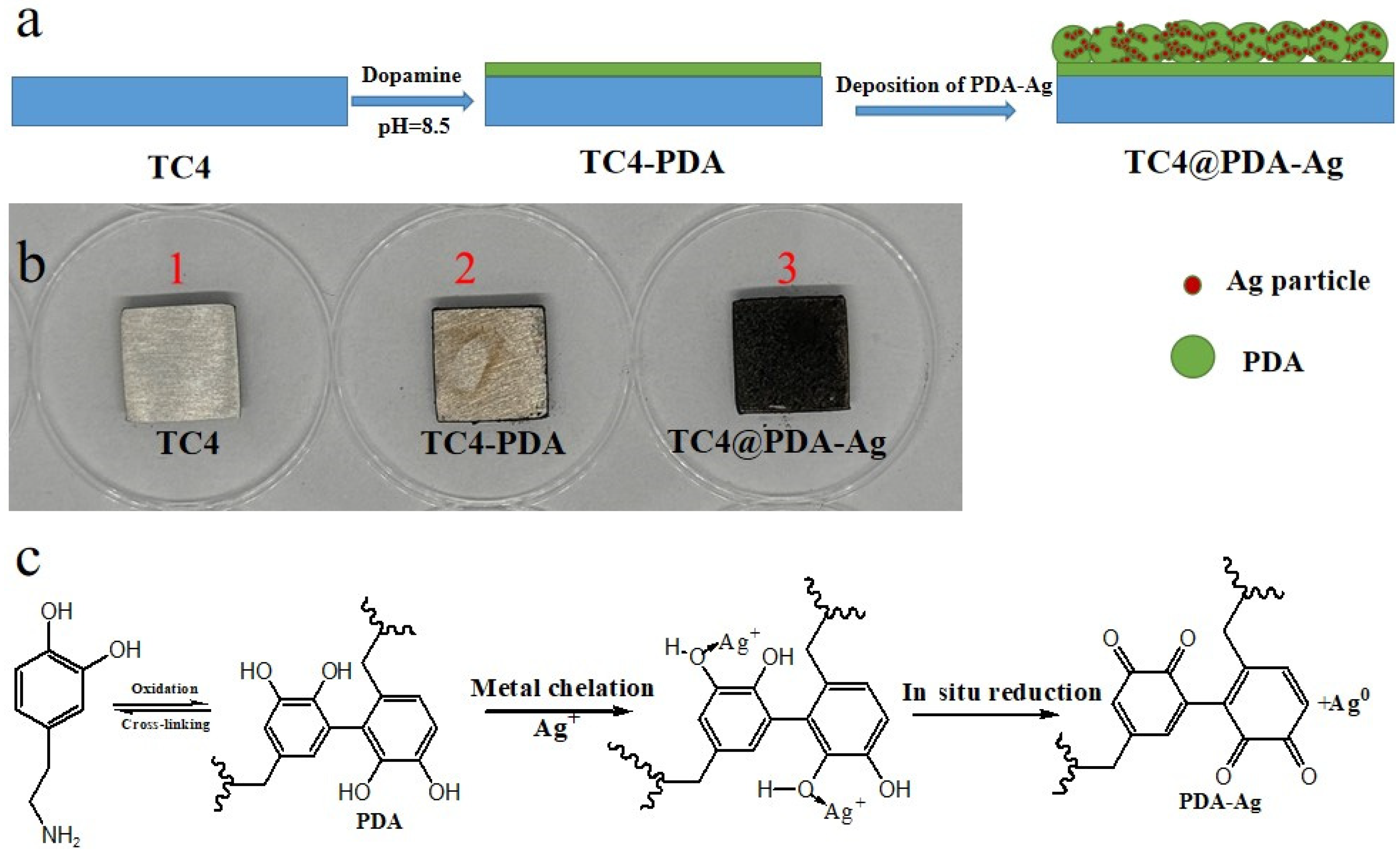
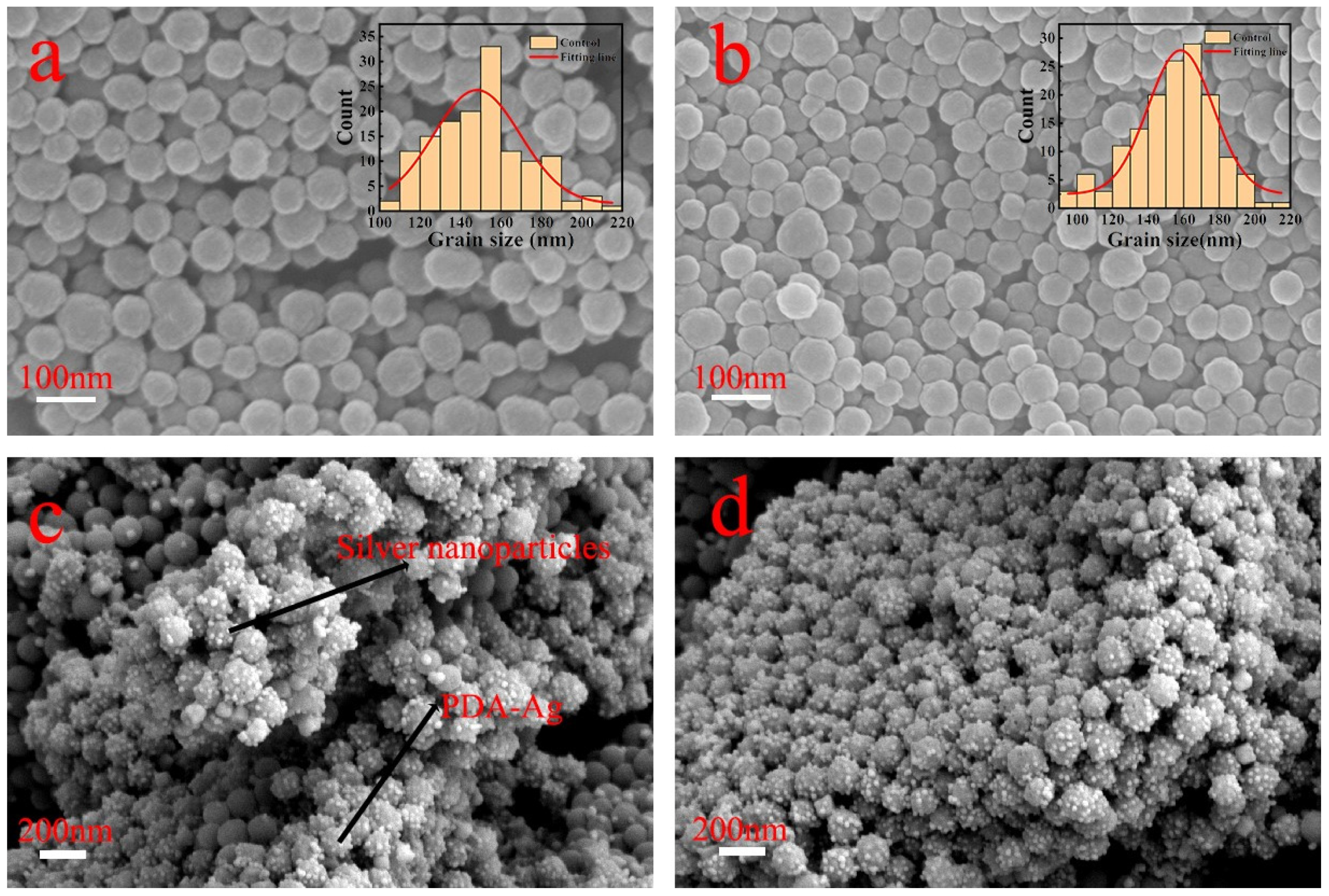
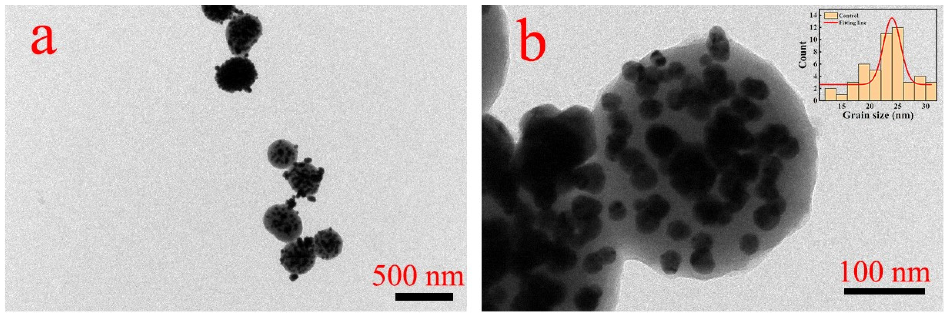
2.2. Preparation of PDA-Ag Composite Particles

Deionized water (90 mL) and ethanol (40 mL) were mixed at room temperature, and the mixture was gently stirred for 30 min after adding an ammonia solution (NH4OH, 2 mL, 25%). Then, 0.5 g of dopamine hydrochloride was dissolved in deionized water (10 mL) and introduced into the mixture [35,36,37]. After 24 h of reaction, a black PDA powder was obtained by centrifugation, washing with deionized water for three times, and freeze-drying.
The obtained PDA was dispersed in deionized water and treated with ultrasound for 15 min. Silver nitrate was dissolved into deionized water (10 mL) to the final concentrations of 0.8, 1.6, 8, and 80 mg/mL. The silver nitrate solutions were then added to the PDA dispersion solution drop by drop. Under the assistance of ultrasound, the reduction reaction was carried out in an ice bath for 10 min, and silver-loaded PDA particles (PDA-Ag) were obtained by centrifugation and washing three times. The product was finally dried 12 h in vacuum at 60 °C for subsequent use.

2.3. Deposition of PDA-Ag onto TC4 Substrates

The titanium alloy substrate was processed into coupons with a thickness of 0.1 cm and a diameter of 1 cm and polished to a smooth state. Then, 0.1 g of the PDA-Ag composite particles was added to 30 mL of deionized water and dispersed uniformly by ultrasound. After this, the pretreated titanium alloy samples were immersed into the prepared solution of PDA-Ag composite particles for 6 h at room temperature under shaking at 30 rpm; the resulting product was denoted as TC4@PDA-Ag. Then, the samples were placed in a 60 °C vacuum drying oven for drying and subsequent use. A schematic diagram of the covalent assembly of PDA-Ag on TC4 is shown in Figure 1.
The surface morphology, particle size, and element distribution of the samples were analyzed by field-emission scanning electron microscopy (SEM, Gemini 300) (ZEISS, Oberkochen, Germany) combined with energy-dispersive X-ray spectroscopy (EDS). The samples were placed on a collection platform after pre-treatment with a gold spray, and their surface morphology was observed after vacuum pumping. Elemental information on the sample surface was collected using the EDS surface scanning mode. A transmission electron microscope (TEM, TECNAI 12) (FEI, Hillsboro, OR, USA) was used to observe the loading of the Ag NPs on the surface of the dopamine particles. Then, 10 µL of a solution containing the PDA-Ag NPs was dropped onto a pretreated copper mesh and air-dried at room temperature. The sample mesh was carefully rinsed with deionized water, followed by ethanol rinsing. Finally, ethanol was absorbed with filter paper, and the sample information was collected using TEM. The surface morphology and roughness of the samples were determined by a three-dimensional optical profilometer (Contour GT-K) (Bruker Corporation, Billerica, MA, USA). The surface elements and their corresponding electronic valence states were analyzed by X-ray photoelectron spectroscopy (XPS, ESCALAB 250Xi) (Thermo Fisher Scientific, Waltham, MA, USA). The samples were placed in the instrument and scanned 64 times during the collection process. A UV–Vis spectrophotometer (L5S, INESA, Shanghai, China) was used to obtain the optical density (OD) value of the sample surface eluate. The adhesion of bacteria to the surface was observed by a confocal scanning microscope (CLSM, TCS SP8 STED) (Leica, Heidelberg, Germany). The samples were placed on the instrument acquisition platform and examined in the fluorescence mode after a rough adjustment using the bright field mode. Two boundaries for image acquisition were determined, and the delimited area was evenly divided to identify five positions; scanned images at the different positions were then collected and merged into the final CLSM image.

2.4. Antimicrobial Properties of TC4@PDA-Ag

Plate counting: Staphylococcus aureus (S. aureus, CMCC (B) 44102) and Escherichia coli (E. coli, CMCC (B) 26003) are the most representative Gram-positive and Gram-negative bacteria; thus, they were selected to carry out antibacterial experiments in this work. The plate counting procedure was described in previous research [38]. Bacterial solutions at concentrations of 107 colony-forming units per microliter (CFU/mL) of S. aureus and E. coli were diluted 100 times with PBS and dropped onto the titanium alloy samples before and after modification (0.1mL/sample). The bacteria were cultured at 37 °C for 6 h. Then, the samples were washed with a 0.9% NaCl solution for 3 times and centrifuged at 4000 rpm for 15 min to recover the bacteria from the sample surface. The bacteria were resuspended in the 0.9% NaCl solution, and the obtained bacteria suspensions (1 mL) were cultured on LB agar plates for 24 h at 37 °C. The antibacterial activity was evaluated according to the following formula:
E = [(N1 − N2)/N1] × 100%
where E is the antibacterial efficiency, N1 is the number of colonies on untreated TC4, and N2 is the number of colonies on TC4 treated with PDA-Ag.
Anti-biofilm test: The anti-biofilm test procedure was described in previous research [2,39]. The samples were placed into bacterial solutions at concentrations of 106 CFU/mL (24-well plates, 1mL/well); the plates were then sealed and incubated under 60 rpm shaking for 24 h at 37 °C. The samples were stained with a 0.1% crystal violet solution (500 μL/well) for 30 min, washed, dried and then washed with a 30% acetic acid solution for 15 min. The washing solution was collected and placed into a colorimetric cup (3 mL/tube) for measuring its OD value using a UV–visible spectrophotometer. A high OD value indicated that a larger biofilm amount was washed away by acetic acid, which means that the bacterial solution produced a greater biofilm amount on the sample surface.
CLSM analysis: The titanium alloy samples before and after the PDA-Ag treatment were placed in the bacterial solution of S. aureus and E. coli at 37 °C for 24 h under shaking at 60 rpm. The bacteria not adhered to the surface were washed with sterile PBS, and the samples were then soaked in a 5% glutaraldehyde solution for 12 h. Half of the samples were stained in a sterile PBS solution with propidium iodide at a concentration of 50 μg/mL for 30 min in the dark and then washed with sterile PBS and stored away from light for the subsequent CLSM observation. Half of the samples were dehydrated with ethanol at concentrations of 25%, 50%, 75%, 90%, and 100% after cleaning and then dried and stored in a constant-temperature drying oven for SEM observation.

2.5. Statistical Analyses

All quantitative measurements were performed in triplicate. The statistical analysis of bacterial adhesion on the sample surface was performed by analyzing the CLSM images via ImageJ software (v1.54j), and the particle size distribution was determined by Nano Measurer software (v1.2.5).

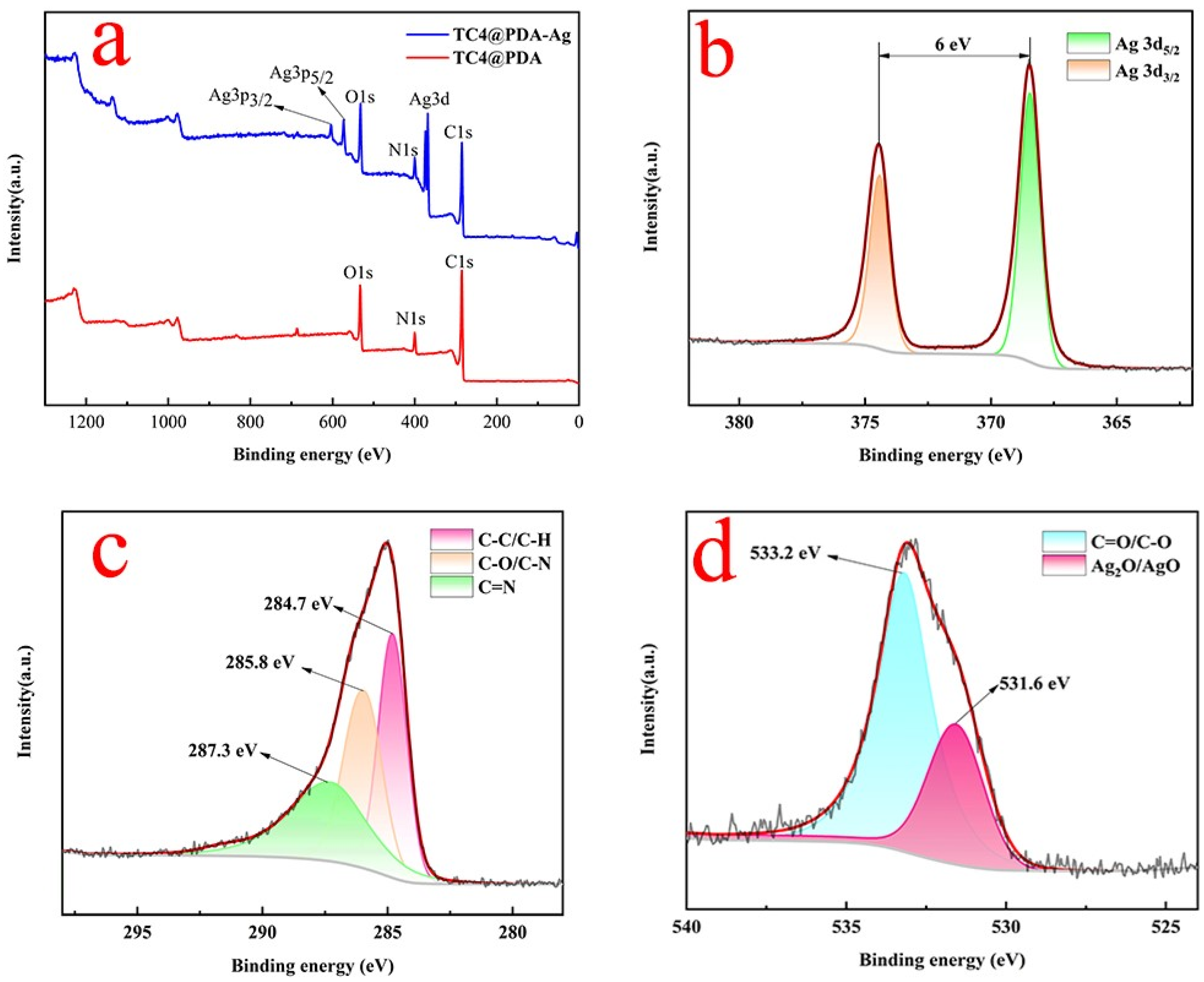
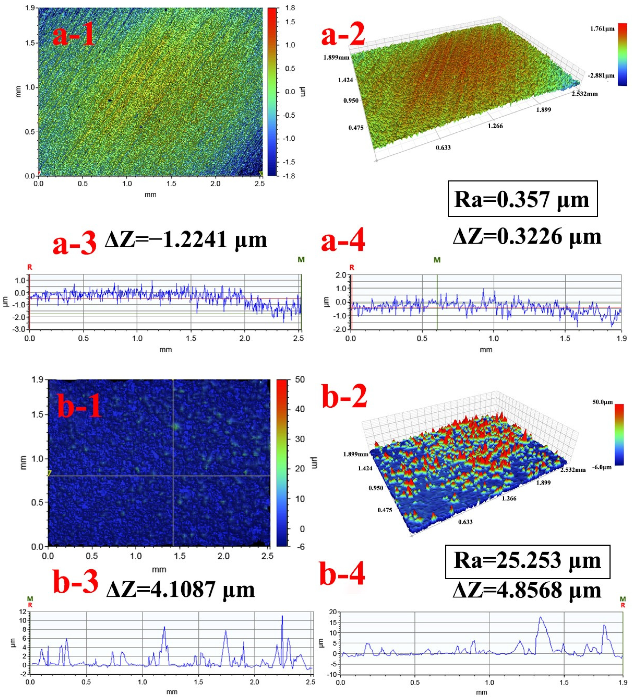
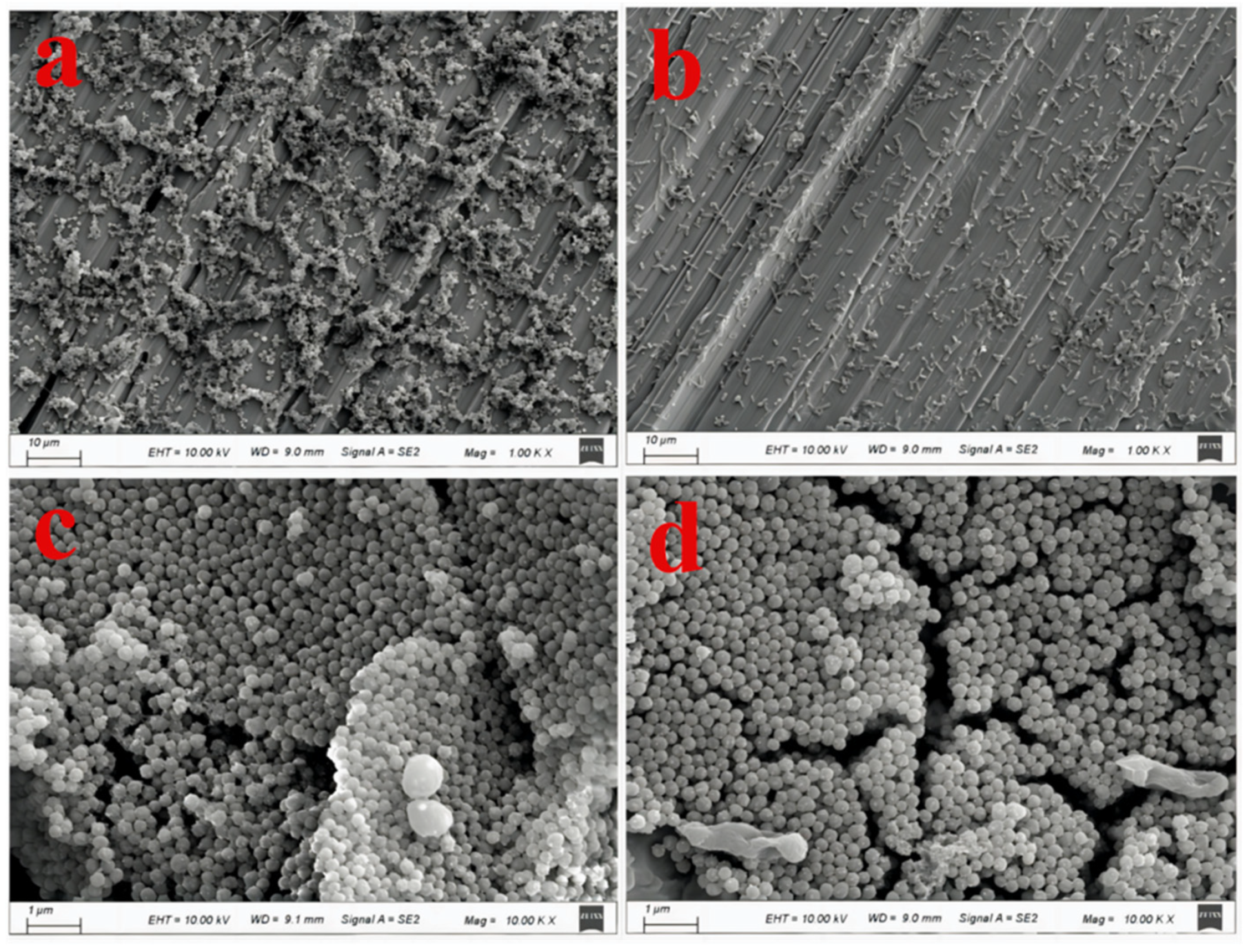
3. Results and Discussion

Dopamine, which is derived from the adhesion protein secreted by the silk foot of marine mussel, is a common coupling agent. It can react with solid surfaces through an oxidative self-polymerization reaction in a weak alkaline solution under oxygen-enriched conditions, and the self-polymerization product PDA, derived by crosslinking via a reverse dismutation reaction, possesses a strong adhesion ability to almost all types of organic and inorganic materials [40]. In this work, dopamine was oxidized and self-polymerized at room temperature in an alkaline water–ethanol system to synthesize PDA NPs. SEM images of PDA NPs polymerized for 24 h and 48 h were obtained. It was found that after 24 h and 48 h of self-polymerization, PDA NPs were successfully prepared and had a relatively uniform size. The size analysis results showed that the size of the PDA NPs was about 160 nm and tended to become stable in 24 h (Figure 2). The contact surface of the PDA NPs with oxygen was reduced, and the subsequent reaction was also hindered due to the reduction of the specific surface area [41].
PDA and its intermediate products have many functional groups, such as catechol, amine, quinone, and imine structures, which reduce silver ions in solution to Ag0 by an in situ reaction. Ag0 was bounded onto the N- and O-sites of PDA, and then slowly grew into silver NPs (Figure 1c) [2]. It was observed by SEM that the concentration of silver nitrate had an impact on the morphology of the PDA-Ag particles. The results showed that as the concentration of silver nitrate increased, the loading amount of silver gradually increased and tended to stabilize at 80 mg/mL (Figure 3). It was also observed from the TEM images that the silver NPs were uniformly distributed on the surface of spherical PDA, with an average diameter of approximately 25 nm (Figure 4a,b). The morphology and particle size of PDA-Ag shown in the TEM images were consistent with those in the SEM images, which proved that Ag was successfully uniformly reduced on the surface of the PDA particles, forming PDA-Ag micro/nano composite particles. The surface morphology of PDA-Ag deposited on the surface of TC4 is shown in Figure 5a,b. It can be seen from the images that under the viscous effect of dopamine, PDA-Ag was uniformly bound to the substrate surface and formed a dense PDA-Ag layer; in addition, there was no change in the particle size of PDA and Ag0, indicating the stability of PDA-Ag in solution during the deposition process.
EDS was utilized to detect the elements of carbon, nitrogen, oxygen, silver, and titanium on TC4@PDA-Ag surface (Figure 6a–d). Nitrogen and silver are not normal components of titanium alloys, and their even distribution after PDA-Ag deposition indicated that PDA and Ag0 had been successfully combined onto the substrate surface. Table 1 reports the EDS analysis results of the TiC4 before and after treatment. It can be seen that the amount of oxygen and titanium elements on TiC4 surface significantly decreased as the surface was covered with PDA-Ag, which caused the C/O mass ratio to increase from 0.116 to 1.015. The C/N mass ratio on the surface of TC4@PDA-Ag reached 5.19, a value very close to the C/N ratio in PDA, indicating that the surface was covered with a dense PDA layer [42].
In order to further identify the substances on the surface of the samples, XPS was applied to detect and quantify the elements. The XPS spectra of TC4@PDA and TC4@PDA-Ag showed that PDA was indeed present on TC4 surface. The N 1s and Ag 3d peaks at around 400 eV and 370 eV in the XPS spectrum indicated that nitrogen from PDA and silver were indeed bonded on the sample surfaces [43]. The high-resolution XPS spectra with the Ag 3d, C 1s, and O 1s peaks are shown in Figure 7 and indicate the element chemical state of TC4@PDA-Ag. The Ag (3d5/2) and Ag (3d3/2) peaks appeared at 368.0 and 374.0 eV, respectively, and the splitting of the 3d doublet of silver was of about 6 eV, which proved the existence of silver on the modified surface [44]. The peaks at 287.3, 285.8, and 284.7 eV in Figure 7c correspond to the C=O bonds or O=C–N bonds of PDA, the carbon atoms in the C–C–O bonds, and the carbon atoms in the C–C and C–H bonds [45]. The peaks at 533.2 and 531.6 eV in Figure 7d were contributed by the C=O and O–H groups of PDA, and the peak at 531.6 eV was, maybe, caused by AgO/Ag2O, due to the partial reduction of Ag+.
The surface morphology and roughness of TC4 and TC4@PDA-Ag were characterized using SEM and 3D optical profilometry. The sample surface was coated by a uniformly distributed micro/nano coating (Figure 5a,b), and the uniform binding of Ag NPs on the surface of PDA particles significantly altered the morphology of the sample surface. The roughness and 3D surface morphology of the TC4 and TC4@PDA-Ag samples were determined and quantified using a 3D optical profiler (Figure 8). The 3D images of TC4@PDA-Ag showed that the surface of the samples was covered with a layer of micro and nano particles, causing a significant change in the roughness of the sample surface. The 3D profile of the surfaces in the X-direction and Y-direction increased from 1.2241 μm and 0.3226 μm to 4.1087 μm and 4.8568 μm, respectively, after PDA-Ag deposition. The surface roughness values (Ra) decreased from 0.357 μm to 25.253 μm. The changes in surface morphology confirmed the successful grafting of PDA-Ag on TiC4 surface.
The antibacterial and antifouling performance of the sample surface was evaluated through plate counting tests. It can be seen that the attachment of S. aureus and E. coli on the surface of the untreated samples corresponded to 1.68 ± 0.13 × 10⁶ colony counts per mL and 0.78 ± 0.07 × 10⁶ colony counts per mL, respectively, through reduction and conversion. It can be seen that the number of S. aureus and E. coli colonies on LB plates decreased significantly after incubation with PDA-Ag-treated surfaces, and the decrease in adhesion percentage for S. aureus and E. coli on TC4@PDA-Ag surface was close to 100% (Figure 9). Crystal violet staining was utilized to assess the surface anti-biofilm ability, and the results showed that a large amount of biofilm was accumulated on the untreated surfaces. The OD values of the untreated surfaces incubated with S. aureus and E. coli were 1.77 ± 0.04 and 1.61 ± 0.04, and the OD values decreased to 0.18 ± 0.03 and 0.76 ± 0.09 after PDA-Ag deposition, which demonstrated that less bacteria attached and less biofilm formed on TC4@PDA-Ag. The above results showed that the reduction in E. coli and its biofilm was significantly higher than that measured for S. aureus, indicating that TC4@PDA-Ag possessed stronger ability to resist E. coli than S. aureus.
Laser confocal scanning microscopy images were used to analyze and quantify bacterial adhesion on the sample surface before and after PDA-Ag deposition (Figure 10). A large number of bacteria adhered to the TC4 surface, while the number of attached bacteria decreased significantly after PDA-Ag deposition. ImageJ analysis showed that the coverage percentages of E. coli and S. aureus on the sample surface decreased from 4.279% and 41.432% to 0.165% and 5.893%, respectively, after PDA-Ag deposition. The bacterial attachment ratio decreased by 96.14% and 85.78%, which illustrated that the PDA-Ag-deposited surfaces possessed strong antibacterial capacity due to the antibacterial performance of Ag. The SEM images showed that a high number of E. coli and S. aureus attached on the untreated surfaces, while few bacteria appeared on the PDA-Ag treated surfaces, which demonstrated that the surfaces exhibited significant antibacterial adhesion performance after PDA-Ag modification (Figure 11). From the above results, it can be seen that the surface modified with PDA-Ag had a stronger ability to resist E. coli than S. aureus, which is consistent with the results of plate counting and biofilm quantification. This result may be related to charge interactions between silver ions and bacterial cell membranes, as well as to the difficulty of Ag NPs entering bacterial cells [46]. Gram-positive bacteria have a much thicker peptidoglycan cell wall structure compared to Gram-negative bacteria, which have a thinner cell wall; the Ag NPs released from TC4@PDA-Ag are more likely to enter Gram-negative bacteria, killing them [47,48].

4. Conclusions

Medical devices and implants are prone to bacterial deposition on their surfaces. PDA particles loaded with Ag NPs were successfully synthesized and homogeneously deposited on the surface of titanium alloy samples to tackle infections caused by bacterial accumulation on the surface of medical supplies. Ag+ was reduced to Ag0 via the chelation of the catechol groups of PDA and Ag+ and grew into NPs with a diameter of approximately 10–20 nm. XPS and EDS analyses demonstrated that PDA-Ag particles were actually deposited onto the TC4 surface, causing significant changes in surface element content and valence state of the substrate surface. The SEM, TEM, and 3D optical profilometer results illustrated that particle morphology and surface topography underwent unexpected changes because of the PDA-Ag modification. Antimicrobial testing of TC4@PDA-Ag was conducted using S. aureus and E. coli; we found that the bacterial attachment ratio decreased by 96.14% and 85.78%, and the biofilm resistance ratio was 89.83% and 52.80%, respectively. The antimicrobial testing results suggest that the PDA-Ag-modified surfaces possessed strong antibacterial and anti-biofilm properties.

Author Contributions

Conceptualization, S.W. and Z.C.; methodology, S.W. and Z.C.; validation, S.W., and F.M.; formal analysis, S.W. and Z.C.; investigation, S.W.; data curation, S.W. and Z.C.; writing—original draft preparation, S.W.; writing—review and editing, F.M. and Z.C.; supervision, Z.C.; funding acquisition, S.W. and Z.C. All authors have read and agreed to the published version of the manuscript.

Funding

This research was funded by the “Qing Lan Project” in Jiangsu Province, Grant Number: 2022.

Institutional Review Board Statement

Not applicable.

Informed Consent Statement

Not applicable.

Data Availability Statement

Data are contained within the article.

Acknowledgments

The authors are grateful for the facilities and other support given by the “Qing Lan Project” in Jiangsu Province.

Conflicts of Interest

Author Fanping Meng was employed by the company Henan Energy HR Group Co. Ltd. The remaining authors declare that the research was conducted in the absence of any commercial or financial relationships that could be construed as a potential conflict of interest.

References

  1. Cheng, Y.F.; Zhang, J.Y.; Wang, Y.B.; Li, C.M.; Lu, Z.S.; Hu, X.F.; Xu, L.Q. Deposition of catechol-functionalized chitosan and silver nanoparticles on biomedical titanium surfaces for antibacterial application. Mater. Sci. Eng. C 2019, 98, 649–656. [Google Scholar] [CrossRef] [PubMed]
  2. Cao, P.; He, X.; Xiao, J.; Yuan, C.; Bai, X. Covalent bonding of AgNPs to 304 stainless steel by reduction in situ for antifouling applications. Appl. Surf. Sci. 2018, 452, 201–209. [Google Scholar] [CrossRef]
  3. Lv, L.; Deegan, A.; Musa, F.; Xu, T.; Yang, Y. The effects of biomimetic ally conjugated VEGF on osteogenesis and angiogenesis of MSCs (human and rat) and HUVECs co-cultu remodels. Colloids Surf. B Biointerfaces 2018, 167, 550–559. [Google Scholar]
  4. Arciola, C.R.; Campoccia, D.; Montanaro, L. Implant infections: Adhesion, biofilm formation and immune evasion. Nat. Rev. Microbiol. 2018, 16, 397–409. [Google Scholar] [CrossRef] [PubMed]
  5. Yavari, S.A.; Castenmiller, S.M.; van Strijp, J.A.G.; Croes, M. Combating implant infections: Shifting focus from bacteria to host. Adv. Mater. 2020, 32, 2002962. [Google Scholar] [CrossRef] [PubMed]
  6. Chu, X.; Yang, F.; Tang, H. Recent advance in polymer coatings combating bacterial adhesion and biofilm formation. Chin. J. Chem. 2022, 40, 2988–3000. [Google Scholar] [CrossRef]
  7. Zhang, Z.; Bai, Y.; Han, R.; Yu, Q.; Yang, R.; Zhang, X. Improving antifouling functions of titanium alloys by robust slippery liquid-infused porous surfaces with tailored multiscale structures. Chem. Eng. J. 2023, 478, 147342. [Google Scholar] [CrossRef]
  8. Wei, Z.; Li, K.; Wang, S.; Wen, L.; Xu, L.; Wang, Y.; Chen, Z.; Li, W.; Qiu, H.; Li, X.; et al. Controllable AgNPs encapsulation to construct biocompatible and antibacterial titanium implant. Front. Bioeng. Biotechnol. 2022, 10, 1056419. [Google Scholar] [CrossRef]
  9. Yue, D.; Jiang, X.; Yu, H.; Sun, D. In-situ fabricated hierarchical nanostructure on titanium alloy as highly stable and durable super-lubricated surface for anti-biofouling in marine engineering. Chem. Eng. J. 2023, 463, 142389. [Google Scholar] [CrossRef]
  10. Higaki, Y.; Kobayashi, M.; Murakami, D.; Takahara, A. Anti-fouling behavior of polymer brush immobilized surfaces. Polym. J. 2016, 48, 325–331. [Google Scholar] [CrossRef]
  11. Song, J.; Zhu, Y.; Zhang, J.; Yang, J.; Du, Y.; Zheng, W.; Wen, C.; Zhang, Y.; Zhang, L. Encapsulation of AgNPs within zwitterionic hydrogels for highly efficient and antifouling catalysis in biological environments. Langmuir 2019, 35, 1563–1570. [Google Scholar] [CrossRef] [PubMed]
  12. Shimabukuro, M.; Morinobu, M.; Tsuchiya, A.; Kishida, R.; Kawashita, M.; Ishikawa, K. Antibacterial and osteogenic thin films on Ti-6Al-4V surface formed bypassivation process in copper hydroxide solution. Sci. Technol. Adv. Mater. 2024, 25, 2303327. [Google Scholar] [CrossRef] [PubMed]
  13. Wu, Y.; Jiang, M.; Zhao, J.; Cai, Y.; Li, X.; Yang, X.; Jiang, H.; Sun, Y.; Wei, N.; Liu, Y.; et al. Polyelectrolyte-based antifouling and pH-responsive multilayer coatings for reverse osmosis membrane. Colloids Surf. A Physicochem. Eng. Asp. 2023, 679, 132642. [Google Scholar] [CrossRef]
  14. Christoff-Tempesta, T.; Deiss-Yehiely, E.; Dromel, P.C.; Uliassi, L.D.; Chazot, C.A.C.; Postelnicu, E.; Hart, A.J.; Spector, M.; Hammond, P.T.; Ortony, J.H. Antifouling surface coatings from self-assembled zwitterionic aramid amphiphile nanoribbons. Adv. Mater. Interfaces 2022, 9, 2200311. [Google Scholar] [CrossRef]
  15. Chen, L.; Duan, Y.; Cui, M.; Huang, R.; Su, R.; Qi, W.; He, Z. Biomimetic surface coatings for marine antifouling: Natural antifoulants, synthetic polymers and surface microtopography. Sci. Total Environ. 2021, 766, 144469. [Google Scholar] [CrossRef] [PubMed]
  16. Ma, Z.; Liu, J.; Shen, G.; Zheng, X.; Pei, Y.; Tang, K. In-situ synthesis and immobilization of silver nanoparticles on microfibrillated cellulose for long-term antibacterial applications. Cellulose 2021, 28, 6287–6303. [Google Scholar] [CrossRef]
  17. He, M.; Li, W.; Chen, J.; Zhang, Z.; Wang, X.; Yang, G. Immobilization of silver nanoparticles on cellulose nanofibrils incorporated into nanofiltration membrane for enhanced desalination performance. mpj Clean Water 2022, 5, 64. [Google Scholar] [CrossRef]
  18. Kumari, R.; Barsainya, M.; Singh, D.P. Biogenic synthesis of silver nanoparticle by using secondary metabolites from Pseudomonas aeruginosa DM1 and its anti-algal effect on Chlorella vulgaris and Chlorella pyrenoidosa. Environ. Sci. Pollut. Res. 2017, 24, 4645–4654. [Google Scholar] [CrossRef]
  19. Debiemme-Chouvy, C.; Cachet, H. Electrochemical (pre)treatments to prevent biofouling. Curr. Opin. Electrochem. 2018, 11, 48–54. [Google Scholar] [CrossRef]
  20. Hirsch, U.M.; Teuscher, N.; Rühl, M.; Heilmann, A. Plasma-enhanced magnetron sputtering of silver nanoparticles on reverse osmosis membranes for improved antifouling properties. Surf. Interfaces 2019, 16, 1–7. [Google Scholar] [CrossRef]
  21. Ferraris, S.; Spriano, S. Antibacterial titanium surfaces for medical implants. Mater. Sci. Eng. C 2016, 61, 965–978. [Google Scholar] [CrossRef] [PubMed]
  22. Suresh, D.; Goh, P.S.; Wong, T.W.; Zhang, L.; Ismail, A.F. In-situ complexation of silver nanoparticle on thin film composite reverse osmosis membrane for improving desalination and anti-biofouling performance. Desalination 2024, 569, 117040. [Google Scholar] [CrossRef]
  23. Qi, L.; Liu, Z.; Wang, N.; Hu, Y. Facile and efficient in situ synthesis of silver nanoparticles on diverse filtration membrane surfaces for antimicrobial performance. Appl. Surf. Sci. 2018, 456, 95–103. [Google Scholar] [CrossRef]
  24. Suriati1, G.; Mariatti, M.; Azizan, A. Synthesis of silver nanoparticles by chemical reduction method: Effect of reducing agent and surfactant concentration. Int. J. Automot. Mech. Eng. 2014, 10, 1920–1927. [Google Scholar] [CrossRef]
  25. Chumachenko, V.; Kutsevol, N.; Rawiso, M.; Schmutz, M.; Blanck, C. In situ formation of silver nanoparticles in linear and branched polyelectrolyte matrices using various reducing agents. Nanoscale Res. Lett. 2014, 9, 164. [Google Scholar] [CrossRef] [PubMed]
  26. Swensson, B.; Ek, M.; Gray, D.G. In situ preparation of silver nanoparticles in paper by reduction with alkaline glucose solutions. ACS Omega 2018, 3, 9449–9452. [Google Scholar] [CrossRef] [PubMed]
  27. Pauzi, N.; Mohamad, S.; Ghazali, S.; Jamari, S.S. Evaluation of glucose reduction for silver nanoparticles synthesis with nanocrystalline cellulose matrix. BioNanoScience 2023, 13, 1695–1702. [Google Scholar] [CrossRef]
  28. Chen, K.; Wang, F.; Liu, S.; Wu, X.; Xu, L.; Zhang, D. In situ reduction of silver nanoparticles by sodium alginate to obtain silver-loaded composite wound dressing with enhanced mechanical and antimicrobial property. Int. J. Biol. Macromol. 2020, 148, 501–509. [Google Scholar] [CrossRef]
  29. Lee, H.; Dellatore, S.M.; Miller, W.M.; Messersmith, P.B. Mussel-inspired surface chemistry for multifunctional coatings. Science 2007, 318, 426. [Google Scholar] [CrossRef]
  30. Wang, J.; Cui, Y.; Zhang, B.; Sun, S.; Xu, H.; Yao, M.; Wu, D.; Wang, Y. Polydopamine-Modified functional materials promote bone regeneration. Mater. Des. 2024, 238, 112655. [Google Scholar] [CrossRef]
  31. Qiu, W.; Wu, G.; Xu, Z. Robust coatings via catechol-amine codeposition: Mechanism, kinetics, and application. ACS Appl. Mater. Interfaces 2018, 10, 5902–5908. [Google Scholar] [CrossRef] [PubMed]
  32. Bekalé, L.; Barazzouk, S.; Hotchandani, S. Nanosilver could usher in nextgeneration photoprotective agents for magnesium porphyrins. Part. Part. Syst. Charact. 2014, 31, 843–850. [Google Scholar] [CrossRef]
  33. Lingamgunta, S.; Xiao, Y.; Choi, H.; Christiea, G.; Fruk, L. Microwave-enhanced antibacterial activity of polydopamine–silver hybrid nanoparticles. RSC Adv. 2024, 14, 8331–8340. [Google Scholar] [CrossRef] [PubMed]
  34. Yao, S.; Guo, D.; Han, S.; Fu, Z.; Lyu, S.; Li, J.; Lu, Y. Polydopamine-assisted immobilization of metallic nanoparticles confined regionally in bamboo microchannels as continuous-flow microreactors for enhanced catalysis. Chem. Eng. J. 2024, 492, 152327. [Google Scholar] [CrossRef]
  35. Luo, H.; Gu, C.; Zheng, W.; Dai, F.; Wang, X.; Zheng, Z. Facile synthesis of novel size-controlled antibacterial hybrid spheres using silver nanoparticles loaded with poly-dopamine spheres. RSC Adv. 2015, 5, 13470–13477. [Google Scholar] [CrossRef]
  36. Wu, C.; Zhang, G.; Xia, T.; Li, Z.; Zhao, K.; Deng, Z.; Guo, D.; Peng, B. Bioinspired synthesis of polydopamine/Ag nanocomposite particles with antibacterial activities. Mater. Sci. Eng. C 2015, 55, 155–165. [Google Scholar] [CrossRef]
  37. Nieto, C.; Marcelo, G.; Vega, M.; del Valle, E.M.M. Antineoplastic behavior of polydopamine nanoparticles prepared in different water/alcohol media. Colloids Surf. B Biointerfaces 2021, 199, 111506. [Google Scholar] [CrossRef] [PubMed]
  38. Bagchi, D.; Bhattacharya, A.; Dutta, T.; Nag, S.; Wulferding, D.; Lemmens, P.; Pal, S.K. Nano MOF entrapping hydrophobic photosensitizer for dual-stimuli-responsive unprecedented therapeutic action against drug-resistant bacteria. ACS Appl. Bio Mater. 2019, 2, 1772–1780. [Google Scholar] [CrossRef]
  39. Niyonshuti, I.I.; Krishnamurthi, V.R.; Okyere, D.; Song, L.; Benamara, M.; Tong, X.; Wang, Y.; Chen, J. Polydopamine surface coating synergizes the antimicrobial activity of silver nanoparticles. ACS Appl. Mater. Interfaces 2020, 12, 39937–40986. [Google Scholar] [CrossRef]
  40. Yu, Y.; Li, X.; Li, J.; Li, D.; Wang, Q.; Teng, W. Dopamine-assisted co-deposition of hydroxyapatite-functionalised nanoparticles of polydopamine on implant surfaces to promote osteogenesis in environments with high ROS levels. Mater. Sci. Eng. C 2021, 131, 112473. [Google Scholar] [CrossRef]
  41. Kim, H.W.; McCloskey, B.D.; Choi, T.H.; Lee, C.; Kim, M.-J.; Freeman, B.D.; Park, H.B. Oxygen concentration control of dopamine-induced high uniformity surface coating chemistry. ACS Appl. Mater. Interfaces 2013, 5, 233–238. [Google Scholar] [CrossRef] [PubMed]
  42. Cao, P.; Du, C.; He, X.; Zhang, C.; Yuan, C. Modification of a derived antimicrobial peptide on steel surface for marine bacterial resistance. Appl. Surf. Sci. 2020, 510, 145512. [Google Scholar] [CrossRef]
  43. Qi, X.; Huang, Y.; You, S.; Xiang, Y.; Cai, E.; Mao, R.; Pan, W.; Tong, X.; Dong, W.; Ye, F.; et al. Engineering robust Ag-decorated polydopamine nano-photothermal platforms to combat bacterial infection and prompt wound healing. Adv. Sci. 2022, 9, 2106015. [Google Scholar] [CrossRef] [PubMed]
  44. Cant, N.E.; Critchley, K.; Zhang, H.-L.; Evans, S.D. Surface functionalisation for the self-assembly of nanoparticle/polymer multilayer films. Thin Solid Film. 2003, 426, 31–39. [Google Scholar] [CrossRef]
  45. Cao, P.; Yuan, C.; Xiao, J.; He, X.; Bai, X. A biofilm resistance surface yielded by grafting of antimicrobial peptides on stainless steel surface. Surf. Interface Anal. 2018, 50, 516–521. [Google Scholar] [CrossRef]
  46. Ahmad, S.A.; Das, S.S.; Khatoon, A.; Ansari, M.T.; Afzal, M.; Hasnain, M.S.; Nayak, A.K. Bactericidal activity of silver nanoparticles: A mechanistic review. Mater. Sci. Energy Technol. 2020, 3, 756–769. [Google Scholar] [CrossRef]
  47. Pasquina-Lemonche, L.; Burns, J.; Turner, R.D.; Kumar, S.; Tank, R.; Mullin, N.; Wilson, J.S.; Chakrabarti, B.; Bullough, P.A.; Foster, S.J.; et al. The architecture of the Gram-positive bacterial cell wall. Nature 2020, 582, 294–297. [Google Scholar] [CrossRef]
  48. Panáček, A.; Kvítek, L.; Smékalová, M.; Večeřová, R.; Kolář, M.; Röderová, M.; Dyčka, F.; Šebela, M.; Prucek, R.; Tomanec, O.; et al. Bacterial resistance to silver nanoparticles and how to overcome it. Nat. Nanotechnol. 2018, 13, 65–71. [Google Scholar] [CrossRef]
Figure 1. A schematic of PDA-Ag formation and the covalent assembly of PDA-Ag on TC4. (a) Preparation process diagram of TC4@PDA-Ag; (b) The actual pictures of TC4, TC4-PDA and TC4@PDA-Ag; (c) The reaction principle diagram of PDA-Ag 2.4. Characterization of TC4-PDA-Ag.
Coatings 14 00887 g001
Figure 2. SEM images of PDA (a,b) and PDA-Ag (c,d) obtained after a reaction time of 24 h (a,c) and 48 h (b,d).
Coatings 14 00887 g002
Figure 3. Optimization of PDA-Ag preparation at different silver nitrate concentrations. (a) 0.8 mg/mL; (b) 1.6 mg/mL; (c) 8 mg/mL; (d) 80 mg/mL.
Coatings 14 00887 g003
Figure 4. (a) TEM images and particle size analysis of PDA-Ag; (b) enlarged image from (a).
Coatings 14 00887 g004
Figure 5. (a) SEM image of TC4@PDA-Ag; (b) enlarged image from (a).
Coatings 14 00887 g005
Figure 6. SEM and EDS images of TC4@PDA-Ag. (a) SEM image; (b) element C; (c) element N; (d) element O; (e) element Ag; (f) EDS spectra.
Coatings 14 00887 g006
Figure 7. (a) Full XPS spectra of TC4@PDA-Ag and TC4@PDA; (b) Ag 3d peaks, (c) N 1s peaks, and (d) O 1s peaks of TC4@PDA-Ag.
Coatings 14 00887 g007
Figure 8. The 3D optical profiler images of TC4 (a) and TC4@PDA-Ag (b); 1: main images; 2: 3D images; 3: X-axis profile; 4: Y-axis profile.
Coatings 14 00887 g008
Figure 9. Bacterial plate counting and anti-biofilm capacity of the samples. (a) TC4 treated with E. coli; (b) TC4@PDA-Ag treated with E. coli; (c) TC4 treated with S. aureus; (d) TC4@PDA-Ag treated with S. aureus; (e) Acetic acid elution solution after CV staining; (f) OD590nm analysis of biofilm eluent.** p < 0.001, very significant.
Coatings 14 00887 g009
Figure 10. CLSM images of surfaces with attached bacteria. (a) E. coli on TC4; (b) S. aureus on TC4; (c) E. coli on TC4@PDA-Ag; (d) S. aureus on TC4@PDA-Ag.
Coatings 14 00887 g010
Figure 11. SEM images of surfaces with attached bacteria. (a): S. aureus on TC4; (b): E. coli on TC4; (c): S. aureus on TC4@PDA-Ag; (d): E. coli on TC4@PDA-Ag.
Coatings 14 00887 g011
Table 1. EDS analysis of the titanium alloy before and after treatment (w%).
ElementsCNOAgTiAl + V
TC44.96042.96044.917.17
TC4@PDA-Ag9.091.758.9680.2000
Disclaimer/Publisher’s Note: The statements, opinions and data contained in all publications are solely those of the individual author(s) and contributor(s) and not of MDPI and/or the editor(s). MDPI and/or the editor(s) disclaim responsibility for any injury to people or property resulting from any ideas, methods, instructions or products referred to in the content.

