Article

Mechanism of Ag-SiO2-TiO2 Nanocomposite Coating Formation on NiTi Substrate for Enhanced Functionalization

1
Łukasiewicz Research Network—Institute of Ceramics and Building Materials, Center of Refractory Materials, Cementowa 8, 31-983 Cracow, Poland
2
Institute of Materials Engineering, University of Silesia, 75 Pułku Piechoty 1A, 41-500 Chorzów, Poland
3
Institute of Physics, Faculty of Science and Technology, University of Silesia in Katowice, 75 Pułku Piechoty 1a, 41-500 Chorzów, Poland
*
Author to whom correspondence should be addressed.
Coatings 2024, 14(8), 1055; https://doi.org/10.3390/coatings14081055
Submission received: 13 July 2024 / Revised: 9 August 2024 / Accepted: 16 August 2024 / Published: 18 August 2024
(This article belongs to the Section Surface Characterization, Deposition and Modification)

Abstract

:
The functionality of the NiTi shape memory alloy was improved through engineering Ag-SiO2-TiO2 nanocomposite coatings. For this purpose, an anaphoretic deposition process, conducted at a constant voltage of 40 V and deposition times ranging from 1 to 10 min, was used. Scanning electron microscopy (SEM) analysis demonstrated that the deposition parameters significantly impacted the morphology of the coatings. Complementary Raman Spectroscopy and X-ray diffraction (XRD) analyses confirmed the successful formation of distinct nanocomposite layers, and revealed the details of their crystalline structure and chemical composition. After that, the adhesion between the NiTi substrate and the electrophoretically deposited ceramic coatings was improved through a post-deposition heat treatment. To prevent excessive shrinkage and cracking of the coating, tests were carried out to characterize the behavior of the coating material at elevated temperatures. The nanocomposite coatings were exposed to a temperature of 800 °C for 2 h. The annealing induced significant structural and morphological transformations, resulting in layers that were distinctly different from both the original materials and those produced solely through electrophoretic deposition. The thermal treatment resulted in the formation of a new kind of nanocomposite structure with enhanced reactivity.

1. Introduction

Recent advancements in nanotechnology have further expanded the use of materials in nanometric form, enhancing their effectiveness and application in medical fields. Nanoparticles offer numerous possibilities due to their small size, high surface area-to-volume ratio, and the ability to tailor their surface properties [1,2,3].
Titanium oxides have extensive applications in regenerative medicine, enhancing tissue integration and cell adhesion, expediting bone healing, stimulating new bone growth, and ensuring implant stability [4]. Additionally, titanium oxides reduce bacterial adhesion and biofilm formation on implants, lowering infection risks [5,6]. They are also used in drug delivery systems for precise, localized release of antimicrobial agents, anti-inflammatory drugs, and tissue-healing compounds [7,8].
Silica (silicon dioxide, SiO2) is another material widely used in regenerative medicine because it improves bone formation, calcification, and fracture healing [9]. Silica nanoparticles are favored in drug delivery for their high surface area, functionalization ease, and biocompatibility. They also serve as anti-biofilm coatings on implants [10,11,12]. Despite its advantages, silica as a coating material may face limitations in high-temperature surface engineering methods due to polymorphic transformations that cause significant volume changes [13]. This phenomenon results in cracks and detachment of the layers, making them unsuitable for applications. However, incorporating nanosilica in small amounts as an additive to ceramic coatings can enhance their adhesion to metallic substrates. This improvement is attributed to the formation of new phases with a glass-like structure during deposition [14].
In recent years, silver nanoparticles (AgNPs) have become one of the most studied nanostructures due to their promising characteristics, making them suitable for various biomedical applications [15]. AgNPs and antibiotics exhibit similar effects on microorganisms, but microorganisms generally do not develop resistance to silver. Silver is highly effective against a broad spectrum of microorganisms, disrupting bacterial cellular structures, enzyme functions, and cell membranes, leading to bacterial destruction and growth inhibition [16,17]. Silver nanoparticles can be used to modify titanium and silica oxides. Nanoparticles can modify titanium oxides and silica to achieve sustained antibacterial effects via controlled release mechanisms [10,18]. Maintaining a low silver content (<1 wt.%) is crucial in designing coatings for implants, as higher concentrations have been linked to increased toxicity, affecting both bacteria and human cells. Moreover, silver concentrations below 1 wt.% in coatings such as Ag-hydroxyapatite on titanium or Ag/SiO2-β-TCP have been shown to promote cell proliferation without causing cytotoxic effects [19].
Nanomaterials are increasingly being used to modify the surfaces of metallic implants, particularly titanium alloys, which are among the most commonly used in orthopedics [20]. Surface modification can involve functionalization to enhance properties such as osseointegration and antibacterial effectiveness. One widely used titanium alloy is NiTi SMA, known for its unique shape memory effects and superelasticity, making it superior to other metallic biomaterials for orthopedic applications [21]. For these materials, surface layers must be neither too stiff nor too thick, as they may be compromised during shape memory effect induction. Thus, nanomaterials hold significant potential due to their distinct mechanical properties compared to large-grain ceramics.
The electrophoretic deposition method (EPD) represents a significant surface modification technique employing nanoparticles, notable for its cost-effectiveness and simplicity, obviating the need for costly equipment. This approach offers rapid, repeatable deposition on substrates of varied geometries. A change in deposition parameters allows precise control over the morphology and topography of deposited coatings [22,23,24]. Consequently, this method affords the production of coatings characterized by unique structures and properties distinct from those of the starting materials, especially if nanomaterials are used [25,26,27,28].
In this study, a novel kind of hybrid coating—Ag-SiO2-TiO2 nanocomposite coating—was applied to the surface of a NiTi shape memory alloy to functionalize it, using the electrophoretic deposition (EPD) method. The innovative aspect of this work lies in the fabrication of unique multifunctional nanocomposite layers on a NiTi alloy, a development that represents an advancement in the field of materials science. There is no literature data regarding the production of such kinds of coatings. This work aimed to fabricate nanocomposite coatings and investigate the influence of different processing parameters on their morphology and structure. By systematically varying the deposition parameters, their influence on layer formation and structural integrity was investigated, providing insight into optimizing these processes to obtain the desired results. Subsequently, heat treatment was employed to enhance their adhesion to the NiTi substrate. The temperature for heat treatment, chosen to prevent shrinkage and cracking of the coatings, was determined through high-temperature tests. The study also examined the formation mechanisms of the coatings. This research introduces a new approach to enhancing the properties of NiTi alloys.

2. Materials and Methods

2.1. Initial Materials

A passivated NiTi alloy was employed as the substrate for the deposition of the layers. The nanomaterial used for the coating, an Ag-SiO2-TiO2 composite, was synthesized chemically. The colloidal TiO2-SiO2 mixture, whose preparation is comprehensively detailed in [28], was incrementally combined with a 10% aqueous solution of AgNO3. The chemical reaction yielded a three-component Ag-SiO2-TiO2 nanocomposite with an average particle size of approximately 30 nm. Following synthesis, the material underwent filtration and drying processes.

2.2. Ag-SiO2-TiO2 Coatings Production

The coatings were deposited using the electrophoretic deposition (EPD) technique, employing a colloidal suspension with a concentration of 0.1 wt.% Ag-SiO2-TiO2 nanocomposite powder in 75% ethanol (Avantor Performance Materials, Gliwice, Poland). Prior to deposition, the suspension was magnetically stirred for 1 h and subsequently treated in an ultrasonic bath for 1 h. The optimal parameters for achieving a stable colloidal suspension were determined by measuring the Zeta potential at different pH levels (Figure 1). The suspension’s pH was precisely adjusted using NaOH and HCl (Avantor Performance Materials, Gliwice, Poland). The measurements indicated that a stable colloidal suspension occurs at pH ca. 8, facilitated by a negative Zeta potential that allows for anaphoretic deposition. Moreover, within this pH range, nanoparticles exhibited a lower tendency to agglomerate compared to conditions with a pH below 5 and pH above 8. Electrophoretic deposition was conducted from the colloidal suspension under an applied voltage of 40 V for durations ranging from 1 to 10 min. Following deposition, the coatings underwent a drying process at ambient temperature for 24 h, succeeded by an annealing at 800 °C for 2 h in a low vacuum.

2.3. Method of Testing

Size and Zeta potential values were taken with a Zetasizer Advance Ultra particle size analyzer (Malvern, UK) using the DLS (Dynamic Light Scattering) and ELS (Electrophoretic Light Scattering) techniques, approximately. The device is equipped with a He-Ne laser with a wavelength of 633 nm and a power of 10 mW. Measurements were carried out using a polystyrene cuvette and dip cell kit (Malvern, UK). The samples were measured three times at a given pH. The measurements were carried out at room temperature.
The scanning electron microscopy (SEM) analysis was performed using a TESCAN Mira 3 LMU instrument (TESCAN, Brno, Czech Republic), coupled with an Oxford Instruments-Aztek Energy Dispersive Spectrometer (EDS, Abingdon, UK) for elemental analysis. Secondary electron (SE) and backscattered electrons (BSE) imaging was utilized to obtain images. Prior to imaging, a carbon layer was sputtered onto the samples using the Quorum Q150T ES sputter coater (Quorum Technologies, East Sussex, UK) to enhance conductivity. Surface chemical composition was analyzed in five distinct regions of the sample, and the average composition was calculated from these measurements.
The structure of the material and phase identification were investigated by an X’PertPro MPD PANalytical X-ray diffractometer (Malvern PANalytical, Almelo, The Netherlands) with CuKα-radiation. Measurements were performed on a silicon holder. The qualitative analysis was performed with HighScore Plus software (version 5.2 Malvern Panalytical, Almelo, The Netherlands) while using the ICDD PDF 5+ database. Lattice parameters were refined using the Rietveld method.
Fourier Transform Infrared (FTIR, Thermo Fisher Scientific, Waltham, MA, USA) and Raman spectroscopy played a pivotal role in validating the coat-forming materials both before and after sintering, particularly in terms of their chemical and structural differentiation. Infrared measurements provided comprehensive bulk information, while Raman spectroscopy allowed for more precise and localized analysis. Consequently, 2D Raman imaging in the X- and Y-directions, as well as the X- and Z-directions, was utilized to analyze the functionalized surface of the NiTi alloy. Additionally, Raman maps were performed in multiple locations to verify the quality of the coating statistically. Detailed information on the measurement parameters used in FTIR and Raman spectroscopy can be found in [28].
The linear changes of the nanocomposite sample were studied using a high-temperature microscope (Leitz) to examine the changes in the sample shape occurring during heat treatment under continuous observation conditions. A sample in the form of a 3 mm cube (prepared by pressing in a die) was heated in the ambient atmosphere to a temperature of 1200 °C at a rate of 7 °C/min. This type of microscope was used for continuous observation of the sample during heating, while simultaneously documenting its course in the form of digital photographs. These photographs, after appropriate processing and determining the relative change in the sample cross-section δ(T) as a function of temperature, allowed for determining, among others, the sample sintering temperature.
Thermomechanical analysis (TMA) was used to determine the sintering temperature and present the sample’s expansion and shrinkage curve. It was performed on a Setaram TMA 92 device (Caluire, France). The test was performed up to 1200 °C, and the air velocity and pressure were 7 °C/min and 0.15 MPa, respectively. Due to its loose form, the sample was placed in a corundum crucible and then loaded with an upper punch with a pressure of 5 g. A correction measurement of the corundum elements corrected the obtained test result.

3. Results and Discussion

3.1. The Influence of Deposition Conditions on the Microstructure and Structure of the Coatings

Ag-SiO2-TiO2 coatings were deposited using the electrophoresis at a constant voltage of 40 V, with varying deposition times of 1, 2, 3, 5, and 10 min. Microscopic images of the coating surfaces are presented in Figure 2a–e. The influence of deposition time on the morphology and quality of the coatings was analyzed. All applied deposition parameters led to the formation of coatings with an island-like morphology, characterized by agglomerated nanoparticles (visible in microscopic photos at high magnifications). With increasing deposition time, there was a noticeable increase in the quantity of these agglomerates. Specifically, extending the deposition time promoted the formation of larger agglomerates within the colloidal suspension, which subsequently deposited onto the NiTi substrate surface. Microscopic observations revealed that up to 3 min of deposition (Figure 2a–c), the surface exhibited an increasing number of small islands. However, at deposition times of 5 min (Figure 2d) and 10 min (Figure 2e), there was a marked shift towards the formation of larger agglomerates. A longer deposition time led to the aggregation of nanoparticles into larger clusters before deposition, thus altering the surface morphology and decreasing the density of smaller agglomerates.
Considering the element distribution maps, it can be concluded that the tiniest fractions from the suspension were initially deposited on the surface of the passivated NiTi alloy. The tests showed that even with short settling times, nanoparticles containing silicon and silver were present between the agglomerates (Figure 2f). These elements were uniformly distributed across the entire alloy surface. The presence of larger agglomerates within the layer is advantageous for biomedical applications due to the increased surface roughness, which can enhance cell adhesion and proliferation. However, this feature is detrimental for alloys used in shape memory applications because cracks can form at the agglomerate boundaries. This cracking can lead to layer delamination and detachment of the agglomerates. Therefore, a coating deposited at 40 V for 3 min was selected for further research. Examination of the element distribution (Figure 2g) demonstrated that, similar to coatings obtained with shorter deposition times (Figure 2f), dispersed silver and silica were observed throughout the coating. Additionally, titanium from both the nanocomposites and the substrate, as well as nickel from the substrate, were visible. The presence of nickel indicates that the resulting coating was relatively thin.
X-ray diffraction studies (Figure 3) confirmed that the predominant phase in the produced layer is rutile, with a tetragonal crystal system (P42/mnm). A slight shift in the diffraction lines relative to the rutile standard, along with minor changes in the lattice parameters (Table 1), especially visible in the sample before heating, indicates structural modifications that are likely due to defects and the substitution of silver ions, as well as distortion of the crystal lattice, resulting in the formation of a solid solution of Ag in TiO2. Lattice parameters of the TiO2 nanocomposite were higher than the rutile standard. At the same time, an octahedron distortion is observed, in contrast to the reference rutile. Four Ti-O distances are lengthened and two are shortened (Table 1).
The second identified phase is metallic silver with a cubic crystal system (Fm-3m). No additional crystalline phases were detected.
Detailed structural characterization of the deposited Ag-SiO2-TiO2 coatings enabled FTIR spectroscopy and Raman spectroscopy. Moreover, these methods enabled the identification of phases that are below the detection threshold of the X-ray diffraction technique.
FTIR spectroscopy showed a weak group of bands between 900 and 1300 cm−1, originating from silica, and a strong signal below 900 cm−1, attributed to titanium dioxide (Figure 4b). This confirmed the SEM-postulated two-phase system, with an excellent distribution of silica as a secondary coat-forming material. The leading silica bands were observed at approximately 1011, 1026, 1064, 1088, and 1114 cm−1, originating from symmetric Si-O-Si and asymmetric siloxane vibrations [30,31]. The low-lying titanium dioxide signals resulted from Ti-O and Ti-O-Ti bonds in TiO2 nanoparticles [32,33].
The strong interaction of tiny silver nanoparticles caused significant distortion in the silica network, activating numerous silica-related bands. Additionally, the interaction of silver nanoparticles with the biphasic silica-titanium dioxide system led to a distortion of titanium octahedra, contributing to the formation of Ti–O–C interconnections, as suggested by bands around 948, 960, and 1048 cm−1 [28,34]. Other studies have reported the 960 cm⁻1 band as evidence of a tetrahedrally coordinated framework within the titanium dioxide structure [35], hydroxyls within the silica network, defective networks, tetrahedrally coordinated Si–O [36,37,38,39] perturbed by Ti atoms [34], or the formation of Si–O–Ti bridges in the presence of carbon incorporated into such connections [31,40,41].
More localized Raman analysis of the deposited coatings showed a complex three-phase system forming the coating, consisting of rutile, silver-silica and silver-affected rutile (Figure 4c). The spectra of silver-affected rutile, found throughout the entire coat-forming material, confirmed uniform coverage of the NiTi surface by rutile nanoparticles (dark spectrum in Figure 4c). Notably, rutile narrow bands occur around 239 cm−1 (multiple-phonon scattering processes, MP), 439 cm−1 (Eg symmetry), and 611 cm−1 (A1g symmetry), with only slight shifts compared to the typical literature-derived spectra [38,42,43,44,45]. These slight discrepancies result from structural distortions of the octahedra and the formation of Ag–O–Ti interconnections [27], which confirm the XRD results.
Conversely, the Raman spectra of silver-silica-affected rutile appear irregularly spread throughout the coating (green spectrum in Figure 4c), featuring a distorted structure that is evidenced by broadening and band shifts toward lower frequencies, compared to the literature [38,42,43,44,45]. The principal Raman features related to MP occur around 247 cm−1, with asymmetric O–Ti–O bending in the {001} plane around 419 cm−1 (Eg symmetry), and symmetric O–Ti–O bending in {001}, {110}, and {−110} planes around 608 cm−1 (A1g symmetry). An observable Eg-band shift toward lower frequencies around 26 cm−1, with a full width at half maximum rise of around 12 cm−1, is strongly correlated with a lower O/Ti ratio than in an ideal rutile, and suggests an oxygen-deficient TiO2 structure [46,47,48]. Furthermore, the observable changes suggest a significant weakening of bond strength, due to the asymmetric bending of O–Ti–O in the {110} plane [45]. All of the observable changes resulted from very reactive silver nanoparticles causing the typical rutile structure to become more disordered. Moreover, the structural distortion of titanium octahedra may stimulate the formation of Ag–O–Ti interconnections, similar to observable structural deformations in anatase [49]. Furthermore, a structural distortion with the presence of a reactive environment of silver ions may lead to the formation of Ag-O-Ag species that favor molecular chemisorption of oxygen species (O*/Ag) [50,51,52].
Another intriguing effect is the presence of bands around 290 and 370 cm−1, indicative of the Ti2O3 structure near the metal–oxide interface, probably due to a thin discontinuous amorphous TiO2 layer. Usually, this phenomenon results from bulk interferences [53], and in the case of metal implants, the Ti2O3 structure is typically masked by the thicker TiO2 layer [54]. Similar effects have been reported for titanium substrate [55,56], though the origin of titanium oxide should be considered with some caution [54].
Interestingly, Raman spectroscopy results exclude the presence of silver carbonates previously reported within the SiO2-TiO2 coatings [28]. However, detailed component analysis of the Ag-SiO2-TiO2 coating revealed the presence of micrometer-sized silver-silica-related islands (red spectrum in Figure 4c). These islands were identified through bands centered around 141 and 198 cm−1, indicative of silver lattice vibrational modes, as well as bands at 274, 359, and 423 cm−1, which resulted from silver-affected deformational SiO2 modes [57].
Raman cross-section data in the X–Z-directions pointed to some fragments with a thin film, and others with a higher thickness coating (Figure 4d). The average coating’s thickness, determined by calculating the full width at half maximum of the Raman signal, was approximately 1.42 ± 0.07 µm, changing from 1.12 ± 0.02 µm in the thinnest parts (white colors in Figure 4a–c) to 2.09 ± 0.09 µm in the areas with the largest particles (red colors in Figure 4a,d). It suggests a two-stage coat-forming process. The initial stage involves the settlement of tiny silver-affected rutile nanoparticles arranged on the passivated NiTi alloy during the electrophoretic deposition process and the formation of the thin interlayer film. As the deposition process continues, larger aggregates (more defected silver-affected SiO2-TiO2) and nanocomposite clusters (Ag-SiO2) tend to form irregularly shaped and sized islands, enhancing the surface roughness (Figure 4e).

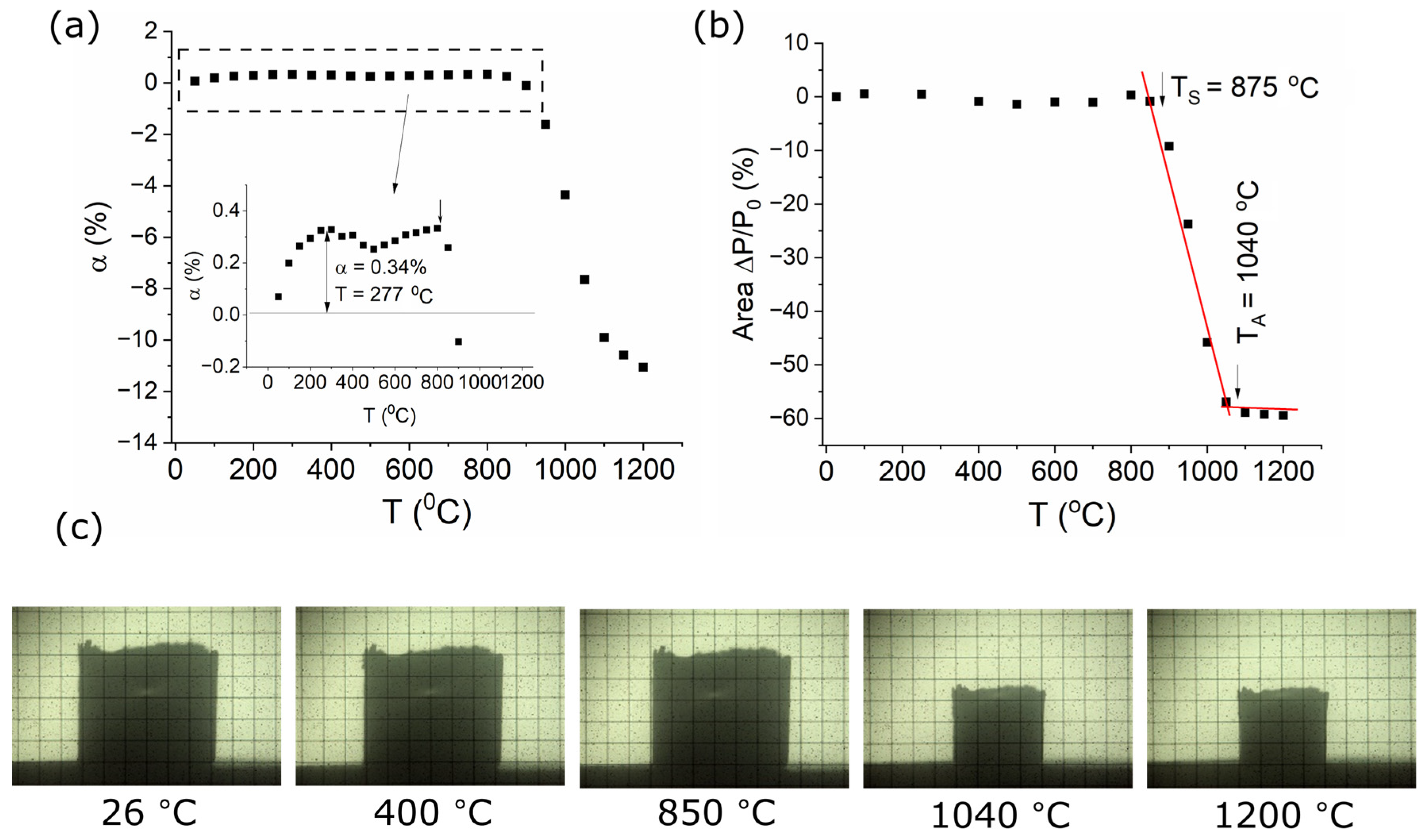
3.2. Thermal Behavior of the Ag-SiO2-TiO2 Nanocomposite

To create a chemical bond and thereby enhance adhesion between electrophoretically deposited nanoceramic coatings and the metallic substrate, heat treatment is necessary [58]. However, the thermal expansion of ceramics and shrinkage cause dimensional changes that may lead to cracks and degradation of the coatings. Therefore, understanding the thermal behavior of the coating material is crucial. To investigate this, research was conducted using a dilatometer and a high-temperature microscope. This combined approach ensured a comprehensive understanding of the thermal properties and stability of the composite coatings, informing the optimization of heat treatment processes to minimize thermal stress and prevent coating failure.
The dilatometry outcomes (Figure 5a) revealed that the sample exhibited a small thermal expansion of 0.34% up to 277 °C, ensued by rapid shrinkage up to 1050 °C, starting from ca. 850 °C. A maximum shrinkage of 11.00% was recorded at 1200 °C, resulting in a total shrinkage of 11.34%. This shrinkage is related to the sintering of the sample.
High-temperature microscope studies enabled the estimation of the size and rate of linear changes accompanying structural transformations in the nanocomposite. Photographs of the sample at selected temperatures, recorded during the examination, along with a graph of linear changes as a function of temperature, are presented in Figure 5b,c.
The significant cross-sectional area change in the Ag-SiO2-TiO2 nanocomposite occurs after reaching a temperature of 800 °C. Between 900 and 1040 °C, the sample shrinks linearly and then flattens out. The starting temperature of sintering was considered to be 875 °C. The percentage difference in shrinkage between the start and end temperatures of sintering was as much as 51%, from the initial 5% to 56% at 1050 °C. At the maximum temperature, the sample shrinkage reached 59%. After the test, a solid, sintered shape was obtained, which suggests the appearance of a liquid phase at the grain boundaries, but not one that is significant enough to be recorded during the test.
The tests indicate that the heat treatment temperature should not exceed 850 to 875 °C to prevent the Ag-SiO2-TiO2 coating from cracking due to shrinkage. This optimal is approximately 50 °C lower than that for the SiO2-TiO2 composite [28], likely due to the addition of silver. Consequently, the coatings deposited at 40 V for 3 min were safely annealed at 800 °C for 2 h.

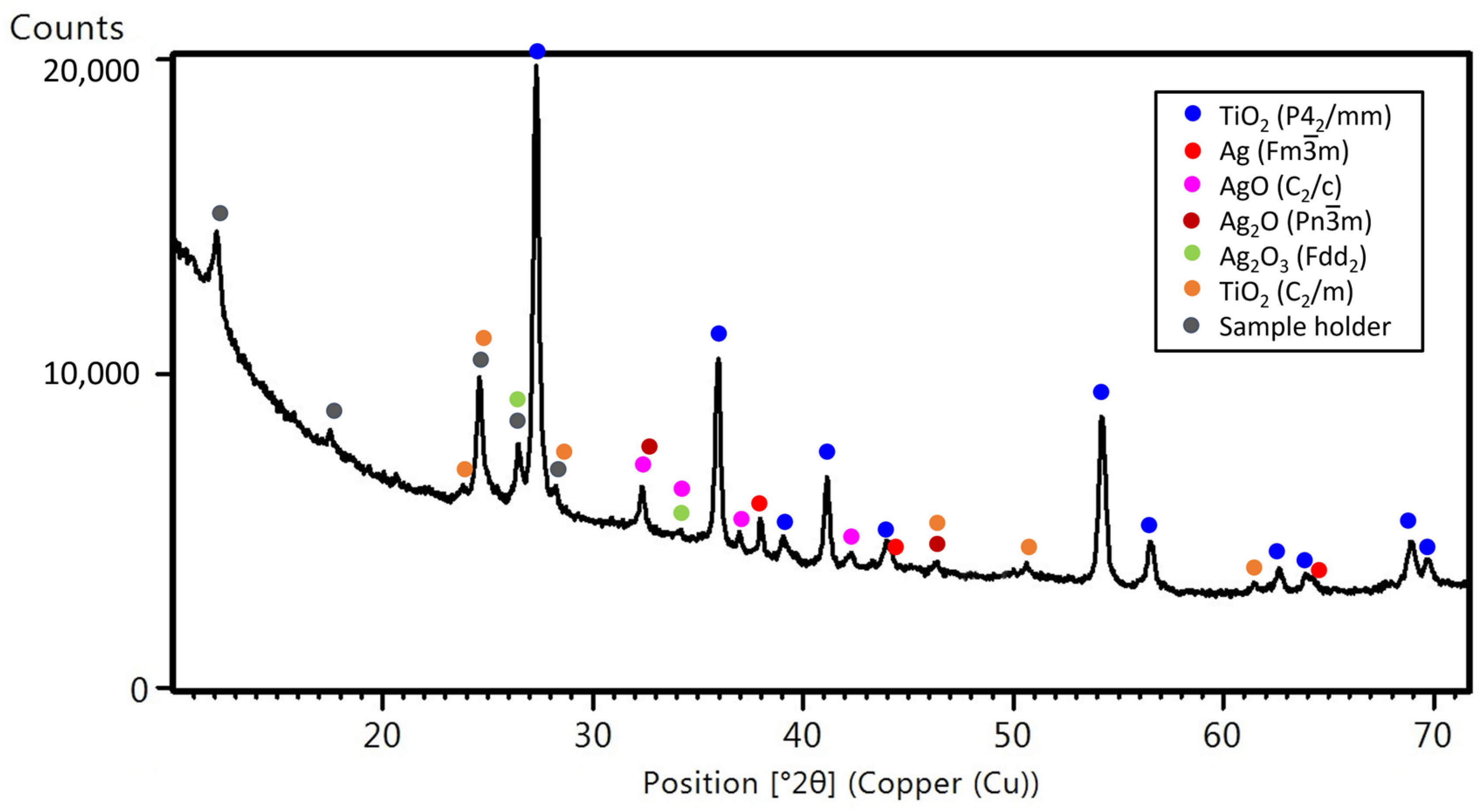
3.3. The Influence of Heating Conditions on the Morphology and Structure of Coatings

Microscopic observations showed that heat treatment contributed to meaningful changes in the microstructure of the deposited Ag-SiO2-TiO2 coatings (Figure 6a). At high magnifications, it was observed that the surface of both of the agglomerates, as well as between them, became rough, which is related to the crystallization of new phases with a different microstructure and grain growth. In the case of SiO2-TiO2 layers, no significant differences in surface morphology were observed after heat treatment [28], while Ag-TiO2 coatings behaved entirely differently than silica-rutile layers [27]. The microstructure changes resembled those identified in Ag-SiO2-TiO2 coatings; therefore, it is assumed that they are related to phase transformations and structural changes related to the presence of reactive silver ions in the nanocomposite.
Analysis of element distribution maps (Figure 6b) showed that the distribution of Ag and Si has not changed. These elements did not segregate and were still dispersed throughout the coating.
X-ray examinations of the coatings post-heat treatment (Figure 7) confirmed the presence of rutile as the predominant phase, like those conducted after electrophoretic deposition (EPD). Minor shifts in the diffraction lines and alterations in the lattice parameters (Table 1) suggest distortions in the crystal lattice and structural changes induced by the elevated temperature. The unit cell parameter a0 decreases, while c0 remains practically unchanged (within the measurement uncertainty), compared to the sample after EPD. The decrease in lattice parameters indicates the precipitation of silver from the solid solution. It is worth noting that the octahedron changes its shape. It is stretched in the plane of the four O2+ ions neighboring Ti4+, and contracted in the axis connecting the vertices of the octahedron. In the sample after EPD, the octahedron was slightly stretched in the axis connecting the vertices of the octahedron. Such Jahn–Teller distortion may be related to another type of defect in the lattice, or even a change in the valence of some part of Ti ions.
The research also revealed modifications related to the silver phase. Structural changes within the coating resulted in the transformation of some metallic silver into silver oxides, including AgO with a monoclinic structure (C2/c), Ag2O with a cubic structure (Pn-3m), and Ag2O3 with a monoclinic structure (Fdd2). Additionally, the formation of another polymorphic form of titanium dioxide, crystallizing in the monoclinic system (C2/m), was identified.
Employing a similar analysis as the one used for the deposited Ag-SiO2-TiO2 coating, spectroscopy techniques were employed to validate hypotheses regarding material consolidation and differences in the chemical and structural composition (Figure 8).
The FTIR spectrum of the Ag-SiO2-TiO2 coating underwent significant modifications, much greater than those observed in comparable coat-forming materials without silver nanoparticles [28]. As a result, titanium dioxide-related bands increased in intensity. In contrast, the silica band arrangement underwent reconstruction with the disappearance of some bands and the appearance of others (Figure 8b). Precisely, the band at 960 cm−1 vanished, indicating the partial or complete decomposition of the Si-OH bonds around the Ti–O network and structural modifications within the SiO2-TiO2 matrix. The main Si-O-Si and siloxane-related bands found at 1013, 1037, 1061, 1090, and 1122 cm−1 remained unaffected by temperature [30,31]. However, new bands appeared around 1159, 1190, and 1254 cm−1, suggesting the rise of structural defects and the appearance of unsaturated SiO-* bonds within the SiO2 network, and favoring the formation of siloxane- or SiO*-C-related bands [30,31].
The infrared data suggest that the segregation of the coat-forming materials caused a solid solution between silica and carbon, leading to the formation of a smooth and continuous interlayer uniformly covering the NiTi substrate.
The structural evolution of the coat-forming system highlights the significant impact of AgNPs on the consolidation and enhancement of the coating’s properties, as confirmed by Raman spectroscopy analysis (Figure 8c). According to the 2D Raman maps and true component analysis, the Ag-SiO2-TiO2 coating emerged as a four-phase system characterized by three dominant spectra of silver-silica-affected titanium dioxide coat-forming phases with bands at approximately 246/254/239 cm−1 (MP), 430/427/430 cm−1 (Eg symmetry), and 606/609/609 cm−1 (A1g symmetry) [42,43,44,45,58] (blue, violet, and dark spectra in Figure 8c). Similar to the as-prepared material, the positions of rutile-related bands in all phases confirm a significant weakening of bond strength due to the asymmetric bending of O–Ti–O in the {110} plane (Eg mode), with minimal impact on the {001}, {110}, or {−110} planes (A1g mode). A shift of the Eg band, indicative of the in-plane mode of the O–O bond, corresponds to the lowering O/Ti ratio relative to the ideal rutile [46,47,48].
More precise analysis reveals significant changes between titanium dioxides, evidenced by the increased intensity of the Ti2O3 interference or silica-titanium dioxide bands (290 and 370 cm−1), and the decreased A1g rutile-related one (Figure 8c). This substantial reduction in bond strength is due to the asymmetric bending of O–Ti–O in the {001}, {110}, or {−110} planes attributed to the reorientation or structural modification of rutile structures under the strong interaction of silver or silica nanoparticles at high-temperature conditions. Consequently, after sintering, disordered rutile structures became the dominant coat-forming materials (blue, violet, and dark spectra in Figure 8c).
An alternative hypothesis related to the deactivation of the A1g vibrations in direct planes suggests the incorporation of carbon into the oxygen-deficient silver-silica-affected rutile structure, with carbon entrapment within rutile nanoparticle cores. Carbon migration into the coating correlates with the chemical bonding of carbon to the coat-forming elements or phases activating Raman bands between 200 and 400 cm−1 (C-Ag bending), and 1300 and 1600 cm−1 (D- and G- planes) [27,59]. Interestingly, carbon-functionalized distorted rutile grouped into isolated and randomly distributed islands increases the roughness of the biomaterial (Figure 8c). Conversely, carbon-free phases formed an interlayer that uniformly covered the NiTi substrate.
The reorganization of silver-silica-affected rutile particles during the sintering (at around ~800 °C) was linked to the formation of a locally occurring phase featuring an unusual band arrangement around 130, 150, and 678, and between 1300 and 1600 cm¹ (green spectrum in Figure 8c). The exact explanation for this band arrangement is not evident, and results from thermal decomposition products, especially Ag-O-Ti interconnections, Ag-O-Ag complexes, or molecularly chemisorbed oxygen species (O*/Ag) [50,51,52]. It is confirmed by the presence of silver oxides identified by XRD (Figure 7).
As for the as-prepared sample, a comparable approach to analyze the depth profiling data applied for the post-sintering Raman cross-section measurements unveiled that the Ag-SiO2-TiO2 coating material exhibited high continuity, with larger structure spread within the proper coating. The average thickness was higher than before sintering, with an estimated value of 2.02 ± 0.03 µm, with thickness that varied from 1.66 ± 0.02 µm in the areas between aggregates to 2.65 ± 0.03 µm in the regions with the most significant objects (depicted in violet colors in Figure 8d,e). We hypothesized the increase in coating thickness resulting from the melting of silica in the titanium dioxide, and amorphous TiO2 from the passivated layer (Figure 8d,e). Such an effect provides coat-forming material consolidation, ultimately forming a new kind of interlayer covering the NiTi alloy substrate. Comparable outcomes at a similar annealing temperature demonstrated a significant enhancement in the bonding strength between the ceramic coatings and the metallic substrate [27,28,60]. On the other hand, the variable coating thickness observed on the X–Z Raman maps due to the high mobility of silica-titanium dioxide particles in the presence of carbon recrystallizes, due to the strong effect of silver, into irregular objects, increasing the surface roughness.

4. Conclusions

Ag-SiO2-TiO2 nanocomposite coatings with island morphology using the electrophoretic deposition method at 40 V for 3 min were developed. Structural studies utilizing X-ray diffraction, Raman spectroscopy, and FTIR spectroscopy provided comprehensive characterization of these coatings. The dominant phase in the coating was defective titanium oxide, which partially formed a solid solution with silver. Additionally, silver was incorporated into the silica structure. The formation of silica-titanium dioxide and silica-titanium dioxide-silver bonds were observed.
The research revealed that the layer formation occurred in several stages. In the first stage, the thin layer, mainly composed of Ti2O3, was formed at the interface with the metallic substrate. Subsequently, an ultrafine fraction of silver-doped rutile nanoparticles was deposited, creating an interlayer. Finally, larger particles forming agglomerates and islands were deposited, consisting of defective rutile, silver-silica oxide-titanium oxide, and silver-silica nanocomposite. The average thickness of the deposited coating was 1.42 ± 0.07 µm. The presence of nanocrystalline metallic silver was also identified within the layer.
Based on the results of thermal tests, heat treatment of the electrophoretically deposited nanocomposite was carried out at 800 °C. It was indicated that temperature-induced changes in morphology and microstructure occurred due to structural transformations. The segregation of materials within the coating resulted in the formation of a continuous SiO2-TiO2 interlayer, uniformly covering the NiTi substrate. The agglomerates were primarily composed of titanium oxide and its solid solution with silver, exhibiting significantly more defects. These increased defects enhance the chemical and biological activity of the material. The presence of silver oxides was also detected in the layer. The average thickness increased to approximately 2.02 ± 0.03 µm, attributable to the structural changes occurring within the layer.
The produced Ag-SiO2-TiO2 layers may be of great potential importance in implant medicine. Future research will focus on a comprehensive characterization of their functional properties, including mechanical properties such as adhesion and deformation resistance related to the induction of the shape memory effect. Additionally, the study will evaluate surface wettability, biocompatibility, antibacterial efficacy, and corrosion resistance to assess the suitability of these coatings for biomedical applications.

Author Contributions

Conceptualization, K.D.; methodology, K.D. and M.D.; validation, K.D., M.D. and J.P.; formal analysis, K.D.; investigation, K.D., M.D., M.K., J.P. and P.R.; resources, K.D., M.D., J.P., M.K. and P.R.; data curation, K.D.; writing—original draft preparation, K.D.; writing—review and editing, K.D., M.D. and J.P.; visualization, K.D. and M.D.; supervision, K.D.; project administration, K.D.; funding acquisition, K.D. All authors have read and agreed to the published version of the manuscript.

Funding

This research was funded by the National Science Center in Poland (NCN), grant number 2020/39/D/ST5/01531.

Institutional Review Board Statement

Not applicable.

Informed Consent Statement

Not applicable.

Data Availability Statement

The data presented in this study are openly available in RepOD https://repod.icm.edu.pl/dataset.xhtml?persistentId=doi:10.18150/64KWUO (accessed on 15 August 2024).

Conflicts of Interest

The authors declare no conflicts of interest. The funders had no role in the design of the study; in the collection, analyses, or interpretation of data; in the writing of the manuscript; or in the decision to publish the results.

References

  1. Murthy, S.K. Nanoparticles in Modern Medicine: State of the Art and Future Challenges. Int. J. Nanomed. 2007, 2, 129–141. [Google Scholar]
  2. Haq Khan, Z.U.; Khan, T.M.; Khan, A.; Shah, N.S.; Muhammad, N.; Tahir, K.; Iqbal, J.; Rahim, A.; Khasim, S.; Ahmad, I.; et al. Brief Review: Applications of Nanocomposite in Electrochemical Sensor and Drugs Delivery. Front. Chem. 2023, 11, 1152217. [Google Scholar] [CrossRef]
  3. Dessale, M.; Mengistu, G.; Mengist, H.M. Nanotechnology: A Promising Approach for Cancer Diagnosis, Therapeutics and Theragnosis. Int. J. Nanomed. 2022, 17, 3735–3749. [Google Scholar] [CrossRef]
  4. Chouirfa, H.; Bouloussa, H.; Migonney, V.; Falentin-Daudré, C. Review of Titanium Surface Modification Techniques and Coatings for Antibacterial Applications. Acta Biomater. 2019, 83, 37–54. [Google Scholar] [CrossRef] [PubMed]
  5. Lorenzetti, M.; Dogša, I.; Stošicki, T.; Stopar, D.; Kalin, M.; Kobe, S.; Novak, S. The Influence of Surface Modification on Bacterial Adhesion to Titanium-Based Substrates. ACS Appl. Mater. Interfaces 2015, 7, 1644–1651. [Google Scholar] [CrossRef] [PubMed]
  6. Liu, L.; Bhatia, R.; Webster, T.J. Atomic Layer Deposition of Nano-TiO2 Thin Films with Enhanced Biocompatibility and Antimicrobial Activity for Orthopedic Implants. Int. J. Nanomed. 2017, 12, 8711–8723. [Google Scholar] [CrossRef]
  7. Łosiewicz, B.; Stróż, A.; Osak, P.; Maszybrocka, J.; Gerle, A.; Dudek, K.; Balin, K.; Łukowiec, D.; Gawlikowski, M.; Bogunia, S. Production, Characterization and Application of Oxide Nanotubes on Ti–6Al–7Nb Alloy as a Potential Drug Carrier. Materials 2021, 14, 6142. [Google Scholar] [CrossRef] [PubMed]
  8. Maher, S.; Mazinani, A.; Barati, M.R.; Losic, D. Engineered Titanium Implants for Localized Drug Delivery: Recent Advances and Perspectives of Titania Nanotubes Arrays. Expert. Opin. Drug Deliv. 2018, 15, 1021–1037. [Google Scholar] [CrossRef]
  9. Shaban, M.; Hasanzadeh, M. Biomedical Applications of Dendritic Fibrous Nanosilica (DFNS): Recent Progress and Challenges. RSC Adv. 2020, 10, 37116–37133. [Google Scholar] [CrossRef]
  10. Massa, M.A.; Covarrubias, C.; Bittner, M.; Fuentevilla, I.A.; Capetillo, P.; Von Marttens, A.; Carvajal, J.C. Synthesis of New Antibacterial Composite Coating for Titanium Based on Highly Ordered Nanoporous Silica and Silver Nanoparticles. Mater. Sci. Eng. C Mater. Biol. Appl. 2014, 45, 146–153. [Google Scholar] [CrossRef]
  11. Selvarajan, V.; Obuobi, S.; Ee, P.L.R. Silica Nanoparticles-A Versatile Tool for the Treatment of Bacterial Infections. Front. Chem. 2020, 8, 602. [Google Scholar] [CrossRef] [PubMed]
  12. Huang, K.; Chen, J.; Nugen, S.R.; Goddard, J.M. Hybrid Antifouling and Antimicrobial Coatings Prepared by Electroless Co-Deposition of Fluoropolymer and Cationic Silica Nanoparticles on Stainless Steel: Efficacy against Listeria Monocytogenes. ACS Appl. Mater. Interfaces 2016, 8, 15926–15936. [Google Scholar] [CrossRef] [PubMed]
  13. Zarys Technologii Materiałów Ogniotrwałych/Franciszek Nadachowski.|Biblioteka Główna. Available online: https://bg.pcz.pl/apiszb/book/3137/Zarys-technologii-materialow-ogniotrwalych-Franciszek-Nadachowski (accessed on 13 July 2024).
  14. Dudek, K.; Dulski, M.; Łosiewicz, B. Functionalization of the NiTi Shape Memory Alloy Surface by HAp/SiO2/Ag Hybrid Coatings Formed on SiO2-TiO2 Glass Interlayer. Materials 2020, 13, 1648. [Google Scholar] [CrossRef] [PubMed]
  15. Burdușel, A.-C.; Gherasim, O.; Grumezescu, A.M.; Mogoantă, L.; Ficai, A.; Andronescu, E. Biomedical Applications of Silver Nanoparticles: An Up-to-Date Overview. Nanomaterials 2018, 8, 681. [Google Scholar] [CrossRef] [PubMed]
  16. Xu, L.; Wang, Y.-Y.; Huang, J.; Chen, C.-Y.; Wang, Z.-X.; Xie, H. Silver Nanoparticles: Synthesis, Medical Applications and Biosafety. Theranostics 2020, 10, 8996–9031. [Google Scholar] [CrossRef] [PubMed]
  17. Shanmuganathan, R.; Karuppusamy, I.; Saravanan, M.; Muthukumar, H.; Ponnuchamy, K.; Ramkumar, V.S.; Pugazhendhi, A. Synthesis of Silver Nanoparticles and Their Biomedical Applications—A Comprehensive Review. Curr. Pharm. Des. 2019, 25, 2650–2660. [Google Scholar] [CrossRef] [PubMed]
  18. Ali, A.; Polepalli, L.; Chowdhury, S.; Carr, M.A.; Janorkar, A.V.; Marquart, M.E.; Griggs, J.A.; Bumgardner, J.D.; Roach, M.D. Silver-Doped Titanium Oxide Layers for Improved Photocatalytic Activity and Antibacterial Properties of Titanium Implants. J. Funct. Biomater. 2024, 15, 163. [Google Scholar] [CrossRef] [PubMed]
  19. Dulski, M.; Dudek, K.; Chalon, D.; Kubacki, J.; Sulowicz, S.; Piotrowska-Seget, Z.; Mrozek-Wilczkiewicz, A.; Gawecki, R.; Nowak, A. Toward the Development of an Innovative Implant: NiTi Alloy Functionalized by Multifunctional β-TCP + Ag/SiO2 Coatings. ACS Appl. Bio Mater. 2019, 2, 987–998. [Google Scholar] [CrossRef]
  20. Rack, H.J.; Qazi, J.I. Titanium Alloys for Biomedical Applications. Mater. Sci. Eng. C 2006, 26, 1269–1277. [Google Scholar] [CrossRef]
  21. Petrini, L.; Migliavacca, F. Biomedical Applications of Shape Memory Alloys. J. Metall. 2011, 2011, 501483. [Google Scholar] [CrossRef]
  22. Boccaccini, A.R.; der Biest, O.V.; Clasen, R.; Uchikoshi, T. Electrophoretic Deposition: Fundamentals and Applications III; Trans Tech Publications Ltd.: Bäch, Switzerland, 2009; ISBN 978-3-03813-267-7. [Google Scholar]
  23. Sarkar, P.; De, D.; Uchikochi, T.; Besra, L. Electrophoretic Deposition (EPD): Fundamentals and Novel Applications in Fabrication of Advanced Ceramic Microstructures. In Electrophoretic Deposition of Nanomaterials; Dickerson, J.H., Boccaccini, A.R., Eds.; Springer: New York, NY, USA, 2012; pp. 181–215. ISBN 978-1-4419-9730-2. [Google Scholar]
  24. Boccaccini, A.R.; Keim, S.; Ma, R.; Li, Y.; Zhitomirsky, I. Electrophoretic Deposition of Biomaterials. J. R. Soc. Interface 2010, 7 (Suppl. 5), S581–S613. [Google Scholar] [CrossRef] [PubMed]
  25. Dudek, K.; Dulski, M.; Podwórny, J.; Kujawa, M.; Gerle, A.; Rawicka, P. Functionalization of the NiTi Shape Memory Alloy Surface through Innovative Hydroxyapatite/Ag-TiO2 Hybrid Coatings. Materials 2024, 17, 604. [Google Scholar] [CrossRef]
  26. Dulski, M.; Gawecki, R.; Sułowicz, S.; Cichomski, M.; Kazek-Kęsik, A.; Wala, M.; Leśniak-Ziółkowska, K.; Simka, W.; Mrozek-Wilczkiewicz, A.; Gawęda, M.; et al. Key Properties of a Bioactive Ag-SiO2/TiO2 Coating on NiTi Shape Memory Alloy as Necessary at the Development of a New Class of Biomedical Materials. Int. J. Mol. Sci. 2021, 22, 507. [Google Scholar] [CrossRef] [PubMed]
  27. Dudek, K.; Dulski, M.; Podwórny, J.; Kujawa, M.; Rawicka, P. Optimization of the Electrophoretic Deposition Parameters and Mechanism of Formation of Ag-TiO2 Nanocoatings on a NiTi Shape Memory Alloy: Part I. Coatings 2024, 14, 44. [Google Scholar] [CrossRef]
  28. Dudek, K.; Dulski, M.; Podwórny, J.; Kujawa, M.; Gerle, A.; Rawicka, P. Synthesis and Characterization of Silica-Titanium Oxide Nano-Coating on NiTi Alloy. Coatings 2024, 14, 391. [Google Scholar] [CrossRef]
  29. Shannon, R.D. Revised Effective Ionic Radii and Systematic Studies of Interatomic Distances in Halides and Chalcogenides. Acta Cryst. A 1976, 32, 751–767. [Google Scholar] [CrossRef]
  30. Wong, J. A Review of Infrared Spectroscopic Studies of Vapor-Deposited Dielectric Glass Films on Silicon. J. Electron. Mater. 1976, 5, 113–160. [Google Scholar] [CrossRef]
  31. Pai, P.G.; Chao, S.S.; Takagi, Y.; Lucovsky, G. Infrared Spectroscopic Study of SiOx Films Produced by Plasma Enhanced Chemical Vapor Deposition. J. Vac. Sci. Technol. A 1986, 4, 689–694. [Google Scholar] [CrossRef]
  32. Verma, R.; Gangwar, J.; Srivastava, A.K. Multiphase TiO2 Nanostructures: A Review of Efficient Synthesis, Growth Mechanism, Probing Capabilities, and Applications in Bio-Safety and Health. RSC Adv. 2017, 7, 44199–44224. [Google Scholar] [CrossRef]
  33. Kadam, A.N.; Dhabbe, R.S.; Kokate, M.R.; Gaikwad, Y.B.; Garadkar, K.M. Preparation of N Doped TiO2 via Microwave-Assisted Method and Its Photocatalytic Activity for Degradation of Malathion. Spectrochim. Acta Part. A Mol. Biomol. Spectrosc. 2014, 133, 669–676. [Google Scholar] [CrossRef]
  34. Kil, H.-S.; Jung, Y.-J.; Moon, J.-I.; Song, J.-H.; Lim, D.-Y.; Cho, S.-B. Glycothermal Synthesis and Photocatalytic Properties of Highly Crystallized Anatase TiO2 Nanoparticles. J. Nanosci. Nanotechnol. 2015, 15, 6193–6200. [Google Scholar] [CrossRef]
  35. Ricchiardi, G.; Damin, A.; Bordiga, S.; Lamberti, C.; Spanò, G.; Rivetti, F.; Zecchina, A. Vibrational Structure of Titanium Silicate Catalysts. A Spectroscopic and Theoretical Study. J. Am. Chem. Soc. 2001, 123, 11409–11419. [Google Scholar] [CrossRef] [PubMed]
  36. Zecchina, A.; Bordiga, S.; Spoto, G.; Marchese, L.; Petrini, G.; Leofanti, G.; Padovan, M. Silicalite Characterization. 1. Structure, Adsorptive Capacity, and IR Spectroscopy of the Framework and Hydroxyl Modes. J. Phys. Chem. 1992, 96, 4985–4990. [Google Scholar] [CrossRef]
  37. Zecchina, A.; Bordiga, S.; Spoto, G.; Marchese, L.; Petrini, G.; Leofanti, G.; Padovan, M. Silicalite Characterization. 2. IR Spectroscopy of the Interaction of Carbon Monoxide with Internal and External Hydroxyl Groups. J. Phys. Chem. 1992, 96, 4991–4997. [Google Scholar] [CrossRef]
  38. Zecchina, A.; Bordiga, S.; Spoto, G.; Scarano, D.; Petrini, G.; Leofanti, G.; Padovan, M.; Areàn, C.O. Low-Temperature Fourier-Transform Infrared Investigation of the Interaction of CO with Nanosized ZSM5 and Silicalite. J. Chem. Soc. Faraday Trans. 1992, 88, 2959–2969. [Google Scholar] [CrossRef]
  39. Bordiga, S.; Ugliengo, P.; Damin, A.A.; Lamberti, C.; Spoto, G.; Zecchina, A.; Spanò, G.; Buzzoni, R.; Dalloro, L.; Rivetti, F. Hydroxyls Nests in Defective Silicalites and Strained Structures Derived upon Dehydroxylation: Vibrational Properties and Theoretical Modelling. Top. Catal. 2001, 15, 43–52. [Google Scholar] [CrossRef]
  40. Larouche, S.; Szymanowski, H.; Klemberg-Sapieha, J.E.; Martinu, L.; Gujrathi, S.C. Microstructure of Plasma-Deposited SiO2/TiO2 Optical Films. J. Vac. Sci. Technol. A 2004, 22, 1200–1207. [Google Scholar] [CrossRef]
  41. de Man, A.J.M.; Sauer, J. Coordination, Structure, and Vibrational Spectra of Titanium in Silicates and Zeolites in Comparison with Related Molecules. An Ab Initio Study. J. Phys. Chem. 1996, 100, 5025–5034. [Google Scholar] [CrossRef]
  42. Balachandran, U.; Eror, N.G. Raman Spectra of Titanium Dioxide. J. Solid. State Chem. 1982, 42, 276–282. [Google Scholar] [CrossRef]
  43. Ocaña, M.; García-Ramos, J.V.; Serna Pereda, C.J. Low-Temperature Nucleation of Rutile Observed by Raman Spectroscopy during Crystallization of TiO2. J. Am. Ceram. Soc. 1992, 75, 2010–2012. [Google Scholar] [CrossRef]
  44. Mazza, T.; Barborini, E.; Piseri, P.; Milani, P.; Cattaneo, D.; Li Bassi, A.; Bottani, C.E.; Ducati, C. Raman Spectroscopy Characterization of TiO2 Rutile Nanocrystals. Phys. Rev. B 2007, 75, 045416. [Google Scholar] [CrossRef]
  45. Zhang, Y.; Harris, C.X.; Wallenmeyer, P.; Murowchick, J.; Chen, X. Asymmetric Lattice Vibrational Characteristics of Rutile TiO2 as Revealed by Laser Power Dependent Raman Spectroscopy. J. Phys. Chem. C 2013, 117, 24015–24022. [Google Scholar] [CrossRef]
  46. Parker, J.C.; Siegel, R.W. Calibration of the Raman Spectrum to the Oxygen Stoichiometry of Nanophase TiO2. Appl. Phys. Lett. 1990, 57, 943–945. [Google Scholar] [CrossRef]
  47. Kernazhitsky, L.; Shymanovska, V.; Gavrilko, T.; Naumov, V.; Fedorenko, L.; Kshnyakin, V.; Baran, J. Laser-Excited Excitonic Luminescence of Nanocrystalline TiO2 Powder. Ukr. J. Phys. 2014, 59, 246. [Google Scholar] [CrossRef]
  48. Wahlbeck, P.G.; Gilles, P.W. Reinvestigation of the Phase Diagram for the System Titanium–Oxygen. J. Am. Ceram. Soc. 1966, 49, 180–183. [Google Scholar] [CrossRef]
  49. Zahornyi, M.M.; Tyschenko, N.I.; Lobunet, T.F.; Kolomys, O.F.; Strelchuk, V.V.; Naumenko, K.S.; Biliavska, L.; Zahorodnia, S.; Kharchuk, M.; Skoryk, S.; et al. The Effect of Ag Content on the Structural, Optical, and Cytotoxicity Properties of TiO2 Nanopowders Grown from TiO(OH)2 Precursor by the Chemical Deposition Method. J. Nano-Electron. Phys. 2021, 19, 06009. [Google Scholar] [CrossRef]
  50. Pettinger, B.; Bao, X.; Wilcock, I.C.; Muhler, M.; Ertl, G. Surface-Enhanced Raman Scattering from Surface and Subsurface Oxygen Species at Microscopically Well-Defined Ag Surfaces. Phys. Rev. Lett. 1994, 72, 1561–1564. [Google Scholar] [CrossRef]
  51. Bao, X.; Muhler, M.; Pettinger, B.; Schlögl, R.; Ertl, G. On the Nature of the Active State of Silver during Catalytic Oxidation of Methanol. Catal. Lett. 1993, 22, 215–225. [Google Scholar] [CrossRef]
  52. Millar, G.J.; Metson, J.B.; Bowmaker, G.A.; Cooney, R.P. In Situ Raman Studies of the Selective Oxidation of Methanol to Formaldehyde and Ethene to Ethylene Oxide on a Polycrystalline Silver Catalyst. J. Chem. Soc. Faraday Trans. 1995, 91, 4149–4159. [Google Scholar] [CrossRef]
  53. Palmquist, A.; Omar, O.M.; Esposito, M.; Lausmaa, J.; Thomsen, P. Titanium Oral Implants: Surface Characteristics, Interface Biology and Clinical Outcome. J. R. Soc. Interface 2010, 7 (Suppl. 5), S515–S527. [Google Scholar] [CrossRef]
  54. Gaintantzopoulou, M.; Zinelis, S.; Silikas, N.; Eliades, G. Micro-Raman Spectroscopic Analysis of TiO2 Phases on the Root Surfaces of Commercial Dental Implants. Dent. Mater. 2014, 30, 861–867. [Google Scholar] [CrossRef] [PubMed]
  55. Sul, Y.-T.; Johansson, C.B.; Petronis, S.; Krozer, A.; Jeong, Y.; Wennerberg, A.; Albrektsson, T. Characteristics of the Surface Oxides on Turned and Electrochemically Oxidized Pure Titanium Implants up to Dielectric Breakdown: The Oxide Thickness, Micropore Configurations, Surface Roughness, Crystal Structure and Chemical Composition. Biomaterials 2002, 23, 491–501. [Google Scholar] [CrossRef] [PubMed]
  56. Lausmaa, J.; Kasemo, B.; Mattsson, H. Surface Spectroscopic Characterization of Titanium Implant Materials. Appl. Surf. Sci. 1990, 44, 133–146. [Google Scholar] [CrossRef]
  57. Chligui, M.; Guimbretiere, G.; Canizares, A.; Matzen, G.; Vaills, Y.; Simon, P. New Features in the Raman Spectrum of Silica: Key-Points in the Improvement on Structure Knowledge 2010. Available online: https://hal.science/hal-00520823/ (accessed on 24 September 2010).
  58. Mali, S.S.; Betty, C.A.; Bhosale, P.N.; Patil, P.S. Hydrothermal Synthesis of Rutile TiO2 with Hierarchical Microspheres and Their Characterization. CrystEngComm 2011, 13, 6349–6351. [Google Scholar] [CrossRef]
  59. Zhang, D.; Zhang, C.; Liu, J.; Chen, Q.; Zhu, X.; Liang, C. Carbon-Encapsulated Metal/Metal Carbide/Metal Oxide Core–Shell Nanostructures Generated by Laser Ablation of Metals in Organic Solvents. ACS Appl. Nano Mater. 2019, 2, 28–39. [Google Scholar] [CrossRef]
  60. Deng, Y.; Sheng, G.; Xu, C. Evaluation of the Microstructure and Mechanical Properties of Diffusion Bonded Joints of Titanium to Stainless Steel with a Pure Silver Interlayer. Mater. Des. (1980–2015) 2013, 46, 84–87. [Google Scholar] [CrossRef]
Figure 1. The Zeta potential and particle size of the colloidal suspension at different pH.
Coatings 14 01055 g001
Figure 2. SEM images at different magnifications (ae), and element distribution (f,g) in the Ag-SiO2-TiO2 coatings deposited at different conditions.
Coatings 14 01055 g002
Figure 3. XRD patterns collected for the deposited coating.
Coatings 14 01055 g003
Figure 4. A chemical and structural differentiation image of the Ag-SiO2-TiO2 coating visualized in (a) X-, Y-, and (b) X-, Z-, with (c) depth scan profiles of the four exemplary places among the AB cross line. Dash-lines on the depth profiles determine the boundary of the layer. (d) FTIR average spectrum gathered for the red dash-line square. (e) Averaged Raman spectra corresponded to individual color-highlighted phases of the Raman maps.
Coatings 14 01055 g004
Figure 5. Change in linear dimensions of the nanocomposite powder with temperature (a), dimensional changes where Ts is a sintering start temperature and TA is the sintering finish temperature, (b) and photographs of the sample during the test in chosen temperatures in the microscope (c).
Coatings 14 01055 g005
Figure 6. SEM images at different magnifications, (a) and elements distribution (b) in the Ag-SiO2-TiO2 after heat treatment.
Coatings 14 01055 g006
Figure 7. XRD patterns collected for the coating after heat treatment.
Coatings 14 01055 g007
Figure 8. A chemical and structural differentiation image of the coating after the heat treatment visualized in (a) X-, Y-, and (b) X-, Z-, with (c) depth scan profiles of the four exemplary places among the AB cross line. Dash-lines on the depth profiles determine the boundary of the Ag-SiO2-TiO2 layer, while the blue-colored area corresponds to the thin interlayer. (d) FTIR average spectrum gathered of the red dash-line square. (e) Averaged Raman spectra corresponded to individual color-highlighted phases of the Raman maps.
Coatings 14 01055 g008
Table 1. Lattice parameters and Ti-O distances in octahedron for TiO2 phase before and after heat treatment obtained from the Rietveld refinement.
TiO2a0 [Å]c0 [Å]Ti-O (x4)Ti-O (x2)Ti-O (Average)
Standard (NIST SRM 674b)4.59402.95891.94981.97831.9593
After EPD4.5950(2)2.9583(2)1.95541.97031.9604
After heat treatment4.5933(4)2.9589(4)1.96971.94761.9623
Theoretical [29] 1.9590
Disclaimer/Publisher’s Note: The statements, opinions and data contained in all publications are solely those of the individual author(s) and contributor(s) and not of MDPI and/or the editor(s). MDPI and/or the editor(s) disclaim responsibility for any injury to people or property resulting from any ideas, methods, instructions or products referred to in the content.

