Article

Coated Biodegradable Zinc Lithium Alloys: Development and Characterization of Co-Doped Strontium Copper Tricalcium Phosphate Coating for Antimicrobial Applications

1
Istituto di Struttura della Materia, Consiglio Nazionale delle Ricerche (ISM-CNR), Via del Fosso del Cavaliere 100, 00133 Rome, Italy
2
Department of Analytical, Physical and Colloid Chemistry, Institute of Pharmacy, I.M. Sechenov First Moscow State Medical University, Trubetskaya 8, Build. 2, Moscow 119048, Russia
3
Dipartimento di Scienze, Università della Basilicata, Via dell’Ateneo Lucano 10, 85100 Potenza, Italy
4
Istituto Zooprofilattico Sperimentale Lazio e Toscana “M. Aleandri”, Via Appia Nuova, 00178 Rome, Italy
5
A.A. Baikov Institute of Metallurgy and Material Science, Russian Academy of Sciences, Leninsky 49, Moscow 119334, Russia
6
Laboratório de Anelasticidade e Biomateriais, UNESP—Universidade Estadual Paulista, Bauru 17033-360, SP, Brazil
7
School of Materials Science and Engineering, Peking University, No. 5 Yi-He-Yuan Road, Hai-Dian District, Beijing 100871, China
*
Author to whom correspondence should be addressed.
Coatings 2024, 14(8), 1073; https://doi.org/10.3390/coatings14081073
Submission received: 25 July 2024 / Revised: 14 August 2024 / Accepted: 20 August 2024 / Published: 22 August 2024

Abstract

:
Zinc biodegradable implants represent a revolutionary advancement in medical technology, offering a promising alternative to titanium and stainless-steel implants and avoiding the need for secondary surgeries for removal. In this study, we aimed to fulfil the clinical demand for biodegradable implant materials by applying a coating of double-doped strontium and copper resorbable tricalcium phosphate (SrCu-TCP) onto a zinc-lithium (Zn-Li) biodegradable alloy using the Pulsed Laser Deposition method. The coated surfaces were thoroughly characterized using X-ray Diffraction, Fourier Transform Infrared Spectroscopy, Atomic Force Microscopy, and Scanning Electron Microscopy coupled with Energy Dispersive X-ray. Microbiology experiments were conducted to assess the inhibitory effects on the growth of various bacteria strains, including gram-positive Staphylococcus aureus and Enterococcus faecalis, gram-negative Pseudomonas aeruginosa and Escherichia coli, as well as the fungus Candida albicans. The obtained results showed that the roughness of the Zn-Li alloy increased from 91.8 ± 29.4 to 651.0 ± 179.5 nm when coated with SrCu-TCP. The thickness of the coating ranged between 3–3.5 µm. The inhibition of growth for all four bacteria strains and the fungus was in the range of 24–35% when cultured on SrCu-TCP coated Zn-Li samples. These findings suggest that the developed coatings are promising candidates for applications requiring inhibition of microorganisms.

1. Introduction

In the past two decades, biodegradable metals have emerged as promising materials for various biomedical implant devices, offering the potential to reduce reliance on permanent metallic implants, such as those made of titanium (Ti) alloys, stainless steel, etc. [1,2,3,4]. This advancement aims to mitigate the need for implant removal surgeries and significantly decrease associated costs [5,6,7,8]. Biodegradable metals, including those used in bone implants, show advantages such as biocompatibility and controlled degradation [9,10,11,12]. However, challenges persist, including the need to address their excessive corrosion rates compared to the healing rate of host bone tissue and to provide implant surface with biomimetic properties [13,14,15,16].
Recent studies indicate that Zinc (Zn) is a crucial micronutrient for human health, particularly in bone tissue development and homeostasis [17,18,19,20]. It contributes to bone tissue composition, collagen synthesis, mineralization, and turnover [21,22,23,24]. Both in vitro and in vivo research demonstrate zinc’s diverse effects: promoting osteoblast proliferation and differentiation while safeguarding osteoblasts from oxidative stress-induced apoptosis, and inhibiting osteoclastogenesis and impacting osteoclast apoptosis [25,26,27]. These properties, combined with its antibacterial activity, have led to successful utilization of zinc in bone substitutes and implant biomaterials [28]. The incorporation of zinc into various calcium phosphates like hydroxyapatite, β-TCP, brushite, or monetite holds promise as a beneficial agent in bone repair [29,30,31].
Binary Zn alloys with alloying elements such as magnesium (Mg), calcium (Ca), strontium (Sr), lithium (Li), manganese (Mn), iron (Fe), copper (Cu), and silver (Ag) are systemically investigated in vitro and in vivo. Li exhibits the most effective strengthening role in Zn, followed by Mg. Alloying leads to accelerated degradation. Adding elements such as Mg, Ca, Sr, and Li into Zn improves the cytocompatibility, osteogenesis, and osseointegration [32,33]. Li becomes toxic from 1.4 mM [34,35], and serum Li quantities are associated with bipolar disorders (10 mg/L) or risk of death (20 mg/L) [36,37]. However, in the quantities in which Li is included in implant alloys, it is not toxic [32,38]. Moreover, Li improved bone mass in mice and enhanced bone formation through the activation of the canonical Wnt pathway [39]. Additionally, maintenance therapy with lithium carbonate increased bone mass [40].
Coating biodegradable metal surfaces such as Mg-Ca [41,42,43] or Zn-Li alloys [44,45,46], with biomimetic calcium phosphates represents a significant strategy for enhancing their properties, including controlling degradation behaviour and improving surface biocompatibility. Tricalcium phosphate (TCP), particularly β-tricalcium phosphate (β-TCP), stands out as a resorbable bioceramic widely used in synthetic bone grafts due to its osteoconductive and osteoinductive properties [47]. To enhance β-TCP’s osteoinductive ability, metal ions like Sr2+ [48], Cu2+ [49], Mg2+ [50], Mn2+ [51,52], Fe2+ [53], and Zn2+ [54] are incorporated, typically by substituting Ca2+ ions. These essential ions have been shown to enhance the biological performance of β-TCP, promoting osteo- and angiogenesis. Additionally, copper (Cu2+) [49], manganese (Mn2+) [51], iron (Fe2+) [53], and zinc (Zn2+) [54] exhibit antimicrobial properties, further bolstering the potential of these coatings in biomedical applications. In particular, copper is known to exert its antimicrobial effect by damaging the cell membrane and inducing the production of reactive oxygen species (ROS) that can, in turn, harm proteins, lipids, and DNA. Additionally, Cu can bind to bacterial enzymes, impairing their functions and preventing bacterial growth and replication [55,56].
Co-substituted β-TCP-based phosphates with Cu2+/Zn2+, Cu2+/Sr2+, and Sr2+/Mn2+ ion pairs were reported in [57]. In the same study, their antibacterial properties were evaluated. The results obtained demonstrated that all synthesized phases exhibit antibacterial effects. Notably, experiments using β-TCP-Cu+Sr, which had the highest concentration of Cu2+ ions among all samples studied and occupied separate crystal sites, showed minimal bacterial and fungal growth. It was concluded that the active dopant ions must be located in different crystal sites to provide higher antimicrobial activity.
It is important to highlight that implant-related infections continue to be a leading cause of implant failure, resulting in significant economic and social burdens [58,59]. Preventing biomaterial-associated infections requires targeted strategies aimed at inhibiting bacterial adhesion and biofilm formation. Alarmingly high rates of septic complications, particularly in high-risk patients and procedures, have been documented [60,61].
Microorganisms such as Staphylococcus aureus (S. aureus), Pseudomonas aeruginosa (P. aeruginosa), Escherichia coli (E. coli), Enterococcus faecalis (E. faecalis), and Candida albicans (C. albicans) are commonly found in hospital environments [62,63]. In particular, S. aureus is typically present on the skin and in the nostrils, posing a risk of contamination to surgical and medical instruments, and is a leading cause of nosocomial infections. P. aeruginosa is present in moist environments, such as sinks, antiseptic solutions, etc. E. coli and E. faecalis are primarily found in the intestinal and urinary tracts and can contaminate surgical surfaces and instruments. Meanwhile, C. albicans is frequently present on the skin and mucous membranes, also posing a contamination risk to medical instruments.
To render implant surfaces antibacterial and mitigate infection risks, coating them with materials possessing antibacterial properties is a viable approach. Among these materials are calcium phosphates substituted with ions, like copper (Cu), manganese (Mn), iron (Fe), and zinc (Zn).
In the present study, for the first time, Zn-Li biodegradable alloy was coated with double doped Sr and Cu resorbable tricalcium phosphate to address the clinical needs for biodegradable implant materials as an alternative to traditional titanium and stainless-steel implants. The alloy was coated by Pulsed Laser Deposition (PLD) to ensure a uniform and well-adhered coating, which is important for the performance and longevity of the implants. Coatings characterization was performed using X-ray Diffraction (XRD), Fourier Transform Infrared Spectroscopy (FTIR), Atomic Force Microscopy (AFM), and Scanning Electron Microscopy (SEM) coupled with Energy Dispersive X-ray (EDX). Microbiological tests were conducted on coated Zn-Li alloy samples, targeting gram-positive bacteria strains such as S. aureus and E. faecalis and gram-negative strains including P. aeruginosa and E. coli, as well as C. albicans fungus.

2. Materials and Methods

2.1. Zn-Li Alloy Preparation

Zn-0.4 wt% Li alloy was synthesized from a Zn (99.99%) and Zn-Li master alloy (6 wt% Li) ingot from the Hunan Rare Earth Metal Material Research Institute (Changsha, China). For casting, a cast iron mould with ZnO coating was applied. The obtained ingot was homogenized at 350 °C for 48 h, followed by water quenching. Then, it was deformed by hot extrusion at 260 °C with an extrusion ratio of 36:1 at 1 mm/s. The obtained samples were cut perpendicular to the extrusion direction, resulting in the following geometry: diameter of 10 mm and a thickness of 3 mm. All the prepared samples were ground to 2000 grit with SiC and then ultrasonically cleaned in acetone, absolute ethanol, and distilled water.

2.2. Synthesis of Bulk Target Material—Double Substituted Sr- and Cu- Tricalcium Phosphate (SrCu-TCP)

(Strontium and Copper)-containing TCP bulk material with formula Ca2.5Cu0.25Sr0.25(PO4)2 was synthesized using the method of mechanochemical activation. The weights of the initial reagents were calculated according to reaction (1):
2.5Ca(NO3)2 + 2(NH4)2HPO4 + 0.25Cu(NO3)2 + 0.25Sr(NO3)2 + 2NH4OH → Ca2.5Sr0.25Cu0.25(PO4)2 + 6NH4NO3 + 2H2O
To begin, 14.00 g CaO, 5.29 g Sr(NO3)2, 6.04 g Cu(NO3)2·3H2O, and 26.41 g (NH4)2HPO4 were placed into the Teflon container with 250 g zirconia balls and activation was carried out in a planetary mill (material: balls ratio of 1:3). The rotation speed was set at 200 rpm for 30 min, after which 200 mL of distilled water was added to the container and grinding was then continued for 30 min. Immediately after the grinding, the resulting precipitate was filtered out on a Buchner funnel and dried at 105 °C until completely dry. After that, the (copper and strontium)-substituted TCP was calcined at 400 °C for 1 h to remove the reaction by-products (ammonium nitrate and water). From the so-obtained powders, disks with a diameter of 15 mm were pressed by the method of uniaxial pressing at a specific pressure of 200 kg/cm2. After that, the disks were sintered in a chamber furnace with silite heaters at 1200 °C for 2 h.
The weight% of Sr and Cu was calculated as follows:
Ca2.5Sr0.25Cu0.25(PO4)2 Mr = 2.5 × 40.08 + 0.25 × 87.62 + 0.25 × 63.55 + (30.97 + 16.00 × 4) × 2 = 327.94
wt% Sr = 87.62 × 0.25 × 100/327.94 = 6.68
wt% Cu = 63.55 × 0.25 × 100/327.94 = 4.84
The content of Sr and Cu in the prepared ceramic targets was determined using atomic absorption spectroscopy on an atomic absorption spectrometer (AAS) manufactured by Thermo Fisher Scientific (Madison, WI, USA), model iCE 3000.

2.3. Pulsed Laser Deposition of Coatings

For thin film deposition, the nanosecond PLD technique supplied with nanosecond Nd:YAG laser source (λ = 532 nm, 10 Hz, 7 ns) was applied. SrCu-TCP discs of 1 cm diameter were applied as targets. The ablation and deposition experiments were carried out in a stainless-steel chamber equipped with a scroll-turbomolecular vacuum system. Films were grown on Zn-Li and monocrystalline Si(100), the latter being necessary for coatings’ FTIR and XRD characterizations. Depositions were performed at a laser fluence of 12 J/cm2 (250 mW) and a pressure of 10−4 Pa for 2 h, with the target substrate distance fixed at 1 cm. The laser beam was incident at an angle of 45° on the target surface and focused by a 35 cm focus lens. All the films were deposited at room temperature of substrate. The disc target was fixed in a rotating holder in order to minimize the laser craterisation effect.

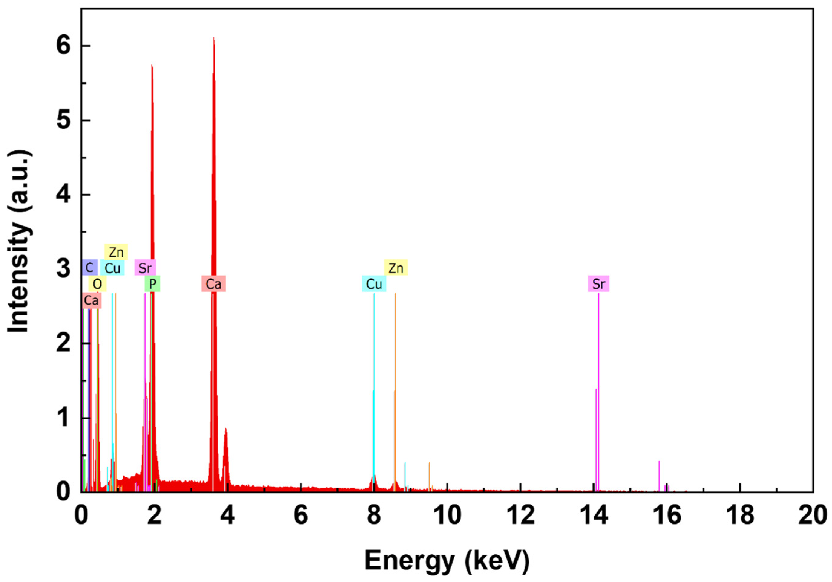
2.4. Physico-Chemical Characterization of Coatings

For XRD, a Siemens 5000D (Bruker, Billerica, MA, USA), operating at 40 kV and 32 mA and using CuKα radiation (λ = 1.5405600 Å, 2θ = 10°–60°, step size 0.040°, time per step 4 s), was employed.
FTIR spectra were acquired by a Jasco 460Plus interferometer (Jasco, Tokyo, Japan). For each sample, 100 spectra were collected at resolution of 4 cm−1 in the 4000–400 cm−1 range.
AFM measurements were performed using a Bruker Dimension Icon system equipped with a Nanoscope V controller. AFM images were acquired in tapping mode, using n-doped Si probes (Bruker RTESP300) with a resonant frequency of 300 kHz at a scan rate 0.3–1 Hz. The scanned areas were from 1 × 1 µm2 to 10 × 10 µm2. Different regions were scanned for each sample.
Morphological characterization and cross-section were made by means of an FE-SEM (Auriga-Zeiss, Oberkochen, Germany) equipped with an EDS analyzer (Bruker).

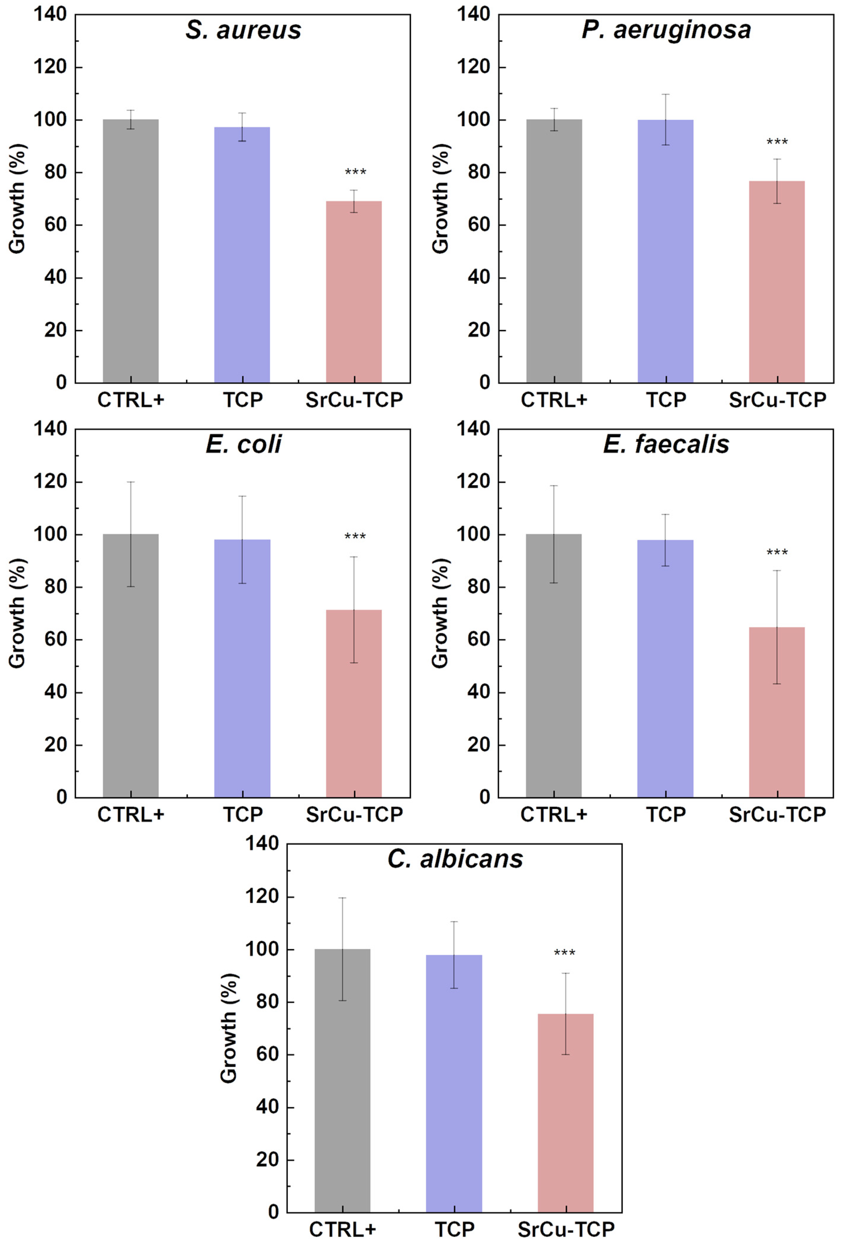
2.5. Microbiology Tests

The antimicrobial activity of pure TCP and SrCu-TCP coatings on Zn-Li substrates was assessed. The samples were autoclaved at 121 °C for 20 min at a pressure of 1.1 bar and then immersed in BHI broth (Brain Heart Infusion, DIFCO, Sparks, NV, USA). Five different microorganisms were used: four bacteria (S. aureus, P. aeruginosa, E. coli, and E. faecalis) and one fungus (C. albicans). The microorganisms were cultured under slow agitation, with bacteria at 37 °C and the fungus at 24 °C. The positive control for each test consisted of the growth of each individual microorganism alone. After 24 h, the growth of the microorganisms was assessed by measuring the OD600 nm of the growth medium using a D30 Biophotometer (Eppendorf, Hamburg, Germany).

2.6. Statistical Analysis

The antimicrobial activity experiment was performed in triplicate. Microorganisms’ growth was quantified as the mean O.D 600 ± standard deviation (S.D.) and analysed using the non-parametric Dunnett test for multiple comparisons (SAS JMP Statistical Discovery software v14 pro, Milan, Italy). p values of ≤0.05, ≤0.01, and ≤0.001 were considered statistically significant, as indicated in the figure legends.

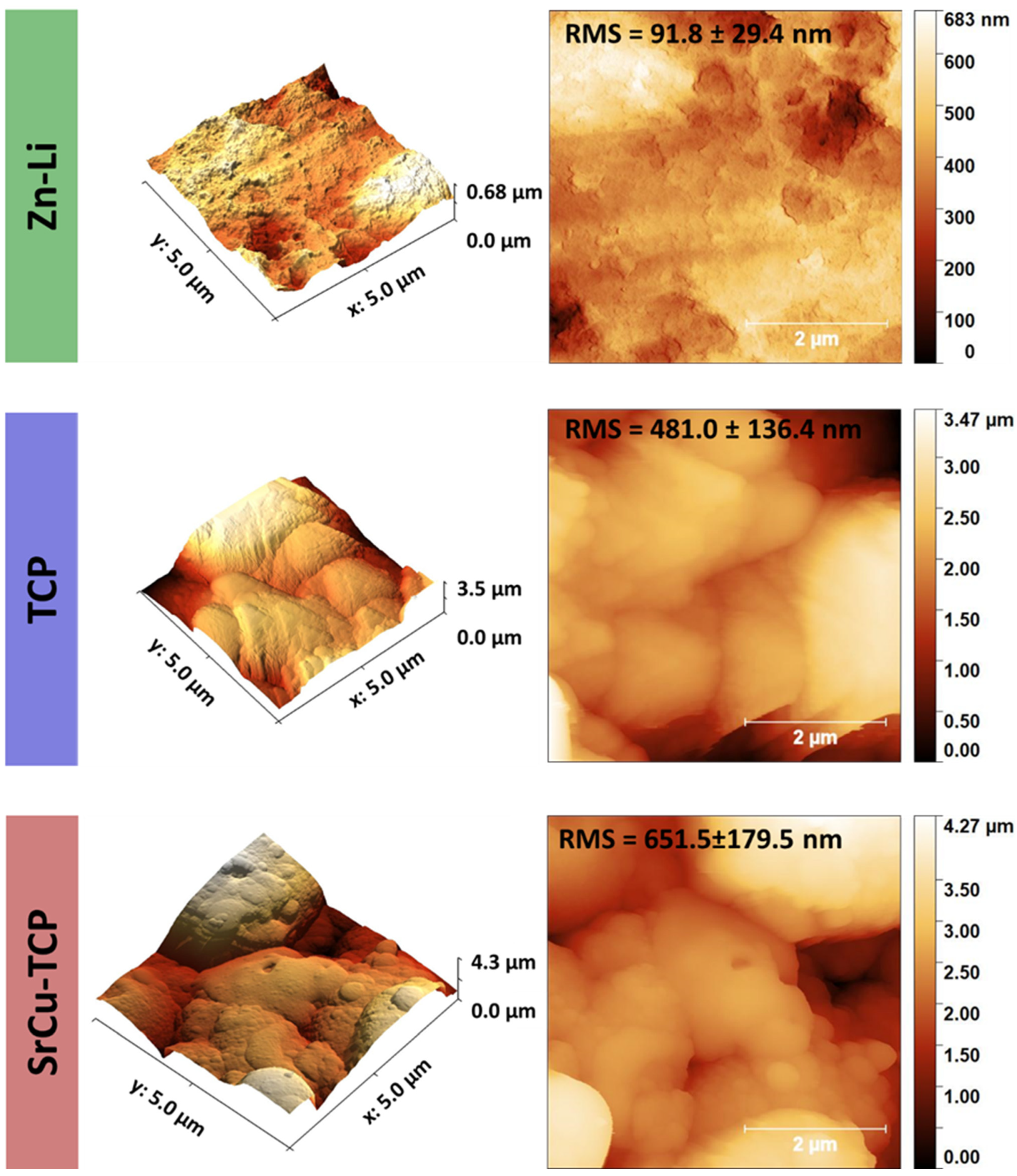
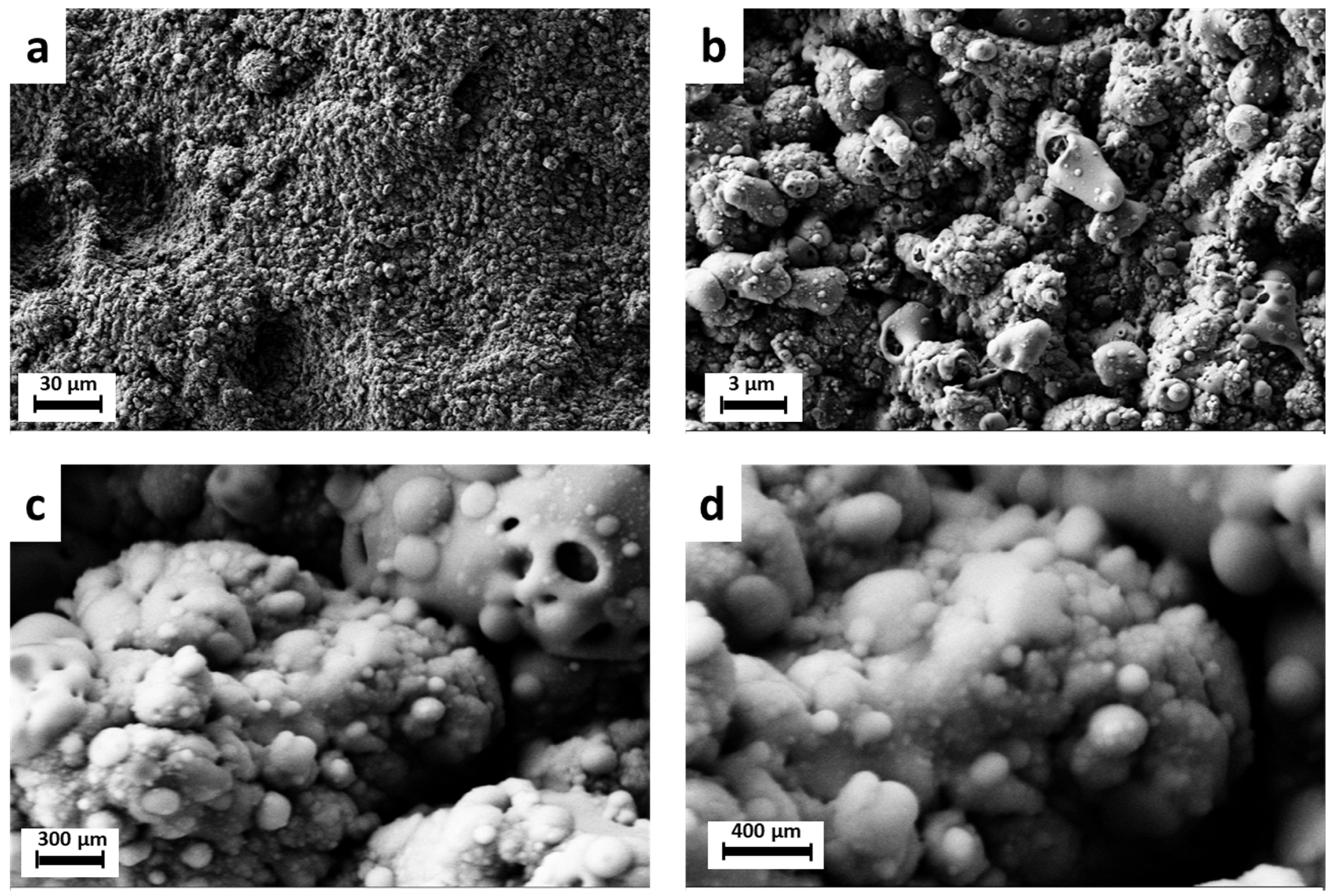
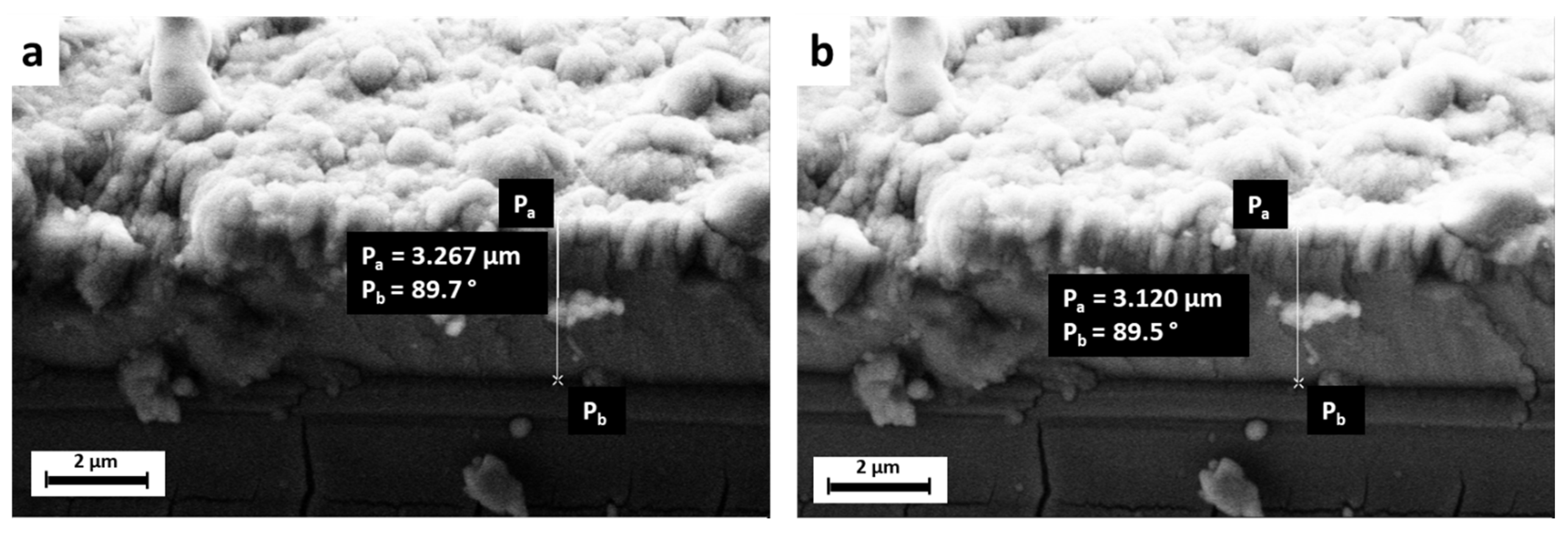
3. Results and Discussion

The coatings were initially deposited on Si(100) substrates to enable more accurate characterization by XRD and FTIR, excluding any interference from the Zn-Li alloy substrate. Additionally, non-substituted tricalcium phosphate (β-TCP) coatings were deposited alongside SrCu-TCP coatings for comparative analysis.
Figure 1 illustrates the XRD diffraction pattern of the TCP coating and its corresponding target material, as well as the SrCu-TCP coating and its corresponding target material. As evident from the diffraction patterns, both coatings exhibit an amorphous background, indicating their partial crystallinity.
Table 1 presents the 2θ position values of the most intense 21 peaks for both β-TCP and SrCu-TCP targets derived from their respective XRD patterns. The table also includes the shift position (Δ2θ) in degrees, calculated by subtracting the SrCu-TCP and β-TCP 2θ peak centre values. Additionally, the hkl Miller Index and the relative intensity (R.I.%) of each reflection are provided [64].
Observing the calculated shift position values (Δ2θ column), it can be inferred that nearly all Bragg reflections of the SrCu-TCP target are shifted toward higher angles, with Δ2θ values ranging from 0° to 0.24°. This suggests that the SrCu-TCP lattice generally exhibits shorter planar distances. Notably, lattice planes with Miller indexes h = 1, k = 0, and l with relatively high values (10, 16) (peak numbers 4, 14, 20) remain unaffected by any angular shift, demonstrating a calculated Δ2θ of 0°.
In Figure 2, the FTIR patterns collected from β-TCP and SrCu-TCP targets (A) and coatings (B), respectively, are presented. Within the main plot of each pattern, an inset showing the spectral window ranging from 500 to 1500 cm−1 is presented, where the most prominent bands are highlighted.
Figure 2A shows a general decreased degeneracy relative to the SrCu-TCP pattern compared to pure TCP. This difference is particularly evident for the IR absorption bands at 945–1087 cm−1, attributable to the ν1 and ν3 modes of the PO43− group, respectively [65,66]. Furthermore, the presence of Cu and Sr ions within the TCP matrix induced a shift in the position of all the observed main bands, as evidenced by the black straight line (TCP) and red dashed line (SrCu-TCP), allowing for the appreciation of the displacement in positions. The specific value for each band shift is reported in Table 2.
In Figure 2B, the patterns collected from TCP and SrCu-TCP coatings are reported and compared. It is possible to observe that the shape of the bands differs from those in the targets’ IR spectra. In particular, both coatings’ FTIR patterns exhibit narrower bands compared to those of the targets. Similar to what was observed for the targets, the presence of Sr and Cu in the TCP coating induced a variation in degeneracy, particularly evident for the bands at 557–604 cm−1, (the ν4 mode of the PO43− group) [66,67] where two sharp bands characterize the SrCu-TCP coating pattern. Finally, no significant shift was detectable from the comparison of pure TCP and SrCu-TCP coatings bands relative to the ν1 mode of the PO43−. For both compositions, the band is centred at around 1038 cm−1.
In Figure 3, typical 2D and 3D topographies of Zn-Li alloy, TCP coatings, and SrCu-TCP coatings on Zn-Li alloys are shown. Due to the deposition of particles of different sizes, it was observed that the surface of the Zn-Li sample is smoother than the others due to the absence of the coating. After coating, the average roughness of the TCP sample increases from 91.8 ± 29.4 to 481.0 ± 136.4 nm. Furthermore, it was found that the coating incorporated with Sr and Cu showed a further increase in roughness, reaching 651.5 ± 179.5 nm.
The morphology of the SrCu-TCP coating on the Zn-Li substrate exhibits the typical characteristics observed in Pulsed Laser Deposited films, featuring droplets of varying sizes dispersed within a compact matrix. SEM images in Figure 4 demonstrate the compact globular morphology of the deposited SrCu-TCP coatings.
To assess the thickness of the deposited coatings, cross-section SEM images were acquired. As depicted in the SEM images shown in Figure 5, the coatings exhibit a thickness within the range of a few micrometres, specifically 3–3.5 µm.
SEM-EDX measurements were conducted to analyse the composition of the coatings, with the results presented in Figure 6 and Table 3. The data indicate that the coatings contain approximately 4.10 ± 0.18 wt% of Cu and 6.54 ± 0.33 wt% of Sr. Additionally, a minor amount of Zn is detected, originating from the Zn-Li substrate.
The results related to the growth of microorganisms (S. aureus, P. aeruginosa, E. coli, E. faecalis, and C. albicans) in the presence and absence of SrCu-TCP coatings on Zn-Li alloys are reported in Table 4 and illustrated in Figure 7. The data provided in the table include the mean OD600 values, standard deviations (SD), percentage of growth, and percentage of growth inhibition derived from three independent experiments. The growth of each organism was assessed after 24 h of incubation at their respective optimal growth temperatures. The microorganisms cultured alone (without the coated substrates) served as the positive control of the experiment (CTR+).
The growth of all microorganisms on TCP is statistically comparable to the growth of the positive control (without samples). Statistically significant growth inhibition was observed in all tested microorganisms when grown in the presence of SrCu-TPC. In particular, the inhibition of the growth of microorganisms is 31.1% for S. aureus, 23.5% for P. aeruginosa, 28.7% for E. coli, 35.3% for E. faecalis, and 24.6% for C. albicans.
In contrast, our previous study [57], focused on double-substituted tricalcium phosphate powder—SrCu-TCP, revealed notably higher growth inhibition rates. The SrCu-TCP powder exhibited a remarkable inhibition of growth, reaching 92.0%, 95.5%, 64.9%, 96.3%, and 70.9% against S. aureus, P. aeruginosa, E. coli, E. faecalis, and C. albicans, respectively. Such a considerable difference could potentially be attributed to the reduced amount of material present in the coating.
In our previous study [44], focusing on double-substituted tricalcium phosphate coatings applied to the same Zn-Li alloys but containing Strontium and Manganese substitution—SrMn-TCP, the inhibition of 12.2% of S. aureus, 9.4% of P. aeruginosa, and 9.0% of E. coli, 9.7% of E. faecalis was registered. The SrMn-TCP-coated Zn-Li alloys were effective in inhibiting the growth of fungus C. albicans, achieving approximately a 50% inhibition rate.
Also in this case, for SrMn-TCP powder [57], a higher inhibition growth was registered compared to the coatings. Indeed, SrMn-TCP powder demonstrated a substantial inhibition of growth, with rates of 57.8% for S. aureus, 92.9% for P. aeruginosa, 19% for E. coli, 67.4% for E. faecalis, and 41.9% for C. albicans.
Table 5 below summarizes the results on microorganisms’ inhibition obtained in the present study and reported in the cited above literature.

4. Conclusions

Partially-crystalline SrCu-TCP coatings were deposited by Pulsed Laser Deposition technique onto the Zn-Li alloy substrates. The composition analysis revealed approximately 6.5 wt% Sr and 4.1 wt% Cu within the coatings. The incorporation of Sr and Cu into the crystal lattice was evidenced by observed shifts in diffraction peaks and IR bands.
Morphological examination exhibited compact micro-globular structures in the coatings. The average surface roughness of the TCP coating was measured at 481.0 ± 136.4 nm, while the SrCu-TCP coating exhibited a further increase, reaching 651.5 ± 179.5 nm. The thickness of the coatings, determined by SEM, ranged between 3–3.5 µm.
Our study showed statistically significant inhibition of the growth in all tested microorganisms in the presence of SrCu-TCP coatings, while microorganisms cultured solely on TCP coatings did not exhibit significant growth inhibition.
The SrCu-TCP surface demonstrated good antimicrobial efficacy, achieving inhibition of the growth ranging between 23.5% and 35.3%. Specifically, for S. aureus, the inhibition was at 31.1%, for P. aeruginosa at 23.5%, for E. coli at 28.7%, for E. faecalis at 35.3%, and for C. albicans at 24.6%. These results highlight the antimicrobial characteristics of the SrCu-TCP surface on Zn-Li alloy, making it a promising candidate for applications requiring inhibition of both bacterial and fungal growth.

Author Contributions

Conceptualization, J.V.R. and Y.Z.; methodology, J.V.R., A.D.B., K.B., M.F., I.V.F., G.C.C., R.T., T.K.S. and Y.Z.; validation, A.D.B., K.B., M.F., I.V.F., G.C.C., R.T. and T.K.S.; formal analysis, M.C., K.B., M.F., G.C.C. and T.K.S.; investigation, A.D.B., M.C., K.B., M.F. and I.V.F.; resources, J.V.R., A.D.B., K.B., I.V.F. and R.T.; data curation, A.D.B., M.C., K.B., M.F., G.C.C. and T.K.S.; writing—original draft preparation, J.V.R.; writing—review and editing, J.V.R. and G.C.C.; visualization, J.V.R. and G.C.C.; supervision, J.V.R.; project administration, J.V.R.; funding acquisition, J.V.R., K.B. and Y.Z. All authors have read and agreed to the published version of the manuscript.

Funding

J.V.R. and Y.Z. thank the bilateral project CNR (Italy)—NSFC (China) 2021–2022 and the International Cooperation and Exchange program of NSFC-CNR (Grant No. 52011530392). Financial support of the Italian Ministry of Health, grants IZSLT 10/22 RC and IZSLT 01/23 RC is gratefully acknowledged.

Institutional Review Board Statement

Not applicable.

Informed Consent Statement

Not applicable.

Data Availability Statement

The experimental data are available upon a reasonable and official request to the corresponding author.

Acknowledgments

Technical assistance of Marco Ortenzi, Massimo Di Menno Di Bucchianico and Luca Imperatori is gratefully acknowledged.

Conflicts of Interest

The authors declare no conflicts of interest.

References

  1. Shi, Z.-Z.; Gao, X.-X.; Zhang, H.-J.; Liu, X.-F.; Li, H.-Y.; Zhou, C.; Yin, Y.-X.; Wang, L.-N. Design biodegradable Zn alloys: Second phases and their significant influences on alloy properties. Bioact. Mater. 2020, 5, 210–218. [Google Scholar] [CrossRef] [PubMed]
  2. Young, J.; Reddy, R.G. Synthesis, mechanical properties, and in vitro corrosion behavior of biodegradable Zn–Li–Cu alloys. J. Alloys Compd. 2020, 844, 156257. [Google Scholar] [CrossRef]
  3. Zhao, S.; Seitz, J.-M.; Eifler, R.; Maier, H.J.; Guillory, R.J.; Earley, E.J.; Drelich, A.; Goldman, J.; Drelich, J.W. Zn-Li alloy after extrusion and drawing: Structural, mechanical characterization, and biodegradation in abdominal aorta of rat. Mater. Sci. Eng. C 2017, 76, 301–312. [Google Scholar] [CrossRef]
  4. Li, H.; Huang, Y.; Ji, X.; Wen, C.; Wang, L.-N. Fatigue and corrosion fatigue behaviors of biodegradable Zn-Li and Zn-Cu-Li under physiological conditions. J. Mater. Sci. Technol. 2022, 131, 48–59. [Google Scholar] [CrossRef]
  5. Chen, Y.; Xu, Z.; Smith, C.; Sankar, J. Recent advances on the development of magnesium alloys for biodegradable implants. Acta Biomater. 2014, 10, 4561–4573. [Google Scholar] [CrossRef]
  6. Staiger, M.P.; Pietak, A.M.; Huadmai, J.; Dias, G. Magnesium and its alloys as orthopedic biomaterials: A review. Biomaterials 2006, 27, 1728–1734. [Google Scholar] [CrossRef]
  7. Farè, S.; Ge, Q.; Vedani, M.; Vimercati, G.; Gastaldi, D.; Migliavacca, F.; Petrini, L.; Trasatti, S. Evaluation of material properties and design requirements for biodegradable magnesium stents. Matéria 2010, 15, 96–103. [Google Scholar] [CrossRef]
  8. Li, H.; Wen, J.; Liu, Y.; He, J.; Shi, H.; Tian, P. Progress in Research on Biodegradable Magnesium Alloys: A Review. Adv. Eng. Mater. 2020, 22, 2000213. [Google Scholar] [CrossRef]
  9. Costantino, M.D.; Schuster, A.; Helmholz, H.; Meyer-Rachner, A.; Willumeit-Römer, R.; Luthringer-Feyerabend, B.J.C. Inflammatory response to magnesium-based biodegradable implant materials. Acta Biomater. 2020, 101, 598–608. [Google Scholar] [CrossRef]
  10. Li, M.; Ren, L.; Li, L.; He, P.; Lan, G.; Zhang, Y.; Yang, K. Cytotoxic Effect on Osteosarcoma MG-63 Cells by Degradation of Magnesium. J. Mater. Sci. Technol. 2014, 30, 888–893. [Google Scholar] [CrossRef]
  11. Henderson, S.E.; Verdelis, K.; Maiti, S.; Pal, S.; Chung, W.L.; Chou, D.-T.; Kumta, P.N.; Almarza, A.J. Magnesium alloys as a biomaterial for degradable craniofacial screws. Acta Biomater. 2014, 10, 2323–2332. [Google Scholar] [CrossRef]
  12. Song, G. Control of biodegradation of biocompatable magnesium alloys. Corros. Sci. 2007, 49, 1696–1701. [Google Scholar] [CrossRef]
  13. Zhang, Z.; Jia, Q.; Wu, X.; Cao, X.; Xu, C.; Zhang, J.; Yang, W. Study on microstructure and dynamic corrosion behavior of degradable Mg–Y–Zn–Zr alloy with different Sr contents. Mater. Res. Express 2019, 6, 125418. [Google Scholar] [CrossRef]
  14. Sanchez, A.H.M.; Luthringer, B.J.C.; Feyerabend, F.; Willumeit, R. Mg and Mg alloys: How comparable are in vitro and in vivo corrosion rates? A review. Acta Biomater. 2015, 13, 16–31. [Google Scholar] [CrossRef] [PubMed]
  15. Nguyen, T.; Waterman, J.; Staiger, M.; Woodfield, T. Controlling in vitro corrosion rate of pure Mg with rough surface texture via biomimetic coating systems. Corros. Eng. Sci. Technol. 2012, 47, 358–364. [Google Scholar] [CrossRef]
  16. Tian, Y.; Shao, Y.; Yu, Q.; Guo, S.; Dang, R.; Du, Y.; Zhu, G.; Lyu, S.; Chen, M. Investigating the corrosion behavior of as-cast Mg-Zn-Ca alloys with the same Zn/Ca atomic ratio by in situ observation. Mater. Corros. 2024. [Google Scholar] [CrossRef]
  17. Hara, T.; Takeda, T.-A.; Takagishi, T.; Fukue, K.; Kambe, T.; Fukada, T. Physiological roles of zinc transporters: Molecular and genetic importance in zinc homeostasis. J. Physiol. Sci. 2017, 67, 283–301. [Google Scholar] [CrossRef]
  18. Lowe, N.M.; Fraser, W.D.; Jackson, M.J. Is there a potential therapeutic value of copper and zinc for osteoporosis? Proc. Nutr. Soc. 2009, 61, 181–185. [Google Scholar] [CrossRef] [PubMed]
  19. Komarova, E.G.; Sharkeev, Y.P.; Sedelnikova, M.B.; Prosolov, K.A.; Khlusov, I.A.; Prymak, O.; Epple, M. Zn- or Cu-Containing CaP-Based Coatings Formed by Micro-arc Oxidation on Titanium and Ti-40Nb Alloy: Part I—Microstructure, Composition and Properties. Materials 2020, 13, 4116. [Google Scholar] [CrossRef]
  20. Hernández-Escobar, D.; Champagne, S.; Yilmazer, H.; Dikici, B.; Boehlert, C.J.; Hermawan, H. Current status and perspectives of zinc-based absorbable alloys for biomedical applications. Acta Biomater. 2019, 97, 1–22. [Google Scholar] [CrossRef]
  21. O’Connor, J.P.; Kanjilal, D.; Teitelbaum, M.; Lin, S.S.; Cottrell, J.A. Zinc as a Therapeutic Agent in Bone Regeneration. Materials 2020, 13, 2211. [Google Scholar] [CrossRef]
  22. Molenda, M.; Kolmas, J. The Role of Zinc in Bone Tissue Health and Regeneration—A Review. Biol. Trace Elem. Res. 2023, 201, 5640–5651. [Google Scholar] [CrossRef]
  23. Cerovic, A.; Miletic, I.; Sobajic, S.; Blagojevic, D.; Radusinovic, M.; El-Sohemy, A. Effects of zinc on the mineralization of bone nodules from human osteoblast-like cells. Biol. Trace Elem. Res. 2007, 116, 61–71. [Google Scholar] [CrossRef]
  24. Hadley, K.B.; Newman, S.M.; Hunt, J.R. Dietary zinc reduces osteoclast resorption activities and increases markers of osteoblast differentiation, matrix maturation, and mineralization in the long bones of growing rats. J. Nutr. Biochem. 2010, 21, 297–303. [Google Scholar] [CrossRef] [PubMed]
  25. Seo, H.-J.; Cho, Y.-E.; Kim, T.; Shin, H.-I.; Kwun, I.-S. Zinc may increase bone formation through stimulating cell proliferation, alkaline phosphatase activity and collagen synthesis in osteoblastic MC3T3-E1 cells. Nutr. Res. Pr. 2010, 4, 356–361. [Google Scholar] [CrossRef] [PubMed]
  26. Moonga, B.S.; Dempster, D.W. Zinc is a potent inhibitor of osteoclastic bone resorption in vitro. J. Bone Miner. Res. 1995, 10, 453–457. [Google Scholar] [CrossRef] [PubMed]
  27. Ma, J.; Zhao, N.; Zhu, D. Endothelial Cellular Responses to Biodegradable Metal Zinc. ACS Biomater. Sci. Eng. 2015, 1, 1174–1182. [Google Scholar] [CrossRef]
  28. Fosca, M.; Streza, A.; Antoniac, I.V.; Vadalà, G.; Rau, J.V. Ion-Doped Calcium Phosphate-Based Coatings with Antibacterial Properties. J. Funct. Biomater. 2023, 14, 250. [Google Scholar] [CrossRef]
  29. Maleki-Ghaleh, H.; Siadati, M.H.; Fallah, A.; Koc, B.; Kavanlouei, M.; Khademi-Azandehi, P.; Moradpur-Tari, E.; Omidi, Y.; Barar, J.; Beygi-Khosrowshahi, Y.; et al. Antibacterial and Cellular Behaviors of Novel Zinc-Doped Hydroxyapatite/Graphene Nanocomposite for Bone Tissue Engineering. Int. J. Mol. Sci. 2021, 22, 9564. [Google Scholar] [CrossRef]
  30. Laskus-Zakrzewska, A.; Zgadzaj, A.; Kolmas, J. Synthesis and physicochemical characterization of Zn-doped brushite. Ceram. Int. 2021, 47, 7798–7804. [Google Scholar] [CrossRef]
  31. Cruz, R.; Calasans-Maia, J.; Sartoretto, S.; Moraschini, V.; Rossi, A.M.; Louro, R.S.; Granjeiro, J.M.; Calasans-Maia, M.D. Does the incorporation of zinc into calcium phosphate improve bone repair? A systematic review. Ceram. Int. 2018, 44, 1240–1249. [Google Scholar] [CrossRef]
  32. Li, Z.; Shi, Z.-Z.; Hao, Y.; Li, H.-F.; Zhang, H.-J.; Liu, X.-F.; Wang, L.-N. Insight into role and mechanism of Li on the key aspects of biodegradable ZnLi alloys: Microstructure evolution, mechanical properties, corrosion behavior and cytotoxicity. Mater. Sci. Eng. C 2020, 114, 111049. [Google Scholar] [CrossRef] [PubMed]
  33. Gong, H.; Wang, K.; Strich, R.; Zhou, J.G. In vitro biodegradation behavior, mechanical properties, and cytotoxicity of biodegradable Zn–Mg alloy. J. Biomed. Mater. Res. Part B Appl. Biomater. 2015, 103, 1632–1640. [Google Scholar] [CrossRef] [PubMed]
  34. Wang, M.; Yang, L.; Zhu, X.; Yang, L.; Shen, J.; Lu, T.; Liu, H.; Song, Z. Corrosion Mechanisms of a Biodegradable Zn-0.4Li Alloy in Simulated Gastrointestinal Environment. Coatings 2023, 13, 529. [Google Scholar] [CrossRef]
  35. Young, W. Review of Lithium Effects on Brain and Blood. Cell Transplant. 2009, 18, 951–975. [Google Scholar] [CrossRef] [PubMed]
  36. Aral, H.; Vecchio-Sadus, A. Toxicity of lithium to humans and the environment—A literature review. Ecotoxicol. Environ. Saf. 2008, 70, 349–356. [Google Scholar] [CrossRef]
  37. Tong, X.; Shen, T.; Zhou, X.; Zeng, J.; Tao, J.; Munir, K.; Li, Y.; Huang, S.; Wu, X.; Ma, J.; et al. Biodegradable Zn–Cu–Li alloys with ultrahigh strength, ductility, antibacterial ability, cytocompatibility, and suitable degradation rate for potential bone-implant applications. Smart Mater. Manuf. 2023, 1, 100012. [Google Scholar] [CrossRef]
  38. Zhang, Y.; Lu, Y.; Xu, X.; Chen, L.; Xiao, T.; Luo, X.; Yan, Y.; Li, D.; Dai, Y.; Yu, K. Microstructure, Corrosion Behaviors in Different Simulated Body Fluids and Cytotoxicity of Zn–Li Alloy as Biodegradable Material. Mater. Trans. 2019, 60, 583–586. [Google Scholar] [CrossRef]
  39. Clément-Lacroix, P.; Ai, M.; Morvan, F.; Roman-Roman, S.; Vayssière, B.; Belleville, C.; Estrera, K.; Warman, M.L.; Baron, R.; Rawadi, G. Lrp5-independent activation of Wnt signaling by lithium chloride increases bone formation and bone mass in mice. Proc. Natl. Acad. Sci. USA 2005, 102, 17406–17411. [Google Scholar] [CrossRef]
  40. Zamani, A.; Omrani, G.R.; Nasab, M.M. Lithium’s effect on bone mineral density. Bone 2009, 44, 331–334. [Google Scholar] [CrossRef]
  41. Salahshoor, M.; Guo, Y. Biodegradable Orthopedic Magnesium-Calcium (MgCa) Alloys, Processing, and Corrosion Performance. Materials 2012, 5, 135–155. [Google Scholar] [CrossRef] [PubMed]
  42. Zhang, C.; Zeng, R.; Liu, C.; Gao, J. Comparison of calcium phosphate coatings on Mg–Al and Mg–Ca alloys and their corrosion behavior in Hank’s solution. Surf. Coat. Technol. 2010, 204, 3636–3640. [Google Scholar] [CrossRef]
  43. Dorozhkin, S.V. Calcium orthophosphate coatings on magnesium and its biodegradable alloys. Acta Biomater. 2014, 10, 2919–2934. [Google Scholar] [CrossRef] [PubMed]
  44. Rau, J.V.; De Bonis, A.; Teghil, R.; Curcio, M.; Fadeeva, I.V.; Barbaro, K.; Di Menno Di Bucchianico, M.; Fosca, M.; Zheng, Y. Double Substituted with Manganese and Strontium Tricalcium Phosphate Coatings on Zinc-Lithium Biodegradable Alloys for Biomedical Implant Applications. Coatings 2023, 13, 36. [Google Scholar] [CrossRef]
  45. Li, H.F.; Shi, Z.Z.; Wang, L.N. Opportunities and challenges of biodegradable Zn-based alloys. J. Mater. Sci. Technol. 2020, 46, 136–138. [Google Scholar] [CrossRef]
  46. Xing, H.; Tang, Y.; Fa, X.; Zhang, H.; Shi, Z.; Yao, S.; Wang, L. Enhanced Biocompatibility and Osteogenic Property of Biodegradable Zn-0.5Li Alloy through Calcium–Phosphorus Coating. Coatings 2024, 14, 350. [Google Scholar] [CrossRef]
  47. Bohner, M.; Santoni, B.L.G.; Döbelin, N. β-tricalcium phosphate for bone substitution: Synthesis and properties. Acta Biomater. 2020, 113, 23–41. [Google Scholar] [CrossRef]
  48. Fadeeva, I.V.; Deyneko, D.V.; Forysenkova, A.A.; Morozov, V.A.; Akhmedova, S.A.; Kirsanova, V.A.; Sviridova, I.K.; Sergeeva, N.S.; Rodionov, S.A.; Udyanskaya, I.L.; et al. Strontium Substituted β-Tricalcium Phosphate Ceramics: Physiochemical Properties and Cytocompatibility. Molecules 2022, 27, 6085. [Google Scholar] [CrossRef]
  49. Fadeeva, I.V.; Lazoryak, B.I.; Davidova, G.A.; Murzakhanov, F.F.; Gabbasov, B.F.; Petrakova, N.V.; Fosca, M.; Barinov, S.M.; Vadalà, G.; Uskoković, V.; et al. Antibacterial and cell-friendly copper-substituted tricalcium phosphate ceramics for biomedical implant applications. Mater. Sci. Eng. C 2021, 129, 112410. [Google Scholar] [CrossRef]
  50. Kazakova, G.; Safronova, T.; Golubchikov, D.; Shevtsova, O.; Rau, J.V. Resorbable Mg2+-Containing Phosphates for Bone Tissue Repair. Materials 2021, 14, 4857. [Google Scholar] [CrossRef]
  51. Rau, J.V.; Fadeeva, I.V.; Fomin, A.S.; Barbaro, K.; Galvano, E.; Ryzhov, A.P.; Murzakhanov, F.; Gafurov, M.; Orlinskii, S.; Antoniac, I.; et al. Sic Parvis Magna: Manganese-Substituted Tricalcium Phosphate and Its Biophysical Properties. ACS Biomater. Sci. Eng. 2019, 5, 6632–6644. [Google Scholar] [CrossRef]
  52. Fadeeva, I.V.; Kalita, V.I.; Komlev, D.I.; Radiuk, A.A.; Fomin, A.S.; Davidova, G.A.; Fursova, N.K.; Murzakhanov, F.F.; Gafurov, M.R.; Fosca, M.; et al. In Vitro Properties of Manganese-Substituted Tricalcium Phosphate Coatings for Titanium Biomedical Implants Deposited by Arc Plasma. Materials 2020, 13, 4411. [Google Scholar] [CrossRef] [PubMed]
  53. Antoniac, I.V.; Filipescu, M.; Barbaro, K.; Bonciu, A.; Birjega, R.; Cotrut, C.M.; Galvano, E.; Fosca, M.; Fadeeva, I.V.; Vadalà, G.; et al. Iron Ion-Doped Tricalcium Phosphate Coatings Improve the Properties of Biodegradable Magnesium Alloys for Biomedical Implant Application. Adv. Mater. Interfaces 2020, 7, 2000531. [Google Scholar] [CrossRef]
  54. Fadeeva, I.V.; Goldberg, M.A.; Preobrazhensky, I.I.; Mamin, G.V.; Davidova, G.A.; Agafonova, N.V.; Fosca, M.; Russo, F.; Barinov, S.M.; Cavalu, S.; et al. Improved cytocompatibility and antibacterial properties of zinc-substituted brushite bone cement based on β-tricalcium phosphate. J. Mater. Sci. Mater. Med. 2021, 32, 99. [Google Scholar] [CrossRef] [PubMed]
  55. Božić Cvijan, B.; Korać Jačić, J.; Bajčetić, M. The Impact of Copper Ions on the Activity of Antibiotic Drugs. Molecules 2023, 28, 5133. [Google Scholar] [CrossRef] [PubMed]
  56. Salah, I.; Parkin, I.P.; Allan, E. Copper as an antimicrobial agent: Recent advances. RSC Adv. 2021, 11, 18179–18186. [Google Scholar] [CrossRef]
  57. Deyneko, D.V.; Fadeeva, I.V.; Borovikova, E.Y.; Dzhevakov, P.B.; Slukin, P.V.; Zheng, Y.; Xia, D.; Lazoryak, B.I.; Rau, J.V. Antimicrobial properties of co-doped tricalcium phosphates Ca3-2x(M′M″)x(PO4)2 (M = Zn2+, Cu2+, Mn2+ and Sr2+). Ceram. Int. 2022, 48, 29770–29781. [Google Scholar] [CrossRef]
  58. Ribeiro, M.; Monteiro, F.J.; Ferraz, M.P. Infection of orthopedic implants with emphasis on bacterial adhesion process and techniques used in studying bacterial-material interactions. Biomatter 2012, 2, 176–194. [Google Scholar] [CrossRef]
  59. Wang, M.; Tang, T. Surface treatment strategies to combat implant-related infection from the beginning. J. Orthop. Transl. 2019, 17, 42–54. [Google Scholar] [CrossRef]
  60. Aggarwal, V.K.; Bakhshi, H.; Ecker, N.U.; Parvizi, J.; Gehrke, T.; Kendoff, D. Organism Profile in Periprosthetic Joint Infection: Pathogens Differ at Two Arthroplasty Infection Referral Centers in Europe and in the United States. J. Knee Surg. 2014, 27, 399–406. [Google Scholar] [CrossRef]
  61. Miquel, S.; Lagrafeuille, R.; Souweine, B.; Forestier, C. Anti-biofilm Activity as a Health Issue. Front. Microbiol. 2016, 7, 592. [Google Scholar] [CrossRef]
  62. Cangui-Panchi, S.P.; Ñacato-Toapanta, A.L.; Enríquez-Martínez, L.J.; Reyes, J.; Garzon-Chavez, D.; Machado, A. Biofilm-forming microorganisms causing hospital-acquired infections from intravenous catheter: A systematic review. Curr. Res. Microb. Sci. 2022, 3, 100175. [Google Scholar] [CrossRef] [PubMed]
  63. Bonadonna, L.; Briancesco, R.; Coccia, A.M. Analysis of Microorganisms in Hospital Environments and Potential Risks. In Indoor Air Quality in Healthcare Facilities; Capolongo, S., Settimo, G., Gola, M., Eds.; Springer International Publishing: Cham, Switzerland, 2017; pp. 53–62. [Google Scholar] [CrossRef]
  64. INORGANIC STRUCTURE CRYSTAL DATABASE (ICSD). Available online: https://icsd.products.fiz-karlsruhe.de/ (accessed on 30 May 2024).
  65. Boanini, E.; Gazzano, M.; Nervi, C.; Chierotti, M.R.; Rubini, K.; Gobetto, R.; Bigi, A. Strontium and Zinc Substitution in β-Tricalcium Phosphate: An X-ray Diffraction, Solid State NMR and ATR-FTIR Study. J. Funct. Biomater. 2019, 10, 20. [Google Scholar] [CrossRef] [PubMed]
  66. Xidaki, D.; Agrafioti, P.; Diomatari, D.; Kaminari, A.; Tsalavoutas-Psarras, E.; Alexiou, P.; Psycharis, V.; Tsilibary, E.C.; Silvestros, S.; Sagnou, M. Synthesis of Hydroxyapatite, β-Tricalcium Phosphate and Biphasic Calcium Phosphate Particles to Act as Local Delivery Carriers of Curcumin: Loading, Release and In Vitro Studies. Materials 2018, 11, 595. [Google Scholar] [CrossRef]
  67. Habelitz, S.; Pascual, L.; Durán, A. Transformation of tricalcium phosphate into apatite by ammonia treatment. J. Mater. Sci. 2001, 36, 4131–4135. [Google Scholar] [CrossRef]
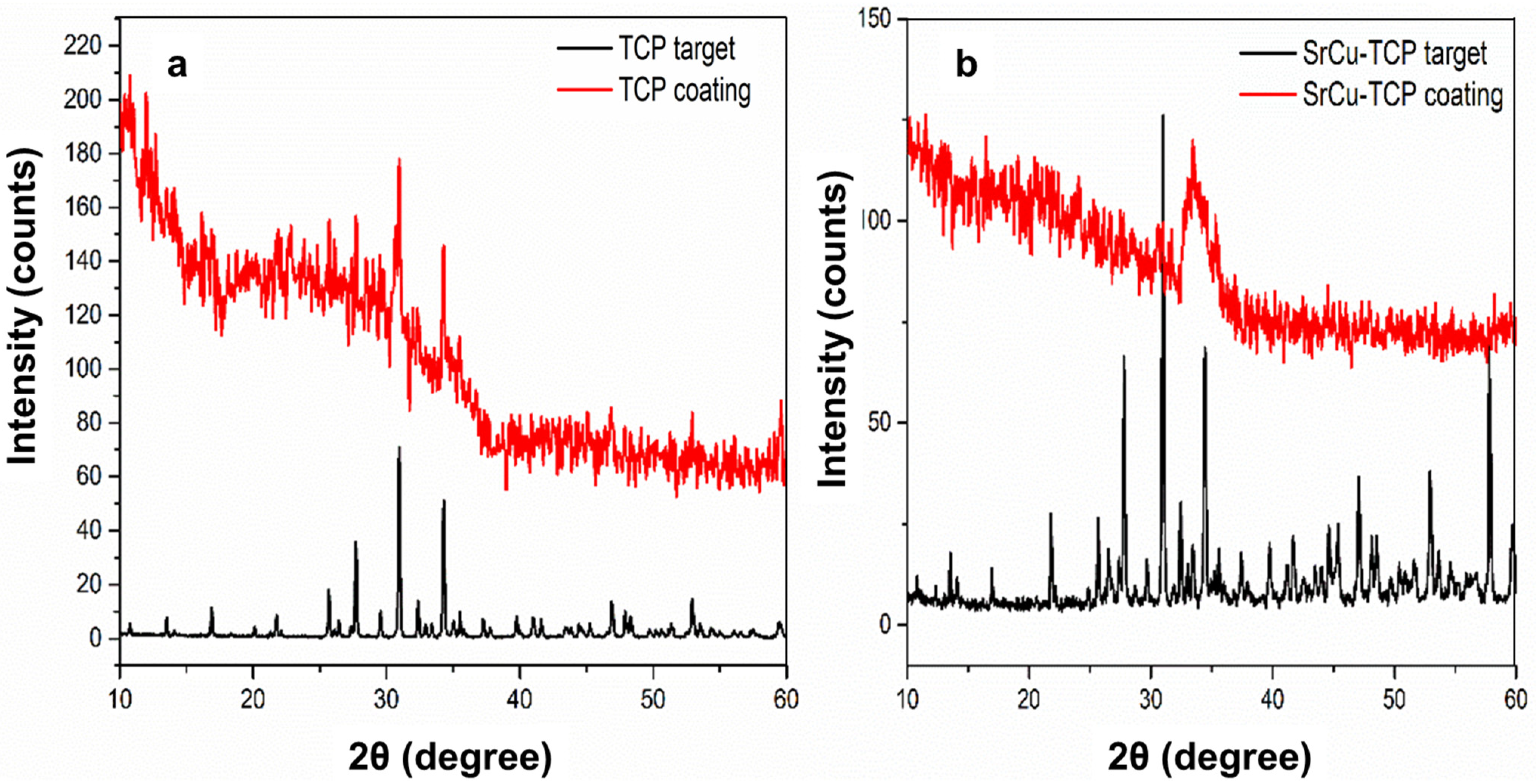
Figure 1. XRD patterns of (a) TCP coating deposited on Si substrate and corresponding target material, (b) SrCu-TCP coating deposited on Si substrate and corresponding target material.
Coatings 14 01073 g001
Figure 2. FTIR spectra collected on TCP and SrCu-TCP compositions as targets (A) and coatings deposited on Si substrate (B). The 500–1500 cm−1 spectral window is highlighted. The centre position of the most significant bands is evidenced by a straight black line (TCP) and a dashed red line (SrCu-TCP).
Coatings 14 01073 g002
Figure 3. AFM 2D and 3D images of Zn-Li substrate and TCP and SrCu-TCP coatings on Zn-Li substrate.
Coatings 14 01073 g003
Figure 4. SEM images of SrCu-TCP coating on Zn-Li substrate at 1 kx (a), 10 kx (b), 50 kx (c), and 100 kx (d) magnifications.
Coatings 14 01073 g004
Figure 5. SEM cross section images of SrCu-TCP coating on Zn-Li substrate. (a) Area 1 and (b) Area 2.
Coatings 14 01073 g005
Figure 6. SEM-EDX spectrum of SrCu-TCP coating on Zn-Li substrate.
Coatings 14 01073 g006
Figure 7. Growth rate of S. aureus, P. aeruginosa, E. coli, and E. faecalis bacteria strains and C. albicans fungus grown in the presence of control sample and TCP and SrCu-TPC coatings on Zn-Li alloy substrate. The growth percentage is calculated on the OD600 resulting from three independent experiments and presented as mean values ± standard deviation (SD), compared to the values of the positive control corresponding to 100%. p values (Dunnett test): p < 0.001 *** compared to positive control.
Coatings 14 01073 g007
Table 1. The shifts for the main Bragg reflections of β-TCP and of double substituted SrCu-TCP.
Peak Numberβ-TCP 2θ Peak Position (°)SrCu-TCP 2θ Peak Position (°)Δ2θhkl Miller Index [64]R.I.%
113.5213.560.041 0 416
216.8816.920.041 1 020
321.7621.800.040 2 416
425.6825.680.001 0 1025
526.4026.520.121 2 210
627.7227.800.082 1 455
729.5629.680.123 0 016
830.9631.000.040 2 10100
932.3632.480.121 2 820
1034.2834.440.162 2 065
1135.0435.220.182 2 38
1235.5235.600.082 1 1012
1337.2437.400.161 2 1110
1439.7639.760.001 0 1610
1541.0041.180.184 0 414
1641.6041.680.083 0 1212
1746.8847.080.204 0 1020
1847.8848.120.242 3 816
1948.2848.520.244 1 614
2052.9252.920.002 0 2025
2159.4859.680.205 1 712
Table 2. FTIR band positions and band shifts for TCP and SrCu-TCP targets.
ModeTCP Target (cm−1)SrCu-TCP Target (cm−1)Band Shift (cm−1)
ν3112011255
ν110421027−15
ν460559015
ν4549554−6
Table 3. Sr and Cu content in targets and deposited SrCu-TCP coatings.
Sr, wt%Cu, wt%
EDX
(Coatings)
AAS (Targets)Theoretical Value, in Ca2.5Sr0.25Cu0.25(PO4)2 PowderEDX
(Coatings)
AAS (Targets)Theoretical Value, in Ca2.5Sr0.25Cu0.25(PO4)2 Powder
6.54 ± 0.336.68 ± 0.056.714.10 ± 0.184.78 ± 0.054.84
Table 4. Growth rate of bacteria (S. aureus, P. aeruginosa, E. coli and E. faecalis) and a fungus (C. albicans) grown in the presence of control sample, TCP, and SrCu-TPC coatings on Zn-Li alloy substrate. The growth percentage was calculated on the OD600 resulting from three independent experiments and presented as mean values ± standard deviation (SD).
OD 600 nmSD% Growth% Inhibition
S. aureus
CTR+1.0090.03100.00.0
TCP0.9800.0597.12.9
SrCu-TCP0.6950.0468.931.1
P. aeruginosa
CTR+0.9840.03100.00.0
TCP0.9830.0999.90.1
SrCu-TCP0.7530.0876.523.5
E. coli
CTR+1.2040.17100.00.0
TCP1.1790.1197.92.1
SrCu-TCP0.8580.2171.328.7
E. faecalis
CTR+0.9950.17100.00.0
TCP0.9730.0797.82.2
SrCu-TCP0.6430.2164.735.3
C. albicans
CTR+0.8100.15100.00.0
TCP0.7920.0997.82.2
SrCu-TCP0.6110.1275.424.6
Table 5. Summary of the results on microbial inhibition obtained in the present study and reported in the literature.
% Inhibition
SampleS. aureusP. aeruginosaE. coliE. faecalisC. albicansReference
SrCu-TCP (coating)31.123.528.735.324.6present study
SrCu-TCP (powder)92.095.564.996.370.9[57]
SrMn-TCP (coating)12.29.49.09.750.0[44]
SrMn-TCP (powder)57.892.919.067.441.9[57]
Disclaimer/Publisher’s Note: The statements, opinions and data contained in all publications are solely those of the individual author(s) and contributor(s) and not of MDPI and/or the editor(s). MDPI and/or the editor(s) disclaim responsibility for any injury to people or property resulting from any ideas, methods, instructions or products referred to in the content.

