Article

Surface Decontamination of Titanium Dental Implants Subjected to Implantoplasty by Treatment with Citric Acid Solutions

by
Pilar Fernández-Garrido
1,
Begoña M. Bosch
2,
Javier Gil
2,* and
Manuel Fernández-Domínguez
1,3
1
Department of Translational Medicine, CEU San Pablo University, Urbanización Montepríncipe, 28925 Alcorcón, Madrid, Spain
2
Bioengineering Institute of Technology, Facultad de Medicina y Ciencias de la Salud, Universitat Internacional de Catalunya, Josep Trueta s/n, 08195 Sant Cugat del Vallés, Spain
3
Facultad de Odontología, Universidad Camilo José Cela, C/Castillo de Alarcón, 49 Urb, Villafranca del Castillo, Villanueva de la Cañada, 28691 Madrid, Spain
*
Author to whom correspondence should be addressed.
Coatings 2024, 14(9), 1093; https://doi.org/10.3390/coatings14091093
Submission received: 21 July 2024 / Revised: 6 August 2024 / Accepted: 8 August 2024 / Published: 27 August 2024
(This article belongs to the Section Bioactive Coatings and Biointerfaces)

Abstract

:
Implantoplasty is one of the most common techniques to remove peri-implantitis from the surface of dental implants. It is a process of mechanization of the titanium surface, causing the loss of the roughness of the dental implant, which leads to difficulty in tissue regeneration. The aim of this research is to apply a decontaminant based on citric acid and add collagen and magnesium cations to promote tissue formation and have a bactericidal character. Eighty commercially pure grade 3 titanium discs were used to perform the implantoplasty protocol, like the one used in dental clinics. They were treated with four different solutions: 25% citric acid, 25% citric acid with the addition of collagen 0.25 g/L, 25% citric acid with the addition of 0.50 g/L and the latter with the addition of 1% Mg (NO3)2. The roughness was determined by confocal microscopy, the contact angle, adhesion and proliferation of HFFs fibroblasts, proliferation of SaOS-2 osteoblasts and bactericidal behavior by culturing very common bacteria in the oral cavity, Gram-positive Streptococcus sanguinis and gordonii and as Gram-negative Pseudomonas aeruginosa. The results showed that the treatment with citric acid slightly increases the roughness and decreases the contact angle from 78 to 13°, making the surface superhydrophilic. Fibroblast proliferation studies show a very significant increase at 24 h, the most favorable solution being the one containing 0.50 g/L of collagen with the presence of magnesium in a 25% citric acid solution. This same solution shows the highest cytocompatibility and osteoblastic proliferation with statistically significant differences with respect to the control and the rest of the solutions. Microbiological studies show a bactericidal effect due to the presence of citric acid, which is especially effective on Gram-positive bacteria. The results allow us to have mouthwashes that can be applied in the patient’s mouth, which will help the regeneration of tissues and avoid new bacterial colonization.

1. Introduction

The success of osseointegration of implanted devices is compromised by the presence of bacterial biofilms that develop on the surface. Therefore, the goal of research in recent years has focused on the development of strategies to inhibit the formation of bacterial biofilms [1,2,3].
Bacterial infections after implant placement are often the cause of high morbidity and mortality rates, as they form biofilms on the implant surfaces, presenting a strong adherence to antibacterial agents. As explained by the World Health Organization (WHO), the high resistance of bacteria to antibacterial agents is one of the most important unknowns in the treatment of infections.
In the past few decades, dental implants have emerged as the preferred solution for restoring both aesthetics and function lost due to missing teeth, given their high success rates [1,2,3]. However, alongside the rising popularity of dental implants, there has been a corresponding increase in the occurrence of biological complications, in particular peri-implantitis, a destructive biofilm-mediated inflammatory condition characterized by inflammation in the peri-implant connective tissue and progressive loss of supporting bone [4].
Given the infectious nature of this condition, the primary therapeutic goal is to modify the environment to promote an aerobic ecosystem, fostering health and stability. In order to accomplish this, it is vital to disrupt biofilm formation on the surface of the affected implant and to address any local factors that may have contributed to the onset and progression of the disease [5]. For this purpose, different surgical and non-surgical measures have been proposed.
Non-surgical therapy has proven effective in managing mucositis [6]; however, as a standalone approach, it often fails to fully resolve peri-implantitis due to its inability to completely mitigate inflammation [7,8]. On the other hand, surgical interventions, such as resective [9,10], reconstructive [11,12] or combined [13,14,15] approaches, have shown superior outcomes in terms of effectively limiting progressive bone loss and attaining optimal soft tissue health. Regarding reconstructive therapy of peri-implantitis, it has been noted that effectively decontaminating the implant surface by means of biofilm removal is crucial for achieving favorable outcomes [15], particularly in terms of resolving inflammation and, whenever feasible, promoting reosseointegration. To achieve this goal, a range of decontamination methods has been meticulously studied and categorized as pharmacological, chemical and mechanical.
Pharmacological adjuncts like local antibiotics have been suggested to reduce the bacterial load. However, evidence has shown that their effectiveness in decontaminating the implant surface is not entirely reliable [16] if it is not administered in conjunction with other chemical or mechanical decontamination methods. Chemical adjuncts, including chlorhexidine, critic acid and hydrogen peroxide, among others, are advised in combination with mechanical methods to reduce bacterial levels, disintegrate organic components of bacteria, and eradicate endotoxins, thus promoting subsequent proliferation of osteoblastic cells [17,18]. Mechanical methods, such as employing ultrasonic devices, curettes or titanium brushes, have shown effectiveness in removing calculus deposits and residual debris [19,20,21]. However, the presence of porosities, undercuts and grooves on modern roughened implant surfaces might complicate achieving complete sterility of the implant surface.
Currently, many clinicians resolve peri-implantitis with mechanical implantoplasty techniques as a way to avoid removal of the infected implant, tissue regeneration and placement of a new implant. Sometimes, the placement of a new implant is not simple because there is not enough space for its placement since it compromises the adjacent teeth. For this reason, some researchers have suggested smoothing and polishing the exposed implant surface (implantoplasty) as a treatment for peri-implantitis. This technique has a dual goal as it removes surface contamination as well as decreases future bacterial colonization. Azzola et al. 2020 showed the effect of implantoplasty on reducing plaque adherence and biofilm formation [22]. They confirmed that implantoplasty significantly produces a decreased growth of biofilm and less mature biofilm in comparison to untreated implants. Therefore, when initial non-surgical treatment fails to produce disease resolution, which is indicated by inflammation, bleeding on probing, and deep probing pocket depths, then surgical treatment is needed. This technique is generally included in surgical treatment whenever implant threads are exposed supra- or subgingivally due to horizontal bone loss [23,24,25,26,27].
It is well known that surfaces that have undergone implantoplasty offer great difficulty for the growth of soft tissue or bone tissue due to the inflammatory processes that have been generated. Kotsakis et al. [28] demonstrate that the TiO2 passivation layer that forms spontaneously on the titanium surface is degraded by the effect of the lack of oxygen in the inflammatory process and generates non-stoichiometric oxides that hinder cell adhesion.
Different studies have been carried out to achieve a bactericidal character in biomaterials used in the oral cavity, one of the most successful strategies being silver nanoparticles [28]. This mechanism is based on the reaction of silver with the sulfur bridges of the DNA of the bacteria, producing death; another theory about the bactericidal mechanism is that the anchoring of silver nanoparticles in the bacterial membrane hinders the metabolic exchange with the medium. In addition, attempts have been made to improve in the case of titanium with passivates of slightly oxidizing acids such as citric acid or dilute acids such as sulfuric acid [29,30].
The aim of this work is to obtain a mouthwash that can be applied in the patient’s mouth to generate a passivation layer with collagen and divalent magnesium cations that favor cell adhesion and tissue differentiation.

2. Materials and Methods

2.1. Materials

For this study, 80 discs of commercially pure titanium (grade 3) supplied by the company SOADCO S.L. (SOADCO, Escaldes Engordany, Andorra) were used. The chemical compositions can be seen in Table 1.
The same researcher (JG) carried out implantoplasty using the drilling protocol. For doing this, a GENTLEsilence LUX 8000B turbine (KaVo Dental GmbH, Biberach an der Riß, Germany) under constant irrigation was used, and the surface was sequentially modified with a fine-grained tungsten carbide bur (reference H379.314. 014 KOMET; GmbH & Co. KG, Lemgo, Germany). Tungsten carbide burs are the main tool for the initial shaping of implant prostheses, with the bur size adjusted to the specific area of treatment. Generally, larger burs are used on the vestibular and palatal sides, while smaller diameter burs are employed in limited access areas or interproximal spaces. These burs effectively eliminate implant threads, providing a smooth surface texture. Moreover, to create a refined finish, a series of polishing drills (from coarse to fine-grained) are used. The references of the silicon carbide polishers are coarse and fine-grained (KOMET; GmbH & Co. KG, Lemgo, Germany) [31,32,33].
The disks were sterilized at a temperature of 121 °C for 30 min, and immersions were performed in six different dissolutions of chemical compositions, as shown in Table 2.

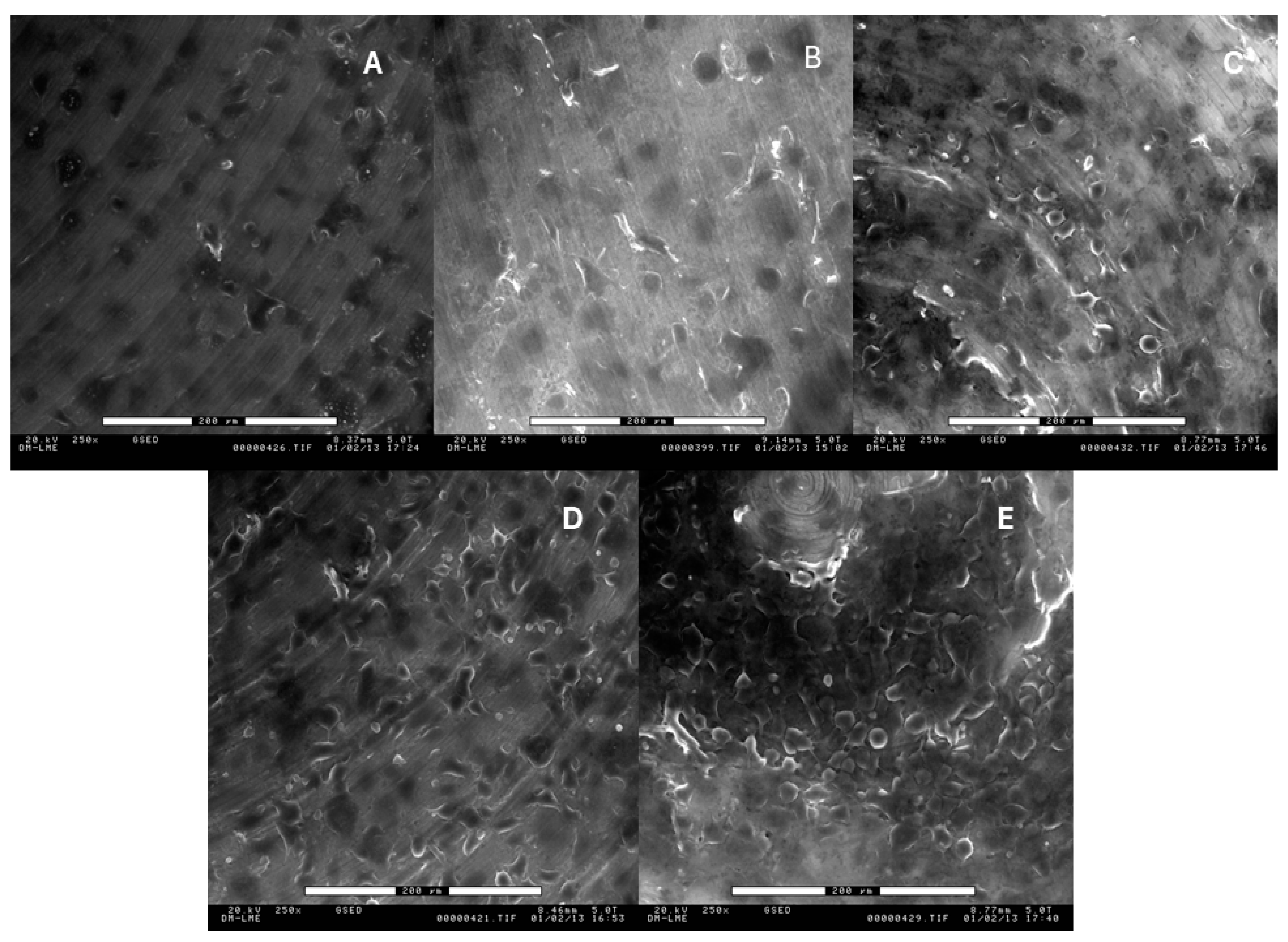
2.2. Roughness Analysis
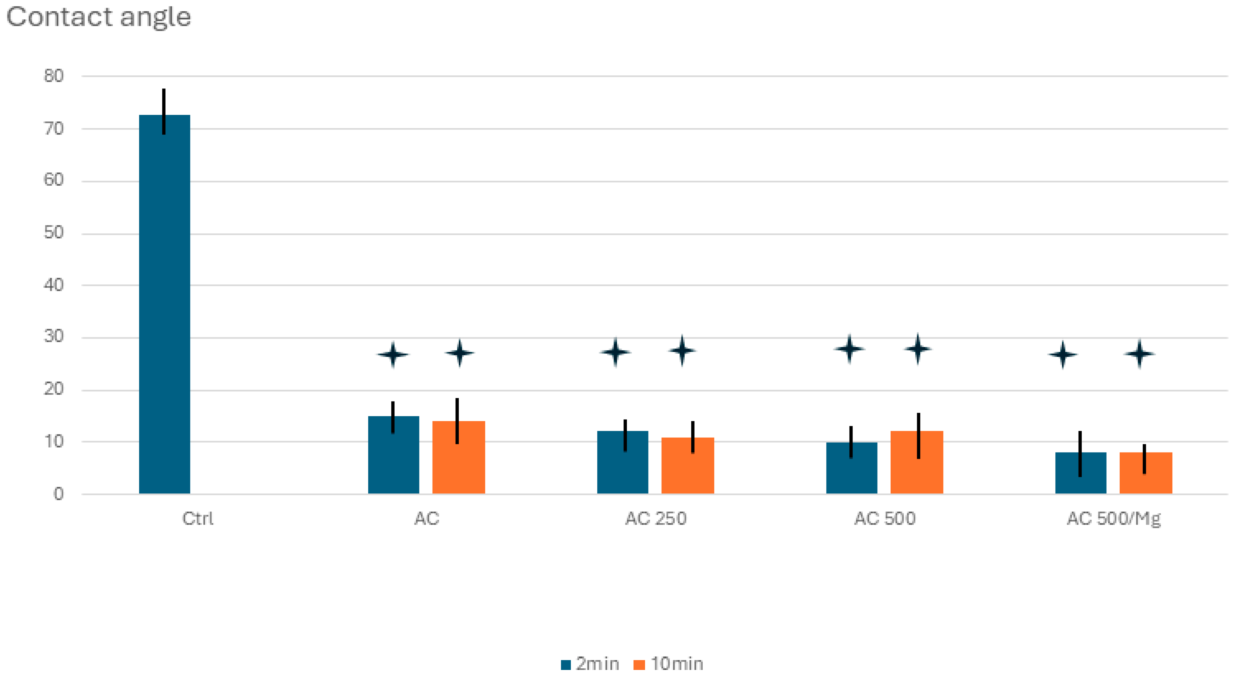
The smooth and micro-roughened surfaces were analyzed using a white light interferometer microscope (Wyko NT9300 Optical Profiler, Veeco Instruments, New York, NY, USA) in vertical scanning interferometry mode. A minimum of three measurements were taken from three different samples of each series. Approximately 230 imaging frames were used, enabling rapid and highly accurate measurements of the grooves. The surface analysis covered areas of 127.7 × 95.8 µm for groove imaging and 63.1 × 47.3 µm for plain regions within the grooves. Data filtering and analysis were conducted with Wyko Vision 4.10 software (Veeco Instruments, Plainview, NY, USA), with a Gaussian filter applied to remove curvature and tilt from every surface analysis. Sa (average roughness) was measured, which represents the arithmetic average of the absolute values of the surface deviations from the mean plane.

2.3. Wettability

The Contact Angle (CA) was determined to evaluate the surface wettability of the titanium with treatments except for the control for 2 and 10 min of immersion, using 5 samples per material. The wettability measurements of the samples were measured using the contact angle system “OCA 15 plus” (Dataphysics Instrument Company, Charlotte, NC, USA) and the results were analyzed with the software “SCA20” (Dataphysics Instrument Company Charlotte, NC, USA).
For droplet deposition, a 1 mL “Braun” syringe was employed in a droplet generation system with micrometer displacement control, allowing for precise dosing volume of 2 μL at a rate of 1 μL/s. The liquid droplets were backlit with LEDs through ground glass, and the contact angle was measured 5 s after the droplets were placed on the surface. MiliQ water was used for contact angle measurement, which was conducted on both untreated and treated samples with a “Citric Acid 25% + Collagen 500” solution after 2 and 10 min of immersion.

2.4. Fibroblast Culture

Human foreskin fibroblasts (Millipore, Billerica, MA, USA) primary cells (HFFs) were cultured in phenol red-free Dulbecco’s Minimum Essential Medium (DMEM; Invitrogen, Carlsbad, CA, USA) supplemented with 10% fetal bovine serum (FBS), L-glutamine (2 mM) and penicillin/streptomycin (50 U/mL and 50 μg/mL, respectively) at 37 °C in a humidified incubator at 5% CO2, with media changed every 2 days. Cells between the sixth and tenth passages were used in all the experiments. Subconfluent cells were trypsinized, centrifuged and seeded at a density of 6 × 10³ cells/disc with serum-free DMEM without phenol red onto different micro-grooved titanium discs in a 48-well microplate with an agarose layer (in order to prevent cell attachment to the dish). Tissue culture polystyrene (TCPS) and polished c.p. titanium served as reference substrates. Cellular analyses were performed at 4 h, 24 h, and 72 h after seeding.
HFFs were cultured on different surfaces. Then, cell adhesion and proliferation were analyzed using the Cell Proliferation Reagent WST-1 (Roche Applied Science, Penzberg, Germany). This colorimetric protocol measures the creation of the formazan dye by cellular activity. The tetrazolium salts incorporated into the medium were cleaved by mitochondrial dehydrogenases of living cells, and the resulting soluble formazan dye analyzed spectrophotometrically. There is a direct correlation between the absorbance of the dye solution and the cell number. Viability was evaluated at the specified culture times by incubating for 2 h with 1:10 WST-1 in serum-free DMEM without phenol red. The optical density (OD) at 440 nm of cell supernatant was evaluated with an ELx800 Universal Microplate Reader (Bio-Tek Instruments, Inc., Winooski, VT, USA). Three different samples for every surface and two different experiments were measured in parallel. A standard curve was performed using cell numbers ranging from 3 × 10³ to 50 × 103.
Non-viable cells were quantified by measuring the released lactate dehydrogenase (LDH) enzyme at the specified culture times. For that purpose, the cell-free culture supernatant was collected, centrifuged at 250× g for 5 min and then analyzed with the Cytotoxicity Detection Kit LDH (Roche Applied Science) as per the manufacturer’s instructions. The reduction of tetrazolium salts into formazan dye by LDH activity was measured spectrophotometrically at 490 nm. TCPS was used as a low-control sample, and lysed cells were utilized as a high-control sample (maximum releasable LDH activity). Three different samples of each series in two experiments were analyzed.

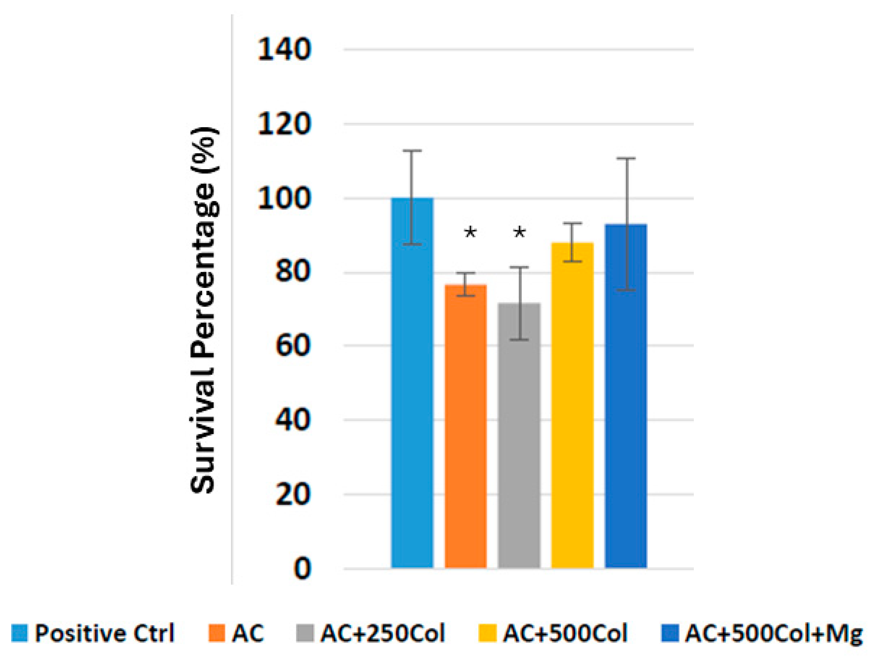
2.5. Osteoblasts Culture

For the cell adhesion assay, osteoblastic SaOS-2 cells, a cell line with epithelial morphology derived from bone, were used. Six to seven cell passages were performed before seeding the cells into the study samples. During cell passages, control of the growth and cell viability was tested. Cells were initially thawed by gently shaking the cryovial in a 37 °C water bath for 1–2 min. From this point onward, everything was performed in sterile conditions. Once thawed, the content was transferred to a falcon with 9 mL of culture medium and centrifuged at 300× g for 3 min. Then, the supernatant was aspirated, and the pellet was resuspended with 1 mL of cell culture medium. Cells were seeded in flasks F175 and were kept at 37 °C with 5% CO2, with the cell culture medium being changed 2–3 times per week.
The cell culture medium for this cell line consists of McCoy’s 5a Medium Modified with L-glutamine 1.5 mM and 2200 mg/L sodium bicarbonate. This medium was supplemented with 15% Fetal Bovine Serum (FBS), 1% Penicillin/Streptomycin (P/S) and 2% Sodium Pyruvate Solution (NaPyr).
Cell passage [46–47] was done at 90% confluence. Therefore, the cells were detached from the flasks by removing the medium, washing twice with 5 mL of PBS (37 °C) to remove dead cells, and then adding 5 mL of 0.05% Trypsin. The flasks were left in the incubator at 37 °C with 5% CO2 for 2–3 min. Afterward, Trypsin was neutralized with 7 mL of cell culture medium (37 °C) and the content was transferred to a Falcon tube and centrifuged for 5 min at 300× g. The supernatant was then aspirated, and the pellet was resuspended with a cell culture medium. Cell counting with TripanBlue was then performed. For this, 10 μL of previously resuspended cells was prepared in an Eppendorf and mixed with Tripan Blue. A volume of 10 μL of the total was transferred to a Neubauer Chamber for counting using Phase Contrast Microscopy. The corresponding calculations were then carried out.
Resazurin Salt assay (Alamar Blue, Paisley, UK) was used to assess cell proliferation and cell viability. The protocol was the following: adding 5 mg Resazurin Salt (Sigma–Aldrich, Burlington, MA, USA) in 1 mL of PBS obtaining a stock solution of 5 mg/mL. Then, 100 μL was transferred to a Falcon with 50 mL of cell culture medium, and this solution was filtered to ensure sterile conditions. The final solution had a concentration of 10 μg/mL. This solution was protected from light.
After three days of cell culture, the culture medium was removed, and each well was washed with 500 μL of pre-warmed PBS. Then, 300 μL of 10 μg/mL Resazurin solution was added to each well and incubated at 37 °C and 5% CO2 for 3 h. Afterward, 200 μL from each well was transferred to a 96-well plate, transparent, and finally, the absorbance was analyzed. The wavelength was 570 nm and 600 nm, and an Infinite® 200 PRO Multimode Absorbance Multimode Microplate Reader (TECAN, Männedorf, Switzerland) was used.

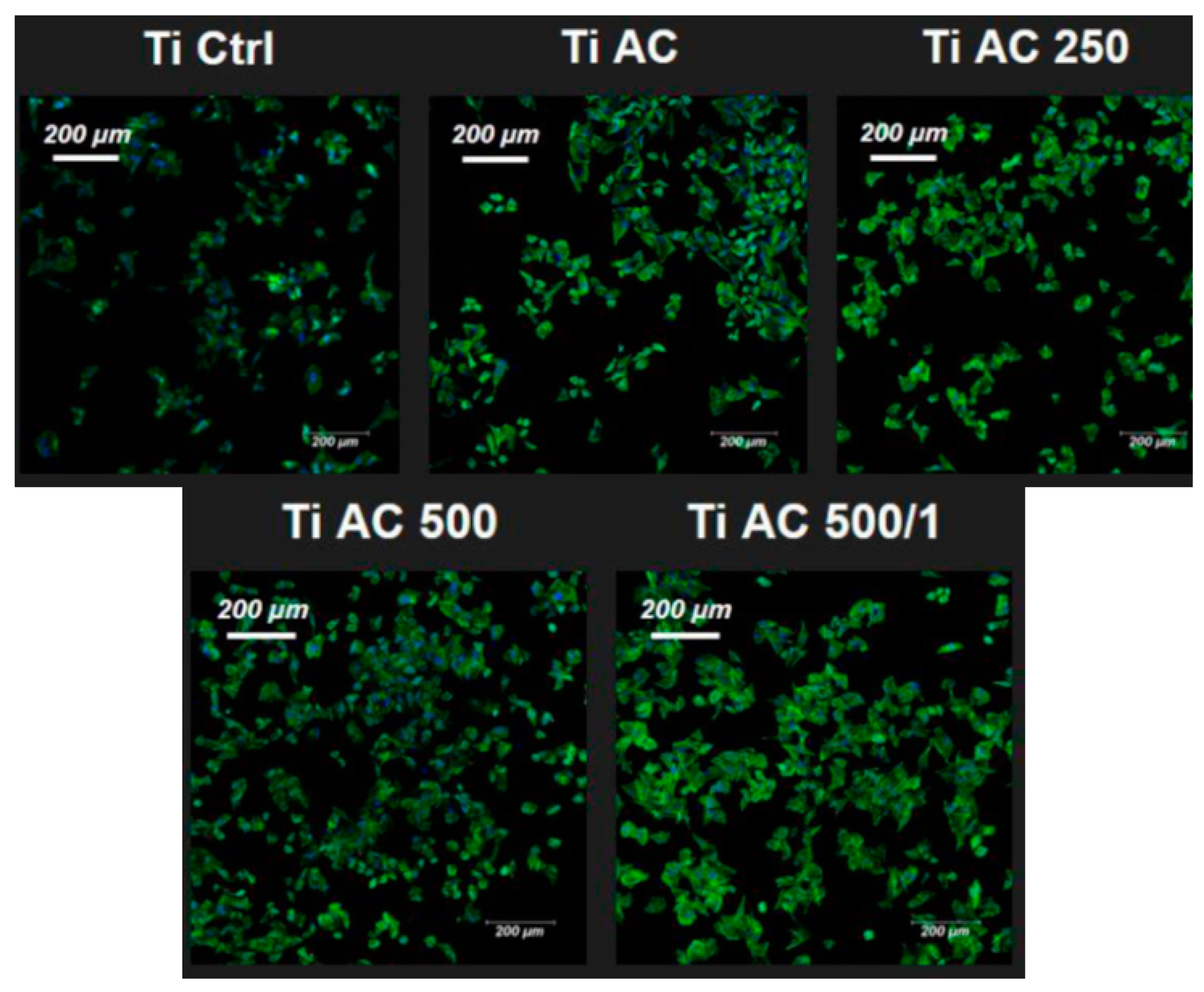
2.6. Immunofluorescence

For the immunofluorescence assay, a working solution of actin 488-stained Phalloidin (100 nM) was prepared by diluting 58.8 μL of the 14 μM stock in 8.4 mL of PBS. Additionally, a DAPI solution was prepared by diluting 10 μL in 10 mL of PBS. Both solutions were kept at room temperature, without light exposure. A 0.1% Triton-X solution was made by diluting 1 mL of Triton-X in 9 mL of PBS. The samples were analyzed using the STELLARIS 5 Cryo Confocal Light Microscope (Molecular Devices, San Jose, CA, USA).
After three days of cell culture, the culture medium was removed, and cells were washed with 500 μL of pre-warmed PBS. Then, 350 μL of 4% PFA/PBS was added to the wells at room temperature in order to fix the cells. Following fixation, cells were washed with 500 μL of PBS, and then permeabilization was performed using 350 μL of 0.1% Triton-X/PBS. After 10 min, the cells were washed with 500 μL of PBS and after, 350 μL of Actin 488-stained Phalloidin was incorporated. Cells were then incubated for 30 min at room temperature without light exposure. After Actin staining, cells were washed three times with 500 μL of PBS and cells were incubated with 350 μL of DAPI solution at room temperature in the dark for 2–3 min. Finally, they were washed again with 500 μL of PBS, and cells were kept with 500 μL of PBS at 4 °C, without light exposure.

2.7. Bacterial Culture

Bacterial assays were performed using two oral pathogens, representing a Gram-negative and a Gram-positive bacterial strain. Pseudomonas aeruginosa, a Gram-negative bacterial strain, was sourced from Colección española de cultivos tipo (CECT 110, Valencia, Spain). For Gram-positive strains, Streptococcus sanguinis and Streptococcus gordonii were used, and were obtained from Culture Collection University of Gothenburg (CCUG 15915, Goteborg, Sweden) and from Colección española de cultivos tipo (CECT 804, Valencia, Spain) respectively.
A total of six samples (n = 6) were used for the bacterial adhesion test, with three samples from each study group dedicated to the Gram-positive and three for the Gram-negative. Prior to the test, the culture media and material (PBS) were sterilized by autoclaving at 121 °C for 30 min using autoclave oven SELECTA model Sterilmax (SELECTA, Abrera, Spain). As previously described, samples were also sterilized by incubating in alcohol three times for 5 min in sterile culture plates. Afterward, the samples were exposed to ultraviolet light for another 30 min [33,34].
Agar plates were incubated at 37 °C for 24 h. The bacterial inoculum was prepared by suspending the bacteria in 5 mL of Brain Heart Infusion Broth (BHI) (Sigma Aldrich, St. Loius, MO, USA) followed by an incubation for 24 h at 37 °C. The medium was then adjusted to an optical density of 0.1 at a wavelength of 600 nm (OD600 = 0.1). For the bacterial adhesion test, 500 µL of the suspension (OD600 = 0.1) was added to each well of the culture plate and incubated at 37 °C for 1 h, using an incubator oven MEMMERT BE500 (MEMMERT Gmbh, Scheabach, Germany). All tests were carried out under static conditions without external stirring.
Then, the samples were rinsed twice with PBS for 5 min each, and then they were fixed with a 2.5% glutaraldehyde solution in PBS for 30 min at 4 °C. Following fixation, the glutaraldehyde solution was removed, and the samples were rinsed three times with PBS for 5 min each.
For viability analysis, confocal microscopy and the LIVE///DEAD Backlight bacterial viability kit (Thermo Fisher, Madrid, Spain) were used [35,36]. A solution was prepared by mixing 1.5 μL of propidium with 1 mL of PBS. Using a micropipette, a drop of this solution (approximately 50 μL/sample) was applied to the surface. After incubating at room temperature without light exposure for 15 min, the samples were rinsed three times with PBS for 5 min. The surfaces were then examined by laser scanning microscopy (CLSM). Three images per sample were captured at 630× magnification. Live and dead bacteria were detected using a wavelength of 488 nm and 561 nm. This analysis enabled both the assessment of bacterial viability on each surface and an initial comparison of the bacterial count present in the different groups of samples.

2.8. Statistical Analysis
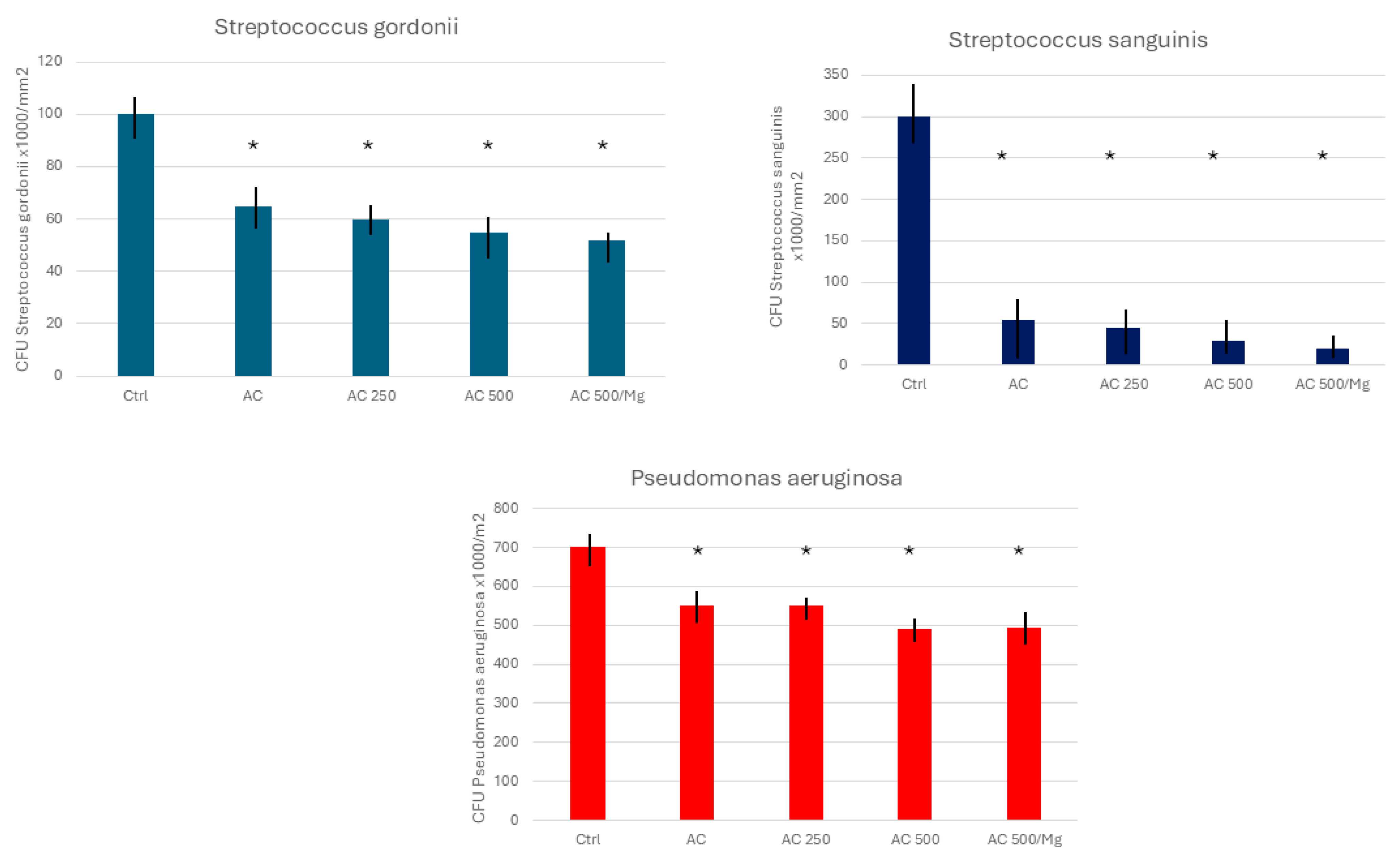
Statistical analysis was carried out using the comparative t-test (with Excel software 365-00123 Microsoft, Redmons, WA, USA). This was done between the different groups at 95% confidence. Therefore, statistically significant differences are with values of (p < 0.05).

3. Results

The roughness measurements (Sa) in Table 3, reveal that the different immersion treatments carried out on the discs slightly increase the roughness, as no statistically significant differences (p < 0.05) were observed with respect to the control group.
Figure 1 shows the contact angle for the different surfaces studied without treatment (Ctrl) and with immersion inside the different solutions for the different times studied. The roughness produced by implantoplasty according to the protocol [31] does not present statistical differences in relation to the control. However, the samples treated with a citric acid solution produced an increase in roughness with statistically significant differences of p < 0.05.
As we can see, the results between the 2 and 10-min times do not present statistically significant differences, and therefore, we will choose the 2 min time for the treatments.
Scanning electron microscope observations (Figure 2) after 4 h of culture showed fibroblasts on the titanium surface with different dissolutions. The cells were flattened and distributed without showing any specific orientation when seeded on TCPS.
Cell proliferation was evaluated by measuring the conversion of tetrazolium salts into soluble formazan dye by metabolically active cells. HFFs were cultured onto different surfaces, and the absorbance at 440 nm after WST-1 addition was measured at 4, 24, and 72 h after cell seeding. A standard curve using serial dilutions of cell numbers was prepared to extrapolate absorbance sample values. After 4 h of culture, there were statistically more living cells on the AC 500/Mg surfaces than on the Ctrl, AC, AC 250, and AC 500 surfaces (Figure 3). After 24 h and 72 h, the number of cells increases, with AC 500/Mg having the highest number of cells and statistically significant differences. Figure 3 permits us to assume that the cells reached culture confluence after 72 h.
Cytotoxicity was assessed by measuring the reduction of tetrazolium salts into formazan dye by LDH activity released by damaged cells. The results can be observed in Figure 4; it can be observed that all the treatments are cytocompatible, with AC 500/Mg performing the best with statistically significant differences for AC and AC 250.
The F-actin (Phalloidin/DAPI) immunofluorescence assay was performed using the Confocal Light Microscope to determine the presence and distribution of osteoblastic cells seeded on the samples and thus define if the surfaces of the titanium are favorable for the adhesion of this type of cells. Figure 5 shows that AC500/Mg treatment shows good cell viability for each treatment.
Figure 6 shows the quantitative analyses of the bacterial adhesion assay performed with Gram-positive Streptococcus gordonii and Streptococcus sanguinis strains, which showed a clear trend toward a reduction in bacterial adhesion with the presence of citric acid solutions. The bacteria CFUs present statistically significant differences in relation to the control with the citric acid treatment solutions. The Gram-negative strain of Pseudomonas aeruginosa shows significant differences (p < 0.05) in the number of bacteria that adhered to the surface of the control in relation to the different immersion treatments. However, the bactericidal effect is lower than that of the Gram-positive strain.
The AC 500/Mg treatment showed lower bacterial adhesion than the other groups for both bacterial strains tested in this study.

4. Discussion

The results were obtained from the control samples, which did not receive any treatment but were sterilized in an autoclave at 121 °C for 30 min. We can observe an important decrease in the contact angles when the samples are treated with citric acid solutions, transforming the surface as hydrophilic [34]. The results obtained from the treated samples, i.e., immersed for 2 and 10 min in the solutions, are very similar. According to the results of the first measurement, we can observe that all the values are within the margins that establish the hydrophilicity and super-hydrophilicity properties for the two treatment times. The two studied times can increase hydrophilicity, but there are no statistically significant differences between them.
The roughness results show that the immersion treatments in the different solutions present statistically significant differences. Solutions based on citric acid produce a slight etching on the surface. It is known from the work of Vilarrasa et al. [35,36] that a passivation layer of about 6 nm of titanium oxide TiO2 is formed, which, as we can see, reproduces the roughness of the control surface.
The initial cell attachment to a surface is a crucial step for their viability. This process is influenced by the surface chemistry but also by its roughness [37,38,39]. While many studies have focused on creating micro and nano-roughness in order to guide cell orientation, an improvement in cell adhesion to modified titanium surfaces has not been fully demonstrated. Our results indicate that cells adhere better and proliferate sooner on AC 500/Mg-treated surfaces compared to the other titanium surfaces tested. After initial cell attachment, cells proliferate in similar patterns on all the surfaces studied until they reach confluence. The micro-etching in the process of immersion of citric acid would favor adhesion, proliferation, and fibroblastic differentiation, generating a biological seal that would prevent or reduce bacterial filtration [38].
Collagen, a common biomaterial used in films, composites and three-dimensional matrices, enhances tissue regeneration and wound protection. Moreover, it is a non-toxic, biodegradable, and bioabsorbable material [40]. Collagen type I, the most abundant type, constitutes 85% of the extracellular matrix (ECM) with well-known proteins like laminins, fibronectin, and vitronectin. An in vivo study performed by Sartori et al. demonstrated that dental implants coated with type I collagen improved bone regeneration and osseointegration in osteopenic rats, providing greater mechanical stability and a higher rate of osseointegration [41]. Other studies have supported the role of type I collagen in promoting osseointegration by stimulating bone formation at the cellular and molecular level [42].
For optimal titanium surface regeneration, soft tissue would be ideal for mimicking the natural tissue orientation. A possible technique will have to be studied, as performing the operation on the dental implant in place is a complex task [37].
Magnesium is used in surface modifications of implants and can enhance bone bonding. A recent study by Veronese et al. that combined titanium dioxide (TiO2) with magnesium imparted anti-inflammatory properties to titanium implants [43]. The inflammatory response plays a critical role in dental implant integration and can influence osteogenesis [44,45]. As explained above, citric acid treatment slightly increases surface roughness, which can boost bacterial adherence [46,47]. However, the acidic nature of the sample surfaces prevents or decreases bacterial colonization [48,49,50]. The concentration of citric acid is related to the antibacterial action it provides and thus causes an alteration (reduction) in the pH of the extracellular matrices. It is hypothesized that the presence of citric acid affects the bacterial membrane permeability, disrupting the hydrogen gradient between intracellular and extracellular environments [51,52]. Furthermore, it possesses an antioxidant capacity that can prevent or delay some types of cell damage and also has a negative effect on mycobacteria [49,50,51]. Furthermore, the treatment provided is capable of forming a biocompatible titanium oxide layer, providing a high resistance to corrosion, thus reducing bacterial activity [51,53,54,55,56,57,58,59,60,61]. Magnesium has also been shown to have an antibacterial effect, which could explain the lower levels of bacterial survival in samples containing Mg.
One of the most effective bactericidal treatments is the silver nanoparticles treatment, as demonstrated by Korniienko et al. [28]. However, we cannot put it in the mouthwash because it would not be anchored in the titanium, and it would be necessary to make an electrolytic treatment to produce the union nanoparticle with titanium having a bactericidal capacity [62,63,64]. Thus, the citric acid that produces a passivation layer of compact titanium oxide and with an oxidizing character causes the oxidation of the bacteria, giving that bactericidal capacity to the titanium surface [65]. Being a weak acid, we have adjusted the concentrations to those that do not cause toxicity to tissues, as demonstrated by experimental results, nor do they cause irritation to soft tissues [66].
This study should be completed in vivo, considering the expected results obtained in vitro. Studies in soft tissues should be carried out to confirm good cell behavior and that there is no irritability in the tissues. Likewise, it should be observed that if the bone tissue is capable of regenerating and bone index contact values are obtained, they can provide a good mechanical fixation to the dental implant. A limitation of the study is that tests have been carried out on isolated bacterial strains, but it would be necessary to colonize a biofilm and determine the bactericidal capacity of the citric acid solution. In addition, the concentrations of the mouthwash should be optimized to optimize the treatment.

5. Conclusions

Treatments on the surface of titanium that underwent the implantoplasty process with citric acid solutions slightly increase the roughness and significantly increase the hydrophilic character. From the studies of the behavior of fibroblasts and osteoblasts, it has been observed that the solution of citric acid at 25% with 0.5 g of collagen/l and 1% of Mg is the one that produces the greatest proliferation and that will enhance tissue regeneration on the surface of the titanium. It has been possible to determine the important bactericidal character of citric acid solutions, especially on Gram-positive bacteria, in order to avoid the possible bacterial recolonization of the surface. The cytocompatibility and the characteristics of the solution allow the use of mouthwash in the mouth, and it could be applied on the surface of the dental implant after implantoplasty treatment.

Author Contributions

Conceptualization, J.G. and M.F.-D.; methodology, B.M.B.; software, P.F.-G.; validation, J.G. and B.M.B.; formal analysis, M.F.-D.; investigation, P.F.-G.; resources, M.F.-D.; data curation, B.M.B.; writing—original draft preparation, J.G.; writing—review and editing, J.G.; visualization, M.F.-D.; supervision, J.G.; project administration, M.F.-D.; funding acquisition, M.F.-D. All authors have read and agreed to the published version of the manuscript.

Funding

The work was funded by the Spanish Government and the Ministry of Science and Innovation of Spain through research projects CONCEPTO PDC2022-133628-C22 (co-funded by the European Regional Development Fund (ERDF), a way to build Europe) and research project MINECO (PID2022-137496OB-I00).

Institutional Review Board Statement

Not applicable.

Informed Consent Statement

Not applicable.

Data Availability Statement

The original contributions presented in the study are included in the article; further inquiries can be directed to the corresponding author.

Acknowledgments

The authors are grateful to Klockner Dental Implants.

Conflicts of Interest

The authors declare no conflicts of interest.

References

  1. Roos-Jansåker, A.M.; Lindahl, C.; Renvert, H.; Renvert, S. Nine- to fourteen-year follow-up of implant treatment. Part I: Implant loss and associations to various factors. J. Clin. Periodontol. 2006, 33, 283–289. [Google Scholar] [CrossRef]
  2. Sailer, I.; Makarov, N.A.; Thoma, D.S.; Zwahlen, M.; Pjetursson, B.E. All-ceramic or metal-ceramic tooth-supported fixed dental prostheses (FDPs)? A systematic review of the survival and complication rates. Part I: Single crowns (SCs). Dent. Mater. 2015, 31, 603–623. [Google Scholar] [CrossRef] [PubMed]
  3. Pjetursson, B.E.; Valente, N.A.; Strasding, M.; Zwahlen, M.; Liu, S.; Sailer, I. A systematic review of the survival and complication rates of zirconia-ceramic and metal-ceramic single crowns. Clin. Oral Implant. Res. 2018, 29 (Suppl. S16), 199–214. [Google Scholar] [CrossRef] [PubMed]
  4. Schwarz, F.; Derks, J.; Monje, A.; Wang, H.L. Peri-implantitis. J. Periodontol. 2018, 89 (Suppl. S1), S267–S290. [Google Scholar] [CrossRef]
  5. Daubert, D.M.; Weinstein, B.F. Biofilm as a risk factor in implant treatment. Periodontology 2000 2019, 81, 29–40. [Google Scholar] [CrossRef] [PubMed]
  6. Renvert, S.; Roos-Jansåker, A.M.; Claffey, N. Non-surgical treatment of peri-implant mucositis and peri-implantitis: A literature review. J. Clin. Periodontol. 2008, 35 (Suppl. S8), 305–315. [Google Scholar] [CrossRef]
  7. Schwarz, F.; Schmucker, A.; Becker, J. Efficacy of alternative or adjunctive measures to conventional treatment of peri-implant mucositis and peri-implantitis: A systematic review and meta-analysis. Int. J. Implant Dent. 2015, 1, 22. [Google Scholar] [CrossRef]
  8. Hentenaar, D.F.M.; De Waal, Y.C.M.; Stewart, R.E.; Van Winkelhoff, A.J.; Meijer, H.J.A.; Raghoebar, G.M. Erythritol airpolishing in the non-surgical treatment of peri-implantitis: A randomized controlled trial. Clin. Oral Implant. Res. 2021, 32, 840–852. [Google Scholar] [CrossRef]
  9. Romeo, E.; Lops, D.; Chiapasco, M.; Ghisolfi, M.; Vogel, G. Therapy of peri-implantitis with resective surgery. A 3-year clinical trial on rough screw-shaped oral implants. Part II: Radiographic outcome. Clin. Oral Implant. Res. 2007, 18, 179–187. [Google Scholar] [CrossRef]
  10. Englezos, E.; Cosyn, J.; Koole, S.; Jacquet, W.; De Bruyn, H. Resective Treatment of Peri-implantitis: Clinical and Radiographic Outcomes After 2 Years. Int. J. Periodontics Restor. Dent. 2018, 38, 729–735. [Google Scholar] [CrossRef]
  11. Tomasi, C.; Regidor, E.; Ortiz-Vigón, A.; Derks, J. Efficacy of reconstructive surgical therapy at peri-implantitis-related bone defects. A systematic review and meta-analysis. J. Clin. Periodontol. 2019, 46 (Suppl. S21), 340–356. [Google Scholar] [CrossRef] [PubMed]
  12. Monje, A.; Pons, R.; Amerio, E.; Wang, H.L.; Nart, J. Resolution of peri-implantitis by means of implantoplasty as adjunct to surgical therapy: A retrospective study. J. Periodontol. 2022, 93, 110–122. [Google Scholar] [CrossRef] [PubMed]
  13. Schwarz, F.; Sahm, N.; Becker, J. Combined surgical therapy of advanced peri-implantitis lesions with concomitant soft tissue volume augmentation. A case series. Clin. Oral Implant. Res. 2014, 25, 132–136. [Google Scholar] [CrossRef]
  14. Monje, A.; Schwarz, F. Principles of Combined Surgical Therapy for the Management of Peri-Implantitis. Clin. Adv. Periodontics 2014 2022, 12, 57–63. [Google Scholar] [CrossRef]
  15. Monje, A.; Amerio, E.; Cha, J.K.; Kotsakis, G.; Pons, R.; Renvert, S.; Sanz-Martin, I.; Schwarz, F.; Sculean, A.; Stavropoulos, A.; et al. LStrategies for implant surface decontamination in peri-implantitis therapy. Int. J. Oral Implantol. 2022, 15, 213–248. [Google Scholar]
  16. Dostie, S.; Alkadi, L.T.; Owen, G.; Bi, J.; Shen, Y.; Haapasalo, M.; Larjava, H.S. Chemotherapeutic decontamination of dental implants colonized by mature multispecies oral biofilm. J. Clin. Periodontol. 2017, 44, 403–409. [Google Scholar] [CrossRef]
  17. Gosau, M.; Hahnel, S.; Schwarz, F.; Gerlach, T.; Reichert, T.E.; Bürgers, R. Effect of six different peri-implantitis disinfection methods on in vivo human oral biofilm. Clin. Oral Implant. Res. 2010, 21, 866–872. [Google Scholar] [CrossRef]
  18. Kotsakis, G.A.; Lan, C.; Barbosa, J.; Lill, K.; Chen, R.; Rudney, J.; Aparicio, C. Antimicrobial Agents Used in the Treatment of Peri-Implantitis Alter the Physicochemistry and Cytocompatibility of Titanium Surfaces. J. Periodontol. 2016, 87, 809–819. [Google Scholar] [CrossRef]
  19. Louropoulou, A.; Slot, D.E.; Van der Weijden, F. The effects of mechanical instruments on contaminated titanium dental implant surfaces: A systematic review. Clin. Oral Implant. Res. 2014, 25, 1149–1160. [Google Scholar] [CrossRef]
  20. de Tapia, B.; Valles, C.; Ribeiro-Amaral, T.; Mor, C.; Herrera, D.; Sanz, M.; Nart, J. The adjunctive effect of a titanium brush in implant surface decontamination at peri-implantitis surgical regenerative interventions: A randomized controlled clinical trial. J. Clin. Periodontol. 2019, 46, 586–596. [Google Scholar] [CrossRef]
  21. Francis, S.; Iaculli, F.; Perrotti, V.; Piattelli, A.; Quaranta, A. Titanium Surface Decontamination: A Systematic Review of In Vitro Comparative Studies. Int. J. Oral Maxillofac. Implant. 2022, 37, 76–84. [Google Scholar] [CrossRef] [PubMed]
  22. Azzola, F.; Ionescu, A.C.; Ottobelli, M.; Cavalli, N.; Brambilla, E.; Corbella, S.; Francetti, L. Biofilm formation on dental implant surface treated by implantoplasty: An in-situ study. Dent. J. 2020, 8, 40. [Google Scholar] [CrossRef] [PubMed]
  23. Yen Nee, W.; Raja Awang, R.A.; Hassan, A. Effects on the Titanium Implant Surface by Different Hygiene Instrumentations: A Narrative Review. Cureus 2022, 14, e30884. [Google Scholar] [CrossRef] [PubMed]
  24. Chen, A.; Ghaffar, H.; Taib, H.; Hassan, A. A Review of Bacterial Colonization on Dental Implants with Various Hygiene Instruments. Cureus 2023, 15, e47483. [Google Scholar] [CrossRef]
  25. Gil, F.J.; Rodriguez, A.; Espinar, E.; Llamas, J.M.; Padullés, E.; Juárez, A. Effect of oral bacteria on the mechanical behavior of titanium dental implants. Int. J. Oral Maxillofac. Implant. 2012, 27, 64–68. [Google Scholar]
  26. Wu, X.; Cai, C.; Gil, J.; Jantz, E.; Al Sakka, Y.; Padial-Molina, M.; Suárez-López del Amo, F. Characteristics of Particles and Debris Released after Implantoplasty: A Comparative Study. Materials 2022, 15, 602. [Google Scholar] [CrossRef]
  27. Toledano-Serrabona, J.; Sánchez-Garcés, M.A.; Gay-Escoda, C.; Valmaseda-Castellon, E.; Camps-Font, O.; Verdeguer, P.; Molmeneu, M.; Gil, F.J. Mechanical properties and corrosión behavior of Ti6Al4V particles obtained by Implatoplasty. An in vivo study. Part II. Materials 2021, 14, 6519. [Google Scholar] [CrossRef]
  28. Korniienko, V.; Husak, Y.; Diedkova, K.; Varava, Y.; Grebnevs, V.; Pogorielova, O.; Bērtiņš, M.; Korniienko, V.; Zandersone, B.; Ramanaviciene, A.; et al. Antibacterial Potential and Biocompatibility of Chitosan/Polycaprolactone Nanofibrous Membranes Incorporated with Silver Nanoparticles. Polymers 2024, 16, 1729. [Google Scholar] [CrossRef]
  29. Punset, M.; Vilarrasa, J.; Nart, J.; Manero, J.M.; Bosch, B.; Padrós, R.; Perez, R.A.; Gil, J. Citric acid passivation of titanium dental implants for minimizing bacterial colonization impact. Coatings 2021, 11, 214. [Google Scholar] [CrossRef]
  30. Cruz, N.; Gil, J.; Punset, M.; Manero, J.M.; Tondela, J.P.; Verdeguer, P.; Aparicio, C.; Rúperez, E. Relevant Aspects of Piranha Passivation in Ti6Al4V Alloy Dental Meshes. Coatings 2022, 12, 154. [Google Scholar] [CrossRef]
  31. Costa-Berenguer, X.; Garcia-Garcia, M.; Sanchez-Torres, A.; Sanz-Alonso, M.; Figueiredo, R.; Valmaseda-Castellon, E. Effect of implantoplasty on fracture resistance and surface roughness of standard diameter dental implants. Clin. Oral Implant. Res. 2018, 29, 46–54. [Google Scholar] [CrossRef] [PubMed]
  32. Toledano-Serrabona, J.; Gil, F.J.; Camps-Font, O.; Valmaseda-Castellón, E.; Gay-Escoda, C.; Sánchez-Garcés, M.Á. Physicochemical and Biological Characterization of Ti6Al4V Particles Obtained by Implantoplasty: An In Vitro Study. Part I. Materials 2021, 14, 6507. [Google Scholar] [CrossRef] [PubMed]
  33. Rupp, F.; Liang, L.; Geis-Gerstorfer, J.; Scheideler, L.; Hüttig, F. Surface characteristics of dental implants: A review. Dent. Mater. 2018, 34, 40–57. [Google Scholar] [CrossRef]
  34. Gil, F.J.; Solano, E.; Pena, J.; Engel, E.; Mendoza, A.; Planell, J.A. Microstructural, mechanical and citotoxicity evaluation of different NiTi and NiTiCu shape memory alloys. J. Mater. Sci. Mater. Med. 2004, 15, 1181–1185. [Google Scholar] [CrossRef] [PubMed]
  35. Vilarrasa, J.; Àlvarez, G.; Soler-Ollé, A.; Gil, J.; Nart, J.; Blanc, V. Bacterial Adhesion of TESPSA and Citric Acid on Different Titanium Surfaces Substrate Roughness: An In Vitro Study with a Multispecies Oral Biofilm Model. Materials 2023, 16, 4592. [Google Scholar] [CrossRef]
  36. Verdeguer, P.; Gil, J.; Punset, M.; Manero, J.M.; Nart, J.; Vilarrasa, J.; Ruperez, E. Citric Acid in the Passivation of Titanium Dental Implants: Corrosion Resistance and Bactericide Behavior. Materials 2022, 15, 545. [Google Scholar] [CrossRef]
  37. Guillem, J.; Delgado, L.; Godoy-Galardo, M.; Pegueroles, M.; Herrero, M.; Gil, F.J. Fibroblast adhesion and activation onto micro-machined titanium surfaces. Clin. Oral Implant. Res. 2013, 24, 770–780. [Google Scholar] [CrossRef]
  38. Zhang, C.N.; Zhou, L.Y.; Qian, S.J.; Gu, Y.X.; Shi, J.Y.; Lai, H.C. Improved response of human gingival fibroblasts to titanium coated with micro-/nano-structured tantalum. Int. J. Implant. Dent. 2021, 7, 36. [Google Scholar] [CrossRef]
  39. Alghamdi, H.S.; Jansen, J.A. The development and future of dental implants. Dent. Mater. J. 2020, 39, 167–172. [Google Scholar] [CrossRef]
  40. Eftekhar Ashtiani, R.; Alam, M.; Tavakolizadeh, S.; Abbasi, K. The Role of Biomaterials and Biocompatible Materials in Implant-Supported Dental Prosthesis. In Evidence-Based Complementary and Alternative Medicine; Hindawi Limited: London, UK, 2021; Volume 2021. [Google Scholar]
  41. Sartori, M.; Giavaresi, G.; Parrilli, A.; Ferrari, A.; Aldini, N.N.; Morra, M.; Cassinelli, C.; Bollati, D.; Fini, M. Collagen type I coating stimulates bone regeneration and osteointegration of titanium implants in the osteopenic rat. Int. Orthop. 2015, 39, 2041–2052. [Google Scholar] [CrossRef]
  42. Silva, R.C.S.; Agrelli, A.; Andrade, A.N.; Mendes-Marques, C.L.; Arruda, I.R.S.; Santos, L.R.L.; Vasconcelos, N.F.; Machado, G. Titanium Dental Implants: An Overview of Applied Nanobiotechnology to Improve Biocompatibility and Prevent Infections. Materials 2022, 15, 3150. [Google Scholar] [CrossRef]
  43. Veronese, N.; Pizzol, D.; Smith, L.; Dominguez, L.J.; Barbagallo, M. Effect of Magnesium Supplementation on Inflammatory Parameters: A Meta-Analysis of Randomized Controlled Trials. Nutrients 2022, 14, 679. [Google Scholar] [CrossRef] [PubMed]
  44. Veronese, N.; Demurtas, J.; Pesolillo, G.; Celotto, S.; Barnini, T.; Calusi, G.; Caruso, M.G.; Notarnicola, M.; Reddavide, R.; Stubbs, B. Magnesium and health outcomes: An umbrella review of systematic reviews and meta-analyses of observational and intervention studies. Eur. J. Nutr. 2020, 59, 263–272. [Google Scholar] [CrossRef] [PubMed]
  45. Nielsen, F.H. Magnesium deficiency and increased inflammation: Current perspectives. J. Inflamm. Res. 2018, 11, 25. [Google Scholar] [CrossRef]
  46. Shibata, Y.; Tanimoto, Y. A review of improved fixation methods for dental implants. Part I: Surface optimization for rapid osseointegration. J. Prosthodont. Res. 2015, 59, 20–33. [Google Scholar] [CrossRef]
  47. Medvedev, A.E.; Neumann, A.; Ng, H.P.; Lapovok, R.; Kasper, C.; Lowe, T.C.; Anumalasetty, V.N.; Estrin, Y. Combined effect of grain refinement and surface modification of pure titanium on the attachment of mesenchymal stem cells and osteoblast-like SaOS-2 cells. Mater. Sci. Eng. C 2017, 71, 483–497. [Google Scholar] [CrossRef]
  48. Godoy-Gallardo, M.; Guillem-Marti, J.; Sevilla, P.; Manero, J.M.; Gil, F.J.; Rodriguez, D. Anhydride-functional silane immobilized onto titanium surfaces induces osteoblast cell differentiation and reduces bacterial adhesion and biofilm formation. Mater. Sci. Eng. C Mater. Biol. Appl. 2016, 59, 524–532. [Google Scholar] [CrossRef]
  49. Godoy-Gallardo, M.; Manzanares-Céspedes, M.C.; Sevilla, P.; Nart, J.; Manzanares, N.; Manero, J.M.; Gil, F.J.; Boyd, S.K.; Rodríguez, D. Evaluation of bone loss in antibacterial coated dental implants: An experimental study in dogs. Mater. Sci. Eng. C Mater. Biol. Appl. 2016, 69, 538–545. [Google Scholar] [CrossRef] [PubMed]
  50. Ferguson, A.B.; Laing, P.G.; Hodge, E.S. The ionization of metal implants in living tissues. J. Bone Jt. Surg. 1960, 42, 77–90. [Google Scholar] [CrossRef]
  51. Ferguson, A.B.; Akahoshi, Y.; Laing, P.G.; Hodge, E.S. Characteristics of trace ion release from embedded metal implants in the rabbit. J. Bone Jt. Surg. 1962, 44, 317–336. [Google Scholar] [CrossRef]
  52. Gil, F.J.; Planell, J.; Proubasta, I.; Vazquez, J. Fundamentos de Biomecánica y Biomateriales; Ergon: Barcelona, Spain, 1997; pp. 125–132. [Google Scholar]
  53. Gil, F.J.; Planell, J.A. Aplicaciones biomédicas del titanio v sus aleaciones. Biomecánica 1993, 1, 34–43. [Google Scholar] [CrossRef]
  54. Lukaszewska-Kuska, M.; Idzior-Haufa, M.; Dorocka-Bobkowska, B. Evaluation of human osteoblast metabolic activity in modified titanium-conditioned medium. Proc. Inst. Mech. Eng. H 2020, 234, 603–611. [Google Scholar] [CrossRef]
  55. Lukaszewska-Kuska, M.; Wirstlein, P.; Majchrowski, R.; Dorocka-Bobkowska, B. The effects of titanium topography and chemical composition on human osteoblast cell. Physiol. Res. 2021, 70, 413–423. [Google Scholar] [CrossRef]
  56. Guizzardi, S.; Galli, C.; Martini, D.; Belletti, S.; Tinti, A.; Raspanti, M.; Taddei, P.; Ruggeri, A.; Scandroglio, R. Different titanium surface treatment influences human mandibular osteoblast response. J. Periodontol. 2004, 75, 273–282. [Google Scholar] [CrossRef] [PubMed]
  57. Sader, M.S.; Balduino, A.; Soares Gde, A.; Borojevic, R. Effect of three distinct treatments of titanium surface on osteoblast attachment, proliferation, and differentiation. Clin. Oral Implant. Res. 2005, 16, 667–675. [Google Scholar] [CrossRef] [PubMed]
  58. Yurttutan, M.E.; Keskin, A. Evaluation of the effects of different sand particles that used in dental implant roughened for osseointegration. BMC Oral Health 2018, 18, 47. [Google Scholar] [CrossRef]
  59. Wang, Q.G.; Zhou, P.; Liu, S.F.; Attarilar, S.; Ma, R.L.W.; Zhong, Y.S.; Wang, L.Q. Multi-scale surface treatments of titanium implants for rapid osseointegration: A review. Nanomaterials 2020, 10, 1244. [Google Scholar] [CrossRef]
  60. Hotchkiss, K.M.; Ayad, N.B.; Hyzy, S.L.; Boyan, B.D.; Olivares-Navarrete, R. Dental implant surface chemistry and energy alter macrophage activation in vitro. Clin. Oral Implant. Res. 2017, 28, 414–423. [Google Scholar] [CrossRef]
  61. Canabarro, A.; Diniz, M.G.; Paciornik, S.; Carvalho, L.; Sampaio, E.M.; Beloti, M.M.; Rosa, A.L.; Fischer, R.G. High concentration of residual aluminum oxide on titanium surface inhibits extracellular matrix mineralization. J. Biomed. Mater. Res. A 2008, 87, 588–597. [Google Scholar] [CrossRef]
  62. Al-asbahi, M.G.S.S.; Al-Ofiry, B.A.; Saad, F.A.A.; Al-Gunaid, M.Q.A. Silver nanoparticles biosynthesis using mixture of Lactobacillus sp. and Bacillus sp. growth and their antibacterial activity. Sci. Rep. 2024, 14, 10224. [Google Scholar] [CrossRef]
  63. Mishra, M.; Ballal, A.; Rath, D.; Rath, A. Novel silver nanoparticle-antibiotic combinations as promising antibacterial and anti-biofilm candidates against multiple-antibiotic resistant ESKAPE microorganisms. Colloids Surf. B Biointerfaces 2024, 236, 113826. [Google Scholar] [CrossRef] [PubMed]
  64. Ortiz-Garcia, I.; Nuñez-Márquez, E.; Moreno-Muñoz, J.; Gil, J.; Delgado, L.M.; Rondón-Romero, J.L.; Monsalve-Guil, L. Silver coating on dental implantabutment connection screws as potential strategy to prevent loosening and minimizing bacteria adhesion. Front. Bioeng. Biotechnol. 2024, 11, 1293582. [Google Scholar] [CrossRef]
  65. Buxadera-Palomero, J.; Calvo, C.; Torrent-Camarero, S.; Gil, F.J.; Mas-Moruno, C.; Canal, C.; Rodríguez, D. Biofunctional polyethylene glycol coatings on titanium: An in vitro-based comparison of functionalization methods. Colloids Surf. B Biointerfaces 2017, 152, 367–375. [Google Scholar] [CrossRef] [PubMed]
  66. Hwang, J.H.; Lee, S.; Lee, H.G.; Choi, D.; Lim, K.M. Evaluation of Skin Irritation of Acids Commonly Used in Cleaners in 3D-Reconstructed Human Epidermis Model, KeraSkinTM. Toxics 2022, 10, 558. [Google Scholar] [CrossRef]
Figure 1. The contact angle of different surfaces studied for the control without treatment, and the samples immersed in solution for 2 and 10 min. Stars mean statistically significant differences of p < 0.05 in relation to the control.
Coatings 14 01093 g001
Figure 2. Fibroblast on the surface of titanium. (A) Control. (B) Citric Acid 25%. (C) Citric Acid 25% with Collagen 250. (D) Citric Acid 25% with Collagen 500. (E) Citric Acid 25% with Collagen 500 and 1% Mg.
Coatings 14 01093 g002
Figure 3. Analysis of cell viability onto the different materials and treatments at 4, 24, and 72 h. WST-1 cell proliferation assay showed that cells adhere better and proliferate earlier on AC 500/mg surfaces, as compared with other titanium surfaces. Asterisks indicate statistically significant differences of p < 0.05. The single asterisk means that it is statistically significantly different from the doubles, triples and quadruples. The double is statistically different from all the others, as well as the triple and quadruple in relation to the other number of asterisks.
Coatings 14 01093 g003
Figure 4. Survival percentage of fibroblasts for different treatments. Asterisks mean statistically significant differences in values of p < 0.05.
Coatings 14 01093 g004
Figure 5. Immunofluorescence assay results of different immersion treatments on the titanium surface showing the presence of cells.
Coatings 14 01093 g005
Figure 6. Quantitative analysis of bacteria for the different treatments. Asterisks mean statistically significant differences of p < 0.05.
Coatings 14 01093 g006
Table 1. Chemical composition in weight percentage.
NitrogenCarbonHydrogenIronOxygenTitanium
0.050.100.120.300.35Balance
Table 2. Citric acid-based dissolutions.
DissolutionChemical Composition
25% Citric acid (AC)Citric acid 25% in volume (v).
25% Citric acid + Collagen 250 (AC 250)Citric acid 25% (v) with 0.25 g Collagen/L.
25% Citric acid + Collagen 500 (AC 500)Citric acid 25% (v) with 0.50 g Collagen/L.
25% Citric acid + Collagen 500 + 1% Mg (AC 500/Mg)Citric acid 25% (v) with 0.50 g Collagen/L. and 10% of Mg(NO3)2·6H2O
Table 3. Roughness parameter values of the titanium-treated samples. Asterisk means statistically significant differences of p < 0.05.
TreatmentRoughness Sa (μm)
As-received0.15 ± 0.09
Implantoplasty (Ctrl)0.25 ± 0.15
AC0.33 ± 0.13 *
AC 2500.30 ± 0.10 *
AC 5000.25 ± 0.11 *
AC 500/Mg0.27 ± 0.10 *
Disclaimer/Publisher’s Note: The statements, opinions and data contained in all publications are solely those of the individual author(s) and contributor(s) and not of MDPI and/or the editor(s). MDPI and/or the editor(s) disclaim responsibility for any injury to people or property resulting from any ideas, methods, instructions or products referred to in the content.

