Review

Carbon Steel Corrosion Induced by Sulfate-Reducing Bacteria: A Review of Electrochemical Mechanisms and Pathways in Biofilms

1
School of Chemistry and Biological Engineering, University of Science and Technology Beijing, Beijing 10083, China
2
Experimental Teaching Center, College of Basic Medical Sciences, Naval Medical University, Shanghai 200433, China
*
Authors to whom correspondence should be addressed.
Coatings 2024, 14(9), 1105; https://doi.org/10.3390/coatings14091105
Submission received: 18 July 2024 / Revised: 20 August 2024 / Accepted: 22 August 2024 / Published: 1 September 2024

Abstract

:
Microbial metal corrosion has become an important topic in metal research, which is one of the main causes of equipment damage, energy loss, and economic loss. At present, the research on microbial metal corrosion focuses on the characteristics of corrosion products, the environmental conditions affecting corrosion, and the measures and means of corrosion prevention, etc. In contrast, the main microbial taxa involved in metal corrosion, their specific role in the corrosion process, and the electron transfer pathway research are relatively small. This paper summarizes the mechanism of microbial carbon steel corrosion caused by SRB, including the cathodic depolarization theory, acid metabolite corrosion theory, and the biocatalytic cathodic sulfate reduction mechanism. Based on the reversible nature of electron transfer in biofilms and the fact that electrons must pass through the extracellular polymers layer between the solid electrode and the cell, this paper focuses on three types of electrochemical mechanisms and electron transfer modes of extracellular electron transfer occurring in microbial fuel cells, including direct-contact electron transfer, electron transfer by conductive bacterial hair proteins or nanowires, and electron shuttling mediated by the use of soluble electron mediators. Finally, a more complete pathway of electron transfer in microbial carbon steel corrosion due to SRB is presented: an electron goes from the metal anode, through the extracellular polymer layer, the extracellular membrane, the periplasm, and the intracellular membrane, to reach the cytoplasm for sulfate allosteric reduction. This article also focuses on a variety of complex components in the extracellular polymer layer, such as extracellular DNA, quinoline humic acid, iron sulfide (FeSX), Fe3+, etc., which may act as an extracellular electron donor to provide electrons for the SRB intracellular electron transfer chain; the bioinduced mineralization that occurs in the SRB biofilm can inhibit metal corrosion, and it can be used for the development of green corrosion inhibitors. This provides theoretical guidance for the diagnosis, prediction, and prevention of microbial metal corrosion.

1. Introduction

Microbiologically influenced corrosion (MIC) refers to the phenomenon of accelerating the corrosion process of metallic materials directly and indirectly due to the life activities of microorganisms themselves and their metabolites [1,2,3]. Statistics show that the corrosion of metallic materials caused by microorganisms accounts for about 20% of the total corrosion of metallic materials [4]. Among them, Sulfate-Reducing Bacteria (SRB) are a group of Gram-negative bacteria capable of utilizing environmental sulfate as a terminal electron acceptor and reducing it to hydrogen sulfide under anaerobic conditions [5], which are widely found in anoxic environments with high concentrations of sulfate such as soils, seawater sediments, oilfields, and industrial equipment [6], and they are considered to be the most common microorganisms associated with anaerobic MIC [7,8].
Regarding the research on the corrosion mechanism of SRB-induced MIC, the corrosion of different metals has different corrosion behaviors and mechanisms. To explain the typical corrosion mechanism of SRB on carbon steel, there are the Acid Metabolite Corrosion Theory and the classic cathodic depolarization theory (CDT) [9]. The cathodic depolarization theory suggests that SRB depolarize the surface of carbon steel using hydrogenases, leading to increased corrosion of the metal [10]. Xu et al. [11] recognized that the cathodic depolarization theory can only be used to explain corrosion caused by hydrogenase-positive SRB. For the case that SRB without hydrogenase can still cause corrosion, the “physical cathode” should be replaced by a “biological cathode”, which is explained by the biocatalytic cathodic sulfate reduction mechanism (BCSR), in which the biofilm of the SRB acts as an electron acceptor directly and accepts the electrons from the metal anode [12]. Biofilm is a mixture of microorganisms, metabolites, and corrosion products as well as some inorganic minerals and adsorbed organic matter [13]. Among them, metabolites, namely extracellular polymers (EPS), are considered to be important components that determine the physicochemical and biological properties of biofilms, affecting metal corrosion and extracellular electron transfer processes. Notably, the biomineralization occurring in EPS can be used for the development of novel and natural green corrosion inhibitors.
Microbiologically influenced corrosion (MIC) is closely related to microbial fuel cells (MFC). The process of extracellular electron transfer (EET) in MFC, including direct electron transport (DET), exemplified by direct electron contact transfer in Shewanella oneidenis MR-1 and conductive bacterial hair proteins or nanowires in Geobacter sulfurreducens, and mediated electron transport (MET), with the examples of flavins and phenazines, can be used to explore the corrosion process of carbon steel caused by SRB, the pathway of transferring extracellular electrons in EPS and the electrochemical mechanism [14,15,16]. The various components of the EPS that can act as electron donors for the SRB intracellular electron-transport chain are further described, as well as the more complete pathways that may exist for electrons to enter the SRB cell, pass through the extracellular membrane, from the periplasm to the intracellular membrane, and arrive at the cytoplasm for sulfate isomerization reduction.
Most of the current studies on MIC are mostly concerned with the corrosion mechanism, the characteristics of corrosion products, the environmental conditions affecting corrosion, and the measures and means of corrosion prevention [3,9,17,18,19]. And based on the biological cathode in MFC, three pathways and ways of extracellular electron transfer between the metal anode and microorganisms were summarized [20]. However, due to the lack of a clear understanding of the key components involved in the complete process of electron transfer from the extracellular to the intracellular, for instance, there is still controversy about the main components of conducting hairs or nanowires involved in long-range electron transfer, and how the extracellular electrons are transported after they reach the extracellular membrane to be finally utilized in the cytoplasm. As well as insufficient consideration of the complex environment of the EPS that influences the corrosion process, there are still many problems in understanding the corrosion of carbon steel by SRB. A PRISMA flowchart of the research and compiling process for the focused questiones is shown in Figure A1. This paper focuses on the specific mechanism of action and electron transfer pathways in the corrosion process and summarizes the corrosion mechanism of SRB causing carbon steel corrosion; the transfer of electrons from solid electrodes to SRB cells and their eventual utilization; and the possible electron transfer reactions in the EPS, extracellular membranes, periplasm, intracellular membranes, and cytoplasm, with a view of providing theoretical guidance for the diagnosis, prediction, and prevention of MIC.

2. Mechanism of SRB-Induced Metal Corrosion

2.1. Corrosion of Carbon Steel

2.1.1. The Cathodic Depolarization Theory (CDT)

The corrosion of iron metal must be accompanied by the anodic dissolution of iron. Under conditions of 1 M solute (1-bar gas) (except H+), 25 °C, and pH = 7, the redox potential (Eo’) value of the couple Fe2+/Fe0 is close to that of the couple CO2+ acetate/lactate. Thus, the coupling of iron oxidation to sulfate reduction is thermodynamically favorable and iron can act as an electron donor for SRB metabolism [11]. Early researchers hypothesized that under anaerobic conditions, when microorganisms are not present in the surrounding environment, hydrogen in the water would act as an electron acceptor and form a “hydrogen film” on the metal surface, ultimately preventing the dissolution of the metal, a phenomenon known as “polarization” [21], as per Equations (1)–(3). And when microorganisms are present in the environment, the theory of cathodic depolarization suggests that SRB with hydrogenase adsorb on the metal surface and use the hydrogenase to break the “hydrogen film”, generating electrons to reduce SO42− to H2S and releasing energy to maintain the growth of SRB. The removal of H2 from the metal surface accelerates the dissolution of the metal, which is called “depolarization”, and SRB act as a depolarizing agent [10,22]. Fe2+ will combine with OH and S2− in solution to form Fe(OH)2 and FeS, shown in Equations (4)–(8). FeS forms iron sulfide (FeSX) complexes on metal surfaces with bare parts not covered by the corrosion layer [23]. Constituting a FeSX/Fe galvanic corrosion electric pair, electron enrichment of the (FeSX) corrosion product layer promotes metal corrosion and transfers electrons to the SRB for sulfate reduction [24].
Fe − 2e → Fe2+
2H2O + 2e → H2 + 2OH
Fe + 2H2O → Fe2+ + H2 +2OH
H2 − 2e → 2H+
SO42− + 8e + 8H+ → S2− + 4H2O
Fe2+ + 2OH → Fe(OH)2
Fe2+ + S2−→ FeS
4Fe + SO42− + 4H2O → 3Fe(OH)2 + FeS + 2OH

2.1.2. Acid Metabolite Corrosion Theory

H2S is one of the metabolites of SRB, and H2S is slightly soluble in water to form HS, and the proportion of its solubility in water affects the corrosion rate of metals [25]. Corrosive H2S will cause localized low pH on the metal surface, and under certain pH conditions, proton reduction coupled with iron oxidation dissolution accelerates the metal corrosion process [26].

2.1.3. The Biocatalytic Cathodic Sulfate Reduction Mechanism (BCSR)

Xu et al. [12] first proposed the biocathodic catalytic reduction theory in 2009, which, from the perspective of bioenergetics, suggests that there is a biofilm with corrosive ability attached to the metal surface and that under the action of a variety of biologically active enzymes secreted by the SRB in the biofilm, the electrons released from the dissolution of the anodic metal material pass through the bacterial cell wall, and they are consumed by the sulfate reduction process occurring in the cytoplasm of the cathodic SRB, as shown in Equations (9)–(11). The complete cathodic reaction occurs in the SRB cell membrane, and the biological membrane directly acts as an electron acceptor, and the “biological cathode” replaces the traditional “physical cathode” [19]. The dissolution of iron (Equation (9)) coupled with sulfate reduction (Equation (10)) produces a thermodynamically more favorable (the Gibbs free energy change) ΔG0′ = −178 kJ/mol as compared to ΔG0′ = −164 kJ/mol for sulfate reduction using lactic acid under standard conditions [27]. Xu et al. [11] pointed out that the CDT is only applicable to hydrogenase-positive SRB, but without hydrogenase in vivo, SRB still corrode metals. However, SRB without hydrogenase in the body still have corrosion phenomena on metals, and the CDT is only a special case of the BCSR theory.
Fe − 2e → Fe2+
SO42− + 8H+ + 8e → HS + OH + 3H2O
4Fe + SO42− + 9H+ → HS + 4H2O + 4Fe2+

2.2. Corrosion of Cu, Zn

There are differences in the mechanism of the corrosion behavior of SRB on different metals. The corrosion of Cu caused by SRB is thermodynamically unfavorable considering that the energy of Cu is much lower than that of Fe, and the oxidation of Cu that produces Cu+ or the direct coupling of Cu2+ with sulfate reduction is thermodynamically unfavorable, and therefore Cu cannot be used as an electron donor for SRB sulfate reduction [28]. Dou et al. [29] stated that the corrosion of Cu is indirectly caused by metabolites of SRB such as sulfides and protons, and that an increase in HS from sulfate reduction will lead to the more severe corrosion of Cu. Zn is a more active metal than Fe, and the corrosion of Zn by SRB is more susceptible to attack by protons in solution compared to carbon steel, in addition to being able to be directly coupled to the sulfate reduction process and be affected by its metabolites, such as H2S. The coupled reaction of the oxidation of Zn and proton reduction is thermodynamically well driven under conditions of PH = 7 [30].

3. Effect of Extracellular Polymers

Biofilm is a mixture of microorganisms, metabolites, and corrosion products as well as some inorganic minerals and adsorbed organic matter [13]. Among them, EPS, the metabolites of microorganisms, account for 50% to 90% of the total organic matter proportion of the biofilm [31] and are considered to be the key components determining the physicochemical and biological properties of the biofilm. An EPS is often found in the form of a gel with a three-dimensional reticulation structure, which is highly hygroscopic and can be solubilized in the charged biofilm matrix [32], resulting in the microbial community fixation, aggregation and mosaic, and irreversible adsorption at the interface or on the surface of the substrate. The components of EPS include polysaccharides, proteins, lipids, humic substances, surface-active substances, and a large number of extracellular nucleic acids (E-DNA) [32,33]. The composition of EPS and its proportion in different microorganisms varies, but they all have complex compositions and diverse functions, which affect microbial metal corrosion and the process of extracellular electron transfer as an important component of the biofilm.

3.1. EPS Affects MIC

3.1.1. Suppression of MIC

The inhibitory effect of EPS on MIC is mainly manifested in four aspects [32]: (1) an EPS leads to competition between different microorganisms, reducing microbial attachment and lowering corrosion rates; (2) the film formed by the adsorption of an EPS on the metal surface isolates the metal from the corrosive environment; (3) an EPS modifies the surface properties of metallic materials to inhibit bacterial adhesion; and (4) biomineralization.
The development of new and natural green corrosion inhibitors based on biomineralized membranes has a wide range of applications. Biomineralization refers to the process of the selective precipitation of inorganic elements from the environment onto specific organic matter with the participation of living organisms, and the formation of minerals under the regulation and induction of biomacromolecular substrates [34]. By definition, biomineralization can be subdivided into two forms: biocontrolled mineralization and biologically induced mineralization. Biocontrolled mineralization is directly synthesized in or on cells at specific locations under the control of physiological activities of organisms, etc., independent of the surrounding physical and chemical environment. Biologically induced mineralization, on the other hand, is a process in which organisms change the physicochemical conditions of the surrounding solution environment through their own physiological and metabolic activities, forming a supersaturated state in the local environment and inducing mineral mineralization outside the cell [35,36]. The process of fixing metal ions by electrostatic interaction between extracellular polymers (EPS) and metal ions, resulting in the nucleation of minerals and the formation of precipitates, which has been studied more frequently, belongs to biologically induced mineralization [37,38].
As an example, SRB utilize the organic matter in EPS to induce the formation of a solid mineralized layer from the surrounding mineralized ions, as shown in Figure 1. Many electronegative functional groups, such as carboxyl and phosphoryl groups, are present in the EPS component of SRB, which is used for adsorption and electrostatic attraction of metal cations (e.g., Ca2+, Cd2+, Pb2+, etc.) to provide nucleation sites and early growth modules [39,40]. During the dissimilatory reduction of SRB, organic matter acts as an electron donor, sulfate ions (SO42−) are reduced to hydrogen sulfide (H2S), and carbonate ions (CO32−) are generated [41]. When H2S escapes from the liquid phase and degasses into the gas phase, the H2S generation reaction moves towards the release of more gas, resulting in a lower concentration of free hydrogen ions and an increase in the pH of the pericellular microenvironment, which promotes the binding of Ca2+ and CO32− [42]. The microbial metabolic and environmental conditions that influence the saturation index of calcium carbonate, referred to as “alkalinity”. Alkalinityand EPS provide the template for carbonate nucleation. They are key components of carbonate biomineralization in microbial mat systems [43]. CaCO3 precipitation nucleation occurs when the saturation index SI > 1 of CaCO3 oversaturates the mineral concentration in a localized area [44]. When H2S is not released in the form of gas, metal ions gather on the cell surface or inside the cell due to the electrostatic adsorption of EPS, a few of them are discharged after combining with intracellular S2− to produce precipitation, and most of them are generated on the cell surface as a precipitate. After metal-S2− precipitation, tiny nanomineralization products are formed by crystallization and nucleation in the presence of EPS and cell-surface functional groups, which in turn aggregate together to form large particles of minerals [36,45]. However, the specific regulatory-inducing mechanism SRB play in this process still needs further investigation. Marques et al. [46] used analytical techniques such as SEM/EDX to characterize the chemistry and structure of the surface modifications of AA5083 immersed in natural seawater and sterilized seawater, respectively, and found that the hydrated magnesium-rich outer layer with extracellular polymers formed during the biomineralization process can improve the corrosion resistance of Al-Mg alloys. Shan et al. [47] used B. velezensis to induce calcium carbonate mineralization to prepare a biomineralized film on the surface of X65 steel, which showed better stability and corrosion resistance in simulated offshore oil field extracted water.

3.1.2. Promotion of MIC

An EPS plays a dual role in MIC, and its MIC-promoting aspects are mainly manifested in [32] (1) the reaction of metal ions with functional groups in EPS breaking the oxide film on the metal surface, leading to accelerated corrosion; (2) an EPS altering the chemistry and morphology of corrosion products, i.e., the formation of non-uniform biofilms and oxygen-concentrated cells that promote the corrosion of metals; and (3) EPS electrochemical reactions that increase metal corrosion, including anodic dissolution and cathodic reduction.
Jin and Guan [18] pointed out that Fe3+ bound to an EPS and its interaction with an EPS at different growth stages can affect anodic or cathodic electrochemical reactions. During biofilm formation, an EPS complexes with Fe3+ and adsorbs on the surface of cast iron, and the complexed Fe3+ catalyzes a new redox reaction at the interface of biofilm and cast iron. On the one hand, the Fe3+-EPS complex membrane will change the surface activity of the metal material, so that the metal ions can be dissociated from the metal surface more easily and accelerate the anodic dissolution. On the other hand, Fe3+ in an EPS does not diffuse easily into the native solution, and the high concentration of Fe3+ acts as a depolarizing agent leading to cathodic depolarization and promoting metal corrosion [48].

3.2. EPS Affects EET

EET refers to the transfer of electrons between electrodes and cells outside the microbial cell. EET is mainly categorized into two types, DET and MET, depending on the electron transfer pathway. In DET, cells rely on conducting proteins in the outer membrane such as c-type cytochromes or extended bacteriorhodopsin or nanowires for transporting electrons. MET mechanisms, in turn, utilize soluble autocrine or exogenous redox electron transfer mediators (ETMs) to facilitate indirect electron transfer [20]. The identified ETMs are mainly endogenous phenazine, flavin, quinones, cytochromes, lysozyme, melanin, and other mediators [49]. The transfer of electrons between cells and extracellular solid electrodes has to pass through the EPS layer, and most of the EPS components have conductive or semiconductive redox properties; therefore, the EPS affects the extracellular electron transfer processes, including the DET and MET pathways.

3.2.1. EPS Influences DET

(1) Significant differences in electrical and electrochemical properties exist between the polysaccharide, protein, and DNA components of EPS, allowing EPS matrices to exhibit different capabilities in different environments [48]. (2) An EPS helps cells to attach to solid electrodes, shortens the gap distance between microorganisms and electrodes, and, for biofilm cells that are located farther away, reduces the energy consumption for the process of reaching electron donors or acceptors. (3) An EPS has an attenuating effect on DET. During EPS growth, most of the cell surface is gradually covered by EPS wrapping, resulting in the inability of multiple c-type cytochromes on the outer membrane of the cells to interact with the electrode surface for direct electron transfer [50].

3.2.2. EPS Influences MET

(1) The c-type cytochromes and flavins are directly present in the EPS matrix [50,51,52]. Functional groups, such as hydroxyl, carboxyl, and keto groups, and extracellular DNA (E-DNA), which are abundantly present in the EPS, can adsorb or bind ETMs, such as flavins and phenazine, in the matrix [48,53]. Xiao et al. [50] detected the DPV peak of the flavin/Mtr C combination only after EPS extraction, probably due to the adsorption or binding of flavin molecules by polysaccharides in EPS, which physically “blocked” the binding of ETMs to the c-type cytochromes of the bacterial outer cell membrane. While the participation of the actual EPS formed a composite electron transfer network, which increased the electron transfer pathway inside the biofilm and improved the electron transfer efficiency in the EPS matrix [32]. (2) An EPS promotes the MET process, and the presence of an EPS allows for the maintenance of high concentration levels of ETMs between the cell and the electron donor or acceptor without easy diffusion [50].

4. MIC and MFC

  • MIC is now an important branch of metals research. Xu et al. [11] classified MIC into two main categories based on the type of anaerobic metabolism and electron transport: I MIC and II MIC. I MIC refers to the fact that the underlying biofilm close to the metal surface, due to the lack of a carbon source, directly uses the metal (e.g., Fe0) as an electron donor to obtain energy [16]. Since the electrons released from metal anodization cannot “swim” as freely as the ions in solution, the electrons must pass through the biofilm on the surface of the iron [27] and then cross the cell membrane into the cytoplasm, reducing the intracellular electron acceptor and releasing the energy. The SRB metal corrosion mechanism of both the CDT and BCSR involves I MIC. II MIC is caused by corrosive metabolites secreted by microorganisms, including protons and (undissociated) organic acids [11]. These oxidants do not require the involvement of biofilms or biocatalysis, with direct extracellular elimination of electrons from the metal anode. The biofilms can act as a diffusion barrier, maintaining high local metabolite concentrations and causing severe localized metal corrosion. H2S corrosion released by SRB metabolism is II MIC [15].
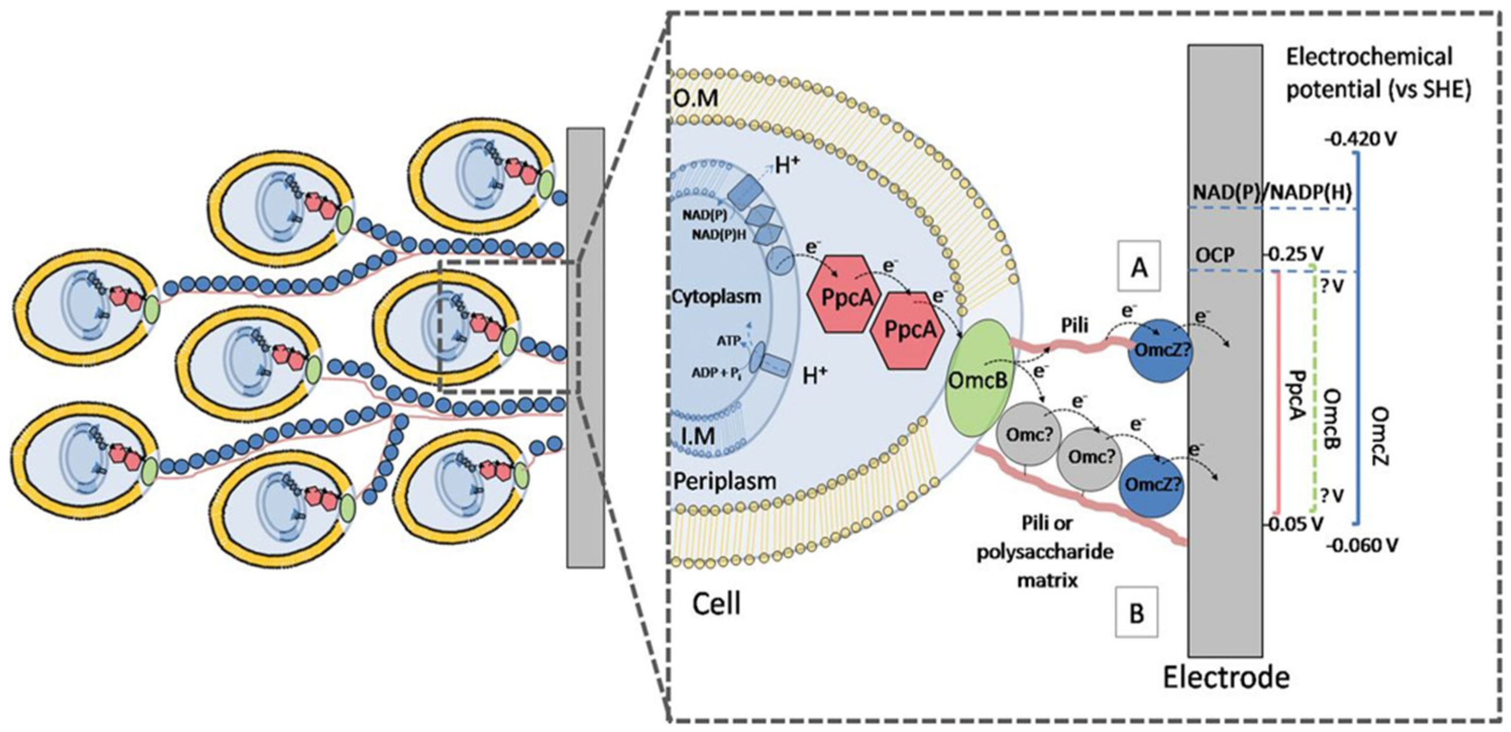
  • The concept of EET is adapted from MFC. An MFC refers to the technology that uses biofilm as a catalyst to directly convert the chemical energy generated by oxidizing organic substances into bioelectric energy [54]. When microorganisms with the ability to produce electricity participate in MFC, the direction of EET is directed outward from the cell to the extracellular electrodes (e.g., insoluble Fe (III), Mn (III), Mn (IV), and other oxides [55,56]), which means that electrons released from the oxidation of organic carbon are transported from the cytoplasm to the metal oxide cathode outside the cell. When there is a localized lack of an organic carbon source as an electron donor within the cells of an electroproducing microorganism, this causes EET reversal, resulting in MIC. Xu et al. [57] compared Desulfovibrio vulgaris with different degrees of MIC caused by carbon-source starvation in media with different carbon contents. Bacteria grown on C1018 carbon steel sheets in the ATCC 1249 medium versus the medium with 90% and 99% reduced organic carbon were found to have the most severe metal corrosion caused by biofilm in the case of starved, but not completely starved, carbon sources. Clauwaert et al. [58] and Huang et al. [59] designed the MFC with a biocathode instead of an oxygen cathode. In this case, electrons are released from the anode into the extracellular environment and enter the biofilm covering the surface of the biocathode before being transferred across the cell wall into the cathode biofilm cells, and the direction of electron transfer from the cathode biofilm to the cathode is the same as in MIC. Therefore, electron transfer in biofilm is often reversible, and the electrochemical mechanism and electron transfer mode in MIC is the same as that in the biocathode of MFC, and the mechanism of how extracellular electrons are imported into the intracellular cell through biofilm can be explored based on the microbial export electron pathway [14,15,16].
EET in MFC is divided into two categories: DET, which includes direct electron contact and bacterial conducting hairs or nanowires, and MET, which includes electron shuttling mediated by ETMs [54].

4.1. Electrical Direct Contact Transmission: Shewanella oneidenis MR-1

Shewanella oneidenis MR-1 can reduce extracellular electron acceptors by direct electron transfer following contact of an extracellular substrate with a redox-active polyheme cytochrome on the cell surface. Polyheme is defined as a two or more c-type heme, namely cytochrome proteins containing multiple iron-containing porphyrin derivatives. The protoporphyrin IX structure allows covalent binding to a peptide chain with a -CXXCH- motif via a thioether bond (where X can be any amino acid), while the motif provides a His axial ligand that allows the iron ions located at the center of the porphyrin ring to be connected in a His/His axial manner [60]. Polyheme cytochromes underlie the respiratory reduction process of the Shewanella oneidenis MR-1 pair of extracellular receptors, supporting rapid electron transfer through a series of intra-protein electron transfer steps involving the Fe3+/Fe2+ redox pair [61].
As shown in Figure 2, oxidation of organic substrates first occurs in the bacterial cytoplasm of Shewanella oneidenis MR-1, and electrons released by this process are transferred through a network of redox proteins originating in the intracellular membrane, spanning the periplasm and extracellular membranes, and extending to the extracellular environment. The soluble electron carrier ubiquinol QH2, present in the intracellular membrane, can bind to transfer electrons; QH2 is produced by the reduction of ubiquinone Q in the cytoplasm, and the two polyhemoglobins, TorC and CymA, catalyze Q regeneration. After the electrons reach the periplasm, terminal electron acceptors, such as NO3, NO2, and fumarate, can cross the extracellular membrane and be reduced in the periplasm by the polyhemoglobin pigments NapAB, NrfA [62], and FccA [61], respectively. For electron acceptors such as graphite anodes, ferric citrate, and flavin (hydrogen) oxides of Fe (III) and Mn (IV), electrons are required to cross the extracellular membrane into the extracellular environment. This process involves the Mtr/OmcA pathway, with the MtrCAB conduit comprising the periplasmic decameric cytochrome (Mtr A), transmembrane pore proteins (Mtr B), and the periplasmic decameric cytochrome (Mtr C) [63]. MtrF, MtrD, and MtrE comprise the MtrFDE complex, which has the same overall structure as that of MtrCAB [61]. Electrons pass sequentially from the periplasm of the cell through MtrA, MtrB, and then MtrB to MtrC or another outer membrane cytochrome, OmcA, to reach the cellular surroundings [64,65].
Polyhemoglobin is a tightly packed heme that is mostly in contact with each other by van der Waals forces [66]. Single-step tunneling of electrons occurs through interconversion between Fe3+ and Fe2+ in the range of 15.5 Å in the iron ion centers of the adjacent heme [61]. Long-range, multi-step electron hopping requires a redox gradient to be driven [48]. Oxidized Fe3+ decreases with distance from the anode surface [65] and the tendency for electrons to be transferred from low-potential Fe2+ to high-potential Fe3+ constitutes a redox gradient in biological membranes. Electrons released from the anode are first transferred to the heme component Fe3+, which is reduced to Fe2+. The Fe3+/Fe2+ potential difference drives electrons to make direct jumps across spatial energy barriers within or between subsequent polyheme cytochromes, as allowed by quantum tunneling effects [67,68,69,70]; that is, electrons jump from one redox center to another along the heme chain, enabling electron transfer over distances of up to tens of nanometers [71].

4.2. Conductive Bacterial Hair Proteins or Nanowires: Geobacter sulfurreducens

Geobacter sulfurreducens requires conducting hairs as biological “pili” to generate high-density currents in MFC, for long-range electron transport to Fe (III) oxides [72]. As shown in Figure 3, the electrons generated from the oxidation of organic matter are first delivered by NADH dehydrogenase to the quinone pool, and then from the quinone pool to the MacA proteins [19], after which the electrons are released from the respiratory chain into the periplasm of the cell and are delivered on the periplasmic cytochrome c peroxidase A (PpcA), which is thought to interact with OmcB in the extracellular membrane. Bonanni et al. [73] discussed two mechanisms of extracellular electron transfer in Geobacter sulfurreducens: the metal-like conductive model and the electron hopping (superexchange) model. The metal-like conductivity model suggests that electrons are transferred along conducting hairs that extend from the cell membrane to the extracellular environment, and that c-type cytochrome-gated electrons are transferred to electrodes, As shown in path A in Figure 3; the electron hopping (superexchange) model, on the other hand, suggests that the bacterial hairs connecting each biofilm cell to the electrodes support an orderly arrangement of c-type cytochromes in the biofilm matrix and that electrons are transferred by tunneling or hopping on redox centers within or between the cytochromes. As shown in path B in Figure 3. Liu et al. [74] showed by direct observation of filaments emanating from Geobacter sulfurreducens cells that conductive hyphae (e-pili) are the most abundant conductive filaments expressed by Geobacter sulfurreducens and are involved in microbial–electrode exchange and long-range extracellular electron transfer in corrosion. Wang et al. [75], on the other hand, pointed out that the c-type cytochrome OmcS on the outer surface of Geobacter sulfurreducens polyhemoglobin can be assembled into filaments, and that the main to cue OmcS filaments rather than e-pili can be used for long-range electron transport in Geobacter sulfurreducens.

4.3. Electron Shuttling Based on Soluble Electron Mediators: Flavins and Phenazines

ETMs are soluble redox-capable electron carriers present in the cellular surroundings. ETMs reversibly act as intermediate electron acceptors or donors, absorbing electrons at different locations in the EPS and releasing them to the next ETM or terminal electron acceptor, thus effectively improving the efficiency of microbial extracellular electron transfer.
Flavin analogs (e.g., riboflavin, flavin adenine nucleotide (FAD), flavin mononucleotide (FMN)) are common ETMs. As shown in Figure 4a, riboflavin is a precursor in the biosynthesis of an FAD and FMN [76], and the isorhodopsin ring in its structure is the main component that determines the electron-binding ability of flavonoids. Flavins can exist in three different forms during redox processes: Flox, Flrad, and Flred2− [77]. Since the midpoint potentials of Flrad and Flred2− differ by only 0.06 V, they are suitable for use as extracellular electron carriers for one- or two-electron reactions with cytochrome or metal interactions [78]. Marsili et al. [78] used electrochemical techniques to investigate the electron transfer process in two strains of Shewanella using extracellular electrodes as electron acceptors for respiration and found the important role played by flavin, which is actively secreted by the cells.
Phenoxazines are a class of nitrogen-containing heterocyclic compounds, including 1-carboxylic acid phenazine (PCA), Pseudomonas aeruginosa (PYO), 1-carboxamidophenazine (PCN), and 1-hydroxyphenazine (1-OHPHZ), and PCA is the synthetic precursor of PYO, PCN, and so on [79], and the physicochemical properties of several phenoxazines are very different due to the different nature and positions of the substituent groups. Phenazines exist in three oxidation states: Phz, Phz, and Phz2−. Each oxidation state has a corresponding protonated form [80]. Phenazine analogs can accept and release electrons as electron carriers, and the reversible electronic redox process that occurs is shown in Figure 4b. Feng et al. [81] introduced the Pseudomonas aeruginosa PCA synthesis pathway heterologously into Escherichia coli, which led to the successful expression of PCA in E. coli and improved the biopower generation and efficiency of EET.
Figure 4. (a) Riboflavin (Vitamin B2) [77]; (b) electron transfer process of phenazine [80].
Coatings 14 01105 g004

5. Electron Transfer in the Corrosion of SRB Microbial Carbon Steel

5.1. Electron Transfer in EPS

The life metabolic process of SRB preferentially uses the organic carbon source in the environment as an electron donor, and when biofilm is formed on the surface of metal materials due to microbial enrichment and propagation, the underlying biofilm cells cannot directly obtain the carbon source from the outside world to maintain the energy needed for cell growth. The SRB cells located in the bottom layer will use the metal as an electron donor and directly utilize the electrons within the biocathode to reduce SO42− and carry out anaerobic respiration, resulting in metal corrosion [82,83]. As shown in Figure 5, microbial carbon steel corrosion is induced by SRB, and the electron transfer process that occurs also involves the same extracellular electron transfer modes and mechanisms described above in MFC. Wang et al. [84] noted that electron-shuttling proteins such as c-type cytochromes on the outer membrane of SRB cells allow the cells to directly utilize electrons from the extracellular insoluble carbon steel and transfer them across the outer cell wall to the intracellular one, resulting in the corrosion of the carbon steel. Sherar et al. [85] found that when there is no organic carbon source in the medium as an electron donor, and the growing hyphae of SRB isolated from oil wells will connect the cells to the carbon steel surface, transferring electrons released from the extracellular anode to the SRB interior for sulfate reduction. Alrammah et al. [86] demonstrated that conductive nanowires significantly accelerated the corrosion of C1020 carbon steel by SRB by facilitating the electron transfer process using various electrochemical testing methods. Both Zhang et al. and Li et al. [15,87] found that SRB accelerated the metal corrosion phenomenon on C1018 carbon steel and 304 stainless steels, respectively, by adding two types of electron mediators, riboflavin and an FAD, to the medium, resulting in more severe pitting craters and the weight loss of hanging sheets.
In addition, considering the complex components in EPS, polysaccharides, proteins, and E-DNA in the matrix all have a certain electrical conductivity [88,89,90], and they may be able to form a mesh structure at the microscopic level, which allows for the direct passage of electrons during part of the extracellular electron transfer or binds a wide range of ETMs, which affects the efficiency of electron transfer [50,53,91]. Humic substances are also important components of EPS, and quinoline humic acids will be reversibly reduced to hydroquinone, which can participate in SRB anaerobic respiration as ETMs [92]. H2S and FeS are two types of metabolic corrosion products secreted by SRB in EPS, which can undergo a reduction reaction on the surface of carbon steel to form electron-rich H2, with there being iron sulfide (FeSX) after the electron to the electron transfer chain on the outer membrane of the SRB cell [23,24,26]. Fe3+ can also be used as an electron donor; there is a variety of redox functional groups in EPS such as carboxyl and amide groups, which will complex with Fe3+ when the distance between the complex and the c-type cytochromes on the surface of the outer membrane of the cell is very small (<2 nm); Fe3+ will be reduced by tunneling electrons to Fe2+; Fe2+ can be combined with glucuronide in EPS to form a new complex, with the complex being unstable and easily degraded; free Fe2+, if it migrates near the outer membrane, can be used as a substrate to be reoxidized by c-type cytochromes; and the electrons then enter the cytosol to be used as a sulfate heterodimerization reduction [18,93].

5.2. Electron Transfer in the Periplasm from the Outer to the Inner Cell Membrane

The SRB-induced MIC process, where electrons from the metal anode are transferred in the periplasm of the cell via the DET or MET pathway across the EPS, the extracellular membrane, requires the participation of multiple cytochromes or electron carriers. Type I tetrameric cytochrome c3 (TpIc3), encoded by the cycA gene, is the most abundant c-type cytochrome in the periplasm of Desulfovibrio spp. With the TpIc3-deficient D. alaskensis G20 mutant, Keller et al. [94] found that when intracellular lactate or pyruvate was used as an electron donor for the process of sulfate dissimilation reduction, the electrons generated from the lactate oxidation product of pyruvate reoxidation or the direct oxidation of pyruvate will partially migrate to the periplasm, where they bind to TpIc3 and are passed to the transmembrane complex QrcABCD, which possesses the activity of methylnaphthoquinone (MQ) reductase [95], generating menaquinone alcohols (MQH2) that reduce the other transmembrane complex, QmoABC. Murali et al. [96] explored the respiratory pathway of symbiotic SRB in which the anaerobic methane-oxidizing archaeon ANME transfers electrons generated from methane oxidation to four major symbiotic SRB taxa. Cytochrome c554, one of the conserved components of SRB that form the electron-transport chain, was found to be present in all four SRB branches. Cytochrome c554, located in the periplasm, transfers electrons to quinone pool reductase in the intracellular membrane to produce menaquinone alcohol or ubiquinol.
TpIc3 and cytochrome c554 are common cytochromes involved in electron transfer in the periplasm of SRB cells, but whether they are involved in the transfer of electrons generated by extracellular metal anodes to the intracellular process and what other definitive cytochromes or electron carriers are involved in the process still require further experimental studies.

5.3. Electron Transfer in SRB Cells

In sulfate respiration under anaerobic conditions in SRB, the process of Dissimilatory Sulfate Reduction (DSR), as shown in Figure 5, involves extracellular SO42− being taken up via trapping by the protein for sulfate uptake (SU) on the cytoplasmic membrane of the cell. In the cytoplasm, it binds to the phosphate group in ATP and is activated by ATP sulfurylase (Sat) to produce adenosine-5′-phosphate sulfate (APS). The reduction of APS to SO32− is then catalyzed by adenylate sulfate reductase (APR) and releases AMP. SO32− is then reduced to S2− by the direct action of sulfite reductase (DSR) or indirectly by the action of three enzymes: sulfite reductase, lienotrisulphate reductase, and thiosulphate reductase [97,98,99], and it diffuses out of the cell membrane in the form of H2S by passive transport. The two terminal reductases, APS reductase (AprAB) and dissimilatory sulfite reductase (DsrAB) in indirect reduction, are soluble and therefore not directly involved in membrane-linked electron transfer [100]. For the exploration of AprAB and DsrAB electron donors, Pires et al. [101] and Mander et al. [102] described for the first time the involvement of the QmoABC complex and the DsrMKJOP complex in the sulfate reduction process.
AprAB is an αβ heterodimeric iron-thioflavonoid enzyme with a catalytic site in the α-subunit that is not covalently bound with an FAD cofactor. The β-subunit contains two [4Fe4S] metal clusters, where the [4Fe4S] buried inside the β-subunit near the cofactor with an FAD is center I, and the other center II is more exposed to the solvent and receives and transmits the electrons from the electron donor to be passed through center I and then to the FAD part of the catalytic site. Diffusion of the substrate in AprAB reaches the encapsulated FAD through the catalytic site that is partially exposed to the solvent [103]. In many sulfate-reducing microorganisms, the qmoABC gene is located close to the aprBA gene as part of the sat-aprBA-qmoABC gene cluster [104], strongly suggesting that QmoABC is involved in AprAB electron transfer. Ramos et al. [105] demonstrated the existence of a direct interaction between the Qmo ABC complex and Apr AB by immunoprecipitation and surface plasmon resonance experiments. The QmoABC complex consists of two cytoplasmic subunits (QmoAB) and one cell membrane subunit (QmoC), of which Qmo A and Qmo B are both soluble iron-thioflavin proteins, Qmo A interacts with Apr AB, and the function of Qmo B remains unclear, with the possibility of other physiological chaperones. The QmoC subunit belongs to the membrane cytochrome b family, and the hydrophobic C-terminal structural domain contains six transmembrane helices that bind two heme b motifs and are responsible for electron exchange with and transfer to the quinone pool in the intracellular membrane [100,105]. Duarte et al. [103] examined protein–protein electron transfer under flipped and unflipped conditions by electrochemical measurements. It was demonstrated that under no-flip conditions, electron transfer from the quinone pool to the AprAB β-subunit and α-subunit via QmoC and QmoA occurs in two consecutive one-electron reductions at the catalytic site FAD.
DsrAB consists of α2β2 4 subunits, with each αβ unit containing two ferroportin cofactors, only one of which is catalytically active. The four ferroportin cofactors are axially coupled [4Fe4S] and immobilized through residues on cysteine [106]. DsrC, on the other hand, acts as a physiological chaperone for DsrAB and contains two highly conserved cysteines (CysA, CysB) with redox activity on its flexible carboxyl-terminal arm [107]. The DsrMKJOP membrane-bound complex is thought to be associated with the electron donor for the sulfate dissimilation reduction process. It includes multiple subunits of the periplasm (triheme cytochrome c DsrJ and multiferroic oxidoreductase protein DsrO), membrane (di-heme cytochrome b DsrM, Dsr P), and cytoplasm (Fe-S protein DsrK) [108], divided into the periplasm-oriented DsrJOP module that may be involved in the exchange of electrons between the periplasm and the quinone pool in the intracellular membrane, and the cytoplasm-oriented DsrMK module that is involved in the exchange of electrons between the intracellular membrane and the cytoplasm [109]. According to the six-electron reaction proposed by Parey et al. [110] proceeding through three two-electron steps, DsrAB sequentially receives two two-electrons to generate [SII] and [S0] at the active center. [S0] will combine with CysA in DsrC to form a persulfide, which CysB later replaces to form an intramolecular disulfide bond bridge. The DsrC trisulfide acts as a substrate to be reduced by the two electrons from the DsrK subunit, ultimately releasing the sulfide [111].

6. Conclusions

MIC has been studied for more than 100 years and many excellent results have been achieved. SRB, widely distributed in nature, are the most common microorganism causing MIC. Regarding the research on the corrosion mechanism of carbon steel by SRB, BCSR was proposed based on the CDT, which innovatively replaces the “physical cathode” with the “biological cathode”, explaining that hydrogenase-negative SRB can still cause corrosion. After the release of electrons from carbon steel anode corrosion, the electron transfer process through the SRB outer cell membrane, periplasm, intracellular membrane, to the cytoplasm and undergoes DSR for eventual utilisation, must cross the EPS, and according to the direction of electron transfer from the biofilm to the cell of the biocathode in the MFC, it is possible to explore the extracellular electron transfer mode and electrochemical mechanism that occurs in the EPS of the SRB.
This paper reviews the complex interactions between SRB, EPS, and carbon steel surfaces in MIC. The following reflections are presented concerning the influence of EPS on MIC and EET under certain conditions, problems with the modes, mechanisms, pathways of intracellular and extracellular electron transfer in MFC and MIC, as well as the actual environmental factors affecting the corrosion process.
  • The bioinduced mineralization occurring in EPS can effectively inhibit metal corrosion, providing new ideas for corrosion protection and the development of new, natural green corrosion inhibitors. The biomineralization induced by SRB will form MeS/MeCO3 mineral precipitates on the cell surface, but how the changes in its own physiological and metabolic activities specifically induce the occurrence of extracellular mineralization needs to be further explored experimentally.
  • Currently, there are more studies on extracellular electron transfer in MFC, and the transfer mechanism where electrons undergo single-step tunneling in the range of 15.5 Å and undergo long-range jumps driven by redox gradients expands the quantum mechanical perspective for probing electron transfer at the bioenergetics level. The elucidation of the mode, mechanism, and pathway of extracellular electron transfer occurring in MIC, on the other hand, only originates from the inference of reversible processes and lacks substantial and more direct experimental evidence.
  • The identification of the types of cytochromes or other electron carriers involved in electron transfer and their roles in the various types of cytochromes present in the EPS layer, extracellular membrane, periplasm, and intracellular membrane during the corrosion of carbon steel induced by SRB still requires further experimental investigation. Molecular tools such as gene mutation, proteomics analysis, and metabolomics studies may play an important role in the identification of key components.
  • The actual corrosion environment is often more complex, whether or how temperature, pH, anions, symbiotic bacteria, etc., affect the corrosion process of SRB carbon steel. Further understanding and discussion are needed to determine whether the electrochemical mechanism and pathway of SRB corrosion on other metal materials, such as carbon steel materials of different concentrations, other alloys and so on, occurring in biofilms are consistent, and whether the corrosion mechanism is the same.

Author Contributions

Conceptualization, L.Q. (Lina Qiu) and N.L.; software, L.Q. (Lijuan Qiu) and N.L.; validation, N.L.; supervision, L.Q. (Lina Qiu). All authors have read and agreed to the published version of the manuscript.

Funding

This work was financially supported by the Chinese National Natural Science Foundation (No. 51701016).

Conflicts of Interest

The authors declare no conflicts of interest.

Appendix A

Figure A1. The figure shows the PRISMA 2020 new system overview flowchart.
Coatings 14 01105 g0a1

References

  1. Khan, M.S.; Yang, K.; Liu, Z.; Zhou, L.; Liu, W.; Lin, S.; Wang, X.; Shang, C. Microorganisms Involved in the Biodegradation and Microbiological Corrosion of Structural Materials. Coatings 2023, 13, 1683. [Google Scholar] [CrossRef]
  2. Rao, P.; Mulky, L. Microbially Influenced Corrosion and Its Control Measures: A Critical Review. J. Bio- Tribo-Corros. 2023, 9, 57. [Google Scholar] [CrossRef]
  3. Rao, P.; Mulky, L. An Overview of Microbiologically Influenced Corrosion on Stainless Steel. ChemBioEng Rev. 2023, 10, 829–840. [Google Scholar] [CrossRef]
  4. Kobisy, A.S.; Nassar, H.N.; Tawfik, S.M.; Elshatoury, E.H.; Aiad, I. Mitigation of Eco-unfriendly and Costly Microbial Induced Corrosion Using Novel Synthesized Schiff Base Cationic Surfactants. J. Chem. Technol. Biotechnol. 2021, 96, 941–952. [Google Scholar] [CrossRef]
  5. Abo Elsoud, M.M.; Abo-Alkasem, M.I. Environmental Sulfate-Reducing Microorganisms. In Application of Microbes in Environmental and Microbial Biotechnology; Inamuddin, Ahamed, M.I., Prasad, R., Eds.; Springer Nature: Singapore, 2022; pp. 625–654. ISBN 9789811622250. [Google Scholar]
  6. Kushkevych, I.; Kovářová, A.; Dordevic, D.; Gaine, J.; Kollar, P.; Vítězová, M.; Rittmann, S.K.-M.R. Distribution of Sulfate-Reducing Bacteria in the Environment: Cryopreservation Techniques and Their Potential Storage Application. Processes 2021, 9, 1843. [Google Scholar] [CrossRef]
  7. Dennis, E.; Julia, G. Corrosion of Iron by Sulfate-Reducing Bacteria: New Views of an Old Problem. Appl. Environ. Microbiol. 2014, 80, 1226–1236. [Google Scholar]
  8. Zhou, L.; Wu, J.; Ji, J.-H.; Gao, J.; Liu, Y.-F.; Wang, B.; Yang, S.-Z.; Gu, J.-D.; Mu, B.-Z. Characteristics of Microbiota, Core Sulfate-Reducing Taxa and Corrosion Rates in Production Water from Five Petroleum Reservoirs in China. Sci. Total Environ. 2023, 858, 159861. [Google Scholar] [CrossRef]
  9. Qiu, L.; Zhang, W.; Gong, A.; Zheng, S.; Zhao, D.; Zhao, W.; Fan, R. Species of corrosive microbes and corrosion mechanisms. Chin. J. Eng. 2023, 45, 927–940. [Google Scholar] [CrossRef]
  10. He, J.; Yang, C.; Li, Z. Research Progress of Microbiologically Influenced Corrosion and Protection in Building Industry. J. Chin. Soc. Corros. Prot. 2021, 41, 151–160. [Google Scholar]
  11. Xu, D.; Li, Y.; Song, F.; Gu, T. Laboratory Investigation of Microbiologically Influenced Corrosion of C1018 Carbon Steel by Nitrate Reducing Bacterium Bacillus licheniformis. Corros. Sci. 2013, 77, 385–390. [Google Scholar] [CrossRef]
  12. Gu, T.; Zhao, K.; Nesic, S. A New Mechanistic Model for Mic Based on A Biocatalytic Cathodic Sulfate Reduction Theory. In Proceedings of the CORROSION 2009, Atlanta, GA, USA, 22–26 March 2009. [Google Scholar]
  13. Muhammad, M.H.; Idris, A.L.; Fan, X.; Guo, Y.; Yu, Y.; Jin, X.; Qiu, J.; Guan, X.; Huang, T. Beyond Risk: Bacterial Biofilms and Their Regulating Approaches. Front. Microbiol. 2020, 11, 928. [Google Scholar] [CrossRef]
  14. Gu, T.; Wang, D.; Lekbach, Y.; Xu, D. Extracellular Electron Transfer in Microbial Biocorrosion. Curr. Opin. Electrochem. 2021, 29, 100763. [Google Scholar] [CrossRef]
  15. Zhang, P.; Xu, D.; Li, Y.; Yang, K.; Gu, T. Electron Mediators Accelerate the Microbiologically Influenced Corrosion of 304 Stainless Steel by the Desulfovibrio vulgaris Biofilm. Bioelectrochemistry 2015, 101, 14–21. [Google Scholar] [CrossRef] [PubMed]
  16. Xia, J.; Xu, D.; Nan, L.; Liu, H.; Li, Q.; Yang, K. Study on Mechanisms of Microbiologically Influenced Corrision of Metal from the Perspective of Bioelectrochemistry and Bio-energetics. Chin. J. Mater. Res. 2016, 30, 161–170. [Google Scholar]
  17. Vengatesh, G.; Ganapathi Sundaram, R.; Punitha, N. Menadione Sodium Bisulfite as an Efficient Anti-Corrosion Additive for Mild Steel in Acid Corrosion: Electrochemical, Surface Morphological and Theoretical Studies. J. Mol. Liq. 2024, 410, 125653. [Google Scholar] [CrossRef]
  18. Jin, J.; Guan, Y. The Mutual Co-Regulation of Extracellular Polymeric Substances and Iron Ions in Biocorrosion of Cast Iron Pipes. Bioresour. Technol. 2014, 169, 387–394. [Google Scholar] [CrossRef]
  19. Huang, H.; Liu, S.; Jiang, C. Microbiologically influenced corrosion and mechanisms. Microbiol. China 2017, 44, 1699–1713. [Google Scholar] [CrossRef]
  20. Jia, R.; Unsal, T.; Xu, D.; Lekbach, Y.; Gu, T. Microbiologically Influenced Corrosion and Current Mitigation Strategies: A State of the Art Review. Int. Biodeterior. Biodegrad. 2019, 137, 42–58. [Google Scholar] [CrossRef]
  21. Dong, X.; Guan, F.; Xu, L.; Duan, J.; Hou, B. A review: Progress on the Corrosion Mechanism of Sulfate-reducing Bacteria in Marine Environment on Metal Materials. J. Chin. Soc. Corros. Prot. 2021, 41, 1–12. [Google Scholar]
  22. Blackwood, D.J. An Electrochemist Perspective of Microbiologically Influenced Corrosion. Corros. Mater. Degrad. 2020, 1, 59–76. [Google Scholar] [CrossRef]
  23. Liu, T.; Liu, H.; Hu, Y.; Zhou, L.; Zheng, B. Growth Characteristics of Thermophile Sulfate-Reducing Bacteria and Its Effect on Carbon Steel. Mater. Corros. 2009, 60, 218–224. [Google Scholar] [CrossRef]
  24. Bao, Q. The Study of Corrosion Mechanism Induced by Sulphate Reducing Bacteria and Composite Antibacterial Materials. Ph.D. Thesis, Graduate School of the Chinese Academy of Sciences (Institute of Oceanography), Qingdao, China, 2013. [Google Scholar]
  25. Dall’Agnol, L.T.; Cordas, C.M.; Moura, J.J.G. Influence of Respiratory Substrate in Carbon Steel Corrosion by a Sulphate Reducing Prokaryote Model Organism. Bioelectrochemistry 2014, 97, 43–51. [Google Scholar] [CrossRef] [PubMed]
  26. Lee, W.; Characklis, W.G. Corrosion of Mild Steel Under Anaerobic Biofilm. Corrosion 1993, 49, 186–199. [Google Scholar] [CrossRef]
  27. Li, Y.; Xu, D.; Chen, C.; Li, X.; Jia, R.; Zhang, D.; Sand, W.; Wang, F.; Gu, T. Anaerobic Microbiologically Influenced Corrosion Mechanisms Interpreted Using Bioenergetics and Bioelectrochemistry: A Review. J. Mater. Sci. Technol. 2018, 34, 1713–1718. [Google Scholar] [CrossRef]
  28. Dou, W.; Pu, Y.; Han, X.; Song, Y.; Chen, S.; Gu, T. Corrosion of Cu by a Sulfate Reducing Bacterium in Anaerobic Vials with Different Headspace Volumes. Bioelectrochemistry 2020, 133, 107478. [Google Scholar] [CrossRef] [PubMed]
  29. Dou, W.; Jia, R.; Jin, P.; Liu, J.; Chen, S.; Gu, T. Investigation of the Mechanism and Characteristics of Copper Corrosion by Sulfate Reducing Bacteria. Corros. Sci. 2018, 144, 237–248. [Google Scholar] [CrossRef]
  30. Wang, D.; Unsal, T.; Kumseranee, S.; Punpruk, S.; Mohamed, M.E.; Saleh, M.A.; Gu, T. Sulfate Reducing Bacterium Desulfovibrio vulgaris Caused Severe Microbiologically Influenced Corrosion of Zinc and Galvanized Steel. Int. Biodeterior. Biodegrad. 2021, 157, 105160. [Google Scholar] [CrossRef]
  31. Lv, M.; Li, Z.; Du, M.; Xiao, F.; Chen, X. Formation, Function and Evolution of Biofilm in Microbiologically Influenced Corrosion. Surf. Technol. 2019, 48, 59–68+139. [Google Scholar] [CrossRef]
  32. Ke, N.; Ni, Y.; He, J.; Liu, H.; Jin, Z.; Liu, H. Research Progress of Metal Corrosion Caused by Extracellular Polymeric Substances of Microorganisms. J. Chin. Soc. Corros. Prot. 2024, 44, 278–294. [Google Scholar]
  33. Pal, M.K.; Lavanya, M. Microbial Influenced Corrosion: Understanding Bioadhesion and Biofilm Formation. J. Bio- Tribo-Corros. 2022, 8, 76. [Google Scholar] [CrossRef]
  34. Huang, L.; Yang, Y.; Li, J. Review of the research on biomineralization. Geol. Resour. 2009, 18, 317–320+297. [Google Scholar]
  35. Chen, Y.; Feng, Y.; Deveaux, J.G.; Masoud, M.A.; Chandra, F.S.; Chen, H.; Zhang, D.; Feng, L. Biomineralization Forming Process and Bio-Inspired Nanomaterials for Biomedical Application: A Review. Minerals 2019, 9, 68. [Google Scholar] [CrossRef]
  36. Wen, Q.; Qin, Y.; Zheng, J.; Wei, Q.; Zhang, Y.; Jiang, Y. Research advances in the fixation of heavy metals in acid mine wastewater by sulfate reducing bacteria. Chem. Ind. Eng. Prog. 2022, 41, 5578–5587. [Google Scholar] [CrossRef]
  37. Huang, M. Research Progress and Application of Biomineralization. Adv. Microbiol. 2021, 10, 175–181. [Google Scholar] [CrossRef]
  38. Hao, X.; Bai, Y.; Lou, Y.; Zhang, D. Research Progress of Microbial Mineralization Impact on Inhibits Metal Corrosion Behavior. Surf. Technol. 2021, 50, 18–29. [Google Scholar] [CrossRef]
  39. Bontognali, T.R.R.; Mckenzie, J.A.; Warthmann, R.J.; Vasconcelos, C. Microbially Influenced Formation of Mg-Calcite and Ca-Dolomite in the Presence of Exopolymeric Substances Produced by Sulphate-Reducing Bacteria. Terra Nova 2014, 26, 72–77. [Google Scholar] [CrossRef]
  40. Kenward, P.A.; Fowle, D.A.; Goldstein, R.H.; Ueshima, M.; González, L.A.; Roberts, J.A. Ordered Low-Temperature Dolomite Mediated by Carboxyl-Group Density of Microbial Cell Walls. AAPG Bull. 2013, 97, 2113–2125. [Google Scholar] [CrossRef]
  41. Wu, Y.; Hu, J.; Zhang, W.; Wang, S.; Liu, W. Research Status Review of Microbial Induced Carbonate Precipitation Technology in Reinforcing Sand. Subgrade Eng. 2018, 5, 6–11. [Google Scholar] [CrossRef]
  42. Mondal, S.; Ghosh, A.D. Review on Microbial Induced Calcite Precipitation Mechanisms Leading to Bacterial Selection for Microbial Concrete. Constr. Build. Mater. 2019, 225, 67–75. [Google Scholar] [CrossRef]
  43. Gadd, G.M. Metals, Minerals and Microbes: Geomicrobiology and Bioremediation. Microbiology 2010, 156, 609–643. [Google Scholar] [CrossRef]
  44. Gallagher, K.L.; Kading, T.J.; Braissant, O.; Dupraz, C.; Visscher, P.T. Inside the Alkalinity Engine: The Role of Electron Donors in the Organomineralization Potential of Sulfate-reducing Bacteria. Geobiology 2012, 10, 518–530. [Google Scholar] [CrossRef] [PubMed]
  45. Guo, B.; Liu, B.; Chen, J.; Jing, C.; Zhong, M.; Shan, Q. Prospect Research on the Diversity of Extracellular Mineralization Process Induced by Mineralizing Microorganisms and Its Use as a Treatment for Soil Pollutants. Sustainability 2023, 15, 4858. [Google Scholar] [CrossRef]
  46. Marques, M.J.F.; Jaume, J.; Mercier, D.; Seyeux, A.; Zanna, S.; Basseguy, R.; Marcus, P. The Positive Impact of Biomineralization for Marine Corrosion Protection of AA5083 Alloy. Corros. Sci. 2024, 233, 112053. [Google Scholar] [CrossRef]
  47. Shan, X.; Wang, J.; Du, M.; Tian, Z. Inhibitory Effect of Marine Bacillus Sp. and Its Biomineralization on the Corrosion of X65 Steel in Offshore Oilfield Produced Water. Bioelectrochemistry 2024, 157, 108659. [Google Scholar] [CrossRef]
  48. Xiao, Y.; Zhao, F. Electrochemical Roles of Extracellular Polymeric Substances in Biofilms. Curr. Opin. Electrochem. 2017, 4, 206–211. [Google Scholar] [CrossRef]
  49. Rabaey, K. Bioelectrochemical Systems: From Extracellular Electron Tranfer to Biotechnological Application; Science Press: Beijing, China, 2012. [Google Scholar]
  50. Xiao, Y.; Zhang, E.; Zhang, J.; Dai, Y.; Yang, Z.; Christensen, H.E.M.; Ulstrup, J.; Zhao, F. Extracellular Polymeric Substances Are Transient Media for Microbial Extracellular Electron Transfer. Sci. Adv. 2017, 3, e1700623. [Google Scholar] [CrossRef]
  51. Li, S.-W.; Sheng, G.-P.; Cheng, Y.-Y.; Yu, H.-Q. Redox Properties of Extracellular Polymeric Substances (EPS) from Electroactive Bacteria. Sci. Rep. 2016, 6, 39098. [Google Scholar] [CrossRef]
  52. Cao, B.; Shi, L.; Brown, R.N.; Xiong, Y.; Fredrickson, J.K.; Romine, M.F.; Marshall, M.J.; Lipton, M.S.; Beyenal, H. Extracellular Polymeric Substances from Shewanella Sp. HRCR-1 Biofilms: Characterization by Infrared Spectroscopy and Proteomics. Environ. Microbiol. 2011, 13, 1018–1031. [Google Scholar] [CrossRef]
  53. Saunders, S.H.; Tse, E.C.M.; Yates, M.D.; Otero, F.J.; Trammell, S.A.; Stemp, E.D.A.; Barton, J.K.; Tender, L.M.; Newman, D.K. Extracellular DNA Promotes Efficient Extracellular Electron Transfer by Pyocyanin in Pseudomonas aeruginosa Biofilms. Cell 2020, 182, 919–932.e19. [Google Scholar] [CrossRef] [PubMed]
  54. Zhou, M.; Wang, H.; Hassett, D.J.; Gu, T. Recent Advances in Microbial Fuel Cells (MFCs) and Microbial Electrolysis Cells (MECs) for Wastewater Treatment, Bioenergy and Bioproducts. J. Chem. Technol. Biotechnol. 2013, 88, 508–518. [Google Scholar] [CrossRef]
  55. Myers, C.R.; Nealson, K.H. Bacterial Manganese Reduction and Growth with Manganese Oxide as the Sole Electron Acceptor. Science 1988, 240, 1319–1321. [Google Scholar] [CrossRef] [PubMed]
  56. Nealson, K.H.; Saffarini, D. Iron and Manganese in Anaerobic Respiration: Environmental Significance, Physiology, and Regulation. Annu. Rev. Microbiol. 1994, 48, 311–343. [Google Scholar] [CrossRef] [PubMed]
  57. Xu, D.; Gu, T. Carbon Source Starvation Triggered More Aggressive Corrosion against Carbon Steel by the Desulfovibrio vulgaris Biofilm. Int. Biodeterior. Biodegrad. 2014, 91, 74–81. [Google Scholar] [CrossRef]
  58. Clauwaert, P.; van de Ha, D.; Boon, N.; Verbeken, K.; Verstraete, M.; Rabaey, K.; Verstraete, W. Open Air Biocathode Enables Effective Electricity Generation with Microbial Fuel Cells. Environ. Sci. Technol. 2007, 41, 7564–7569. [Google Scholar] [CrossRef] [PubMed]
  59. Huang, L.; Regan, J.M.; Quan, X. Electron Transfer Mechanisms, New Applications, and Performance of Biocathode Microbial Fuel Cells. Bioresour. Technol. 2011, 102, 316–323. [Google Scholar] [CrossRef] [PubMed]
  60. Bertini, I.; Cavallaro, G.; Rosato, A. Cytochrome c:  Occurrence and Functions. Chem. Rev. 2006, 106, 90–115. [Google Scholar] [CrossRef]
  61. Breuer, M.; Rosso, K.M.; Blumberger, J.; Butt, J.N. Multi-Haem Cytochromes in Shewanella oneidensis MR-1: Structures, Functions and Opportunities. J. R. Soc. Interface 2015, 12, 20141117. [Google Scholar] [CrossRef]
  62. Gao, H.; Yang, Z.K.; Barua, S.; Reed, S.B.; Romine, M.F.; Nealson, K.H.; Fredrickson, J.K.; Tiedje, J.M.; Zhou, J. Reduction of Nitrate in Shewanella oneidensis Depends on Atypical NAP and NRF Systems with NapB as a Preferred Electron Transport Protein from CymA to NapA. ISME J. 2009, 3, 966–976. [Google Scholar] [CrossRef]
  63. Piper, S.E.H.; Edwards, M.J.; van Wonderen, J.H.; Casadevall, C.; Martel, A.; Jeuken, L.J.C.; Reisner, E.; Clarke, T.A.; Butt, J.N. Bespoke Biomolecular Wires for Transmembrane Electron Transfer: Spontaneous Assembly of a Functionalized Multiheme Electron Conduit. Front. Microbiol. 2021, 12, 714508. [Google Scholar] [CrossRef]
  64. Shi, L.; Squier, T.C.; Zachara, J.M.; Fredrickson, J.K. Respiration of Metal (Hydr)Oxides by Shewanella and Geobacter: A Key Role for Multihaem c-Type Cytochromes. Mol. Microbiol. 2007, 65, 12–20. [Google Scholar] [CrossRef]
  65. Zacharoff, L.A.; El-Naggar, M.Y. Redox Conduction in Biofilms: From Respiration to Living Electronics. Curr. Opin. Electrochem. 2017, 4, 182–189. [Google Scholar] [CrossRef]
  66. Smith, L.J.; Kahraman, A.; Thornton, J.M. Heme Proteins—Diversity in Structural Characteristics, Function, and Folding. Proteins Struct. Funct. Bioinform. 2010, 78, 2349–2368. [Google Scholar] [CrossRef]
  67. Gray, H.B.; Winkler, J.R. Long-Range Electron Transfer. Proc. Natl. Acad. Sci. USA 2005, 102, 3534–3539. [Google Scholar] [CrossRef]
  68. Berg, J.; Tymoczko, J.; Stryer, L. Biochemistry, 5th ed.; National Center Biotechnology Informationõs Bookshelf; Palgrave MacMillan: London, UK, 2002; pp. 93–126. [Google Scholar] [CrossRef]
  69. Voet, D.; Voet, J.G.; Pratt, C.W. Fundamentals of Biochemistry: Life at the Molecular Level; Wiley: Hoboken, NJ, USA, 2016; p. 484. [Google Scholar]
  70. Page, C.C.; Moser, C.C.; Chen, X.; Dutton, P.L. Natural Engineering Principles of Electron Tunnelling in Biological Oxidation–Reduction. Nature 1999, 402, 47–52. [Google Scholar] [CrossRef]
  71. Xu, S.; Barrozo, A.; Tender, L.M.; Krylov, A.I.; El-Naggar, M.Y. Multiheme Cytochrome Mediated Redox Conduction through Shewanella oneidensis MR-1 Cells. J. Am. Chem. Soc. 2018, 140, 10085–10089. [Google Scholar] [CrossRef] [PubMed]
  72. Lovley, D.R.; Walker, D.J.F. Geobacter Protein Nanowires. Front. Microbiol. 2019, 10, 2078. [Google Scholar] [CrossRef] [PubMed]
  73. Bonanni, P.S.; Schrott, G.D.; Busalmen, J.P. A Long Way to the Electrode: How Do Geobacter Cells Transport Their Electrons? Biochem. Soc. Trans. 2012, 40, 1274–1279. [Google Scholar] [CrossRef]
  74. Liu, X.; Walker, D.J.F.; Nonnenmann, S.S.; Sun, D.; Lovley, D.R. Direct Observation of Electrically Conductive Pili Emanating from Geobacter Sulfurreducens. mBio 2021, 12, e0220921. [Google Scholar] [CrossRef]
  75. Wang, F.; Gu, Y.; O’Brien, J.P.; Yi, S.M.; Yalcin, S.E.; Srikanth, V.; Shen, C.; Vu, D.; Ing, N.L.; Hochbaum, A.I.; et al. Structure of Microbial Nanowires Reveals Stacked Hemes That Transport Electrons over Micrometers. Cell 2019, 177, 361. [Google Scholar] [CrossRef] [PubMed]
  76. Bailey, M.R.; Schultz, Z.D. SERS Speciation of the Electrochemical Oxidation-Reduction of Riboflavin. The Analyst 2016, 141, 5078–5087. [Google Scholar] [CrossRef]
  77. Tan, S.L.J.; Webster, R.D. Electrochemically Induced Chemically Reversible Proton-Coupled Electron Transfer Reactions of Riboflavin (Vitamin B2). J. Am. Chem. Soc. 2012, 134, 5954–5964. [Google Scholar] [CrossRef] [PubMed]
  78. Marsili, E.; Baron, D.B.; Shikhare, I.D.; Coursolle, D.; Gralnick, J.A.; Bond, D.R. Shewanella Secretes Flavins That Mediate Extracellular Electron Transfer. Proc. Natl. Acad. Sci. USA 2008, 105, 3968–3973. [Google Scholar] [CrossRef] [PubMed]
  79. Ding, A.; Zheng, P.; Zhang, M. Progress in research of electron transfer mediator(ETM). J. Zhejiang Univ. Life Sci. 2016, 42, 573–581. [Google Scholar]
  80. Chen, J.-J.; Chen, W.; He, H.; Li, D.-B.; Li, W.-W.; Xiong, L.; Yu, H.-Q. Manipulation of Microbial Extracellular Electron Transfer by Changing Molecular Structure of Phenazine-Type Redox Mediators. Environ. Sci. Technol. 2013, 47, 1033–1039. [Google Scholar] [CrossRef]
  81. Feng, J.; Qian, Y.; Wang, Z.; Wang, X.; Xu, S.; Chen, K.; Ouyang, P. Enhancing the Performance of Escherichia Coli-Inoculated Microbial Fuel Cells by Introduction of the Phenazine-1-Carboxylic Acid Pathway. J. Biotechnol. 2018, 275, 1–6. [Google Scholar] [CrossRef]
  82. Wei, B.; Xv, J.; Gao, L.; Qin, Q.; Fu, Q.; Yu, C.; Sun, C.; Wang, Z. Research Progress on Sulfate Reducing Bacteria Induced Corrosion of Pipeline Steel in Soil Environment. Surf. Technol. 2021, 50, 30–44. [Google Scholar] [CrossRef]
  83. Ding, L. Study on Corrosion Resistance of Anti-Microbial Tubing and Casing Steel. Bachelor’s Thesis, Xi’an Shiyou University, Xi’an, China, 2024. [Google Scholar]
  84. Wang, D.; Yang, C.; Saleh, M.A.; Alotaibi, M.D.; Mohamed, M.E.; Xu, D.; Gu, T. Conductive Magnetite Nanoparticles Considerably Accelerated Carbon Steel Corrosion by Electroactive Desulfovibrio Vulgaris Biofilm. Corros. Sci. 2022, 205, 110440. [Google Scholar] [CrossRef]
  85. Sherar, B.W.A.; Power, I.M.; Keech, P.G.; Mitlin, S.; Southam, G.; Shoesmith, D.W. Characterizing the Effect of Carbon Steel Exposure in Sulfide Containing Solutions to Microbially Induced Corrosion. Corros. Sci. 2011, 53, 955–960. [Google Scholar] [CrossRef]
  86. Alrammah, F.; Xu, L.; Patel, N.; Kontis, N.; Rosado, A.; Gu, T. Conductive Magnetic Nanowires Accelerated Electron Transfer between C1020 Carbon Steel and Desulfovibrio vulgaris Biofilm. Sci. Total Environ. 2024, 925, 171763. [Google Scholar] [CrossRef]
  87. Li, H.; Xu, D.; Li, Y.; Feng, H.; Liu, Z.; Li, X.; Gu, T.; Yang, K. Extracellular Electron Transfer Is a Bottleneck in the Microbiologically Influenced Corrosion of C1018 Carbon Steel by the Biofilm of Sulfate-Reducing Bacterium Desulfovibrio vulgaris. PLoS ONE 2015, 10, e0136183. [Google Scholar] [CrossRef]
  88. Tan, Y.; Adhikari, R.Y.; Malvankar, N.S.; Pi, S.; Ward, J.E.; Woodard, T.L.; Nevin, K.P.; Xia, Q.; Tuominen, M.T.; Lovley, D.R. Synthetic Biological Protein Nanowires with High Conductivity. Small 2016, 12, 4481–4485. [Google Scholar] [CrossRef] [PubMed]
  89. Zhuravel, R.; Huang, H.; Polycarpou, G.; Polydorides, S.; Motamarri, P.; Katrivas, L.; Rotem, D.; Sperling, J.; Zotti, L.A.; Kotlyar, A.B.; et al. Backbone Charge Transport in Double-Stranded DNA. Nat. Nanotechnol. 2020, 15, 836–840. [Google Scholar] [CrossRef]
  90. Finkenstadt, V.L. Natural Polysaccharides as Electroactive Polymers. Appl. Microbiol. Biotechnol. 2005, 67, 735–745. [Google Scholar] [CrossRef]
  91. Schulten, H.-R.; Plage, B.; Schnitzer, M. A Chemical Structure for Humic Substances. Sci. Nat. 1991, 78, 311–312. [Google Scholar] [CrossRef] [PubMed]
  92. Hernandez, M.E.; Newman, D.K. Extracellular Electron Transfer. Cell. Mol. Life Sci. CMLS 2001, 58, 1562–1571. [Google Scholar] [CrossRef] [PubMed]
  93. Medvedev, D.; Stuchebrukhov, A.A. DNA Repair Mechanism by Photolyase: Electron Transfer Path from the Photolyase Catalytic Cofactor FADH(-) to DNA Thymine Dimer. J. Theor. Biol. 2001, 210, 237–248. [Google Scholar] [CrossRef]
  94. Keller, K.L.; Rapp-Giles, B.J.; Semkiw, E.S.; Porat, I.; Brown, S.D.; Wall, J.D. New Model for Electron Flow for Sulfate Reduction in Desulfovibrio Alaskensis G20. Appl. Environ. Microbiol. 2014, 80, 855–868. [Google Scholar] [CrossRef]
  95. Venceslau, S.S.; Lino, R.R.; Pereira, I.A.C. The Qrc Membrane Complex, Related to the Alternative Complex III, Is a Menaquinone Reductase Involved in Sulfate Respiration. J. Biol. Chem. 2010, 285, 22774–22783. [Google Scholar] [CrossRef]
  96. Murali, R.; Yu, H.; Speth, D.R.; Wu, F.; Metcalfe, K.S.; Crémière, A.; Laso-Pèrez, R.; Malmstrom, R.R.; Goudeau, D.; Woyke, T.; et al. Physiological Potential and Evolutionary Trajectories of Syntrophic Sulfate-Reducing Bacterial Partners of Anaerobic Methanotrophic Archaea. PLoS Biol. 2023, 21, e3002292. [Google Scholar] [CrossRef]
  97. Diao, M.; Dyksma, S.; Koeksoy, E.; Ngugi, D.K.; Anantharaman, K.; Loy, A.; Pester, M. Global Diversity and Inferred Ecophysiology of Microorganisms with the Potential for Dissimilatory Sulfate/Sulfite Reduction. FEMS Microbiol. Rev. 2023, 47, fuad058. [Google Scholar] [CrossRef]
  98. Zhang, H.; Li, Y.; Wang, W.; Wang, L. Research progress of the mircobial sulfur-cycling network. Acta Microbiol. Sin. 2021, 61, 1567–1581. [Google Scholar] [CrossRef]
  99. Jørgensen, B.B.; Findlay, A.J.; Pellerin, A. The Biogeochemical Sulfur Cycle of Marine Sediments. Front. Microbiol. 2019, 10, 849. [Google Scholar] [CrossRef] [PubMed]
  100. Ramos, A.R.; Keller, K.L.; Wall, J.D.; Pereira, I.A.C. The Membrane QmoABC Complex Interacts Directly with the Dissimilatory Adenosine 5´-Phosphosulfate Reductase in Sulfate Reducing Bacteria. Front. Microbiol. 2012, 3, 137. [Google Scholar] [CrossRef] [PubMed]
  101. Pires, R.H.; Lourenço, A.I.; Morais, F.; Teixeira, M.; Xavier, A.V.; Saraiva, L.M.; Pereira, I.A.C. A Novel Membrane-Bound Respiratory Complex from Desulfovibrio Desulfuricans ATCC 27774. Biochim. Biophys. Acta (BBA)—Bioenerg. 2003, 1605, 67–82. [Google Scholar] [CrossRef]
  102. Mander, G.J.; Duin, E.C.; Linder, D.; Stetter, K.O.; Hedderich, R. Purification and Characterization of a Membrane-bound Enzyme Complex from the Sulfate-reducing Archaeon Archaeoglobus fulgidus Related to Heterodisulfide Reductase from Methanogenic Archaea. Eur. J. Biochem. 2002, 269, 1895–1904. [Google Scholar] [CrossRef]
  103. Duarte, A.G.; Santos, A.A.; Pereira, I.A.C. Electron Transfer between the QmoABC Membrane Complex and Adenosine 5′-Phosphosulfate Reductase. Biochim. Biophys. Acta (BBA)—Bioenerg. 2016, 1857, 380–386. [Google Scholar] [CrossRef]
  104. Pereira, I.A.C.; Ramos, A.R.; Grein, F.; Marques, M.C.; Da Silva, S.M.; Venceslau, S.S. A Comparative Genomic Analysis of Energy Metabolism in Sulfate Reducing Bacteria and Archaea. Front. Microbiol. 2011, 2, 69. [Google Scholar] [CrossRef]
  105. Pereira, I.A.C. Respiratory Membrane Complexes of Desulfovibrio. In Proceedings of the Microbial Sulfur Metabolism; Dahl, C., Friedrich, C.G., Eds.; Springer: Berlin/Heidelberg, Germany, 2008; pp. 24–35. [Google Scholar]
  106. Ferreira, D.; Barbosa, A.C.C.; Oliveira, G.P.; Catarino, T.; Venceslau, S.S.; Pereira, I.A.C. The DsrD Functional Marker Protein Is an Allosteric Activator of the DsrAB Dissimilatory Sulfite Reductase. Proc. Natl. Acad. Sci. USA 2022, 119, e2118880119. [Google Scholar] [CrossRef]
  107. Santos, A.A.; Venceslau, S.S.; Grein, F.; Leavitt, W.D.; Dahl, C.; Johnston, D.T.; Pereira, I.A.C. A Protein Trisulfide Couples Dissimilatory Sulfate Reduction to Energy Conservation. Science 2015, 350, 1541–1545. [Google Scholar] [CrossRef]
  108. Barbosa, A.C.C.; Venceslau, S.S.; Pereira, I.A.C. DsrMKJOP Is the Terminal Reductase Complex in Anaerobic Sulfate Respiration. Proc. Natl. Acad. Sci. USA 2024, 121, e2313650121. [Google Scholar] [CrossRef]
  109. Venceslau, S.S.; Stockdreher, Y.; Dahl, C.; Pereira, I.A.C. The “Bacterial Heterodisulfide” DsrC Is a Key Protein in Dissimilatory Sulfur Metabolism. Biochim. Biophys. Acta BBA—Bioenerg. 2014, 1837, 1148–1164. [Google Scholar] [CrossRef] [PubMed]
  110. Parey, K.; Warkentin, E.; Kroneck, P.M.H.; Ermler, U. Reaction Cycle of the Dissimilatory Sulfite Reductase from Archaeoglobus fulgidus. Biochemistry 2010, 49, 8912–8921. [Google Scholar] [CrossRef] [PubMed]
  111. Oliveira, T.F.; Vonrhein, C.; Matias, P.M.; Venceslau, S.S.; Pereira, I.A.C.; Archer, M. The Crystal Structure of Desulfovibrio vulgaris Dissimilatory Sulfite Reductase Bound to DsrC Provides Novel Insights into the Mechanism of Sulfate Respiration. J. Biol. Chem. 2008, 283, 34141. [Google Scholar] [CrossRef] [PubMed]
Figure 1. Schematic diagram of SRB-induced biomineralization process [36].
Coatings 14 01105 g001
Figure 2. Multi-heme cytochromes from Shewanella oneidenis MR-1 are illustrated schematically to indicate their cellular location and roles [61].
Coatings 14 01105 g002
Figure 3. Schematic representation of the electron-transport process from the inner membrane to the electrode in Geobacter sulfurreducens biofilms [73].
Coatings 14 01105 g003
Figure 5. Schematic representation of the complete process of sulfate dissimilation reduction in microbial carbon steel corrosion induced by SRB, where electrons are transferred from the metal anode to the EPS; the outer, periplasmic, and inner membranes; and then reach the cytoplasm.
Coatings 14 01105 g005
Disclaimer/Publisher’s Note: The statements, opinions and data contained in all publications are solely those of the individual author(s) and contributor(s) and not of MDPI and/or the editor(s). MDPI and/or the editor(s) disclaim responsibility for any injury to people or property resulting from any ideas, methods, instructions or products referred to in the content.

