Article

Superhydrophilic Surface Creation and Its Temporal Transition to Hydrophobicity on Copper via Femtosecond Laser Texturing

by
Jeonghong Ha
Smart Forming Process Group, Korea Institute of Industrial Technology (KITECH), Ulsan 44776, Republic of Korea
Coatings 2024, 14(9), 1107; https://doi.org/10.3390/coatings14091107
Submission received: 30 July 2024 / Revised: 25 August 2024 / Accepted: 28 August 2024 / Published: 2 September 2024
(This article belongs to the Section Laser Coatings)

Abstract

:
We analyzed a process to fabricate a superhydrophilic surface on copper by forming various laser-induced periodic surface structures (LIPSS) using a Ti/sapphire femtosecond laser. For these structured surfaces, the correlation between the surface structure and the wetting characteristics was analyzed by scanning electron microscopy, atomic force microscopy, and contact angle (CA) measurement. X-ray photoelectron spectroscopy (XPS) was also employed to analyze variation of the elemental composition of the surfaces. The laser treatment produced micro/nanostructures composed of ripples whose length and width are in microscale and nanoscale, respectively. At specific conditions, the CA of a water droplet was reduced to less than 1°. The superhydrophilcity is attributed to the effect of nanoholes and nanoclusters, which consist of copper (II) oxide and copper hydroxide, having a hydrophilic effect on LIPSS. However, the pristine superhydrophilic surface spontaneously became hydrophobic after being exposed to air at room temperature for about 10 days. According to XPS analysis, the surface’s transition to hydrophobic is attributed not only to the decomposition of Cu(OH)2 but also to the adsorption of oxygen molecules and/or airborne organic molecules containing carbon, which further influences the wettability.

1. Introduction

Wettability of a solid surface is a subject of fundamental and practical importance, and extensive investigations have thus been conducted on the subject [1,2,3]. Especially, various new techniques to modify the surface wettability have been developed with the aid of micro- and nanoscale fabrication processes [4,5,6,7]. Generally, the wettability of a surface, represented by the static contact angle (CA) of a water droplet, is determined by the surface chemistry and the geometric structure of the surface [8,9]. Therefore, controlling the chemistry and surface topography are two main approaches to tailor the wettability of a surface. Recently, superhydrophilicity has attracted substantial attention owing to its practical applications [10,11,12]. In particular, superhydrophilic surfaces made of metal, especially those made of copper due to its excellent thermal conductivity, have become important in the area of heat transfer because superhydrophilicity can enhance the boiling heat transfer performance substantially [3,4,6,7]. Previous studies have shown that micro/nanostructured surfaces with superhydrophilicity exhibit a much higher critical heat flux compared to untreated surfaces [13,14].
Fabricating superhydrophilic surfaces can be achieved through a number of micro- and nanofabrication techniques, including nanoparticle deposition [4], photolithography [5], and anodic oxidation [15]. Among those surface modification techniques, laser treatment using a femtosecond laser source is a unique process to modify the wettability by generating nanoscale surface topography, i.e., without producing micro- or larger-scale surface roughness or affecting the properties of subsurface bulk material significantly. Laser processing techniques can be applied to non-silicon materials without requiring complicated process steps [16,17,18,19,20,21]. The non-contact nature of laser processing makes it flexible in practical manufacturing and enables selective treatment of a surface. Because many applications require complex patterning of nanostructured surfaces, the capability for selective processing is an important advantage of laser processing. For example, the performance of pool-boiling heat transfer can be substantially enhanced by selectively altering the wetting characteristics of heated surfaces [22,23].
Numerous studies have been conducted to fabricate hydrophobic or hydrophilic surfaces using various lasers [16,17,18,19,20,21,24,25,26,27]. However, relatively little attention has been paid to technologies to generate hydrophilicity. Especially, only a few reports have been published regarding fabrication of superhydrophilic surfaces on metal substrates [16,17,18,19,20,21]. Among the process, some generate surface topography on microscale or larger scales, which sets constraints in the applications of the surfaces [14,15,16,17,18,19,20,21,22,23,24,25,26]. Several investigations reported formation of superhydrophilic surfaces on metal alloys [20] and ceramics [28] by femtosecond laser irradiation. Kietzig et al. [29] reported enhanced hydrophilicity on pure metal surfaces, including copper, by femtosecond laser treatment. However, the fabricated surface was only moderately hydrophilic, with CAs between 20 and 40°.
Laser texturing on copper surfaces has been extensively explored, leading to a variety of functional surface properties, such as enhanced hydrophobicity, superhydrophilicity, and selective wettability transitions. For example, Allahyari et al. [30] demonstrated how varying laser fluence and environmental conditions can tune the wettability of copper surfaces, making them suitable for applications in anti-icing [31], heat transfer [32], and microfluidic devices [33]. Additionally, research by Ta et al. [34] and Long et al. [35] focused on creating superhydrophobic copper surfaces through the formation of hierarchical structures that mimic natural systems like the lotus leaf. Moze et al. initially created superhydrophilic surfaces on copper using nanosecond laser texturing and later transitioned these surfaces to a superhydrophobic state by introducing self-assembled monolayers (SAMs) for specific applications in heat transfer. These studies typically achieved the superhydrophobic state by introducing hydrophobic molecules through SAMs or other chemical treatments after the laser texturing. However, despite the significant advances in this area, the direct creation of superhydrophilic surfaces on copper using femtosecond laser texturing and in-depth examination of their temporal transitions have not been extensively reported.
In this work, laser-induced periodic surface structures (LIPSS) were fabricated on a copper surface for different laser parameters. Extensive studies on LIPSS have been performed for a variety of materials, and the physical mechanisms are relatively well known. Under certain processing conditions, laser irradiation generates LIPSS typically composed of micro/nano dual-scale structures with periodicity and amplitude equal to or smaller than the wavelength of the laser beam [24,25,26,27,28,29]. LIPSS can manifest as either low-spatial-frequency LIPSS (LSFL) or high-spatial-frequency LIPSS (HSFL), depending on the laser fluence and material properties. LSFL typically forms near the material’s ablation threshold, with periodicities close to the laser wavelength, driven by the interference between the incident laser light and surface-scattered waves [36]. In contrast, HSFL occurs at lower fluences, producing finer, nanometer-scale features that may result from subsurface plasmon polaritons [37]. The process parameters, such as pulse duration and temporal and spatial pulse spacing, play critical roles in determining the formation speed and stability of these structures, which develop progressively due to the cumulative effects of each pulse [38,39]. Moreover, the ability to control the spatial parameters of the resulting structures, such as their periodicity and orientation, is of significant interest. The spatial characteristics can be finely tuned by adjusting the laser parameters, such as wavelength, angle of incidence, and polarization, within the capabilities of the experimental setup, thereby enabling precise control over the surface patterning process [40,41].
The dual-scale structures, which combine nanometer and micrometer features, are crucial in dictating the wettability of surfaces [27]. The nanometer-scale structures enhance initial hydrophilicity by increasing the surface area and providing numerous active sites for water interaction [42]. Meanwhile, micrometer-scale structures contribute to the overall texture of the surface, stabilizing the wetting state by facilitating rapid water spreading or trapping air, depending on the surface energy dynamics [43]. Optimal wettability is achieved through a hierarchical arrangement, where nanostructures are densely packed on micrometer-scale features, providing a synergistic effect that enhances the superhydrophilic nature of the surface [44]. In this work, laser generation of superhydrophilic surfaces (CA < 1°) on pure copper with micro/nano dual-scale structures and its temporal transition to hydrophobicity were analyzed experimentally. This method relies on the controlled formation of micro/nano-scale structures and their interaction with environmental factors such as oxidation to induce a stable hydrophobic state over time.

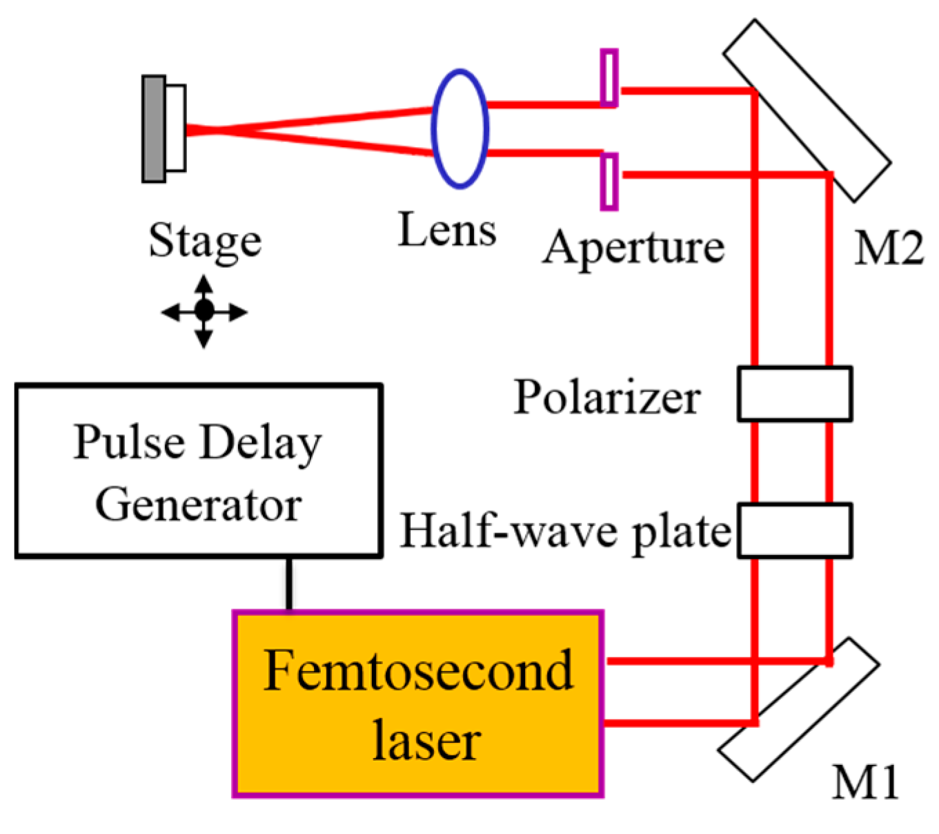
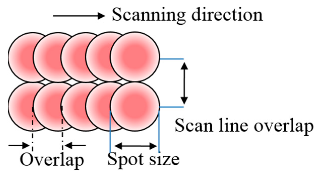
2. Materials and Methods

The copper samples were prepared by mechanically polishing commercially available copper sheets (15 mm × 15 mm × 0.3 mm, purity > 99.9%) using SiC grinding papers; the initial surface roughness obtained was <25 nm. The polished samples were cleaned in an acetone ultrasonic bath for 15 min and washed in an ultrasonic bath filled with de-ionized (DI) water for 15 min before drying.
An fs Ti/sapphire laser with a regenerative amplification system (wavelength λ = 800 nm, full width at half maximum = 50 fs, pulse energy E < 3.5 mJ, repetition rate f = 1 kHz) was employed in the experiment (Figure 1). The laser beam was linearly polarized with a Gaussian energy distribution (M square factor < 1.2). A half-wave plate and a polarizer were used to change the laser energy. After passing through a circular aperture of diameter 1 mm, the laser beam was focused by a lens of focal length 150 mm. The sample was placed perpendicular to the laser beam, and the measured laser spot size was in the range of 75 μm under different experimental conditions. The laser beam scanned the sample surface by moving the sample stage with a computer-controlled three-dimensional micro stage (Figure 2). Scanning speed was varied in the range of 75~500 μm/s at a 1 kHz pulse repetition rate. Correspondingly, each spot was irradiated with 150~1000 laser pulses by the beam scanning method summarized in Table 1. The scan line overlap was set to be 33%. Key factors that determine the morphology of LIPSS include laser fluence, scan speed, and polarization direction. The laser fluence controls the amount of energy delivered to the surface area, directly influencing the formation and characteristics of ripples. Adjusting the scan speed and spot size allows for precise tuning of the density and distribution of these structures. Additionally, the polarization direction of the laser beam plays a critical role in determining the orientation and symmetry of the LIPSS. By carefully controlling these parameters, it is possible to engineer the desired surface topography with a high degree of precision.
The topography of the surface structures was analyzed using SEM (JEOL JSM-6390, JSM-7401 for high resolution images, Tokyo, Japan) and AFM (Seiko SPI3800N Pro, Chiba, Japan). Energy dispersive spectroscopy (EDS) analysis was also conducted to analyze the surface chemistry. Elemental surface analysis was also performed by XPS (VG Scientific ESCALAB 250, Waltham, MA, USA). In the XPS analysis, a monochromatized AlKα source (1486.6 eV) was used, and the base pressure was maintained at 2 × 10 7 Pa. The pass energy and the step size for survey scans were set to 50 eV and 1.0 eV, respectively, while the step size for narrow scans was adjusted to 0.1 eV. All the spectra were charge corrected by set C 1s peak to 284.8 eV. The peak fit analysis was performed using the XPS PEAK program (version 4.1). In the characterization of the surface wettability, the static CA of a DI water droplet was measured using a commercial apparatus (Krüss DSA 100, Hamburg, Germany). All CA measurements were conducted at room temperature conditions (25 °C) using a sessile drop of 3 μL in volume and were repeated at four different locations on the sample surface. Although the sessile drop method was employed in this study to measure contact angles, it is acknowledged that for extremely low contact angles, such as those approaching 0°, the capillary bridge method could provide higher precision [45]. All the laser texturing, measurement, and sample aging were observed at room temperature under ambient air and 30% humidity.

3. Results and Discussion

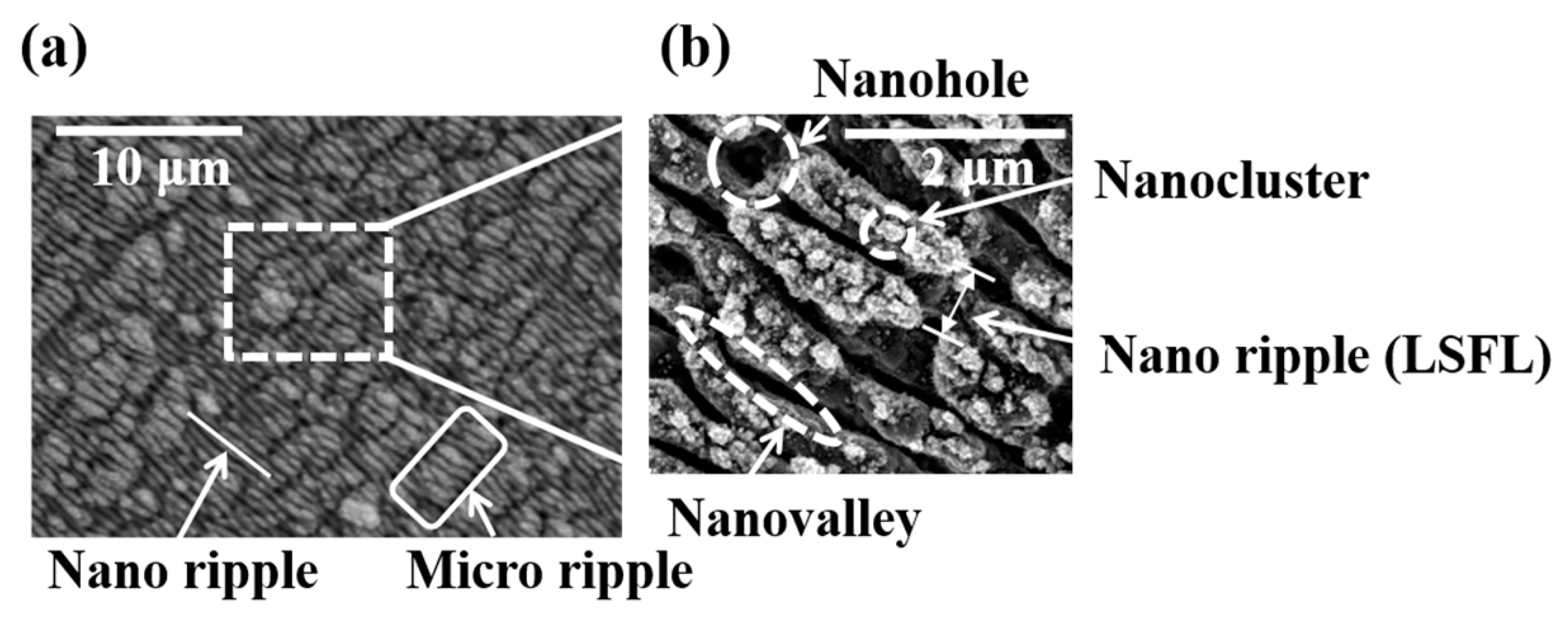
3.1. LIPSS Varying with Laser Parameters

The topographic variations of the LIPSS were observed for different values of pulse number N (number of overlapped pulses on each spot) and laser fluence F. To adjust N and F, the scan speed, spot size, and output energy were changed as described above. At laser fluences of 1.0, 1.3, 1.5, and 1.8 J/cm2, N was adjusted to 150, 400, 700, and 1000 pulses/spot, respectively. SEM images of the laser process surface revealed the topographic variation induced by laser pulse irradiation (Figure 3). At F ≈ 1 J/cm2, so-called “ripples” started to appear from the center part of the laser spot, where the local fluence is the highest. As more pulses were overlapped (N > 200), the entire surface was covered with the LIPSS.
Figure 4 shows details of the typical LIPSS. The ripple lines had a periodicity of 500~600 nm, which corresponds to the low-spatial-frequency LIPSS (LSFL) of copper [46]. These “nano ripples” are separated by nanoscale valleys (“nanovalleys”) of tens to hundreds nm in depth (Figure 4 and Figure 5). These LSFL were perpendicular to the direction of laser beam polarization. In the direction parallel to the polarization, microscale ripples occurred; these had a period of 1~5 μm. At the ends of each ripple, i.e., the depression lines dividing the ripples on a microscale, deep holes (“nanoholes”) with depths larger than the average depth of the nanovalley were produced (Figure 4 and Figure 5). These dual-scale structures (LSFLs and “micro ripple”) have been observed in LIPSS on a variety of other materials, and the LIPSS generated on copper were not fundamentally different from those. The formation of LSFL can be explained by the interference effect between the incident laser beam and surface-scattered electromagnetic waves [47]. The physical origin of the micro ripples has been attributed to the combined action of the confined laser-induced plasma (LIP) in deep and narrow cavities (“nanoholes” in this work) and self-organizing effect of the structure [48,49]. According to the analysis, deep cavities are necessary to generate the confined LIP, which causes rapid heating, melting, and subsequent hydrodynamic phenomena of the substrate. Then, highly-oriented micro-scale periodic structures are formed by fast cooling of the surface.

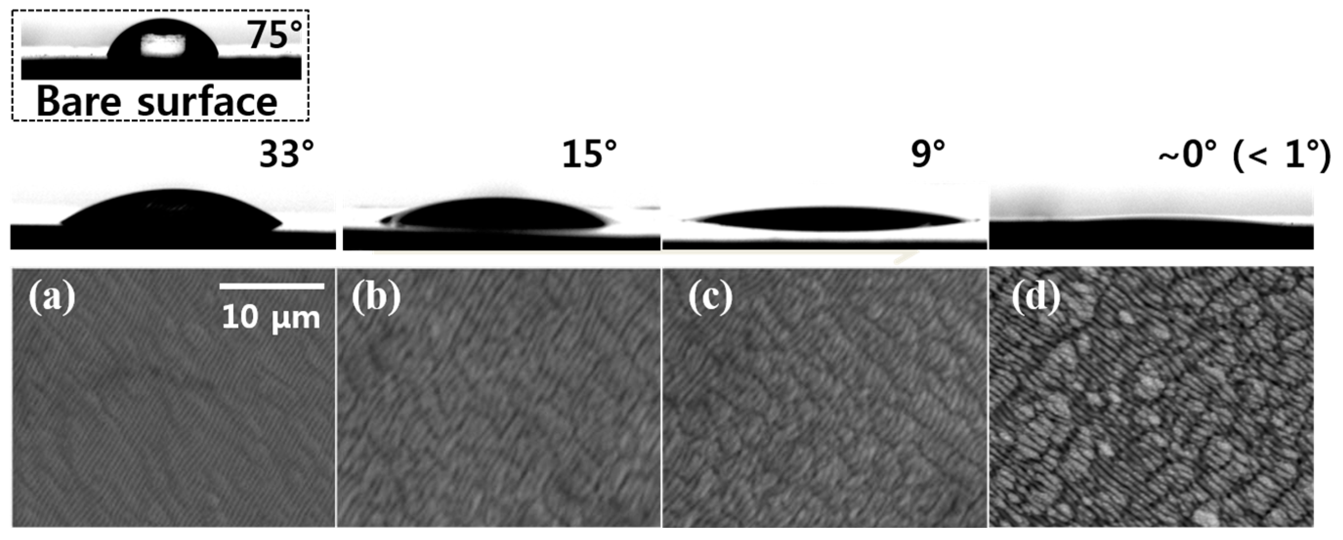
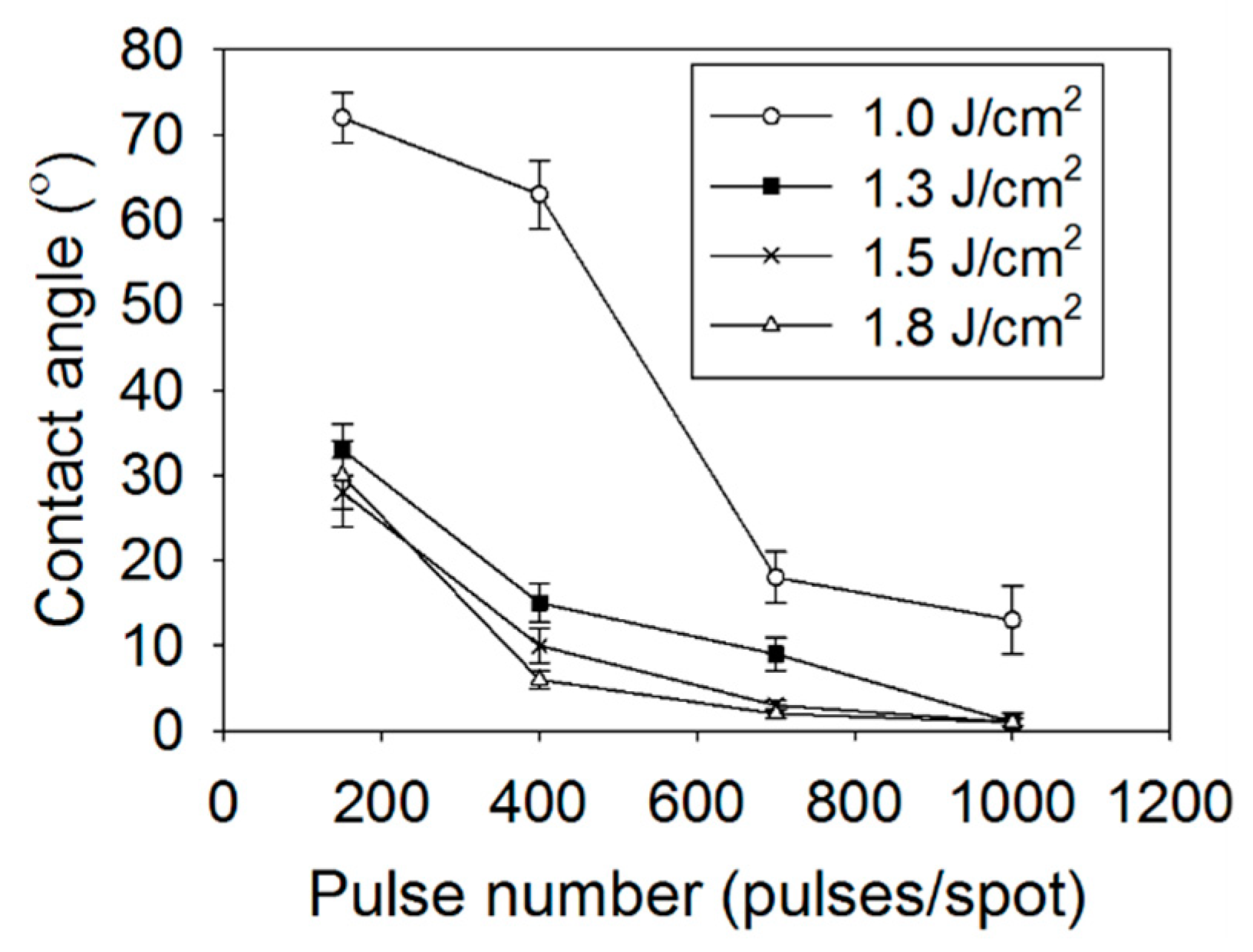
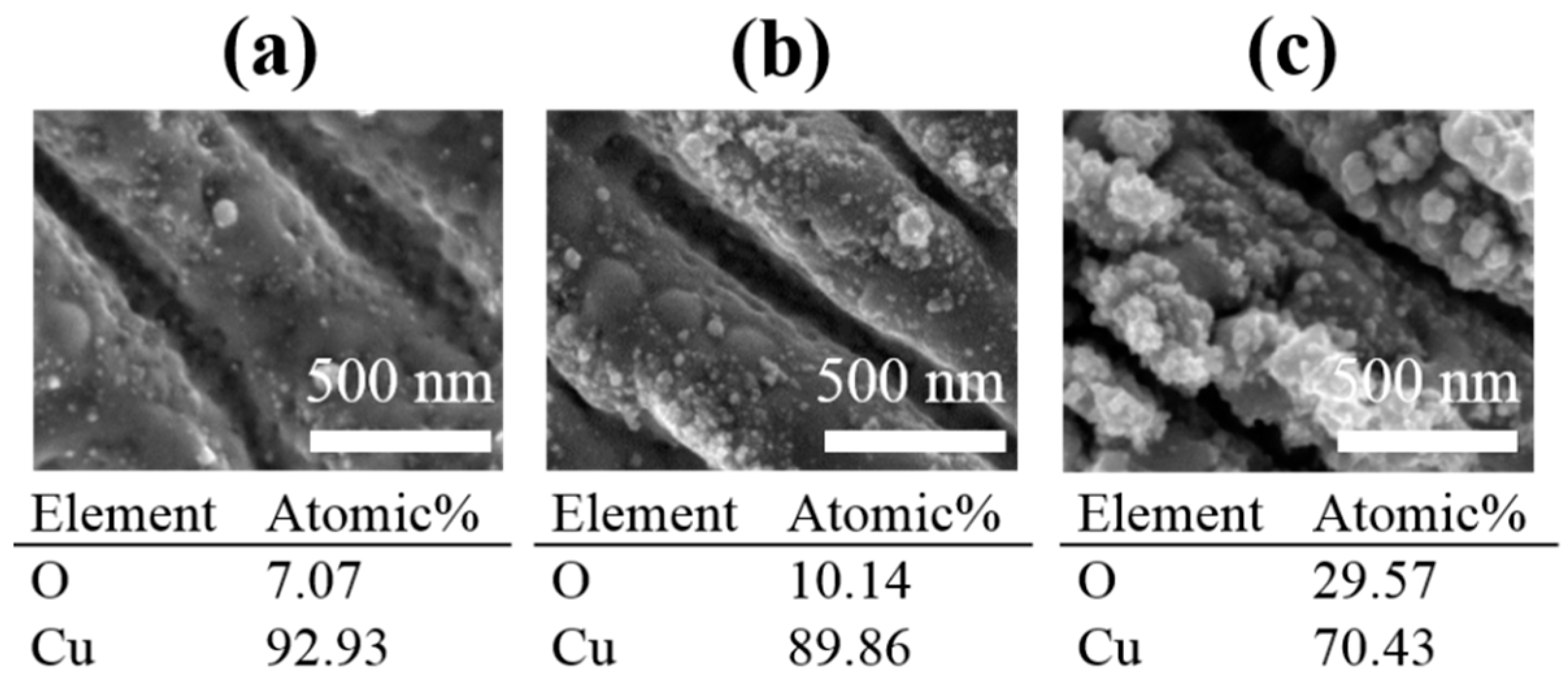
3.2. Wettability of the Laser-Modified Surfaces

The wetting properties of the laser-modified copper surface were evaluated by measuring the static CA of water droplets. The CA decreased rapidly with the development of the LIPSS, i.e., with the pulse number (Figure 6). The CA was ~75° for a bare copper surface. Successive irradiation of the laser pulse decreased the CA (Figure 6a–c), and the surface became superhydrophilic (CA < 1°) after ~1000 pulses/spot at F = 1.3 J/cm2 (Figure 6d). The effects of F and N on the CA were measured by varying them (Figure 7). In the near-threshold fluence region (F = 1.0~1.3 J/cm2), the CA was critically dependent on F, but it hardly affected the CA in the high-fluence region (F = 1.3~1.8 J/cm2). Therefore, a detailed analysis on the effect of N was conducted at F = 1.3 J/cm2.
The results exhibited in Figure 7 suggest that the laser-induced structures modified the wettability. At a fixed fluence F = 1.3 J/cm2, with increasing N from 150 to 400, the length of the ripple drastically decreased, and the number density of the nanoholes at the ends of the ripples thus increased (Figure 8). Figure 5 indicates that both the nanovalleys and nanoholes became deeper from 47 to 377 nm and from 389 to 769 nm, respectively. Correspondingly, the CA decreased from 33 to 15°. These observations indicate that the nanoholes contributed significantly to the reduction in the CA. As N was further increased from 400 to 700, neither the length of the nano ripples nor the number density of nanoholes increased significantly. Nevertheless, the increased pulse number changed the wettability from hydrophilic (CA~15°) to superhydrophilic (CA~0°). This transition to superhydrophilicity is attributed to formation of “nanoclusters” on LSFL, as shown in Figure 4b and Figure 9c. It is known that added nanostructures on a hydrophilic surface can make the surface superhydrophilic (<1°) [50,51]. Kietzig et al. [29] fabricated a hydrophilic surface with CA~20° by femtosecond laser irradiation with a fixed pulse number (120 pulses/spot), which agrees with the result of this work (F = 1.3 J/cm2, 150 pulses/spot). However, further increase in the pulse number up to 1000 pulses/spot (F = 1.3 J/cm2) generated nanoclusters on the LIPSS, which is believed to cause the superhydrophilicity of the surface.
The wettability of a surface is not only a function of the morphological structure of the surface but also a function of surface chemistry. Especially, an oxide layer makes the surface of a metal more hydrophilic than that of the base metal (Cu, in this work) [2]. The fs laser irradiation on the Cu sample generated an oxide layer on the surface, which was confirmed by EDS (Figure 9). The oxygen content was much higher on the laser-treated surface than on the bare Cu surface (<1 atomic%). Oxygen content increased from 7 to 30 atomic% as the pulse number N was enlarged from 150 to 1000 at F = 1.3 J/cm2. To analyze the chemical composition in detail, XPS was performed and detected three elements: Cu, O, and C. Figure 10 shows the high-resolution XPS spectra over the binding energy range 928–940 eV, corresponding to the Cu 2p3/2 spectra [52]. Three components corresponding to the three peaks in the spectra, namely Cu/Cu2O (932.7 eV), CuO (933.8 eV), Cu(OH)2 (935.1 eV), were identified as listed in Table 2. These results confirm the formation of copper (II) oxide (CuO) and copper (II) hydroxide (Cu(OH)2) by oxidation of copper in successive irradiation of fs laser pulses. The wetting characteristics of CuO and Cu(OH)2 are both hydrophilic. Therefore, surfaces composed of those layers can become superhydrophilic (<1°) when nanostructures are formed on them [50,51].
The superhydrophilic nature of the copper surface is primarily due to the formation of dual-scale structures, such as nanoholes and nanoclusters, which increase the surface area and enhance water adhesion. The presence of copper oxide and copper hydroxide in these nanostructures further contributes to the hydrophilic properties, as these compounds are inherently hydrophilic. The combination of increased surface roughness and the chemical nature of the oxidized layers can create a surface that promotes rapid water spreading.

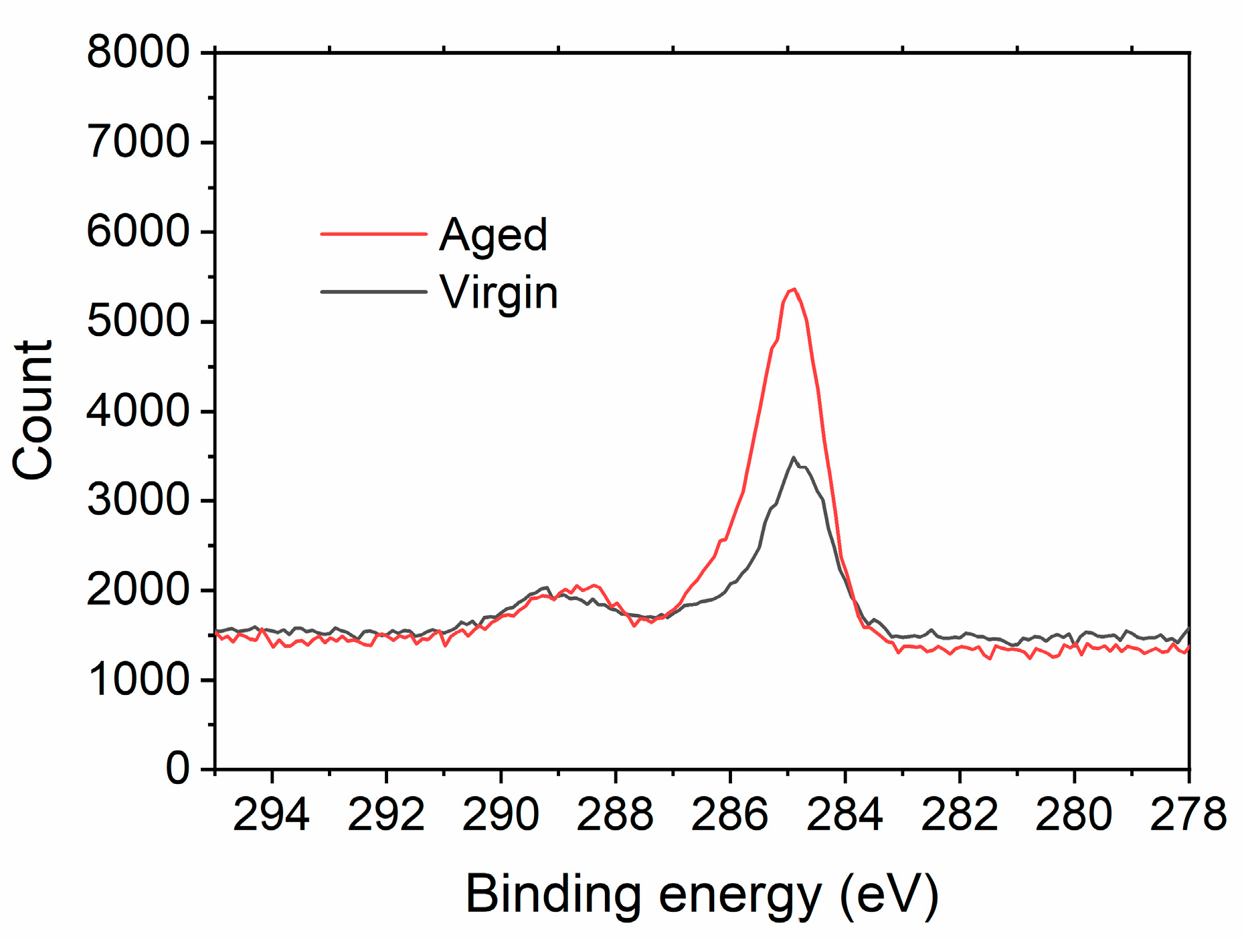
3.3. Aging Effect: Transition from Superhydrophilic to Hydrophobic Surface

Immediately after the laser treatment, the surface exhibited superhydrophilic characteristic (F = 1.3 J/cm2, N = 1000 pulses/spot). However, the contact angle increased over time and reached 118° after 10 days (Figure 11). The sample immediately after the laser irradiation, named “virgin sample”, had Cu(OH)2 and CuO. However, Cu(OH)2 was spontaneously decomposed to CuO and water at exposure of air condition, as it is metastable [53].
Cu(OH)2 → CuO + H2O,
Figure 10 and Table 2 show such trends over time, i.e., the decomposition with increasing CuO signal and decreasing Cu(OH)2 signal. The sample exposed to air for 10 days, named “aged sample”, thus had a relatively large CuO content. Wang et al. observed the transition of a superhydrophilic CuO surface to a superhydrophobic surface by aging and attributed the transition to the effect of physically adsorbed oxygen [54]. Similarly, the adsorbed oxygen molecules reduced the hydrophilicity in this work. The observed O 1s spectra (Figure 12) were composed of five peaks: (1) CuO at 529.9 eV, (2) Cu2O at 530.5 eV, (3) Cu(OH)2 at 531.7 eV, (4) physically adsorbed oxygen molecules O2 at 531.5 eV, and (5) physically adsorbed H2O at 533.0 eV [54,55]. Figure 12 confirms that the peak of physically adsorbed oxygen increased significantly in the aged sample.
The XPS analysis of aged copper samples revealed a substantial increase in the C 1s peak value compared to virgin samples, indicating the accumulation of carbon-based compounds on the surface (Figure 13). This observation is consistent with the findings of Shirazy et al., who demonstrated that the loss of hydrophilicity in copper metal foams is primarily driven by the adsorption of volatile organic compounds (VOCs) rather than oxidation [56]. Their study showed that while copper oxide content remained relatively stable, the increase in surface carbon content led to a reduction in metallic copper and a subsequent transition to hydrophobicity. This suggests that the adsorption of VOCs on the active copper surface lowers the surface energy, contributing to the observed wettability transition.
The transition from hydrophilicity to hydrophobicity is influenced not only by the oxidation of copper but also by environmental factors such as temperature and humidity. High humidity levels can slow the decomposition of Cu(OH)2 to CuO, as the presence of moisture can stabilize the hydroxide phase, delaying the transition to a hydrophobic state. Additionally, the adsorption of airborne molecules further contributes to this transition. The reduction in surface free energy, associated with the growth of the copper oxide layer and the adsorption of these molecules, plays a key role in facilitating the surface’s shift to a hydrophobic state. Over time, the surface of the structured copper undergoes oxidation, leading to the formation of a layer of copper oxide. This oxidation alters the surface chemistry, making it less hydrophilic and eventually hydrophobic as the oxide layer thickens and as organic molecules adsorb onto the surface.
Maintaining superhydrophilicity has been a significant focus in materials science, especially for applications requiring consistent wetting properties over extended periods. Polymer coatings, for instance, have been effectively used to prevent oxidation and contamination [57]. Environmental control, such as reducing humidity and oxygen exposure, can also slow down oxidation and help maintain hydrophilicity [54]. While applying long-term-stable superhydrophilic SAMs is a promising approach, it still remains challenging due to the susceptibility of these coatings to various degradation factors, including oxygen adsorption, mechanical wear, humidity, and chemical exposure. This difficulty highlights the need for further research to develop more resilient coatings that can withstand environmental stressors and extend the lifespan of superhydrophilic surfaces.

4. Conclusions

We developed an fs laser irradiation process to fabricate superhydrophilic surface on copper with micro/nanoscale periodic structures. The effect of various laser parameters on the LIPSS was analyzed by SEM and AFM. Repeated irradiation of the laser pulse decreased the length of the ripple but increased the number density of the nanoholes at the ends of the ripples; the depths of these nanoholes also increased with N. At F = 1.3 J/cm2, the development of the nanoholes reduced the CA of water droplets. Further increase in N did not enlarge the density or depth of the nanoholes but generated nanoclusters on LIPSS, which reduced the contact angle from hydrophilic (CA~15°) to superhydrophilic (CA < 1°). Through the chemical analysis using XPS, the superhydrophilic surface was composed of CuO and Cu(OH)2, which exhibit superhydrophilic wetting characteristics when nanostructures are formed. The fs laser-induced superhydrophilic surface on copper was not permanent but spontaneously changed to be hydrophobic after exposure to air at room temperature. Chemical analysis of the surface indicate that the reduced hydrophilicity is due to decomposition of Cu(OH)2 and the adsorption of oxygen molecule and/or airborne organic molecules. Further studies are needed to enhance the long-term stability of the superhydrophilic properties over extended periods for various applications.

Funding

This work was supported by the Ministry of Science and ICT (Project Number: 2024-22030005-20) and Commercialization Promotion Agency for R&D Outcomes (COMPA) and Korea Institute of Industrial Technology (JA-24-0007).

Institutional Review Board Statement

Not applicable.

Informed Consent Statement

Not applicable.

Data Availability Statement

Data are contained within the article.

Conflicts of Interest

The author declares no conflicts of interest. The funders had no role in the design of the study; in the collection, analyses, or interpretation of data; in the writing of the manuscript; or in the decision to publish the results.

References

  1. Feng, X.J.; Jiang, L. Design and Creation of Superwetting/Antiwetting Surfaces. Adv. Mater. 2006, 18, 3063–3078. [Google Scholar] [CrossRef]
  2. Zhai, W.; Zhou, W.; Nai, S.M.L. Effect of Interface Wettability on Additively Manufactured Metal Matrix Composites: A Case Study of 316L-Y2O3 Oxide Dispersion-Strengthened Steel. Metals 2024, 14, 170. [Google Scholar] [CrossRef]
  3. Paul, D.; Zaplotnik, R.; Primc, G.; Vesel, A.; Mozetič, M. Evolution of the Surface Wettability of Vertically Oriented Multilayer Graphene Sheets Deposited by Plasma Technology. Nanomaterials 2024, 14, 1023. [Google Scholar] [CrossRef] [PubMed]
  4. Meng, X.; Zhao, D.; Zhang, J.; Shen, D.; Lu, Y.; Dong, L.; Xiao, Z.; Liu, Y.; Fan, X. Wettability conversion on ZnO nanowire arrays surface modified by oxygen plasma treatment and annealing. Chem. Phys. Lett. 2005, 413, 450–453. [Google Scholar] [CrossRef]
  5. Kim, H.; Kim, M.H.; Kim, J. Wettability of dual-scaled surfaces fabricated by the combination of a conventional silicon wet-etching and a ZnO solution method. J. Micromech. Microeng. 2009, 19, 095002. [Google Scholar] [CrossRef]
  6. McDonald, B.T.; Cui, T. Superhydrophilic surface modification of copper surfaces by layer-by-layer self-assembly and liquid phase deposition of TiO2 thin film. J. Colloid Interface Sci. 2011, 354, 1–6. [Google Scholar] [CrossRef]
  7. Ngo, C.-V.; Liu, Y.; Li, W.; Yang, J.; Guo, C. Scalable Wettability Modification of Aluminum Surface through Single-Shot Nanosecond Laser Processing. Nanomaterials 2023, 13, 1392. [Google Scholar] [CrossRef] [PubMed]
  8. Wróblewski, P. The theory of the surface wettability angle in the formation of an oil film in internal combustion piston engines. Materials 2023, 16, 4092. [Google Scholar] [CrossRef]
  9. Papadopoulou, E.L.; Barberoglou, M.; Zorba, V.; Manousaki, A.; Pagkozidis, A.; Stratakis, E.; Fotakis, C. Reversible photoinduced wettability transition of hierarchical ZnO structures. J. Phys. Chem. C 2009, 113, 2891–2895. [Google Scholar] [CrossRef]
  10. Eiamchai, P.; Chindaudom, P.; Horprathum, M.; Patthanasettakul, V.; Limsuwan, P. Design and investigation of photo-induced super-hydrophilic materials for car mirrors. Mater. Des. 2009, 30, 3428–3435. [Google Scholar] [CrossRef]
  11. Shi, J.J.; Yang, E.L. Non-UV driven self-cleaning and anti-fogging glasses prepared by ultrasonic nebulization of TiO2 hydrosol. Adv. Mater. Res. 2012, 549, 674–678. [Google Scholar] [CrossRef]
  12. Betz, A.R.; Jenkins, J.; Attinger, D. Boiling heat transfer on superhydrophilic, superhydrophobic, and superbiphilic surfaces. Int. J. Heat Mass Transf. 2013, 57, 733–741. [Google Scholar] [CrossRef]
  13. Kim, S.; Kim, H.D.; Kim, H.; Ahn, H.S.; Jo, H.; Kim, J.; Kim, M.H. Effects of nano-fluid and surfaces with nano structure on the increase of CHF. Exp. Therm. Fluid Sci. 2010, 34, 487–495. [Google Scholar] [CrossRef]
  14. Ahn, H.S.; Lee, C.; Kim, H.; Jo, H.; Kang, S.; Kim, J.; Shin, J.; Kim, M.H. Pool boiling CHF enhancement by micro/nanoscale modification of zircaloy-4 surface. Nucl. Eng. Des. 2010, 240, 3350–3360. [Google Scholar] [CrossRef]
  15. Ye, J.; Yin, Q.; Zhou, Y. Superhydrophilicity of anodic aluminum oxide films: From “honeycomb” to “bird’s nest”. Thin Solid Film. 2009, 517, 6012–6015. [Google Scholar] [CrossRef]
  16. Lawrence, J.; Li, L. Wettability characteristics of a modified mild steel with CO2, Nd: YAG, excimer and high power diode lasers. J. Phys. D Appl. Phys. 1999, 32, 2311. [Google Scholar] [CrossRef]
  17. Tseng, S.-F.; Hsiao, W.-T.; Chen, M.-F.; Huang, K.-C.; Hsiao, S.-Y.; Lin, Y.-S.; Chou, C.-P. Surface wettability of silicon substrates enhanced by laser ablation. Appl. Phys. A 2010, 101, 303–308. [Google Scholar] [CrossRef]
  18. Chang, T.-L.; Tsai, T.-K.; Yang, H.-P.; Huang, J.-Z. Effect of ultra-fast laser texturing on surface wettability of microfluidic channels. Microelectron. Eng. 2012, 98, 684–688. [Google Scholar] [CrossRef]
  19. Kam, D.; Bhattacharya, S.; Mazumder, J. Control of the wetting properties of an AISI 316L stainless steel surface by femtosecond laser-induced surface modification. J. Micromech. Microeng. 2012, 22, 105019. [Google Scholar] [CrossRef]
  20. Kietzig, A.-M.; Hatzikiriakos, S.G.; Englezos, P. Patterned superhydrophobic metallic surfaces. Langmuir 2009, 25, 4821–4827. [Google Scholar] [CrossRef] [PubMed]
  21. Caputo, G.; Nobile, C.; Kipp, T.; Blasi, L.; Grillo, V.; Carlino, E.; Manna, L.; Cingolani, R.; Cozzoli, P.D.; Athanassiou, A. Reversible wettability changes in colloidal TiO2 nanorod thin-film coatings under selective UV laser irradiation. J. Phys. Chem. C 2008, 112, 701–714. [Google Scholar] [CrossRef]
  22. Zhang, L.; Shoji, M. Nucleation site interaction in pool boiling on the artificial surface. Int. J. Heat Mass Transf. 2003, 46, 513–522. [Google Scholar] [CrossRef]
  23. Chatpun, S.; Watanabe, M.; Shoji, M. Experimental study on characteristics of nucleate pool boiling by the effects of cavity arrangement. Exp. Therm. Fluid Sci. 2004, 29, 33–40. [Google Scholar] [CrossRef]
  24. Borowiec, A.; Haugen, H. Subwavelength ripple formation on the surfaces of compound semiconductors irradiated with femtosecond laser pulses. Appl. Phys. Lett. 2003, 82, 4462–4464. [Google Scholar] [CrossRef]
  25. Wang, J.; Guo, C. Ultrafast dynamics of femtosecond laser-induced periodic surface pattern formation on metals. Appl. Phys. Lett. 2005, 87. [Google Scholar] [CrossRef]
  26. Wagner, R.; Gottmann, J.; Horn, A.; Kreutz, E.W. Subwavelength ripple formation induced by tightly focused femtosecond laser radiation. Appl. Surf. Sci. 2006, 252, 8576–8579. [Google Scholar] [CrossRef]
  27. Vorobyev, A.; Makin, V.; Guo, C. Periodic ordering of random surface nanostructures induced by femtosecond laser pulses on metals. J. Appl. Phys. 2007, 101. [Google Scholar] [CrossRef]
  28. Das, S.K.; Dasari, K.; Rosenfeld, A.; Grunwald, R. Extended-area nanostructuring of TiO2 with femtosecond laser pulses at 400 nm using a line focus. Nanotechnology 2010, 21, 155302. [Google Scholar] [CrossRef]
  29. Kietzig, A.-M.; Negar Mirvakili, M.; Kamal, S.; Englezos, P.; Hatzikiriakos, S.G. Laser-patterned super-hydrophobic pure metallic substrates: Cassie to Wenzel wetting transitions. J. Adhes. Sci. Technol. 2011, 25, 2789–2809. [Google Scholar] [CrossRef]
  30. Allahyari, E.; Nivas, J.J.; Oscurato, S.L.; Salvatore, M.; Ausanio, G.; Vecchione, A.; Fittipaldi, R.; Maddalena, P.; Bruzzese, R.; Amoruso, S. Laser surface texturing of copper and variation of the wetting response with the laser pulse fluence. Appl. Surf. Sci. 2019, 470, 817–824. [Google Scholar] [CrossRef]
  31. Li, J.; Zhou, Y.; Wang, W.; Xu, C.; Ren, L. Superhydrophobic copper surface textured by laser for delayed icing phenomenon. Langmuir 2020, 36, 1075–1082. [Google Scholar] [CrossRef] [PubMed]
  32. Može, M.; Zupančič, M.; Steinbücher, M.; Golobič, I.; Gjerkeš, H. Nanosecond laser-textured copper surfaces hydrophobized with self-assembled monolayers for enhanced pool boiling heat transfer. Nanomaterials 2022, 12, 4032. [Google Scholar] [CrossRef] [PubMed]
  33. Tong, W.; Xiong, D. Direct laser texturing technique for metal surfaces to achieve superhydrophobicity. Mater. Today Phys. 2022, 23, 100651. [Google Scholar] [CrossRef]
  34. Ta, D.V.; Dunn, A.; Wasley, T.J.; Kay, R.W.; Stringer, J.; Smith, P.J.; Connaughton, C.; Shephard, J.D. Nanosecond laser textured superhydrophobic metallic surfaces and their chemical sensing applications. Appl. Surf. Sci. 2015, 357, 248–254. [Google Scholar] [CrossRef]
  35. Long, J.; He, Z.; Zhou, C.; Xie, X.; Cao, Z.; Zhou, P.; Zhu, Y.; Hong, W.; Zhou, Z. Hierarchical micro-and nanostructures induced by nanosecond laser on copper for superhydrophobicity, ultralow water adhesion and frost resistance. Mater. Des. 2018, 155, 185–193. [Google Scholar] [CrossRef]
  36. Bonse, J.; Rosenfeld, A.; Krüger, J. On the role of surface plasmon polaritons in the formation of laser-induced periodic surface structures upon irradiation of silicon by femtosecond-laser pulses. J. Appl. Phys. 2009, 106. [Google Scholar] [CrossRef]
  37. Li, Z.; Wu, Q.; Jiang, X.; Zhou, X.; Liu, Y.; Hu, X.; Zhang, J.; Yao, J.; Xu, J. Formation mechanism of high spatial frequency laser-induced periodic surface structures and experimental support. Appl. Surf. Sci. 2022, 580, 152107. [Google Scholar] [CrossRef]
  38. Bonse, J.; Krüger, J.; Höhm, S.; Rosenfeld, A. Femtosecond laser-induced periodic surface structures. J. Laser Appl. 2012, 24. [Google Scholar] [CrossRef]
  39. Bonse, J.; Krüger, J. Pulse number dependence of laser-induced periodic surface structures for femtosecond laser irradiation of silicon. J. Appl. Phys. 2010, 108. [Google Scholar] [CrossRef]
  40. Huang, M.; Zhao, F.; Cheng, Y.; Xu, N.; Xu, Z. Origin of laser-induced near-subwavelength ripples: Interference between surface plasmons and incident laser. ACS Nano 2009, 3, 4062–4070. [Google Scholar] [CrossRef]
  41. Razi, S.; Asghari, M.; Mollabashi, M. Angle-and polarization-dependent reflection and backscattering from FS-LIPSS covered stainless steel surfaces: Experimental study. Eur. Phys. J. Plus 2020, 135, 1–19. [Google Scholar] [CrossRef]
  42. Xiao, C.; Shi, P.; Yan, W.; Chen, L.; Qian, L.; Kim, S.H. Thickness and structure of adsorbed water layer and effects on adhesion and friction at nanoasperity contact. Colloids Interfaces 2019, 3, 55. [Google Scholar] [CrossRef]
  43. Bhushan, B.; Jung, Y.C.; Koch, K. Micro-, nano-and hierarchical structures for superhydrophobicity, self-cleaning and low adhesion. Philos. Trans. R. Soc. A Math. Phys. Eng. Sci. 2009, 367, 1631–1672. [Google Scholar] [CrossRef]
  44. Su, Y.; Ji, B.; Zhang, K.; Gao, H.; Huang, Y.; Hwang, K. Nano to micro structural hierarchy is crucial for stable superhydrophobic and water-repellent surfaces. Langmuir 2010, 26, 4984–4989. [Google Scholar] [CrossRef]
  45. Nagy, N. Capillary bridges on hydrophobic surfaces: Analytical contact angle determination. Langmuir 2022, 38, 6201–6208. [Google Scholar] [CrossRef] [PubMed]
  46. Sakabe, S.; Hashida, M.; Tokita, S.; Namba, S.; Okamuro, K. Mechanism for self-formation of periodic grating structures on a metal surface by a femtosecond laser pulse. Phys. Rev. B—Condens. Matter Mater. Phys. 2009, 79, 033409. [Google Scholar] [CrossRef]
  47. Sipe, J.; Young, J.F.; Preston, J.; Van Driel, H. Laser-induced periodic surface structure. I. Theory. Phys. Rev. B 1983, 27, 1141. [Google Scholar] [CrossRef]
  48. Bizi-Bandoki, P.; Valette, S.; Audouard, E.; Benayoun, S. Effect of stationary femtosecond laser irradiation on substructures’ formation on a mold stainless steel surface. Appl. Surf. Sci. 2013, 270, 197–204. [Google Scholar] [CrossRef]
  49. Kim, S.H.; Sohn, I.-B.; Jeong, S. Parallel ripple formation during femtosecond laser grooving of ceramic. Appl. Phys. A 2011, 103, 1053–1057. [Google Scholar] [CrossRef]
  50. Chang, F.-M.; Cheng, S.-L.; Hong, S.-J.; Sheng, Y.-J.; Tsao, H.-K. Superhydrophilicity to superhydrophobicity transition of CuO nanowire films. Appl. Phys. Lett. 2010, 96. [Google Scholar] [CrossRef]
  51. Pei, M.-D.; Wang, B.; Li, E.; Zhang, X.-h.; Song, X.-m.; Yan, H. The fabrication of superhydrophobic copper films by a low-pressure-oxidation method. Appl. Surf. Sci. 2010, 256, 5824–5827. [Google Scholar] [CrossRef]
  52. Zeng, D.; Yung, K.C.; Xie, C. UV Nd: YAG laser ablation of copper: Chemical states in both crater and halo studied by XPS. Appl. Surf. Sci. 2003, 217, 170–180. [Google Scholar] [CrossRef]
  53. Cudennec, Y.; Lecerf, A. The transformation of Cu (OH) 2 into CuO, revisited. Solid State Sci. 2003, 5, 1471–1474. [Google Scholar] [CrossRef]
  54. Wang, G.; Zhang, T.-Y. Oxygen adsorption induced superhydrophilic-to-superhydrophobic transition on hierarchical nanostructured CuO surface. J. Colloid Interface Sci. 2012, 377, 438–441. [Google Scholar] [CrossRef]
  55. Platzman, I.; Brener, R.; Haick, H.; Tannenbaum, R. Oxidation of polycrystalline copper thin films at ambient conditions. J. Phys. Chem. C 2008, 112, 1101–1108. [Google Scholar] [CrossRef]
  56. Shirazy, M.R.; Blais, S.; Fréchette, L.G. Mechanism of wettability transition in copper metal foams: From superhydrophilic to hydrophobic. Appl. Surf. Sci. 2012, 258, 6416–6424. [Google Scholar] [CrossRef]
  57. Conradi, M.; Sever, T.; Gregorčič, P.; Kocijan, A. Short-and long-term wettability evolution and corrosion resistance of uncoated and polymer-coated laser-textured steel surface. Coatings 2019, 9, 592. [Google Scholar] [CrossRef]
Figure 1. Schematic overview of the experimental setup (M1, M2: mirrors).
Coatings 14 01107 g001
Figure 2. Schematic diagram of laser beam scanning parameters.
Coatings 14 01107 g002
Figure 3. SEM images of topographic variation on copper surface with various laser fluences and number of overlapped pulses on each spot.
Coatings 14 01107 g003
Figure 4. SEM images of dual-scale structures: (a) an overview; (b) a magnified image.
Coatings 14 01107 g004
Figure 5. Depth profiles of nanovalleys after (a) 150, (b) 400, and (c) 1000 laser pulses and those of nanoholes after (d) 150, (e) 400, and (f) 1000 laser pulses at F = 1.3 J/cm2.
Coatings 14 01107 g005
Figure 6. Variation of surface structures with increasing pulse number at F = 1.3 J/cm2 and corresponding contact angle; (a) 150, (b) 400, (c) 700, and (d) 1000 pulse number.
Coatings 14 01107 g006
Figure 7. Contact angle on copper surface with varying laser fluence and pulse number.
Coatings 14 01107 g007
Figure 8. Quantified topographic variation of ripple at F = 1.3 J/cm2.
Coatings 14 01107 g008
Figure 9. High-resolution SEM images of topographic variation and the chemical composition obtained by EDS on nano ripples (LSFL) after (a) 150, (b) 400, and (c) 1000 laser pulses at F = 1.3 J/cm2.
Coatings 14 01107 g009
Figure 10. XPS spectra of a Cu 2p3/2 region: (a) a virgin sample and (b) an aged sample.
Coatings 14 01107 g010
Figure 11. Variation of the contact angle of the laser-irradiated surface with the time exposed to air.
Coatings 14 01107 g011
Figure 12. XPS spectra of an O 1s region: (a) a virgin sample and (b) an aged sample.
Coatings 14 01107 g012
Figure 13. XPS spectra of a C 1s region of a virgin sample and an aged one.
Coatings 14 01107 g013
Table 1. Overlapped pulse numbers for different scanning speeds.
Scanning Speed
[μm/s]
Overlapped Pulse Number
[Pulses/Spot]
500150
187.5400
107.1700
751000
Table 2. Peak parameters of XPS Cu 2p3/2 spectra.
Cu/Cu2OCuOCu(OH)2
Peak
(eV)
Area
(%)
FWHM
(eV)
Peak
(eV)
Area
(%)
FWHM
(eV)
Peak
(eV)
Area
(%)
FWHM
(eV)
Virgin932.776.760.97933.88.312.00935.114.932.40
Aged932.772.521.10933.819.852.00935.17.632.40
Disclaimer/Publisher’s Note: The statements, opinions and data contained in all publications are solely those of the individual author(s) and contributor(s) and not of MDPI and/or the editor(s). MDPI and/or the editor(s) disclaim responsibility for any injury to people or property resulting from any ideas, methods, instructions or products referred to in the content.

