Review

Exploring the Role of Nanoparticles in Dental Materials: A Comprehensive Review

by
Rim Bourgi
1,2,*,†,
Zahra Doumandji
3,†,
Carlos Enrique Cuevas-Suárez
4,
Teissir Ben Ammar
2,
Chloé Laporte
2,3,5,
Naji Kharouf
2,5 and
Youssef Haikel
2,3,5
1
Department of Restorative Dentistry, School of Dentistry, Saint-Joseph University, Beirut 1107 2180, Lebanon
2
Department of Biomaterials and Bioengineering, INSERM UMR_S 1121, University of Strasbourg, 67000 Strasbourg, France
3
Pôle de Médecine et Chirurgie Bucco-Dentaire, Hôpital Civil, Hôpitaux Universitaire de Strasbourg, 67000 Strasbourg, France
4
Dental Materials Laboratory, Academic Area of Dentistry, Autonomous University of Hidalgo State, San Agustín Tlaxiaca 42160, Mexico
5
Department of Endodontics and Conservative Dentistry, Faculty of Dental Medicine, University of Strasbourg, 67000 Strasbourg, France
*
Author to whom correspondence should be addressed.
These authors contributed equally to this work.
Coatings 2025, 15(1), 33; https://doi.org/10.3390/coatings15010033
Submission received: 11 December 2024 / Revised: 22 December 2024 / Accepted: 25 December 2024 / Published: 2 January 2025

Abstract

:
In recent decades, the integration of nanotechnology into dentistry has led to groundbreaking advancements in dental materials and applications. This article explores the role of nanoparticles (NPs) in modern dentistry, highlighting their definitions, unique properties, and various applications. The introduction establishes the significance of nanotechnology in dental health care, emphasizing the potential of NPs to transform traditional practices. The overview includes a discussion of the fundamental properties of NPs, which contribute to their effectiveness in dental applications. The article categorizes NPs into three main groups: antimicrobial, therapeutic, and material property-improving NPs, detailing their clinical uses and mechanisms of action. Furthermore, it addresses current innovations in dental products incorporating NPs and examines emerging trends in the field. The research for this review was conducted using high-quality, peer-reviewed scientific databases, including PubMed, Scopus, Web of Science, and Google Scholar, with no time restriction as an inclusion criterion. These databases were selected for their credibility and comprehensive collections of relevant studies. In conclusion, NPs represent a promising avenue for innovation in dental materials and therapeutics. Their unique properties enable the development of enhanced antimicrobial agents, effective drug delivery systems, and improved material performance. However, the risks associated with cytotoxicity and stability must be carefully managed to ensure safe and effective use. Ongoing research is essential to fully understand and optimize the applications of NPs in dentistry, balancing their benefits against potential health risks. As the field advances, the integration of NPs into clinical practice will likely revolutionize approaches to dental care and treatment.

1. Introduction

Nanotechnology has emerged as one of the most dynamic fields of research in recent decades, particularly within the realm of health sciences [1]. Maintaining oral health presents significant challenges in dentistry. Various materials have been employed to address different dental conditions; however, the success of these treatments is often constrained by the properties of the biomaterials used. To address these limitations, the incorporation of nanoparticles (NPs) into dental applications offers promising solutions in fields such as endodontics, periodontics, tissue engineering, oral surgery, and imaging [2,3,4,5].
NPs, defined as ultrafine units with dimensions ranging from 1 to 100 nm, exhibit unique physical, chemical, mechanical, and biological properties that distinguish them from macro materials [6]. Their small size contributes to a high surface-to-volume ratio, enhancing their reactivity and allowing for novel applications across various fields, including medicine, engineering, and environmental remediation [7]. Within the dental sector, the integration of NPs has catalyzed significant advancements in dental materials and practices.
Prior research has highlighted the diverse applications of NPs in dentistry, encompassing areas such as tissue regeneration, antimicrobial strategies, and enhancement of material properties [8]. NPs can be categorized into several types based on their origin—natural or synthetic—and composition, including organic (e.g., dendrimers, liposomes) and inorganic (e.g., metal or metal oxide-based) [9]. While many NPs possess advantageous characteristics such as biocompatibility, chemical reactivity, and mechanical strength [10], not all NPs are inherently biocompatible. For instance, certain plate-like structures can pose risks to human health due to their cytotoxic or inflammatory potential. This underscores the need for careful evaluation and selection of NPs to ensure their safe use in dental applications.
In restorative dentistry, NPs are incorporated into composites to enhance mechanical strength, reduce shrinkage, and improve aesthetics [11]. Antimicrobial NPs, such as silver and zinc oxide (ZnO), are used to inhibit bacterial growth, thus reducing the risk of infections [12]. Additionally, NPs are instrumental in endodontics, where they are employed for tissue regeneration, drug delivery systems, and the effective elimination of biofilms and bacteria, ultimately aiming to improve overall oral health [13].
In preventive dentistry, NPs like hydroxyapatite and calcium phosphate are recognized for their ability to remineralize enamel and prevent caries [14]. Moreover, in the context of dental implants, NPs enhance osseointegration and surface adhesion, contributing to better treatment outcomes [15]. Recent advancements have also seen the use of NPs in developing adhesives that reduce biofilm accumulation and promote oral health [16].
Despite the numerous advantages that NPs present, their long-term effects and potential toxicity in biological systems necessitate careful consideration in both research and clinical applications [2,17]. Regulatory challenges and safety assessments remain critical for the integration of nanotechnology into dental practice. A thorough understanding of the physicochemical properties of NPs and their interactions within the oral environment is essential for optimizing their use in dentistry [2,3,4].
Specifically, the incorporation of NPs in dentistry not only enhances the efficacy of treatments but also holds the potential to revolutionize patient care by promoting personalized and targeted therapeutic strategies. For instance, with advancements in drug delivery systems utilizing NPs, there is the capability to deliver medications directly to the site of action, thus minimizing systemic exposure and side effects. This targeted approach could improve the effectiveness of treatments for conditions such as periodontal disease and oral cancer [18,19]. Furthermore, the multifunctional nature of NPs allows for the simultaneous delivery of therapeutic agents and diagnostic imaging, paving the way for novel applications in precision dentistry [2,20,21].
As research continues to uncover the versatile applications of NPs, their integration into clinical practice could lead to more effective, efficient, and patient-centered care in the field of dentistry [2,17,22]. Thus, the objective of this comprehensive review is to explore the role of NPs in dental materials by analyzing their properties, applications, and innovations. This review aims to provide a thorough understanding of the current state of knowledge regarding NPs in dentistry, identify emerging trends, and discuss potential future applications and challenges associated with their use.

2. Overview of Nanoparticles in Dentistry

2.1. Definition of Nanoparticles

NPs are typically defined as particles with at least one dimension ranging between 1 and 100 nm, which corresponds to one billionth of a meter (1 nm = 10⁻⁹ m) [23,24]. This definition is generally accepted across scientific disciplines, though specific regulations or organizations may have slight variations in their interpretation of this size range [25]. NPs occupy a unique size domain, larger than individual atoms yet smaller than cells, placing them in a category where they exhibit distinctive properties that bulk materials lack [26]. Their high surface area, quantum effects, and enhanced chemical reactivity provide them with unique physicochemical characteristics, such as increased mechanical strength, conductivity, and reactivity, making them highly suitable for various applications across industries [27].
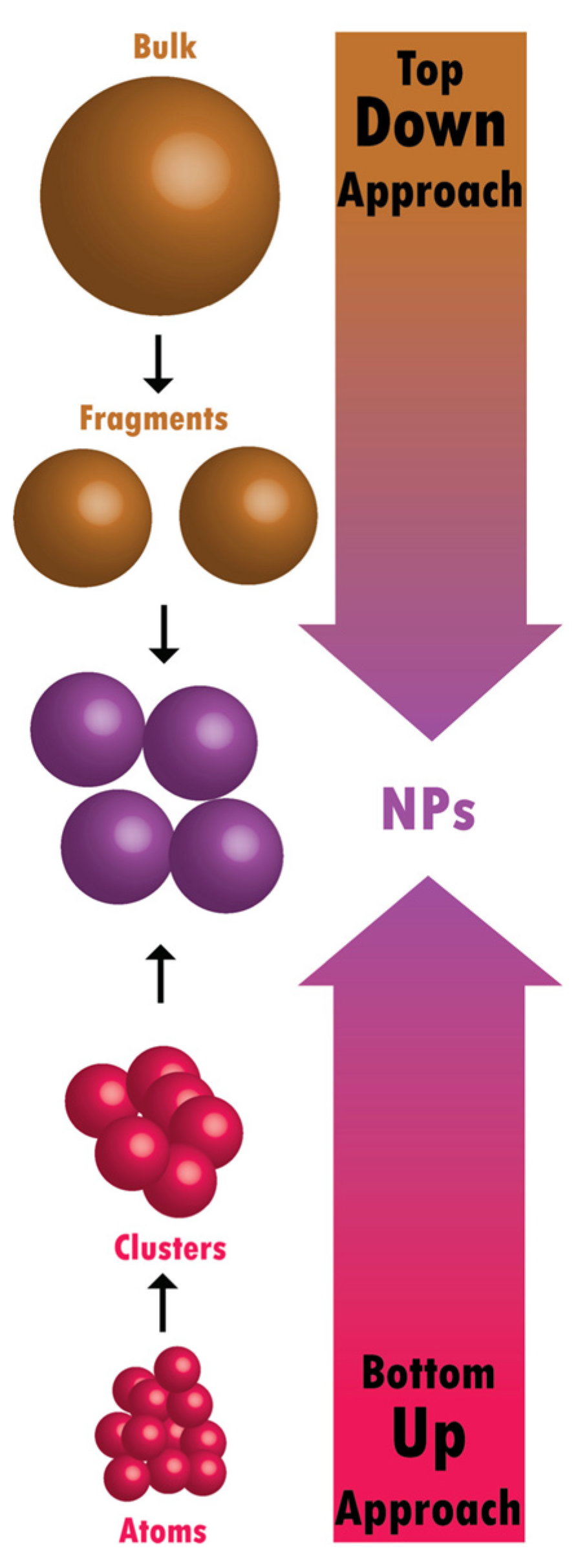
NPs can occur naturally (e.g., volcanic ash, ocean spray, and fine dust) or be engineered synthetically to meet specific needs [28]. Synthetic NPs are created using two primary approaches: top-down or bottom-up methods [29]. In the top-down approach, larger bulk materials are broken down into NPs via processes like milling or lithography. In contrast, the bottom-up approach involves assembling NPs from atomic or molecular units through processes like chemical vapor deposition, sol-gel synthesis, or self-assembly [30]. As a result, NPs can exist in various forms, including powders, gels, or colloidal solutions, and are typically tailored to specific applications based on their size, shape, and surface properties [31,32,33,34,35,36,37,38].
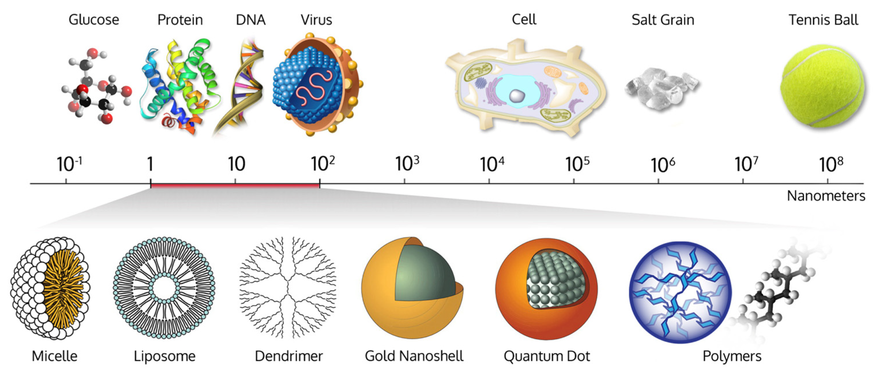
Figure 1 represents the definition of NPs.
Figure 2 illustrates the comparative scale of NPs.
NPs have a wide range of applications, spanning from medicine to aerospace and cosmetics [41]. Their unique size allows them to impart properties that are otherwise unattainable at larger scales. For instance, gold, which is chemically inert in its bulk form, acts as an efficient catalyst at the nanoscale due to the increased surface area and altered electronic properties [42]. However, despite these advantages, NPs also pose potential risks to both health and the environment. Their small size enables them to penetrate biological barriers, potentially leading to adverse effects such as cytotoxicity, oxidative stress, and inflammation in living organisms [43,44]. Additionally, the environmental impact of nanoparticle accumulation and their behavior in ecosystems remain critical concerns [30,45]. Ongoing research is crucial to balance the beneficial applications of NPs with the need to mitigate their potential risks [46].

2.2. Nanotechnology

In recent years, the primary focus of utilizing nanotechnologies in dental materials has been to enhance mechanical properties, improve abrasion resistance, reduce shrinkage, and optimize optical and aesthetic aspects [47]. Today, it is widely recognized that NPs possess a broad range of indispensable properties, including bioactive and antimicrobial characteristics [48]. Moreover, in the field of dentistry, specifically in endodontics, NPs are employed for tissue regeneration and drug delivery. Enhancing oral health is the primary objective, with the eradication of biofilms and bacteria using NPs at the core of ongoing research [49].
In this context, the incorporation of NPs into certain dental treatment materials has shown the potential to reduce biofilm accumulation, restore a safe level of oral pH, and promote remineralization while improving the durability of the material [50].
Biofilm refers to a complex community of microorganisms, primarily bacteria, that adhere to surfaces such as teeth or dental materials, embedded within a self-produced extracellular matrix. This protective layer makes biofilms highly resistant to conventional antimicrobial treatments, contributing to persistent infections, inflammation, and tooth decay. The dangers of biofilm formation in the oral cavity include an increased risk of periodontal disease, caries, and other oral infections. Additionally, biofilms can facilitate bacterial resistance to antibiotics, complicating treatment efforts. Furthermore, protein corona refers to the layer of proteins that adsorb onto the surface of NPs once they enter a biological environment. This interaction alters the NPs’ surface properties and influences their behavior in the body. The protein corona can affect how NPs interact with cells, tissues, and the immune system, potentially influencing their effectiveness and safety. Understanding the protein corona is crucial for predicting the biological interactions of NPs, as it may impact their biocompatibility and therapeutic outcomes. With an in-depth understanding of the physical principles of nanomaterials, their chemical properties, strengths, and specific advantages, as well as knowledge of their limitations, nanotechnology research seeks to harness the potential of NPs to further enhance dental practices, allowing the practitioner to overcome some of the inadequacies found in currently available products [51].
Nanotechnology can be defined as a technology that deals with small structures or materials of very small size [52]. It is the science of designing, manufacturing, and applying products at the molecular level, continually developing more efficient materials through the unique properties permitted by the nanoscale. Nanoscience involves the exploration of NPs to understand their implications on a macroscopic scale. As such, nanotechnology builds upon the principles established by nanoscience.
Nanotechnology and nanoscience have proven highly effective for applications in the biological sciences, giving rise to the concept of nanobiotechnology. This emerging field represents a distinct and specialized scientific domain, integrating methods, techniques, and protocols from diverse disciplines such as nanotechnology, biology, and biochemistry. This convergence has led to the development of unique and innovative methodologies and materials [53].
Similarly, NPs’ ability to penetrate cellular membranes and interact with cellular organelles enables them to induce specific effects, which may contribute to cytotoxicity and inflammation [53,54]. Consequently, nanostructures play a key role in the development of drug delivery systems, contrast agents, photothermal phenomena, and medical imaging techniques [55].
In dental surgery, the applications of nanotechnology are vast, ranging from diagnostics and prevention to treatment materials across different specialties, including endodontics, restorative care, periodontics, aesthetics, and even implantology [56].
However, although nanoscience and nanotechnology represent rapidly growing and intrinsically interesting scientific and technological fields, the addition of the “nano” prefix does not always guarantee better quality. Sometimes, this prefix is used to market dental products without sufficient clinical evidence proving that the nanometric version of a material is significantly superior to its conventional counterpart. Nonetheless, it seems that adding the “nano” prefix can boost sales, suggesting that the motive may sometimes be purely commercial [51].

2.3. Design of Nanoparticles

A variety of synthesis approaches for NPs have been developed, which can be categorized into either the top-down or bottom-up methods, as illustrated in Figure 3. These techniques allow for precise control over the size, shape, and surface properties of the NPs, contributing to their diverse applications in various fields [57,58,59].

2.3.1. Top-Down Approach

In the top-down approach, also known as the “descending method,” nanoparticle design begins with bulk materials or larger structures that are gradually reduced to nanoscale dimensions. This method employs advanced miniaturization techniques to transform larger materials into NPs with tailored properties. Techniques such as ultrasonication, laser ablation, thermal decomposition, lithography, ion etching, and mechanical milling are commonly used to break down bulk materials into nanoscale particles [60,61,62]. These methods, particularly mechanical grinding and mechano-synthesis rely on the application of high energy or pressure [61,63].
For instance, mechanical milling and ball milling are effective techniques among the top-down approaches that can reduce particle dimensions to the nanoscale range (typically in the 1–5-micron range) through grinding or shearing [62,63]. While these techniques may not always produce NPs in the strictest sense (e.g., sub-100 nm sizes), they remain important for achieving controlled particle sizes and are commonly used for large-scale production, providing consistent and reproducible results, which are crucial for industrial and commercial applications, as noted by Ahmed et al. [62].
Furthermore, the top-down approach offers precise control over the size, shape, and surface properties of the NPs, which is beneficial for various fields, including biotechnology, materials science, and pharmaceuticals. However, one potential limitation of the top-down method, apart from the price, is the possible generation of defects during the milling or etching processes, which can impact the quality of the NPs produced [64].

2.3.2. Bottom-Up Approach

The bottom-up approach, widely used in research centers and nanoscience laboratories, involves synthesizing NPs by accumulating materials from atoms to aggregates, gradually forming nanoscale structures through chemical and biological processes [65]. Methods such as photochemical reduction, chemical precipitation, microemulsion, microbial reduction, and hydrothermal methods are commonly employed in the synthesis of NPs. Both approaches have distinct advantages and drawbacks. The bottom-up method offers significant flexibility in terms of control and diversity of nanoparticle structures, while the top-down approach is more suited for large-scale production, though controlling the particle size can be more challenging. The bottom-up approach predominantly relies on chemical and physical processes, whereas the top-down method favors mechanical processes [61].

3. Properties of Nanoparticles

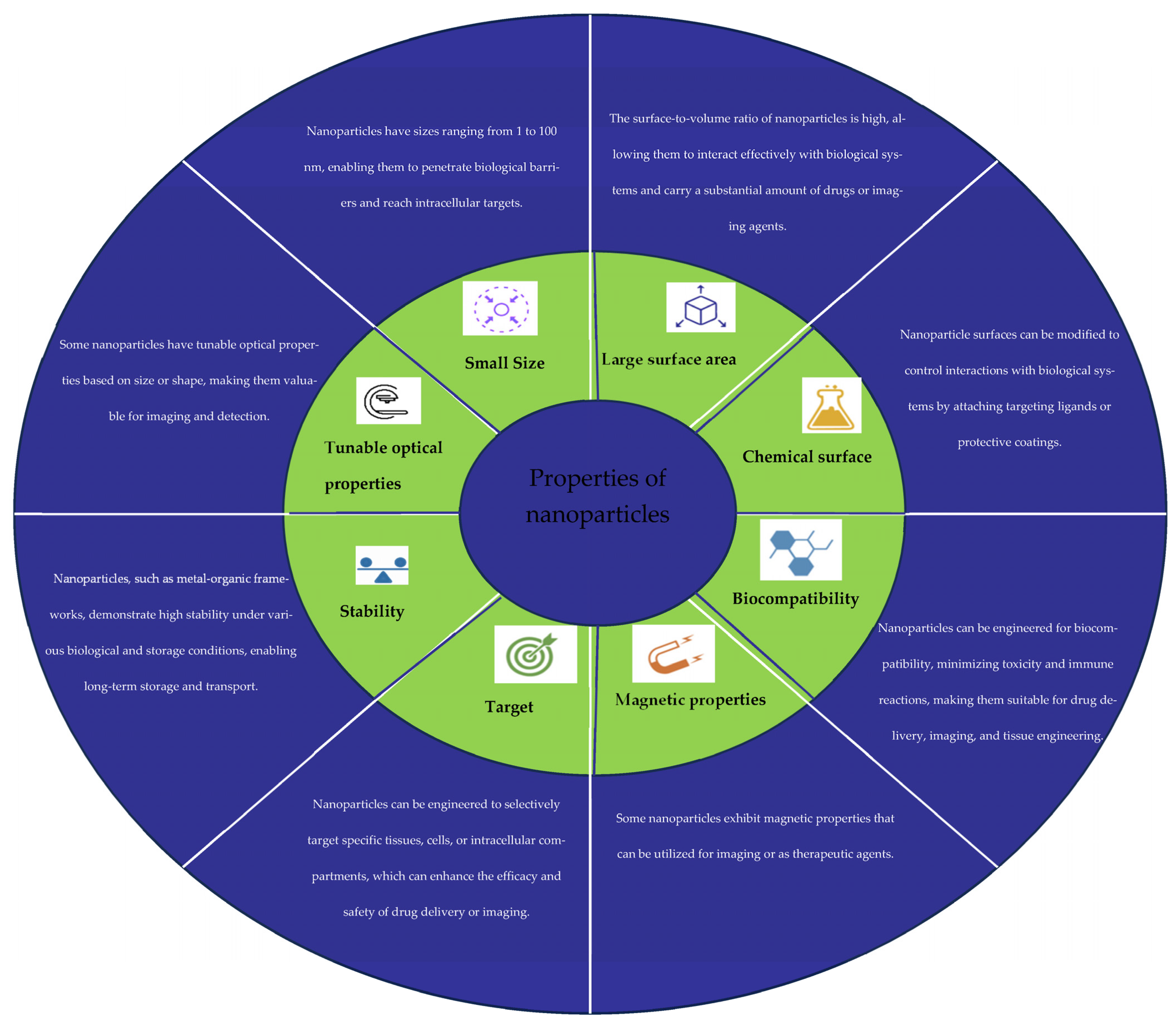
The properties of NPs are summarized in Figure 4.

3.1. Morphological Properties

3.1.1. Size

The properties of NPs are often influenced by their morphological characteristics, which have garnered significant interest. Various techniques are available to characterize the size and morphology of NPs, but transmission electron microscopy (TEM) and scanning electron microscopy (SEM) are the most commonly used methods [66].
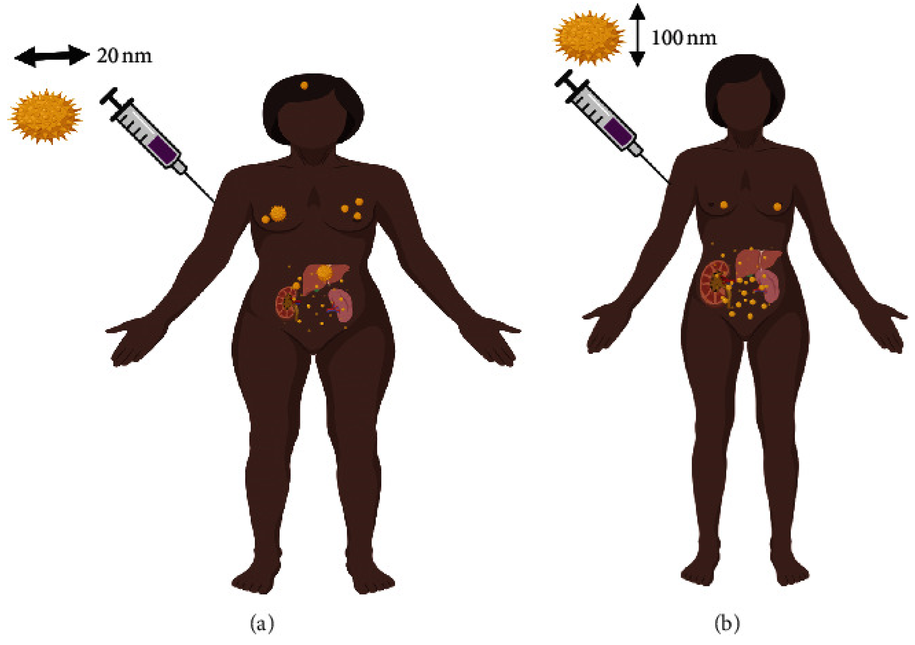
Several studies have evaluated the biodistribution of NPs of varying sizes in the human body following exposure [66,67]. For instance, Sonavane et al. [66] intravenously injected NPs ranging from 15 to 200 nm and observed that the accumulation of NPs in tissues such as the liver, lungs, spleen, and kidneys depended on their size. The smaller NPs were able to cross the blood–brain barrier [66,67].
It has also been demonstrated that the size of NPs affects their clearance from circulation. Particles with diameters smaller than 5–6 nm are rapidly cleared by the kidneys, while larger particles (over 200 nm) are more efficiently removed by the liver and spleen [68]. Larger particles, particularly those exceeding 100 nm, are cleared by the mononuclear phagocyte system (MPS), which is mainly facilitated by hepatic, splenic, and bone marrow cells [69,70].
The cytotoxicity of NPs is also closely linked to their size, with smaller NPs generally being more toxic. This was demonstrated by Gao et al. [71] who compared the cellular response to exposure to NPs with diameters of 8 nm and 37 nm. The results indicated that the toxicity was significantly higher after cellular exposure to the 8 nm NPs. The biodistribution of NPs can be clearly seen in Figure 5.

3.1.2. Shapes of Nanoparticles

The size of NPs is the primary determinant of their absorption by macrophages; however, their shape also significantly contributes to regulating this absorption. NPs can have different forms:
  • One-dimensional (1D) NPs include nanotubes, nanowires, and nanofilaments.
  • Two-dimensional (2D) NPs are found as sheets or disks.
  • Three-dimensional (3D) NPs can take non-spherical shapes [73].

3.1.3. Interaction with Macrophages

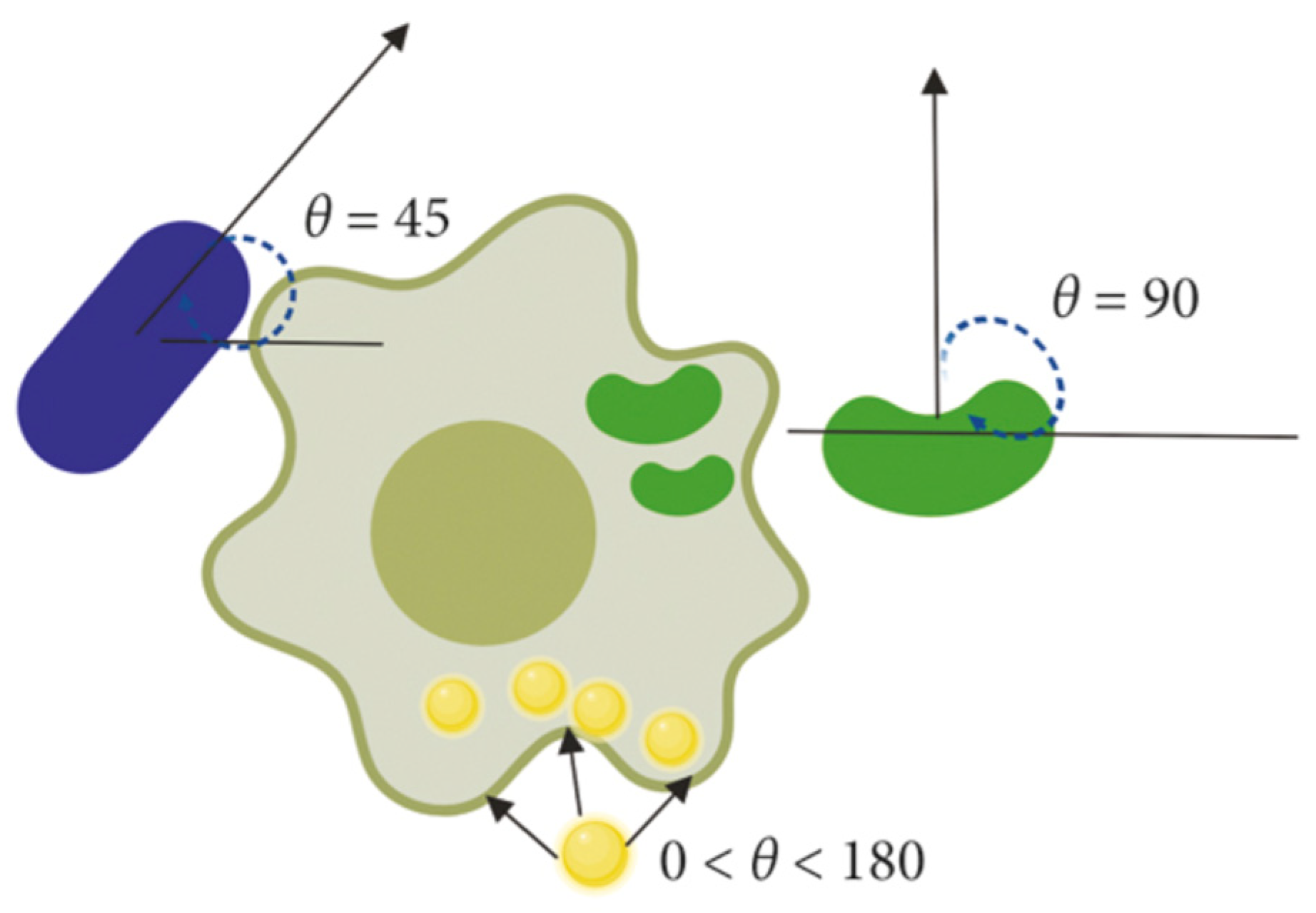
When these non-spherical NPs interact with macrophages, the initial contact angle (CA) plays a crucial role in determining the internalization rate [73,74]. NPs that align with their major axis parallel to the cell membrane are internalized more slowly than those aligned with their minor axis. Furthermore, filamentous NPs are internalized more rapidly when they are perpendicular to the cellular axis (θ = 90°). The internalization rate decreases when NPs are tangential to the macrophage membrane. For spherical NPs, the internalization rate is independent of the angle (θ) due to their symmetrical nature [72,73,74,75] (Figure 6).

3.1.4. Structural Characterization of Nanoparticles

The structural characterization of NPs is crucial before any compositional study, as it provides essential information about the overall properties of the material being studied. Various methods exist for evaluating the shape of NPs, including the following:
  • X-ray diffraction (XRD);
  • Energy dispersive X-ray spectroscopy (EDX);
  • X-ray photoelectron spectroscopy (XPS);
  • Infrared spectroscopy (IR);
  • Raman spectroscopy;
  • Brunauer–Emmett–Teller (BET) surface analysis;
  • Zetasizer analysis;
  • TEM;
  • SEM.
Certain types of NPs can induce toxicity, which, in some cases, is related to the shape of the particles. In this regard, Auclair and Gagné demonstrated that silver NPs (AgNPs) exhibit cytotoxicity depending on their morphology. Their study evaluated the toxicity of three distinct shapes (spherical, cubic, and prismatic) of AgNPs on Hydra vulgaris. To focus on morphology-related effects, the NPs were kept within the same size range and had identical surface coatings. Shape-dependent toxicity was observed, with spherical AgNPs being the most toxic, followed by prismatic AgNPs, while no significant toxicity was detected with cubic AgNPs [76].
Similarly, Champion and Mitragotri compared various shapes of polymeric NPs and concluded that elongated particles with a higher length-to-width ratio are less likely to be cleared by the immune system. Furthermore, elongated particles exhibited longer blood circulation times and avoided phagocytosis, depending on the CA when interacting with macrophages [77,78].

3.1.5. Specific Surface Area

Due to their minute size, NPs possess a large surface area, a characteristic that offers a wide range of applications. Consequently, measuring the surface area of nanomaterials would enhance the understanding of their properties, behaviors, and potential hazards. The BET method is considered the paramount technique for determining the surface area of particulate materials. This technique relies on the principles of adsorption and desorption, as well as the BET theorem.
It has been reported that, at equal unit mass, smaller NPs allow more particles and a larger surface area to participate in biological actions compared to larger particles, which could be the source of their more significant toxic effects. Lu et al. [79] injected silica particles of 30, 70, and 300 nm into mice intravenously at different doses. The results showed that when the surfaces of the injected particles were similar, despite the differing number of particles, the extent of hepatic lesions was also similar. This indicates that while the number of particles may exert some influence on in vivo toxicity, surface area could be a more critical factor for toxicity in both nanometric and micrometric particles. Supporting conclusions were drawn from the study by Nemmar et al. [80], which revealed that vascular homeostasis alteration was more pronounced in the group treated with 50 nm NPs than in the group treated with 500 nm particles after intraperitoneal administration. The authors attributed this to the high surface-to-volume ratio, which decreases inversely with size and favors biological interactions, leading to superior vascular and systemic toxicity [81,82,83,84].

3.2. Chemical Properties

3.2.1. Charge

The surface charge is often measured as the zeta potential (ξ), which is quantified using a Zetasizer. Several studies indicate that surface charge determines the fate of NPs. Surface charge plays a crucial role in the stability of particles and directly influences their level of toxicity. In fact, positively charged NPs exhibit a higher affinity for cell membranes compared to those that are negatively charged or neutral [85,86]. It appears that the surface characteristics of particles also play an important role regarding toxicity, as they determine the initial direct contact with biological materials and cell surfaces, along with their components.
It was demonstrated that gold NPs with a positive charge induce cell death through apoptosis, while neutral gold NPs cause necrosis in human adult low calcium temperature (HaCaT) cell lines [87]. It is important to note that surface charge influences the composition of biological proteins on the surface of NPs, thereby conditioning all subsequent chemical and biochemical interactions [88].

3.2.2. (Bio)Chemical Surface

The chemical or biochemical surface of NPs is formed upon their interaction with biological materials. As a result, NPs become coated with a diverse array of proteins, collectively known as the “protein corona”. This corona can significantly modify the properties of NPs by obscuring their inherent surface characteristics [89,90]. Furthermore, it has been established that the duration of exposure in the bloodstream is a critical factor that influences the biomolecular composition of NPs. The novel properties imparted to NPs by the corona also become a primary determinant of their nanotoxicity and/or therapeutic efficacy within the body [91]. To date, considerable research has focused on elucidating the composition and biological implications of the protein corona [92]. A comprehensive understanding of NP–protein interactions is vital for the advancement of targeted delivery systems for nanomaterials in healthcare [93].
Extensive studies have been conducted to investigate the formation of protein coronas on various forms of NPs [94,95]. These investigations have revealed that multiple factors can significantly affect the thickness and composition of protein coronas. These factors include the physicochemical properties of NPs, such as surface chemistry, charge, size, shape, solubility, protein binding affinities, and exposure duration [89,96,97].

3.2.3. Biocompatibility

The concept of biocompatibility is based on the appropriate interaction between a material and its biological environment, characterized by the absence of toxic or immune responses from the treated biomaterial (cell, tissue, or organism) [98,99]. Biocompatibility is often described as the ability of a specific material or device to be compatible with living tissue or organisms. It is achieved when the interaction between the nanomaterial and the host does not lead to undesirable outcomes, such as oxidative stress, Deoxyribonucleic Acid (DNA) damage, mutagenesis, or apoptosis [100,101]. Cytotoxicity is generally linked to adverse effects on a specific cell line and is typically assessed first through specific in vitro tests, followed by in vivo evaluations. In practice, cytotoxicity and biocompatibility are influenced by several factors, including the inherent physicochemical properties of the NPs and their delivery methods within the body [102].
There are surface modification methods available for NPs aimed at optimizing their biocompatibility. The surfaces of most NPs can be functionalized with polymer linkers, hybridized DNA, proteins, cell membranes, or inorganic chemicals such as metals and ceramic coatings [103]. However, the role of surface coatings remains unclear to date [104]. Improving the specific interactions of different coatings and their delivery mechanisms is still a work in progress [72,105].

3.2.4. Stability

The term “stability of NPs” refers to the preservation of specific properties of a nanostructure, such as size, surface chemistry, aggregation, and shape. This stability is maintained only for a limited period, as all nanostructures are intrinsically thermodynamically and energetically unfavorable compared to larger structures with the same chemical composition [106].
The nature and concentration of reducing and stabilizing agents significantly influence the size distribution and shape of NPs during their synthesis, largely determining their functional properties. Widoniak et al. [107] described the preparation of colloidal silver solutions with average sizes ranging from 1 nm to 6 μm and various shapes (spheres, plates, needles, or sheets) through the reduction of silver ions using different reducing agents and various polymeric stabilizing agents. The control of nanoparticle shape and size distribution can also be achieved by carefully adjusting other experimental conditions, such as temperature, stirring speed, agitation, and reaction time [108]. Some studies have indicated that the stability of NPs could be preserved long-term when stored under standard conditions: at room temperature, protected from light, and shielded from humidity [109,110].
Surface modification of NPs can also effectively maintain their stability [111,112]. Encapsulation provides complex protection for the payload core and allows for modulation of cytotoxicity, as demonstrated by the work of Lv et al. [113]. The encapsulation of therapeutic gold NPs in hydrogel nanospheres led to increased stability and enhanced cellular uptake efficiency while significantly reducing oxidative stress levels in mesenchymal stem cells.

3.3. Magnetic Properties

3.3.1. Targeting

The majority of NPs exhibit magnetic properties, providing significant advantages. These properties allow for selective attachment to functional molecules, impart magnetic characteristics to the target, and facilitate manipulation and transportation to a desired location through the control of a magnetic field produced by an electromagnet or permanent magnet. Magnetic nanoparticle carriers consist of three functional components: a magnetic core, a surface coating, and a functionalized outer layer [114].
Targeting capability is a crucial aspect of various nanoparticle applications. Specifically, metallic NPs can direct therapeutic agents to specific sites within the body, thereby enhancing the efficacy of medical treatments [115].

3.3.2. Optical Properties

The optical properties of metallic NPs are largely influenced by the collective excitation of conduction electrons when interacting with electromagnetic radiation. This behavior is particularly evident in NPs made of gold, silver, and copper, owing to their conduction electron availability. When the electric field from the incident radiation interacts with these particles, it generates an electric dipole, leading to a force that counteracts this effect at a specific resonance frequency. Notably, the optical characteristics are influenced by the size, shape, and composition of the NPs [116,117,118].

4. Categories of Nanoparticles in Dental Applications

4.1. Antimicrobial Nanoparticles

4.1.1. Types (e.g., Silver, Zinc Oxide)

Antimicrobial NPs are increasingly utilized in dentistry to combat microbial infections. The most prominent types include the following:
  • AgNPs: These are widely recognized for their broad-spectrum antimicrobial activity. They disrupt bacterial cell walls, interfere with metabolic processes, and can generate reactive oxygen species (ROS) that are harmful to microbial cells [119,120].
  • ZnO NPs: ZnO NPs exhibit antibacterial properties by producing ROS and direct interaction with bacterial membranes, leading to cell death [121,122]. They also promote wound healing and have biocompatibility, making them suitable for dental applications.

4.1.2. Mechanisms of Action and Applications in Preventing Infections

The mechanisms of action for antimicrobial NPs typically involve the following:
  • Membrane disruption: antimicrobial NPs can bind to the microbial cell membrane, leading to structural damage and eventual cell lysis.
  • ROS: NPs like silver and ZnO can induce oxidative stress within microbial cells, resulting in DNA damage and apoptosis [123,124].
In dental applications, antimicrobial NPs are employed in various products:
  • Dental cements: incorporation of AgNPs and ZnO NPs enhances the antimicrobial properties of dental cements, reducing the risk of secondary infections.
  • Composite resins: these NPs are added to restorative materials to prevent biofilm formation on dental surfaces [125,126].

4.2. Therapeutic Nanoparticles

Drug Delivery Systems and Their Role in Pain Management and Healing

Therapeutic NPs play a crucial role in the targeted delivery of drugs, improving the effectiveness and reducing the side effects of treatments. Key features include the following:
  • Enhanced bioavailability: NPs improve the solubility and stability of therapeutic agents, allowing for lower doses and minimizing systemic toxicity [127].
  • Controlled release: NPs can be engineered to release drugs at a controlled rate, providing sustained therapeutic effects over time [128].
  • Pain management: for dental procedures, therapeutic NPs can deliver analgesics directly to the site of pain, enhancing pain relief while minimizing the need for systemic medication [129].

4.3. Material Property-Improving Nanoparticles

Enhancements in Mechanical Strength, Wear Resistance, and Aesthetics of Dental Materials
NPs are incorporated into dental materials to improve their physical properties:
  • Mechanical strength: NPs such as silica and alumina can significantly enhance the mechanical strength of composite resins and dental cements, making them more durable under occlusal forces [130].
  • Wear resistance: the addition of ceramic NPs improves the wear resistance of restorative materials, prolonging their lifespan and maintaining their functionality [131].
  • Aesthetics: NPs can improve the optical properties of dental materials, providing better color matching and translucency, which is critical for aesthetic restorations [132,133].

5. Current Applications and Innovations

5.1. Dental Products Incorporating Nanoparticles

5.1.1. Dental Composites

Modern dental composites integrate NPs like nanosilica to improve filler content, which enhances wear resistance, reduces polymerization shrinkage, and increases the composite’s aesthetic quality and mechanical durability. These modifications result in more durable restorations, with reduced wear and cracking over time [134]. The incorporation of NPs also optimizes translucency, allowing composites to better mimic the appearance of natural teeth and improving their longevity and color stability [134,135]. In recent years, the integration of NPs into dental materials has significantly enhanced their mechanical properties. For instance, the incorporation of graphene platelets into epoxy nanocomposites has led to an increase in Young’s modulus and an improvement in tensile strength compared to pristine epoxy. Similarly, the addition of nanosilica particles to cementitious composites reinforced with polyvinyl alcohol fibers has resulted in a 38% increase in tensile strength. These findings underscore the substantial impact of NPs on enhancing the mechanical performance of dental materials [136,137].

5.1.2. Antimicrobial Agents

AgNPs are commonly incorporated into dental materials like sealants, adhesives, and cements due to their antimicrobial capabilities, which help prevent biofilm formation, reduce bacterial colonization, and mitigate the risk of secondary infections [135,138]. These materials release Ag ions that interact with bacterial membranes, leading to cellular damage and limiting microbial growth [139]. ZnO NPs, another common antimicrobial nanoparticle, exhibit ROS generation, directly inhibiting bacterial activity and offering biocompatibility suitable for dental applications [140,141].

5.1.3. Endodontic Materials

In endodontics, NPs such as ZnO NPs and AgNPs are used in root canal sealers to enhance antibacterial effects, reduce leakage, and improve sealing properties essential for successful long-term outcomes. ZnO NPs, in particular, are known to target endodontic pathogens like Enterococcus faecalis, offering improved healing post procedure. The addition of ZnO NPs into root canal sealers has been shown to enhance antibacterial activity while maintaining biocompatibility with surrounding tissues, which helps in the prevention of reinfection and improves the overall success rate of endodontic treatments [142,143].

5.1.4. Dental Implants

Nanotechnology has significantly advanced dental implant surfaces, with nanocoatings applied to titanium and other metals to promote osseointegration and reduce infection risks. Nanostructured surfaces on implants enhance cell attachment, bone growth, and implant stability, thereby improving overall success rates and reducing healing times [144]. Titanium dioxide (TiO2) NPs have shown promising results in creating antibacterial surfaces, reducing biofilm formation, and enhancing biocompatibility [145,146]. These modifications contribute to a faster healing process and lower rejection rates. The introduction of nanostructures to implant surfaces has been found to increase surface roughness at the nanoscale level, facilitating improved bone formation and promoting direct bone–implant contact, which is crucial for implant stability [2,3,4].

5.1.5. Amalgam

While dental amalgam is traditionally a blend of mercury with silver, tin, copper, and zinc, recent research has explored the addition of NPs like silver–copper and TiO2 to improve the amalgam’s properties. These enhancements have demonstrated improved antimicrobial activity, reduced cytotoxicity, and increased strength, making amalgam restorations safer and more effective. For example, the incorporation of TiO2 NPs into dental amalgams has demonstrated the potential for reducing mercury content, improving the material’s resistance to corrosion, and enhancing its overall durability [147,148].

5.1.6. Glass Ionomer Cements (GICs)

NPs such as chitosan, hydroxyapatite, TiO2, silicone dioxide (SiO2), and zirconia (ZrO2) are added to GICs to enhance aesthetics, bond strength, and antimicrobial properties. In resin-modified GICs, fluoroaluminosilicate NPs are widely used, and silica nanofillers improve wear resistance and curing efficiency. Chitosan NPs increase fluoride release and material strength, while TiO2 NPs inhibit biofilm formation and enhance physical properties, contributing to better clinical outcomes [149]. The addition of these NPs has shown to improve the overall longevity and functional performance of GICs, making them more versatile in clinical dentistry [2,3,4].

5.1.7. Dental Prosthetics

NPs are incorporated into dental prosthetics to enhance wear resistance, aesthetic qualities, and surface smoothness, which reduces bacterial adhesion. Nanosized fillers improve the flexural strength and elasticity of prosthetic materials, which is critical for enduring the stresses of mastication. ZrO2 NPs, in particular, improve toughness and translucency, making them favorable for prosthetic frameworks and crowns [150].
In addition to their known roles, NPs are being explored for other innovative uses in dentistry. For example, NPs have been integrated into repair resins to enhance the mechanical properties of denture repairs, offering improved durability and resistance to fractures [151].

5.1.8. Periodontal Applications

NPs are used in periodontal treatments to facilitate drug delivery for targeted antibacterial action and tissue regeneration. Hydroxyapatite NPs, for example, support bone regrowth and are compatible with periodontal ligament cells, promoting the repair of periodontal defects. Additionally, silver and ZnO NPs serve as local antimicrobials, contributing to infection control and improving healing rates in periodontal surgeries [152]. Research is also exploring the potential of nano-hydroxyapatite in repairing damaged bone tissues and preventing periodontal disease progression [2,3,4].

5.1.9. Whitening Agents

NPs in whitening products are utilized for their effectiveness and reduced damage to enamel. Carbamide peroxide polymeric NPs enhance stability and efficacy in whitening agents, while nano-hydroxyapatite helps with remineralization and provides lasting whiteness in oral hygiene products. Nano-encapsulated sodium metabisulfite allows for safe, gradual whitening through liposomal enclosures, which reduces enamel erosion and sensitivity [153,154]. This application of NPs has been shown to significantly improve whitening effects while ensuring that enamel integrity is preserved during the treatment process.

5.1.10. Enamel Repair and Remineralization

NPs have shown promising potential in enamel repair and remineralization, offering innovative solutions to prevent and reverse early carious lesions. Among these, bioactive glass NPs have gained attention for their ability to release ions, such as calcium and phosphate, which are essential for remineralizing enamel surfaces. A recent study by Raszewski et al. [155] demonstrated the efficacy of bioactive glass-infused gels in restoring enamel mineral content in vitro, highlighting their potential as a non-invasive treatment option for enamel demineralization. These NPs not only promote the regeneration of lost mineral structure but also enhance the surface properties of enamel, providing added protection against future demineralization. Such advancements position bioactive glass NPs as a pivotal component of therapeutic strategies aimed at strengthening dental tissues and improving overall oral health.

5.2. Highlight Recent Advancements in Nanoparticle Technology

5.2.1. Targeted Drug Delivery

Advances in targeted drug delivery systems using NPs allow for the localized release of therapeutics in dental applications, enhancing treatment efficacy while minimizing systemic side effects [156].

5.2.2. Smart Biomaterials

The development of smart biomaterials that respond to environmental stimuli (e.g., pH, temperature) is a significant innovation. These materials can release drugs in a controlled manner when triggered by specific conditions in the oral environment [157].

5.2.3. Enhanced Imaging Techniques

NPs, particularly quantum dots and gold NPs, are being utilized to improve imaging techniques such as fluorescence imaging, enhancing the visualization of dental tissues and the detection of oral diseases [158].

6. Future Directions and Challenges

6.1. Potential Future Applications of Nanoparticles in Dentistry

6.1.1. Regenerative Dentistry

NPs may play a crucial role in regenerative approaches, such as stem cell therapy and tissue engineering, to promote the regeneration of dental tissues and structures [159].

6.1.2. Personalized Dental Care

The integration of NPs in diagnostic tools could lead to personalized treatment plans based on the specific needs of patients, enhancing the effectiveness of dental care [160].

6.1.3. Nanoparticle-Based Vaccines

Research into nanoparticle-based vaccines targeting oral diseases (e.g., periodontal disease, dental caries) is promising and could revolutionize preventive dental care [161].
The incorporation of NPs in dental applications presents a wealth of opportunities to enhance therapeutic effectiveness, improve material properties, and develop innovative solutions for oral health. However, ongoing research is essential to address potential challenges, including biocompatibility, safety, and regulatory hurdles [2,3,4].

6.2. Consideration of Risks and Regulatory Challenges in Their Use

The cytotoxic effects of nanomaterials are primarily observed when they exist in free form—meaning they are not integrated into the material’s structure—or when they are released into the surrounding tissue. In dentistry, this cytotoxicity can be strategically utilized against bacterial and fungal pathogens that are resistant to traditional antibiotic treatments. By modifying dental sealers with NPs to impart antimicrobial properties, their effectiveness can be significantly enhanced through molecular interactions [2,3,4].
In a study evaluating three different commercial endodontic sealants modified with nanostructured silver vanadate (AgVO3), cytotoxicity assessments revealed that two sealants, Sealer 26 and Endometasone N, exhibited cytotoxic effects both in their pure forms and when combined with NPs. Conversely, the AH Plus sealant demonstrated cytotoxicity only when used in conjunction with the nanomaterial, impacting the viability of human gingival fibroblasts (HGF) as the sole tested material [162]. The mechanisms of cytotoxicity associated with AgNPs on HGF have been further explored, illustrating the dual nature of their therapeutic and toxic potentials [107,163].
While NPs offer a plethora of therapeutic applications, they also pose risks related to cytotoxicity and potential immune responses. For example, magnetic NPs (MNPs) can enhance the translucency and abrasion resistance of dental composites without introducing significant risk. However, using MNPs in free form, especially in dental adhesives aimed at providing anti-biofilm properties or in drug delivery systems for caries and periodontal disease management, raises concerns about triggering undesirable immune responses [2,164].
Striking a balance between the benefits and risks associated with nanoparticle use presents a challenge for clinicians. Despite their cytotoxic potential, MNPs often exhibit cellular activity that is targeted, which helps to mitigate systemic toxicity. Consequently, in recent years, the application of NPs and magnetic forces in dentistry has gained traction for delivering drugs aimed at the prevention and treatment of dental diseases [165,166].
It is essential for future research to focus on overcoming these challenges by developing standardized testing protocols to evaluate the long-term performance and safety of nanoparticle-based materials in clinical settings. Additionally, innovations in manufacturing technologies will be crucial for producing NPs at scale while maintaining their unique properties [167]. Future studies should also explore the synergistic effects of combining different types of NPs (e.g., antimicrobial and material-enhancing) in a single product. Finally, the development of regulatory frameworks to ensure the safe incorporation of NPs in dental products will be vital for their successful commercialization. As these advancements occur, the research anticipates that NPs will play an increasingly significant role in revolutionizing dental care and treatment.

7. Conclusions

In conclusion, NPs represent a promising avenue for innovation in dental materials and therapeutics. Their unique properties enable the development of enhanced antimicrobial agents, effective drug delivery systems, and improved material performance. However, the risks associated with cytotoxicity and stability must be carefully managed to ensure safe and effective use. Ongoing research is essential to fully understand and optimize the applications of NPs in dentistry, balancing their benefits against potential health risks. As the field advances, the integration of NPs into clinical practice will likely revolutionize approaches to dental care and treatment.

Author Contributions

Conceptualization, R.B. and Y.H.; methodology, R.B.; software, Z.D., C.L. and N.K.; validation, Y.H., C.E.C.-S. and R.B.; formal analysis, Y.H., Z.D., R.B. and C.E.C.-S.; investigation, R.B., Y.H., C.L. and T.B.A.; resources, R.B.; data curation, R.B.; writing—original draft preparation, R.B.; writing—review and editing, Y.H., C.E.C.-S., N.K. and T.B.A.; visualization, T.B.A., R.B. and Z.D.; supervision, Y.H.; project administration, Y.H.; funding acquisition, R.B. and Y.H. All authors have read and agreed to the published version of the manuscript.

Funding

This research received no external funding.

Institutional Review Board Statement

Not applicable.

Informed Consent Statement

Not applicable.

Data Availability Statement

Data sharing is not applicable. No new data were created or analyzed in this study.

Conflicts of Interest

The authors declare no conflicts of interest.

References

  1. Yakop, F.; Abd Ghafar, S.A.; Yong, Y.K.; Saiful Yazan, L.; Mohamad Hanafiah, R.; Lim, V.; Eshak, Z. Silver nanoparticles Clinacanthus Nutans leaves extract induced apoptosis towards oral squamous cell carcinoma cell lines. Artif. Cells Nanomed. Biotechnol. 2018, 46, 131–139. [Google Scholar] [CrossRef]
  2. Gronwald, B.; Kozłowska, L.; Kijak, K.; Lietz-Kijak, D.; Skomro, P.; Gronwald, K.; Gronwald, H. Nanoparticles in Dentistry—Current Literature Review. Coatings 2023, 13, 102. [Google Scholar] [CrossRef]
  3. Moraes, G.; Zambom, C.; Siqueira, W.L. Nanoparticles in Dentistry: A Comprehensive Review. Pharmaceuticals 2021, 14, 752. [Google Scholar] [CrossRef] [PubMed]
  4. Bapat, R.A.; Joshi, C.P.; Bapat, P.; Chaubal, T.V.; Pandurangappa, R.; Jnanendrappa, N.; Gorain, B.; Khurana, S.; Kesharwani, P. The Use of Nanoparticles as Biomaterials in Dentistry. Drug Discov. Today 2019, 24, 85–98. [Google Scholar] [CrossRef] [PubMed]
  5. Pecci-Lloret, M.P.; Gea-Alcocer, S.; Murcia-Flores, L.; Rodríguez-Lozano, F.J.; Oñate-Sánchez, R.E. Use of Nanoparticles in Regenerative Dentistry: A Systematic Review. Biomimetics 2024, 9, 243. [Google Scholar] [CrossRef] [PubMed]
  6. Auffan, M.; Rose, J.; Bottero, J.-Y.; Lowry, G.V.; Jolivet, J.-P.; Wiesner, M.R. Towards a Definition of Inorganic Nanoparticles from an Environmental, Health and Safety Perspective. Nat. Nanotechnol. 2009, 4, 634–641. [Google Scholar] [CrossRef] [PubMed]
  7. McNamara, K.; Tofail, S.A. Nanoparticles in Biomedical Applications. Adv. Phys. X 2017, 2, 54–88. [Google Scholar] [CrossRef]
  8. Ghadiri, M.; Stokes, R. Nanotechnology in Dentistry: A Comprehensive Review. Materials 2020, 13, 2700. [Google Scholar] [CrossRef]
  9. An, K.; Somorjai, G.A. Size and Shape Control of Metal Nanoparticles for Reaction Selectivity in Catalysis. ChemCatChem 2012, 4, 1512–1524. [Google Scholar] [CrossRef]
  10. Arms, L.; Smith, D.W.; Flynn, J.; Palmer, W.; Martin, A.; Woldu, A.; Hua, S. Advantages and Limitations of Current Techniques for Analyzing the Biodistribution of Nanoparticles. Front. Pharmacol. 2018, 9, 802. [Google Scholar] [CrossRef]
  11. ElSheikh, S.K.; Eid, E.G.; Abdelghany, A.M.; Abdelaziz, D. Physical/Mechanical and Antibacterial Properties of Composite Resin Modified with Selenium Nanoparticles. BMC Oral Health 2024, 24, 1245. [Google Scholar] [CrossRef] [PubMed]
  12. Mercan, D.A.; Niculescu, A.G.; Grumezescu, A.M. Nanoparticles for Antimicrobial Agents Delivery—An Up-to-Date Review. Int. J. Mol. Sci. 2022, 23, 13862. [Google Scholar] [CrossRef] [PubMed]
  13. Capuano, N.; Amato, A.; Dell’Annunziata, F.; Giordano, F.; Folliero, V.; Di Spirito, F.; More, P.R.; De Filippis, A.; Martina, S.; Amato, M.; et al. Nanoparticles and Their Antibacterial Application in Endodontics. Antibiotics 2023, 12, 1690. [Google Scholar] [CrossRef] [PubMed]
  14. Bossù, M.; Saccucci, M.; Salucci, A.; Di Giorgio, G.; Bruni, E.; Uccelletti, D.; Sarto, M.S.; Familiari, G.; Relucenti, M.; Polimeni, A. Enamel Remineralization and Repair Results of Biomimetic Hydroxyapatite Toothpaste on Deciduous Teeth: An Effective Option to Fluoride Toothpaste. J. Nanobiotechnol. 2019, 17, 17. [Google Scholar] [CrossRef] [PubMed]
  15. Yıldız, C.; Kılıç, E.; Kurt, K.; Özdemir, H.; Korkmaz, A. Nanoparticles for Dental Implant Applications: Enhancing Osseointegration. Mater. Today Proc. 2021, 46, 3469–3472. [Google Scholar] [CrossRef]
  16. Gutiérrez, M.F.; Alegría-Acevedo, L.F.; Méndez-Bauer, L.; Bermudez, J.; Dávila-Sánchez, A.; Buvinic, S.; Hernández-Moya, N.; Reis, A.; Loguercio, A.D.; Farago, P.V.; et al. Biological, Mechanical, and Adhesive Properties of Universal Adhesives Containing Zinc and Copper Nanoparticles. J. Dent. 2019, 82, 45–55. [Google Scholar] [CrossRef]
  17. Ekrikaya, S.; Yilmaz, E.; Arslan, S.; Karaaslan, R.; Ildiz, N.; Celik, C.; Ocsoy, I. Dentin Bond Strength and Antimicrobial Activities of Universal Adhesives Containing Silver Nanoparticles Synthesized with Rosa canina Extract. Clin. Oral Investig. 2023, 27, 6891–6902. [Google Scholar] [CrossRef] [PubMed]
  18. Sahu, A.; Pramanik, K.; Mohapatra, A.; Dandapat, S.; Sinha, A.; Patra, S. Nanoparticle-based targeted drug delivery systems: Applications in cancer therapy. Nanomaterials 2020, 10, 883. [Google Scholar]
  19. Chen, Z.; Zhang, H.; Yang, X.; Zhang, Y.; Zhu, G.; Yang, J.; Zhang, M. Multifunctional nanoparticles for cancer diagnosis and therapy. Front. Chem. 2020, 8, 212. [Google Scholar] [CrossRef]
  20. Lu, H.; Li, J.; Huang, X.; Yan, H.; Gao, H.; Liu, Y.; Wu, Y.; Chen, Z.; Wang, Y. The application of nanoparticles in oral drug delivery systems. Int. J. Nanomed. 2021, 16, 1233–1248. [Google Scholar]
  21. Sadeghi, A.; Dastjerdi, R.; Asgarian, A.; Shafiei, M.; Moudi, M.; Shahmoradi, K. Nanotechnology in dentistry: A review of the current literature. Int. J. Dent. 2020, 2020, 4568019. [Google Scholar]
  22. Ramesh, R.; Babu, R.S.; Manickam, P.; Shyamaladevi, R.; Raja, V.S. Recent advancements in nanoparticle-based drug delivery systems for cancer therapy: A review. Biotechnol. Rep. 2020, 25, e00424. [Google Scholar]
  23. Christian, P.; Von der Kammer, F.; Baalousha, M.; Hofmann, T. Nanoparticles: Structure, Properties, Preparation and Behaviour in Environmental Media. Ecotoxicology 2008, 17, 326–343. [Google Scholar] [CrossRef] [PubMed]
  24. Kreuter, J. Nanoparticles—A Historical Perspective. Int. J. Pharm. 2007, 331, 1–10. [Google Scholar] [CrossRef] [PubMed]
  25. Iavicoli, I.; Leso, V.; Fontana, L. Esposizione a Nanoparticelle nei Laboratori di Ricerca [Nanoparticle Exposure in Research Laboratories]. G. Ital. Med. Lav. Ergon. 2019, 41, 349–353. [Google Scholar]
  26. Rahman, A.; Ghosh, M. Nanoparticles and Their Applications in Dental Materials: An Overview. Nanomaterials 2021, 11, 674. [Google Scholar] [CrossRef]
  27. Niu, L.N.; Zhang, Y.; Tsoi, J.K.H.; Matinlinna, J.P. Nanotechnology in Dental Applications. J. Dent. Res. 2019, 98, 337–347. [Google Scholar]
  28. Sharma, V.K.; Filip, J.; Zboril, R.; Varma, R.S. Natural Inorganic Nanoparticles—Formation, Fate, and Toxicity in the Environment. Chem. Soc. Rev. 2015, 44, 8410–8423. [Google Scholar] [CrossRef]
  29. Ghosh, A.; Banerjee, S.; Paul, P. A Review on the Use of Nanoparticles for Dental Tissue Regeneration. J. Biomater. Sci. Polym. Ed. 2020, 31, 797–816. [Google Scholar]
  30. Zhang, J.; Wang, Y.; Zhao, Y.; Chen, H. The Role of Nanoparticles in Restorative Dentistry. Front. Mater. 2021, 8, 583934. [Google Scholar]
  31. Xu, X.; Liu, Y.; Li, J.; Wang, Y. Recent Advances in Protein-Repellent Adhesives Using Nanotechnology. J. Adhes. Sci. Technol. 2021, 35, 490–507. [Google Scholar]
  32. Hasan, S. A Review on Nanoparticles: Their Synthesis and Types. Res. J. Recent Sci. 2015, 2277, 2502. [Google Scholar]
  33. Tuncer, M.; Büyükyılmaz, T.; Tütüncü, M.; Korkmaz, Y. Nanoparticles in Endodontics: Current Trends and Future Directions. J. Endod. 2021, 47, 1204–1216. [Google Scholar] [CrossRef]
  34. Daraee, H.; Eatemadi, A.; Abbasi, E.; Fekri Aval, S.; Kouhi, M.; Akbarzadeh, A. Application of Gold Nanoparticles in Biomedical and Drug Delivery. Artif. Cells Nanomed. Biotechnol. 2016, 44, 410–422. [Google Scholar] [CrossRef] [PubMed]
  35. Zhang, Y.; Wang, Y.; Wang, Y.; Yang, X. Nanotechnology for Controlled Drug Delivery in Dental Treatments. Dent. Mater. J. 2019, 38, 80–86. [Google Scholar] [CrossRef]
  36. Naguib, G.; Maghrabi, A.A.; Mira, A.I.; Mously, H.A.; Hajjaj, M.; Hamed, M.T. Influence of Inorganic Nanoparticles on Dental Materials’ Mechanical Properties: A Narrative Review. BMC Oral Health 2023, 23, 897. [Google Scholar] [CrossRef] [PubMed]
  37. Kim, D.; Shin, K.; Kwon, S.G.; Hyeon, T. Synthesis and Biomedical Applications of Multifunctional Nanoparticles. Adv. Mater. 2018, 30, e1802309. [Google Scholar] [CrossRef] [PubMed]
  38. Gao, W.; Zhang, Y.; Zhang, Q.; Zhang, L. Nanoparticle-Hydrogel: A Hybrid Biomaterial System for Localized Drug Delivery. Ann. Biomed. Eng. 2016, 44, 2049–2061. [Google Scholar] [CrossRef]
  39. European Commission. Commission Recommendation (EU) 2022/1089 of 14 June 2022 on Defining Nanomaterials. Off. J. Eur. Union 2022, L176, 1–4. Available online: https://eur-lex.europa.eu/legal-content/EN/TXT/?uri=CELEX%3A32022H0614%2801%29 (accessed on 23 December 2024).
  40. Size-Comparison-Bio-Nanoparticles. Size Comparison of Bio-Nanoparticles: Nanometer Scale Comparison and Nanoparticle Size Comparison Nanotechnology Chart Ruler. Nanotechnology Chart Ruler. 2017. Available online: https://www.wichlab.com/nanometer-scale-comparison-nanoparticle-size-comparison-nanotechnology-chart-ruler-2/ (accessed on 23 December 2024).
  41. Ghosh, A.; Banerjee, S. Nanoparticles in Medicine: Current Status and Future Directions. Molecules 2019, 24, 747. [Google Scholar] [CrossRef]
  42. Baran, I.; Alavi, S.; Baghery, A.; Vatanpour, M. Potential Toxicity of Nanoparticles in Dental Applications: A Review. Int. J. Mol. Sci. 2020, 21, 5003. [Google Scholar] [CrossRef]
  43. Grande, F.; Tucci, P. Titanium Dioxide Nanoparticles: A Risk for Human Health? Mini Rev. Med. Chem. 2016, 16, 762–769. [Google Scholar] [CrossRef]
  44. Khan, Y.; Ali, S.; Zia, A.; Murtaza, G.; Ali, I.; Khan, M.N. Antimicrobial Nanoparticles in Dental Applications. Crit. Rev. Microbiol. 2020, 46, 421–438. [Google Scholar]
  45. Bundschuh, M.; Filser, J.; Lüderwald, S.; McKee, M.S.; Metreveli, G.; Schaumann, G.E.; Schulz, R.; Wagner, S. Nanoparticles in the Environment: Where Do We Come from, Where Do We Go To? Environ. Sci. Eur. 2018, 30, 6. [Google Scholar] [CrossRef] [PubMed]
  46. Meena, K.; Kumar, V.; Kumar, A.; Yadav, A.; Kumar, D. Hydroxyapatite Nanoparticles for Remineralization of Dental Enamel. Mater. Sci. Eng. C 2020, 110, 110704. [Google Scholar] [CrossRef]
  47. Horikoshi, S.; Serpone, N. Introduction to Nanotechnology. In Nanotechnology for Environmental Decontamination; Springer: New York, NY, USA, 2013. [Google Scholar]
  48. Takallu, M.; Mohammadi, M.; Hajikhani, M. Advances in Nanoparticles for Endodontic Regeneration. J. Nanomater. Dent. 2024, 12, 85–97. [Google Scholar]
  49. Hayat, K.; Malik, A.; Khan, A. Role of nanoparticles in combating oral biofilms. Int. J. Nanomed. 2022, 17, 521–533. [Google Scholar]
  50. Zhang, N.; Ma, J.; Li, Y. Remineralization of enamel with nanohydroxyapatite and its role in dentistry. J. Dent. Sci. 2018, 13, 170–180. [Google Scholar] [CrossRef]
  51. Jandt, K.D.; Watts, D.C. Nanotechnology in dentistry: Present and future perspectives. J. Dent. Res. 2020, 99, 1242–1249. [Google Scholar]
  52. Cao, G. Nanostructures and Nanomaterials: Synthesis, Properties and Applications; Imperial College Press: London, UK, 2004. [Google Scholar]
  53. Jain, K.K. Nanobiotechnology and its applications. Pharm. Nanotechnol. 2012, 4, 215–229. [Google Scholar]
  54. Banerjee, A.; Dutta, K.; Panda, A. Nanomaterials in dentistry: Applications and toxicological risks. Mater. Sci. Eng. C 2022, 125, 112086. [Google Scholar] [CrossRef]
  55. Kaur, A.; Thombre, R. Nanoparticle-based drug delivery systems in dentistry. J. Cont. Release 2021, 330, 42–57. [Google Scholar] [CrossRef]
  56. AlKahtani, R. Nanotechnology applications in dentistry: A review of recent advances. Saudi Dent. J. 2018, 30, 107–116. [Google Scholar] [CrossRef]
  57. Bhushan, B. Springer Handbook of Nanotechnology, 4th ed.; Springer: Berlin, Germany, 2017. [Google Scholar] [CrossRef]
  58. Altammar, K. A review on nanoparticles: Characteristics, synthesis, applications, and challenges. Front Microbiol. 2023, 14, 1155622. [Google Scholar] [CrossRef] [PubMed]
  59. La, D.D.; Truong, T.N.; Pham, T.Q.; Vo, H.T.; Tran, N.T.; Nguyen, T.A.; Nguyen, T.B.; Pham, H.D.; Tran, V.T.; Nguyen, D.D. Scalable Fabrication of Modified Graphene Nanoplatelets as an Effective Additive for Engine Lubricant Oil. Nanomaterials 2020, 10, 877. [Google Scholar] [CrossRef]
  60. Subhan, M.A.; Alharthi, A.I.; Kumar, M.; Bhowmik, S. Nanoparticle fabrication using various approaches. Adv. Colloid Sci. 2022, 83, 69–94. [Google Scholar]
  61. Baig, U.; Kamal, S.; Gondal, M.A. Top-down and bottom-up approaches for the synthesis of nanomaterials: A review. Nanomater. Sci. Eng. 2021, 10, 224–230. [Google Scholar]
  62. Ahmed, K.; Rashid, A.; Numan, A. Recent trends in nanoparticle synthesis via top-down approaches. J. Nanotechnol. 2021, 22, 331–345. [Google Scholar]
  63. Kalaiselvan, S.; Malek, M.; Al-Abed, S. Advances in top-down nanoparticle fabrication techniques: Applications and challenges. Int. J. Nanotechnol. 2020, 17, 101–110. [Google Scholar]
  64. Sinha, R.; Shukla, P.; Singh, A. Top-down nanofabrication techniques: Advancements and applications. J. Appl. Nanotechnol. 2022, 13, 58–75. [Google Scholar]
  65. Vijayaram, T.R.; Sundaresan, R.; Shanmugam, K. Nanoparticle synthesis: Bottom-up approaches and their advantages in nanotechnology. J. Nanomater. Res. 2023, 18, 114–126. [Google Scholar]
  66. Sonavane, G.; Tomoda, K.; Makino, K. Biodistribution of colloidal gold nanoparticles after intravenous administration: Effect of particle size. Colloids Surf. B Biointerfaces 2008, 66, 274–280. [Google Scholar] [CrossRef] [PubMed]
  67. Sarin, H.; Kanevsky, A.S.; Wu, H.; Brimacombe, K.R.; Fung, S.H.; Sousa, A.A.; Auh, S.; Wilson, C.M.; Sharma, K.; Aronova, M.A.; et al. Physiologic upper limit of pore size in the blood-tumor barrier of malignant solid tumors. J. Transl. Med. 2009, 7, 51. [Google Scholar] [CrossRef] [PubMed]
  68. Longmire, M.; Choyke, P.L.; Kobayashi, H. Clearance properties of nano-sized particles and molecules as imaging agents: Considerations and caveats. Nanomedicine 2008, 3, 703–717. [Google Scholar] [CrossRef] [PubMed]
  69. Sm, P.; Qin, Y.; Jia, Z.; Wang, Y.; Tian, H. Clearance and biodistribution of nanoparticles in vivo. Pharm. Biomed. Sci. 2001, 15, 337–343. [Google Scholar]
  70. Xu, M.; Zhao, X.; Huang, Y.; Yang, Y. Size-dependent biodistribution and clearance of nanoparticles. Int. J. Nanomed. 2023, 18, 2279–2290. [Google Scholar]
  71. Gao, X.; Yin, L.; Zhang, Y.; Yuan, Y.; Wang, Y.; Li, Y. Toxicity of 8 nm and 37 nm silica nanoparticles in murine macrophages. J. Hazard. Mater. 2011, 195, 228–233. [Google Scholar]
  72. Zein, R.; Sharrouf, W.; Selting, K. Physical Properties of Nanoparticles That Result in Improved Cancer Targeting. J. Oncol. 2020, 2020, 5194780. [Google Scholar] [CrossRef]
  73. Zein, I.; Mosa, A.; Alkhazaleh, M. Nanoparticle shape and its influence on cellular uptake and internalization. J. Nanotechnol. 2020, 15, 125–134. [Google Scholar]
  74. Zhang, L.; Chen, K.; Wang, Y. The role of nanoparticle morphology in cellular uptake. Adv. Drug Deliv. Rev. 2015, 95, 57–67. [Google Scholar] [CrossRef]
  75. Jarai, B.M.; Fromen, C.A. Nanoparticle Internalization Promotes the Survival of Primary Macrophages. Adv. Nanobiomed Res. 2022, 2, 2100127. [Google Scholar] [CrossRef] [PubMed]
  76. Auclair, K.; Gagné, F. Toxicity of silver nanoparticles: Influence of morphology. Environ. Sci. Pollut. Res. 2022, 29, 123–134. [Google Scholar]
  77. Champion, J.A.; Mitragotri, S. Role of nanoparticle size, shape, and surface chemistry in oral drug delivery. Adv. Drug Deliv. Rev. 2009, 61, 1032–1045. [Google Scholar]
  78. Shukla, R.; Cerniglia, G.; Wang, H. Impact of nanoparticle geometry on cellular uptake and cytotoxicity. Int. J. Nanomed. 2013, 8, 1897–1913. [Google Scholar]
  79. Lu, J.; Liong, M.; Zink, J.I.; Tamanoi, F. Biocompatible silica nanoparticles for cancer therapy. Small 2010, 6, 1787–1790. [Google Scholar] [CrossRef] [PubMed]
  80. Nemmar, A.; Albarwani, S.; Beegam, S.; Yuvaraju, P.; Yasin, J.; Attoub, S.; Ali, B.H. Amorphous silica nanoparticles impair vascular homeostasis and induce systemic inflammation. Int. J. Nanomed. 2014, 9, 2779–2789. [Google Scholar] [CrossRef]
  81. Liu, Y.; Hardie, J.; Zhang, X.; Rotello, V.M. Effects of engineered nanoparticles on the innate immune system. Semin. Immunol. 2017, 34, 25–32. [Google Scholar] [CrossRef] [PubMed]
  82. Greulich, C.; Kittler, S.; Epple, M.; Muhr, G.; Köller, M. Studies on the biocompatibility and the interaction of silver nanoparticles with human mesenchymal stem cells (hMSCs). Langenbecks Arch. Surg. 2009, 394, 495–502. [Google Scholar] [CrossRef]
  83. Yazdimamaghani, M.; Moos, P.J.; Dobrovolskaia, M.A.; Ghandehari, H. Genotoxicity of amorphous silica nanoparticles: Status and prospects. Nanomedicine 2019, 16, 106–125. [Google Scholar] [CrossRef]
  84. Nejati, K.; Dadashpour, M.; Gharibi, T.; Mellatyar, H.; Akbarzadeh, A. Biomedical applications of functionalized gold nanoparticles: A review. J. Cluster Sci. 2021, 1–16. [Google Scholar] [CrossRef]
  85. Fröhlich, E. The role of surface charge in the interactions of nanoparticles with biological systems. Nanotoxicology 2012, 6, 120–130. [Google Scholar]
  86. Gwinn, M.R.; Vallyathan, V. Nanoparticles: Health effects—Pros and cons. Environ. Health Perspect. 2006, 114, 1818–1825. [Google Scholar] [CrossRef]
  87. Sun, H.; Jia, J.; Jiang, C.; Zhai, S. Gold Nanoparticle-Induced Cell Death and Potential Applications in Nanomedicine. Int. J. Mol. Sci. 2018, 19, 754. [Google Scholar] [CrossRef] [PubMed]
  88. Kopac, T. The influence of surface charge on the biological interactions of nanoparticles. J. Nanobiotechnol. 2021, 19, 43. [Google Scholar]
  89. Lundqvist, M.; Stigler, J.; Elia, G.; Dawson, K. The evolution of the protein corona around nanoparticles. Nat. Nanotechnol. 2008, 3, 392–397. [Google Scholar]
  90. Nel, A.E.; Madler, L.; Velegol, D.; Xia, T.; Hoek, E.M.V.; Somasundaran, P.; Klaessig, F.; Castranova, V. Understanding biophysicochemical interactions at the nano-bio interface. Nat. Mater. 2009, 8, 543–557. [Google Scholar] [CrossRef]
  91. Yallapu, M.M.; Chauhan, N.; Othman, S.F.; Khalilzad-Sharghi, V.; Ebeling, M.C.; Khan, S.; Jaggi, M.; Chauhan, S.C. Implications of Protein Corona on Physico-Chemical and Biological Properties of Magnetic Nanoparticles. Biomaterials 2015, 46, 1–12. [Google Scholar] [CrossRef]
  92. Nienhaus, K.; Nienhaus, G.U. Mechanistic Understanding of Protein Corona Formation Around Nanoparticles: Old Puzzles and New Insights. Small 2023, 19, 2301663. [Google Scholar] [CrossRef]
  93. Ghosh, G.; Panicker, L. Protein–Nanoparticle Interactions and a New Insight. Soft Matter 2021, 17, 3855–3875. [Google Scholar] [CrossRef]
  94. Del Pino, P.; Pelaz, B.; Zhang, Q.; Maffre, P.; Nienhaus, G.U.; Parak, W.J. Protein Corona Formation Around Nanoparticles–From the Past to the Future. Mater. Horiz. 2014, 1, 301–313. [Google Scholar] [CrossRef]
  95. García-Álvarez, R.; Hadjidemetriou, M.; Sánchez-Iglesias, A.; Liz-Marzán, L.M.; Kostarelos, K. In Vivo Formation of Protein Corona on Gold Nanoparticles. The Effect of Their Size and Shape. Nanoscale 2018, 10, 1256–1264. [Google Scholar] [CrossRef]
  96. Lynch, I.; Dawson, K.A. Protein–Nanoparticle Interactions. In Nano-Enabled Medical Applications; Elsevier: Amsterdam, The Netherlands, 2020; pp. 231–250. [Google Scholar]
  97. Kurtz-Chalot, A.; Villiers, C.; Pourchez, J.; Boudard, D.; Martini, M.; Marche, P.N.; Cottier, M.; Forest, V. Impact of Silica Nanoparticle Surface Chemistry on Protein Corona Formation and Consequential Interactions with Biological Cells. Mater. Sci. Eng. C 2017, 75, 16–24. [Google Scholar] [CrossRef]
  98. Black, J. Biological Performance of Materials: Fundamentals of Biocompatibility; CRC Press: Boca Raton, FL, USA, 2005. [Google Scholar]
  99. Ratner, B.D. The Biocompatibility of Implant Materials. In Host Response to Biomaterials; Academic Press: Cambridge, MA, USA, 2015; pp. 37–51. [Google Scholar]
  100. Kaur, J.; Tikoo, K. Evaluating Cell Specific Cytotoxicity of Differentially Charged Silver Nanoparticles. Food Chem. Toxicol. 2013, 51, 1–14. [Google Scholar] [CrossRef]
  101. Suresh, A.K.; Pelletier, D.A.; Wang, W.; Morrell-Falvey, J.L.; Gu, B.; Doktycz, M.J. Cytotoxicity Induced by Engineered Silver Nanocrystallites Is Dependent on Surface Coatings and Cell Types. Langmuir 2012, 28, 2727–2735. [Google Scholar] [CrossRef] [PubMed]
  102. Han, D.W.; Woo, Y.I.; Lee, M.H.; Lee, J.H.; Lee, J.; Park, J.C. In-Vivo and In-Vitro Biocompatibility Evaluations of Silver Nanoparticles with Antimicrobial Activity. J. Nanosci. Nanotechnol. 2012, 12, 5205–5209. [Google Scholar] [CrossRef] [PubMed]
  103. Sanità, G.; Carrese, B.; Lamberti, A. Nanoparticle Surface Functionalization: How to Improve Biocompatibility and Cellular Internalization. Front. Mol. Biosci. 2020, 7, 587012. [Google Scholar] [CrossRef] [PubMed]
  104. Malvindi, M.A.; Matteis, V.D.; Galeone, A.; Brunetti, V.; Anyfantis, G.C.; Athanassiou, A.; Cingolani, R.; Pompa, P.P. Toxicity Assessment of Silica Coated Iron Oxide Nanoparticles and Biocompatibility Improvement by Surface Engineering. PLoS ONE. 2014, 9, e85835. [Google Scholar] [CrossRef]
  105. Kyriakides, T.R.; Raj, A.; Tseng, T.H.; Xiao, H.; Nguyen, R.; Mohammed, F.S.; Halder, S.; Xu, M.; Wu, M.J.; Bao, S.; et al. Biocompatibility of Nanomaterials and Their Immunological Properties. Biomed. Mater. 2021, 16, 042005. [Google Scholar] [CrossRef] [PubMed]
  106. Phan, T.T.; Haes, A.J. Stability of nanoparticles: Fundamentals and applications. Nanomaterials 2019, 9, 30. [Google Scholar]
  107. Widoniak, J.; Eiden-Assmann, S.; Maret, G. Silver Particles Tailoring of Shapes and Sizes. Colloids Surf. A Physicochem. Eng. Asp. 2005, 270, 340–344. [Google Scholar] [CrossRef]
  108. Pinto, V.V.; Ferreira, M.J.; Silva, R.; Santos, H.A.; Silva, F.; Pereira, C.M. Long-Time Effect on the Stability of Silver Nanoparticles in Aqueous Medium: Effect of the Synthesis and Storage Conditions. Colloids Surf. A Physicochem. Eng. Asp. 2010, 364, 19–25. [Google Scholar] [CrossRef]
  109. Korshed, P.; Li, L.; Ngo, D.-T.; Wang, T. Effect of Storage Conditions on the Long-Term Stability of Bactericidal Effects for Laser Generated Silver Nanoparticles. Nanomaterials 2018, 8, 218. [Google Scholar] [CrossRef]
  110. Popa, M.; Pradell, T.; Crespo, D.; Calderón-Moreno, J.M. Stable Silver Colloidal Dispersions Using Short Chain Polyethylene Glycol. Colloids Surf. Physicochem. Eng. Asp. 2007, 303, 184–190. [Google Scholar] [CrossRef]
  111. Mahato, K.; Nagpal, S.; Shah, M.A.; Srivastava, A.; Maurya, P.K.; Roy, S.; Jaiswal, A.; Singh, R.; Chandra, P. Gold Nanoparticle Surface Engineering Strategies and Their Applications in Biomedicine and Diagnostics. 3 Biotech 2019, 9, 57. [Google Scholar] [CrossRef]
  112. Sani, A.; Cao, C.; Cui, D. Toxicity of Gold Nanoparticles (AuNPs): A Review. Biochem. Biophys. Rep. 2021, 26, 100991. [Google Scholar] [CrossRef]
  113. Lv, Y.; Yu, C.; Li, X.; Bao, H.; Song, S.; Cao, X.; Lin, H.; Huang, J.; Zhang, Z. ROS-Activatable Nanocomposites for CT Imaging Tracking and Antioxidative Protection of Mesenchymal Stem Cells in Idiopathic Pulmonary Fibrosis Therapy. J. Control. Release. 2023, 357, 249–263. [Google Scholar] [CrossRef]
  114. Vatta, L.L.; Sanderson, R.D.; Koch, K.R. Magnetic Nanoparticles: Properties and Potential Applications. Pure Appl. Chem. 2006, 78, 1793–1801. [Google Scholar] [CrossRef]
  115. Mody, V.V.; Siwale, R.; Singh, A.; Mody, H.R. Introduction to Metallic Nanoparticles. J. Pharm. Bioallied Sci. 2010, 2, 282–289. [Google Scholar] [CrossRef] [PubMed]
  116. Fu, H.B.; Yao, J.N. Size Effects on the Optical Properties of Organic Nanoparticles. J. Am. Chem. Soc 2001, 123, 1434–1439. [Google Scholar] [CrossRef]
  117. Kelly, K.L.; Coronado, E.; Zhao, L.L.; Schatz, G.C. The Optical Properties of Metal Nanoparticles: The Influence of Size, Shape, and Dielectric Environment. J. Phys. Chem. B 2003, 107, 668–677. [Google Scholar] [CrossRef]
  118. Scholes, G.D. Controlling the Optical Properties of Inorganic Nanoparticles. Adv. Funct. Mater. 2008, 18, 1157–1172. [Google Scholar] [CrossRef]
  119. Khan, S.T.; Al-Khedhairy, A.A.; Musarrat, J. ZnO and TiO2 Nanoparticles as Novel Antimicrobial Agents for Oral Hygiene: A Review. J. Nanopart. Res. 2015, 17, 276. [Google Scholar] [CrossRef]
  120. More, P.R.; Pandit, S.; Filippis, A.; Franci, G.; Mijakovic, I.; Galdiero, M. Silver Nanoparticles: Bactericidal and Mechanistic Approach against Drug Resistant Pathogens. Microorganisms 2023, 11, 369. [Google Scholar] [CrossRef] [PubMed]
  121. Grenho, L.; Salgado, C.L.; Fernandes, M.H.; Monteiro, F.J.; Ferraz, M.P. Antibacterial Activity and Biocompatibility of Three-Dimensional Nanostructured Porous Granules of Hydroxyapatite and Zinc Oxide Nanoparticles—An In Vitro and In Vivo Study. Nanotechnology 2015, 26, 315101. [Google Scholar] [CrossRef] [PubMed]
  122. Lallo da Silva, B.; Abuçafy, M.P.; Berbel Manaia, E.; Oshiro Junior, J.A.; Chiari-Andréo, B.G.; Pietro, R.C.R.; Chiavacci, L.A. Relationship between Structure and Antimicrobial Activity of Zinc Oxide Nanoparticles: An Overview. Int. J. Nanomed. 2019, 14, 9395–9410. [Google Scholar] [CrossRef]
  123. Liu, X.; Lu, B.; Fu, J.; Zhu, X.; Song, E.; Song, Y. Amorphous Silica Nanoparticles Induce Inflammation via Activation of NLRP3 Inflammasome and HMGB1/TLR4/MYD88/NF-kB Signaling Pathway in HUVEC Cells. J. Hazard. Mater. 2021, 404, 124050. [Google Scholar] [CrossRef]
  124. Gharpure, S.; Ankamwar, B. Synthesis and Antimicrobial Properties of Zinc Oxide Nanoparticles. J. Nanosci. Nanotechnol. 2020, 20, 5977–5996. [Google Scholar] [CrossRef] [PubMed]
  125. Noronha, V.T.; Paula, A.J.; Durán, G.; Galembeck, A.; Cogo-Müller, K.; Franz-Montan, M.; Durán, N. Silver Nanoparticles in Dentistry. Dent. Mater. 2017, 33, 1110–1126. [Google Scholar] [CrossRef] [PubMed]
  126. Mahamuni-Badiger, P.P.; Patil, P.M.; Badiger, M.V.; Patel, P.R.; Thorat-Gadgil, B.S.; Pandit, A.; Bohara, R.A. Biofilm Formation to Inhibition: Role of Zinc Oxide-Based Nanoparticles. Mater. Sci. Eng. C. 2020, 108, 110319. [Google Scholar] [CrossRef] [PubMed]
  127. Wang, Y.; Pi, C.; Feng, X.; Hou, Y.; Zhao, L.; Wei, Y. The Influence of Nanoparticle Properties on Oral Bioavailability of Drugs. Int. J. Nanomed. 2020, 15, 6295–6310. [Google Scholar] [CrossRef]
  128. Wang, J.J.; Sanderson, B.J.S.; Wang, H. Cytotoxicity and Genotoxicity of Ultrafine Crystalline SiO₂ Particulate in Cultured Human Lymphoblastoid Cells. Environ. Mol. Mutagen. 2007, 48, 151–157. [Google Scholar] [CrossRef] [PubMed]
  129. Khan, I.; Saeed, K.; Khan, I. Nanoparticles: Properties, Applications and Toxicities. Arab. J. Chem. 2019, 12, 908–931. [Google Scholar] [CrossRef]
  130. Priyadarsini, S.; Mukherjee, S.; Mishra, M. Nanoparticles Used in Dentistry: A Review. J. Oral Biol. Craniofac. Res. 2018, 8, 58–67. [Google Scholar] [CrossRef]
  131. Yesil, Z.D.; Alapati, S.; Johnston, W.; Seghi, R.R. Evaluation of the Wear Resistance of New Nanocomposite Resin Restorative Materials. J. Prosthet. Dent. 2008, 99, 435–443. [Google Scholar] [CrossRef] [PubMed]
  132. Silikas, N.; Masouras, K.; Satterthwaite, J.; Watts, D.C. Effect of Nanofillers on Adhesive and Aesthetic Properties of Dental Resin-Composites. Int. J. Nano Biomater. 2007, 1, 116–127. [Google Scholar] [CrossRef]
  133. De Souza, G.M. Nanoparticles in Restorative Materials. In Nanotechnology in Endodontics: Current and Potential Clinical Applications; Springer: Berlin/Heidelberg, Germany, 2015; pp. 139–171. [Google Scholar]
  134. Jongrungsomran, S.; Pissuwan, D.; Yavirach, A.; Rungsiyakull, C.; Rungsiyakull, P. The Integration of Gold Nanoparticles into Dental Biomaterials as a Novel Approach for Clinical Advancement: A Narrative Review. J. Funct. Biomater. 2024, 15, 291. [Google Scholar] [CrossRef] [PubMed]
  135. Subramani, K.; Ahmed, W. Nanobiomaterials in Clinical Dentistry; Elsevier: Amsterdam, The Netherlands, 2012. [Google Scholar]
  136. Yusuf, J.; Sapuan, S.M.; Rashid, U.; Ilyas, R.A.; Hassan, M.R. Thermal, Mechanical, Thermo-Mechanical, and Morphological Properties of Graphene Nanoplatelets Reinforced Green Epoxy Nanocomposites. Polym. Compos. 2024, 45, 1998–2011. [Google Scholar] [CrossRef]
  137. Gkaliou, K. Developing Nanocomposites with Highly Aligned Nanoscale Reinforcement. Doctoral Dissertation, Cardiff University, Cardiff, Wales, 2021. [Google Scholar]
  138. Mallineni, S.K.; Sakhamuri, S.; Kotha, S.L.; AlAsmari, A.R.G.M.; AlJefri, G.H.; Almotawah, F.N.; Mallineni, S.; Sajja, R. Silver Nanoparticles in Dental Applications: A Descriptive Review. Bioengineering 2023, 10, 327. [Google Scholar] [CrossRef] [PubMed]
  139. Radzig, M.A.; Nadtochenko, V.A.; Koksharova, O.A.; Kiwi, J.; Lipasova, V.A.; Khmel, I.A. Antibacterial Effects of Silver Nanoparticles on Gram-Negative Bacteria: Influence on the Growth and Biofilm Formation, Mechanisms of Action. Colloids Surf. B Biointerfaces 2013, 102, 300–306. [Google Scholar] [CrossRef]
  140. Mandal, A.K.; Katuwal, S.; Tettey, F.; Gupta, A.; Bhattarai, S.; Jaisi, S.; Parajuli, N. Current Research on Zinc Oxide Nanoparticles: Synthesis, Characterization, and Biomedical Applications. Nanomaterials 2022, 12, 3066. [Google Scholar] [CrossRef] [PubMed]
  141. Mishra, P.K.; Mishra, H.; Ekielski, A.; Talegaonkar, S.; Vaidya, B. Zinc Oxide Nanoparticles: A Promising Nanomaterial for Biomedical Applications. Drug Discovery Today 2017, 22, 1825–1834. [Google Scholar] [CrossRef] [PubMed]
  142. Samiei, M.; Farjami, A.; Dizaj, S.M.; Lotfipour, F. Nanoparticles for Antimicrobial Purposes in Endodontics: A Systematic Review of In Vitro Studies. Mater. Sci. Eng. C. 2016, 58, 1269–1278. [Google Scholar] [CrossRef] [PubMed]
  143. Ibrahim, A.I.O.; Petrik, L.; Moodley, D.S.; Patel, N. Use of Antibacterial Nanoparticles in Endodontics. S. Afr. Dent. J. 2017, 72, 105–112. [Google Scholar]
  144. Parnia, F.; Yazdani, J.; Javaherzadeh, V.; Dizaj, S.M. Overview of Nanoparticle Coating of Dental Implants for Enhanced Osseointegration and Antimicrobial Purposes. J. Pharm. Pharm. Sci. 2017, 20, 148–160. [Google Scholar] [CrossRef]
  145. Tomsia, A.P.; Launey, M.E.; Lee, J.S.; Mankani, M.H.; Wegst, U.G.; Saiz, E. Nanotechnology Approaches for Better Dental Implants. Int. J. Oral Maxillofac. Implants 2011, 26, 25. [Google Scholar]
  146. Thomas, B.; Ramesh, A. Nanotechnology in Dental Implantology. In Nanomaterials in Dental Medicine; Springer Nature Singapore: Singapore, 2023; pp. 159–175. [Google Scholar]
  147. Tolou, N.B.; Fathi, M.H.; Monshi, A.; Mortazavi, V.S.; Shirani, F.; Mohammadi, M. The Effect of Adding TiO2 Nanoparticles on Dental Amalgam Properties. Iranian J. Mater. Sci. Eng. 2013, 10, 46–56. [Google Scholar]
  148. Rajih, A.K.; Al-Sultani, K.F.; Al-Kinani, M.A. Mechanical Properties Improvement of Dental Amalgam Using TiO2 and ZnO. Life Sci. J. 2015, 12, 86–90. [Google Scholar]
  149. Amin, F.; Rahman, S.; Khurshid, Z.; Zafar, M.S.; Sefat, F.; Kumar, N. Effect of Nanostructures on the Properties of Glass Ionomer Dental Restoratives/Cements: A Comprehensive Narrative Review. Materials 2021, 14, 6260. [Google Scholar] [CrossRef] [PubMed]
  150. Nikkerdar, N.; Golshah, A.; Mobarakeh, M.S.; Fallahnia, N.; Azizie, B.; Shoohanizad, E. Recent Progress in Application of Zirconium Oxide in Dentistry. J. Med. Pharm. Chem. Res. 2024, 6, 1042–1071. [Google Scholar]
  151. Basmacı, F.; Avukat, E.N.; Akay, C.; Aykent, F. Effect of Graphene Oxide Incorporation on the Strength of Denture Repair Resin. ECS J. Solid State Sci. Technol. 2024, 13, 061004. [Google Scholar] [CrossRef]
  152. Kachoei, M.; Divband, B.; Tabriz, F.D.; Helali, Z.N.; Esmailzadeh, M. A Comparative Study of Antibacterial Effects of Mouthwashes Containing Ag/ZnO or ZnO Nanoparticles with Chlorhexidine and Investigation of Their Cytotoxicity. Nanomed. J. 2018, 5, 102–110. [Google Scholar]
  153. Lima, F.V.; Mendes, C.; Zanetti-Ramos, B.G.; Nandi, J.K.; Cardoso, S.G.; Bernardon, J.K.; Silva, M.A.S. Carbamide Peroxide Nanoparticles for Dental Whitening Application: Characterization, Stability and In Vivo/In Situ Evaluation. Colloids Surf. B Biointerfaces 2019, 179, 326–333. [Google Scholar] [CrossRef] [PubMed]
  154. Shang, R.; Kunzelmann, K.-H. Biomimetic Tooth-Whitening Effect of Hydroxyapatite-Containing Mouthrinses after Long-Term Simulated Oral Rinsing. Am. J. Dent. 2021, 34, 307–312. [Google Scholar]
  155. Raszewski, Z.; Chojnacka, K.; Mikulewicz, M. Investigating Bioactive-Glass-Infused Gels for Enamel Remineralization: An In Vitro Study. J. Funct. Biomater. 2024, 15, 119. [Google Scholar] [CrossRef] [PubMed]
  156. Javidi, M.; Zarei, M.; Naghavi, N.; Mortazavi, M.; Nejat, A.H. Zinc Oxide Nano-Particles as Sealer in Endodontics and Its Sealing Ability. Contemp. Clin. Dent. 2014, 5, 20–24. [Google Scholar] [PubMed]
  157. Montoya, C.; Roldan, L.; Yu, M.; Valliani, S.; Ta, C.; Yang, M.; Orrego, S. Smart Dental Materials for Antimicrobial Applications. Bioact. Mater. 2023, 24, 1–19. [Google Scholar] [CrossRef]
  158. Joseph, B. Nanotechnology in Oral and Dental Diagnosis. In Nanomaterials in Dental Medicine; Springer Nature Singapore: Singapore, 2023; pp. 33–49. [Google Scholar]
  159. Makvandi, P.; Josic, U.; Delfi, M.; Pinelli, F.; Jahed, V.; Kaya, E.; Ashrafizadeh, M.; Zarepour, A.; Rossi, F.; Zarrabi, A.; et al. Drug Delivery (Nano) Platforms for Oral and Dental Applications: Tissue Regeneration, Infection Control, and Cancer Management. Adv. Sci. 2021, 8, 2004014. [Google Scholar] [CrossRef]
  160. Alghamdi, M.A.; Fallica, A.N.; Virzì, N.; Kesharwani, P.; Pittalà, V.; Greish, K. The Promise of Nanotechnology in Personalized Medicine. J. Pers. Med. 2022, 12, 673. [Google Scholar] [CrossRef] [PubMed]
  161. Elizabeth, P.S.; Néstor, M.M.; David, Q.G. Nanoparticles as Dental Drug-Delivery Systems. In Nanobiomaterials in Clinical Dentistry; Elsevier: Amsterdam, The Netherlands, 2019; pp. 567–593. [Google Scholar]
  162. Bapat, R.A.; Parolia, A.; Chaubal, T.; Dharamadhikari, S.; Abdulla, A.M.; Sakkir, N.; Kesharwani, P. Recent Update on Potential Cytotoxicity, Biocompatibility, and Preventive Measures of Biomaterials Used in Dentistry. Biomater. Sci. 2021, 9, 3244–3283. [Google Scholar] [CrossRef]
  163. Sun, X.; Wang, Z.; Zhai, S.; Cheng, Y.; Liu, J.; Liu, B. In Vitro Cytotoxicity of Silver Nanoparticles in Primary Rat Hepatic Stellate Cells. Mol. Med. Rep. 2013, 8, 1365–1372. [Google Scholar] [CrossRef] [PubMed]
  164. Neagu, C.S.; Cojocariu, A.C.; Zaharia, C.; Romînu, M.; Negruţiu, M.L.; Duma, V.F.; Sinescu, C. The Evaluation of Dental Adhesives Augmented with Magnetic Nanoparticles. In Advances in 3OM: Opto-Mechatronics, Opto-Mechanics, and Optical Metrology; SPIE: Bellingham, WA, USA, 2022; Volume 12170, pp. 127–135. [Google Scholar]
  165. Kim, J.E.; Shin, J.Y.; Cho, M.H. Magnetic Nanoparticles: An Update of Application for Drug Delivery and Possible Toxic Effects. Arch. Toxicol. 2012, 86, 685–700. [Google Scholar] [CrossRef] [PubMed]
  166. Mathew, D.M.; Pushpalatha, C.; Anandakrishna, L. Magnetic Nanoparticles: A Novel Adjunct for Dentistry. Mater. Today Proc. 2022, 50, 173–180. [Google Scholar] [CrossRef]
  167. Karunakaran, H.; Krithikadatta, J.; Doble, M. Local and Systemic Adverse Effects of Nanoparticles Incorporated in Dental Materials—A Critical Review. Saudi Dent. J. 2024, 36, 158–167. [Google Scholar] [CrossRef] [PubMed]
Figure 1. Nanoparticle means a natural, incidental or manufactured material consisting of solid particles that are present, either on their own or as identifiable constituent particles in aggregates or agglomerates, and where 50% or more of these particles in the number-based size distribution fulfil at least one of the following conditions presented in this figure [39].
Coatings 15 00033 g001
Figure 2. Comparative scale of nanoparticles (NPs)-(“Size-comparison of Bio-NPs: nanometer scale comparison and nanotechnology chart ruler”, 2017) [40].
Coatings 15 00033 g002
Figure 3. Diagram illustrating the two approaches for synthesizing nanoparticles: the top-down method and the bottom-up method [58].
Coatings 15 00033 g003
Figure 4. Schematic summarizing the properties of nanoparticles, adapted from Altammar et al. [58].
Coatings 15 00033 g004
Figure 5. The diagram illustrates the biodistribution of nanoparticles in the human body based on size, specifically 20 nm (a) and 100 nm (b) [72].
Coatings 15 00033 g005
Figure 6. Effect of contact angle on internalization efficiency [72].
Coatings 15 00033 g006
Disclaimer/Publisher’s Note: The statements, opinions and data contained in all publications are solely those of the individual author(s) and contributor(s) and not of MDPI and/or the editor(s). MDPI and/or the editor(s) disclaim responsibility for any injury to people or property resulting from any ideas, methods, instructions or products referred to in the content.