Article

Surface Modification of Bioactive Glasses by Femtosecond and CO2 Lasers

1
CINTECX, LaserON Research Group, Universidade de Vigo, 36310 Vigo, Spain
2
Galicia Sur Health Research Institute (IIS Galicia Sur), SERGAS-UVIGO, 36312 Vigo, Spain
*
Author to whom correspondence should be addressed.
These authors contributed equally to this work.
Coatings 2025, 15(2), 195; https://doi.org/10.3390/coatings15020195
Submission received: 31 December 2024 / Revised: 27 January 2025 / Accepted: 2 February 2025 / Published: 6 February 2025

Abstract

:
This study explores the potential of laser surface modification (LSM) to enhance the biological properties of melt-derived bioactive glasses, specifically 45S5 and ICIE16, which are key in medical implants due to their bone-regenerating capabilities. Despite their bioactivity, these materials have limitations in cellular adhesion due to their smooth surfaces. LSM enables the creation of precise surface patterns that could improve interactions with biological environments. This study involved surface texturing bioactive glass (BG) samples using CO2 and femtosecond (fs) laser systems, modifying the laser average power, scanning speed, line spacing, and number of passes. Characterization methods included optical and stereoscopic microscopy, profilometry, and solubility tests in Tris-HCl buffer to evaluate surface roughness evolution, morphology, and bioactive behavior. The findings demonstrated significant modifications in surface properties post-texturing. The CO2 laser-treated surfaces preserve the increased roughness values after 75 days of immersion in Tris-HCl buffer for both 45S5 and ICIE16 melt-quenched bioactive glasses, showing a potential long-term osteoconductivity enhancement. On the contrary, the femtosecond laser-treated surfaces revealed a preferential apatite precipitation ability at the pattern grooves. Femtosecond laser modification stands as a suitable technique to provide preferential osteoconductivity characteristics when conducted on the surface of bioactive glass with moderate reactivity, such as ICIE16 bioactive glass.

1. Introduction

The field of bioactive glasses has witnessed considerable advancements over recent decades, driven by an increased demand for innovative solutions to address musculoskeletal disorders and the growing necessity for reliable bone graft substitutes. This is because these disorders represent a significant global health concern, particularly in aging populations where they are becoming increasingly prevalent. As indicated by global health data, these disorders currently affect hundreds of millions of individuals worldwide. Projections suggest that there will be a 115% increase in cases by 2050, emphasizing the urgency for effective management strategies within healthcare systems [1]. The treatment of these disorders frequently necessitates surgical procedures, such as bone grafting, which remains one of the most efficacious solutions for addressing critical-sized defects or promoting bone regeneration. However, conventional approaches, such as autografts and allografts, are associated with significant limitations. Autografts are regarded as the gold standard due to their biological compatibility. However, they are associated with donor site morbidity, limited availability, and prolonged recovery times for the patient [2,3]. Similarly, allografts introduce several risks, including the potential for immune rejection, disease transmission, and variability in graft quality [4]. This highlights the need for reliable, synthetic alternatives to address these concerns.
Melt-derived bioactive glasses (BGs) emerged as transformative materials in the field of tissue engineering, offering a solution to the previous challenges. In particular, the 45S5 and ICIE16 composition families have been the subject of considerable interest due to their capacity for promoting bone regeneration and integration with biological tissues [5,6]. These materials, distinguished by their amorphous and non-crystalline composition, display distinctive surface characteristics that promote bioactivity [7]. It is noteworthy that they facilitate the formation of hydroxycarbonate apatite (HCA) layers upon contact with physiological fluids, thereby enhancing integration with bone tissue and osteogenesis. The capacity of BGs to promote bone regeneration while maintaining structural integrity makes them a promising alternative to traditional grafting techniques. This is giving rise to a growing interest in BGs, driven by technological advancements and an increasing demand for healthcare services.
It has been established that bioactive glasses, such as 45S5, can form a hydroxycarbonate apatite (HCA) layer when in contact with physiological fluids. This reaction is critical for bonding these materials to bone tissues [7]. However, intrinsic limitations, including lower fracture toughness and restricted mechanical strength, present challenges for broader clinical applications [8]. The introduction of ICIE16, with an altered composition that includes potassium oxide (K2O), is intended to address these limitations by extending the sintering window and thereby enhancing the material’s mechanical properties (such as low fracture toughness) and bioactive properties [9]. This composition maintains its amorphous structure while providing greater flexibility in processing and improved resistance to crystallization during sintering [10]. These developments have rendered it a promising alternative for applications that necessitate both mechanical stability and bioactivity.
The smooth surface morphology of cast bioactive glasses often presents challenges for optimal cell adhesion, proliferation, and differentiation, which are essential for successful bone regeneration. As a result, surface modifications are necessary to enhance their functional properties. It is, therefore, of significant importance to improve these materials, which can be achieved through laser surface modification (LSM). Alternative techniques to laser-based modification can be used to modify the surface stereometry, such as chemical etching, water pulsating jets, abrasive water jets, and other mechanical methods, such as grit blasting or sandblasting. Chemical etching uses chemical solutions to remove material, creating textures or patterns, but lacks the spatial precision of LST and results in less controlled surface morphologies. Additionally, the use of organic solvents in this technique can reduce the ability of cells to form new tissues in vivo [11]. Water pulsating jet uses can be applied to surface roughening, but the surface substrate must be able to withstand the high mechanical stresses due to the water jet impact [12]. Moreover, the spatial resolution of the water jet would be one or two orders of magnitude higher than that obtained by LSM. The abrasive water jet incorporates abrasive particles into the high-pressure water stream, with an additional problem of surface contamination. Mechanical texturing, with techniques like grit blasting or sandblasting, creates rough surfaces when applied to tough substrates. These methods are simple and quick, but they lack the spatial precision and replicability of LSM [13]. While there are other techniques for surface texturing, laser surface modification provides a unique combination of precision and control over the dimensions and morphology, without the use of potentially toxic or reaction-inducing substances and without subjecting the substrate to impacts or mechanical loads. Thus, laser surface modification is a suitable method for modifying bioactive glasses for biomedical applications. LSM encompasses techniques, such as ultrashort (femtosecond) and long (CO2) pulse laser processing, which has emerged as a powerful method for modifying material surfaces with high precision [14,15]. The process enables the creation of precisely controlled micro- and nanoscale surface features that can markedly influence the material’s mechanical and biological performance [16,17]. These techniques have distinct effects on material surfaces. The use of femtosecond lasers ensures minimal thermal impact on the surrounding material, thereby preserving the integrity of the underlying microstructure while achieving the desired surface textural modifications with precision [18,19]. This approach maintains the material’s intrinsic bioactivity and mechanical stability. In a study conducted by Shaikh et al. (2017), it was demonstrated that femtosecond lasers possess an enhanced capacity to reduce discontinuities, which in turn enhances bacterial resistance and improves cellular response [20]. However, this minimal impact can limit the depth and definition of the topographical features necessary for enhanced osteointegration.
In contrast, the use of longer pulse durations with a CO2 laser represents a different approach, whereby thermal and physical changes are induced on the surface, resulting in alterations to the surface characteristics. This can enhance roughness and topographical features but may also carry the risk of undesired effects, such as thermal damage, or, in the case of BGs, such as 45S5, which are sensitive to the thermal impacts, undesirable crystallization [21]. Sharma et al. (2016) demonstrated that varying the laser fluence (energy per unit area) using CO2 lasers resulted in different porosities and roughness levels. They found that higher fluences promoted enhanced HA formation in simulated body fluids (SBFs) [11].
While surface roughness is linked to improved bioactivity, in some studies, such as the one conducted by Korovessis et al. (2002), it is suggested that there may be diminishing returns or adverse effects at extreme roughness levels. It has been demonstrated that this process can reduce enzyme activity, such as alkaline phosphatase, which may potentially inhibit cell proliferation [22]. This duality highlights the necessity of optimizing laser parameters to achieve an equilibrium between roughness and bioactivity without compromising mechanical integrity.
The modification of BG surfaces through laser-based approaches has the potential to optimize their roughness and microstructure, thereby fostering a favorable environment for cell attachment, proliferation, and subsequent bone tissue formation [23,24]. This is of great importance for the enhancement of the biointegration of implants and the reduction in potential complications related to implant failure or insufficient osteointegration [25] to improve the quality of life of patients on a global scale. This study aims to conduct a comprehensive investigation into the impact of femtosecond and CO2 laser surface texturing on the roughness and microstructural characteristics of melt-derived bioactive glasses under varying operational parameters, including power, speed, and lattice spacing. Therefore, this work aims to contribute to a comprehensive understanding of the interaction between laser-induced surface modifications and material properties, thereby supporting future innovations in implant technology and customized biomedical solutions [21]. These solutions are designed to meet the growing demands of personalized and regenerative medicine.

2. Materials and Methods

2.1. Sample Preparation and Laser Surface Texturing

The melt-derived bioactive glass compositions studied in this work are mentioned in Table 1, including the glasses’ network connectivity (NC). The reagents utilized to make the glass batches were analytical grade SiO2, CaCO3, CaHPO4·2H2O, Na2CO3, and K2CO3. The bioactive glass with a composition equivalent to 45S5 Bioglass was obtained from the mixture’s decarbonation and melting (3 h at 1250 °C) and subsequent refinement at 1400 °C for 1 h in a Pt crucible. ICIE16 bioactive glass was subjected to refinement at 1420 °C for 1.5 h. All glass melts were cast in graphite molds to obtain parallelepipeds with approximate dimensions of 20 × 20 × 11 mm3, and they were annealed at 520 °C for 1 h. The surfaces of the investigated samples are low-porosity surfaces.
Two distinct laser systems, a femtosecond laser source (ytterbium active laser medium) and a CO2 laser (pulse length in the micro- and millisecond range), were employed to process the bioactive glass samples by laser surface texturing. A ytterbium femtosecond (fs) laser (LASING MICROSYSTEMS, LS-CS-19001, LASING S.A., Madrid, Spain) equipped with a 25W source and a wavelength reducer to 532 nm was employed. Surface texturing patterns, linear grooves, and grids were created with the power set to 100%, a repetition rate of 20,000 Hz, and a focal height of 9.5 mm. CO2 laser surface texturing was conducted using a Rofin Coherent Micro Laser (Coherent Munich GmbH & Co. KG, Gilching, Germany) with a 25 W Synrad laser source (Synrad Inc., Mukilteo, WA, USA). Patterns were developed with different power and processing speed settings, with single passes. Both laser sources are coupled to a galvanometric scanner and focused by an F-Theta lens. The ranges of explored parameters for each laser source are listed in Table 2.

2.2. Surface Characterization

Surface roughness and topographical features were evaluated using the Profilm3D optical interferometric profilometer (OIP, Filmetrics Inc., San Diego, CA, USA), with a vibration isolation system (ACCURION NANO_SERIES, Accurion GmbH, Göttingen, Germany), ×10 and ×20 magnification, and a pixel area of 1/16″. The parameters measured included the root mean square roughness (Sq) and interfacial area ratio (Sdr) following the ISO 25178-2:2021 standard [27]. The roughness values corresponding to the root mean square height and interfacial development ratio were obtained for five repetitions for each melt-derived bioactive glass composition. The intrinsic roughness of the samples in the absence of any surface texturing treatment was measured for reference.
For optical visualization, a Nikon SMZ1000 stereomicroscope (SM) (Nikon Corp., Tokyo, Japan) was utilized, equipped with a Canon (Canon Inc., Tokyo, Japan) camera and a Nikon Plan Apo 1X WD 70 lens (Nikon Corp.) for detailed imaging of surface structures. Surface observation and elemental composition analysis were also performed by scanning electron microscopy (SEM) using an JEOL JSM-6510 (JEOL Ltd., Tokyo, Japan) equipped with an INCA Penta FETx3 EDS module (Oxford Instruments, NanoAnalysis, High Wycombe, UK). The software utilized for data acquisition and analysis was INCA Energy.
Raman reflection spectra were acquired by means of a spectrometer (Horiba Jobin Yvon LAbRam-HR800, HORIBA France SAS, Villeneuve d’Ascq, France) provided with an Ar laser excitation source (488 nm) and coupled with a microscope. They were acquired between 900 and 1100 cm−1 at 40 s/scan and 15 average scans per measured range.
Measurements of the contact angle using bi-distilled water were performed via the sessile drop technique to determine the wettability of the treated areas. A liquid drop was placed on the surface using a calibrated syringe. Then, the contact angle was measured using a goniometer measuring system (Ossila Contact Angle Goniometer, Ossila Ltd., Sheffield, UK) with a minimum of 10 measurements per sample.
The Vickers hardness was measured adjacent (100 μm fringe) to the laser-modified surface using a Shimadzu hardness tester (Shimadzu, Kyoto, Japan). A force of 2942 N was applied over 18 s. Immediately after indentation, the fracture toughness KIc and fracture surface energy γf were evaluated from the well-developed radial cracks according to the method established by Anstis and Miyoshi [28,29,30]. Values of 78 GPa and 0.27 were employed for Young’s modulus and Poisson’s ratio [31].
The surface responses to simulated physiological conditions were evaluated through a dissolution test conducted in 0.05 M Tris-HCl buffer (Tris(hydroxyl)methyl-aminomethane-HCl) of pH 7.4 at 36.5 °C (standard UNE-EN ISO 10993-14:2001 [32]). This process was conducted using an Orbital Shaker-Incubator ES-20 incubator from Grant-Bio (Grant Instruments Ltd., Cambridge, UK). The buffer was prepared in accordance with the methodology delineated in the protocol (Table 3) and maintained at 4 °C to ensure consistency. The dissolution process was monitored at designated intervals, including 0.25 h (15 min), 0.5 h (30 min), 1 h, 3 h, 6 h, 8 h, and 24 h. The sample surfaces were observed at 24 h intervals for a period of 1 day, 2 days, 3 days, 5 days, 7 days, and 75 days, respectively. Both optical microscopy and optical interferometric profilometry techniques were used to assess alterations in surface morphology and roughness. The fluid devoid of the sample was maintained at a controlled temperature and agitation throughout the duration of the measurements. This methodology enables the observation of apatite formation in the samples, as well as the effects of body fluid on the surface, including alterations in morphology, degradation, and roughness. Furthermore, it allows for the quantification of the evolution of the topography over time as melt-derived bioactive glass dissolution occurs.

3. Results and Discussion

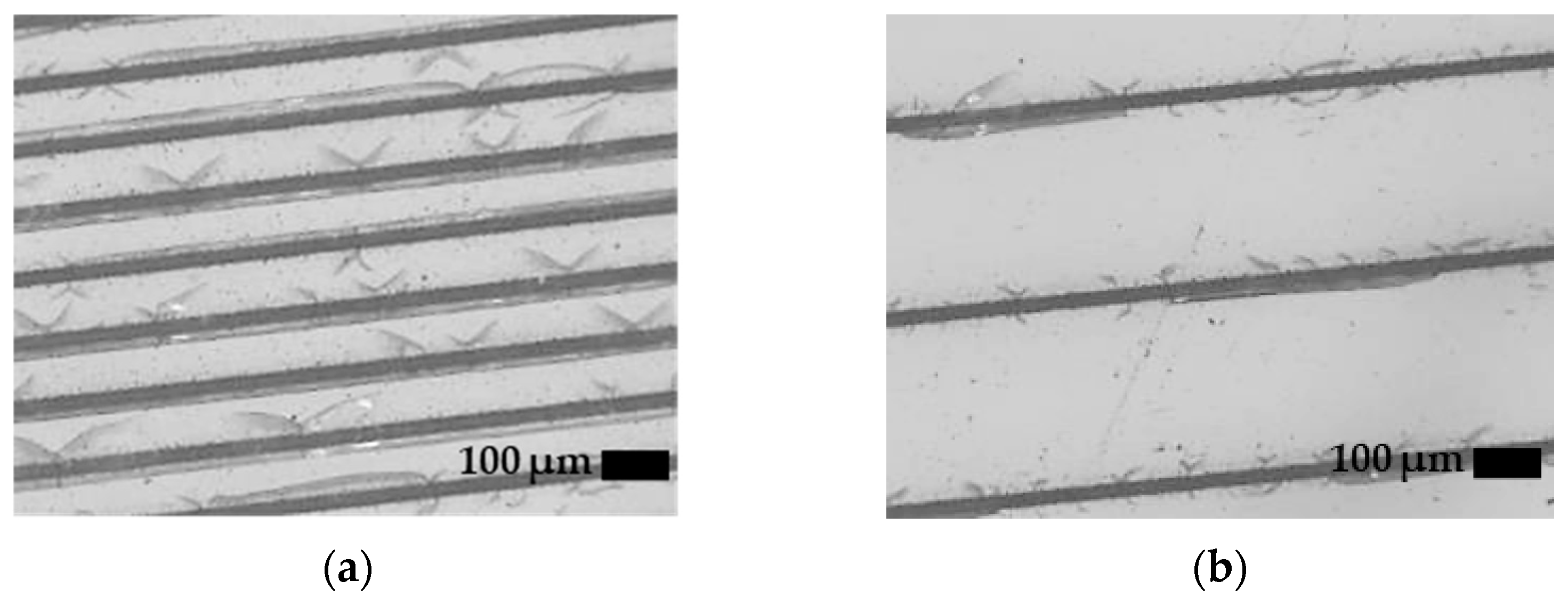
3.1. CO2 Laser Surface Modification. Surface Roughness Dependence on Processing Parameters and Topography

To assess the roughness dependence on the different processing parameters involved in laser surface modification, a series of tests were conducted on 45S5 bioactive glass surfaces. The objective of these tests was to determine the optimal power, speed, and number of passes. As shown in Figure 1, the dependence of the surface root mean square roughness (Sq) and interfacial area ratio (Sdr) are functions of the processing parameters. These results were obtained by laser surface modification using the CO2 laser system. The intrinsic roughness of the samples, measured in the absence of any texturing treatment, was used as a reference. The average Sq and Sdr values were 1.14 µm and 0.19% for the 45S5 cast bioactive glass and 1.08 µm and 0.13% for the ICIE16 cast bioactive glass.
Figure 1a shows that the surface roughness exhibits a strong dependence on the scanning speed. High roughness values, up to Sq and equal to 10 μm, are obtained with intermediate–low values of the scanning speeds. Within this range of scanning speeds, the Sq roughness differences are not statistically significant. At high values of the scanning speed, the produced roughness notably decreases but remains above the reference values. Scanning speeds from 200 to 500 mm/s produce Sq values dropping from approximately 8 to 4 μm. Significant differences are observed for these scanning speed values. At the lower limit range of the scanning speed, the surface roughness increment is limited, and this effect is attributed to glass remelting produced at elevated surface power density. In addition to surface power density influenced by the scanning speed, the pulsed nature of the energy provided by the CO2 laser source in the micro- to millisecond range plays a significant role. Regarding the interfacial area ratio, as shown in Figure 1b, it is observed that scanning speeds between 100 and 200 mm/s led to Sdr values above 20%. The Sdr parameter gives an indication of the area increment due to laser surface modification.
Figure 1c shows the roughness dependence on the average optical power of the laser radiation during processing. Low optical power values produce a very low effect on the surface roughness, with Sq values remarkably similar to unprocessed surfaces. An increment of surface roughness is observed when increasing optical power in the range evaluated. Nevertheless, there is a threshold above which the effect on roughness is much more noticeable. The Sdr parameter is more sensitive to determine this power threshold (see Figure 1d), revealing statistically significant increments of the interfacial area at 20 W, while the Sq value remains close to reference values.
The effect of the separation between the consecutive paths followed by the scanned laser beam is shown in Figure 1e,f. It is observed that the maximum roughness for separations is similar to the spot size. Increasing the pitch reduces the measured roughness because of the rising presence of untreated areas within the area where the roughness is measured. On the other hand, an excessively low pitch also limits the roughness due to partial overlapping of the consecutive laser tracks. From the analysis of the influence of these processing parameters, it is possible to select a specific set of processing parameters for a particular roughness target value. This approach is very valuable for applications where the modification of a wide surface area is required. Mainly, the Sdr values, representative of the relative increment of surface area for a fixed projected area, can be related to the available surface to interact with the surrounding media. In addition, it is also of great interest to study the treated surface in detail as this will provide insight into the potential behavior of the surface during the distinct stages of the osseointegration process.
To understand the laser interaction with the bioactive glass surface, it is necessary to assess in detail the morphology of the processed surface. With the purpose of studying the pattern, and subsequently analyzing the morphology evolution as a function of time in immersion in Tris-buffer, the following parameters were taken as reference: a spacing of 250 μm, a scanning speed of 100 mm/s, and an average optical power of 25 W. The topography of 45S5 bioactive glass processed under these conditions is shown in Figure 2. The 3D map obtained by interferometric profilometry shows well-defined remelted zones where the surface was swept by the center of the laser energy Gaussian profile. The discontinuities observed in the remelted glass areas are attributed to the pulsed laser radiation provided by the CO2 laser source. Also, the presence of cracks that develop between the remelted tracks is observed. This effect is associated with the thermoelastic phenomena that occur during laser surface modification. These cracks stitch the irradiated paths and have an oblique arrangement between paths. This crack pattern is better seen by employing optical microscopy, as shown in Figure 3. The crack tips show a direction coinciding with the irradiated paths in cases where the origin is a discontinuity of the remelted zone. On the other hand, they present a direction preferentially perpendicular to the path of the laser beam when the crack originates in a continuously remelted area. Cracking is driven by stresses generated during processing, and residual stresses are presented in non-cracked glass areas. The nucleation of the cracks is determined by the stress concentration zones, and the development of the cracks is guided by the thermal gradient.

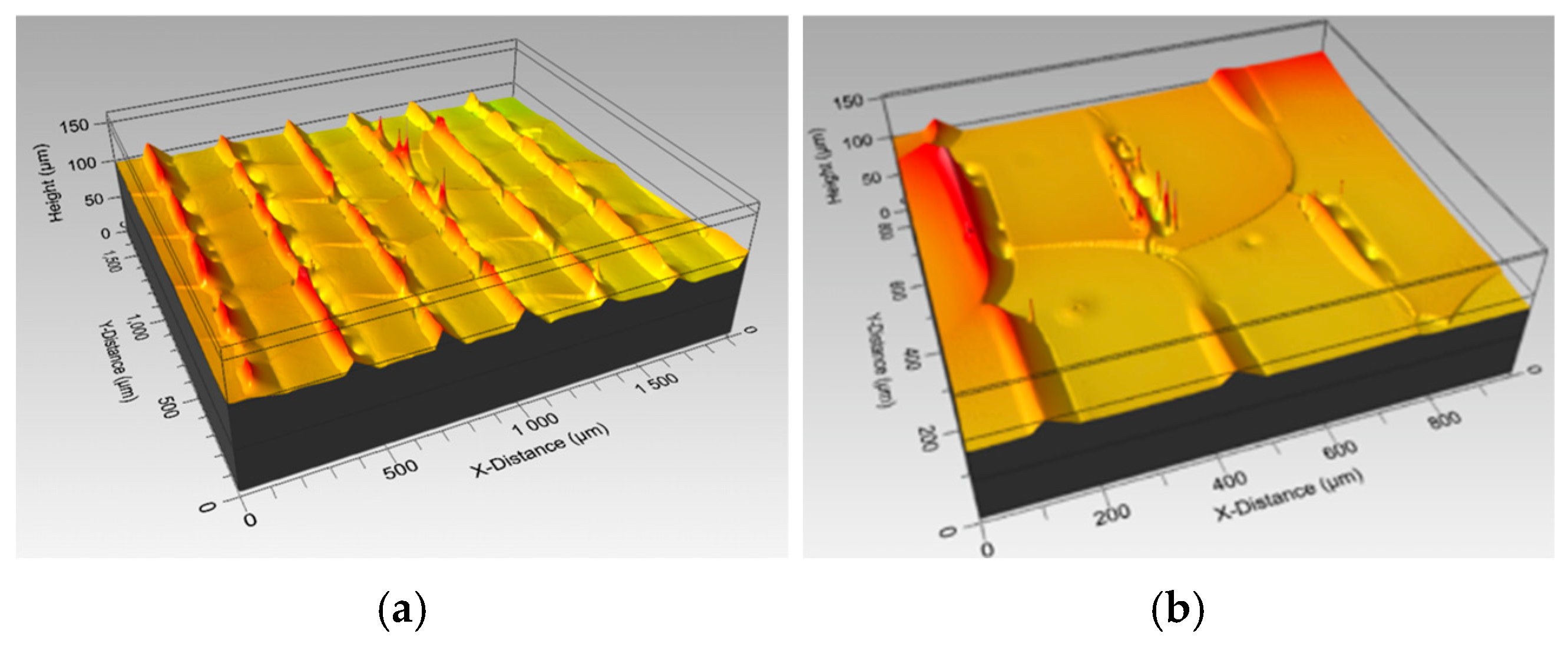
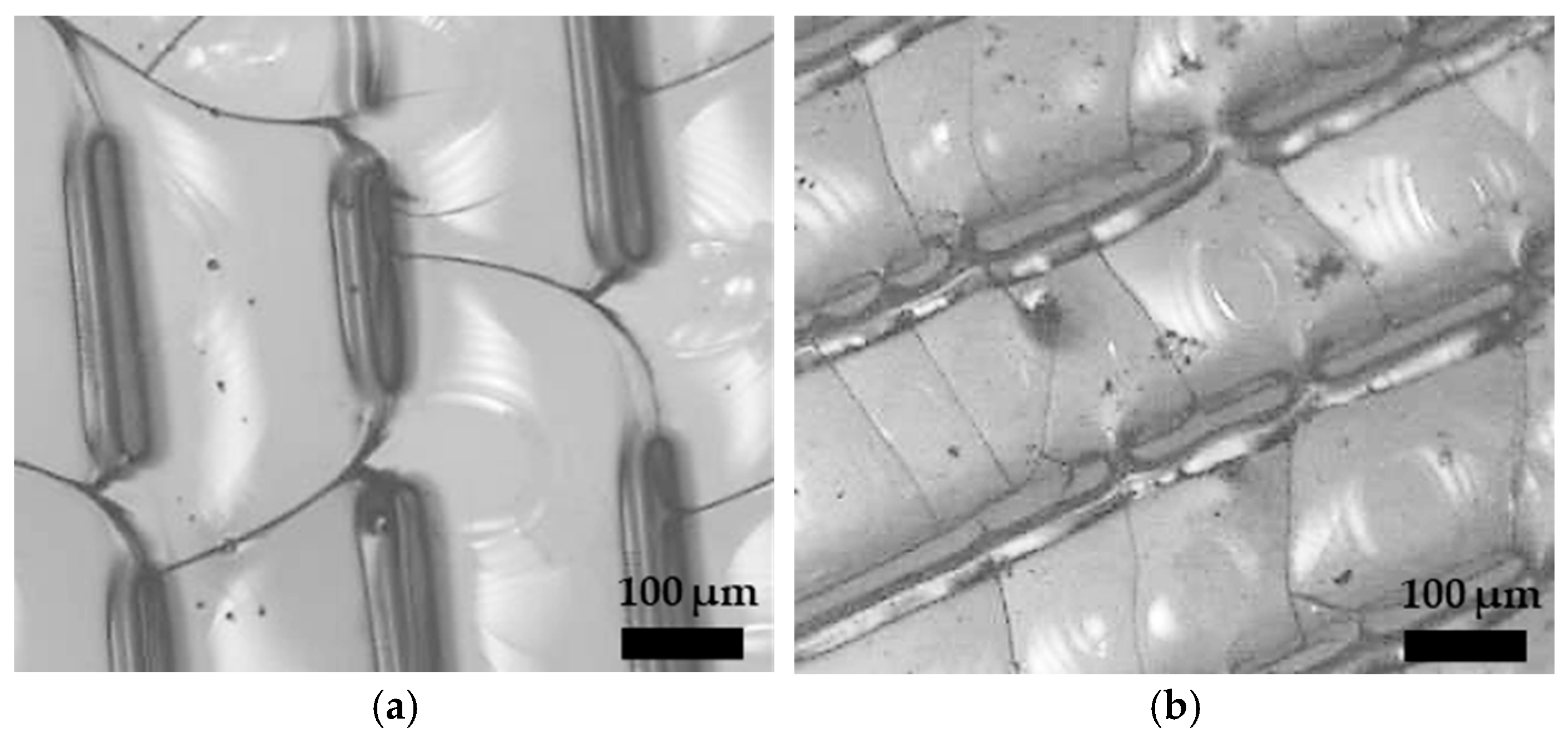
3.2. Femtosecond Laser Surface Modification. Surface Roughness Dependence on Pass Number and Topography

The interaction between the femtosecond laser and the bioactive glass surface differs from the surface interaction of the CO2 laser radiation; both are motivated by the different wavelengths and the different pulse lengths. Consequently, the practical operation and process parameters of interest are also different. To understand this complementary operating regime, Figure 4 shows the dependence of the roughness measured on the surface processed with a femtosecond laser as a function of the number of repetitions on the same scanned path. The Sq values for each number of passes present statistically significant differences. The specific Sq and Sdr clearly depend on the size of the measurement area in relation to the processed area, which is much smaller (see Figure 5). Therefore, these values cannot be extrapolated in absolute value, but they do allow for a qualitative assessment of the impact of the number of passes for the specific processing conditions. Thus, it is observed that the number of repetitions on the same scanned path increases the roughness by means of increasing the depth of the produced groove. This increment is continuous from 1 to 10 passes and then decreases to 20 and 50. The effect on the roughness is explained by a depth growth that turns into a widening of the bottom of the groove after 10 repetitions. Consequently, the Sdr also stabilizes and stops growing at the same point.
Figure 5 shows the morphology of the grooves generated after scanning a straight single pass (Figure 5a) and after five repetitions (Figure 5b). The V-type groove is well defined, and the remelted material deposited around the upper edges is extremely low. The depth reached by a single pass is 15 μm and reaches 23 μm after five repetitions. The groove’s whole angle is approximately 80 degrees for a single pass and decreases to 57 degrees after five repetitions. For repetitions above five, the ablated material redeposits around the upper edges, leading to much less defined patterns. The micrographs obtained by an optical microscope from the patterns generated by the femtosecond laser on 45S5 bioactive glass are shown in Figure 6. Some cracking is produced around the groove’s upper edges, but there is limited crack development. The ablative nature of the process leads to surfaces free from remelted glass and redeposited glass for these operation conditions.

3.3. Surface Hardness and Fracture Toughness
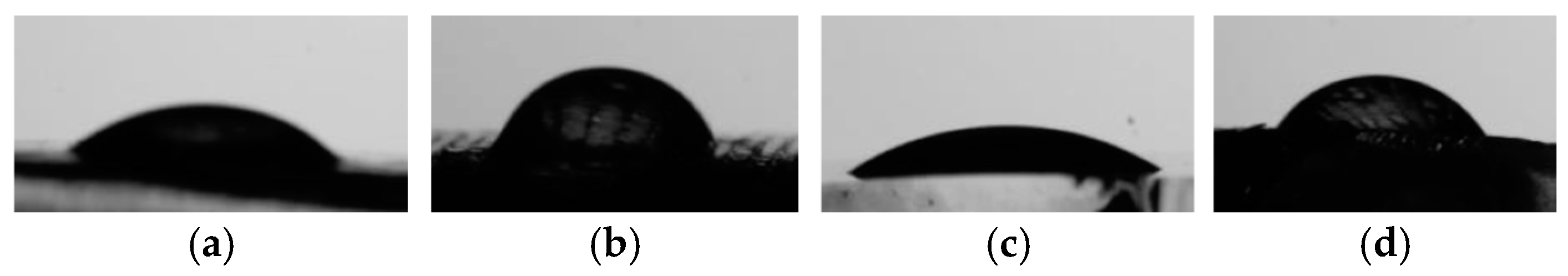
The average hardness of the laser-modified surfaces is depicted in Table 4. It is observed that the hardness measured from CO2 laser-textured surface samples does not present significant differences with respect to the reference surfaces, neither for the 45S5 BG (573 HV0.3) nor for the ICIE16 BG (561 HV0.3). Nevertheless, a greater variability of the measured values is observed and can be associated with the heterogeneity of the treated surface. A higher degree of hardness is evident in the remelted glass areas, while the values are lower than average in the most cracked areas. Similarly, the fracture toughness determination from the radial cracks after indentation and the fracture surface energy reveal similar values to those of the reference glasses and high dispersion. On the contrary, the surface patterning performed by the femtosecond laser led to a reduction in hardness for both the 45S5 BG (464 HV0.3) and ICIE16 BG (482 HV0.3). The fracture toughness and fracture surface energy in these surfaces do not follow the same trend, mainly due to the crack arrest effect of the generated grooves.
Previous works on CO2 laser modification of glasses report hardness average values similar to or slightly higher than those of the reference glasses and with higher dispersion of measured values [31]. The behavior observed for the CO2 laser-textured surfaces of this work agrees with the reported findings. The irradiation of bioactive and inert silicate glasses with a long pulse and quasi-continuous laser radiation leads to thermal phenomena and glass melting in different degrees, depending on energy density (optical energy per unit area) used for the specific purpose, such as surface modification [33,34], sintering/melting to generate a surface coating [35,36], or complete melting of the BG [31]. These laser-assisted processes employ comparatively low irradiances and large interaction times, thus enabling glass heating, melting, and cooling [37]. The thermal process induced by surface modification under the processing conditions reported in this work also produces a low portion of remelted material with remarkably high quenching speed, causing the observed dispersion in hardness values.
During glass processing by short laser pulses, glass ablation is produced by irradiation several orders of magnitude higher [37]. The remaining glass between the ablated patterns is not substantially modified, but residual stress can lead to different hardness values than the parent glass. Increments in negative residual stress have been reported in the internal ablation of quartz using a femtosecond laser, related to the internal plane strain state [38]. However, the process of a free surface as addressed in the present work, related to a plane state of stress, would not lead to the generation of such compressive residual stresses. Moreover, when the ablated depths are comparable to the indentation size, higher permanent deformation is enabled as the pattern spacing is reduced due to the lower material confinement at measurement boundaries. Lower hardness values and high dispersion after femtosecond laser exposure have also been reported for silicate glass surfaces [39].

3.4. Contact Angle

The wettability for distilled water was significantly decreased after the modification of the CO2 laser surface. The average measured contact angle for the selected processing conditions (250 μm spacing, 100 mm/s, 25 W) was 63.2° for 45S5 BG-modified surfaces and 59.1° for ICIE16 BG-modified surfaces. In contrast, the contact angles for the as-received glass surface were 39.9° and 36.0°, respectively. A different behavior was observed for the femtosecond laser-modified surface (spacing 100 μm; 100 mm/s; 25 W; five repetitions), where the differences in the contact angle are not statistically significant. The average values of the contact angles are listed in Table 4. Figure 7 illustrates the different behaviors regarding the distilled water wettability.
Surface modification by CO2 laser radiation can change the wettability of the distilled water of silicate glasses, but the modification towards hydrophobic or hydrophilic behavior depends on several factors such as initial wettability, surface chemistry, pattern, laser overlapping, and other processing conditions. Continuous broad texturing of parallel patterns by a defocused CO2 laser has been reported to increase the hydrophilic behavior of silicate glass surfaces [33]. Higher water affinity is achieved by overlapping the hydrophilic scanned surfaces. Nevertheless, if the alternating arrays of surfaces with different water affinities are created, the fluid of water droplets is restrained, leading to a less hydrophilic surface. Different pattern spacing has been implemented by CO2 laser treatment of silicate flat glass to obtain hydrophobic–superhydrophilic patterns with drop retention ability [40]. In the bioactive glass surfaces modified here by a CO2 laser, the pattern formed by parallel scanning clearly restrains the flow of the distilled water droplet, leading to higher wetting angles than the untreated BG surfaces. Similarly, the capability of the femtosecond laser to modify the surface wettability also depends on a number of factors. For example, low pattern spacing with overlapped grooves leads to double-hierarchical surfaces that importantly reduce the wetting angles in borosilicate glass surfaces [41]. Nevertheless, the hydrophilic behavior of the surface remains unmodified when pattern spacing is higher and ablated features do not overlap.

3.5. Roughness Evolution During Immersion in Tris-HCl Buffer

The results presented in Figure 8 elucidate the impact of laser texturing on surface roughness (Sq) and developed interfacial area ratio (Sdr) across varying time intervals during the immersion in Tris-HCl buffer. First, it is observed how the surface roughness of the untreated melt-quenched bioactive glasses increases during immersion, owing to the formation of the silica-rich layer and the apatite precipitation. The reference glasses reach Sq roughness values close to 5 μm after 7 days of immersion. The roughness evolution is faster for the 45S5 BG in comparison to the reference ICIE16 BG. The observed Sq differences for the different immersion times are statistically significant. Two differentiated stages are evident in the 45S5 BG sample. The initial stage manifests immediately following immersion, while the subsequent one emerges between 12 h and 2 days of immersion. In the case of the laser-modified 45S5 BG (Figure 8a,b), the CO2 laser results in surface roughness values that are significantly higher compared to those observed for the untreated bioactive glass. The CO2 laser-treated samples show a notable increase in Sq and Sdr values over time, particularly beyond 6 h of immersion, with a maximum Sq value of 35 μm, and after 7 days of immersion, they reach 25 μm. After 75 days of immersion in Tris-HCl buffer, the measured roughness at a long time presents an Sq value of 20 μm. The surface complexity evolution during immersion does not present a clear dependence on the CO2 laser surface modification.
During the first 8 h of immersion in Tris-HCl buffer, the CO2 laser-textured surfaces present evidence of a leaching process, which homogeneously affects the entire surface. The elevated areas of remelted bioactive glass do not show a faster dissolution than the rest of the surface, which would be expected given its morphology and high specific surface area. It has been previously observed that the CO2 laser-melted 45S5 BG can produce a postponement in the concentration peak of released phosphorous in Tris-HCl buffer [31,42]. This fact evidences a delay of the silica-rich layer formation and the subsequent apatite precipitation in comparison to the bioactive glass that is not remelted, which can explain that the CO2 laser-textured surfaces increase the surface roughness during the expected time of silica-rich layer formation in the 45S5 BG. Following this, apatite precipitation starts at the plane zone of the textured surface, leading to a roughness reduction after 24 h of immersion. The onset of apatite precipitation in the elevated zones of remelted bioactive glass is not evidenced until 72 h after immersion, which produces a recovery of the surface roughness. As illustrated in Figure 9a,b, the CO2 laser-textured surfaces of the 45S5 BG, prior to and after immersion in Tris-HCl buffer, are shown in the long term. It is observed that after 75 days of immersion, the surface is almost wholly covered with precipitated apatite. The initial roughness of the CO2 laser-textured surfaces is retained, and the overall roughness is increased by the inherent BG apatite precipitation process.
The femtosecond laser-textured 45S5 BG surfaces present quite different roughness evolution during soaking in Tris-HCl buffer. The roughness values are comparable to those previously measured on reference BG surfaces. Notably, even lower roughness values are registered for 300 μm pattern spacing. The roughness starts to decrease at the expected onset of apatite precipitation in this bioactive glass. After 7 days, the Sq roughness was 10 μm and 5 μm for 100 and 300 μm pattern spacing, respectively. In the long term, after 75 days of immersion, the Sq roughness value is stabilized at 6 μm, and no differences among pattern spacings are observed. Figure 9c,d show the femtosecond laser-textured surfaces of the 45S5 BG prior to and after immersion in Tris-HCl buffer in the long run. This is evidenced by the fact that the precipitated apatite has almost filled the grooves generated by the femtosecond laser radiation. Intermediate observations show how the apatite precipitation is progressively filling the grooves. Consequently, the roughness induced by the femtosecond laser’s topography is gradually lost by the 45S5 BG surfaces, and in the long run, only the roughness component due to precipitated apatite is present.
Regarding the soaking time influence on the developed interfacial area ratio Sdr, in the long run, the complexity of the 45S5 BG surfaces does not depend on the laser surface treatment. However, it is determined by the apatite layer morphology, leading to high Sdr values (between 500 and 800%). On the contrary, the ICIE16 BG surfaces exhibit a surface complexity that still depends on the laser treatment, as the apatite layer does not cover the complete surface patterns. After 75 days of immersion, the Sdr is 150% and 400% for 100 and 300 μm femtosecond pattern spacing, respectively, and 400% for the CO2 laser-textured surface.
The roughness evolution of the laser-modified ICIE16 BG (Figure 8c,d) shows Sq values notably higher than those of the untreated BG. The CO2 laser-treated samples show the maximum Sq values around 20 μm, which remain stable from 8 h onwards. The slower kinetics of apatite precipitation of this bioactive glass produce a more progressive evolution of the surface roughness and play a more significant role in the surface texture generated by the laser treatments. Figure 10a,b illustrate the surfaces of the CO2 laser-textured surfaces of the ICIE16 BG prior to and after immersion in Tris-HCl buffer in the long run of 75 days. It is observed that the apatite layer is much thinner than in the case of 45S5 glass, and the features of the laser surface texturing are still present. The initial roughness of the CO2 laser-textured surfaces is retained during immersion.
The femtosecond laser treatment on the ICIE16 BG surfaces presents a sustained growth of the roughness Sq at different immersion times. After 7 days, Sq roughness values of 18 μm and 13 μm were noticed for 100 and 300 μm pattern spacing, respectively. After 75 days of immersion, the Sq roughness value is stabilized at 5 μm, and no differences among pattern spacings are again observed. Meanwhile, the surface complexity Sdr remains close to or below the reference bioactive glass evolution, depending on the pattern spacing. Figure 10c,d show the femtosecond laser-textured surfaces of the ICIE16 BG prior to and after immersion in Tris-HCl buffer in the long term. The laser-induced grooves suffered a progressive widening during the bioactive glass leaching. The apatite precipitation is preferentially produced within the grooves, limiting the available phosphate groups for the plane areas. The degree of apatite precipitation in the plane areas is lower for the specimens with 100 μm pattern spacing, as revealed by comparing the SEM micrographs in Figure 10d and Figure 11b. Fewer grooves per surface area lead to a higher covering of the apatite layer. Thus, the femtosecond laser-textured surfaces in the ICIE16 BG are provided with a pattern of preferential apatite precipitation, whose character is also maintained in the long term.
Figure 11 presents SEM images of the femtosecond laser-textured surfaces with a pattern spacing of 300 μm after immersion in Tris-HCl buffer for 75 days. The comparison between the laser-treated 45S5 BG and the laser-treated ICIE16 BG allows us to understand that a suitable combination of a produced pattern and bioactivity, in terms of apatite precipitation kinetics, is required for a specific response at determinate immersion times. While the preferential osteoconductive capability of the femtosecond laser-treated ICIE16 BG is preserved after 75 days, the same surface treatment in the 45S5 BG loses this property after approximately 72 h from the onset of the bioactive response. Raman spectra between 900 and 1100 cm−1 have been acquired from the surfaces of the laser-modified surfaces after immersion in Tris-HCl buffer (Figure 12). The apatite formation is evidenced by the growth of a band at 962 cm−1 related to the stretching vibration mode of the PO43− group over the reference band at 944 cm−1 related to Si-O(NBO) stretching. In the spectra corresponding to the 45S5 and ICIE16 reference BGs, prior to immersion, the different ratios of the bands related to Si-O(NBO) and Si-O-Si stretching between 1050 and 1100 cm−1 are observed. The ICIE16 lower relative ratio between the areas below the non-bonding and bonding Si-O bands is related to its higher network connectivity and its slower apatite precipitation ability. The lower bioactivity leads to a weaker intensity of the PO43− peak both in the bare and the laser-treated surfaces of the ICIE16 bioactive glass.
The bioactive glass surface modification with the CO2 laser preserves the apatite precipitation ability for both bioactive glasses. Although delays of leaching and apatite precipitation onset can be produced due to the presence of remelted bioactive glass, differences in reaction kinetics are limited to the short term. No differences are found regarding apatite precipitation in the CO2 laser-treated surfaces in the long run. The potential to provide apatite spatial preferential precipitation of these patterns is discarded. Nevertheless, the high roughness in the long run and the increased wetting angle provide the CO2 laser-treated BGs with a high potential for a preferential biological response in terms of cellular attachment and proliferation. The CO2 laser modification of both highly reactive and more stable melt-derived bioactive glasses, such as the 45S5 BG and ICIE16 BG, would be the preferred option when relatively high surface areas are involved.
Regarding the complexity of surfaces modified by the CO2 laser, the development of the interfacial area ratio, Sdr, is ultimately determined by apatite precipitation in the context of the high bioactivity 45S5 BG. The flake morphology characteristic of biological-like precipitated apatite with a high specific surface area explains the high measured values for the surface complexity after long-term immersion in Tris-HCl buffer. In the case of moderate bioactivity, the ICIE16 BG, the complexity of surfaces in the long term is attributable to a combination of the textured pattern and apatite precipitation.
When testing the evolution of the surface in Tris-HCl buffer, the effect of the surface modification by the femtosecond laser is sensitive to the bioactive glass reactivity. The immersion tests in Tris-HCl of the surfaces revealed loss of the patterning in femtosecond-treated 45S5 surfaces. The apatite formation is preferentially produced in the grooves, which are progressively filled by precipitated apatite, and no impact of the laser treatment on the surface roughness is observed in the long term. After 75 days of soaking, the grooves are almost occult by the apatite layer. Therefore, femtosecond surface modification of highly reactive melt-derived bioactive glasses would be interesting only when a specific short-term response is sought, e.g., short-term, space-resolved preferential osteoconductivity. In the work developed by Shaikh et al., a 45S5 BG surface modified by a femtosecond laser was observed to completely lose the induced topography after 5 days of immersion in SBF [20].
Conversely, when immersed in Tris-HCl buffer, the femtosecond-treated surfaces in the ICIE16 BG exhibited preferential apatite precipitation in the pattern entities at low and long terms. The grooves are progressively widened and coated by the precipitated apatite and, simultaneously, the calcium phosphate deposit in the plane areas is regulated by pattern spacing. The combination of the highly resolved femtosecond laser treatment with a more stable ICIE16 BG demonstrates short- and long-term space-resolved preferential apatite formation ability. This good response observed opens the door for multiple potential applications, given that the ICIE16 BG is one of the bioactive glasses that provides a larger working window, enabling the generation of different elements for bone tissue engineering and at the same time showing a very good biological response [26,43]. In addition, other more stable bioactive glass compositions, such as the 13–93 bioactive glass, are of interest in order to maintain the long-term effect of the laser surface treatment [44,45].
Prior research has underscored the pivotal influence of surface topography on cellular behavior, including adhesion, differentiation, and proliferation [14]. The creation of micro- and nano-patterned surfaces using LSM techniques has demonstrated potential in emulating the natural extracellular matrix (ECM) and improving tissue regeneration [46]. Increases in surface roughness, similar to those observed in this study, have been demonstrated to directly correlate with enhanced osteoblastic adhesion and proliferation, which, in turn, lead to improved bone tissue integration [47]. Moreover, properties of roughness and topography influence other functions required during the osseointegration process, such as angiogenesis, for which the study of the impact of the surface modifications presented here would be of great interest [34]. This work contributes to the expanding field of biomaterial engineering by offering a comprehensive examination of the impact of laser parameters, including power, speed, and line spacing, on surface roughness and reactive behavior in Tris-HCl buffer. Further studies will aim to assess the interaction between modified surfaces and osteoblastic cells and the potential enhancement of bone osseointegration.

4. Conclusions

The bioactive glass surface modification with the CO2 laser produces average roughness dependent on processing parameters. The surface texturing process is governed by thermal and thermoelastic effects. The absorption of laser radiation in a thin surface layer of silicate glass, coupled with the relatively long pulses of optical energy, results in the formation of rough topographies characterized by remelted glass and cracked surfaces. An increased wetting angle in distilled water was observed for the CO2 laser-modified 45S5 BG and ICIE16 BG surfaces. This behavior is explained by the generation of alternate parallel bands with different water affinity, which restrains the flow of water droplets.
The CO2 laser surface texturing notably increases Sq roughness in the 45S5 BG and ICIE16 BG surfaces. The long-term roughness is preserved; after 75 days of immersion in Tris-HCl buffer, an Sq value of 20 μm is observed. The long-term roughness results of the combination of high roughness increase due to the CO2 laser treatment and the roughness increase due to the inherent apatite precipitation of the melt-quenched bioactive glasses. In the long term, the CO2 laser-treated surfaces preserve the apatite precipitation ability. These surfaces present a high potential for a preferential biological response due to the high roughness and the increased wetting angles.
The bioactive glass surface modification by the femtosecond laser with a 532 nm wavelength produces highly resolved patterns composed of grooves. The ablation produces clean entities using one to five repetitions, with depths up to 23 μm and V groove full angles down to 57 degrees. The roughening capability of the femtosecond laser is increased by the repetition number and the reduction in the pattern spacing. Still, a repetition number over five produces an increased deposition of ablated material in the groove edges.
Femtosecond surface modification of highly reactive melt-derived bioactive glasses has an impact only in the short-term response when immersed in Tris-HCl buffer. On the contrary, the femtosecond-treated surfaces in the ICIE16 BG exhibited preferential apatite precipitation in the pattern entities in the short and long terms. The combination of the highly resolved femtosecond laser treatment with more stable melt-derived bioactive glasses, such as the ICIE16 BG, demonstrates that it is a good choice to provide short- and long-term space-resolved preferential osteoconductivity. The results of this study offer insights into understanding the responses of different BG glasses to other laser treatments and contribute to identifying the optimal parameters for achieving the desired surface characteristics.

Author Contributions

Conceptualization, A.R., R.C. and B.G.-V.; methodology, M.G.-Q., B.G.-V., A.R. and R.C.; formal analysis, M.G.-Q. and R.C.; investigation, M.G.-Q., B.G.-V. and E.C.-G.; resources, A.R., R.C. and J.P.; data curation, B.G.-V., E.C.-G. and H.S.; writing—original draft preparation, M.G.-Q. and B.G.-V.; writing—review and editing, E.C.-G. and R.C.; visualization, M.G.-Q., E.C.-G. and H.S.; supervision, A.R. and J.P.; project administration, A.R. and R.C.; funding acquisition, A.R., R.C. and J.P. All authors have read and agreed to the published version of the manuscript.

Funding

This research was partially supported by the Government of Spain (PID2022-138763OA-I00, funded by MCIN/AEI/10.13039/501100011033, FEDER, UE, and FSE+, UE) and by Xunta de Galicia (ED431C 2023/25).

Institutional Review Board Statement

Not applicable.

Informed Consent Statement

Not applicable.

Data Availability Statement

The data supporting the findings of this study are not publicly available but can be obtained from the corresponding author upon reasonable request.

Acknowledgments

The Center for Scientific and Technological Support to Research (CACTI) of the University of Vigo is gratefully acknowledged.

Conflicts of Interest

The authors declare no conflicts of interest.

References

  1. Gill, T.K.; Mittinty, M.M.; March, L.M.; Steinmetz, J.D.; Culbreth, G.T.; Cross, M.; Kopec, J.A.; Woolf, A.D.; Haile, L.M.; Hagins, H.; et al. Global, Regional, and National Burden of Other Musculoskeletal Disorders, 1990–2020, and Projections to 2050: A Systematic Analysis of the Global Burden of Disease Study 2021. Lancet Rheumatol. 2023, 5, e670–e682. [Google Scholar] [CrossRef] [PubMed]
  2. Kooy, C.E.V.W.; Jakobsen, R.B.; Fenstad, A.M.; Persson, A.; Visnes, H.; Engebretsen, L.; Ekås, G.R. Major Increase in Incidence of Pediatric ACL Reconstructions From 2005 to 2021: A Study from the Norwegian Knee Ligament Register. Am. J. Sports Med. 2023, 51, 2891. [Google Scholar] [CrossRef] [PubMed]
  3. Chiarello, E.; Cadossi, M.; Tedesco, G.; Capra, P.; Calamelli, C.; Shehu, A.; Giannini, S. Autograft, Allograft and Bone Substitutes in Reconstructive Orthopedic Surgery. Aging Clin. Exp. Res. 2013, 25, 101–103. [Google Scholar] [CrossRef]
  4. Ferraz, M.P. Bone Grafts in Dental Medicine: An Overview of Autografts, Allografts and Synthetic Materials. Materials 2023, 16, 4117. [Google Scholar] [CrossRef]
  5. Hench, L.L. Bioceramics: From Concept to Clinic. J. Am. Ceram. Soc. 1991, 74, 1487–1510. [Google Scholar] [CrossRef]
  6. Greenspan, D.C. Glass and Medicine: The Larry Hench Story. Int. J. Appl. Glass Sci. 2016, 7, 134–138. [Google Scholar] [CrossRef]
  7. Yadav, S.; Yadav, D.; Kumar, P.; Yadav, A.; Nirala, G.; Yadav, S. Bioactive Glass for Biomedical Application: An Overview. In Defects Engineering in Electroceramics for Energy Applications; Springer: Singapore, 2024; pp. 305–327. [Google Scholar] [CrossRef]
  8. Jain, S.; Raghavendra, G.; Naik, R.H.; Daloji, L.; Azeem, P.A. Exploring the Versatility of Phosphate-Based Bioactive Glass for Biomedical Applications. In Recent Advances in Mechanical Engineering, Volume 1 (ICMech-REC 2023); Springer: Singapore, 2024; pp. 673–685. [Google Scholar] [CrossRef]
  9. Islam, M.T.; Felfel, R.M.; Abou Neel, E.A.; Grant, D.M.; Ahmed, I.; Hossain, K.M.Z. Bioactive Calcium Phosphate–Based Glasses and Ceramics and Their Biomedical Applications: A Review. J. Tissue Eng. 2017, 8, 2041731417719170. [Google Scholar] [CrossRef] [PubMed]
  10. Ruiz-Aguilar, C. Porous Phosphate-Based Bioactive Glass/β-TCP Scaffold for Tooth Remineralization. PLoS ONE 2023, 18, e0284885. [Google Scholar] [CrossRef] [PubMed]
  11. Sharma, K.; Kedia, S.; Singh, A.K.; Basak, C.B.; Chauhan, A.K.; Basu, S.; Sinha, S. Morphology and Structural Studies of Laser Treated 45S5 Bioactive Glass. J. Non Cryst. Solids 2016, 440, 43–48. [Google Scholar] [CrossRef]
  12. Szada-Borzyszkowska, M.; Kacalak, W.; Szada-Borzyszkowski, W.; Borkowski, P.J.; Laskowska, D.; Szafraniec, F. Topography of Textured Surfaces Using an Abrasive-Water Jet Technology. Arch. Civ. Mech. Eng. 2024, 24, 226. [Google Scholar] [CrossRef]
  13. Maximov, M.; Maximov, O.C.; Craciun, L.; Ficai, D.; Ficai, A.; Andronescu, E. Bioactive Glass—An Extensive Study of the Preparation and Coating Methods. Coatings 2021, 11, 1386. [Google Scholar] [CrossRef]
  14. Taye, M.B.; Ningsih, H.S.; Shih, S.J. Exploring the Advancements in Surface-Modified Bioactive Glass: Enhancing Antibacterial Activity, Promoting Angiogenesis, and Modulating Bioactivity. J. Nanopart. Res. 2024, 26, 28. [Google Scholar] [CrossRef]
  15. Kargozar, S.; Kermani, F.; Beidokhti, S.M.; Hamzehlou, S.; Verné, E.; Ferraris, S.; Baino, F. Functionalization and Surface Modifications of Bioactive Glasses (BGs): Tailoring of the Biological Response Working on the Outermost Surface Layer. Materials 2019, 12, 3696. [Google Scholar] [CrossRef]
  16. Jonušauskas, L.; Martínez Vázquez, R.; Bragheri, F.; Paiè, P. 3D Manufacturing of Glass Microstructures Using Femtosecond Laser. Micromachines 2021, 12, 499. [Google Scholar] [CrossRef]
  17. Ferraris, S.; Vitale-Brovarone, C.; Bretcanu, O.; Cassinelli, C.; Vernè, E. Surface Functionalization of 3D Glass–Ceramic Porous Scaffolds for Enhanced Mineralization in vitro. Appl. Surf. Sci. 2013, 271, 412–420. [Google Scholar] [CrossRef]
  18. Shivakoti, I.; Kibria, G.; Cep, R.; Pradhan, B.B.; Sharma, A. Laser Surface Texturing for Biomedical Applications: A Review. Coatings 2021, 11, 124. [Google Scholar] [CrossRef]
  19. Pani, R.; Roy, S.; Behera, R.R.; Chakraborty, S.; Das, D.; Mahanta, K. Laser Texturing for Biomedical Application: A State of Art. AIP Conf. Proc. 2024, 3007, 100026. [Google Scholar] [CrossRef]
  20. Shaikh, S.; Kedia, S.; Singh, A.K.; Sharma, K.; Sinha, S. Surface Treatment of 45S5 Bioglass Using Femtosecond Laser to Achieve Superior Growth of Hydroxyapatite. J. Laser Appl. 2017, 29, 022004. [Google Scholar] [CrossRef]
  21. Schille, J.; Schneider, L.; Mauersberger, S.; Szokup, S.; Höhn, S.; Pötschke, J.; Reiß, F.; Leidich, E.; Löschner, U. High-Rate Laser Surface Texturing for Advanced Tribological Functionality. Lubricants 2020, 8, 33. [Google Scholar] [CrossRef]
  22. Korovessis, P.G.; Deligianni, D.D. Role of Surface Roughness of Titanium versus Hydroxyapatite on Human Bone Marrow Cells Response. J. Spinal Disord. Tech. 2002, 15, 175–183. [Google Scholar] [CrossRef] [PubMed]
  23. Deligianni, D.D.; Katsala, N.D.; Koutsoukos, P.G.; Missirlis, Y.F. Effect of Surface Roughness of Hydroxyapatite on Human Bone Marrow Cell Adhesion, Proliferation, Differentiation and Detachment Strength. Biomaterials 2000, 22, 87–96. [Google Scholar] [CrossRef] [PubMed]
  24. Hussain, Z.; Mehmood, S.; Liu, X.; Liu, Y.; Wang, G.; Pei, R. Decoding Bone-Inspired and Cell-Instructive Cues of Scaffolds for Bone Tissue Engineering. Eng. Reg. 2024, 5, 21–44. [Google Scholar] [CrossRef]
  25. Saiz, E.; Boccaccini, A.R.; Chevalier, J.; Peroglio, M. Editorial on Bioceramics for Bone Repair. J. Eur. Ceram. Soc. 2018, 38, 821–822. [Google Scholar] [CrossRef]
  26. Elgayar, I.; Aliev, A.E.; Boccaccini, A.R.; Hill, R.G. Structural Analysis of Bioactive Glasses. J. Non Cryst. Solids 2005, 351, 173–183. [Google Scholar] [CrossRef]
  27. ISO 25178-2; Geometrical Product Specifications (GPS)—Surface Texture: Areal—Part 2: Terms, Definitions and Surface Texture Parameters. International Organization for Standardization (ISO): Geneva, Switzerland, 2021.
  28. Anstis, G.R.; Chantikul, P.; Lawn, B.R.; Marshall, D.B. A Critical Evaluation of Indentation Techniques for Measuring Fracture Toughness: I, Direct Crack Measurements. J. Am. Ceram. Soc. 1981, 64, 533–538. [Google Scholar] [CrossRef]
  29. Miyoshi, T.; Sagawa, N.; Sassa, T. Study on fracture toughness evaluation for structural ceramics. Trans. Jpn. Soc. Mech. Eng. A 1985, 51, 2489–2497. [Google Scholar] [CrossRef]
  30. Quinn, G.D.; Bradt, R.C. On the Vickers indentation fracture toughness test. J. Am. Ceram. Soc. 2007, 90, 673–680. [Google Scholar] [CrossRef]
  31. Del Val, J.; López-Cancelos, R.; Riveiro, A.; Badaoui, A.; Lusquiños, F.; Quintero, F.; Comesaña, R.; Boutinguiza, M.; Pou, J. On the fabrication of bioactive glass implants for bone regeneration by laser assisted rapid prototyping based on laser cladding. Ceram. Int. 2016, 42, 2021–2035. [Google Scholar] [CrossRef]
  32. ISO 10993-14; Biological Evaluation of Medical Devices—Part 14: Identification and Quantification of Degradation Products from Ceramics. International Organization for Standardization (ISO): Geneva, Switzerland, 2001.
  33. Comesaña, R.; Riveiro, A.; del Val, J.; Badaoui, A.; Penide, J.; Quintero, F.; Boutinguiza, M.; Lusquiños, F.; Pou, J. Laser Surface Modification of Structural Glass for Anti-Slip Applications. Ceram. Int. 2019, 45, 24734–24741. [Google Scholar] [CrossRef]
  34. Calvo-García, E.; Comesaña, R.; Riveiro, A.; del Val, J.; Badaoui, A.; Quintero, F.; Boutinguiza, M.; Lusquiños, F.; Pou, J. Experimental Study of Strength and Fracture Toughness of Laser-Blasted Glass. Procedia Struct. Integr. 2022, 42, 251–258. [Google Scholar] [CrossRef]
  35. Bajda, S.; Cholewa-Kowalska, K.; Krzyzanowski, M.; Dziadek, M.; Kopyscianski, M.; Liu, Y.; Rai, A. Laser-Directed Energy Deposition of Bioactive Glass on Ti-6Al-7Nb Titanium Alloy Substrate with Highly Refined Grain Structure. Surf. Coat. Technol. 2024, 485, 130904. [Google Scholar] [CrossRef]
  36. Bajda, S.; Liu, Y.; Tosi, R.; Cholewa-Kowalska, K.; Krzyzanowski, M.; Dziadek, M.; Kopyscianski, M.; Dymek, S.; Polyakov, A.V.; Semenova, I.P.; et al. Laser Cladding of Bioactive Glass Coating on Pure Titanium Substrate with Highly Refined Grain Structure. J. Mech. Behav. Biomed. Mater. 2021, 119, 104519. [Google Scholar] [CrossRef]
  37. Comesaña, R.; del Val, J.; Quintero, F.; Riveiro, A.; Arias-González, F.; Boutinguiza, M.; Lusquiños, F.; Pou, J. Laser Cladding and Laser Direct Glass Deposition of Bioactive Glass and Glass-Ceramics. In Bioactive Glasses and Glass-Ceramics: Fundamentals and Applications; John Wiley & Sons, Inc.: New York, NY, USA, 2022; pp. 311–340. [Google Scholar] [CrossRef]
  38. Wang, Y.; Fan, G.; Qu, S.; Dong, S. Plastic Residual Strains under Femtosecond Laser Microexplosion Initiated High Pressure inside Quartz. Int. J. Nanomanuf. 2013, 9, 10–18. [Google Scholar] [CrossRef]
  39. Locker, S.; Sundaram, S.K. Ultrafast Modification of Oxide Glass Surface Hardness. Appl. Phys. B 2019, 125, 225. [Google Scholar] [CrossRef]
  40. Alqurashi, T.; Alnufaili, M.; Hassan, M.U.; Aloufi, S.; Yetisen, A.K.; Butt, H. Laser Inscription of Microfluidic Devices for Biological Assays. ACS Appl. Mater. Interfaces 2019, 11, 12253–12260. [Google Scholar] [CrossRef]
  41. Kostal, E.; Stroj, S.; Kasemann, S.; Matylitsky, V.; Domke, M. Fabrication of Biomimetic Fog-Collecting Superhydrophilic-Superhydrophobic Surface Micropatterns Using Femtosecond Lasers. Langmuir 2018, 34, 2933–2941. [Google Scholar] [CrossRef] [PubMed]
  42. Comesaña, R.; Lusquiños, F.; Del Val, J.; López-Álvarez, M.; Quintero, F.; Riveiro, A.; Boutinguiza, M.; De Carlos, A.; Jones, J.R.; Hill, R.G.; et al. Three-Dimensional Bioactive Glass Implants Fabricated by Rapid Prototyping Based on CO2 Laser Cladding. Acta Biomater. 2011, 7, 3476–3487. [Google Scholar] [CrossRef]
  43. Westhauser, F.; Hohenbild, F.; Arango-Ospina, M.; Schmitz, S.I.; Wilkesmann, S.; Hupa, L.; Moghaddam, A.; Boccaccini, A.R. Bioactive Glass (BG) ICIE16 Shows Promising Osteogenic Properties Compared to Crystallized 45S5-BG. Int. J. Mol. Sci. 2020, 21, 1639. [Google Scholar] [CrossRef]
  44. Groh, D.; Döhler, F.; Brauer, D.S. Bioactive Glasses with Improved Processing. Part 1. Thermal Properties, Ion Release and Apatite Formation. Acta Biomater. 2014, 10, 4465–4473. [Google Scholar] [CrossRef] [PubMed]
  45. Nawaz, Q.; de Pablos-Martín, A.; Contreras Jaimes, A.T.; Scheffler, F.; Wagner, T.; Brauer, D.S.; Boccaccini, A.R. Comparison of Microstructure, Sintering Behavior, and Biological Response of Sol-Gel and Melt-Derived 13–93 Bioactive Glass Scaffolds. Open Ceram. 2023, 15, 100407. [Google Scholar] [CrossRef]
  46. Liang, W.; Wu, X.; Dong, Y.; Shao, R.; Chen, X.; Zhou, P.; Xu, F. In Vivo Behavior of Bioactive Glass-Based Composites in Animal Models for Bone Regeneration. Biomater. Sci. 2021, 9, 1924–1944. [Google Scholar] [CrossRef]
  47. Gough, J.E.; Notingher, I.; Hench, L.L. Osteoblast Attachment and Mineralized Nodule Formation on Rough and Smooth 45S5 Bioactive Glass Monoliths. J. Biomed. Mater. Res. A 2004, 68A, 640–650. [Google Scholar] [CrossRef] [PubMed]
Figure 1. Root mean square height of the surface roughness (a,c,e) and developed interfacial area ratio (b,d,f) for the samples texturized using laser CO2 radiation as a function of scanning speed (a,b), optical power (c,d), and lattice spacing (e,f). Fixed processing parameters: a scanning speed of 100 mm/s (c,d,e,f); an optical power of 25 W (a,b,e,f); a spacing of 100 µm (ad).
Coatings 15 00195 g001
Figure 2. Topological 3D map of the 45S5 bioactive glass surface processed by a CO2 laser: (a) 10× magnification; (b) 20× magnification (processing parameters: lattice spacing 250 μm, scanning speed 100 mm/s, average optical power 25 W).
Coatings 15 00195 g002
Figure 3. Optical micrographs at ×8 magnification from the surface texturing of a CO2 laser with a 250 μm lattice spacing, a scanning speed of 100 mm/s, and an average optical power of 25 W: (a) 45S5 bioactive glass; (b) ICIE 16 bioactive glass.
Coatings 15 00195 g003
Figure 4. (a) Root mean square height Sq of the surface roughness and (b) developed interfacial area ratio Sdr for the samples texturized using laser femtosecond radiation as a function of the number of passes (an average optical power of 25 W, a scanning speed of 100 mm/s).
Coatings 15 00195 g004
Figure 5. A 3D map of the groove generated by femtosecond laser irradiation scanned along a rectilinear path obtained by interferometric profilometry: (a) single pass; (b) 5 passes (processing conditions: a scanning speed of 100 mm/s, an average optical power of 25 W).
Coatings 15 00195 g005
Figure 6. Optical micrographs of the textured pattern obtained by femtosecond laser surface modification of 45S5 bioactive glass: (a) pattern spacing 100 μm; (b) pattern spacing 300 μm (scanning speed 100 mm/s, average optical power of 25 W, five repetitions).
Coatings 15 00195 g006
Figure 7. Contact angle with distilled water for (a) 45S5 BG reference surface; (b) CO2 laser surface-modified 45S5 BG, (c) ICIE16 BG reference surface; (d) CO2 laser surface-modified ICIE16 BG.
Coatings 15 00195 g007
Figure 8. Root mean square height Sq of the surface roughness and the developed interfacial area ratio for the specimens subjected to immersion in Tris-HCl buffer during different immersion times. (a) Sq roughness for laser-treated 45S5 BG and reference 45S5 BG; (b) Sdr surface complexity for laser-treated 45S5 BG and reference 45S5 BG; (c) Sq roughness for laser-treated ICIE16 BG and reference ICIE16 BG; (d) Sdr surface complexity for laser-treated ICIE16 BG and reference ICIE16 BG.
Coatings 15 00195 g008aCoatings 15 00195 g008b
Figure 9. SEM images of the laser surface-modified 45S5 BG, taken prior to immersion in Tris-HCL. (a) CO2 laser-textured surface prior to immersion in Tris-HCl buffer; (b) CO2 laser-textured surface after immersion in Tris-HCl buffer for 75 days; (c) femtosecond laser-textured surface prior to immersion in Tris-HCl buffer; (d) femtosecond laser-textured surface after immersion in Tris-HCl buffer for 75 days.
Coatings 15 00195 g009
Figure 10. SEM images of the laser surface-modified ICIE16 BG, taken prior to immersion in Tris-HCL. (a) CO2 laser-textured surface prior to immersion in Tris-HCl buffer; (b) CO2 laser-textured surface after immersion in Tris-HCl buffer for 75 days; (c) femtosecond laser-textured surface prior to immersion in Tris-HCl buffer; (d) femtosecond laser-textured surface after immersion in Tris-HCl buffer for 75 days.
Coatings 15 00195 g010aCoatings 15 00195 g010b
Figure 11. SEM images of the sample surface after immersion in Tris-HCl buffer for 75 days: (a) femtosecond laser-textured 45S5 BG; (b) femtosecond laser-textured ICIE16 BG (pattern spacing 300 μm).
Coatings 15 00195 g011
Figure 12. Raman spectra corresponding to laser surface-modified BGs after immersion in Tris-HCl buffer for 75 days: (a) laser surface-modified 45S5 BG; (b) laser surface-modified ICIE16 BG.
Coatings 15 00195 g012
Table 1. Composition and network connectivity of 45S5 and ICIE16 bioactive glasses [26].
Composition (wt%)
OxideICIE1645S5
SiO248.045
CaO 32.924.5
P2O52.56.0
K2O 10.0-
Na2O 6.624.5
NC2.041.90
Table 2. Configuration and ranges of explored parameters for CO2 and femtosecond lasers.
Laser Source
Parameters (Units)CO2fs Yb
Wavelength (nm)10,600532
Power (W)6–2525
Scanning speed (mm/s)40–500100
Lattice spacing (μm)30–500100 and 300
Number of Passes1.01–50
Focal distance (mm)280.09.5
Table 3. Specific components and steps for the preparation of 1 L of Tris-HCl buffer.
StepsProcedures (Materials, Quantities, and Task)
1Prepare 800 mL of distilled water in an appropriate container.
2Add 121.14 g of Tris base (molecular weight 121.14 g/mol) to the solution.
3Adjust the solution to the desired pH using HCl (pH ≈ 7.4).
4Add distilled water until the total volume reaches 1 L.
Table 4. Hardness, fracture toughness, fracture surface energy, and contact angle of the laser surface-modified BG specimens and the reference BGs.
SpecimenHardness (HV0.3)KIc (MPa·m1/2) Miyoshi [29]KIc (MPa·m1/2) Anstis [28]Fracture Surface Energy (J/m2)Contact Angle (°)
45S5 Ref.524 ± 160.55 ± 0.100.49 ± 0.090.72 ± 0.2439.9 ± 8.9
ICIE16 Ref.562 ± 110.58 ±0.070.52 ± 0.060.80 ± 0.1836.0 ± 7.6
CO2 45S5573 ± 390.54 ± 0.220.48 ± 0.200.71 ± 0.6163.2 ± 8.1
CO2 ICIE16561 ± 350.54 ± 0.170.49 ± 0.150.76 ± 0.4959.1 ± 6.4
fs 45S5464 ± 370.56 ± 0.030.50 ± 0.030.73 ± 0.0937.4 ± 2.0
fs ICIE16482 ± 210.54 ± 0.160.45 ± 0.070.60 ± 0.2038.7 ± 7.8
Disclaimer/Publisher’s Note: The statements, opinions and data contained in all publications are solely those of the individual author(s) and contributor(s) and not of MDPI and/or the editor(s). MDPI and/or the editor(s) disclaim responsibility for any injury to people or property resulting from any ideas, methods, instructions or products referred to in the content.

