Review

Toxicity, Irritation, and Allergy of Metal Implants: Historical Perspective and Modern Solutions

by
Grzegorz Szczęsny
1,
Mateusz Kopec
2,* and
Zbigniew L. Kowalewski
2
1
Department of Orthopaedic Surgery and Traumatology, Medical University, 4 Lindleya Str., 02-005 Warsaw, Poland
2
Institute of Fundamental Technological Research, Polish Academy of Sciences, Pawińskiego 5B, 02-106 Warsaw, Poland
*
Author to whom correspondence should be addressed.
Coatings 2025, 15(3), 361; https://doi.org/10.3390/coatings15030361
Submission received: 24 February 2025 / Revised: 13 March 2025 / Accepted: 18 March 2025 / Published: 20 March 2025
(This article belongs to the Collection Review Papers Collection for Bioactive Coatings)

Abstract

:
The widespread adoption of metal implants in orthopaedics and dentistry has revolutionized medical treatments, but concerns remain regarding their biocompatibility, toxicity, and immunogenicity. This study conducts a comprehensive literature review of traditional biomaterials used in orthopaedic surgery and traumatology, with a particular focus on their historical development and biological interactions. Research articles were gathered from PubMed and Web of Science databases using keyword combinations such as “toxicity, irritation, allergy, biomaterials, corrosion, implants, orthopaedic surgery, biocompatible materials, steel, alloys, material properties, applications, implantology, and surface modification”. An initial pool of 400 articles was screened by independent reviewers based on predefined inclusion and exclusion criteria, resulting in 160 relevant articles covering research from 1950 to 2025. This paper explores the electrochemical processes of metals like iron, titanium, aluminium, cobalt, molybdenum, nickel, and chromium post-implantation, which cause ion release and wear debris formation. These metal ions interact with biological molecules, triggering localized irritation, inflammatory responses, and immune-mediated hypersensitivity. Unlike existing reviews, this paper highlights how metal–protein interactions can form antigenic complexes, contributing to delayed hypersensitivity and complications such as peri-implant osteolysis and implant failure. While titanium is traditionally considered bioinert, emerging evidence suggests that under certain conditions, even inert metals can induce adverse biological effects. Furthermore, this review emphasizes the role of oxidative stress, illustrating how metal ion release and systemic toxicity contribute to long-term health risks. It also uncovers the underappreciated genotoxic and cytotoxic effects of metal ions on cellular metabolism, shedding light on potential long-term repercussions. By integrating a rigorous methodological approach with an in-depth exploration of metal-induced biological responses, this paper offers a more nuanced perspective on the complex interplay between metal implants and human biology, advancing the discourse on implant safety and material innovation.

1. Introduction: Historical Overview

The interaction between metals and their alloys with human body has been known for millennia [1]. However, military applications were the first typical cases, not medical activities. It directly followed from the fact that the first tools manufactured by human beings were used for hunting and fighting, and that these tools were made of materials available at that time: wood, flint, and metals [2].
It is presumed that gold and silver found in their pure form served as the first metals that were used to manufacture objects, as plenty of artifacts made with them have been found worldwide [1,2]. Nevertheless, they were a rarity, and as a consequence, their limited availability constricted their application. They were always available only for special purposes and only for a limited group of people. Their softness, malleability, ductility, and low melting point simplified processing, and their high chemical resistance provided adequate durability, essential for a plethora of products manufactured from these metals [3]. Among the most popular applications of gold and silver is the manufacturing of precious objects either for religious or prestigious purposes. Even nowadays, they symbolize the dignity and majesty of their users [3].
Copper, tin, and meteoric iron could also be found in their natural forms, which enabled their usage by people throughout history [4]. They are much more abundant in comparison to gold and silver and thus have much greater availability. And, more importantly, several metals could be quite easily obtained from their deposits and ores in the process of smelting (heating), increasing their availability [5]. Silver and lead were probably the first metals produced from ores, but the number of metals acquired this way soon increased [5,6]. Compared to native forms, much higher quantities of metals that could be obtained from ores provided better availability for craft and, later on, industrial applications [5]. Several of them were found to be excellent for manufacturing tools for everyday use.
The first occurrences of metal object insertion into the human body were probably accidental and during military events [7]. These events allowed us to observe the interactions of metals (in most cases, arrowheads or tips of spears) with tissues [8]. In the vast majority of cases, iron (as a cast iron) and copper (and bronze) were used in such situations. Cast iron containing more than 2%–4% of carbon was very popular at that time due to its low melting temperature and ductility, which simplified production and processing. Bronze, as an alloy consisting of copper and tin (ca. 12%), often also with the addition of other metals (aluminium, zinc, nickel, or manganese), non-metals (phosphorus), and metalloids (arsenic, silicon), was so popular that it became the name of a whole era of human history, i.e., the Bronze Age [8].
Cast iron and bronze were used to manufacture tools for hunting, fighting, food production, and craft needed for everyday life. Unfortunately, their ability to interact with biological ions and substances of high oxidative potential makes them highly susceptible to corrosion and tissue irritation, thus leading to suppuration when inserted into viable tissues [7]. Copper itself is bactericidal, and its salts are even poisonous [9]. But it also became obvious that precious metals, namely silver and gold, did not corrode nor produce tissue irritation and suppuration, which became the basis for their use to manufacture items having close contact with the human body, like earrings, bracelets, and rings [3,4]. The high demand for raw material for the production of medical and dental tools and implants has led to interest in the possibilities of using metals and their alloys in medicine. These are gold, silver, and other metals and their alloys based on lead, mercury, nickel, chromium, copper, zinc, and iron. Others, including beryllium, palladium, platinum, ruthenium, rhodium, and iridium, were introduced later, with some being added to gold. They replaced metals used previously due to their better physical and chemical properties, increased resistance, lower toxicity, and lower irritancy. However, many of them quickly showed unfavourable properties when in contact with living tissues.
Nowadays, only a few metals and their alloys are regularly applied in medicine. One can mention here stainless steel, nickel–chromium, cobalt–chromium, which is also used with molybdenum (CoCr, CoCrMo), and titanium alloys, which are also added to aluminium, vanadium, niobium, nickel, iron, and other metals (i.e., Ti6Al4V, Ti6Al7Nb, Ti5Al2.5Fe, Ti12Mo6Zr2Fe). Silver amalgam, being a mixture of copper, silver, tin, zinc, and inorganic mercury, has lost popularity due to the content of mercury [10]. As of 1 January 2025, the use of dental amalgam was banned in the European Union [11]. Nevertheless, it is still popular in several countries [12].
These metals are used in their highest quantities in dentistry and orthopaedics for crowns and bridges, “stifts” for mounting porcelain crowns, for joint prostheses, and for other orthopaedic and trauma implants. Unfortunately, the high mass of materials used to manufacture these implants, especially orthopaedic ones, weighing hundreds of grams and even more, bring a high risk of exposure to the metals. Their considerable prevalence has become the cause of a significant and growing risk of exposure to metal ions released from medical implants, becoming for their users a threat that outweighs exposure coming from other sources, especially soil, water, and air, even when polluted [13], and more importantly, evoking unwanted side effects.

2. Motivation and Methodology

The increasing reliance on metallic implants in orthopaedics and dentistry has led to significant advancements in medical treatments, yet concerns regarding their biocompatibility, toxicity, and long-term stability remain unresolved. While numerous reviews and studies have examined various aspects of metal implants, the existing literature often lacks a comprehensive discussion integrating the complex interplay between corrosion mechanisms, metal ion release, immunogenic responses, and systemic toxicity. Many previous publications focus on individual topics, such as implant corrosion or metal allergies, without providing a holistic perspective on how these factors collectively influence implant performance and patient outcomes.
Furthermore, several prior reviews predominantly discuss traditional biomaterials, without addressing emerging nanotechnology-based surface modifications that are actively being developed to enhance implant longevity, biocompatibility, and safety. Additionally, there is a lack of critical evaluation regarding regulatory standards, diagnostic advancements, and the clinical implications of metal ion accumulation in patients with comorbidities such as autoimmune disorders. Given the growing body of evidence linking oxidative stress, inflammation, and systemic toxicity to metal ion release, there is a pressing need for a comprehensive and up-to-date analysis that synthesizes these findings into a cohesive framework.
This review aims to fill these gaps by providing a historical and mechanistic overview of metal use in medical implants, critically evaluating the biological interactions of metal ions, and exploring cutting-edge advancements in surface coatings and implant technology. Additionally, it seeks to address clinical concerns including diagnostic limitations, implant failure risks, and patient-specific factors that influence implant success or complications. By offering an integrated and multidisciplinary perspective, this review will contribute valuable insights for researchers, clinicians, and biomedical engineers working toward the development of safer, more durable, and patient-specific implant solutions.
This study involved a comprehensive literature review on traditional biomaterials used in orthopaedic surgery and traumatology, with a particular focus on their historical development. The review was conducted using PubMed and Web of Science databases, with keyword combinations such as “toxicity, irritation, allergy, biomaterials, corrosion, implants, orthopaedic surgery, biocompatible materials, steel, alloys, material properties, applications, implantology, and surface modification”. A total of 400 articles were initially identified from the databases. These were evaluated by independent reviewers who screened each article’s title, abstract, and summary based on predefined inclusion and exclusion criteria. After applying these criteria, 160 articles were deemed relevant for this review (Figure 1). The focus of this study was on providing a historical perspective, so review papers on recent developments in biomaterials were not included. The review primarily covers key research from 1950 to 2025.

3. The Interactions of Metals Within a Biological Environment

Metal ions may be released from medical implants as free metallic ions, colloidal complexes, oxides, or inorganic/organic salts [13,14]. Moreover, a relatively large amount of metals are released in the form of wear debris, that is as micro and nanoparticles torn out from the interacting surfaces of the implant [15]. The release of ions proceeds due to electrochemical and galvanic processes taking place in viable tissue [13]. As a consequence, metal cations are released to the adjacent tissues. Particles of diameters from 1 to 100 nm can form colloids that disperse in intracellular structures, and larger (microparticles) particles constitute wear debris, forming deposits in the surrounding tissues. The formation of salts with anionic proteins enables the presentation of metals to the immune system as antigens [9].
The biological environment is chemically aggressive for metals and their alloys. This is due to oxidative elements, chemical groups, and enzymes involved in biological processes [15]. Biological fluids, including blood, urine, saliva, milk, and tissue fluid itself, contain substances of high oxidative potential coming from oxygen and its reduced forms (hydrogen and lipid peroxides), as well as anions (e.g., hydroxide, bicarbonate, chloride, phosphate, and sulphate) [15,16]. They are produced intracellularly and released extracellularly, so they can interact with implants releasing metal ions from their surfaces and forming appropriate salts. Chloride is particularly corrosive to almost all metals, since it has an enormously high oxidizing potential [17]. In order to improve the resistance of metallic materials against chloride, admixtures of other elements are used. For example, the addition of molybdenum to stainless steel significantly improves its resistance against chloride, thus giving rise to the production of the family of alloys resistant against sea water and biological fluids (so-called surgical steel) [18,19].
The interaction of implant material begins as soon as an implant is introduced into the body. Disruption of the microvasculature during implantation causes a rapid and drastic drop in pH on the one hand and impacts the organic and non-organic oxidative anions of bodily fluids on the other. Both effects lead to the release of metallic ions from the implant. Metal cations may leave the implant due to elution and the formation of inorganic (chloride, carbonic, sulphuric, nitrous anions) and organic (proteins that bind metals, i.e., ferritin, transferrin, metallothionein, ceruloplasmin, CTR1 (high-affinity copper uptake protein 1 that belongs to metallochaperons)) substances, forming salts. Its volume depends on several factors, of which the external contact surface and the aggressiveness of the neighbouring biological environment play the most important roles. It has to be emphasized that matted materials release much more ions compared to smooth materials. The process is highly accelerated by mechanical stimuli, which are responsible for the production of wear debris that highly increases the surface of contact between the implant and the biological environment [20,21].
Surgical trauma that appears during the implant’s insertion into the bone disrupts its microvasculature and forms hematoma, resulting in hypoxia and acidosis. Hypoxia originates from the lowering of the oxygen partial pressure due to its consumption by tissue and acidosis—resulting from an increased accumulation of carbon dioxide. The process is much more intensified when the surgical field around the implant becomes contaminated with bacteria [22]. Consequently, the pH around it may drop to 5.5 [23] or even below when the implant is infected. Biofilm pH measurement has allowed us to conclude that the protection of the infected loci by the biofilm highly depends on the antibiotics and detergents used, in addition to the extracellular polymeric substances and lipids that protect microbes from antimicrobial proteins, antibiotics, and detergents [24]. Such a process allows pathogens to be protected against the counteraction of the immune system and the toxic effect of antimicrobial pharmaceuticals [25]. Moreover, it promotes their multiplication, as rpoS gene encoding sigma factor (σS) initiates transcription of the bacterial genome expressed under acidic conditions, i.e., when the pH drops from physiological 7 to 5 [26].
Another factor promoting the release of metal ions from implants depends on the activation of the thrombosis cascade at the lodge of its insertion. Blood platelets that become activated in the clot interact with metal ions participating in their metabolism [27]. Additionally, several metals (e.g., iron) are able to be directly bound with fibrin [28,29]. Furthermore, the presence of comorbidities, particularly autoimmune disorders, can significantly increase the risk of systemic toxicity from metal implants [30]. Autoimmune diseases, such as rheumatoid arthritis or lupus, often involve immune system dysregulation, leading to heightened sensitivity to metal ions like cobalt, chromium, or nickel. This can result in exaggerated inflammatory responses, impaired metal ion clearance, and a higher risk of chronic inflammation, oxidative stress, and tissue damage [31]. Patients with autoimmune conditions may also experience allergic reactions to metals, further complicating the implant’s success. Additionally, weakened or altered immune responses in these patients may lead to the accumulation of metal ions in vital organs, contributing to systemic toxicity. Consequently, such patients are at a greater risk of complications like implant loosening, bone loss, and systemic poisoning, requiring more careful monitoring and potentially alternative materials or coatings to reduce these risks [32].
Metal particles, their oxides, and their salts are phagocyted by phagocytizing cells, namely neutrophils, macrophages, mast cells, and dendritic cells, becoming activated to synthesize and release proinflammatory cytokines, molecules, and growth factors. Consequently, peri-implant inflammation and osteoclastic osteolysis become activated around the implant [33]. Moreover, metal ions accumulate intracellularly, dysregulating cellular metabolism and function. The most obvious macroscopic manifestation of this fact is metallosis around the implant, appearing as a dark grey or even black tint (Figure 2).
Separate from phagocytosis, however, elementary metals may even cross biological membranes due to their capability to dissolve in lipids [30]. Intracellularly, metals accumulate in several organelles and bind to several proteins once entering the intracellular environment, thus interacting with their function. It has been shown that lysosomes absorb several metals, including iron, coper, zinc, cobalt, and molybdenum, thus participating in metal homeostasis [34,35,36]. It has also become obvious that metals interfere with various biological processes that are crucial for their metabolism, gene transcription, and protein synthesis [37]. Such processes alter cellular metabolism and signalling, causing dysregulation of several biochemical processes, including those crucial for cell vitality, and this may lead to apoptosis and cell death [38,39]. Activation of a local inflammatory reaction shows the irritant nature of metals, but this may also be present when immune cells activate a metal-driven immune response and thus become immunogenic, possibly interfering with cellular metabolism and exposing their toxic nature (Figure 3). Each of the above-mentioned processes leads to the onset of clinically manifesting severe unwanted side effects mitigating the use of implants manufactured with these metals.
The mechanisms of metal ion release and corrosion in metal implants are driven by complex electrochemical interactions and wear-induced damage [13]. Metal implants, typically made of materials like titanium, stainless steel, and cobalt–chromium alloys, are designed to withstand the harsh environment of the human body. However, over time, these materials can degrade due to corrosion, leading to the release of metal ions into surrounding tissues. This corrosion process is initially mitigated by a thin, passive oxide layer that forms naturally on the metal surface. This oxide layer serves as a protective barrier, preventing further oxidation. However, physical damage or chemical attacks from bodily fluids can disrupt this protective layer, exposing the underlying metal to corrosion.
Corrosion is primarily an electrochemical process. When an implant is submerged in an electrolyte, such as bodily fluids, the metal surface may undergo galvanic corrosion [16]. In this process, the surface is divided into anodic and cathodic regions. In the anodic regions, the metal undergoes oxidation, releasing metal ions into the surrounding fluids. For example, in stainless steel implants, iron can be oxidized to form iron ions which are released into the surrounding tissues. These metal ions can have harmful biological effects, including causing local tissue irritation, allergic reactions, or even systemic toxicity if the release is significant. The cathodic regions of the implant surface experience a reduction reaction, often involving the reduction of oxygen or hydrogen ions from bodily fluids. This electrochemical interaction continues until the implant surface is fully exposed to the electrolyte, leading to greater corrosion and ion release.
In addition to electrochemical corrosion, mechanical wear plays a critical role in the degradation of metal implants. Over time, implants experience mechanical stresses due to movement, loading, and friction with adjacent tissues or bones. This wear can physically damage the protective oxide layer, allowing corrosion to proceed more rapidly. Wear particles produced during this process, such as tiny fragments of metal, can further exacerbate corrosion by increasing surface area exposure to bodily fluids. These particles may also trigger an inflammatory response or cause foreign body reactions, which can lead to complications like osteolysis (bone resorption) or chronic inflammation. Furthermore, some metal ions released through corrosion, like chromium, cobalt, and nickel, can interact with biological molecules, leading to cellular damage and affecting tissue function. For example, elevated levels of chromium ions may interfere with cellular respiration, while cobalt ions can disrupt the structure of proteins or DNA.

4. Irritancy

Irritation, by definition, is local inflammation with discomfort or pain which occurs in response to chemical or physical stimuli causing allergy or cell-lining damage. Several agents may irritate tissues, including the chemical compounds listed in [40], metals, and their salts (chromic acid, chromates, bichromates, nickel salts, mercury compounds), as do physical factors, such as ionizing radiation and ultraviolet light [41]. Their action over viable tissues results in the activation of a local inflammatory response of a wide spectrum of intensity, from mild to crucial, resulting in cellular damage and necrosis. Clinically, irritation may manifest as contact dermatitis or respiratory insufficiency when inhaled [42]. Clinical manifestations of irritants include symptoms such as local redness, itching, and discomfort, or more expressed symptoms like severe pain and oedema to tissue necrosis. Usually, they affect the skin, cornea, and mucosa, causing erosions, eczema, rush, intertrigo, and perspiration, but may also produce cough, dyspnoea, chest tightness, and asthmatic reactions. Their common mechanism of activation depends on, as opposed to the immune reaction, a non-specific response to irritant stimuli, whereas the immune response depends on mounting an immune response that is specific to the allergen. Nevertheless, both mechanisms may coexist, as irritation, especially when chronic, may have immune consequences. Nevertheless, mounting of the immune reaction usually requires a much less evoking agent compared to irritant agents [43,44].
Metal ions that enter the human body via an implant and are gradually released from it accumulate in the surrounding tissues, evoking pathological processes. They activate the inflammatory response of phagocytic cells (macrophages, mast cells, neutrophils) after phagocytosis, resulting in the synthesis and release of oxidative stress (i.e., hydrogen peroxide), signalling (nitric oxide) molecules, proinflammatory cytokines (Interleukins 1α and β, 2, 6, and 18, and TNF-α), and several other biologically active substances, including histamine, serotonin, and bradykinin [45]. Together, they activate a local inflammatory reaction affecting the tissue, resulting in oedema, itching, pain, and increased blood perfusion (redness, local tissue warming). But, alone or together with a provoked mechanical counteraction, they may also lead to tissue injury with cell disruption, apoptosis, and death [46].

5. Immune Response to Metals

Sensitivity to metals becomes increasingly important due to the widespread popularity of alloys in the human environment. It is known that human beings may develop allergy to almost all metals used to manufacture implants, including chromium, nickel, cobalt, molybdenum, and titanium [47]. Even iron may be allergic under certain circumstances, that is when transfused intravenously as a salt bound to sugars: high- and low-molecular-weight dextrans, carboxymaltose, isomaltose, gluconate, or sucrose [44]. The risk of acute hypersensitivity reaction is especially high when iron is bound to dextrans [48,49]. Living organisms only tolerate sodium, potassium, calcium, and magnesium well. These are metals that are widely distributed throughout the body and whose concentration in each particular biological compartment is under strict control by sufficient, regulatory mechanisms. All others may evoke an immune reaction.
Unfortunately, allergy to metals concerns metals that are, or were, used to manufacture medical implants: lead and mercury, aluminium, chromium, nickel, cobalt, vanadium, and even titanium (Figure 4) [47,50,51,52,53,54,55,56]. So far, others like tantalum, niobium, and molybdenum are believed to harness activation of the local inflammatory process, the immune reaction to other metals, and oxidative stress. Tantalum decreases the expression of proinflammatory cytokines [57], niobium reduces allergy to metallic implants [58], and molybdenum, as an oxide, demonstrates pH-dependent oxidative degradation properties [59], thus being used as additives to alloys that suppress immune and inflammatory reactions. Even precious metals, such as silver, gold, platinum, and palladium, may become immunogenic [60,61].
Nevertheless, being sensitive to a particular metal does not always correspond to the activation of the immune reaction to it. The immune reaction to metals belongs to Type I or Type IV cell-mediated hypersensitivity [62]. The first one proceeds due to the release of specific antigens targeted to IgE antibodies by B lymphocytes stimulated by Th1 cells. The second is targeted against cells that are marked with an antigen bound to MHC class II on their surfaces that becomes recognized by helper CD4+ T-lymphocytes [63,64]. Metals, including nickel, cobalt, and palladium, are also capable of directly binding to MHC class II molecules [65] and can trigger toll-like receptors. Nickel, for instance, triggers TLR-4 [66]. In complexes with MHC-associated peptides, they serve as haptens mounting the immune reaction and bounding to T-cell receptors [67]. In neighbouring lymph nodes, these complexes are presented to naïve T-lymphocytes. As a result, the proliferation and differentiation of several subsets of T-lymphocytes proceeds, including cytotoxic, natural killer, regulatory, helper, and memory (central, effector, tissue resident, and virtual) cells [68,69]. When released into the circulation, they mount and regulate the immune response targeted against the presented hapten, which in this case is the metal. It has been demonstrated that nowadays up to 20% of the population of industrialized countries is sensitive to metals, of which chromium and nickel salts are the most popular [70].
The capability of metal ions to bind to biomolecules depends on their reactivity. Highly active metals, like titanium, zirconium, niobium, and tantalum, easily bind to tissue fluid oxygen and inorganic anions, forming oxides and inorganic salts. Thus, their potential to bind with proteins is lower than that of less reactive metals possessing higher redox potential. Metals providing ions of higher reactivity, like chromium and nickel, more often bind to proteins activating an immune reaction [71]. Natrium, potassium, calcium, and magnesium, as metals of extremely high reactivity, do not form tight connections with biomolecules, persisting thus in ionized form. This is probably the reason why the immune reaction against them could not be mounted. Meanwhile, metals immobilized in tight connections in ceramics (aluminium and zirconia oxides) that possess very low solubility and do not ionize also avoid presentation to antigen-presenting cells, thus being bioinert. Nevertheless, in nanoparticles, they may also evoke an immune reaction [72]. Early detection of implant corrosion or hypersensitivity complications before clinical symptoms arise is crucial for preventing long-term implant failure and ensuring better patient outcomes [73]. Utilizing biomarkers to identify these complications can offer valuable insights into the status of the implant and the surrounding tissues. These biomarkers can be categorized into corrosion biomarkers (which indicate metal ion release) and hypersensitivity biomarkers (which reflect immune system responses). Corrosion biomarkers primarily include metal ions released into the bloodstream or local tissues due to implant degradation. For instance, elevated levels of cobalt, chromium, titanium, or nickel ions in the blood or urine can indicate ongoing corrosion of metallic implants [74]. These ion concentrations can be regularly monitored using blood tests or urinary assays. Consistently high levels of these ions could signal that the implant is undergoing excessive wear or corrosion, potentially before clinical symptoms such as pain or implant loosening appear [75]. If caught early, these elevated metal ion levels can prompt the physician to investigate the implant’s integrity, consider imaging studies, or adjust treatment strategies to prevent further degradation. Hypersensitivity biomarkers, on the other hand, can detect early immune responses to metals, especially in patients who may be prone to metal allergies or immune-mediated reactions. Tests such as the Lymphocyte Transformation Test (LTT) or specific blood assays can measure the activation of immune cells when exposed to specific metals or metal ions [76]. Elevated levels of proinflammatory cytokines (like IL-1β, TNF-α) or specific T-cell responses can indicate hypersensitivity reactions at a subclinical level [77]. These biomarkers allow clinicians to identify early signs of an immune response, such as a delayed-type hypersensitivity reaction, before visible symptoms like skin rashes, localized swelling, or pain occur around the implant. Both metal ion monitoring and immune response profiling can be integrated into patient follow-up protocols to track the condition of the implant over time, particularly in individuals at a high risk of complications due to prior reactions or genetic predispositions [78]. By detecting corrosion or hypersensitivity early, medical professionals can intervene sooner, adjusting implant materials, providing anti-inflammatory treatments, or opting for revision surgery, potentially averting more severe outcomes like implant loosening or systemic toxicity. Additionally, these biomarkers can serve as a tool for identifying patients who might be better suited for non-metallic alternatives in future surgeries.
Individual patient factors such as age, sex, and genetics play a crucial role in the biocompatibility of metal implants and can influence the likelihood of adverse reactions. These factors impact how the body responds to metal materials, how the implant integrates with the bone, and how metal ions are processed by the body [79]. As patients age, physiological changes such as decreased bone density and slower healing rates can affect the success of metal implants. Older adults often experience conditions like osteoporosis, which reduces the quality of bone and can impact the implant’s ability to properly integrate. Inadequate osseointegration may increase the risk of implant failure or loosening. Additionally, the aging immune system becomes less efficient, which can reduce the body’s ability to clear metal particles or ions that leach from the implant. This could result in chronic inflammation around the implant and an increased likelihood of soft tissue reactions or osteolysis (bone resorption). Older patients may also have slower metabolic rates, which could result in a delayed clearance of metal ions, leading to potential systemic toxicity over time [80]. Sexual differences also play a significant role in how implants interact with the body. Post-menopausal women, for instance, experience a decline in estrogen levels, which can lead to decreased bone density and weaker bone healing capacity. This makes implant integration more challenging and increases the risk of loosening or failure. Furthermore, women tend to have a stronger immune response, which can heighten the risk of metal hypersensitivity reactions, such as skin rashes or chronic inflammation, especially to metals like nickel. These reactions may be more pronounced in women compared to men, contributing to an increased risk of adverse outcomes like local tissue reactions and pain around the implant. The immune response also tends to be more active in females, which may result in an elevated likelihood of inflammatory responses to metal ions over time [80]. Genetic factors also play a critical role in how individuals react to metal implants. Certain genetic variations can influence immune responses and predispose individuals to conditions such as metal allergies or increased inflammation around implants. Genetic differences in bone metabolism can also affect how well the bone heals and integrates with the implant, influencing long-term implant stability. For example, individuals with genetic predispositions to autoimmune disorders or increased inflammatory responses may be more susceptible to adverse reactions, such as chronic inflammation or implant loosening. Genetic factors can also influence how the body processes and eliminates metal ions, which could either increase or decrease the risk of systemic toxicity from metals like cobalt or chromium [81].

6. Metal Toxicity

The toxicity of several metals is commonly known. The main problems are related to exposure to lead, cadmium, mercury, and arsenic. They appear due to contact with several products that are in use these days, like water pipes (lead) and dishes, as they are produced with alloys that contain these metals and metalloids as additives, e.g., arsenic in bronze [82,83]. With growing knowledge of their negative influence on human health, these are even used as an “effective” poison (arsenic oxide). Nevertheless, they can also be found in soil, poisoning drinking water and as such evoking endemically a poisoning effect on populations living in endangered areas (arsenic) [84]. Metal toxicity research has increased markedly with growing interest in metallurgy and the increased popularity of metal products in craft, industry, and households. Today, heavy metals are drastically removed from the household in well-developed, industrialized societies. Thus, they may evoke their negative influence mostly via the pollution of water, soil, and air [85]. Their content in food and drinking water has been drastically reduced due to several regulations. Nevertheless, they still exist in some food products (i.e., mercury in fish, cadmium in tobacco fume). But, the population of developing countries is still highly exposed due to the lack of regulations protecting from people from the release of a huge quantity of metals in a gaseous state (fumes) and as soluble salts as a result of unavoidable production technological processes [86]. Some metals are also released as secondary pollution as a result of inappropriate disposal of used products (i.e., cadmium from batteries) [87,88]. Since the mechanism of toxicity differs for each metal, the most hazardous ones are described below.
Lead was (probably) the first metal whose wide application led to health hazards in the ancient world. As a metal that is soluble in lipids, it easily transmits through biological barriers, accumulating primarily in red blood cells (erythrocytes), and later on redistributes to soft tissues and finally to hair and bones [89]. Its half-life in erythrocytes is between 30 and 60 days, but in bones it may persist much longer, from 20 to 30 years. Its total removal from skeletal tissue and hair is very problematic and probably impossible during a single life-time. The main toxicity mechanisms include inhibition of haem biosynthesis, affecting thus the synthesis of haem-originating proteins, including haemoglobin, myoglobin, and cytochromes, as well as other non-haem-related proteins. At the molecular level, lead competes with other metals (iron, calcium, zinc) in binding to metal-binding proteins, like haemoglobin, δ-aminolevulinic acid dehydratase, and several enzymes participating in the synthesis of antioxidants, including glutathione disulphide, thus producing oxidative stress [90]. It also binds to metal–DNA-binding domains, influencing gene expression and protein synthesis [91,92]. Clinically, the accumulation of lead in soft tissues results in irreversible damage to the gastrointestinal tract, liver, kidney, and brain. The main clinical symptoms include abdominal pain, nausea, vomiting, diarrhoea, constipation, haemoglobinuria, oliguria, headache, fatigue, vertigo, insomnia, and irritation. It may result in liver and renal insufficiency leading to hypoproteinaemia, hypoalbuminemia, and hypovolemia with hypovolemic shock [93]. Lead intoxication affects secretory, gastrointestinal, immune, and reproductive systems but also possesses carcinogenic capabilities, promoting cell proliferation and thus the ingrowth of several carcinomatous tumours [94,95].
Mercury is the only metal that occurs in liquid form at room temperature. It was well known and widely used in ancient times Moreover, its compounds, i.e., oxide (HgO) and sulphide (HgS), are used these days as valuable and wanted dyes, known as montroydite (HgO) and vermillion (HgS). From the beginning, mercury has been acquired from its sulphide known as cinnabar. Its Greek name, hydrargyrum, means “watery silver” or “quicksilver”, named after its shiny, silver colour and its ability to spill out on surfaces. In the past, silver has been used for several purposes, including the extraction of noble metals, as dyes, for various measuring instruments, including thermometers, sphygmomanometers, barometers, etc., and for medical use, i.e., in dentistry and several other applications including the treatment of syphilis [96]. Its wide usage for thousands of years brought about the risk of intoxication that is currently very well known. Thus, the current use of mercury, as well as lead, is very restricted. The first case of massive mercury intoxication probably occurred in ancient times [97]. In the modern era, it was the middle of the 19th century that brought about massive mercury intoxication caused by significant spread of the use of mercury–lead amalgam in dentistry [98]. Its popularity lasted for the next one hundred years, but nowadays, dental amalgams pose a significant risk of intoxication in the elderly population. In modern times, mercury is mostly used for industrial purposes and is subject to strict regulations. Every source or instrument containing mercury or its oxides or salts is removed from households, thus reducing the risk of intoxication. Even mercury lamps (bulbs), batteries, and medicines are removed. Currently, mercury has a practically historical meaning in medicine, although several even very sophisticated measuring instruments still use it, i.e., plethysmometers [99]. Nevertheless, their occurrence is sporadic, being used in laboratory and not clinical medicine, and does not exert an impact in medicine. Mercury currently possesses no value in clinical use and as such could be considered only historical. Mercury, especially in its organic (methylmercury) and vapour forms, is highly toxic to the kidneys, as well as the nervous and musculoskeletal systems. Chronic exposure can lead to neuromuscular symptoms such as tremor, muscle weakness, and ataxia, which impair locomotor efficiency and thus mobility. Additionally, mercury disrupts protein synthesis and collagen formation, potentially impairing bone healing and osseointegration of implants, which are critical for their stability that determines the successfulness of orthopaedic procedures. Mercury toxicity is also known to cause oxidative stress and inflammation, increasing the risk of delayed wound healing, infection, and implant loosening due to compromised bone remodelling. Furthermore, mercury can interfere with calcium metabolism, leading to bone demineralization and increasing the risk of osteoporosis and fractures, which are major concerns in orthopaedic patients.
Cobalt is known from the Bronze Age, being used to dye glass, ceramic glazes, and ceramics by the Egyptians, Persians, and Chinese, although it was only smelted for the first time in 1735 by G. Brandt [100]. It became the first metal, apart from those known from ancient times, to be extracted by humans. It was, and still is, widely used as a pigment (cobalt blue) for dying fabrics, glass, ceramic, and paints and for several other purposes. In the last few decades, cobalt has also found a wide range of applications in manufacturing batteries and solar cells. Its medical use is mostly limited to the cobalt–chromium–molybdenum alloy that, due to its properties, including high corrosion resistance, durability, and lack of ferromagnetic properties (it does not warm up in alternative magnetic fields), is widely used to manufacture medical implants. Nevertheless, it has also found several other medical applications, i.e., in the treatment of anaemia (as CoCl2) and neoplasms [101]. Due to its natural occurrence in vital molecules (vitamin B12; cobalamin) and its derivates, cobalt is considered a microelement that is necessary for several organisms, especially plants, algae, and fungi, and is also necessary in nano-quantities for humans [102]. The toxic effects of cobalt are known from the 1950s, or at least, these have been reported in the literature since then [103], although the first known report of the toxicity of cobalt to viable (non-human) organisms is dated 1908 [104]. Presumably, research on its toxic effect reflects an increasing interest in this metal as a component of cobalt–chromium alloys whose extremely high hardness, high resistance to corrosion, and lack of ferromagnetic properties predispose them to be used as manufactured parts of machines subjected to high loads and aggressive, chemical environments. The increasing need for such an alloy for industrial and military use (and later on also for medical applications) became the reason for the growth of cobalt mining and metallurgy that rapidly spread after the Second World War. Thus, the toxicity of the cobalt has not been considered for the first half of the century, despite the fact that CoCr alloys have been known since 1900, probably due to the very low quantities of the alloy produced and thus the very rare possibility of coming into contact with it. But its increased production and use have resulted in the visualization of its toxic properties. The name of cobalt comes from the German word Kobald, which was used to refer to the toxic effect of this metal in miners, but, in fact, this effect was related more to the release of poisonous arsenic trioxide (As4O6) during the process of its smelting from the ore than to the toxicity of the cobalt itself. According to old myths, toxicity was a punishment for the goblins to replace the silver that they had stolen from the ore, leaving behind the worthless metal later called cobalt [105]. Originally, reported cobalt intoxications originated from exposure to fumes containing high concentrations of cobalt compounds, usually affecting men working in cobalt mining and metallurgy, or just living close enough to production plants processing this metal to become exposed to intoxication. A very interesting observation arose during the 1950–70s, when so-called “cobalt-induced cardiomyopathy”, which was later on recognized as “beer drinkers’ cardiomyopathy”, was understood to in fact be caused by intoxication with cobalt sulphate added to the beer to stabilize its foam [106]. But it was the popularization of implants manufactured with CoCr and CoCrMo alloys that brought about the high and relevant risk of intoxication with this metal due to the introduction of alloys into the human body. The most endangered became orthopaedic patients, as their implants possess the highest mass of implanted material. Other specialities, using a few, instead of hundreds, of grams in implants have a much lower risk of being exposed to this metal. The problem that was primarily reported in 1973 [106] was widely popularized by a British MHRA report warning that cobalt released from some types (metal-on-metal) of hip prostheses threatens severe intoxication with this metal [107]. In this particular case, the problem mostly lies in high-grade abrasion of the working elements of implants rubbing against each other. Nevertheless, other types of articular prostheses can also pose a risk of intoxication [108].
Molybdenum, despite the fact that its salts have been known to our civilization for thousands of years, was isolated in 1781 as a chemical element. It is currently used mostly as an additive that increases the hardness, corrosion resistance, and weldability of steel (molybdenum steel), but is also used for several other industrial applications. So far, it has not found application in medicine apart from supplements, since it is an essential trace dietary element. It can be found in as many as 50 animal and plant enzymes, i.e., in several mammalian oxidases, mitochondrial amidoxime reductase, and a pterin-based molybdenum cofactor [109,110]. Its toxicity has been established mostly to farm animals, which could be intoxicated by a molybdenum-rich diet [111]. In humans, oral intake of molybdenum absorbs at a rate of 28%–77%, but 17%–80% is excreted with urine [112]. This is important since most routes of exposure to molybdenum are through food. Others routes, especially industrial intoxication with molybdenum-rich dusts and fumes, are occupational poisoning occurring in employees working with molybdenum and its alloys [113]. Since exposure to molybdenum is still relatively slight, the risk of becoming intoxicated with it is insignificant. Nevertheless, the risk will probably increase as the quantity of this metal used in industry increases. One should mention that excessive molybdenum ion release, often resulting from implant wear or corrosion, can lead to systemic toxicity and localized tissue reactions [114]. High concentrations of molybdenum ions in the body have been associated with cytotoxic effects, disrupting cellular metabolism, impairing enzymatic functions, and inducing oxidative stress. In orthopaedic patients, this may contribute to delayed bone healing, impaired osteoblast function, and an inflammatory reaction around the implant. Molybdenum exposure has also been linked to joint pain, chronic inflammation, and hypersensitivity reactions, which can compromise implant integration and longevity. Additionally, systemic molybdenum toxicity may interfere with copper metabolism, potentially leading to secondary deficiencies affecting connective tissue health and immune function [113].
Aluminium is the most popular metal in the crest of the Earth. It constitutes 8.23% of all metals. Due to its physical properties, namely low density, hardness, malleability, and high corrosion resistance due to the formation of oxides on its outer layer, it is currently widely used in industry, aviation, and households. It has also found several other applications, like in drink containers (cans), cutlery and dishes, frame tubes for different applications, etc. Several companies even produce car bodies with aluminium. It is the second most popular metal, localized just after the iron metal in modern industry [115]. So far, aluminium has no known role in viable organisms, since under the natural pH for natural water reservoirs it precipitates as hydroxide and is not biologically available. Due to these properties, aluminium is considered a non-toxic and non-carcinogenic element. After oral intake, most of it is simply excreted with faeces, and a small quantity, which has been absorbed into the blood stream, is excreted with urine. Thus, intoxications with aluminium are unlikely, but still possible. The highest chance of exposure to aluminium is through oral intake. Singular examples of intoxication with aluminium-rich water (namely aluminium sulphate) are known, but without clear symptoms of poisoning [116]. Its participation in the manufacturing of several medicines, such as antacids and vaccines, brings about a risk of intoxication with this metal. In antacids, the properties of aluminium hydroxide (and magnesium carbonate) that allow it to neutralize the acidity of gastric juice are used. An increasing risk of intoxication comes from orthopaedic use of aluminium as an alloy component (namely Ti6Al4V, one of the most widely used alloys in orthopaedic surgery) used for the manufacturing of various implants, including joint prostheses. In these cases, in contrary to vaccination, the quantity of metal introduced into the body and slowly but constantly released from brings about a real risk of intoxication that has to be considered [117,118]. In several studies, the correlation of brain degenerative processes with aluminium intoxication has also been postulated [119], but without, so far, any obvious evidence pointing to these processes being caused by aluminium-containing vaccines [120]. Prolonged exposure or degradation of aluminium-containing implants may result in aluminium ion release, which can accumulate in tissues and organs. Systematically, aluminium toxicity is linked to neurotoxicity, potentially contributing to cognitive decline and encephalopathy [121]. Additionally, aluminium can interfere with bone metabolism by inhibiting osteoblast function and promoting osteoclast activity, leading to poor bone mineralization and osteomalacia, which can compromise implant stability and fracture healing. Locally, aluminium deposits in tissues can trigger chronic inflammation, fibrosis, and impaired wound healing, increasing the risk of implant failure. Furthermore, excessive aluminium levels in the bloodstream may affect kidney function, particularly in patients with renal impairment, as aluminium is primarily excreted through the kidneys.
So far, titanium is generally believed to be a non-toxic, non-irritant, and as such, ideal metal for medical applications. Its high resistance to corrosion, excellent durability-to-weight ratio, and mechanical properties (Young’s modulus) similar to those of bone predispose it to be used in the production of dental and orthopaedic implants [122]. Despite the fact that titanium has been known from the end of 18th century, it only found wide applications in the mid-20th century. Currently, it is mostly used in aviation as a durable, light, and high-temperature-resistant material for the production of jet engines, as well as in maritime industries for submarine production as a non-ferromagnetic, corrosion-resistant, and durable material. Also, several other branches are using titanium for plenty of applications. Titanium oxide has displaced lead oxide as a white dye and an additive to other metals (e.g., steel), improving their mechanical and chemical properties, and moreover, it has been used as a corrosion-resistant and abrasion-resistant coating for several other materials used in metallurgy, jewellery, and architecture. It also has applications in biomedical engineering [123]. An increasing number of applications have resulted in increased contact with human bodies, giving rise to allergic reactions against titanium. In dentistry, interactions have been found in the oral cavity, and in orthopaedic surgery, they have been observed in deep tissues surrounding titanium implants but also in regional lymph nodes and lungs, pointing to the fact that titanium is released from implants as micro- and nano-particles due to friction between working parts, but also as a result of ionic exchange. Moreover, titanium causes specific tissue incrustation and the formation of granulomas known as metallosis, which clearly shows that in fact titanium interacts with biological processes [124,125]. These interactions cause corrosion of the implant, increased metal ion release, and adverse reactions macroscopically occurring in the form of peri-implant osteolysis and implant loosing [126]. An increasing interest in titanium toxicity has also been observed, as titanium, its oxides, and its salts appear not to be totally bioinert [127].
Strontium, vanadium, niobium, antimony, and beryllium are not as popular as the metals listed above; however, they may also be present in alloys used in medicine. Strontium, due to its similarities to calcium enabling its embedding into the hydroxyapatite, is used in the regulation of bone turn-over in the treatment of osteoporosis [128]. Its application as a component of alloys used in orthopaedics and dentistry is aimed at increasing the strength of the connection between the implant’s surface and the surrounding bone, thus increasing its stability [129]. Vanadium serves as a compound increasing the strength of several ferritic and non-ferritic alloys, mostly used in machinery and military, but not medical applications. It is also postulated, however, that it can serve as a component of various compounds that may find an application in medicine [130]. Niobium is being introduced into medical applications due to its osteoconductive properties. Hence, several alloys containing niobium have been introduced into orthopaedic surgery and dentistry [131,132]. Antimony compounds have been used for medical purposes as emetic, laxative, and antiparasitic drugs [133]. Beryllium, due its high irritancy and toxicity, is not used in medicine. However, it may be present as very small additives in other alloys that have found applications in medicine [134]. Beryllium and its salts are highly poisonous, carcinogenic, and irritant materials. It is used industrially due to the fact that its small quantity in alloys with other metals markedly improves their physical properties. Exposure to beryllium may be dietary, in the case of contaminated food and water, or industrial, through inhalation of beryllium vapours or direct contact of alloys containing beryllium or its salts with the skin [135].
It should be highlighted that there are threshold levels of metal ions in the bloodstream beyond which systemic toxicity becomes a concern. For example, elevated levels of cobalt (greater than 5 µg/L) have been linked to serious health issues such as cardiomyopathy, neurological damage, and thyroid dysfunction. Chromium ions with concentrations exceeding 1 µg/L can also pose risks associated with kidney and liver toxicity. Nickel sensitivity is more variable; however, levels above 5 µg/L can trigger allergic reactions and inflammatory responses in some individuals. The safe threshold varies depending on the metal and individual factors, such as age, genetic predisposition, and overall health. Chronic exposure to high levels of metal ions can lead to long-term systematic effects, including organ damage and immune system dysfunction. Monitoring blood metal ion concentrations is critical to detecting early signs of toxicity and preventing adverse outcomes, especially in patients with metal implants [136].
One should mention that metals can have significant effects on osteoblasts and osteoclasts, influencing bone formation and resorption [137]. Titanium is generally considered highly biocompatible and promotes osteoblast activity, encouraging the attachment, proliferation, and differentiation of these bone-forming cells. This makes titanium an ideal material for orthopaedic implants, as it supports bone integration and healing. In contrast, metals like cobalt–chromium alloys, while strong and fatigue-resistant, can negatively impact osteoblast function. These metals can induce oxidative stress and inflammatory responses, which may hinder osteoblast activity and bone formation, when they degrade or release ions. Over time, this can lead to impaired bone healing or integration, especially in high-stress environments. Nickel, commonly found in stainless steel, can also adversely affect osteoblasts. Nickel exposure has been linked to reduced osteoblast differentiation and bone formation, which could compromise implant success, especially in a long-term usage. However, magnesium alloys, although still in the research phase, show promise in promoting osteoblast activity due to their biodegradable nature. These alloys are expected to degrade as the bone heals, potentially reducing the need for implant removal surgery. However, their lower strength and poor corrosion resistance may limit their use in high-stress applications. Regarding osteoclasts, metals like cobalt and chromium can stimulate osteoclast activity, leading to increased bone resorption. This is particularly concerning for patients with weakened bone structures, such as older individuals or those with osteoporosis, as it may exacerbate bone loss around the implant site. Inflammatory responses triggered by metal ion release can also enhance osteoclast activity, contributing to implant loosening or failure. Overall, while certain metals support osteoblast function and bone integration, others can induce negative effects, highlighting the importance of choosing the right material for specific patient needs and implant types.

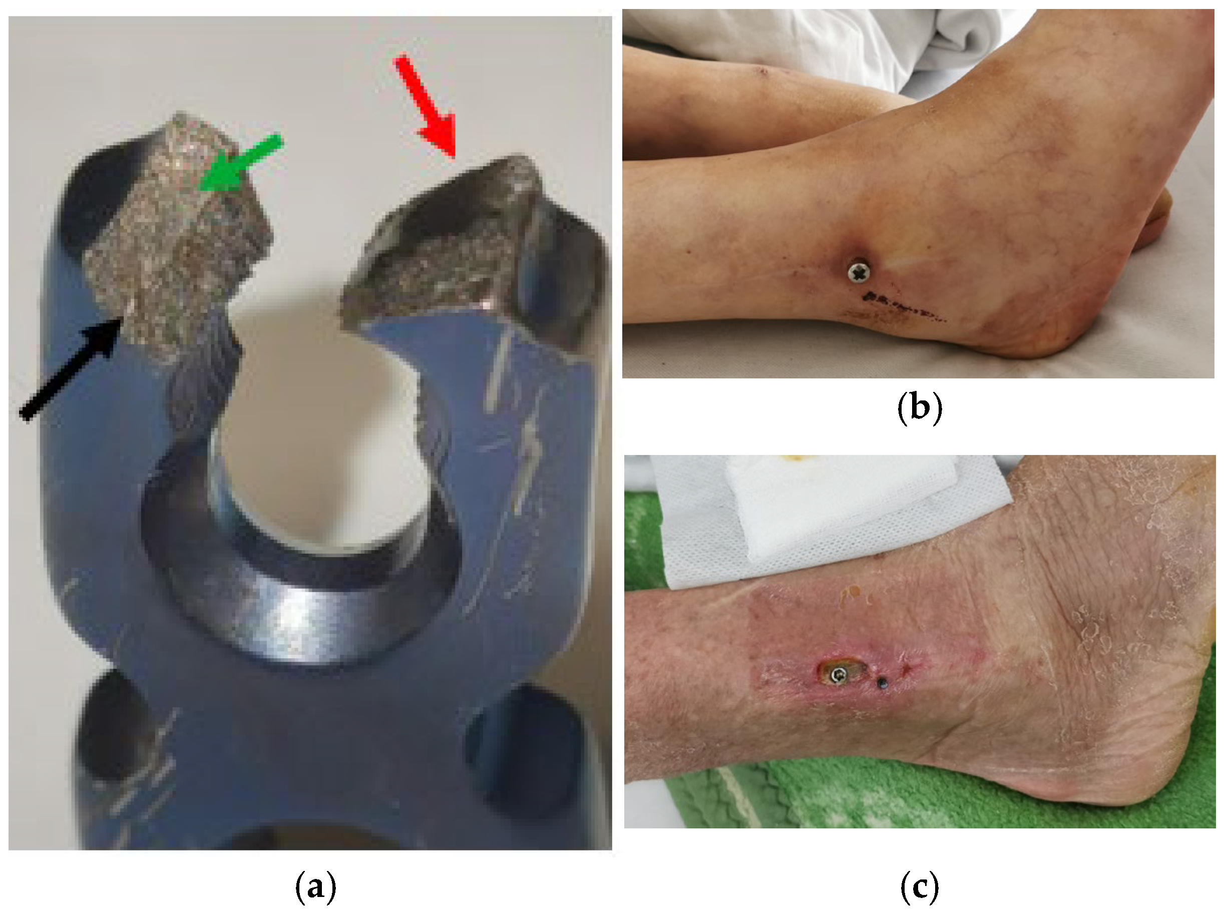
7. Macroscopic Observations of Interacted Surfaces of Removed Implants—Author Observations

The design of an implant significantly influences its surface area and, consequently, the rate at which metal ions are released [138]. Complex geometries with more exposed surfaces or sharp edges may have higher ion release rates compared to smoother, more compact designs. This is because larger surface areas allow for more interaction with bodily fluids, which can accelerate corrosion and the subsequent release of ions. The corrosion rate and metal ion release from implants are significantly influenced by such factors as pH, mechanical stress, electrochemical environment, and temperature. In acidic environments, often found in inflamed or infected tissue, corrosion is accelerated as a lower pH increases the electrochemical reactivity of metals, promoting oxidation and metal ion release. For instance, titanium and cobalt–chromium alloys corrode more rapidly under acidic conditions, leading to a higher release of ions like titanium or cobalt into the bloodstream [75]. Mechanical stress also plays a crucial role, as repetitive loading or weight-bearing on implants can cause wear and tear on the metal surface, exacerbating corrosion by exposing fresh surfaces to the body’s electrolytic environment. Temperature influences the rate of corrosion, with higher body temperatures generally accelerating electrochemical reactions. Additionally, electrolyte concentration (such as chloride ions in bodily fluids) can enhance the corrosion process, while surface roughness and protective coatings can mitigate corrosion by providing a barrier against ion release. Collectively, these factors contribute to the degree of degradation and the potential for systematic toxicity due to the release of harmful metal ions. Furthermore, the porosity of an implant can also play an important role. Implants designed with higher porosity might have more surface area exposed to the surrounding biological environment, increasing the likelihood of ion release. For example, porous titanium implants may exhibit more ion release than solid ones due to their larger exposed area. Material choices in implant design also affect ion release, with materials like cobalt–chromium and stainless steel being more prone to corrosion compared to titanium alloys. The surface finish of an implant plays a crucial role in determining its resistance to corrosion and, therefore, the rate of ion release [139]. Smooth surfaces generally reduce the likelihood of wear and tear and thus the release of ions. Polished surfaces, which are often applied to implants, can form a protective oxide layer that acts as a barrier to further corrosion. In contrast, rough or scratched surfaces expose more of the material to bodily fluids, which can lead to higher rates of ion release. Additionally, surface coatings, such as diamond-like carbon or ceramic coatings, can act as a protective barrier, reducing the amount of metal ion that leaches out of the implant. Anodization processes, commonly used on titanium implants, also help to increase the thickness of the natural oxide layer, thereby minimizing corrosion and ion release. The mechanical loading conditions placed on an implant significantly influence its ion release rate. Dynamic loading, such as the forces generated during joint movement, can cause wear, micro-motion, or deformation of the implant’s surface. These mechanical stresses may disrupt the protective oxide layer, promoting corrosion and thus ion release. For example, joint replacements experience cyclic loads during normal movement, which can gradually wear down the implant surface and lead to ion release [140]. Both fretting (micromovement between contacting surfaces) and abrasion (due to friction between the implant and surrounding tissue) can increase the release of ions. Stress corrosion cracking can also occur under high-loading conditions, further accelerating ion release. Implants under high-load-bearing conditions, such as in weight-bearing joints, are more likely to experience wear and corrosion than those under lower load conditions.
The presented implants exhibit significant surface changes, including discolouration, corrosion, and material degradation, which can have serious biological implications (Figure 5). Several examples of damage to the surface of orthopaedic implants on bone plates and intramedullary nails have been presented. Discolouration and abrasions of the surface of the plate highlight the friction between the implant and the underlying bone fragments. Abrasions on its inner surface, as shown in Figure 5a, probably originate from the relatively high-amplitude movements between the plate and stabilized bone fragments, whereas the oxide layer wiped off from the inner surface of the plate points to the low-amplitude, repeated friction between the implant and the underlying bone (Figure 5b,b1). Areas with abraded surface oxide spots resulting from the repeated, but small in size, relatively mild abrasions show minor friction of tissues (muscles, tendons) sliding over the implant during limb movements. Deep scratches on the outer layer probably originate from surgical intervention during the implant’s removal (Figure 5b1). Three deep parallel scratches are the most probable iatrogenic damages produced during the implant’s removal (green arrows). The broken intramedullary gamma nail (Figure 5d) shows the reason for this (wiped-off oxide showing the high-magnitude, repeated friction between the implant and stabilized bone; red arrows), resulting in its break (red dotted line). Deep scratches on the outer layer of the nail seen on the lower fragment on the right side are iatrogenic and were produced during the implant’s removal (green arrow). Electrochemical interactions on the implants’ surfaces are seen as discolouration (Figure 5e,e1) and corrosion causing deeper pits (Figure 5f) (arrows).
The blue, brown, and golden hues observed on the implants suggest excessive interaction with human tissues, leading to alterations in metal surface composition. Such changes can increase the likelihood of metal ion release (e.g., nickel, chromium, and cobalt), which may contribute to cytotoxicity, systemic toxicity, and local irritation when implanted in the body. Metal ions released into surrounding tissues can trigger inflammatory responses and, in some cases, lead to osteolysis or implant failure. Furthermore, the visible surface damage, such as pitting, cracks, and structural degradation, indicates corrosion or mechanical wear. Corrosion can weaken the implant structure while simultaneously releasing metallic debris into the body. These particles can accumulate in surrounding tissues, potentially leading to chronic inflammation, pain, and hypersensitivity reactions. For patients sensitive to certain metals, such as nickel or cobalt, the presence of corroded or worn implants may increase the risk of allergic responses, including dermatitis, swelling, and localized tissue damage. Additionally, the rough and porous structures suggest material degradation or additive manufacturing techniques that may have undergone wear over time. While porous surfaces can promote osseointegration, excessive degradation can lead to implant instability and bacterial colonization, increasing infection risks.
The compromised integrity of these implants may further exacerbate inflammatory responses, ultimately leading to implant failure (Figure 6). Areas of brown–red coatings point to the corrosion occurring in areas of slowly, gradually progressing implant breakage. They point to the presence of redox reactions occurring on the metal surface not protected by an oxide coating as a result of the interactions between the implant and the surrounding biological environment These observations highlight the importance of implant surface stability to minimize toxicity, irritation, and allergic reactions, ensuring long-term biocompatibility and patient safety.
To minimize adverse reactions associated with metal implants, advancements such as surface modifications, coatings, and the development of novel alloys have become key strategies in improving biocompatibility and implant longevity [141]. These technologies aim to reduce corrosion, enhance osseointegration, and minimize the release of metal ions that could trigger inflammatory or allergic responses in the body.
Surface modification techniques have gained significant attention in recent years as a means to enhance the biocompatibility of metal implants [142]. A widely used method is anodization, which forms a thicker, more stable oxide layer on materials like titanium. This layer acts as a barrier that prevents corrosion and reduces the release of metal ions into the surrounding tissue. For example, titanium implants with anodized surfaces show increased resistance to wear, decreased ion release, and better bone integration. Another approach is plasma spray coating, which can be applied to create rougher surfaces or deposit bioactive materials like hydroxyapatite (HA), a compound similar to the mineral content of bone. This enhances osseointegration, making the implant more stable and reducing the risk of failure due to loosening. Laser surface treatment is another technique that can improve the mechanical properties and corrosion resistance of implants by creating microstructures on the surface that mimic natural bone.
Implant coatings are another promising advancement that helps minimize adverse reactions [142]. Bioceramic coatings, such as hydroxyapatite and tricalcium phosphate, are often used on metal implants to improve bone attachment and reduce the risk of implant loosening. These coatings not only enhance osseointegration but also form a protective layer that minimizes direct contact between the metal and body tissues, decreasing the likelihood of metal ion release and allergic reactions. Furthermore, polymeric coatings, such as those made from plasma-polymerized thin films or biodegradable polymers, can be used to reduce friction and wear at the implant surface, which is especially useful in joint replacements where movement is frequent. Additionally, coatings that release pharmaceuticals such as antibiotics or anti-inflammatory agents could be employed to reduce the risk of infection or chronic inflammation around the implant site.
The development of novel alloys is a key way to reduce adverse reactions. Traditional materials like stainless steel, cobalt–chromium, and titanium alloys have known limitations in terms of corrosion resistance or biocompatibility. Researchers are now focusing on creating bioinert or biodegradable alloys with improved properties. For instance, titanium-based alloys with a more refined composition, such as titanium–niobium or titanium–molybdenum, are being developed to provide better strength and corrosion resistance while maintaining excellent biocompatibility. Magnesium alloys, which are biodegradable, are also being explored for temporary implants, where the material gradually dissolves in the body, reducing the long-term risks associated with permanent implants, such as chronic inflammation or wear debris. The development of cobalt–chromium alternatives with a reduced nickel content is also underway to address the growing concern over nickel allergies, which can cause severe adverse reactions in some patients.

8. Discussion and Summary

The use of metal implants in modern medicine, particularly in orthopaedics and dentistry, has revolutionized patient care by providing durable and effective solutions for bone and joint replacements, dental reconstructions, and trauma management. However, despite their widespread use, metal implants pose significant biological challenges, primarily due to their interactions with the human body. The three major concerns—toxicity, irritation, and allergic reactions—have been extensively documented, raising questions about the long-term safety and viability of these materials, as presented in Table 1. As implant-related complications continue to surface, recent trends in biomedical research have focused on mitigating these risks through material modifications, alternative biomaterials, and personalized medicine approaches.
One of the primary concerns with metal implants is the release of metal ions through corrosion, wear, and electrochemical reactions with bodily fluids. These ions can enter systemic circulation and accumulate in tissues, leading to inflammatory responses and toxicity. Recent studies have demonstrated that metal-on-metal implants, such as cobalt–chromium (CoCr) hip prostheses, generate high levels of metal wear debris, which can induce adverse local tissue reactions and systemic toxicity, including cobalt poisoning. This has led to a shift toward ceramic-on-metal or polymer-on-metal implant designs that aim to reduce wear and ion release. Moreover, surface engineering techniques have gained traction as a method to enhance implant biocompatibility. Advances in nanocoatings, plasma spraying, and anodization have been employed to modify implant surfaces to reduce corrosion and wear. For instance, hydroxyapatite coatings mimic the natural bone matrix, improving osseointegration while acting as a protective layer against metal ion release. Similarly, titanium oxide nanotube coatings have shown promise in reducing bacterial adhesion and inflammation, making implants more resistant to infections. The development of bioactive coatings that incorporate anti-inflammatory and antibacterial agents is another growing trend. For example, silver nanoparticle coatings have demonstrated antibacterial properties, reducing the risk of implant-associated infections while limiting silver toxicity. Additionally, the incorporation of zinc, niobium, and tantalum into titanium alloys has been investigated for their ability to suppress inflammation and enhance biocompatibility.
Despite these advancements, metal implants continue to pose risks due to individual variations in immune responses. While titanium alloys (such as Ti6Al4V) are widely considered biocompatible, recent studies have identified cases of titanium hypersensitivity, leading to implant failure due to persistent inflammation, osteolysis, and pain. This is particularly concerning given titanium’s extensive use in dental and orthopaedic applications. Moreover, the long-term effects of nano-sized metal particles released from implants remain poorly understood, raising concerns about their potential to cross biological barriers and induce neurotoxicity or organ damage. Another significant challenge is implant-related metal allergies, with nickel, cobalt, and chromium being among the most common allergens. Contact dermatitis, chronic inflammation, and Type IV hypersensitivity reactions have been reported in patients with orthopaedic and dental implants. While pre-implantation allergy screening has been suggested, the lack of standardized testing protocols makes it difficult to predict which patients are at risk. The need for personalized implant selection based on genetic and immunological profiling is therefore an area that requires further exploration. Additionally, the impact of mechanical forces on implant degradation is a growing concern. High-load-bearing implants, such as hip and knee replacements, experience constant friction, leading to the formation of metallic debris and reactive oxygen species (ROS). This accelerates oxidative stress and inflammatory reactions, contributing to implant loosening and failure. Innovations in tribology (the study of friction, wear, and lubrication) are being applied to improve implant longevity, but a universally accepted solution is yet to be developed. To address these ongoing issues, researchers are exploring alternative biomaterials that can either replace or complement metal implants. One promising direction is the development of ceramic-based implants, particularly zirconia (ZrO2) and alumina (Al2O3) ceramics, which offer high strength, wear resistance, and excellent biocompatibility. However, their brittleness remains a limiting factor, particularly in load-bearing applications. The use of polymers and composite materials is also gaining attention. Polyether ether ketone (PEEK) has emerged as a strong contender due to its lightweight nature, chemical stability, and radiolucency, making it particularly useful in spinal and orthopaedic implants. Additionally, carbon fibre-reinforced PEEK (CFR-PEEK) is being explored to enhance mechanical properties, potentially providing a viable alternative to traditional metal implants [143].
Another emerging strategy is the incorporation of bioresorbable metal implants, such as magnesium-based alloys, which degrade over time and are gradually replaced by natural bone [144]. This eliminates long-term exposure to metal ions while supporting temporary load-bearing functions. However, challenges related to controlling degradation rates and maintaining structural integrity need to be addressed before these materials can be widely adopted. Three-dimensional printing and personalized implant designs are also paving the way for the next generation of medical implants. Using additive manufacturing, patient-specific implants with tailored porosity and surface modifications can be created, enhancing osseointegration and reducing complications. Furthermore, smart implants embedded with sensors to monitor biomechanical and biochemical changes in real time are being developed, offering a new frontier in implant technology [145]. One can also mention nanotechnology-based surface coatings that have the potential to represent a significant breakthrough in creating bioinert implants with enhanced long-term stability. Nanotechnology allows for the development of highly precise, functionalized surfaces that can improve the biocompatibility, corrosion resistance, and wear resistance of metal implants. These coatings can be engineered at the nanoscale to mimic the natural extracellular matrix, enhancing cell adhesion and osseointegration while reducing the risk of immune responses or inflammation.
The state of the art in nanotechnology coatings includes the development of materials such as nano-structured titanium oxide, hydroxyapatite (HA), carbon nanotubes, nanocomposite coatings, and biopolymer-based nanocoatings. These materials can be tailored to control the release of metal ions, prevent biofilm formation, and reduce the risk of implant-associated infections. For instance, nano-hydroxyapatite coatings can promote bone growth by improving the attachment and proliferation of osteoblasts around the implant, facilitating faster integration and reducing the likelihood of implant loosening [146,147,148]. Additionally, nano-silver and nano-zinc coatings have been explored for their antibacterial properties, which could significantly reduce infection rates, particularly in patients with compromised immune systems. Furthermore, smart coatings are being developed that can respond to changes in the local environment, such as pH or mechanical stress, to release therapeutic agents (e.g., antibiotics or anti-inflammatory drugs) in a controlled manner, further enhancing the safety and longevity of implants [149,150,151].
While these coatings have shown great promise in pre-clinical studies, challenges remain in ensuring consistent manufacturing, scalability, and long-term stability under physiological conditions. Due to their small size, nanoparticles exhibit high surface reactivity and unique physicochemical properties that can lead to unintended biological interactions, raising concerns about cytotoxicity, immune system activation, oxidative stress, and systemic toxicity [152,153]. One of the primary concerns with nanotechnology-based coatings is the potential detachment of nanoparticles from the implant surface. During mechanical stress, wear, or corrosion, nanoparticles can be released into surrounding tissues and enter systemic circulation. Once in the bloodstream, they may accumulate in various organs such as the liver, kidneys, lungs, and brain, leading to systemic toxicity [154]. Studies have shown that certain nanoparticles, including titanium dioxide (TiO2) and silver (Ag) nanoparticles, can cross biological barriers, such as the blood–brain barrier, potentially leading to neurological effects [155]. These particles may also disrupt normal cellular function, interfere with enzymatic activity, and contribute to metabolic imbalances. Many nanomaterials, particularly metal-based nanoparticles, can induce oxidative stress by generating reactive oxygen species (ROS) [156]. ROS can damage cellular components, including lipids, proteins, and DNA, leading to apoptosis (programmed cell death) or necrosis. Silver nanoparticles, widely used for their antimicrobial properties, are particularly known for their ability to generate ROS, which can cause oxidative stress in human cells [157]. While this effect is beneficial for preventing infections, prolonged exposure to high concentrations of silver nanoparticles may lead to cellular toxicity, DNA fragmentation, and mitochondrial dysfunction. Similarly, titanium dioxide nanoparticles, despite their biocompatibility in bulk form, may induce oxidative damage at the nanoscale, potentially impairing tissue healing and osseointegration [154]. The immune system plays a critical role in determining the biocompatibility of nanocoatings. Some nanomaterials, particularly carbon nanotubes and metal nanoparticles, have been shown to activate immune responses, leading to chronic inflammation [155]. This immune activation can result in prolonged tissue irritation, fibrosis, and, in some cases, osteolysis (bone degradation). For instance, cobalt–chromium nanoparticles released from wear-resistant coatings have been linked to hypersensitivity reactions and peri-implant inflammation [156]. Chronic inflammation not only compromises implant stability but also increases the risk of implant failure due to excessive bone resorption and fibrotic tissue formation. Moreover, certain nanocoatings may act as haptens, binding to proteins in the body and triggering an allergic response [157]. Nickel nanoparticles, for example, have been reported to provoke hypersensitivity reactions in individuals with metal allergies [158]. This raises concerns about the widespread use of nickel-containing alloys in medical implants and emphasizes the need for alternative coatings that minimize immune activation. While nanocoatings are designed to enhance osseointegration, some nanoparticles may interfere with bone cell activity, leading to adverse effects on implant stability [159]. Osteoblasts (bone-forming cells) and osteoclasts (bone-resorbing cells) are highly sensitive to changes in their microenvironment. Excessive nanoparticle exposure can disrupt their normal function, impairing bone healing and integration. For example, studies suggest that high concentrations of cobalt, chromium, or silver nanoparticles can inhibit osteoblast proliferation and differentiation while promoting osteoclast activity [160]. This imbalance may contribute to implant loosening and failure over time. However, controlled-release nanocoatings have been developed to deliver bioactive molecules that promote bone regeneration while minimizing toxicity. For instance, nanocoatings incorporating calcium phosphate or hydroxyapatite have been shown to improve osteoblast adhesion and mineralization, enhancing implant integration. The challenge lies in designing coatings that provide these benefits without the unintended release of toxic nanoparticles into the surrounding tissues.
While metal implants continue to play an essential role in modern medicine, their long-term interactions with the human body remain a subject of concern. Recent advancements in material science and bioengineering have focused on reducing metal toxicity, irritation, and allergic reactions through surface modifications, alternative materials, and smart implant designs. However, challenges persist in terms of implant degradation, immune responses, and mechanical wear, necessitating further research into novel biomaterials and personalized medicine approaches. The future of implant technology lies in the integration of multidisciplinary research, combining materials science, immunology, bioengineering, and nanotechnology to develop safer, more effective, and patient-specific solutions. As new materials and technologies emerge, the goal should be to strike a balance between mechanical durability and biological compatibility, ensuring that medical implants not only restore function but also minimize long-term health risks.

9. Conclusions

This review highlights the transformative impact of metal implants in orthopaedics and dentistry, underscoring the complexity of their interactions with biological systems. Despite their durability and mechanical strength, metals such as titanium, cobalt, chromium, and nickel pose significant challenges due to corrosion, ion release, and immune responses. This paper reveals that metal ions can trigger local and systemic effects, including inflammation, oxidative stress, and hypersensitivity reactions, which may lead to implant failure or chronic health conditions. The biological environment’s aggressive nature, coupled with mechanical wear, accelerates these processes, raising concerns about long-term implant safety. Emerging evidence even questions the bioinert status of widely used materials like titanium, highlighting the need for a more nuanced understanding of their impact on cellular metabolism and genetic stability.
Future research should prioritize the development of advanced surface modifications, including nanotechnology-based coatings, to mitigate corrosion and reduce ion release. Exploring bioinert and biodegradable alloys tailored to patient-specific conditions could offer safer alternatives, particularly for high-risk populations with autoimmune disorders or metal sensitivities. Additionally, integrating smart implants with real-time monitoring capabilities to detect early signs of corrosion or inflammatory responses could revolutionize patient care. Personalized implant design, powered by additive manufacturing and informed by genetic and immunological profiling, promises to optimize biocompatibility and longevity. Ultimately, the next generation of metal implants should strive not only for mechanical excellence but also for harmonious integration with the human body, minimizing adverse effects while promoting long-term health and functionality. This interdisciplinary approach, bridging materials science, immunology, and clinical medicine, will be pivotal in shaping the future of implant technology and improving patient outcomes worldwide.

Author Contributions

Conceptualisation, G.S.; methodology, G.S., M.K. and Z.L.K.; validation, M.K. and Z.L.K.; formal analysis, G.S., M.K. and Z.L.K.; investigation, G.S. and M.K.; resources, G.S. and M.K.; data curation, G.S.; writing—original draft preparation, G.S. and Z.L.K.; writing—review and editing, Z.L.K. and M.K.; visualisation, G.S. and M.K.; supervision, G.S.; project administration, G.S. All authors have read and agreed to the published version of the manuscript.

Funding

This research received no external funding.

Institutional Review Board Statement

Not applicable.

Data Availability Statement

All data are available upon request. The data presented in this study are available on request from the corresponding author.

Conflicts of Interest

The authors declare no conflicts of interest.

References

  1. Bugaj, U.; Nejbert, K.; Ilnicki, S.; Wieciński, P.; Onyszczuk, T.; Garbacz, H.; Włodarczak, P. Copper sulphosalts in early metallurgy (2600–1900 BC)—Chemical-mineralogical investigation of artefacts from southern Poland. Geol. Q. 2019, 63, 302–318. [Google Scholar] [CrossRef]
  2. Radivojević, M.; Roberts, B.W. Early Balkan Metallurgy: Origins, Evolution and Society, 6200–3700 BC. J. World Prehistory 2021, 34, 195–278. [Google Scholar] [CrossRef]
  3. Adams, M. Advances in Gold Processing; Elsevier B.V.: Perth, Australia, 2005. [Google Scholar]
  4. Murr, L.E. A Brief History of Metals. In Handbook of Materials Structures, Properties, Processing and Performance; Springer: Cham, Switzerland, 2015. [Google Scholar] [CrossRef]
  5. Killick, D. From Ores to Metals. In Archaeometallurgy in Global Perspective; Roberts, B., Thornton, C., Eds.; Springer: New York, NY, USA, 2014. [Google Scholar] [CrossRef]
  6. Winder, C. The History of Lead, Part 3. 1994. ISSN 1324-6011. Available online: https://lead.org.au/lanv2n3/lanv2n3-22.html (accessed on 10 February 2025).
  7. Bartoníček, J. Early history of operative treatment of fractures. Arch. Orthop. Trauma Surg. 2010, 130, 1385–1396. [Google Scholar] [CrossRef] [PubMed]
  8. Walsh, S. Identity as Process: An Archaeological and Osteological Study of Early Bronze Age Burials in Northern England. Ph.D. Thesis, University of Central Lancashire, Preston, UK, 2013. [Google Scholar]
  9. Salah, I.; Parkin, I.P.; Allan, E. Copper as an antimicrobial agent: Recent advances. RSC Adv. 2021, 11, 18179–18186. [Google Scholar] [CrossRef] [PubMed] [PubMed Central]
  10. Sinha, N.; Hamre, H.J.; Musial, F.; Werner, E.L.; Björkman, L. Health complaints before and at one and five years after removal of dental amalgam restorations—Data from a prospective cohort study in Norway. Acta Odontol. Scand. 2024, 83, 219–229. [Google Scholar] [CrossRef] [PubMed] [PubMed Central]
  11. Council of the European Union. Council Signs Off on Measures to Make the EU Mercury-Free. Available online: https://www.consilium.europa.eu/en/press/press-releases/2024/05/30/council-signs-off-on-measures-to-make-the-eu-mercury-free/ (accessed on 10 February 2025).
  12. Björkman, L.; Musial, F.; Alræk, T.; Werner, E.L.; Hamre, H.J. Mercury, silver and selenium in serum before and after removal of amalgam restorations: Results from a prospective cohort study in Norway. Acta Odontol. Scand. 2022, 81, 298–310. [Google Scholar] [CrossRef] [PubMed]
  13. Brown, R.P.; Fowler, B.A.; Fustinoni, S.; Nordberg, M. Handbook on the Toxicology of Metals, 4th ed.; Academic Press: Cambridge, MA, USA, 2015; pp. 113–122. ISBN 9780444594532. [Google Scholar] [CrossRef]
  14. Hall, J.E. Guyton and Hall Textbook of Medical Physiology, 13th ed.; Elsevier: Philadephia, PA, USA, 2016. [Google Scholar]
  15. Ottaviano, F.G.; Handy, D.E.; Loscalzo, J. Redox Regulation in the Extracellular Environment. Circ. J. 2008, 72, 1–16. [Google Scholar] [CrossRef] [PubMed]
  16. Eliaz, N. Corrosion of Metallic Biomaterials: A Review. Materials 2019, 12, 407. [Google Scholar] [CrossRef] [PubMed] [PubMed Central]
  17. Song, Y.; Jiang, G.; Chen, Y.; Zhao, P.; Tian, Y. Effects of chloride ions on corrosion of ductile iron and carbon steel in soil environments. Sci. Rep. 2017, 7, 6865. [Google Scholar] [CrossRef]
  18. Hashimoto, K.; Asami, K.; Teramoto, K. An X-ray photo-electron spectroscopic study on the role of molybdenum in increasing the corrosion resistance of ferritic stainless steels in HCl. Corros. Sci. 1979, 19, 3–14. [Google Scholar] [CrossRef]
  19. Gotman, I. Characteristics of Metals Used in Implants. J. Endourol. 1997, 11, 383–389. [Google Scholar] [CrossRef] [PubMed]
  20. Knecht, C.; Polakof, L.; Behrens, J.; Goodman, S.B. Wear debris in metal-on-metal bearings and modular junctions: What have we learned from the last decades? Orthopadie 2023, 52, 206–213. [Google Scholar] [CrossRef] [PubMed]
  21. Wang, Y.; Yan, Y.; Su, Y.; Qiao, L. Release of metal ions from nano CoCrMo wear debris generated from tribo-corrosion processes in artificial hip implants. J. Mech. Behav. Biomed. Mater. 2017, 68, 124–133. [Google Scholar] [CrossRef] [PubMed]
  22. Aro, H.; Eerola, E.; Aho, A.; Niinikoski, J. Tissue oxygen tension in externally stabilized tibial fractures in rabbits during normal healing and infection. J. Surg. Res. 1984, 37, 202–207. [Google Scholar] [CrossRef] [PubMed]
  23. Laing, P.G. Compatibility of biomaterials. Orthop. Clin. N. Am. 1973, 4, 249–273. [Google Scholar] [CrossRef] [PubMed]
  24. Behbahani, S.B.; Kiridena, S.D.; Wijayaratna, U.N.; Taylor, C.; Anker, J.N.; Tzeng, T.-R.J. pH variation in medical implant biofilms: Causes, measurements, and its implications for antibiotic resistance. Front. Microbiol. 2022, 13, 1028560. [Google Scholar] [CrossRef] [PubMed] [PubMed Central]
  25. Lebeaux, D.; Chauhan, A.; Létoffé, S.; Fischer, F.; de Reuse, H.; Beloin, C.; Ghigo, J.-M. pH-Mediated Potentiation of Aminoglycosides Kills Bacterial Persisters and Eradicates In Vivo Biofilms. J. Infect. Dis. 2014, 210, 1357–1366. [Google Scholar] [CrossRef] [PubMed]
  26. Hengge-Aronis, R. Signal Transduction and Regulatory Mechanisms Involved in Control of the σS(RpoS) Subunit of RNA Polymerase. Microbiol. Mol. Biol. Rev. 2002, 66, 373–395. [Google Scholar] [CrossRef] [PubMed] [PubMed Central]
  27. Blair, T.A.; Frelinger, A.L. Platelet surface marker analysis by mass cytometry. Platelets 2020, 31, 633–640. [Google Scholar] [CrossRef] [PubMed]
  28. Pretorius, E.; Vermeulen, N.; Bester, J.; Lipinski, B.; Kell, D.B. A novel method for assessing the role of iron and its functional chelation in fibrin fibril formation: The use of scanning electron microscopy. Toxicol. Mech. Methods 2013, 23, 352–359. [Google Scholar] [CrossRef] [PubMed]
  29. Steven, F.S.; Griffin, M.M.; Brown, B.S.; Hulley, T.P. Aggregation of fibrinogen molecules by metal ion. Int. J. Biol. Macromol. 1982, 4, 367–369. [Google Scholar] [CrossRef]
  30. Goodman, S.B.; Gallo, J.; Gibon, E.F.; Takagi, M. Diagnosis and management of implant debris-associated inflammation. Expert Rev. Med. Devices 2020, 17, 41–56. [Google Scholar] [CrossRef] [PubMed] [PubMed Central]
  31. Roach, K.; Roberts, J. A comprehensive summary of disease variants implicated in metal allergy. J. Toxicol. Environ. Health B Crit. Rev. 2022, 25, 279–341. [Google Scholar] [CrossRef] [PubMed] [PubMed Central]
  32. Sansone, V.; Pagani, D.; Melato, M. The effects on bone cells of metal ions released from orthopaedic implants. A review. Clin. Cases Miner. Bone Metab. 2013, 10, 34–40. [Google Scholar] [CrossRef] [PubMed] [PubMed Central]
  33. Noronha Oliveira, M.; Schunemann, W.V.H.; Mathew, M.T.; Henriques, B.; Magini, R.S.; Teughels, W.; Souza, J.C.M. Can degradation products released from dental implants affect peri-implant tissues? J. Periodontal Res. 2018, 53, 1–11. [Google Scholar] [CrossRef] [PubMed]
  34. Simkiss, K. Lipid solubility of heavy metals in saline solutions. J. Mar. Biol. Assoc. U. K. 1983, 63, 1–7. [Google Scholar] [CrossRef]
  35. Rizzollo, F.; More, S.; Vangheluwe, P.; Agostinis, P. The lysosome as a master regulator of iron metabolism. Trends Biochem. Sci. 2021, 46, 960–975. [Google Scholar] [CrossRef] [PubMed]
  36. Britton, R.S. Metal-Induced Hepatotoxicity. Semin. Liver Dis. 1996, 16, 3–12. [Google Scholar] [CrossRef] [PubMed]
  37. Blaby-Haas, C.E.; Merchant, S.S. Lysosome-related Organelles as Mediators of Metal Homeostasis. J. Biol. Chem. 2014, 289, 28129–28136. [Google Scholar] [CrossRef] [PubMed] [PubMed Central]
  38. Jaishankar, M.; Tseten, T.; Anbalagan, N.; Mathew, B.B.; Beeregowda, K.N. Toxicity, mechanism and health effects of some heavy metals. Interdiscip. Toxicol. 2014, 7, 60–72. [Google Scholar] [CrossRef] [PubMed] [PubMed Central]
  39. Granchi, D.; Cenni, E.; Ciapetti, G.; Savarino, L.; Stea, S.; Gamberini, S.; Gori, A.; Pizzoferrato, A. Cell death induced by metal ions: Necrosis or apoptosis? J. Mater. Sci. Mater. Med. 1998, 9, 31–37. [Google Scholar] [CrossRef] [PubMed]
  40. European Commission Taxation and Customs Union. Skin Irritants Health and Safety. Available online: https://ec.europa.eu/taxation_customs/dds2/SAMANCTA/EN/Safety/SkinIrritants_EN.htm (accessed on 10 February 2025).
  41. Lechuga, M.; Fernández-Serrano, M.; Núñez-Olea, J.; Martínez-Gallegos, J.F.; Ríos, F. Optimization of Toxicity, Biodegradability, and Skin Irritation in Formulations Containing Mixtures of Anionic and Nonionic Surfactants Combined with Silica Nanoparticles. Toxics 2025, 13, 43. [Google Scholar] [CrossRef] [PubMed]
  42. Nishida, C.; Yatera, K. The Impact of Ambient Environmental and Occupational Pollution on Respiratory Diseases. Int. J. Environ. Res. Public Health 2022, 19, 2788. [Google Scholar] [CrossRef] [PubMed] [PubMed Central]
  43. Deng, L.; He, S.; Guo, N.; Tian, W.; Zhang, W.; Luo, L. Molecular mechanisms of ferroptosis and relevance to inflammation. Inflamm. Res. 2023, 72, 281–299. [Google Scholar] [CrossRef] [PubMed] [PubMed Central]
  44. Tramontana, M.; Hansel, K.; Bianchi, L.; Sensini, C.; Malatesta, N.; Stingeni, L. Advancing the understanding of allergic contact dermatitis: From pathophysiology to novel therapeutic approaches. Front. Med. 2023, 10, 1184289. [Google Scholar] [CrossRef] [PubMed] [PubMed Central]
  45. Cadosch, D.; Chan, E.; Gautschi, O.P.; Filgueira, L. Metal is not inert: Role of metal ions released by biocorrosion in aseptic loosening—Current concepts. J. Biomed. Mater. Res. A 2009, 91, 1252–1262. [Google Scholar] [CrossRef] [PubMed]
  46. Teschke, R. Aluminum, Arsenic, Beryllium, Cadmium, Chromium, Cobalt, Copper, Iron, Lead, Mercury, Molybdenum, Nickel, Platinum, Thallium, Titanium, Vanadium, and Zinc: Molecular Aspects in Experimental Liver Injury. Int. J. Mol. Sci. 2022, 23, 12213. [Google Scholar] [CrossRef] [PubMed] [PubMed Central]
  47. Pacheco, K.A. Allergy to Surgical Implants. Clin. Rev. Allergy Immunol. 2019, 56, 72–85. [Google Scholar] [CrossRef] [PubMed]
  48. Lim, W.; Afif, W.; Knowles, S.; Lim, G.; Lin, Y.; Mothersill, C.; Nistor, I.; Rehman, F.; Song, C.; Xenodemetropoulos, T. Canadian expert consensus: Management of hypersensitivity reactions to intravenous iron in adults. Vox Sang. 2019, 114, 363–373. [Google Scholar] [CrossRef] [PubMed] [PubMed Central]
  49. Caimmi, S.; Crisafulli, G.; Franceschini, F.; Liotti, L.; Bianchi, A.; Bottau, P.; Mori, F.; Triggiano, P.; Paglialunga, C.; Saretta, F.; et al. Hypersensitivity to Intravenous Iron Preparations. Children 2022, 9, 1473. [Google Scholar] [CrossRef] [PubMed] [PubMed Central]
  50. Szebeni, J.; Fishbane, S.; Hedenus, M.; Howaldt, S.; Locatelli, F.; Patni, S.; Rampton, D.; Weiss, G.; Folkersen, J. Hypersensitivity to intravenous iron: Classification, terminology, mechanisms and management. Br. J. Pharmacol. 2015, 172, 5025–5036. [Google Scholar] [CrossRef] [PubMed] [PubMed Central]
  51. Xu, H.; Ohgami, N.; Sakashita, M.; Ogi, K.; Hashimoto, K.; Tazaki, A.; Tong, K.; Aoki, M.; Fujieda, S.; Kato, M. Intranasal levels of lead as an exacerbation factor for allergic rhinitis in humans and mice. J. Allergy Clin. Immunol. 2021, 148, 139–147.e10. [Google Scholar] [CrossRef] [PubMed]
  52. Bjørklund, G.; Peana, M.; Dadar, M.; Chirumbolo, S.; Aaseth, J.; Martins, N. Mercury-induced autoimmunity: Drifting from micro to macro concerns on autoimmune disorders. Clin. Immunol. 2020, 213, 108352. [Google Scholar] [CrossRef] [PubMed]
  53. Baseri, M.; Radmand, F.; Hamedi, R.; Yousefi, M.; Kafil, H.S. Immunological Aspects of Dental Implant Rejection. BioMed Res. Int. 2020, 2020, 7279509. [Google Scholar] [CrossRef] [PubMed] [PubMed Central]
  54. D’ambrosi, R.; Nuara, A.; Mariani, I.; Di Feo, F.; Ursino, N.; Hirschmann, M. Titanium Niobium Nitride Mobile-Bearing Unicompartmental Knee Arthroplasty Results in Good to Excellent Clinical and Radiographic Outcomes in Metal Allergy Patients With Medial Knee Osteoarthritis. J. Arthroplast. 2021, 36, 140–147.e2. [Google Scholar] [CrossRef] [PubMed]
  55. Contuzzi, N.; Casalino, G.; Boccaccio, A.; Ballini, A.; Charitos, I.A.; Bottalico, L.; Santacroce, L. Metals Biotribology and Oral Microbiota Biocorrosion Mechanisms. J. Funct. Biomater. 2022, 14, 14. [Google Scholar] [CrossRef] [PubMed] [PubMed Central]
  56. Sun, Y.; Liu, T.; Hu, H.; Xiong, Z.; Zhang, K.; He, X.; Liu, W.; Lei, P.; Hu, Y. Differential effect of tantalum nanoparticles versus tantalum micron particles on immune regulation. Mater. Today Bio 2022, 16, 100340. [Google Scholar] [CrossRef] [PubMed] [PubMed Central]
  57. Zhang, B.; Su, Y.; Zhou, J.; Zheng, Y.; Zhu, D. Toward a Better Regeneration through Implant-Mediated Immunomodulation: Harnessing the Immune Responses. Adv. Sci. 2021, 8, e2100446. [Google Scholar] [CrossRef] [PubMed] [PubMed Central]
  58. Qiu, N.; Yang, X.; Zhang, Y.; Zhang, J.; Ji, J.; Zhang, Y.; Kong, X.; Xi, Y.; Liu, D.; Ye, L.; et al. A molybdenum oxide-based degradable nanosheet for combined chemo-photothermal therapy to improve tumor immunosuppression and suppress distant tumors and lung metastases. J. Nanobiotechnol. 2021, 19, 428. [Google Scholar] [CrossRef]
  59. Mascarenhas-Melo, F.; Mathur, A.; Murugappan, S.; Sharma, A.; Tanwar, K.; Dua, K.; Singh, S.K.; Mazzola, P.G.; Yadav, D.N.; Rengan, A.K.; et al. Inorganic nanoparticles in dermopharmaceutical and cosmetic products: Properties, formulation development, toxicity, and regulatory issues. Eur. J. Pharm. Biopharm. 2023, 192, 25–40. [Google Scholar] [CrossRef] [PubMed]
  60. Möller, H. Contact allergy to gold as a model for clinical-experimental research. Contact Dermat. 2010, 62, 193–200. [Google Scholar] [CrossRef] [PubMed]
  61. Zemelka-Wiacek, M. Metal Allergy: State-of-the-Art Mechanisms, Biomarkers, Hypersensitivity to Implants. J. Clin. Med. 2022, 11, 6971. [Google Scholar] [CrossRef] [PubMed] [PubMed Central]
  62. Dispenza, M.C. Classification of hypersensitivity reactions. Allergy Asthma Proc. 2019, 40, 470–473. [Google Scholar] [CrossRef] [PubMed]
  63. Loh, J.; Fraser, J. Metal-derivatized Major Histocompatibility Complex: Zeroing in on contact hypersensitivity. J. Exp. Med. 2003, 197, 549–552. [Google Scholar] [CrossRef]
  64. Büdinger, L.; Hertl, M. Immunologic mechanisms in hypersensitivity reactions to metal ions: An overview. Allergy 2000, 55, 108–115. [Google Scholar] [CrossRef] [PubMed]
  65. Riedel, F.; Aparicio-Soto, M.; Curato, C.; Thierse, H.-J.; Siewert, K.; Luch, A. Immunological Mechanisms of Metal Allergies and the Nickel-Specific TCR-pMHC Interface. Int. J. Environ. Res. Public Health 2021, 18, 10867. [Google Scholar] [CrossRef] [PubMed] [PubMed Central]
  66. Schmidt, M.; Raghavan, B.; Müller, V.; Vogl, T.; Fejer, G.; Tchaptchet, S.; Keck, S.; Kalis, C.; Nielsen, P.J.; Galanos, C.; et al. Crucial role for human Toll-like receptor 4 in the development of contact allergy to nickel. Nat. Immunol. 2010, 11, 814–819. [Google Scholar] [CrossRef] [PubMed]
  67. Thierse, H.-J.; Gamerdinger, K.; Junkes, C.; Guerreiro, N.; Weltzien, H.U. T cell receptor (TCR) interaction with haptens: Metal ions as non-classical haptens. Toxicology 2005, 209, 101–107. [Google Scholar] [CrossRef] [PubMed]
  68. Nguyen, Q.P.; Deng, T.Z.; Witherden, D.A.; Goldrath, A.W. Origins of CD4+ circulating and tissue-resident memory T-cells. Immunology 2019, 157, 3–12. [Google Scholar] [CrossRef]
  69. Shin, H.; Iwasaki, A. Tissue-resident memory T cells. Immunol. Rev. 2013, 255, 165–181. [Google Scholar] [CrossRef]
  70. Fransway, A.F.; Zug, K.A.; Belsito, D.V.; DeLeo, V.A.; Fowler, J.F.; Maibach, H.I.; Marks, J.G.; Mathias, C.T.; Pratt, M.D.; Rietschel, R.L.; et al. North American Contact Dermatitis Group Patch Test Results for 2007–2008. Dermatitis 2013, 24, 10–21. [Google Scholar] [CrossRef] [PubMed]
  71. Hanawa, T. Degradation of dental implants. In Degradation of Implant Materials; Eliaz, N., Ed.; Springer: New York, NY, USA, 2012; Chapter 3; pp. 57–78. [Google Scholar]
  72. Simón-Vázquez, R.; Lozano-Fernández, T.; Dávila-Grana, A.; González-Fernández, Á. Metal oxide nanoparticles interact with immune cells and activate different cellular responses. Int. J. Nanomed. 2016, 11, 4657–4668. [Google Scholar] [CrossRef] [PubMed] [PubMed Central]
  73. Nagay, B.E.; Cordeiro, J.M.; Barao, V.A.R. Insight into Corrosion of Dental Implants: From Biochemical Mechanisms to Designing Corrosion-Resistant Materials. Curr. Oral Health Rep. 2022, 9, 7–21. [Google Scholar] [CrossRef] [PubMed] [PubMed Central]
  74. Delaunay, C.; Petit, I.; Learmonth, I.; Oger, P.; Vendittoli, P. Metal-on-metal bearings total hip arthroplasty: The cobalt and chromium ions release concern. Orthop. Traumatol. Surg. Res. 2010, 96, 894–904. [Google Scholar] [CrossRef] [PubMed]
  75. Dikici, B.; Esen, Z.; Duygulu, O.; Gungor, S. Corrosion of Metallic Biomaterials. In Advances in Metallic Biomaterials; Springer Series in Biomaterials Science and Engineering; Niinomi, M., Narushima, T., Nakai, M., Eds.; Springer: Berlin/Heidelberg, Germany, 2015; Volume 3. [Google Scholar] [CrossRef]
  76. Grivennikov, S.I.; Greten, F.R.; Karin, M. Immunity, inflammation, and cancer. Cell 2010, 140, 883–899. [Google Scholar] [CrossRef] [PubMed] [PubMed Central]
  77. Matharu, G.S.; Berryman, F.; Judge, A.; Reito, A.; McConnell, J.; Lainiala, O.; Young, S.; Eskelinen, A.; Pandit, H.G.; Murray, D.W. Blood Metal Ion Thresholds to Identify Patients with Metal-on-Metal Hip Implants at Risk of Adverse Reactions to Metal Debris: An External Multicenter Validation Study of Birmingham Hip Resurfacing and Corail-Pinnacle Implants. J. Bone Jt. Surg. 2017, 99, 1532–1539. [Google Scholar] [CrossRef]
  78. Přikrylová, J.; Procházková, J.; Podzimek, Š. Side Effects of Dental Metal Implants: Impact on Human Health (Metal as a Risk Factor of Implantologic Treatment). BioMed Res. Int. 2019, 2019, 2519205. [Google Scholar] [CrossRef] [PubMed] [PubMed Central]
  79. Clark, D.; Nakamura, M.; Miclau, T.; Marcucio, R. Effects of Aging on Fracture Healing. Curr. Osteoporos. Rep. 2017, 15, 601–608. [Google Scholar] [CrossRef] [PubMed] [PubMed Central]
  80. Ortona, E.; Pagano, M.T.; Capossela, L.; Malorni, W. The Role of Sex Differences in Bone Health and Healing. Biology 2023, 12, 993. [Google Scholar] [CrossRef] [PubMed] [PubMed Central]
  81. Müller-Heupt, L.K.; Schiegnitz, E.; Kaya, S.; Jacobi-Gresser, E.; Kämmerer, P.W.; Al-Nawas, B. Diagnostic tests for titanium hypersensitivity in implant dentistry: A systematic review of the literature. Int. J. Implant. Dent. 2022, 8, 29. [Google Scholar] [CrossRef] [PubMed] [PubMed Central]
  82. Lechtman, H.; Klein, S. The Production of Copper–Arsenic Alloys (Arsenic Bronze) by Cosmelting: Modern Experiment, Ancient Practice. J. Archaeol. Sci. 1999, 26, 497–526. [Google Scholar] [CrossRef]
  83. Spoto, G. Secondary ion mass spectrometry in art and archaeology. Thermochim. Acta 2000, 365, 157–166. [Google Scholar] [CrossRef]
  84. Ro, S.-H.; Bae, J.; Jang, Y.; Myers, J.F.; Chung, S.; Yu, J.; Natarajan, S.K.; Franco, R.; Song, H.-S. Arsenic Toxicity on Metabolism and Autophagy in Adipose and Muscle Tissues. Antioxidants 2022, 11, 689. [Google Scholar] [CrossRef] [PubMed] [PubMed Central]
  85. Sarker, A.; Kim, J.-E.; Islam, A.R.M.T.; Bilal, M.; Rakib, R.J.; Nandi, R.; Rahman, M.M.; Islam, T. Heavy metals contamination and associated health risks in food webs—A review focuses on food safety and environmental sustainability in Bangladesh. Environ. Sci. Pollut. Res. 2022, 29, 3230–3245. [Google Scholar] [CrossRef] [PubMed] [PubMed Central]
  86. Mathec, A.; von Schirnding, Y.; Montgomery, M.; Röllin, H. Lead Poisoning in South African Children: The Hazard is at Horne. Rev. Environ. Health 2021, 19, 347–362. [Google Scholar] [CrossRef] [PubMed]
  87. Boscolo, M.; Antonucci, S.; Volpe, A.R.; Carmignani, M.; Di Gioacchino, M. Acute mercury intoxication and use of chelating agents. J. Biol. Regul. Homeost. Agents 2009, 23, 217–223. [Google Scholar] [PubMed]
  88. Bjørklund, G.; Crisponi, G.; Nurchi, V.M.; Cappai, R.; Djordjevic, A.B.; Aaseth, J. A Review on Coordination Properties of Thiol-Containing Chelating Agents Towards Mercury, Cadmium, and Lead. Molecules 2019, 24, 3247. [Google Scholar] [CrossRef] [PubMed] [PubMed Central]
  89. Thomas, O.; Le Bot, B.; Verrey, D.; Durand, S.; Harpet, C.; Froment, A.; Jégou, B. High lead level in the Alps in XIXth century, learning from the analysis of 138 historical hair stands. Chemosphere 2022, 286, 131658. [Google Scholar] [CrossRef] [PubMed]
  90. Charkiewicz, A.E.; Backstrand, J.R. Lead Toxicity and Pollution in Poland. Int. J. Environ. Res. Public Health 2020, 17, 4385. [Google Scholar] [CrossRef]
  91. Sun, M.; Ju, J.; Ding, Y.; Zhao, C.; Tian, C. The signaling pathways regulated by KRAB zinc-finger proteins in cancer. Biochim. Biophys. Acta Rev. Cancer 2022, 1877, 188731. [Google Scholar] [CrossRef]
  92. Guo, H.; Liu, H.; Wu, H.; Cui, H.; Fang, J.; Zuo, Z.; Deng, J.; Li, Y.; Wang, X.; Zhao, L. Nickel Carcinogenesis Mechanism: DNA Damage. Int. J. Mol. Sci. 2019, 20, 4690. [Google Scholar] [CrossRef] [PubMed]
  93. Markiewicz-Górka, I.; Januszewska, L.; Michalak, A.; Prokopowicz, A.; Januszewska, E.; Pawlas, N.; Pawlas, K. Effects of chronic exposure to lead, cadmium, and manganese mixtures on oxidative stress in rat liver and heart. Arch. Ind. Hyg. Toxicol. 2015, 66, 51–62. [Google Scholar] [CrossRef] [PubMed]
  94. Ge, E.J.; Bush, A.I.; Casini, A.; Cobine, P.A.; Cross, J.R.; DeNicola, G.M.; Dou, Q.P.; Franz, K.J.; Gohil, V.M.; Gupta, S.; et al. Connecting copper and cancer: From transition metal signalling to metalloplasia. Nat. Rev. Cancer 2022, 22, 102–113. [Google Scholar] [CrossRef] [PubMed] [PubMed Central]
  95. Shen, L.; Zhou, Y.; He, H.; Chen, W.; Lenahan, C.; Li, X.; Deng, Y.; Shao, A.; Huang, J. Crosstalk between Macrophages, T Cells, and Iron Metabolism in Tumor Microenvironment. Oxidative Med. Cell. Longev. 2021, 2021, 8865791. [Google Scholar] [CrossRef]
  96. Kłys, M. Z rtecia (i…) przez stulecia [Mercury (and…) through the centuries]. Arch. Med. Sadowej Kryminol. 2010, 60, 298–307. (In Polish) [Google Scholar] [PubMed]
  97. Michaleas, S.N.; Laios, K.; Tsoucalas, G.; Androutsos, G. Theophrastus Bombastus Von Hohenheim (Paracelsus) (1493–1541): The eminent physician and pioneer of toxicology. Toxicol. Rep. 2021, 8, 411–414. [Google Scholar] [CrossRef] [PubMed] [PubMed Central]
  98. Soler, J.I.; Ellacuria, J.; Triana, R.; Guinea, E.; Osborne, J.W. A history of dental amalgam. J. Hist. Dent. 2002, 50, 109–116. [Google Scholar] [PubMed]
  99. Motevalian, M.; Shiri, M.; Shiri, S.; Shiri, Z.; Shiri, H. Anti-inflammatory activity of Elaeagnus angustifolia fruit extract on rat paw edema. J. Basic Clin. Physiol. Pharmacol. 2017, 28, 377–381. [Google Scholar] [CrossRef] [PubMed]
  100. Gratuze, B.; Pactat, I.; Schibille, N. Changes in the Signature of Cobalt Colorants in Late Antique and Early Islamic Glass Production. Minerals 2018, 8, 225. [Google Scholar] [CrossRef]
  101. Chen, R.J.; Lee, V.R. Cobalt Toxicity. In StatPearls [Internet]; StatPearls Publishing: Treasure Island, FL, USA, 2024. [Google Scholar] [PubMed]
  102. Zohoori, F.V.; Duckworth, R.M. Chapter 5: Microelements: Part II: F, Al, Mo and Co. Monogr. Oral Sci. 2020, 28, 48–58. [Google Scholar] [CrossRef] [PubMed]
  103. Munari, M.; Tinazzi, V. Ricerche sperimentali sull’intossicazione da cobalto e suo trattamento con Ca EDTA Na2 [Experimental research on cobalt poisoning and its treatment with calcium disodium edathamil]. Folia Med. (Napoli) 1956, 39, 260–266. (In Italian) [Google Scholar] [PubMed]
  104. Moore, B.; Hawkes, J.L. An Investigation of the Toxic Actions of Dilute Solutions of the Salts of certain Heavy Metals (viz.: Copper, Iron, Nickel, Cobalt, Manganese, Zinc, Silver, and Lead) upon the Bacillus Typhosus, with a view to practical application in the Purification of Shell-fish. Biochem. J. 1908, 3, 313–345. [Google Scholar] [CrossRef] [PubMed] [PubMed Central]
  105. Lv, Y.; Wang, Y.; Zhang, C.; Wu, C.; Xu, X.; Xiao, K.; Zhao, Z.; Zhang, H. The Impact of Cobalt Species on the Hazardous Characteristics of Cobalt-Leaching Residue: A Case Study from Guangdong Province, China. Water 2024, 16, 2953. [Google Scholar] [CrossRef]
  106. Coleman, R.F.; Herrington, J.; Scales, J.T. Concentration of Wear Products in Hair, Blood, and Urine after Total Hip Replacement. BMJ 1973, 1, 527–529. [Google Scholar] [CrossRef] [PubMed] [PubMed Central]
  107. Medicines and Healthcare products Regulatory Agency. Medical Device Alert, Ref: MDA/2012/036; Medicines and Healthcare products Regulatory Agency: London, UK, 2012. Available online: https://assets.publishing.service.gov.uk/media/5485abf6ed915d4c10000273/con155767.pdf (accessed on 10 February 2025).
  108. Liu, Y.K.; Deng, X.X.; Yang, H. Cytotoxicity and genotoxicity in liver cells induced by cobalt nanoparticles and ions. Bone Jt. Res. 2016, 5, 461–469. [Google Scholar] [CrossRef] [PubMed] [PubMed Central]
  109. Kirk, M.L.; Hille, R. Spectroscopic Studies of Mononuclear Molybdenum Enzyme Centers. Molecules 2022, 27, 4802. [Google Scholar] [CrossRef] [PubMed] [PubMed Central]
  110. Colston, K.J.; Basu, P. Synthesis, Redox and Spectroscopic Properties of Pterin of Molybdenum Cofactors. Molecules 2022, 27, 3324. [Google Scholar] [CrossRef] [PubMed] [PubMed Central]
  111. Novotny, J.A.; A Peterson, C. Molybdenum. Adv. Nutr. Int. Rev. J. 2018, 9, 272–273. [Google Scholar] [CrossRef] [PubMed] [PubMed Central]
  112. Vyskocil, A.; Viau, C. Assessment of molybdenum toxicity in humans. J. Appl. Toxicol. 1999, 19, 185–192. [Google Scholar] [CrossRef] [PubMed]
  113. Agency for Toxic Substances and Disease Registry (US). Toxicological Profile for Molybdenum; Agency for Toxic Substances and Disease Registry (US): Atlanta, GA, USA, 2020. [Google Scholar] [PubMed]
  114. Zhong, Q.; Pan, X.; Chen, Y.; Lian, Q.; Gao, J.; Xu, Y.; Wang, J.; Shi, Z.; Cheng, H. Prosthetic Metals: Release, Metabolism and Toxicity. Int. J. Nanomed. 2024, 19, 5245–5267. [Google Scholar] [CrossRef] [PubMed] [PubMed Central]
  115. 7 Most Abundant Metal Elements in Earth’s Crust. Available online: https://www.refractorymetal.org/7-most-abundant-metal-elements-in-earths-crust/ (accessed on 10 February 2025).
  116. Powell, J.J.; Greenfield, S.M.; Thompson, R.P.H.; Cargnello, J.A.; Kendall, M.D.; Landsberg, J.P.; Watt, F.; Delves, H.T.; House, I. Assessment of toxic metal exposure following the Camelford water pollution incident: Evidence of acute mobilization of lead into drinking water. Analyst 1995, 120, 793–798. [Google Scholar] [CrossRef] [PubMed]
  117. Tervaert, J.W.C.; Martinez-Lavin, M.; Jara, L.J.; Halpert, G.; Watad, A.; Amital, H.; Shoenfeld, Y. Autoimmune/inflammatory syndrome induced by adjuvants (ASIA) in 2023. Autoimmun. Rev. 2023, 22, 103287. [Google Scholar] [CrossRef] [PubMed]
  118. Willis, J.; Li, S.; Crean, S.J.; Barrak, F.N. Is titanium alloy Ti-6Al-4 V cytotoxic to gingival fibroblasts—A systematic review. Clin. Exp. Dent. Res. 2021, 7, 1037–1044. [Google Scholar] [CrossRef] [PubMed] [PubMed Central]
  119. Huat, T.J.; Camats-Perna, J.; Newcombe, E.A.; Valmas, N.; Kitazawa, M.; Medeiros, R. Metal Toxicity Links to Alzheimer’s Disease and Neuroinflammation. J. Mol. Biol. 2019, 431, 1843–1868. [Google Scholar] [CrossRef] [PubMed] [PubMed Central]
  120. Bryliński, Ł.; Kostelecka, K.; Woliński, F.; Duda, P.; Góra, J.; Granat, M.; Flieger, J.; Teresiński, G.; Buszewicz, G.; Sitarz, R.; et al. Aluminium in the Human Brain: Routes of Penetration, Toxicity, and Resulting Complications. Int. J. Mol. Sci. 2023, 24, 7228. [Google Scholar] [CrossRef] [PubMed] [PubMed Central]
  121. Armstrong, R.A. Risk factors for Alzheimer’s disease. Folia Neuropathol. 2019, 57, 87–105. [Google Scholar] [CrossRef] [PubMed]
  122. De Viteri, V.S.; Fuentes, E. Titanium and Titanium Alloys as Biomaterials. In Tribology—Fundamentals and Advancements; Gegner, J., Ed.; IntechOpen: London, UK, 2013; Volume 55, pp. 561–565. [Google Scholar]
  123. Singh, R.; Lehl, G.; Bin Hussain, A.; Abhang, T.N.; Kulkarni, M.M.; Elagib, M.F.A.; Tiwari, R.V.C. Prevalence of Titanium Hypersensitivity in Patients with Titanium Implants: A Systematic Review and Meta-analysis. J. Pharm. Bioallied Sci. 2021, 13 (Suppl. S2), S1345–S1349. [Google Scholar] [CrossRef] [PubMed] [PubMed Central]
  124. Goutam, M.; Giriyapura, C.; Mishra, S.; Gupta, S. Titanium allergy: A literature review. Indian J. Dermatol. 2014, 59, 630. [Google Scholar] [CrossRef] [PubMed] [PubMed Central]
  125. Watanabe, M.; Liu, L.; Ichikawa, T. Are Allergy-Induced Implant Failures Actually Hypersensitivity Reactions to Titanium? A Literature Review. Dent. J. 2023, 11, 263. [Google Scholar] [CrossRef] [PubMed] [PubMed Central]
  126. Wawrzynski, J.; Gil, A.; Goodman, A.D.; Waryasz, G.R. Hypersensitivity to Orthopedic Implants: A Review of the Literature. Rheumatol. Ther. 2017, 4, 45–56. [Google Scholar] [CrossRef] [PubMed] [PubMed Central]
  127. Kim, K.T.; Eo, M.Y.; Nguyen, T.T.H.; Kim, S.M. General review of titanium toxicity. Int. J. Implant. Dent. 2019, 5, 10. [Google Scholar] [CrossRef] [PubMed] [PubMed Central]
  128. Tanski, W.; Kosiorowska, J.; Szymanska-Chabowska, A. Osteoporosis—Risk factors, pharmaceutical and non-pharmaceutical treatment. Eur. Rev. Med. Pharmacol. Sci. 2021, 25, 3557–3566. [Google Scholar] [CrossRef] [PubMed]
  129. Dali, S.S.M.; Wong, S.K.; Chin, K.-Y.; Ahmad, F. The Osteogenic Properties of Calcium Phosphate Cement Doped with Synthetic Materials: A Structured Narrative Review of Preclinical Evidence. Int. J. Mol. Sci. 2023, 24, 7161. [Google Scholar] [CrossRef] [PubMed]
  130. Pessoa, J.C.; Etcheverry, S.; Gambino, D. Vanadium compounds in medicine. Coord. Chem. Rev. 2015, 301, 24–48. [Google Scholar] [CrossRef] [PubMed] [PubMed Central]
  131. Safavi, M.S.; Walsh, F.C.; Visai, L.; Khalil-Allafi, J. Progress in Niobium Oxide-Containing Coatings for Biomedical Applications: A Critical Review. ACS Omega 2022, 7, 9088–9107. [Google Scholar] [CrossRef] [PubMed] [PubMed Central]
  132. Chézeau, L.; Tchinda, A.; Pierson, G.; Bravetti, P.; Ferrari, L.; Joubert, O.; Zaiou, M.; Rihn, B.H. In Vitro Molecular Study of Titanium-Niobium Alloy Biocompatibility. Biomedicines 2022, 10, 1898. [Google Scholar] [CrossRef] [PubMed] [PubMed Central]
  133. de Santana, F.R.; Dalboni, L.C.; Nascimento, K.F.; Konno, F.T.; Alvares-Saraiva, A.M.; Correia, M.S.; Bomfim, M.D.C.; Casarin, R.C.; Perez, E.C.; Lallo, M.A.; et al. High dilutions of antimony modulate cytokines production and macrophage—Leishmania (L.) amazonensis interaction in vitro. Cytokine 2017, 92, 33–47. [Google Scholar] [CrossRef] [PubMed]
  134. Stark, M.; Lerman, Y.; Kapel, A.; Pardo, A.; Schwarz, Y.; Newman, L.; Maier, L.; Fireman, E. Biological Exposure Metrics of Beryllium-Exposed Dental Technicians. Arch. Environ. Occup. Health 2013, 69, 89–99. [Google Scholar] [CrossRef] [PubMed] [PubMed Central]
  135. Stearney, E.R.; Jakubowski, J.A.; Regina, A.C. Beryllium Toxicity. In StatPearls [Internet]; StatPearls Publishing: Treasure Island, FL, USA, 2024. [Google Scholar] [PubMed]
  136. Moulis, J.-M.; Bulat, Z.; Djordjevic, A.B. Threshold in the toxicology of metals: Challenges and pitfalls of the concept. Curr. Opin. Toxicol. 2020, 19, 28–33. [Google Scholar] [CrossRef]
  137. Mine, Y.; Makihira, S.; Nikawa, H.; Murata, H.; Hosokawa, R.; Hiyama, A.; Mimura, S. Impact of titanium ions on osteoblast-, osteoclast- and gingival epithelial-like cells. J. Prosthodont. Res. 2010, 54, 1–6. [Google Scholar] [CrossRef] [PubMed]
  138. Kligman, S.; Ren, Z.; Chung, C.-H.; Perillo, M.A.; Chang, Y.-C.; Koo, H.; Zheng, Z.; Li, C. The Impact of Dental Implant Surface Modifications on Osseointegration and Biofilm Formation. J. Clin. Med. 2021, 10, 1641. [Google Scholar] [CrossRef] [PubMed] [PubMed Central]
  139. Robles, D.; Brizuela, A.; Fernández-Domínguez, M.; Gil, J. Corrosion Resistance and Titanium Ion Release of Hybrid Dental Implants. Materials 2023, 16, 3650. [Google Scholar] [CrossRef] [PubMed] [PubMed Central]
  140. Delgado-Ruiz, R.; Romanos, G. Potential Causes of Titanium Particle and Ion Release in Implant Dentistry: A Systematic Review. Int. J. Mol. Sci. 2018, 19, 3585. [Google Scholar] [CrossRef] [PubMed] [PubMed Central]
  141. Li, J.; Fan, H.; Li, H.; Hua, L.; Du, J.; He, Y.; Jin, Y. Recent Advancements in the Surface Modification of Additively Manufactured Metallic Bone Implants. Addit. Manuf. Front. 2025, 4, 200195. [Google Scholar] [CrossRef]
  142. Szczęsny, G.; Kopec, M.; Politis, D.J.; Kowalewski, Z.L.; Łazarski, A.; Szolc, T. A Review on Biomaterials for Orthopaedic Surgery and Traumatology: From Past to Present. Materials 2022, 15, 3622. [Google Scholar] [CrossRef]
  143. Arevalo, S.; Arthurs, C.; Molina, M.I.E.; Pruitt, L.; Roy, A. An overview of the tribological and mechanical properties of PEEK and CFR-PEEK for use in total joint replacements. J. Mech. Behav. Biomed. Mater. 2023, 145, 105974. [Google Scholar] [CrossRef] [PubMed]
  144. Antoniac, I.; Paltanea, V.M.; Antoniac, A.; Paltanea, G. Magnesium-based alloys with adapted interfaces for bone implants and tissue engineering. Regen. Biomater. 2023, 10, rbad095. [Google Scholar] [CrossRef] [PubMed] [PubMed Central]
  145. Viswanathan, V.K.; Jain, V.K.; Sangani, C.; Botchu, R.; Iyengar, K.P.; Vaishya, R. SMART (self-monitoring analysis and reporting technology) and sensor based technology applications in trauma and orthopaedic surgery. J. Orthop. 2023, 44, 113–118. [Google Scholar] [CrossRef] [PubMed] [PubMed Central]
  146. Kasai, H.; Bergamo, E.; Balderrama, Í.; Imamura, K.; Witek, L.; Jalkh, E.; Bonfante, E.; Inoue, K.; Coelho, P.; Yamano, S. The effect of nano hydroxyapatite coating implant surfaces on gene expression and osseointegration. Med. Oral Patol. Oral Y Cirugia Bucal 2024, 29, e326–e333. [Google Scholar] [CrossRef] [PubMed] [PubMed Central]
  147. Liao, Z.; Zhang, L.; Lan, W.; Du, J.; Hu, Y.; Wei, Y.; Hang, R.; Chen, W.; Huang, D. In situ titanium phosphate formation on a titanium implant as ultrahigh bonding with nano-hydroxyapatite coating for rapid osseointegration. Biomater. Sci. 2023, 11, 2230–2242. [Google Scholar] [CrossRef] [PubMed]
  148. Watanabe, R.; Takahashi, H.; Matsugaki, A.; Uemukai, T.; Kogai, Y.; Imagama, T.; Yukata, K.; Nakano, T.; Sakai, T. Novel nano-hydroxyapatite coating of additively manufactured three-dimensional porous implants improves bone ingrowth and initial fixation. J. Biomed. Mater. Res. Part B Appl. Biomater. 2023, 111, 453–462. [Google Scholar] [CrossRef] [PubMed] [PubMed Central]
  149. Gamna, F.; Spriano, S. Vitamin E: A Review of Its Application and Methods of Detection When Combined with Implant Biomaterials. Materials 2021, 14, 3691. [Google Scholar] [CrossRef] [PubMed] [PubMed Central]
  150. Meng, F.; Yin, Z.; Ren, X.; Geng, Z.; Su, J. Construction of Local Drug Delivery System on Titanium-Based Implants to Improve Osseointegration. Pharmaceutics 2022, 14, 1069. [Google Scholar] [CrossRef] [PubMed] [PubMed Central]
  151. Xuan, L.; Ju, Z.; Skonieczna, M.; Zhou, P.; Huang, R. Nanoparticles-induced potential toxicity on human health: Applications, toxicity mechanisms, and evaluation models. Medcomm 2023, 4, e327. [Google Scholar] [CrossRef] [PubMed] [PubMed Central]
  152. Yao, Y.; Zang, Y.; Qu, J.; Tang, M.; Zhang, T. The Toxicity Of Metallic Nanoparticles On Liver: The Subcellular Damages, Mechanisms, And Outcomes. Int. J. Nanomed. 2019, 14, 8787–8804. [Google Scholar] [CrossRef] [PubMed] [PubMed Central]
  153. Wang, Y.-L.; Lee, Y.-H.; Chou, C.-L.; Chang, Y.-S.; Liu, W.-C.; Chiu, H.-W. Oxidative stress and potential effects of metal nanoparticles: A review of biocompatibility and toxicity concerns. Environ. Pollut. 2024, 346, 123617. [Google Scholar] [CrossRef]
  154. Trouiller, B.; Reliene, R.; Westbrook, A.; Solaimani, P.; Schiestl, R.H. Titanium Dioxide Nanoparticles Induce DNA Damage and Genetic Instability In vivo in Mice. Cancer Res. 2009, 69, 8784–8789. [Google Scholar] [CrossRef] [PubMed] [PubMed Central]
  155. Aljabali, A.A.; Obeid, M.A.; Bashatwah, R.M.; Serrano-Aroca, Á.; Mishra, V.; Mishra, Y.; El-Tanani, M.; Hromić-Jahjefendić, A.; Kapoor, D.N.; Goyal, R.; et al. Nanomaterials and Their Impact on the Immune System. Int. J. Mol. Sci. 2023, 24, 2008. [Google Scholar] [CrossRef] [PubMed] [PubMed Central]
  156. Skjöldebrand, C.; Tipper, J.L.; Hatto, P.; Bryant, M.; Hall, R.M.; Persson, C. Current status and future potential of wear-resistant coatings and articulating surfaces for hip and knee implants. Mater. Today Bio 2022, 15, 100270. [Google Scholar] [CrossRef]
  157. Erkes, D.A.; Selvan, S.R. Hapten-Induced Contact Hypersensitivity, Autoimmune Reactions, and Tumor Regression: Plausibility of Mediating Antitumor Immunity. J. Immunol. Res. 2014, 2014, 175265. [Google Scholar] [CrossRef] [PubMed] [PubMed Central]
  158. Baumann, C.A.; Crist, B.D. Nickel allergy to orthopaedic implants: A review and case series. J. Clin. Orthop. Trauma 2020, 11, S596–S603. [Google Scholar] [CrossRef] [PubMed]
  159. Hakim, L.K.; Yari, A.; Nikparto, N.; Mehraban, S.H.; Cheperli, S.; Asadi, A.; Darehdor, A.A.; Nezaminia, S.; Dortaj, D.; Nazari, Y.; et al. The current applications of nano and biomaterials in drug delivery of dental implant. BMC Oral Health 2024, 24, 126. [Google Scholar] [CrossRef] [PubMed] [PubMed Central]
  160. Drynda, A.; Drynda, S.; Kekow, J.; Lohmann, C.H.; Bertrand, J. Differential Effect of Cobalt and Chromium Ions as Well as CoCr Particles on the Expression of Osteogenic Markers and Osteoblast Function. Int. J. Mol. Sci. 2018, 19, 3034. [Google Scholar] [CrossRef] [PubMed]
Figure 1. Schematics of review methodology.
Coatings 15 00361 g001
Figure 2. Metallic wear debris in soft tissues surrounding hip prosthesis with infiltration of HLA-DR-positive cells around it. Magnification 400×.
Coatings 15 00361 g002
Figure 3. A skin allergy around a postoperative scar. Redness, swelling, and itching around the locations of subcutaneous sutures suggest an allergy to the surgical suture rather than the implant used for the fixation (a). Dehiscence of a postoperative wound. Leakage of purulent content together with increased serum concentration of molecular indicators of inflammation point to the infectious aetiology of the process. Nevertheless, both metal allergy and wound infection could coexist (b). Fistula of the lower pole of a postoperative wound after shoulder arthroplasty. Primarily sterile, this is subject to secondary infection by skin and environmental flora (c).
Coatings 15 00361 g003aCoatings 15 00361 g003b
Figure 4. Local skin allergy around postoperative scar in patient stabilized with angularly stable titanium plate.
Coatings 15 00361 g004
Figure 5. Several examples of surface damage of orthopaedic implants which could be classified as discoloration and wiping off of the surface oxide. These highlight the repeated, but low-magnitude, relatively mild abrasions, and the deeper scratches result from more aggressive damage affecting not only the outer layers but also the deeper layers of the implant (ae) with enlarged areas marked as (b1,c1,e1). Areas of corrosion showing the electrochemical interactions of the implant with the surrounding biological environment, causing deeper pits (f).
Coatings 15 00361 g005
Figure 6. A removed broken implant (bone plate) showing areas of brittle (red arrow) and fatigue (green arrow) fractures. (a). Bone screw migration causing decubital skin lesions leading to implant exposure (b). Secondary peri-implant infection spreading at the site of metal allergy in a patient sensitive to chromium (c).
Coatings 15 00361 g006
Table 1. Summary of metal elements’ effects, toxicity, irritation, and allergy potential on human tissues.
ElementEffects and UsesToxicityIrritationAllergyMitigation Strategies Ref.
Gold (Au)Used in medical and dental implants due to its non-corrosive nature; historically used in jewellery and coins; possesses some antimicrobial properties.Considered non-toxic; does not corrode; is bioinert in most cases.Very low; does not commonly cause irritation.Rare but possible sensitization reactions in some individuals.Use inert forms when possible. Limit exposure to reactive gold compounds. Monitor for signs of allergic reactions in medical settings.[13]
Silver (Ag)Used in dental implants, wound dressings, and coatings for medical devices due to its antimicrobial properties.Low toxicity, but long-term exposure can lead to argyria (blue-grey skin discoloration).Generally low; can cause irritation, if absorbed in excess.Rare, but silver allergies have been reported.Adhere to regulated dosages in medicinal applications. Ensure proper handling in industrial processes to avoid ingestion or prolonged exposure.[13]
Copper (Cu)Used in alloys such as bronze and brass; has bactericidal properties; is currently not used for dental or orthopaedic applications, but is still in use in gynaecology (contraceptive spirals).Toxic in high amounts; can cause liver and kidney damage (Wilson’s disease) and neurological symptoms.High irritation potential; copper salts can cause skin and mucosal irritation.Can trigger contact dermatitis, particularly in individuals sensitive to metal jewellery.Monitor dietary and environmental copper levels. Apply chelation therapy in poisoning cases. Control contamination in water supplies and industrial settings.[13]
Iron (Fe)Essential for red blood cell production; used in stainless steel implants, surgical instruments, and devices.Generally safe, but excess iron (hemochromatosis) can lead to organ failure and oxidative stress.Mild; iron supplements may cause gastrointestinal irritation.Rare, but intravenous iron infusions may trigger allergic reactions.Use iron chelators (e.g., deferoxamine) and phlebotomy for overload conditions. Regulate iron supplementation and monitor body iron levels.[38]
Nickel (Ni)Common in stainless steel and orthopaedic implants; is an austenite stabilizer; improves corrosion resistance and the strength of an alloy; is highly allergenic.Moderate to high toxicity; nickel exposure can lead to systemic toxicity, organ damage, and carcinogenic effects.Strong irritant; causes skin irritation and can provoke chronic conditions like eczema.One of the most common metal allergens; causes nickel dermatitis, itching, and rashes.Limit exposure (especially in sensitized individuals) through substitution in products.
Use appropriate personal protective equipment (PPE) in industrial settings.
[13,16]
Chromium (Cr)Found in stainless steel, dental, and orthopaedic implants; improves corrosion resistance.Hexavalent chromium is highly toxic and carcinogenic; can cause lung and kidney damage.Strong irritant; chromate salts cause severe skin and respiratory irritation.Can provoke immune responses and hypersensitivity reactions in some individuals.Replace or reduce Cr(VI) with Cr(III) when possible. Enforce strict industrial controls and PPE. Implement remediation strategies for contaminated sites.[16,38]
Cobalt (Co)Mechanically very hard; used in cobalt–chromium-molybdenum (CoCrMo) alloys to manufacture various medical implants; is also essential in vitamin B12 (cobalamin).Toxic in high exposure; linked to cardiomyopathy, neurological disorders, and thyroid dysfunction.Moderate; can cause dermatitis, rashes, and respiratory irritation.High allergenic potential; can trigger cobalt dermatitis and asthma.Enforce industrial hygiene practices and limit airborne exposure. Use PPE and continuous monitoring in occupational settings.[13,16]
Molybdenum (Mo)Strengthens stainless steel and CoCrMo alloys; is an essential trace dietary element.Low toxicity; rare cases of molybdenum poisoning exist, often occupational.Mild irritant, particularly in dust or fume form.Rare allergic reactions, though not commonly recognized as an allergen.Monitor exposure in occupational environments. Ensure balanced dietary intake to avoid imbalances with copper levels.[13,16]
Aluminium (Al)Used in alloys, implants, vaccines, antacids, and food packaging; is lightweight and corrosion-resistant.Generally considered non-toxic, but possible links between aluminium poisoning and Alzheimer’s disease and neurodegeneration are debated. Mild; can cause skin irritation and granulomas, when implanted.Possible, but rare aluminium hypersensitivity reactions exist.Reduce exposure through water treatment and controlled use in consumer products. Use alternative materials in medical applications (e.g., dialysis fluids).[16,38]
Titanium (Ti)Used extensively in orthopaedic and dental implants; is bioinert and corrosion-resistant.Low toxicity, but some concerns over long-term accumulation in tissues.Low irritation; metallosis can occur in rare cases around implants.Rare cases of titanium hypersensitivity reported, leading to implant rejection.Control nanoparticle release in industrial settings. Use adequate ventilation and PPE to limit inhalation exposure.[13]
Lead (Pb)In the past, used in dental amalgams and anti-infectious medicines (i.e., syphilis); also used in paints, plumbing, and batteries but is highly toxic to humans.Highly toxic; causes neurological damage, developmental disorders, kidney failure, and anaemia.Strong irritant; can cause severe skin and mucosal inflammation.Not typically allergenic, but exposure can affect the immune system.Remove lead sources from environments (e.g., lead abatement programs). Apply chelation therapy when necessary. Enforce strict industrial and public health regulations.[34]
Mercury (Hg)In the past, used in dental amalgams and thermometers; is highly toxic.Neurotoxic; affects the central nervous system, kidneys, and immune system.Strong irritant; can cause burns, ulcers, and respiratory issues.Rare, but mercury exposure can sometimes provoke immune reactions.Limit consumption of high-mercury fish and control industrial emissions. Use chelation therapy for mercury poisoning. Monitor and remediate environmental contamination.[38]
Strontium (Sr)Used in bone-strengthening treatments (strontium ranelate) and some medical alloys.Low toxicity; large amounts can disrupt calcium metabolism.Mild irritant in high doses.Rare, but can theoretically trigger immune responses.Monitor and regulate industrial and environmental exposures.
Remediate radioactive contamination and use safe handling practices.
[128]
Vanadium (V)Found in some orthopaedic alloys; is considered for medical applications.Moderate toxicity; excessive exposure can cause neurotoxicity and respiratory issues.High irritation potential, particularly in airborne forms.Rare, but sensitization reactions have been reported.Limit exposure through strict industrial standards. Monitor environmental levels and enforce the use of PPE.[9,79]
Niobium (Nb)Used in orthopaedic implants to enhance biocompatibility.Low toxicity; well-tolerated by human tissues.Low irritation; does not commonly provoke adverse effects.May reduce allergic reactions to other metals in alloys.Follow standard industrial hygiene protocols. Monitor exposure where applicable and promote further research on long-term effects.[53]
Antimony (Sb)Historically used in medicine for antiparasitic and emetic treatments.Toxic in excess; affects the liver, heart, and respiratory system.Strong irritant; can cause skin inflammation and mucosal damage.Possible allergic reactions, particularly in occupational exposure.Employ strict industrial controls and proper PPE.
Monitor air quality and ensure safe handling/disposal of antimony compounds.
[13]
Beryllium (Be)Industrially used but highly toxic; in medicine, used as a radiographic dye (BaSO4).Very toxic; causes lung disease (berylliosis) and is carcinogenic.Strong irritant; beryllium compounds cause severe skin and respiratory inflammation.Highly allergenic; can trigger chronic immune disorders (beryllium sensitization).Implement rigorous industrial controls and respiratory protection.
Substitute with less toxic materials when possible.
Regular health screening for exposed workers.
[13,131]
Disclaimer/Publisher’s Note: The statements, opinions and data contained in all publications are solely those of the individual author(s) and contributor(s) and not of MDPI and/or the editor(s). MDPI and/or the editor(s) disclaim responsibility for any injury to people or property resulting from any ideas, methods, instructions or products referred to in the content.

