Article

Comparative Analysis of Biological Characteristics among P0 Proteins from Different Brassica Yellows Virus Genotypes

1
School of Agriculture, Ludong University, Yantai 264025, China
2
State Key Laboratory for Agrobiotechnology, Ministry of Agriculture and Rural Affairs Key Laboratory of Pest Monitoring and Green Management, China Agricultural University, Beijing 100193, China
3
Department of Plant Biology and The Genome Center, College of Biological Sciences, University of California, Davis, CA 95616, USA
*
Author to whom correspondence should be addressed.
These authors contributed equally to this work.
Biology 2021, 10(11), 1076; https://doi.org/10.3390/biology10111076
Submission received: 23 September 2021 / Revised: 17 October 2021 / Accepted: 19 October 2021 / Published: 21 October 2021
(This article belongs to the Special Issue Recent Advances in Molecular Plant Pathology)

Abstract

:

Simple Summary

Polerovirus P0 proteins are multifunctional proteins. Besides their viral suppressor of RNA silencing (VSR) functions, several P0 proteins can induce a cell death phenotype within the infiltrated region of Nicotiana benthamiana or Nicotiana glutinosa. Recently, the Brassica yellows virus (BrYV) genotype A P0 protein (P0BrA) was identified as a strong viral suppressor of RNAi. In this study, we compared the features of the P0 proteins encoded by different genotypes of BrYV and revealed their difference in inducing cell death in N. benthamiana. Key residues in P0BrA for inducing cell death were also identified. We also showed that all three BrYV genotypes had synergistic interaction with PEMV 2 in N. benthamiana. This study provides theoretical guidance for controlling the viral disease caused by poleroviruses in the future.

Abstract

Brassica yellows virus (BrYV) is a tentative species of the genus Polerovirus, which has at least three genotypes (A, B, and C) in China. The P0 protein of BrYV-A (P0BrA) has been identified as a viral suppressor of RNA silencing (VSR), which can also induce cell death in infiltrated Nicotiana benthamiana leaves. In this study, we demonstrated that the cell death induced by P0BrA was accompanied by the accumulation of reactive oxygen species (ROS) and increased Pathogenesis-related protein genes-1 (PR1) expression. Meanwhile, this cell death phenotype was delayed by salicylic acid (SA) pretreatment. Biological function comparison of the three P0 proteins showed that transiently expressed P0BrB or P0BrC induced a significantly delayed and milder cell death response compared with P0BrA. However, like P0BrA, they also suppressed local and systemic RNA silencing. Six residues of P0BrA essential for inducing cell death were identified by comparative analysis and amino acid substitution assay. We also show that all three BrYV genotypes have synergistic interactions with pea enation mosaic virus 2 (PEMV 2) in N. benthamiana. This study provides theoretical guidance for controlling the viral disease caused by poleroviruses in the future.

1. Introduction

RNA silencing is an important immune strategy used by plants against plant viruses [1]. To suppress the host anti-viral RNA silencing, many plant viruses have evolved viral suppressor of RNA silencing (VSR) proteins [2,3,4,5]. The P0 proteins have been identified as VSR in many poleroviruses, including Turnip yellows virus (TuYV), Cucurbit aphid-borne yellows virus (CABYV), Potato leafroll virus (PLRV), Beet mild yellowing virus (BMYV), Cotton leafroll dwarf virus (CLRDV), Sugarcane yellow leaf virus (ScYLV), Melon aphid-borne yellows virus (MABYV), Maize yellow mosaic virus (MaYMV), Cereal yellow dwarf virus (CYDV), Wheat yellow dwarf virus-GPV isolate (WYDV-GPV), Maize yellow dwarf virus-RMV 2 (MYDV-RMV2), and Pea mild chlorosis virus (PMCV) [6,7,8,9,10,11,12,13,14,15,16,17,18,19,20,21]. The amino acid sequence of P0 proteins throughout the genus shows low identity and the RNA silencing suppressor (RSS) activity of the P0 proteins varies between different viruses. Among the P0 proteins mentioned above, those encoded by PLRV, ScYLV, CYDV, MaYMV, and MYDV-RMV2 display systemic silencing suppression activity, while P0 proteins of TuYV, CABYV, and CLRDV do not have systemic silencing suppression activity [7,8,14,17]. Different isolates of the same virus (BMYV and PLRV) can exhibit various efficiencies in RSS activity [11,14,22]. P0 proteins of several polerovirus members can trigger ARGONAUTE1 degradation via the autophagy pathway to suppress RNA silencing [6,16,23,24]. Studies have shown that P0 proteins of TuYV, CABYV, CYDV-RPV, CYDV-RPS, and PMCV have a consensus F-box-like domain, through which P0 proteins interact with S-phase kinase-associated protein 1 (SKP1), a component of the SKP1–Cullin F box (SCF) family of the E3 ubiquitin ligases complex [10,18]. Although PLRV and PMCV P0 proteins contain a conserved F-box-like domain and suppress RNA silencing, they fail to interact with SKP1 [6,11].
Several polerovirus P0s are multifunctional proteins. Besides their VSR functions, the P0 proteins of TuYV (P0Tu), CABYV (P0CA), PLRV (P0PL), ScYLV (P0Sc), and CYDV (P0CY) are reported to induce the cell death phenotype within the infiltration region in Nicotiana species [14,16,17,25]. The cell death phenotype induced by P0 proteins varies among different viruses. Under the same experimental conditions, the P0CY protein triggered an obvious cell death phenotype of about 7 dpi; however, P0PL triggered necrosis that began developing around 14 dpi [14]. A previous study by Mangwende et al. demonstrated that P0Sc triggered the cell death phenotype as early as 1 dpi. P0Sc and P0Tu induced a dosage-dependent cell death phenotype in infiltrated N. benthamiana plants while this phenotype was not observed in P0PL or P0CA [16,17]. Meanwhile, P0Tu, P0PL, and P0CA were reported to elicit hypersensitive responses (HR) in Nicotiana glutinosa, and genetic analysis revealed the recognition of P0Tu by the RPO1 resistance gene [25].
Brassica yellows virus (BrYV) is a tentative species of the genus Polerovirus, which is widespread throughout China, South Korea, and Japan [26,27,28,29]. Its positive-sense single-stranded RNA (+ssRNA) genome is approximately 5.7 kb in size and includes seven open reading frames (ORFs) encoding seven proteins [30]. The BrYV has at least three different genotypes (A, B, and C) and the infectious cDNA clones of the three BrYVs have been successfully developed [31,32]. A large-scale survey of the incidence and prevalence of BrYV in China showed that the three BrYV genotypes displayed differences in incidence rates and host species [26]. Recently, the BrYV genotype A P0 protein (P0BrA) was identified as a strong viral suppressor, exhibiting both local and systemic RSS activity. The P0BrA can interact with SKP1, which is beneficial for P0BrA stabilization. This ensures efficient RSS activity that is required for the BrYV effective systemic infection of N. benthamiana plants [30]. Moreover, P0BrA can induce cell death within the region of infiltration [30]. According to previous sequence comparisons, the 5′-proximal ORF0 of the three genotypes shares 90.4–92.5% nucleotide sequence identity and 86.7–90.8% amino acid sequence identity [32]. Therefore, there may be some differences in their biological functions, but whether these biological functions are related to differences in incidence levels of the three BrYV genotypes is not known. This study aimed to investigate whether P0 of the three BrYV genotypes displayed similar biological functions. We assessed and compared the local and systemic RNA silencing suppression activities of different BrYV genotypes. In addition, several residues in P0BrA essential for inducing cell death were also identified through P0 protein comparative analysis.

2. Materials and Methods

2.1. Plant Material and Growth Conditions

Wild-type N. benthamiana and green fluorescent protein (GFP) transgenic N. benthamiana line 16c were grown in a climate chamber with a 16-h photoperiod at 24 °C.

2.2. Plasmid Constructs

The primers used in this study are shown in Supplementary Table S1. The pGD, pGDG, and pGD-P0BrA-3Flag vectors were used for transient expression [30,33]. P0BrB, P0BrC, and P0BrA mutants were amplified by PCR and cloned into to pGD-3Flag vector, which fused with a C-terminal 3 × Flag tag to produce the pGD-P0BrB-3Flag, pGD-P0BrC-3Flag, and desired P0BrA mutants [34], respectively. For the PVX constructs, heterologously expressing P0BrA was amplified with primers BrAP0ClaF/BrAP0SalhisR and then cloned between the SalI and ClaI sites of the pND108 vector [35]. Full-length cDNA infectious clones of BrYV (pCaBrA, pCaBr5B3A, and pCaBrC) and PEMV 2 (pCaPE2) were constructed as described [31,36].

2.3. Transient Co-Expression Assay and GFP Fluorescence Observation

The constructs were transformed into the C58CI strain of Agrobacterium tumefaciens using the freeze–thaw method [33]. Co-infiltration assays were performed as described previously [37,38]. A. tumefaciens cultures containing pGDG expressing GFP and A. tumefaciens cultures containing pGD-P0BrB-3Flag, pGD-P0BrC-3Flag, or pGD-P0BrA-3Flag were mixed and co-infiltrated into N. benthamiana leaves. For the mutagenesis experiments, A. tumefaciens cultures harboring the relevant binary plasmids were mixed prior to infiltration. The concentration for mixed infiltrations at OD600 was 0.5 for each. GFP fluorescence was measured under a long-wavelength UV lamp and images were recorded with a digital camera under a yellow filter at 2 and 14 dpi, respectively.

2.4. Agrobacterium-Mediated Inoculation of Virus

The pCaBrA, pCaBr5B3A, pCaBrC, or pCaPEMV 2 plasmids were transformed into A. tumefaciens strain GV3101 and infiltrated into 4-week-old N. benthamiana plants. For single virus infiltration, the concentration of cell suspension was 0.5 at OD600. For mixed infiltrations, the OD600 was 0.5 for each.
For PVX and PVX.P0BrA inoculation, the plasmids were transformed into the A. tumefaciens strain GV3101 and infiltrated into 4-week-old N. benthamiana plants. The infiltration was at a concentration of 0.1 at OD600. Upper leaves were harvested at 6 dpi and analyzed. PVX.P0BrA was a recombinant PVX-based vector expressing the P0BrA protein. The empty PVX vector was used as the control.

2.5. Western Blot Analysis

Total protein extraction and Western blotting were performed as described [11]. Protein samples were separated by electrophoresis in 12.5% SDS-PAGE and transferred onto a Nitrocellulose Membrane (GE Healthcare, Chicago, IL, USA). The antiserum against GFP was used to detect GFP expression, and the Flag antibody (Sigma-Aldrich) was used to detect expression of the P0BrA, P0BrB, P0BrC, or P0BrA mutants. After incubation with primary antibody and washing, the membrane was incubated with goat anti-rabbit alkaline phosphatase-conjugated secondary antibody (Sigma-Aldrich, St. Louis, MO, USA) followed by NBT (0.33 mg/mL)/BCIP (0.165 mg/mL) staining.

2.6. Diaminobenzidine (DAB) Staining

Agroinfiltrated leaves of N. benthamiana were harvested and incubated in 1 mg/mL DAB in the dark for 8 h and destained with 96% ethanol.

2.7. Reverse Transcription PCR and Northern Blotting

Plant total RNA was prepared by an SDS-phenol/chloroform extraction and a reverse transcription (RT) reaction was conducted as previously described [26,30]. BrYVs and PEMV 2 were detected using the primers BrYA484F/BrYB88F/BrYC257F/BrY761R and PEM2797F/PEM3202R, respectively. After PCR amplification, the products were separated in 1.5% agarose gels and stained with ethidium bromide.
Northern blot was performed as described [36]. RNAs used for BrYV (3 μg) and PEMV 2 (5 μg) were separated in a 1.2% formaldehyde-agarose gel and then transferred onto a Hybond-N+ nylon membrane. Prehybridization was performed for 5 h at 65 °C. The [α-32P] dCTP-labeled DNA probe specific for BrYV or PEMV 2 was generated using the Prime-a-Gene labeling system (Promega, Madison, WI, USA). Hybridization was carried out at 65 °C for 16 h. After washing, the nylon membrane was exposed to a storage phosphor screen (GE healthcare).

2.8. Real-Time Quantitative PCR

Total RNA was extracted using Trizol Reagent (Invitrogen, Waltham, MA, USA) according to the manufacturer’s protocol. cDNA was synthesized from 3 μg total RNA using an oligo(dT) primer and M-MLV reverse transcriptase (Promega). The gene fragments were amplified using 2 × SsoFastTM EvaGreen Supermix (Bio-Rad, Hercules, CA, USA). Primers 212F22/341R23 shown in Table S1 were used for amplification of the NbPR1 gene [39]. 18S ribosomal RNA gene served as an internal control by using the primers 18S-1/18S-2 [39]. NbPR1 gene expression was normalized to the 18S ribosomal RNA gene and the data were analyzed using CFX MANGE software (Bio-Rad).

2.9. Salicylic Acid (SA) Pre-Treatment Assay

Five-week-old N. benthamiana plants were sprayed and soil drenched with a solution of 1.1 mM SA in 0.11% ethanol 4 days prior to infiltration. On Day 5, Agrobacterium harboring the construct PVX.P0BrA or an empty vector was infiltrated into the treated leaves and sprayed with SA again 4 h later. Samples were collected 2 days after SA treatment. Control plants were sprayed with 0.11% ethanol.

3. Results

3.1. Cell Death Induced by P0BrA Was Accompanied by Increased Production of ROS and Induction of PR1 Expression

To investigate whether the cell death caused by P0BrA is a plant immune response, we analyzed the markers of plant immunity. DAB staining detected the accumulation of ROS, a physiological response associated with the onset of HR, in the infiltration regions of N. benthamiana leaves expressing the P0BrA protein (Figure 1A). Similar to the Bc1-2-associated X protein (Bax, positive control), the expression of which triggered cell death phenotype and ROS accumulation [40], transient expression of P0BrA protein was also followed by ROS accumulation. The phytohormone salicylic acid (SA) plays an important role in regulating plant immunity and its accumulation induced the expression of a series of downstream genes, including Pathogenesis-related protein genes-1 (PR1). In the systemic leaves of N. benthamiana plants infected with PVX.P0BrA (a recombinant PVX-based vector expressing the P0BrA protein), the PR1 gene transcription level was significantly increased compared with the control plants (Figure 1B). Interestingly, we observed a significantly delayed cell death response in the N. benthamiana plants with exogenous SA pretreatment compared with the control plant pretreated with ethanol (Figure 1C). These results suggest that the cell death induced by P0BrA in N. benthamiana may be the plant immune response to the virus.

3.2. Both P0BrB and P0BrC Suppressed Local and Systemic RNA Silencing

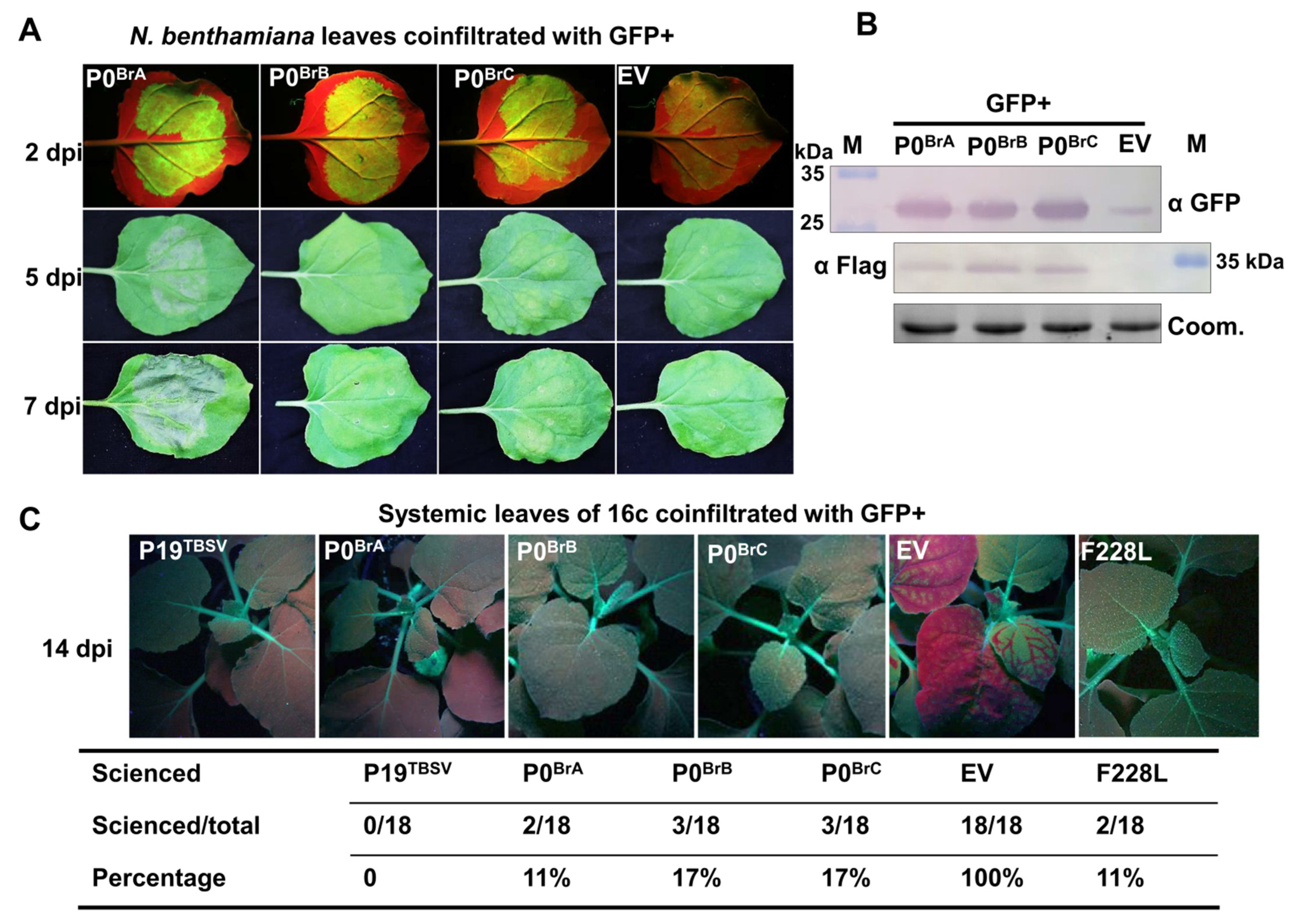
BrYV was identified to have at least three genotypes in China [26,31]. We previously identified BrYV-A P0 as a strong viral suppressor of RNA silencing, with both local and systemic RNA silencing suppression activity [30]. To confirm whether P0 encoded by the BrYV genotypes B and C (P0BrB and P0BrC) are also RNA silencing suppressors, P0BrA, P0BrB, or P0BrC were transiently coexpressed together with a green fluorescent protein (GFP) in N. benthamiana leaves [38]. Under the long-wavelength UV light at 2 days post-infiltration (dpi), the leaf patches co-infiltrated with GFP and the empty vector (EV) showed faint GFP fluorescence, suggesting that GFP RNA silencing was effectively induced (Figure 2A). In contrast, the GFP/P0BrB or GFP/P0BrC co-infiltrated leaf patches showed strong GFP fluorescence, similar to GFP/P0BrA (Figure 2A). The accumulation of the GFP protein was detected using Western blot. In GFP/EV-infiltrated leaf patches, the GFP proteins were rarely detected. Furthermore, a high accumulation of GFP was detected in GFP/P0BrB- and GFP/P0BrC-infiltrated leaf patches, similar to the GFP/P0BrA (Figure 2B). These results suggest that P0BrB and P0BrC suppressed local RNA silencing in N. benthamiana.
To test whether P0BrB and P0BrC could prevent the spread of the systemic silencing signal, a GFP co-infiltration assay was conducted in transgenic N. benthamiana line 16c. In this experiment, GFP transient expression in the lower leaves resulted in GFP silencing in the newly emerging leaves, which could be monitored by UV illumination [41]. At 14 dpi, the fluorescence signals of GFP in the upper leaves were observed under the long-wavelength UV light (Figure 2C). By calculating the percentage of systemic silencing suppression, the result showed that all the plants co-infiltrated with GFP/EV (negative control) exhibited systemic RNA silencing. Conversely, no plants co-infiltrated with GFP and P19TBSV, which served as the positive control, exhibited systemic RNA silencing at 14 dpi [42]. Only 11, 17, and 17% of the plants infiltrated with GFP/P0BrA, GFP/P0BrB, and GFP/P0BrC exhibited systemic RNA silencing, respectively (Figure 2C). These data showed that P0BrB and P0BrC display a systemic RNA silencing suppressor activity similar to P0BrA.

3.3. P0BrB and P0BrC Induce Significantly Delayed and Milder Cell Death in N. benthamiana Compared to P0BrA

It was previously shown that P0BrA induces cell death in infiltrated N. benthamiana leaves [30]. To confirm whether P0BrB or P0BrC also induce cell death, we transiently expressed them in N. benthamiana plants. At 5 dpi, the P0BrA protein triggered an obvious cell death phenotype and rapidly developed severe necrosis by 7 dpi (Figure 2A). However, under the same conditions, the cell death phenotype was not evident in leaves expressing P0BrB or P0BrC at 5 dpi. Even at 7 dpi, both P0BrB and P0BrC only triggered a mild cell death response, which was significantly weaker than the P0BrA-induced cell death (Figure 2A). Immunoblotting results showed that the accumulation level of P0BrB or P0BrC was equivalent to P0BrA, indicating that dosage was not the reason for the compromised cell death (Figure 2B).

3.4. Identification of Key Amino Acid Residues in P0BrA That Affect the Induction of Cell Death in N. benthamiana

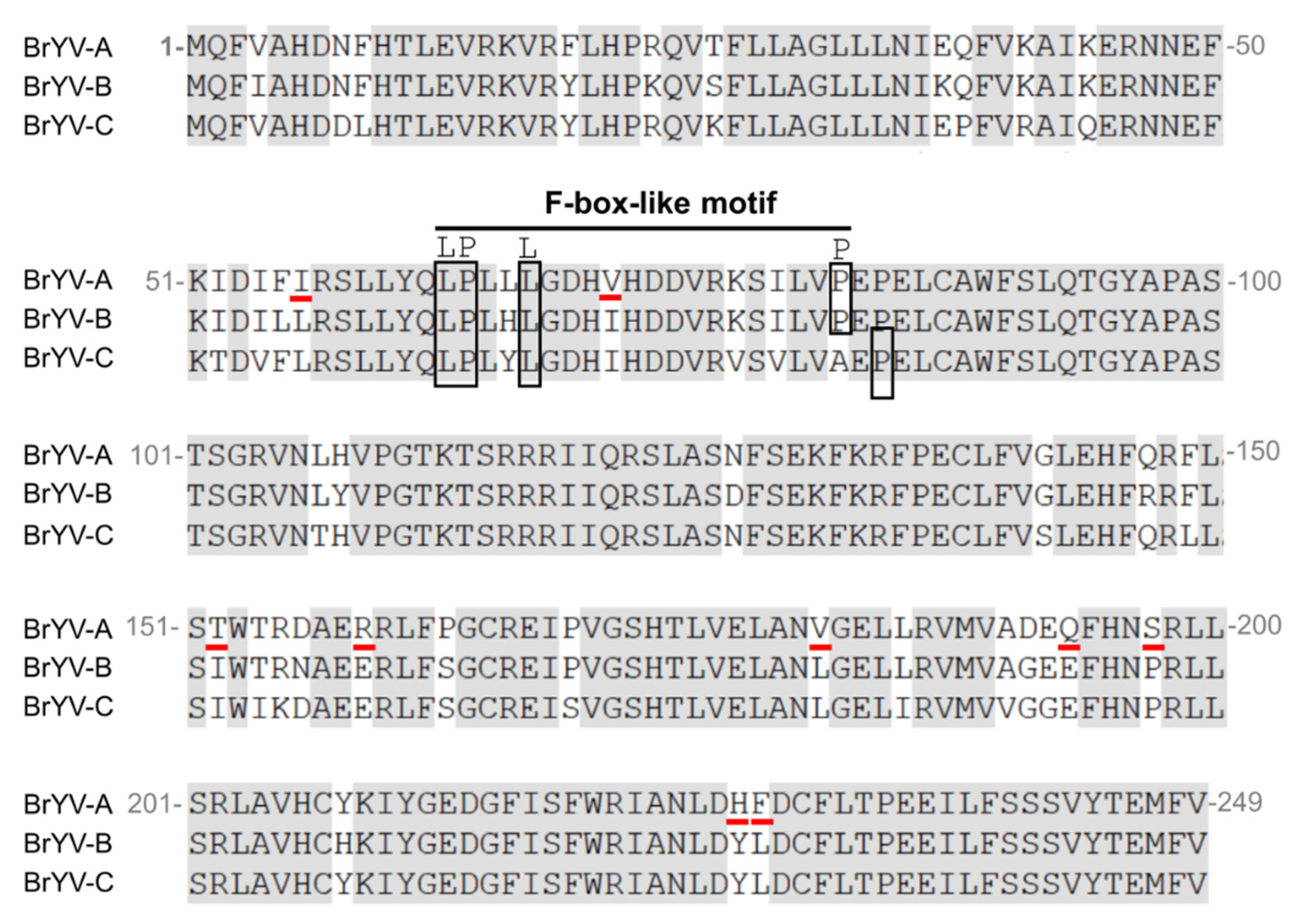
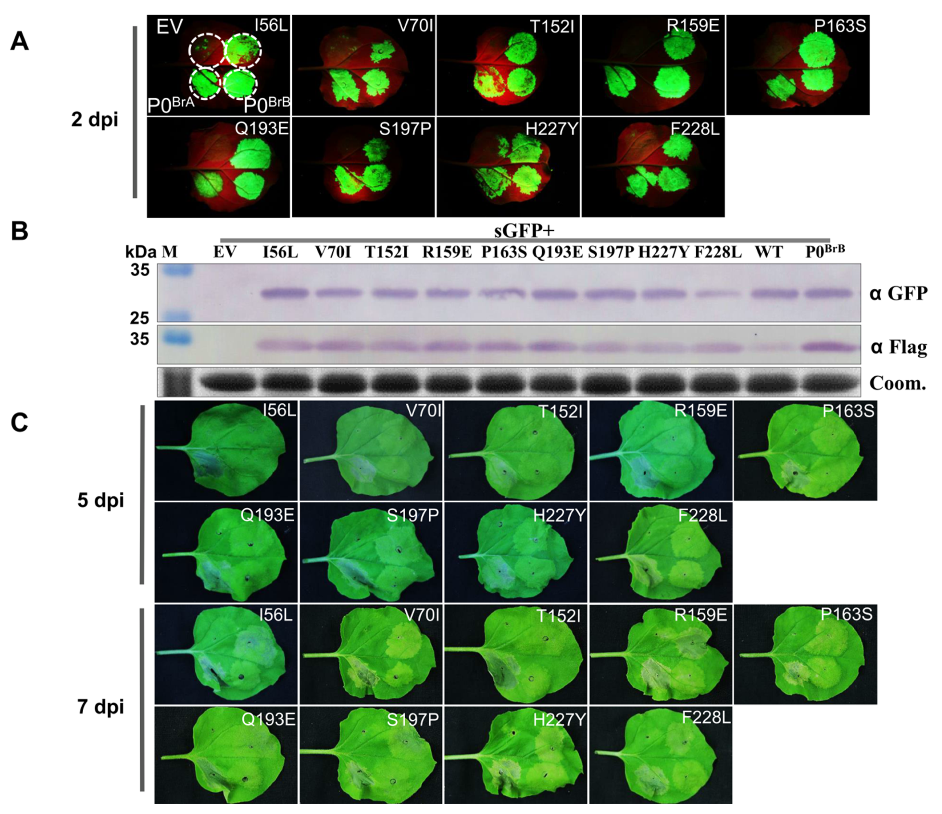
Sequence comparison of P0BrA, P0BrB, and P0BrC showed that the shared amino acid sequence identity ranged from 86.7 to 90.8%. The amino acid sequence of P0BrB and P0BrC had nine residues, different from P0BrA (Figure 3). In order to identify the P0BrA key amino acids that function in inducing cell death, a single amino acid substitution in P0BrA, mutagenesis experiments were conducted for the following nine residues: Leu substitution of Ile56 (I56L), Ile substitution of Val70 (V70I), Ile substitution of Thr152 (T152I), Glu substitution of Arg159 (R159E), Ser substitution of Pro163 (P163S), Glu substitution of Gln193 (Q193E), Pro substitution of Ser197 (S197P), Tyr substitution of His227 (H227Y), and Leu substitution of Phe 228 (F228L) (Figure 3). The P0BrA mutants were transiently coexpressed together with GFP in wild-type N. benthamiana leaves. Under the long-wavelength UV light, we found that the GFP fluorescence in the leaf patches co-infiltrated with pGDG and all nine mutants were as strong as the co-infiltration with pGDG and the wild-type P0BrA at 2 dpi (Figure 4A). The GFP protein accumulation (Figure 4B) detected by Western blot corresponded to the GFP fluorescence levels (Figure 4A), indicating the nine substitutions in P0BrA did not affect the local RNA silencing suppressor activity.
We then compared the cell death induction capacity of the nine mutants with P0BrA. At 5 dpi, the leaves co-infiltrated with P0BrA showed a more prominent cell death phenotype than leaves infiltrated with mutants (Figure 4C). At 7 dpi, cell death was observed in parts of leaves expressing mutants I56L, R159E, and S197P (Figure 4C), indicating that the three mutations delayed the onset of rapid and strong cell death induced by P0BrA. The cell death phenotypes were not evident in infiltrations with the remaining six mutations V70I, T152I, P163S, Q193E, H227Y, and F228L (Figure 4C), suggesting that the Val70, Thr152, Pro163, Gln193, His227, and Phe 228 of P0BrA were key residues essential for inducing cell death in N. benthamiana but not for suppressing local RNA silencing.

3.5. All Three BrYV Genotypes Have Synergistic Interaction with PEMV 2 Resulting in Increased Accumulation of BrYV and Causing Severe Symptoms in N. benthamiana

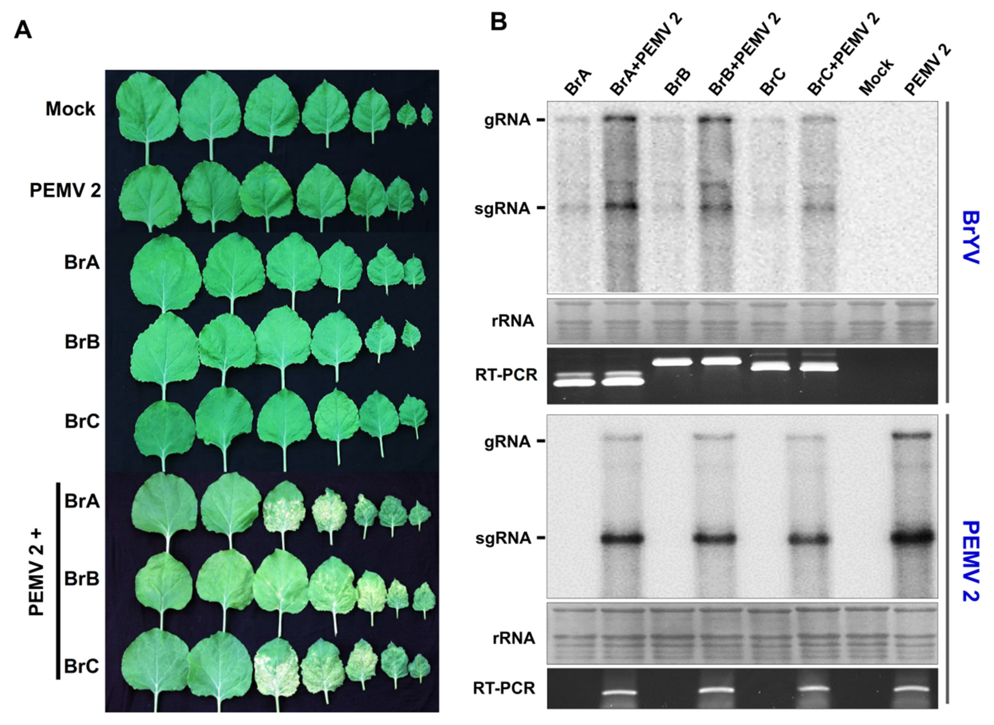
As the BrYV is restricted to the host phloem tissue, the Pea enation mosaic virus 2 (PEMV 2), which belongs to Umbravirus, can help BrYV move out of the phloem into nonvascular tissues and be transmitted mechanically [36]. Meanwhile, the synergistic infection of BrYV and PEMV 2 on N. benthamiana upper leaves produced severe symptoms and significantly increased the BrYV titer [36]. To further investigate whether the three BrYV genotypes display differences in synergism with PEMV 2, the wild-type plants were infiltrated with BrA, BrA + PEMV 2, BrB, BrB + PEMV 2, BrC, BrC + PEMV 2, empty vector (Mock), or PEMV 2. At 21 dpi, N. benthamiana upper leaves infected with BrA + PEMV 2, BrB + PEMV 2, and BrC + PEMV 2 all displayed necrosis and leaf-curling symptoms. In contrast, no obvious symptoms were observed in the plants infected with BrA, BrB, BrC, or PEMV 2 (Figure 5A). Northern blot analysis showed an increased accumulation of BrYVs (BrA, BrB, or BrC) in coinfected plants in contrast to BrYV-only infected plants (Figure 5B). In this experiment, sequence progeny for BrA, BrB, or BrC in the upper leaves was determined by multiplex RT-PCR followed by amplicon sequencing. The PEMV 2 infection was confirmed by RT-PCR and Northern blot detection (Figure 5B). Taken together, PEMV 2 displayed a synergistic interaction with all of the three BrYV genotypes, resulting in increased accumulation of BrYV and causing severe symptoms in N. benthamiana.

4. Discussion

P0 proteins have been previously identified as viral suppressors of RNA silencing in many poleroviruses. Recently, we showed that BrYV-A P0 is a strong VSR that can suppress both local and systemic RNA silencing [30]. Besides the VSR functions, the P0 proteins of TuYV, ScYLV, CABYV, and PLRV were reported to cause cell death phenotypes in some Nicotiana species. P0Tu, in particular, elicited the HR in N. glutinosa, which is recognized by the RPO1 resistance gene, a likely immune receptor of P0 [16,17,25]. HR is a form of programmed cell death that accompanies defense reactions such as the generation of ROS, including H2O2, the expression of pathogenesis-related (PR) genes, and the accumulation of phytohormones, including salicylic acid (SA), jasmonic acid, and ethylene [43,44]. In this study, we discovered that transiently expressed P0BrA in N. benthamiana leaves induced a cell death response. This rapid and restricted cell death phenotype is accompanied by the accumulation of ROS and the induction of the marker gene PR1 expression, which is similar to the HR-like cell death in plants. The phytohormone SA plays an important role in the regulation of plant immunity [45]. Our study showed the P0BrA-induced cell death phenotype could be delayed by SA pretreatment, indicating that the SA pathway may be involved in cell death initiation, but we cannot rule out the involvement of other defense pathways. Generally, the development of HR resistance is associated with the recognition of the viral protein by the corresponding resistance (R) proteins of plants in a host-specific manner [46]. The rapid cell death induced by P0BrA in the agroinfiltrated region of N. benthamiana leaves may be the result of P0BrA recognition by an R protein from Nicotiana species, but further experiments are required to confirm this.
BrYV has a widespread distribution and prevalence in crucifer crops throughout China. According to the multiplex RT-PCR amplification, the three BrYV genotypes had differences in incidence rates and host species [26]. Among all the BrYV infection types, genotype C was detected at the greatest proportion, followed by genotype B and A. BrYV-C had the greatest incidence rates in five Brassica species: B. rapa var. pekinensis, B. oleracea var. capitata, B. oleracea var. botrytis, B. oleracea var. alboglbra, and B. napus [26]. Raphanus sativus and B. juncea plants have been previously infected with BrYV-A and BrYV-B genotypes [26]. As the three genotype sequences of BrYV ORF0 have a high diversity, a comparative analysis of the biological activities of their respective P0 proteins was done. We found that the P0 proteins of the two other BrYV genotypes (P0BrB and P0BrC) also suppressed both local and systemic RNA silencing, such as P0BrA. However, transiently-expressed P0BrB or P0BrC induced a significantly delayed and milder cell death response compared with P0BrA. As the VSR of BrYV, P0 can suppress the host anti-viral RNA silencing and benefit the accumulation of virus. However, this pro-viral factor may possibly be recognized by plant hosts as an effector and elicit HR-like cell death response to restrict the spread of virus. Although P0BrA, P0BrB, and P0BrC have similar VSR activities, P0BrB or P0BrC induced a significantly delayed and milder cell death response compared with P0BrA. This may imply the escape of BrYV genotypes C and B from the perception by host innate immune receptors, which may further facilitate their infection in plants. Consistent with this presumption, our previous study had shown that the BrYV genotype C and B have a higher incidence in nature compared with genotype A, which indicated that BrYV-C and B gradually become the dominant genotypes of BrYV during virus evolution. Moreover, it cannot rule out the possibility that the aphids are more likely to feed on the plants infected with BrYV-C or B in fields, which further facilitate the virus transmission. However, whether the aphid viral acquisition rates are related to P0’s cell death induction function needs further study.
Previous mutational analyses of P0BrA revealed that the LP63-64AA mutant abolished local RNA silencing suppression (RSS) activity but retained the systemic RSS and cell death induction activity. In contrast, the L184A mutant retained local RSS activity but eliminated the systemic RSS and cell death induction activity, suggesting that the cell death induction of P0 is independent of the local suppression activity [30]. In this study, the six mutations V70I, T152I, P163S, Q193E, H227Y, and F228L suppressed local PTGS but failed to induce the obvious cell death phenotype. The mutant F228L in particular also suppressed systemic PTGS (Figure 2C), suggesting that the cell death induction activity was not directly required for P0 to suppress systemic RNA silencing. Since the Phe228 plays an important role in cell death induction, the screening of host proteins interacting with the F228L mutant, but not with the wild-type protein, may provide clues for the underlying mechanism of P0BrA in inducing cell death. However, further research is also needed to determine whether the F228L mutation affected the viral infection.
Earlier studies have shown that the P0 proteins in poleroviruses have a conserved F-box-like motif [LPXX(L/I)X10–13P] and FWR motif [(K/R) IYGEDGX3FWR], indispensable to the silencing suppression activity [6,8,9,10,11,12,14,15,17,18,24,30]. Multiple alignments of the P0 proteins from three BrYV genotypes showed that they shared an amino acid sequence identity, ranging from 86.7 to 90.8% [31]. A highly conserved FWR motif (209-KIYGEDGFISFWRIA-223) was present in the three P0Br proteins (Figure 2). P0BrA and P0BrB had a similar F-box-like motif (63-LPL(L/H)LGDH(V/I)HDDVRKSILVP-82). Interestingly, the amino acid residue 82 of P0BrC in the F-box-like motif was an alanine (A) instead of the consensus proline (P), but residue 84 of P0BrC was a P (Figure 2). We speculate that 84P is the key site for the F-box-like motif of P0BrC, but further investigation is required. Our previous studies have shown that P0BrA interacts with SKP1 via its F-box-like motif to stabilize itself, ensuring efficient RSS activity of P0BrA for BrYV infection [30]. Studies have shown that both P0PL and P0PM have conserved F-box-like motif and suppress RNA silencing but fail to interact with SKP1. Therefore, it will be interesting to investigate the difference in SKP1-interacting abilities among P0BrA, P0BrB, and P0BrC.
Viral synergism is common in nature [47,48,49,50,51,52,53,54]. The synergistic infection of phloem-restricted poleroviruses and umbraviruses has destructive effects on crop plants. Previous research has shown that in the presence of PEMV 2, BrYV can move out of the phloem to invade the mesophyll tissues and be transmitted mechanically in N. benthamiana [36]. Our results presented that the three BrYVs have no obvious difference in synergism with PEMV 2. All three BrYV genotypes have synergistic interactions with PEMV 2, resulting in increased accumulation of BrYV and thus causing more severe symptoms in N. benthamiana. As an RNA silencing suppressor, P0 is needed for BrYV systemic infection [30]. However, whether the P0 protein is necessary for synergism between BrYV and PEMV 2 is uncertain. Further investigation is also needed to identify if the P0 protein acts as a pathogenicity determinant that leads to severe symptoms during the co-infection of BrYV and PEMV 2.

5. Conclusions

In this study, we demonstrated that the cell death induced by P0BrA was accompanied by the accumulation of ROS and induction of PR1 gene expression. We also showed that this cell death phenotype could be delayed by SA pretreatment. The comparison of the biological functions of the P0 proteins from the three BrYV genotypes demonstrated that transiently expressed P0BrB or P0BrC induced a significantly delayed and milder cell death response compared with P0BrA. However, they can also suppress local and systemic RNA silencing, such as P0BrA. Several residues in P0BrA (Val70, Thr152, Pro163, Gln193, His227, and Phe228) essential for inducing cell death were identified by comparative analysis of the P0 proteins. Here, we also showed that all three BrYV genotypes had a synergistic interaction with PEMV 2 in N. benthamiana. This study provides theoretical guidance on controlling the viral disease caused by poleroviruses in the future.

Supplementary Materials

The following are available online at https://www.mdpi.com/article/10.3390/biology10111076/s1, Table S1: Sequences of primers used in this study.

Author Contributions

Conceptualization, C.-G.H., X.-Y.Z. and Y.-Y.L.; methodology, X.-Y.Z. and Y.-Y.L.; software, X.-Y.Z. and Y.-Y.L.; validation, C.-G.H., X.-Y.Z. and Y.-Y.L.; formal analysis, X.-Y.Z.; investigation, X.-Y.Z.; resources, Y.W., D.-W.L., J.-L.Y. and C.-G.H.; data curation, X.-Y.Z. and Y.-Y.L.; writing—original draft preparation, X.-Y.Z.; writing—review and editing, X.-Y.Z., Y.-Y.L., Y.W., D.-W.L., J.-L.Y. and C.-G.H.; visualization, X.-Y.Z.; supervision, Y.W., D.-W.L., J.-L.Y. and C.-G.H.; project administration, C.-G.H.; funding acquisition, X.-Y.Z. and C.-G.H. All authors have read and agreed to the published version of the manuscript.

Funding

This work was supported in part by the National Natural Science Foundation of China (31900139 and 31972240).

Institutional Review Board Statement

Not applicable.

Informed Consent Statement

Not applicable.

Data Availability Statement

The data presented in this study are available within the article.

Acknowledgments

We thank David C. Baulcombe (University of Cambridge, UK) for providing wild-type N. benthamiana and GFP transgenic N. benthamiana line 16c, Xiaorong Tao (Nanjing Agricultural University, China) for providing the pCB301-2x35S-MCS-HDVRZ-NOS vector, Andrew O. Jackson (University of California, USA) for providing the pGD, pGDG, and pGDP19TBSV vectors, Bo Zhou (Zhejiang University, China) for providing the pT-Bax vector, and Daowen Wang (China Academy of Science, China) for providing C58CI. We also thank Xianbing Wang and Yongliang Zhang (China Agricultural University, China) for critical reading and fruitful discussions of this work.

Conflicts of Interest

The authors declare no conflict of interest.

References

  1. Lindbo, J.A.; Dougherty, W.G. Plant pathology and RNAi: A brief history. Annu. Rev. Phytopathol. 2005, 43, 191–204. [Google Scholar] [CrossRef]
  2. Burgyán, J.; Havelda, Z. Viral suppressors of RNA silencing. Trends Plant. Sci. 2011, 16, 265–272. [Google Scholar] [CrossRef]
  3. Wu, Q.; Wang, X.; Ding, S.W. Viral suppressors of RNA-based viral immunity: Host targets. Cell Host Microbe 2010, 8, 12–15. [Google Scholar] [CrossRef] [Green Version]
  4. Reis, R.S.; Litholdo, C.G.; Bally, J.; Roberts, T.H.; Waterhouse, P.M. A conditional silencing suppression system for transient expression. Sci. Rep. 2018, 8, 9426. [Google Scholar] [CrossRef]
  5. Incarbone, M.; Dunoyer, P. RNA silencing and its suppression: Novel insights from in planta analyses. Trends Plant. Sci. 2013, 18, 382–392. [Google Scholar] [CrossRef] [PubMed]
  6. Sun, Q.; Zhuo, T.; Zhao, T.Y.; Zhou, C.J.; Li, Y.Y.; Wang, Y.; Li, D.W.; Yu, J.L.; Han, C.G. Functional Characterization of RNA Silencing Suppressor P0 from Pea Mild Chlorosis Virus. Int. J. Mol. Sci. 2020, 21, 7163. [Google Scholar] [CrossRef]
  7. Wang, F.; Zhao, X.; Dong, Q.; Zhou, B.L.; Gao, Z.L. Characterization of an RNA silencing suppressor encoded by maize yellow dwarf virus-RMV2. Virus Genes 2018, 54, 570–577. [Google Scholar] [CrossRef]
  8. Chen, S.; Jiang, G.Z.; Wu, J.X.; Liu, Y.; Qian, Y.J.; Zhou, X.P. Characterization of a novel polerovirus infecting maize in China. Viruses 2016, 8, 120. [Google Scholar] [CrossRef] [PubMed] [Green Version]
  9. Cascardo, R.S.; Arantes, I.L.; Silva, T.F.; Sachetto-Martins, G.; Vaslin, M.F.; Corrêa, R.L. Function and diversity of P0 proteins among cotton leafroll dwarf virus isolates. Virol. J. 2015, 12, 123. [Google Scholar] [CrossRef] [PubMed]
  10. Almasi, R.; Miller, W.A.; Ziegler-Graff, V.; Vachon, V.K.; Conn, G.L. Mild and severe cereal yellow dwarf viruses differ in silencing suppressor efficiency of the P0 protein. Virus Res. 2015, 208, 199–206. [Google Scholar] [CrossRef] [PubMed]
  11. Zhuo, T.; Li, Y.Y.; Xiang, H.Y.; Wu, Z.Y.; Wang, X.B.; Wang, Y.; Zhang, Y.L.; Li, D.W.; Yu, J.L.; Han, C.G. Amino acid sequence motifs essential for P0-mediated suppression of RNA silencing in an isolate of potato leafroll virus from inner mongolia. Mol. Plant. Microbe Interact. 2014, 27, 515–527. [Google Scholar] [CrossRef]
  12. Delfosse, V.C.; Agrofoglio, Y.; Casse, M.F.; Kresic, I.B.; Hopp, H.E.; Ziegler-Graff, V.; Distéfano, A.J. The P0 protein encoded by cotton leafroll dwarf virus (CLRDV) inhibits local but not systemic RNA silencing. Virus Res. 2014, 180, 70–75. [Google Scholar] [CrossRef]
  13. Liu, Y.; Zhai, H.; Zhao, K.; Wu, B.; Wang, X. Two suppressors of RNA silencing encoded by cereal-infecting members of the family Luteoviridae. J. Gen. Virol. 2012, 93, 1825–1830. [Google Scholar] [CrossRef]
  14. Fusaro, A.F.; Corrêa, R.L.; Nakasugi, K.; Jackson, C.; Kawchuk, L.M.; Vaslin, M.F.; Waterhouse, P.M. The Enamovirus P0 protein is a silencing suppressor which inhibits local and systemic RNA silencing through AGO1 degradation. Virology 2012, 426, 178–187. [Google Scholar] [CrossRef]
  15. Kozlowska-Makulska, A.; Guilley, H.; Szyndel, M.S.; Beuve, M.; Lemaire, O.; Herrbach, E.; Bouzoubaa, S. P0 proteins of European beet-infecting poleroviruses display variable RNA silencing suppression activity. J. Gen. Virol. 2010, 91, 1082–1091. [Google Scholar] [CrossRef] [PubMed]
  16. Csorba, T.; Lózsa, R.; Hutvágner, G.; Burgyán, J. Polerovirus protein P0 prevents the assembly of small RNA-containing RISC complexes and leads to degradation of ARGONAUTE1. Plant J. 2010, 62, 463–472. [Google Scholar] [CrossRef]
  17. Mangwende, T.; Wang, M.L.; Borth, W.; Hu, J.; Moore, P.H.; Mirkov, T.E.; Albert, H.H. The P0 gene of Sugarcane yellow leaf virus encodes an RNA silencing suppressor with unique activities. Virology 2009, 384, 38–50. [Google Scholar] [CrossRef] [Green Version]
  18. Pazhouhandeh, M.; Dieterle, M.; Marrocco, K.; Lechner, E.; Berry, B.; Brault, V.; Hemmer, O.; Kretsch, T.; Richards, K.E.; Genschik, P.; et al. F-box-like domain in the polerovirus protein P0 is required for silencing suppressor function. Proc. Natl. Acad. Sci. USA 2006, 103, 1994–1999. [Google Scholar] [CrossRef] [Green Version]
  19. Stevens, M.; Freeman, B.; Liu, H.Y.; Herrbach, E.; Lemaire, O. Beet poleroviruses: Close friends or distant relatives? Mol. Plant Pathol. 2005, 6, 1–9. [Google Scholar] [CrossRef] [PubMed] [Green Version]
  20. Taliansky, M.; Mayo, M.A.; Barker, H. Potato leafroll virus: A classic pathogen shows some new tricks. Mol. Plant Pathol. 2003, 4, 81–89. [Google Scholar] [CrossRef] [PubMed]
  21. Pfeffer, S.; Dunoyer, P.; Heim, F.; Richards, K.E.; Jonard, G.; Ziegler-Graff, V. P0 of beet western yellows virus is a suppressor of posttranscriptional gene silencing. J. Virol. 2002, 76, 6815–6824. [Google Scholar] [CrossRef] [Green Version]
  22. Zarghani, S.N.; Shams-Bakhsh, M.; Zand, N.; Sokhandan-Bashir, N.; Pazhouhandeh, M. Genetic analysis of Iranian population of Potato leafroll virus based on ORF0. Virus Genes 2012, 45, 567–574. [Google Scholar] [CrossRef]
  23. Derrien, B.; Baumberger, N.; Schepetilnikov, M.; Viotti, C.; De Cillia, J.; Ziegler-Graff, V.; Isono, E.; Schumacher, K.; Genschik, P. Degradation of the antiviral component ARGONAUTE1 by the autophagy pathway. Proc. Natl. Acad. Sci. USA 2012, 109, 15942–15946. [Google Scholar] [CrossRef] [PubMed] [Green Version]
  24. Bortolamiol, D.; Pazhouhandeh, M.; Marrocco, K.; Genschik, P.; Ziegler-Graff, V. The polerovirus F box protein P0 targets ARGONAUTE1 to suppress RNA silencing. Curr. Biol. 2007, 17, 1615–1621. [Google Scholar] [CrossRef] [Green Version]
  25. Wang, K.D.; Empleo, R.; Nguyen, T.T.V.; Moffett, P.; Sacco, M.A. Elicitation of hypersensitive responses in Nicotiana glutinosa by the suppressor of RNA silencing protein P0 from poleroviruses. Mol. Plant Pathol. 2015, 16, 435–448. [Google Scholar] [CrossRef]
  26. Zhang, X.Y.; Peng, Y.M.; Xiang, H.Y.; Wang, Y.; Li, D.W.; Yu, J.L.; Han, C.G. Incidence and prevalence levels of three aphid-transmitted viruses in crucifer crops in China. J. Integr. Agr. 2021, 20, 2–9. [Google Scholar]
  27. Yoshida, N.; Tamada, T. Host range and molecular analysis of Beet leaf yellowing virus, Beet western yellows virus-JP and Brassica yellows virus in Japan. Plant Pathol. 2019, 68, 1045–1058. [Google Scholar] [CrossRef]
  28. Kamitani, M.; Nagano, A.J.; Honjo, M.N.; Kudoh, H. RNA-Seq reveals virus-virus and virus-plant interactions in nature. FEMS. Microbiol. Ecol. 2016, 92, fiw176. [Google Scholar] [CrossRef] [PubMed] [Green Version]
  29. Lim, S.; Yoo, R.H.; Igori, D.; Zhao, F.; Kim, K.H.; Moon, J.S. Genome sequence of a recombinant brassica yellows virus infecting Chinese cabbage. Arch. Virol. 2015, 160, 597–600. [Google Scholar] [CrossRef]
  30. Li, Y.Y.; Sun, Q.; Zhao, T.Y.; Xiang, H.Y.; Zhang, X.Y.; Wu, Z.Y.; Zhou, C.J.; Zhang, X.; Wang, Y.; Zhang, Y.L.; et al. Interaction between Brassica yellows virus silencing suppressor P0 and plant SKP1 facilitates stability of P0 in vivo against degradation by proteasome and autophagy pathways. New Phytol. 2019, 222, 1458–1473. [Google Scholar] [CrossRef] [PubMed] [Green Version]
  31. Zhang, X.Y.; Dong, S.W.; Xiang, H.Y.; Chen, X.R.; Li, D.W.; Yu, J.L.; Han, C.G. Development of three full-length infectious cDNA clones of distinct brassica yellows virus genotypes for agrobacterium-mediated inoculation. Virus Res. 2015, 197, 13–16. [Google Scholar] [CrossRef]
  32. Zhang, X.Y.; Xiang, H.Y.; Zhou, C.J.; Li, D.W.; Yu, J.L.; Han, C.G. Complete genome sequence analysis identifies a new genotype of brassica yellows virus that infects cabbage and radish in China. Arch. Virol. 2014, 159, 2177–2180. [Google Scholar] [CrossRef]
  33. Goodin, M.; Dietzgen, R.G.; Schichnes, D.; Ruzin, S.; Jackson, A.O. pGD vectors: Versatile tools for the expression of green and red fluorescent protein fusions in agroinfiltrated plant leaves. Plant J. 2002, 31, 375–383. [Google Scholar] [CrossRef]
  34. Geier, G.E.; Modrich, P. Recognition sequence of the dam methylase of Escherichia coli K12 and mode of cleavage of Dpn I endonuclease. J. Biol. Chem. 1979, 254, 1408–1413. [Google Scholar] [CrossRef]
  35. Zhang, L.D.; Wang, Z.H.; Wang, X.B.; Zhang, W.H.; Li, D.W.; Han, C.G.; Zhai, Y.F.; Yu, J.L. Functional identification of two RNA silencing suppressors from two plant viruses. Sci. Bull. 2005, 50, 219–224. (In Chinese) [Google Scholar]
  36. Zhou, C.J.; Zhang, X.Y.; Liu, S.Y.; Wang, Y.; Li, D.W.; Yu, J.L.; Han, C.G. Synergistic infection of BrYV and PEMV 2 increases the accumulations of both BrYV and BrYV-derived siRNAs in Nicotiana benthamiana. Sci. Rep. 2017, 7, 45132. [Google Scholar] [CrossRef] [PubMed] [Green Version]
  37. Johansen, L.K.; Carrington, J.C. Silencing on the spot. Induction and suppression of RNA silencing in the Agrobacterium-mediated transient expression system. Plant Physiol. 2001, 126, 930–938. [Google Scholar] [CrossRef] [PubMed] [Green Version]
  38. Voinnet, O.; Rivas, S.; Mestre, P.; Baulcombe, D. An enhanced transient expression system in plants based on suppression of gene silencing by the p19 protein of tomato bushy stunt virus. Plant J. 2003, 33, 949–956. [Google Scholar] [CrossRef] [PubMed] [Green Version]
  39. Liu, D.S.; Shi, L.D.; Han, C.G.; Yu, J.L.; Li, D.W.; Zhang, Y.L. Validation of reference genes for gene expression studies in virus-infected Nicotiana benthamiana using quantitative real-time PCR. PLoS ONE 2012, 7, e46451. [Google Scholar] [CrossRef] [PubMed]
  40. Ruiz, M.T.; Voinnet, O.; Baulcombe, D.C. Initiation and maintenance of virus-induced gene silencing. Plant Cell 1998, 10, 937–946. [Google Scholar] [CrossRef]
  41. Lacomme, C.; Santa Cruz, S. Bax-induced cell death in tobacco is similar to the hypersensitive response. Proc. Natl. Acad. Sci. USA 1999, 96, 7956–7961. [Google Scholar] [CrossRef] [Green Version]
  42. Bragg, J.N.; Jackson, A.O. The C-terminal region of the Barley stripe mosaic virusγb protein participates in homologous interactions and is required for suppression of RNA silencing. Mol. Plant Pathol. 2004, 5, 465–481. [Google Scholar] [CrossRef] [PubMed]
  43. Nürnberger, T.; Brunner, F.; Kemmerling, B.; Piater, L. Innate immunity in plants and animals: Striking similarities and obvious differences. Immunol. Rev. 2004, 198, 249–266. [Google Scholar] [CrossRef]
  44. Hammond-Kosack, K.E.; Jones, J.D. Plant disease resistance genes. Annu. Rev. Plant Physiol. Plant Mol. Biol. 1997, 48, 575–607. [Google Scholar] [CrossRef] [PubMed] [Green Version]
  45. Yang, D.L.; Yang, Y.N.; He, Z.H. Roles of plant hormones and their interplay in rice immunity. Mol. Plant 2013, 6, 675–685. [Google Scholar] [CrossRef] [PubMed] [Green Version]
  46. Garcia-Ruiz, H. Host factors against plant viruses. Mol. Plant Pathol. 2019, 20, 1588–1601. [Google Scholar] [CrossRef]
  47. Tatineni, S.; Graybosch, R.A.; Hein, G.L.; Wegulo, S.N.; French, R. Wheat cultivar-specific disease synergism and alteration of virus accumulation during co-infection with Wheat streak mosaic virus and Triticum mosaic virus. Phytopathology 2010, 100, 230–238. [Google Scholar] [CrossRef] [Green Version]
  48. Stenger, D.C.; Young, B.A.; Qu, F.; Morris, T.J.; French, R. Wheat streak mosaic virus lacking helper component-proteinase is competent to produce disease synergism in double infections with Maize chlorotic mottle virus. Phytopathology 2007, 97, 1213–1221. [Google Scholar] [CrossRef] [PubMed] [Green Version]
  49. Srinivasan, R.; Alvarez, J.M. Effect of mixed viral infections (Potato virus Y-Potato leafroll virus) on biology and preference of vectors Myzus persicae and Macrosiphum euphorbiae (Hemiptera: Aphididae). J. Econ. Entomol. 2007, 100, 646–655. [Google Scholar] [CrossRef]
  50. Murphy, J.F.; Bowen, K.L. Synergistic disease in pepper caused by the mixed infection of Cucumber mosaic virus and Pepper mottle virus. Phytopathology 2006, 96, 240–247. [Google Scholar] [CrossRef] [PubMed] [Green Version]
  51. Karyeija, R.F.; Kreuze, J.F.; Gibson, R.W.; Valkonen, J.P. Synergistic interactions of a potyvirus and a phloem-limited crinivirus in sweet potato plants. Virology 2000, 269, 26–36. [Google Scholar] [CrossRef] [PubMed] [Green Version]
  52. Scheets, K. Maize chlorotic mottle machlomovirus and wheat streak mosaic rymovirus concentrations increase in the synergistic disease corn lethal necrosis. Virology 1998, 242, 28–38. [Google Scholar] [CrossRef] [PubMed] [Green Version]
  53. Vance, V.B. Replication of potato virus X RNA is altered in coinfections with potato virus Y. Virology 1991, 182, 486–494. [Google Scholar] [PubMed]
  54. Rochow, W.F.; Ross, A.F. Virus multiplication in plants doubly infected by potato viruses X and Y. Virology 1955, 1, 10–27. [Google Scholar] [CrossRef]
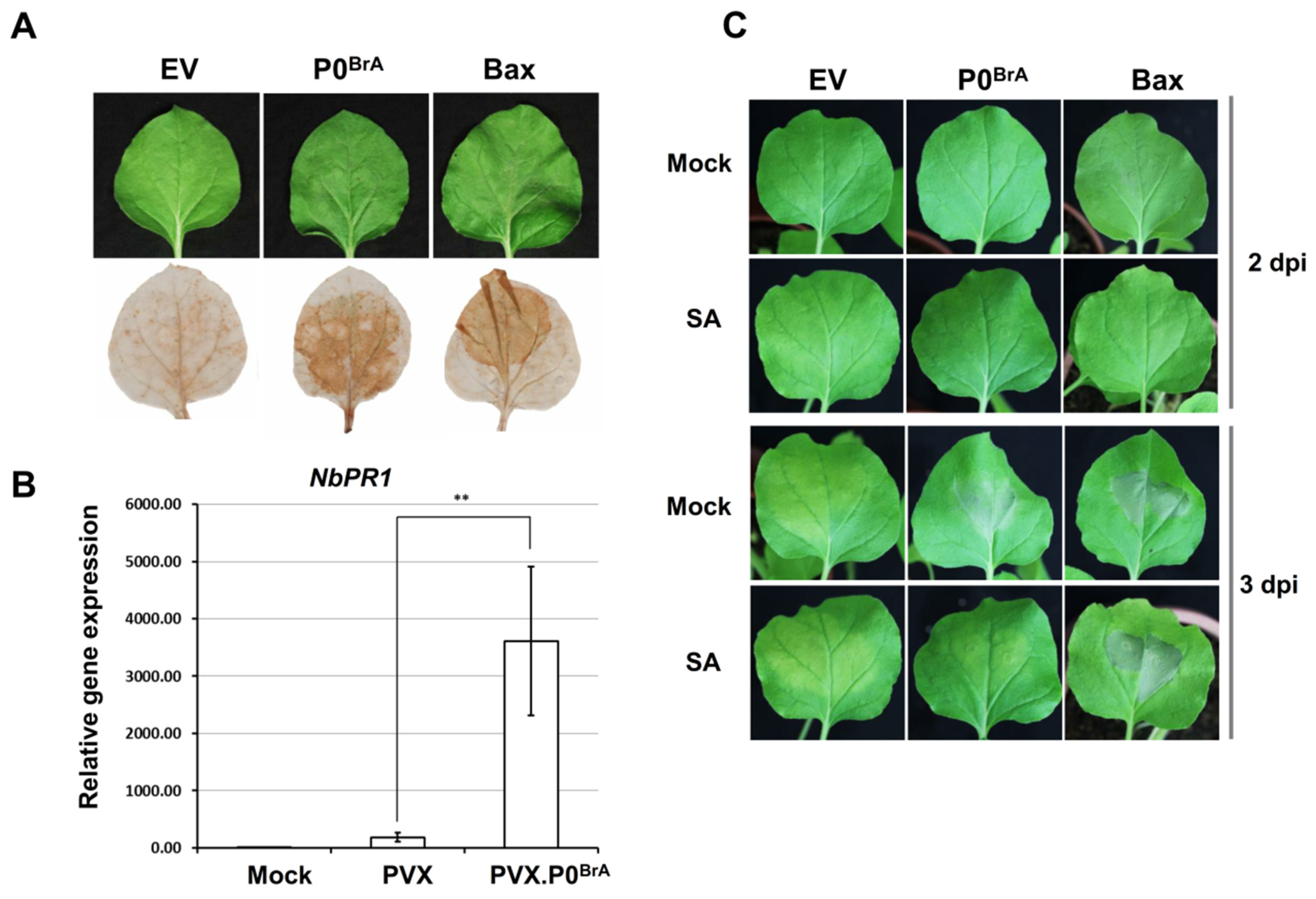
Figure 1. Transient overexpression of P0BrA-triggered HR-like cell death in N. benthamiana leaves. (A) DAB staining in N. benthamiana leaves transiently expressing P0BrA at 48 h post infiltration. Empty vector (EV), negative control; Bc1-2-associated X protein (Bax), positive control. (B) Analysis of PR1 transcript accumulation in N. benthamiana plants infected with PVX or PVX.P0BrA by qRT-PCR. Values are presented as the mean ± standard error (n = 3). ** p < 0.01; asterisks show significant differences as determined using the Student’s t-test. Error bars represent standard errors of the means. (C) Effect of SA treatment on cell death phenotypes triggered by P0BrA. EV, negative control. Bax, positive control. A 0.11% ethanol treatment (Mock) was used as the solvent control.
Biology 10 01076 g001
Figure 2. Suppression of local and systemic RNA silencing by P0BrB and P0BrC. (A) Suppression of local GFP silencing in N. benthamiana-infiltrated leaves. Agroinfiltration with GFP and an empty vector pGD (EV), P0BrA, P0BrB, or P0BrC. The leaves infiltrated with EV were included as the negative control. (B) Western blot analysis of GFP and three Flag-tagged P0Br protein levels in infiltrated N. benthamiana leaf patches using an antibody specific for GFP and Flag, respectively. Coomassie-stained (Coom.) gels are the loading control. M, molecular weights of protein marker. (C) Systemic RNA silencing suppression activity of P0BrB and P0BrC. Transiently coexpressed GFP and P0 were in leaves of N. benthamiana line 16c. P19TBSV and EV were used as positive and negative controls, respectively. The efficiency of systemic silencing is presented below the images. The ratio shows the number of systemic silencing plants to the number of infiltrated plants, scored in three independent experiments.
Biology 10 01076 g002
Figure 3. Multiple alignments of amino acid sequences from P0BrA, P0BrB, and P0BrC. All the conserved residues are indicated in gray background. The mutated amino acids of the nine mutants are presented by red underlines. Landmark residues of the F-box-like motif are highlight in the boxes. The P0 sequences used are as follows: BrYV-A (Accession No. HQ388348), BrYV-B (Accession No. HQ388351), and BrYV-C (Accession No. KF015269). The numbers correspond to the positions of amino acids within the P0Br sequence.
Biology 10 01076 g003
Figure 4. Suppression of local RNA silencing by P0BrA mutants. (A) Agroinfiltration with GFP plus empty vector pGD (EV), P0BrA, P0BrA mutants, or P0BrB in N. benthamiana leaves. (B) Western blot analysis of GFP and P0BrA mutants in N. benthamiana-infiltrated patches from (A). (B) GFP and P0BrA mutants were detected using an antibody specific for GFP and Flag, respectively. Coomassie-stained (Coom.) gels are the loading control. Molecular weights of protein marker (M) are shown on the left. (C) Cell death phenotypes in N. benthamiana leaves with transiently-expressed P0BrA or its mutants. Photographs were taken under white light to show cell death at 5 or 7 dpi. Empty vector (EV) was used as the negative control.
Biology 10 01076 g004
Figure 5. Systemic infection of the three BrYV genotypes and PEMV 2. (A) The symptoms of N. benthamiana upper leaves were induced by coinfection with BrYVs and PEMV 2. The upper leaves were photographed at 21 dpi. (B) Accumulation of BrYV and PEMV 2 in N. benthamiana upper leaves was analyzed using Northern blotting. The viral genomic RNAs (gRNA) and subgenomic RNAs (sgRNA) are shown on the left of the panel. Methylene blue-stained ribosomal RNA (rRNA) was used as the loading control.
Biology 10 01076 g005
Publisher’s Note: MDPI stays neutral with regard to jurisdictional claims in published maps and institutional affiliations.