Review

NRSF/REST-Mediated Epigenomic Regulation in the Heart: Transcriptional Control of Natriuretic Peptides and Beyond

by
Hideaki Inazumi
1 and
Koichiro Kuwahara
2,*
1
Department of Cardiovascular Medicine, Graduate School of Medicine, Kyoto University, 54 Shogoin-kawahara-cho, Sakyo-ku, Kyoto 606-8507, Japan
2
Department of Cardiovascular Medicine, School of Medicine, Shinshu University, 3-1-1 Asahi, Nagano 390-8621, Japan
*
Author to whom correspondence should be addressed.
Biology 2022, 11(8), 1197; https://doi.org/10.3390/biology11081197
Submission received: 22 July 2022 / Revised: 7 August 2022 / Accepted: 9 August 2022 / Published: 10 August 2022

Abstract

:

Simple Summary

Reactivation of the fetal cardiac gene program, such as those encoding atrial and brain natriuretic peptides (ANP and BNP, respectively), is a characteristic feature of failing hearts. We previously revealed that a transcriptional repressor, neuron-restrictive silencer factor (NRSF), also called repressor element-1-silencing transcription factor (REST), plays a crucial role in the transcriptional control of ANP, BNP and other fetal cardiac genes through collaboration with various other transcription factors to maintain physiological cardiac function and electrical stability. Increased production of ANP and BNP prevents the progression of heart failure, but reactivation of Gαo and fetal-type cardiac ion channels (T-type Ca2+ and HCN channels) leads to deteriorated cardiac function and lethal arrhythmias observed in mice with disturbed NRSF function. Epigenetic regulators with which NRSF forms a complex modify histone acetylation and methylation, thereby participating in NRSF-mediated transcriptional regulation. Further comprehensive studies will lead to clarification of the molecular mechanisms underlying the development of cardiac dysfunction and heart failure.

Abstract

Reactivation of fetal cardiac genes, including those encoding atrial natriuretic peptide (ANP) and brain natriuretic peptide (BNP), is a key feature of pathological cardiac remodeling and heart failure. Intensive studies on the regulation of ANP and BNP have revealed the involvement of numerous transcriptional factors in the regulation of the fetal cardiac gene program. Among these, we identified that a transcriptional repressor, neuron-restrictive silencer factor (NRSF), also named repressor element-1-silencing transcription factor (REST), which was initially detected as a transcriptional repressor of neuron-specific genes in non-neuronal cells, plays a pivotal role in the transcriptional regulation of ANP, BNP and other fetal cardiac genes. Here we review the transcriptional regulation of ANP and BNP gene expression and the role of the NRSF repressor complex in the regulation of cardiac gene expression and the maintenance of cardiac homeostasis.

1. Introduction

Heart failure remains a leading cause of morbidity and mortality worldwide [1,2]. Under conditions of pathological stress or tissue injury, cardiac myocytes develop hypertrophy as an initially adaptive response. The sustained stress, however, makes the hypertrophy pathological and provokes pathological cardiac remodeling, which finally causes heart failure. Increased cell size, increased protein synthesis and sarcomere assembly, and altered gene expression are characteristic features of cardiomyocyte hypertrophy. Among the cardiac gene reprogramming during the pathological cardiac remodeling, there is reactivation of “fetal” cardiac genes; such genes encoding atrial natriuretic peptide (ANP) and brain natriuretic peptide (BNP), fetal isoforms of contractile proteins (skeletal α-actin and β-myosin heavy chain), fetal-type cardiac ion channels and some smooth muscle proteins (smooth muscle α-actin and smooth muscle 22α) are robust markers of cardiac hypertrophy and pathological cardiac remodeling [3]. Fetal ventricles abundantly express these genes, but as the heart matures, the expression is decreased. Reactivation of the fetal gene program plays an important role in the molecular processes underlying pathological cardiac remodeling that alters cardiac structure and function besides acting as a marker of pathological conditions [3]. Indeed, the expression of ANP and BNP in cardiomyocytes is remarkably increased in failing hearts and the elevation of their plasma levels is widely recognized as a prognostic marker of disease severity [4,5].
As a means of better understanding the underlying molecular mechanisms of heart failure, much effort has been made to elucidate the mechanisms regulating expression of fetal cardiac genes, including NPPA and NPPB, the genes respectively encoding ANP and BNP. It is now known, for example, that fetal cardiac gene programs are regulated by numerous transcriptional factors [6]. Among these, we have been studying the role played by the transcriptional repressor neuron-restrictive silencer factor (NRSF), also named repressor element-1-silencing transcription factor (REST). Normally, NRSF negatively regulates the transcription of NPPA and NPPB [7,8]. NRSF also represses expression of other fetal cardiac genes, including HCN2 and −4 and CACNA1H, which respectively encode hyperpolarization-activated cyclic nucleotide-gated (HCN) channels and the T-type Ca2+ channel. By forming a complex with several corepressors, NRSF is involved in various molecular pathways that affect the expression of fetal cardiac genes. Consistent with these findings, cardiac-specific inactivation of NRSF by overexpression of a dominant-negative mutant of NRSF (dnNRSF), driven by the cardiac-specific α-MHC promoter (dnNRSF-Tg mice) or cardiac-specific NRSF knockout (NRSF cKO), induces reactivation of fetal cardiac gene expression in the ventricle. Moreover, both dnNRSF-Tg and NRSF cKO mice show deteriorated cardiac function with dilation of the left ventricle and sudden death due to ventricular arrhythmias, indicating that NRSF maintains cardiac integrity by governing the cardiac gene program [9,10]. In this review article, we describe the transcriptional regulation of ANP and BNP mediated through NRSF and other transcriptional factors and the role played by the NRSF repressor complex in maintaining normal cardiac homeostasis.

2. Transcriptional Regulation of ANP and BNP in the Heart

ANP and BNP are cardiac-derived peptide hormones that comprise the natriuretic peptide system [11,12]. These two peptides have similar structures, but are synthesized at different sites. ANP is usually synthesized in the atria, while BNP is synthesized mainly in the ventricles [12]. They are cardiac differentiation markers, and their expression is tightly regulated in a spatiotemporal manner during cardiac development. In fact, the analysis of ANP and BNP promoters and their activity made a tremendous contribution to our current understanding of the transcriptional regulation during cardiac development. As for the postnatal period, the expression of ANP and BNP is elevated in the ventricle in various pathological states of the heart, and plasma concentrations of ANP and BNP are remarkably increased in heart failure patients [4]. In fact, plasma concentrations of ANP and BNP are measured clinically to diagnose heart failure, assess prognosis and determine therapeutic strategies [13]. Therefore, the molecular pathways underlying the reactivation of NPPA and NPPB are thought to be closely related to adaptive or maladaptive signaling pathways evoked by pathological stimuli in the heart. Because of the important role played by ANP and BNP in cardiac physiology and pathology, many studies have been carried out to investigate the molecular mechanisms involved in the regulation of NPPA and NPPB during cardiac development and disease progression. The regulation of NPPA during embryonic development was one of the subjects of early in situ gene expression studies [14]. Those studies showed that NPPA expression in the embryonic mouse heart is finely controlled; it is strongly expressed in the atria, while ventricular expression is restricted to the outer “working” myocardium of the left and right ventricles. Later, during embryonic development, strong NPPA expression is maintained in the atria, while its ventricular expression becomes limited to the trabeculae. Thus, NPPA shows a dynamic expression pattern during embryonic development that is highly restricted to the heart. Research into molecular mechanisms underlying the atrial-specific expression of NPPA has promoted the search for cardiac transcription factors, and the ANP promoter has been extensively analyzed by using molecular biological methods.
The proximal part of the 5′ flanking region (5′-FR) of NPPA has been shown to be sufficient to recapitulate the spatiotemporal expression of the endogenous gene and to contain sequences crucial for the regulation of NPPA expression [15,16]. Expression of a reporter gene driven by the NPPA proximal 5′-FR from atrial or ventricular cardiac myocytes obtained at various developmental stages revealed the regions that mediate the proper spatial and temporal expression of the ANP promoter [17,18]. In regard to that point, the NPPA proximal 5′-FR contains three T-box binding elements (TBE), two GATA sites, two CArG boxes, two NK-homeobox binding elements (NKE), an A/T-rich element and a phenylephrine-responsive element (PERE), to which the transcriptional factors Tbx5, GATA4/6, SRF, NKX2.5, MEF2C and Zfp260 have all been shown to bind [19,20] and to contribute singly or cooperatively to the basal and inducible activation of ANP promoter in cardiac myocytes [20,21,22,23,24,25,26]. Furthermore, those sequences have been highly conserved in evolutionarily diverse organisms [27]. However, there are some differences in the expression pattern between the proximal 5′-FR of NPPA and the intact endogenous NPPA, suggesting that regions outside of the proximal 5′-FR region also have a regulatory capacity [19,28]. Investigation of the NPPA locus in mouse disclosed that distal regulatory elements are also necessary for fine-tuned regulation of NPPA expression during embryonic development [28]. It is reported that the neuron-restrictive silencer element (NRSE), glucocorticoid responsive element (GRE) and hypoxia-response element (HRE), all of which are located outside the proximal promoter, are also involved in the induction of NPPA transcription [29,30].
Similarly, the proximal region of the human BNP promoter is sufficient to mediate ventricle-specific NPPB expression [31,32]. NPPB contains an AT-rich region in its 3′-untranslated region (UTR), which makes the gene unstable, indicating that BNP expression is also regulated post-transcriptionally [33]. Indeed, NPPB has a shorter half-life than NPPA. Moreover, the region extending from −127 to −40 of the human NPPB 5′-FR is found to be necessary for cardiac-specific expression by deletion analysis [32]. This human BNP promoter proximal region contains potential GATA, CArG, AP-1/CRE-like and M-CAT elements, which are conserved among humans, mice and rats [32,34,35,36]. All of these elements have been shown to control the cardiac selectivity of gene expression [35,36,37,38,39,40] and to mediate both basal and inducible expression of NPPB [35,36,39,40,41]. Other sites in the distal regions of human NPPB 5′-FR, such as NRSE, NF-AT binding sites, SSREs and TRE, have also been found to play important roles in the inducible activation of the human BNP promoter [42,43,44,45].
NPPA is located within an insulated chromatin structure called the topologically associating domain (TAD), and NPPB and several other genes are located in its vicinity. As TADs generally create a local gene-regulatory environment, it is likely that the specific elements that regulate NPPA are situated within this TAD. Further investigation of the NPPB-NPPA genomic cluster in mice unraveled the specific elements required for cardiac expression of the two genes and demonstrated that physical contact between the genes and their regulatory elements was likely an important component of their co-regulatory expression [46]. For instance, the BNP promoter was shown to be required for stress-induced expression of NPPA [46].
Pathological stress including mechanical stress on the heart increases ANP and BNP expression and secretion in both ventricular and atrial myocytes [47,48,49]. During that process, a variety of neurohumoral factors, cytokines and growth factors are induced and cooperatively affect ANP and BNP expression by mediation through various signaling pathways [50]. We previously identified that Rho- and actin-treadmill-dependent nuclear accumulation of myocardin-related transcription factor A (MRTF-A), a coactivator of SRF, contributes to the transduction of mechanical stress to the transcriptional activation of Nppb via SRF-responsive element [36].

3. NRSF Is a Transcriptional Repressor of Fetal Cardiac Genes, including NPPA and NPPB

The neuron-restrictive silencer element (NRSE), which is also named as repressor element 1 (RE-1), has been identified as a negatively acting DNA regulatory element that represses neuronal gene expression in non-neuronal cells and in undifferentiated neuronal cells [51,52]. NRSF, also known as REST, has been identified as a protein that binds to NRSE [53]. NRSF is one of the zinc finger transcriptional factor family members and is globally expressed in most non-neuronal tissues, including the heart [52]. Subsequently, it was shown that NRSF also suppresses non-neuronal genes containing NRSE, implying that NRSF is a transcriptional regulator of both neuronal and non-neuronal genes containing NRSE in non-neuronal tissues [54]. As described above, the activity of the 5′-FR of human NPPA is strongly suppressed by a fragment containing the 3′-UTR of the gene. The 3′-UTR of NPPA contains a conserved sequence similar to NRSE. When the NRSE-like sequence is mutated in the human NPPA 3′-UTR, the repressor activity is completely lost. Mutant NRSE also reduces the response of ANP promoter activity to endothelin-1 through the constitutive activation in cultured ventricular myocytes. This indicates that the hypertrophic stimulus-inducible ANP expression in cardiac myocytes is likely attributed to attenuation of NRSE-mediated repression, at least in part [7].
We also found that the 5′-FR of NPPB contains a cis-acting negative regulatory element, which has 90% homology to the consensus sequence of NRSE. This element, called the fibronectin-inducible element, is well conserved among species and mediates the increase in BNP promoter activity by fibronectin in cardiomyocytes. We demonstrated that the element binds NRSF, and its mutation significantly increases human BNP promoter activity. These results indicate that NRSF represses transcription of Nppa as well as Nppb [8]. Indeed, infection of recombinant adenovirus expressing dnNRSF results in an increase of both Nppa and Nppb expression in cultured ventricular myocytes [9]. Both Nppa and Nppb expression in ventricles of dnNRSF-Tg are also increased. Adenovirus-mediated expression of dnNRSF in cardiomyocytes prevents hypertrophic stimulation-induced increases in ANP and BNP expression. Furthermore, pressure-load-induced increases in ANP and BNP expression, which are normally seen in wild-type mice, are markedly suppressed in the ventricles of dnNRSF-Tg mice. On the other hand, forced recruitment of NRSF to the ANP promoter in ventricular myocytes represses basal promoter activity, but enhances inducible activity in response to endothelin-1 [7]. Collectively, removal of NRSF-mediated repression likely contributes to the inducible ANP and BNP expression in response to pathological stimuli.
The skeletal α-actin gene is expressed in the fetal ventricle, but its expression level in the ventricle declines after birth and is reactivated only when exposed to pathological stress [9]. 3′-UTR of the skeletal α-actin gene also contains NRSE [55]. This implies that NRSF participates in the maintenance of normal cardiac structure and function by regulating the expression of multiple fetal cardiac genes (Figure 1). As well as NPPA and NPPB, skeletal α-actin gene expression is significantly elevated in the ventricle of dnNRSF-Tg mice. Intriguingly, dnNRSF-Tg mice exhibit reduced cardiac contractility with left ventricular dilatation, ventricular arrhythmias and early lethality. These indicate that NRSF plays an essential role in maintaining normal myocardial integrity through regulation of the cardiac gene program. Moreover, genetic deletion of guanylyl cyclase-A (GC-A), a common receptor for ANP and BNP, by crossing with GC-A knock out mice, exacerbates the pathological cardiac remodeling observed in dnNRSF-Tg mice, demonstrating that the cardiac dysfunction and lethal arrhythmia observed in dnNRSF-Tg mice are not caused by the increased expression of ANP or BNP [56]. Taking the involvement of NRSF in the reactivation of the fetal cardiac gene program into account, further investigation of the molecular mechanisms by which dnNRSF-Tg mice exhibit cardiac dysfunction and ventricular arrhythmias may bring about the discovery of novel molecular mechanisms underlying the progression to pathological cardiac remodeling and heart failure.

4. NRSF Regulates Fetal Cardiac Ion Channels and Maintains Electrical Stability in the Heart

Electrical instability caused by alterations in ion channel activity is likely responsible for the malignant ventricular arrhythmias and sudden arrhythmic death observed in dnNRSF-Tg mice [57]. It has been demonstrated that two types of fetal cardiac ion channel, T-type Ca2+ and HCN channels, are potentially responsible for the increased incidence of arrhythmias in dnNRSF-Tg mice (Figure 1). T-type Ca2+ channel is one of the well-studied ion channels in fetal myocardium [58]. Voltage-gated Ca2+ channels are the main sources of Ca2+ influx in excitable cells and are classified into several types: L-(long-lasting), T-(transient), N-(neuronal), P/Q-(Purkinje) and R-(residual-drug-resistant). Among these types of Ca2+ channels, cardiac myocytes express only L- and T-type channels. In mature cardiomyocytes, L-type Ca2+ channels are the major subtype and play an important role in excitation-contraction coupling [59]. Conversely, T-type Ca2+ channels are abundantly expressed in the embryonic ventricle, but their ventricular expression is suppressed during the maturation of the heart, so that their expression is restricted to the conduction system in the adult ventricle [60,61]. However, they are reactivated in hypertrophied and failing ventricles [62]. There are two α1 subunits of T-type Ca2+ channel expressed in the heart, α1G (CACNA1G) and α1H (CACNA1H) [58]. CACNA1H contains an NRSE-like sequence within its first intron. This NRSE-like sequence has 93% homology to the NRSE consensus sequence and is well preserved among different mammalian species, including humans. NRSF binds to that sequence, which indicates NRSF-mediated negative regulation of CACNA1H expression in cardiac ventricular myocytes [9]. Consistent with this notion, CACNA1H expression and T-type Ca2+ currents are increased in ventricles of dnNRSF-Tg mice. This suggests that the NRSF-NRSE system is involved in the increased cardiac expression of T-type Ca2+ channels observed under pathological conditions. Pharmacological inhibition of T-type Ca2+ currents by efonidipine or R(−)-isomer efonidipine significantly prolongs lifespan among dnNRSF-Tg mice and mice with acute myocardial infarction by suppressing electrical abnormalities in ventricular myocytes and consequently ventricular arrhythmias. Although further investigation is necessary, this suggests inhibition of T-type Ca2+ channels could be a clinically useful approach to prevent malignant arrhythmias in heart failure patients [63].
The HCN ion channel family (HCN1-4) carries the If current in the heart [64,65]. In the adult mammalian heart, HCN channels are predominantly expressed in the conduction system, especially in the sinus node, where HCN4 controls cardiac rhythmicity as the major isoform [65]. In ventricular myocytes, HCN2 is expressed as the major isoform, though expression level of HCN channels in the healthy adult ventricular myocardium is generally much lower than that in the conduction system. Indeed, If currents are rarely detectable in normal ventricular myocytes [65].
During the developmental process, HCN channels are highly expressed in the ventricular myocardium of the fetus, but their expression gradually decreases after birth and becomes almost exclusively restricted to the conduction system in the adult heart [66]. However, HCN channels, especially HCN2 and HCN4, are re-induced in hypertrophied and failing hearts in both rodents and humans, which leads to an increase in If currents [67,68]. The transcriptional activator MEF-2 activates HCN4 promoter activity in cardiomyocytes through its binding sequence located in the first intron of the HCN4 gene [69]. Increased HCN2 and HCN4 expression are also observed in dnNRSF-Tg hearts. HCN4 contains a conserved NRSE-like sequence within its first intron [9]. In neonatal rat ventricular myocytes, a part of the first intron of HCN4 gene containing NRSE-like sequence significantly suppresses the activity of HCN4 promoter in an NRSF-dependent fashion [70]. In addition, during cardiac development, the NRSF expression profile was inversely correlated with the HCN4 expression profile, suggesting that NRSF regulates HCN expression in the developmental stage. Moreover, the NRSE-like sequence seems to be important for the reactivation of HCN4 induced by hypertrophic stimuli [9,70]. Thus, NRSF appears to regulate both basal and inducible expression of the HCN2 and HCN4 genes in ventricular myocytes.
To clarify the role of reactivated HCN channels in the increased arrythmicity associated with heart failure, dnNRSF-Tg mice were treated with ivabradine, a specific HCN channel blocker [71]. Ivabradine given orally at doses as low as 7 mg/kg/day significantly prolonged the life span and reduced incidences of malignant arrhythmias without affecting heart rate and cardiac function or structure. Likewise, in ventricular myocytes isolated from dnNRSF-Tg mice, ivabradine suppressed ventricular arrhythmias by inhibiting pathologically increased automaticity. Reciprocally, cardiac-specific overexpression of HCN2 channels increases susceptibility to arrhythmias induced by β-adrenergic stimulation in mice. These results indicate that increased HCN channel expression in dnNRSF-Tg ventricles likely contributes to increased arrhythmogenicity. From a clinical point of view, it is noteworthy that HCN2 and HCN4 gene expression is increased in hypertrophied and failing human ventricles. The SHIFT study revealed that the beneficial effect of ivabradine in heart failure patients is correlated with heart rate reduction, although there is still a possibility that ivabradine may exert cardioprotective effects independent of heart rates [72,73].

5. NRSF Maintains Ca2+ Homeostasis and Systolic Function in the Heart

Progressive impairment in cardiac function and dilatation of cardiac chambers are observed in both dnNRSF-Tg mice and NRSF cKO mice; this indicates the essential role played by NRSF in maintaining cardiac integrity. Analysis comparing the gene expression profiles of ventricles between dnNRSF-Tg mice and NRSF cKO mice showed that gene expression of Gnao1, which encodes Gαo, was increased in both types of mouse, while expression of Gnai2, which encodes Gαi2, was unchanged.
o and Gαi2 belong to the Gαi/o family of heterotrimeric GTP-binding proteins (G proteins). Heterotrimeric G proteins fall into four major families: Gαs, Gαi/o, Gαq and Gα12/13 [74,75,76]. Though Gαi/o activity is reported to be increased in the failing human heart [77,78], its pathophysiological role in failing ventricles remains poorly understood. Within the normal heart, Gαi2 is the dominant subtype, whereas Gαo is about half the abundance of Gαi2 [78,79,80,81]. The role played by Gαi2 in pathological cardiac remodeling remains controversial [79,82,83,84], and little is known about the function of Gαo in the pathophysiology of heart failure. We found that Gαo is transcriptionally regulated by NRSF [10]. Furthermore, genetic reduction of Gαo by mating with Gnao1 knockout mice prevents the progression of pathological cardiac remodeling and cardiac dysfunction in dnNRSF-Tg mice and NRSF cKO mice (Figure 1). Similarly, in two other models of heart failure, mice with chronic pressure overload induced by transverse aortic constriction [85] and mice carrying a cardiac troponin T mutation [86], both show attenuated heart failure progression under conditions where Gαo is genetically reduced. We also demonstrated that increased expression of Gαo in the heart causes cardiac dysfunction. Mechanistically, Gαo increases surface sarcolemmal L-type Ca2+ channel activity in ventricular cardiomyocytes, which, in turn, activates pathological Ca2+ signaling such as CaMKII activation, thereby impairing SR function and leading to pathological cardiac remodeling [10]. Single-cell RNA sequence data from human ventricular myocytes, which are publicly available, show higher expression of Gnao1 and other NRSF target genes, including NPPA, ACTA1 and CACNA1H, in dilated cardiomyopathy patients than in healthy control subjects [87]. These findings demonstrate that the NRSF-GNAO1 transcriptional pathway may broadly underlie the development of human heart failure, implying that selective inhibition of Gαo could be a novel and effective strategy for heart failure.

6. Epigenetic Regulators Associated with NRSF

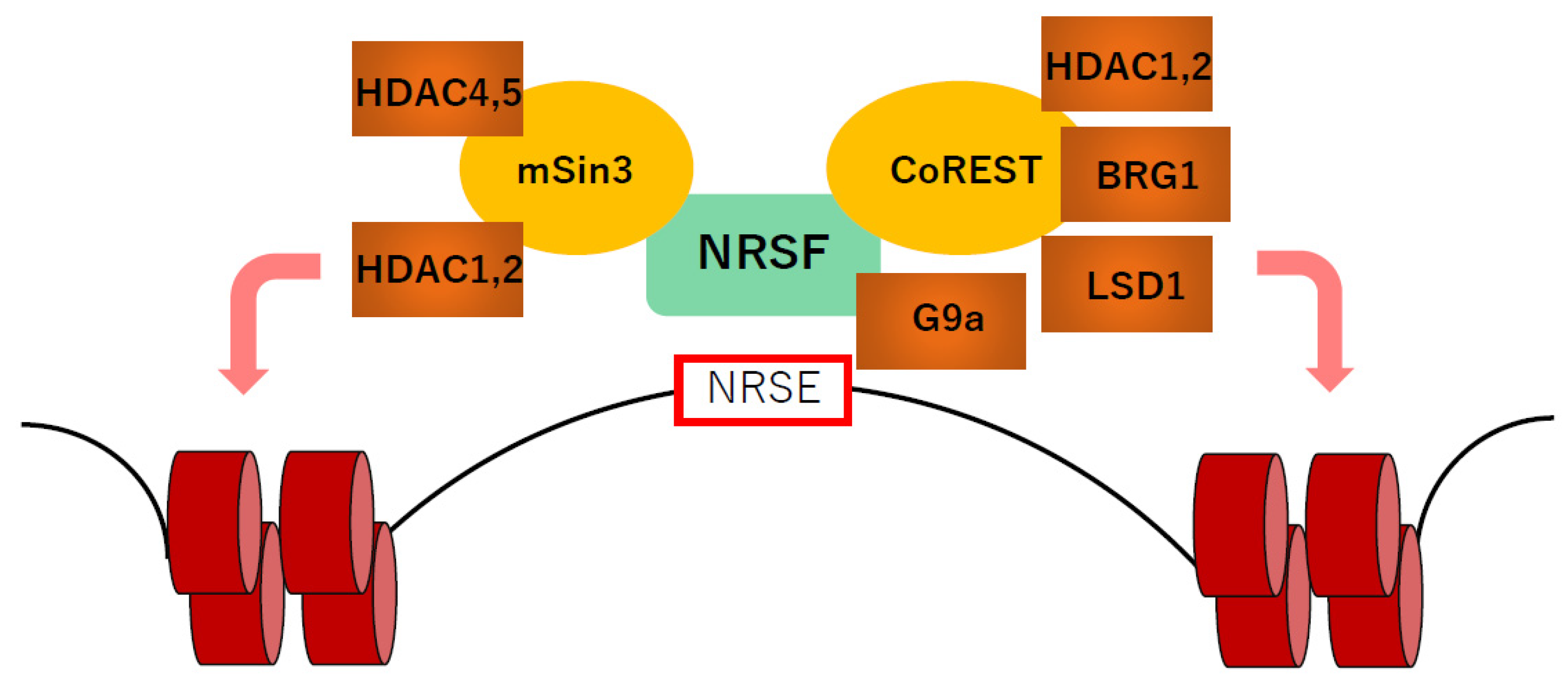
A number of studies performed in non-cardiac cells have revealed that NRSF forms a complex with corepressors to repress transcription (Figure 2). NRSF has two repressor domains at the N- and C-terminal ends, respectively. The N-terminal repressor domain interacts with the mSin3A and mSin3B complexes that associate with histone deacetylases (HDAC) [7,88,89]. On the other hand, the C-terminal repressor domain interacts with the corepressor CoREST, which forms a complex with HDACs, the histone demethylase LSD1 and the ATP-dependent chromatin remodeling enzyme BRG1 [88]. The C-terminal repressor domain of NRSF also interacts with the histone methylase G9a independently of CoREST [90]. HDACs remove acetyl groups from histone tails, resulting in transcriptional repression by chromatin compaction [91]. LSD1 demethylates mono- and dimethylated H3 lysines K4 and K9, thereby repressing gene expression [92]. G9a monomethylates and dimethylates histone H3K9 (H3K9me1 and H3K9me2) and, to a lesser extent, H3K27 [93]. In cardiomyocytes, NRSF forms a complex with Class I HDACs and the Class IIa HDACs, HDAC4 and HDAC5, and suppresses NPPA and NPPB expression by modifying histone acetylation [7,89]. Class I HDACs are relatively ubiquitously expressed, while Class IIa HDACs, including HDAC4, HDAC5, HDAC7 and HDAC9, are expressed in a tissue-specific manner, most abundantly in heart, brain and skeletal muscle. In the heart, Class IIa HDACs have been reported to act as a signal-responsive suppressor of cardiac hypertrophy [94]. Phosphorylation at two conserved sites by Ca2+/calmodulin-dependent kinase and/or protein kinase D induced by hypertrophic stimuli causes nuclear export of Class IIa HDACs, resulting in the attenuation of NRSF-mediated repression of the fetal gene program during cardiac remodeling [89]. These findings suggest that NRSF needs association with epigenetic machinery at least in part for regulation of cardiac gene expression.
The actions of HDACs have been studied in a variety of heart failure models. In a mouse model of chronic pressure overload induced by transverse aortic constriction, treatment with a pan-HDAC inhibitor (HDACi) blunted the hypertrophic response and slowed the progression toward heart failure [95,96]. Cardiac fibrotic markers were also reduced in the ventricle, and HDACi application reduced collagen production in isolated cardiac fibroblasts [96]. HDACi treatment also ameliorated established hypertrophy [95]. Similar cardioprotective effects were produced through cardiomyocyte-specific deletion of Class I HDACs (HDAC1 and HDAC2) [97,98]. On the other hand, cardiomyocyte-specific deletions of Class II HDACs promoted hypertrophic responses [99]. In addition, administration of a clinical-stage HDACi (givinostat) improved cardiac function in two diastolic dysfunction models; the Dahl salt-sensitive hypertensive rat and normotensive diastolic dysfunction mice induced by aging [100]. The HDACi-mediated improvement seen with these models appears to be unrelated to blood pressure, cardiac hypertrophy, changes in the isoform of expressed cardiac sarcomeric proteins, or regulation of gene transcription. Instead, the improvement correlates with enhanced relaxation of myocardial fibers mediated by direct deacetylation of sarcomeric proteins [100]. In a recent study using a feline model of diastolic dysfunction and heart failure induced by pressure overload, another HDACi (vorinostat) had a similar effect on relaxation properties of isolated myocardial fibers [101]. Moreover, vorinostat ameliorated cardiac hypertrophy and cardiac fibrosis in a manner similar to that seen in mouse models of pressure-overload-induced hypertrophy [96,101]. Suppression of HDACs appears to reduce cardiac hypertrophy, protect against oxidative damage, reduce inflammation, inhibit fibrosis and modulate the composition extracellular matrix [97,102]. However, taking into consideration that cardiac-specific deletion of HDAC1 and HDAC2 results in neonatal death, with arrhythmia and dilated cardiomyopathy [91], more precise studies will be needed before establishment of a clinical application.
The role of LSD1 has also been studied in various heart failure models. LSD1 deletion in cardiac myofibroblasts attenuates the remodeling induced by transverse aortic constriction, whereas LSD1 deletion in cardiomyocytes triggers mild cardiac hypertrophy and dysfunction [103]. Knockdown of LSD1 prevents cardiac fibroblast activation by inhibiting the TGFβ pathway, as evidenced by downregulation of Ang-II-induced TGFβ1 expression and Smad2/3, p38, ERK and JNK phosphorylation. On the other hand, LSD1 deletion from cardiomyocytes downregulated CoREST and NRSF accompanied with reactivation of ANP and BNP. LSD1 seems to maintain the integrity of the CoREST complex, so that deletion of LSD1 causes downregulation of CoREST protein [104]. CoREST reduction triggers a decrease in the level of LSD1 and de-repression of NRSF-responsive gene expression [105]. How LSD1 deletion downregulates NRSF in cardiomyocytes and how LSD1 affects the TGFβ pathway in cardiac fibroblasts remain unknown, however.
G9a forms a heteromeric complex with GLP (G9a-like protein, also known as EHMT1 or KMT1D), another enzyme catalyzing the methylation at H3K9 in vivo [106]. Genetic analysis showed that, in vivo, the histone methyl-transferase activity of G9a is more important than that of GLP and that neither can compensate for the loss of the activity of the other [107]. G9a and GLP are essential for normal development, and KO mice deficient in either gene die in utero (at E9.5) due to severe growth defects [106]. H3K9me2 accumulates during cardiac development and keeps adult cardiomyocytes locked into their terminally differentiated state in which the fetal gene program is suppressed. However, pathological hypertrophic stimuli increase expression of miR-217, which downregulates G9a/GLP and leads to a reduction in H3K9me2. This decrease in H3K9me2 causes cardiomyocytes to be in a less differentiated state and de-represses the fetal gene program [108]. It remains unclear whether the effects of G9a described above are mediated by NRSF, however.
All these studies demonstrate that alterations in histone acetylation and/or histone H3 lysine methylation play an important role in the regulation of cardiac genes mediated by NRSF [109]. We anticipate that further studies into how each component of the epigenetic regulators contributes to the regulation of the cardiac gene program via NRSF and cardiac homeostasis will lead to a better understanding of the molecular mechanisms underlying the pathological cardiac remodeling and heart failure.

Author Contributions

H.I. and K.K. wrote the manuscript. All authors have read and agreed to the published version of the manuscript.

Funding

This research was supported by Grants-in-Aid for Scientific Research from the Japan Society for the Promotion of Science 20K17077 (Inazumi), 20H03673 (Kuwahara), and by grants from the Japan Heart Foundation, the Takeda Science Foundation, the Uehara Memorial Foundation and the SENSHIN Medical Research Foundation (to Kuwahara).

Acknowledgments

We thank Ayako Ohta and Mebae Kobayashi for their excellent secretarial work.

Conflicts of Interest

The authors declare no conflict of interest.

References

  1. Ahmad, F.B.; Anderson, R.N. The Leading Causes of Death in the US for 2020. JAMA 2021, 325, 1829–1830. [Google Scholar] [CrossRef] [PubMed]
  2. Tsao, C.W.; Aday, A.W.; Almarzooq, Z.I.; Alonso, A.; Beaton, A.Z.; Bittencourt, M.S.; Boehme, A.K.; Buxton, A.E.; Carson, A.P.; Commodore-Mensah, Y.; et al. Heart Disease and Stroke Statistics-2022 Update: A Report From the American Heart Association. Circulation 2022, 145, e153–e639. [Google Scholar] [CrossRef] [PubMed]
  3. McKinsey, T.A.; Olson, E.N. Toward transcriptional therapies for the failing heart: Chemical screens to modulate genes. J. Clin. Investig. 2005, 115, 538–546. [Google Scholar] [CrossRef] [PubMed] [Green Version]
  4. Mukoyama, M.; Nakao, K.; Hosoda, K.; Suga, S.; Saito, Y.; Ogawa, Y.; Shirakami, G.; Jougasaki, M.; Obata, K.; Yasue, H.; et al. Brain natriuretic peptide as a novel cardiac hormone in humans. Evidence for an exquisite dual natriuretic peptide system, atrial natriuretic peptide and brain natriuretic peptide. J. Clin. Investig. 1991, 87, 1402–1412. [Google Scholar] [CrossRef] [PubMed]
  5. Kjaer, A.; Hesse, B. Heart failure and neuroendocrine activation: Diagnostic, prognostic and therapeutic perspectives. Clin. Physiol. 2001, 21, 661–672. [Google Scholar] [CrossRef] [Green Version]
  6. Oka, T.; Xu, J.; Molkentin, J.D. Re-employment of developmental transcription factors in adult heart disease. Semin. Cell Dev. Biol. 2007, 18, 117–131. [Google Scholar] [CrossRef] [Green Version]
  7. Kuwahara, K.; Saito, Y.; Ogawa, E.; Takahashi, N.; Nakagawa, Y.; Naruse, Y.; Harada, M.; Hamanaka, I.; Izumi, T.; Miyamoto, Y.; et al. The neuron-restrictive silencer element-neuron-restrictive silencer factor system regulates basal and endothelin 1-inducible atrial natriuretic peptide gene expression in ventricular myocytes. Mol. Cell. Biol. 2001, 21, 2085–2097. [Google Scholar] [CrossRef] [Green Version]
  8. Ogawa, E.; Saito, Y.; Kuwahara, K.; Harada, M.; Miyamoto, Y.; Hamanaka, I.; Kajiyama, N.; Takahashi, N.; Izumi, T.; Kawakami, R.; et al. Fibronectin signaling stimulates BNP gene transcription by inhibiting neuron-restrictive silencer element-dependent repression. Cardiovasc. Res. 2002, 53, 451–459. [Google Scholar] [CrossRef] [Green Version]
  9. Kuwahara, K.; Saito, Y.; Takano, M.; Arai, Y.; Yasuno, S.; Nakagawa, Y.; Takahashi, N.; Adachi, Y.; Takemura, G.; Horie, M.; et al. NRSF regulates the fetal cardiac gene program and maintains normal cardiac structure and function. EMBO J. 2003, 22, 6310–6321. [Google Scholar] [CrossRef]
  10. Inazumi, H.; Kuwahara, K.; Nakagawa, Y.; Kuwabara, Y.; Numaga-Tomita, T.; Kashihara, T.; Nakada, T.; Kurebayashi, N.; Oya, M.; Nonaka, M.; et al. NRSF-GNAO1 Pathway Contributes to the Regulation of Cardiac Ca2+ Homeostasis. Circ. Res. 2022, 130, 234–248. [Google Scholar] [CrossRef]
  11. Kuwahara, K.; Nakao, K. Regulation and significance of atrial and brain natriuretic peptides as cardiac hormones. Endocr. J. 2010, 57, 555–565. [Google Scholar] [CrossRef] [PubMed] [Green Version]
  12. Goetze, J.P.; Bruneau, B.G.; Ramos, H.R.; Ogawa, T.; de Bold, M.K.; de Bold, A.J. Cardiac natriuretic peptides. Nat. Rev. Cardiol. 2020, 17, 698–717. [Google Scholar] [CrossRef] [PubMed]
  13. Heidenreich, P.A.; Bozkurt, B.; Aguilar, D.; Allen, L.A.; Byun, J.J.; Colvin, M.M.; Deswal, A.; Drazner, M.H.; Dunlay, S.M.; Evers, L.R.; et al. 2022 AHA/ACC/HFSA Guideline for the Management of Heart Failure: A Report of the American College of Cardiology/American Heart Association Joint Committee on Clinical Practice Guidelines. Circulation 2022, 145, e895–e1032. [Google Scholar] [CrossRef]
  14. Zeller, R.; Bloch, K.D.; Williams, B.S.; Arceci, R.J.; Seidman, C.E. Localized expression of the atrial natriuretic factor gene during cardiac embryogenesis. Genes Dev. 1987, 1, 693–698. [Google Scholar] [CrossRef] [PubMed] [Green Version]
  15. Field, L.J. Atrial natriuretic factor-SV40 T antigen transgenes produce tumors and cardiac arrhythmias in mice. Science 1988, 239, 1029–1033. [Google Scholar] [CrossRef] [PubMed]
  16. Knowlton, K.U.; Rockman, H.A.; Itani, M.; Vovan, A.; Seidman, C.E.; Chien, K.R. Divergent pathways mediate the induction of ANF transgenes in neonatal and hypertrophic ventricular myocardium. J. Clin. Investig. 1995, 96, 1311–1318. [Google Scholar] [CrossRef]
  17. Argentin, S.; Ardati, A.; Tremblay, S.; Lihrmann, I.; Robitaille, L.; Drouin, J.; Nemer, M. Developmental stage-specific regulation of atrial natriuretic factor gene transcription in cardiac cells. Mol. Cell. Biol. 1994, 14, 777–790. [Google Scholar] [CrossRef]
  18. LaPointe, M.C.; Wu, J.P.; Greenberg, B.; Gardner, D.G. Upstream sequences confer atrial-specific expression on the human atrial natriuretic factor gene. J. Biol. Chem. 1988, 263, 9075–9078. [Google Scholar] [CrossRef]
  19. Houweling, A.C.; van Borren, M.M.; Moorman, A.F.; Christoffels, V.M. Expression and regulation of the atrial natriuretic factor encoding gene Nppa during development and disease. Cardiovasc. Res. 2005, 67, 583–593. [Google Scholar] [CrossRef] [Green Version]
  20. Debrus, S.; Rahbani, L.; Marttila, M.; Delorme, B.; Paradis, P.; Nemer, M. The zinc finger-only protein Zfp260 is a novel cardiac regulator and a nuclear effector of α1-adrenergic signaling. Mol. Cell. Biol. 2005, 25, 8669–8682. [Google Scholar] [CrossRef] [Green Version]
  21. Sprenkle, A.B.; Murray, S.F.; Glembotski, C.C. Involvement of multiple cis elements in basal- and α-adrenergic agonist-inducible atrial natriuretic factor transcription. Roles for serum response elements and an SP-1-like element. Circ. Res. 1995, 77, 1060–1069. [Google Scholar] [CrossRef] [PubMed]
  22. Lee, Y.; Shioi, T.; Kasahara, H.; Jobe, S.M.; Wiese, R.J.; Markham, B.E.; Izumo, S. The cardiac tissue-restricted homeobox protein Csx/Nkx2.5 physically associates with the zinc finger protein GATA4 and cooperatively activates atrial natriuretic factor gene expression. Mol. Cell. Biol. 1998, 18, 3120–3129. [Google Scholar] [CrossRef] [PubMed] [Green Version]
  23. Durocher, D.; Chen, C.Y.; Ardati, A.; Schwartz, R.J.; Nemer, M. The atrial natriuretic factor promoter is a downstream target for Nkx-2.5 in the myocardium. Mol. Cell. Biol. 1996, 16, 4648–4655. [Google Scholar] [CrossRef] [PubMed] [Green Version]
  24. Shiojima, I.; Komuro, I.; Oka, T.; Hiroi, Y.; Mizuno, T.; Takimoto, E.; Monzen, K.; Aikawa, R.; Akazawa, H.; Yamazaki, T.; et al. Context-dependent transcriptional cooperation mediated by cardiac transcription factors Csx/Nkx-2.5 and GATA-4. J. Biol. Chem. 1999, 274, 8231–8239. [Google Scholar] [CrossRef] [Green Version]
  25. Morin, S.; Charron, F.; Robitaille, L.; Nemer, M. GATA-dependent recruitment of MEF2 proteins to target promoters. EMBO J. 2000, 19, 2046–2055. [Google Scholar] [CrossRef] [PubMed] [Green Version]
  26. Harris, A.N.; Ruiz-Lozano, P.; Chen, Y.F.; Sionit, P.; Yu, Y.T.; Lilly, B.; Olson, E.N.; Chien, K.R. A novel A/T-rich element mediates ANF gene expression during cardiac myocyte hypertrophy. J. Mol. Cell. Cardiol. 1997, 29, 515–525. [Google Scholar] [CrossRef]
  27. Small, E.M.; Krieg, P.A. Transgenic analysis of the atrialnatriuretic factor (ANF) promoter: Nkx2-5 and GATA-4 binding sites are required for atrial specific expression of ANF. Dev. Biol. 2003, 261, 116–131. [Google Scholar] [CrossRef] [Green Version]
  28. Horsthuis, T.; Houweling, A.C.; Habets, P.E.; de Lange, F.J.; el Azzouzi, H.; Clout, D.E.; Moorman, A.F.; Christoffels, V.M. Distinct regulation of developmental and heart disease-induced atrial natriuretic factor expression by two separate distal sequences. Circ. Res. 2008, 102, 849–859. [Google Scholar] [CrossRef] [Green Version]
  29. Argentin, S.; Sun, Y.L.; Lihrmann, I.; Schmidt, T.J.; Drouin, J.; Nemer, M. Distal cis-acting promoter sequences mediate glucocorticoid stimulation of cardiac atrial natriuretic factor gene transcription. J. Biol. Chem. 1991, 266, 23315–23322. [Google Scholar] [CrossRef]
  30. Chun, Y.S.; Hyun, J.Y.; Kwak, Y.G.; Kim, I.S.; Kim, C.H.; Choi, E.; Kim, M.S.; Park, J.W. Hypoxic activation of the atrial natriuretic peptide gene promoter through direct and indirect actions of hypoxia-inducible factor-1. Biochem. J. 2003, 370, 149–157. [Google Scholar] [CrossRef] [Green Version]
  31. He, Q.; Wang, D.; Yang, X.P.; Carretero, O.A.; LaPointe, M.C. Inducible regulation of human brain natriuretic peptide promoter in transgenic mice. Am. J. Physiol.-Heart Circ. Physiol. 2001, 280, H368–H376. [Google Scholar] [CrossRef] [PubMed]
  32. LaPointe, M.C.; Wu, G.; Garami, M.; Yang, X.P.; Gardner, D.G. Tissue-specific expression of the human brain natriuretic peptide gene in cardiac myocytes. Hypertension 1996, 27, 715–722. [Google Scholar] [CrossRef] [PubMed]
  33. Nakagawa, O.; Ogawa, Y.; Itoh, H.; Suga, S.; Komatsu, Y.; Kishimoto, I.; Nishino, K.; Yoshimasa, T.; Nakao, K. Rapid transcriptional activation and early mRNA turnover of brain natriuretic peptide in cardiocyte hypertrophy. Evidence for brain natriuretic peptide as an “emergency” cardiac hormone against ventricular overload. J. Clin. Investig. 1995, 96, 1280–1287. [Google Scholar] [CrossRef] [PubMed] [Green Version]
  34. Steinhelper, M.E. Structure, expression, and genomic mapping of the mouse natriuretic peptide type-B gene. Circ. Res. 1993, 72, 984–992. [Google Scholar] [CrossRef] [PubMed] [Green Version]
  35. Thuerauf, D.J.; Hanford, D.S.; Glembotski, C.C. Regulation of rat brain natriuretic peptide transcription. A potential role for GATA-related transcription factors in myocardial cell gene expression. J. Biol. Chem. 1994, 269, 17772–17775. [Google Scholar] [CrossRef]
  36. Kuwahara, K.; Kinoshita, H.; Kuwabara, Y.; Nakagawa, Y.; Usami, S.; Minami, T.; Yamada, Y.; Fujiwara, M.; Nakao, K. Myocardin-related transcription factor A is a common mediator of mechanical stress-and neurohumoral stimulation-induced cardiac hypertrophic signaling leading to activation of brain natriuretic peptide gene expression. Mol. Cell. Biol. 2010, 30, 4134–4148. [Google Scholar] [CrossRef] [Green Version]
  37. Akazawa, H.; Komuro, I. Roles of cardiac transcription factors in cardiac hypertrophy. Circ. Res. 2003, 92, 1079–1088. [Google Scholar] [CrossRef] [Green Version]
  38. Shimizu, N.; Smith, G.; Izumo, S. Both a ubiquitous factor mTEF-1 and a distinct muscle-specific factor bind to the M-CAT motif of the myosin heavy chain β gene. Nucleic Acids Res. 1993, 21, 4103–4110. [Google Scholar] [CrossRef]
  39. Molkentin, J.D.; Olson, E.N. GATA4: A novel transcriptional regulator of cardiac hypertrophy? Circulation 1997, 96, 3833–3835. [Google Scholar]
  40. Grépin, C.; Dagnino, L.; Robitaille, L.; Haberstroh, L.; Antakly, T.; Nemer, M. A hormone-encoding gene identifies a pathway for cardiac but not skeletal muscle gene transcription. Mol. Cell. Biol. 1994, 14, 3115–3129. [Google Scholar] [CrossRef]
  41. LaPointe, M.C. Molecular regulation of the brain natriuretic peptide gene. Peptides 2005, 26, 944–956. [Google Scholar] [CrossRef] [PubMed]
  42. Liang, F.; Gardner, D.G. Mechanical strain activates BNP gene transcription through a p38/NF-kappaB-dependent mechanism. J. Clin. Investig. 1999, 104, 1603–1612. [Google Scholar] [CrossRef] [PubMed] [Green Version]
  43. Ogawa, E.; Saito, Y.; Harada, M.; Kamitani, S.; Kuwahara, K.; Miyamoto, Y.; Ishikawa, M.; Hamanaka, I.; Kajiyama, N.; Takahashi, N.; et al. Outside-in signalling of fibronectin stimulates cardiomyocyte hypertrophy in cultured neonatal rat ventricular myocytes. J. Mol. Cell. Cardiol. 2000, 32, 765–776. [Google Scholar] [CrossRef] [PubMed]
  44. Molkentin, J.D.; Lu, J.R.; Antos, C.L.; Markham, B.; Richardson, J.; Robbins, J.; Grant, S.R.; Olson, E.N. A calcineurin-dependent transcriptional pathway for cardiac hypertrophy. Cell 1998, 93, 215–228. [Google Scholar] [CrossRef] [Green Version]
  45. Liang, F.; Webb, P.; Marimuthu, A.; Zhang, S.; Gardner, D.G. Triiodothyronine increases brain natriuretic peptide (BNP) gene transcription and amplifies endothelin-dependent BNP gene transcription and hypertrophy in neonatal rat ventricular myocytes. J. Biol. Chem. 2003, 278, 15073–15083. [Google Scholar] [CrossRef] [Green Version]
  46. Sergeeva, I.A.; Hooijkaas, I.B.; Ruijter, J.M.; van der Made, I.; de Groot, N.E.; van de Werken, H.J.; Creemers, E.E.; Christoffels, V.M. Identification of a regulatory domain controlling the Nppa-Nppb gene cluster during heart development and stress. Development 2016, 143, 2135–2146. [Google Scholar] [CrossRef] [Green Version]
  47. de Bold, A.J.; Bruneau, B.G.; Kuroski de Bold, M.L. Mechanical and neuroendocrine regulation of the endocrine heart. Cardiovasc. Res. 1996, 31, 7–18. [Google Scholar] [CrossRef] [Green Version]
  48. Harada, M.; Saito, Y.; Kuwahara, K.; Ogawa, E.; Ishikawa, M.; Nakagawa, O.; Miyamoto, Y.; Kamitani, S.; Hamanaka, I.; Kajiyama, N.; et al. Interaction of myocytes and nonmyocytes is necessary for mechanical stretch to induce ANP/BNP production in cardiocyte culture. J. Cardiovasc. Pharmacol. 1998, 31 (Suppl. 1), S357–S359. [Google Scholar] [CrossRef]
  49. Kinnunen, P.; Vuolteenaho, O.; Uusimaa, P.; Ruskoaho, H. Passive mechanical stretch releases atrial natriuretic peptide from rat ventricular myocardium. Circ. Res. 1992, 70, 1244–1253. [Google Scholar] [CrossRef] [Green Version]
  50. Sergeeva, I.A.; Christoffels, V.M. Regulation of expression of atrial and brain natriuretic peptide, biomarkers for heart development and disease. Biochim. Biophys. Acta 2013, 1832, 2403–2413. [Google Scholar] [CrossRef] [Green Version]
  51. Mori, N.; Schoenherr, C.; Vandenbergh, D.J.; Anderson, D.J. A common silencer element in the SCG10 and type II Na+ channel genes binds a factor present in nonneuronal cells but not in neuronal cells. Neuron 1992, 9, 45–54. [Google Scholar] [CrossRef]
  52. Schoenherr, C.J.; Anderson, D.J. The neuron-restrictive silencer factor (NRSF): A coordinate repressor of multiple neuron-specific genes. Science 1995, 267, 1360–1363. [Google Scholar] [CrossRef] [PubMed]
  53. Chong, J.A.; Tapia-Ramírez, J.; Kim, S.; Toledo-Aral, J.J.; Zheng, Y.; Boutros, M.C.; Altshuller, Y.M.; Frohman, M.A.; Kraner, S.D.; Mandel, G. REST: A mammalian silencer protein that restricts sodium channel gene expression to neurons. Cell 1995, 80, 949–957. [Google Scholar] [CrossRef] [Green Version]
  54. Ooi, L.; Wood, I.C. Chromatin crosstalk in development and disease: Lessons from REST. Nat. Rev. Genet. 2007, 8, 544–554. [Google Scholar] [CrossRef]
  55. Schoenherr, C.J.; Paquette, A.J.; Anderson, D.J. Identification of potential target genes for the neuron-restrictive silencer factor. Proc. Natl. Acad. Sci. USA 1996, 93, 9881–9886. [Google Scholar] [CrossRef] [Green Version]
  56. Yasuno, S.; Usami, S.; Kuwahara, K.; Nakanishi, M.; Arai, Y.; Kinoshita, H.; Nakagawa, Y.; Fujiwara, M.; Murakami, M.; Ueshima, K.; et al. Endogenous cardiac natriuretic peptides protect the heart in a mouse model of dilated cardiomyopathy and sudden death. Am. J. Physiol.-Heart Circ. Physiol. 2009, 296, H1804–H1810. [Google Scholar] [CrossRef] [Green Version]
  57. Takano, M.; Kinoshita, H.; Shioya, T.; Itoh, M.; Nakao, K.; Kuwahara, K. Pathophysiological remodeling of mouse cardiac myocytes expressing dominant negative mutant of neuron restrictive silencing factor. Circ. J. 2010, 74, 2712–2719. [Google Scholar] [CrossRef] [Green Version]
  58. Kuwahara, K.; Takano, M.; Nakao, K. Pathophysiological significance of T-type Ca2+ channels: Transcriptional regulation of T-type Ca2+ channel—Regulation of CACNA1H by neuron-restrictive silencer factor. J. Pharmacol. Sci. 2005, 99, 211–213. [Google Scholar] [CrossRef] [Green Version]
  59. Ono, K.; Iijima, T. Cardiac T-type Ca2+ channels in the heart. J. Mol. Cell. Cardiol. 2010, 48, 65–70. [Google Scholar] [CrossRef]
  60. Yasui, K.; Niwa, N.; Takemura, H.; Opthof, T.; Muto, T.; Horiba, M.; Shimizu, A.; Lee, J.K.; Honjo, H.; Kamiya, K.; et al. Pathophysiological significance of T-type Ca2+ channels: Expression of T-type Ca2+ channels in fetal and diseased heart. J. Pharmacol. Sci. 2005, 99, 205–210. [Google Scholar] [CrossRef] [Green Version]
  61. Niwa, N.; Yasui, K.; Opthof, T.; Takemura, H.; Shimizu, A.; Horiba, M.; Lee, J.K.; Honjo, H.; Kamiya, K.; Kodama, I. Cav3.2 subunit underlies the functional T-type Ca2+ channel in murine hearts during the embryonic period. Am. J. Physiol.-Heart Circ. Physiol. 2004, 286, H2257–H2263. [Google Scholar] [CrossRef] [PubMed] [Green Version]
  62. Clozel, J.P.; Ertel, E.A.; Ertel, S.I. Voltage-gated T-type Ca2+ channels and heart failure. Proc. Assoc. Am. Physicians 1999, 111, 429–437. [Google Scholar] [CrossRef] [PubMed]
  63. Kinoshita, H.; Kuwahara, K.; Takano, M.; Arai, Y.; Kuwabara, Y.; Yasuno, S.; Nakagawa, Y.; Nakanishi, M.; Harada, M.; Fujiwara, M.; et al. T-type Ca2+ channel blockade prevents sudden death in mice with heart failure. Circulation 2009, 120, 743–752. [Google Scholar] [CrossRef] [PubMed] [Green Version]
  64. Robinson, R.B.; Siegelbaum, S.A. Hyperpolarization-activated cation currents: From molecules to physiological function. Annu. Rev. Physiol. 2003, 65, 453–480. [Google Scholar] [CrossRef] [Green Version]
  65. Wahl-Schott, C.; Biel, M. HCN channels: Structure, cellular regulation and physiological function. Cell. Mol. Life Sci. 2009, 66, 470–494. [Google Scholar] [CrossRef]
  66. Cerbai, E.; Mugelli, A. If in non-pacemaker cells: Role and pharmacological implications. Pharmacol. Res. 2006, 53, 416–423. [Google Scholar] [CrossRef]
  67. Yasui, K.; Liu, W.; Opthof, T.; Kada, K.; Lee, J.K.; Kamiya, K.; Kodama, I. If current and spontaneous activity in mouse embryonic ventricular myocytes. Circ. Res. 2001, 88, 536–542. [Google Scholar] [CrossRef] [Green Version]
  68. Stillitano, F.; Lonardo, G.; Zicha, S.; Varro, A.; Cerbai, E.; Mugelli, A.; Nattel, S. Molecular basis of funny current (If) in normal and failing human heart. J. Mol. Cell. Cardiol. 2008, 45, 289–299. [Google Scholar] [CrossRef]
  69. Kuratomi, S.; Ohmori, Y.; Ito, M.; Shimazaki, K.; Muramatsu, S.; Mizukami, H.; Uosaki, H.; Yamashita, J.K.; Arai, Y.; Kuwahara, K.; et al. The cardiac pacemaker-specific channel Hcn4 is a direct transcriptional target of MEF2. Cardiovasc. Res. 2009, 83, 682–687. [Google Scholar] [CrossRef] [Green Version]
  70. Kuratomi, S.; Kuratomi, A.; Kuwahara, K.; Ishii, T.M.; Nakao, K.; Saito, Y.; Takano, M. NRSF regulates the developmental and hypertrophic changes of HCN4 transcription in rat cardiac myocytes. Biochem. Biophys. Res. Commun. 2007, 353, 67–73. [Google Scholar] [CrossRef]
  71. Kuwabara, Y.; Kuwahara, K.; Takano, M.; Kinoshita, H.; Arai, Y.; Yasuno, S.; Nakagawa, Y.; Igata, S.; Usami, S.; Minami, T.; et al. Increased expression of HCN channels in the ventricular myocardium contributes to enhanced arrhythmicity in mouse failing hearts. J. Am. Heart Assoc. 2013, 2, e000150. [Google Scholar] [CrossRef] [PubMed] [Green Version]
  72. Heusch, G. Heart rate and heart failure. Not a simple relationship. Circ. J. 2011, 75, 229–236. [Google Scholar] [CrossRef] [PubMed] [Green Version]
  73. Swedberg, K.; Komajda, M.; Böhm, M.; Borer, J.S.; Ford, I.; Dubost-Brama, A.; Lerebours, G.; Tavazzi, L. Ivabradine and outcomes in chronic heart failure (SHIFT): A randomised placebo-controlled study. Lancet 2010, 376, 875–885. [Google Scholar] [CrossRef]
  74. Hurowitz, E.H.; Melnyk, J.M.; Chen, Y.J.; Kouros-Mehr, H.; Simon, M.I.; Shizuya, H. Genomic characterization of the human heterotrimeric G protein α, β, and γ subunit genes. DNA Res. 2000, 7, 111–120. [Google Scholar] [CrossRef] [PubMed] [Green Version]
  75. Strathmann, M.P.; Simon, M.I. G α12 and G α13 subunits define a fourth class of G protein α subunits. Proc. Natl. Acad. Sci. USA 1991, 88, 5582–5586. [Google Scholar] [CrossRef] [Green Version]
  76. He, J.C.; Neves, S.R.; Jordan, J.D.; Iyengar, R. Role of the Go/i signaling network in the regulation of neurite outgrowth. Can. J. Physiol. Pharmacol. 2006, 84, 687–694. [Google Scholar] [CrossRef]
  77. Neumann, J.; Schmitz, W.; Scholz, H.; von Meyerinck, L.; Doring, V.; Kalmar, P. Increase in myocardial Gi-proteins in heart failure. Lancet 1988, 2, 936–937. [Google Scholar] [CrossRef]
  78. Feldman, A.M.; Cates, A.E.; Veazey, W.B.; Hershberger, R.E.; Bristow, M.R.; Baughman, K.L.; Baumgartner, W.A.; Van Dop, C. Increase of the 40,000-mol wt pertussis toxin substrate (G protein) in the failing human heart. J. Clin. Investig. 1988, 82, 189–197. [Google Scholar] [CrossRef] [Green Version]
  79. DeGeorge, B.R., Jr.; Gao, E.; Boucher, M.; Vinge, L.E.; Martini, J.S.; Raake, P.W.; Chuprun, J.K.; Harris, D.M.; Kim, G.W.; Soltys, S.; et al. Targeted inhibition of cardiomyocyte Gi signaling enhances susceptibility to apoptotic cell death in response to ischemic stress. Circulation 2008, 117, 1378–1387. [Google Scholar] [CrossRef] [Green Version]
  80. Li, Y.; Mende, U.; Lewis, C.; Neer, E.J. Maintenance of cellular levels of G-proteins: Different efficiencies of αs and αo synthesis in GH3 cells. Biochem. J. 1996, 318 Pt 3, 1071–1077. [Google Scholar] [CrossRef]
  81. Asano, T.; Morishita, R.; Semba, R.; Itoh, H.; Kaziro, Y.; Kato, K. Identification of lung major GTP-binding protein as Gi2 and its distribution in various rat tissues determined by immunoassay. Biochemistry 1989, 28, 4749–4754. [Google Scholar] [CrossRef] [PubMed]
  82. Bauer, A.; McDonald, A.D.; Nasir, K.; Peller, L.; Rade, J.J.; Miller, J.M.; Heldman, A.W.; Donahue, J.K. Inhibitory G protein overexpression provides physiologically relevant heart rate control in persistent atrial fibrillation. Circulation 2004, 110, 3115–3120. [Google Scholar] [CrossRef] [PubMed] [Green Version]
  83. Keller, K.; Maass, M.; Dizayee, S.; Leiss, V.; Annala, S.; Koth, J.; Seemann, W.K.; Muller-Ehmsen, J.; Mohr, K.; Nurnberg, B.; et al. Lack of Gαi2 leads to dilative cardiomyopathy and increased mortality in β1-adrenoceptor overexpressing mice. Cardiovasc. Res. 2015, 108, 348–356. [Google Scholar] [CrossRef] [PubMed] [Green Version]
  84. Kaur, K.; Parra, S.; Chen, R.; Charbeneau, R.A.; Wade, S.M.; Jay, P.Y.; Neubig, R.R. Gα2 signaling: Friend or foe in cardiac injury and heart failure? Naunyn-Schmiedeberg’s Arch. Pharmacol. 2012, 385, 443–453. [Google Scholar] [CrossRef] [Green Version]
  85. Nishiga, M.; Horie, T.; Kuwabara, Y.; Nagao, K.; Baba, O.; Nakao, T.; Nishino, T.; Hakuno, D.; Nakashima, Y.; Nishi, H.; et al. MicroRNA-33 Controls Adaptive Fibrotic Response in the Remodeling Heart by Preserving Lipid Raft Cholesterol. Circ. Res. 2017, 120, 835–847. [Google Scholar] [CrossRef]
  86. Du, C.K.; Morimoto, S.; Nishii, K.; Minakami, R.; Ohta, M.; Tadano, N.; Lu, Q.W.; Wang, Y.Y.; Zhan, D.Y.; Mochizuki, M.; et al. Knock-in mouse model of dilated cardiomyopathy caused by troponin mutation. Circ. Res. 2007, 101, 185–194. [Google Scholar] [CrossRef] [Green Version]
  87. Nomura, S.; Satoh, M.; Fujita, T.; Higo, T.; Sumida, T.; Ko, T.; Yamaguchi, T.; Tobita, T.; Naito, A.T.; Ito, M.; et al. Cardiomyocyte gene programs encoding morphological and functional signatures in cardiac hypertrophy and failure. Nat. Commun. 2018, 9, 4435. [Google Scholar] [CrossRef]
  88. Bingham, A.J.; Ooi, L.; Wood, I.C. Multiple chromatin modifications important for gene expression changes in cardiac hypertrophy. Biochem. Soc. Trans. 2006, 34, 1138–1140. [Google Scholar] [CrossRef]
  89. Nakagawa, Y.; Kuwahara, K.; Harada, M.; Takahashi, N.; Yasuno, S.; Adachi, Y.; Kawakami, R.; Nakanishi, M.; Tanimoto, K.; Usami, S.; et al. Class II HDACs mediate CaMK-dependent signaling to NRSF in ventricular myocytes. J. Mol. Cell. Cardiol. 2006, 41, 1010–1022. [Google Scholar] [CrossRef]
  90. Roopra, A.; Qazi, R.; Schoenike, B.; Daley, T.J.; Morrison, J.F. Localized domains of G9a-mediated histone methylation are required for silencing of neuronal genes. Mol. Cell. 2004, 14, 727–738. [Google Scholar] [CrossRef]
  91. Montgomery, R.L.; Davis, C.A.; Potthoff, M.J.; Haberland, M.; Fielitz, J.; Qi, X.; Hill, J.A.; Richardson, J.A.; Olson, E.N. Histone deacetylases 1 and 2 redundantly regulate cardiac morphogenesis, growth, and contractility. Genes Dev. 2007, 21, 1790–1802. [Google Scholar] [CrossRef] [PubMed] [Green Version]
  92. Klose, R.J.; Zhang, Y. Regulation of histone methylation by demethylimination and demethylation. Nat. Rev. Mol. Cell Biol. 2007, 8, 307–318. [Google Scholar] [CrossRef] [PubMed]
  93. Tachibana, M.; Sugimoto, K.; Nozaki, M.; Ueda, J.; Ohta, T.; Ohki, M.; Fukuda, M.; Takeda, N.; Niida, H.; Kato, H.; et al. G9a histone methyltransferase plays a dominant role in euchromatic histone H3 lysine 9 methylation and is essential for early embryogenesis. Genes Dev. 2002, 16, 1779–1791. [Google Scholar] [CrossRef] [PubMed] [Green Version]
  94. McKinsey, T.A.; Zhang, C.L.; Lu, J.; Olson, E.N. Signal-dependent nuclear export of a histone deacetylase regulates muscle differentiation. Nature 2000, 408, 106–111. [Google Scholar] [CrossRef] [PubMed]
  95. Kee, H.J.; Sohn, I.S.; Nam, K.I.; Park, J.E.; Qian, Y.R.; Yin, Z.; Ahn, Y.; Jeong, M.H.; Bang, Y.J.; Kim, N.; et al. Inhibition of histone deacetylation blocks cardiac hypertrophy induced by angiotensin II infusion and aortic banding. Circulation 2006, 113, 51–59. [Google Scholar] [CrossRef]
  96. Kong, Y.; Tannous, P.; Lu, G.; Berenji, K.; Rothermel, B.A.; Olson, E.N.; Hill, J.A. Suppression of class I and II histone deacetylases blunts pressure-overload cardiac hypertrophy. Circulation 2006, 113, 2579–2588. [Google Scholar] [CrossRef]
  97. Gillette, T.G.; Hill, J.A. Readers, writers, and erasers: Chromatin as the whiteboard of heart disease. Circ. Res. 2015, 116, 1245–1253. [Google Scholar] [CrossRef] [Green Version]
  98. Morales, C.R.; Li, D.L.; Pedrozo, Z.; May, H.I.; Jiang, N.; Kyrychenko, V.; Cho, G.W.; Kim, S.Y.; Wang, Z.V.; Rotter, D.; et al. Inhibition of class I histone deacetylases blunts cardiac hypertrophy through TSC2-dependent mTOR repression. Sci. Signal. 2016, 9, ra34. [Google Scholar] [CrossRef] [Green Version]
  99. Zhang, C.L.; McKinsey, T.A.; Chang, S.; Antos, C.L.; Hill, J.A.; Olson, E.N. Class II histone deacetylases act as signal-responsive repressors of cardiac hypertrophy. Cell 2002, 110, 479–488. [Google Scholar] [CrossRef] [Green Version]
  100. Jeong, M.Y.; Lin, Y.H.; Wennersten, S.A.; Demos-Davies, K.M.; Cavasin, M.A.; Mahaffey, J.H.; Monzani, V.; Saripalli, C.; Mascagni, P.; Reece, T.B.; et al. Histone deacetylase activity governs diastolic dysfunction through a nongenomic mechanism. Sci. Transl. Med. 2018, 10, eaao0144. [Google Scholar] [CrossRef] [Green Version]
  101. Wallner, M.; Eaton, D.M.; Berretta, R.M.; Liesinger, L.; Schittmayer, M.; Gindlhuber, J.; Wu, J.; Jeong, M.Y.; Lin, Y.H.; Borghetti, G.; et al. HDAC inhibition improves cardiopulmonary function in a feline model of diastolic dysfunction. Sci. Transl. Med. 2020, 12, eaay7205. [Google Scholar] [CrossRef] [PubMed]
  102. Travers, J.G.; Wennersten, S.A.; Peña, B.; Bagchi, R.A.; Smith, H.E.; Hirsch, R.A.; Vanderlinden, L.A.; Lin, Y.H.; Dobrinskikh, E.; Demos-Davies, K.M.; et al. HDAC Inhibition Reverses Preexisting Diastolic Dysfunction and Blocks Covert Extracellular Matrix Remodeling. Circulation 2021, 143, 1874–1890. [Google Scholar] [CrossRef] [PubMed]
  103. Huo, J.L.; Jiao, L.; An, Q.; Chen, X.; Qi, Y.; Wei, B.; Zheng, Y.; Shi, X.; Gao, E.; Liu, H.M.; et al. Myofibroblast Deficiency of LSD1 Alleviates TAC-Induced Heart Failure. Circ. Res. 2021, 129, 400–413. [Google Scholar] [CrossRef]
  104. Foster, C.T.; Dovey, O.M.; Lezina, L.; Luo, J.L.; Gant, T.W.; Barlev, N.; Bradley, A.; Cowley, S.M. Lysine-specific demethylase 1 regulates the embryonic transcriptome and CoREST stability. Mol. Cell. Biol. 2010, 30, 4851–4863. [Google Scholar] [CrossRef] [PubMed] [Green Version]
  105. Lee, M.G.; Wynder, C.; Cooch, N.; Shiekhattar, R. An essential role for CoREST in nucleosomal histone 3 lysine 4 demethylation. Nature 2005, 437, 432–435. [Google Scholar] [CrossRef] [PubMed]
  106. Tachibana, M.; Ueda, J.; Fukuda, M.; Takeda, N.; Ohta, T.; Iwanari, H.; Sakihama, T.; Kodama, T.; Hamakubo, T.; Shinkai, Y. Histone methyltransferases G9a and GLP form heteromeric complexes and are both crucial for methylation of euchromatin at H3-K9. Genes Dev. 2005, 19, 815–826. [Google Scholar] [CrossRef] [PubMed] [Green Version]
  107. Tachibana, M.; Matsumura, Y.; Fukuda, M.; Kimura, H.; Shinkai, Y. G9a/GLP complexes independently mediate H3K9 and DNA methylation to silence transcription. EMBO J. 2008, 27, 2681–2690. [Google Scholar] [CrossRef]
  108. Thienpont, B.; Aronsen, J.M.; Robinson, E.L.; Okkenhaug, H.; Loche, E.; Ferrini, A.; Brien, P.; Alkass, K.; Tomasso, A.; Agrawal, A.; et al. The H3K9 dimethyltransferases EHMT1/2 protect against pathological cardiac hypertrophy. J. Clin. Investig. 2017, 127, 335–348. [Google Scholar] [CrossRef] [Green Version]
  109. Bingham, A.J.; Ooi, L.; Kozera, L.; White, E.; Wood, I.C. The repressor element 1-silencing transcription factor regulates heart-specific gene expression using multiple chromatin-modifying complexes. Mol. Cell. Biol. 2007, 27, 4082–4092. [Google Scholar] [CrossRef] [Green Version]
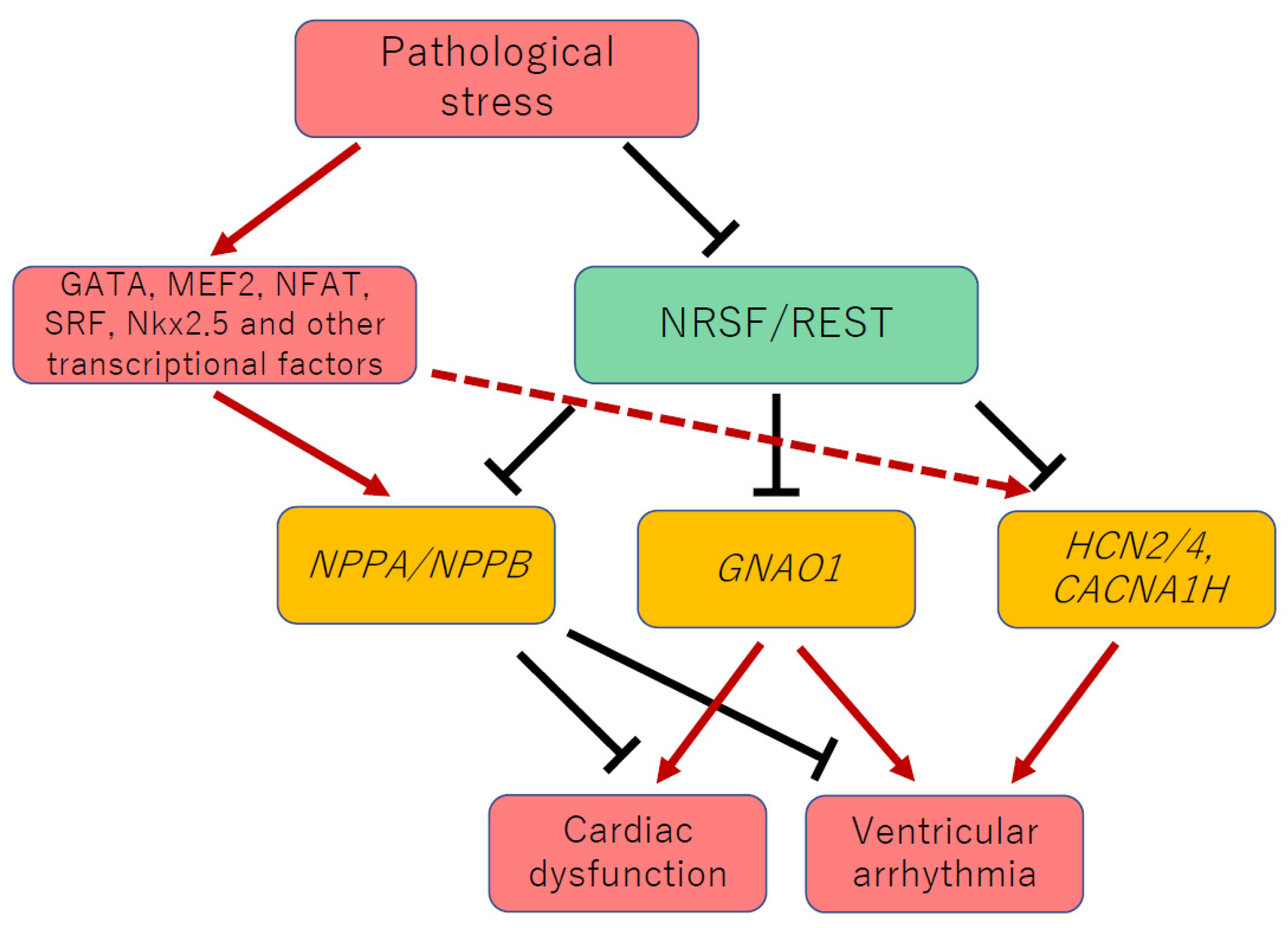
Figure 1. NRSF maintains normal cardiac function and electrical stability by regulating the cardiac gene program with other transcription factors. Whereas ANP and BNP protect against heart failure development, reactivation of Gαo and fetal-type cardiac ion channels, such as T-type Ca2+ and HCN channels, leads to cardiac dysfunction and ventricular arrhythmia in dnNRSF-Tg mice and NRSF cKO mice. HCN, hyperpolarization-activated cyclic nucleotide; NFAT, nuclear factor of T cells; MEF2, myocytes enhancer factor 2; SRF, serum response factor; NRSF, neuron-restrictive silencer factor; REST, repressor element-1 silencing factor.
Biology 11 01197 g001
Figure 2. NRSF forms a repressor complex with epigenetic modifiers and cooperatively regulates gene expression. HDAC, histone deacetylase; LSD1, lysine (K)-specific demethylase 1A; NRSE, neuron-restrictive silencer element; NRSF, neuron-restrictive silencer factor.
Biology 11 01197 g002
Publisher’s Note: MDPI stays neutral with regard to jurisdictional claims in published maps and institutional affiliations.